
مقدمه و سازمان سیستم عصبی؛ مرور مبانی و اصول فصل اول نوروآناتومی بالینی اسنل


دعای مطالعه [ نمایش ]
بِسْمِ الله الرَّحْمنِ الرَّحیمِ
اَللّهُمَّ اَخْرِجْنى مِنْ ظُلُماتِ الْوَهْمِ
خدايا مرا بيرون آور از تاريكىهاى وهم،
وَ اَكْرِمْنى بِنُورِ الْفَهْمِ
و به نور فهم گرامى ام بدار،
اَللّهُمَّ افْتَحْ عَلَيْنا اَبْوابَ رَحْمَتِكَ
خدايا درهاى رحمتت را به روى ما بگشا،
وَانْشُرْ عَلَيْنا خَزائِنَ عُلُومِكَ بِرَحْمَتِكَ يا اَرْحَمَ الرّاحِمينَ
و خزانههاى علومت را بر ما باز كن به امید رحمتت اى مهربانترين مهربانان.
آیندهنگاران مغز: «ما مغز را میشناسیم، تا آینده را بسازیم.»
کتاب «نوروآناتومی بالینی اسنل» اثر دکتر ریچارد اس. اسنل، با ویرایش جدید دکتر رایان اسپلیتگربر، همچنان بهعنوان مرجع طلایی این حوزه شناخته میشود. این اثر با تلفیق تازهترین پژوهشها و کاربردهای بالینی، پلی استوار میان دانش پایه و عمل پزشکی بنا میکند.
تلاشهای علمی و سالها تدریس ارزشمند استاد گرانقدر آقای دکتر محمدتقی جغتایی الهامبخش ترجمه و گسترش این دانش در ایران بوده است.
ترجمه دقیق این کتاب توسط برند علمی آیندهنگاران مغز به مدیریت داریوش طاهری، امکان دسترسی فارسیزبانان به مرزهای نوین علوم اعصاب را فراهم کرده و رسالتی علمی در جهت شناخت عمیقتر مغز و ترسیم آیندهای روشنتر بر عهده گرفته است.
»» فصل ۱: مقدمه و سازماندهی سیستم عصبی؛ قسمت مبانی و اصول
»» Chapter 1: Introduction and Organization of the Nervous System
CHAPTER OBJECTIVES
• To understand the basic organization of the main structures that form the nervous system.
• To gain a three-dimensional appreciation of the parts of the brain and their relative positions to one another.
اهداف فصل
• درک سازماندهی اساسی ساختارهای اصلی که سیستم عصبی را تشکیل میدهند.
• درک سهبعدی از بخشهای مغز و موقعیت نسبی آنها نسبت به یکدیگر.
A 23-year-old student is driving home from a party and crashes his car head-on into a tree. On examination in the emergency department of the local hospital, he has a fracture dislocation of the 7th thoracic vertebra, with signs and symptoms of severe damage to the spinal cord. Later, he is found to have paralysis of the left leg. Testing of cutaneous sensibility reveals a band of cutaneous hyperesthesia (increased sensitivity) extending around the abdominal wall on the left side at the level of the umbilicus. Just below this, he has a narrow band of anesthesia and analgesia. On the right side, he has total analgesia, thermoan- esthesia, and partial loss of touch sensation of the skin of the abdominal wall below the level of the umbilicus and involving the whole of the right leg.
یک دانشجوی ۲۳ ساله در حال رانندگی از یک مهمانی به خانه است که ناگهان ماشینش به درخت برخورد میکند. در معاینه در بخش اورژانس بیمارستان محلی، او دچار شکستگی و دررفتگی مهره هفتم قفسه سینه با علائم و نشانههای آسیب شدید به نخاع شده است. بعداً مشخص میشود که او فلج پای چپ دارد. آزمایش حساسیت پوستی، نواری از هیپراستزی پوستی (افزایش حساسیت) را نشان میدهد که در اطراف دیواره شکم در سمت چپ و در سطح ناف امتداد یافته است. درست زیر این نوار، او یک نوار باریک از بیحسی و بیدردی دارد. در سمت راست، او بیدردی کامل، بیحسی حرارتی و از دست دادن نسبی حس لامسه پوست دیواره شکم در زیر سطح ناف و درگیر کردن کل پای راست دارد.
With knowledge of anatomy, a clinician knows that a fracture dislocation of the 7th thoracic vertebra can result in severe damage to the 10th thoracic segment of the spinal cord. Because of the small size of the vertebral foramen in the thoracic region, such an injury inevitably results in damage to the spinal cord. Knowledge of the vertebral levels of the various segments of the spinal cord enables the clinician to determine the likely neurologic deficits. The unequal sensory and motor losses on the two sides indicate a left hemisection of the cord. The band of anesthesia and analgesia was caused by the destruction of the cord on the left side at the level of the 10th thoracic segment; all afferent nerve fibers entering the cord at that point were interrupted. The loss of pain and thermal sensibilities and the loss of light touch below the level of the umbilicus on the right side were caused by the interruption of the lateral and anterior spinothalamic tracts on the left side of the cord.
با دانش آناتومی، یک پزشک میداند که شکستگی و دررفتگی مهره هفتم سینهای میتواند منجر به آسیب شدید به قطعه دهم سینهای نخاع شود. به دلیل کوچک بودن سوراخ مهرهای در ناحیه سینهای، چنین آسیبی ناگزیر منجر به آسیب به نخاع میشود. آگاهی از سطوح مهرهای قطعات مختلف نخاع، پزشک را قادر میسازد تا نقایص عصبی احتمالی را تعیین کند. از دست دادن حس و حرکت نابرابر در دو طرف، نشاندهنده قطع شدن نیمه چپ نخاع است. نوار بیحسی و بیدردی ناشی از تخریب نخاع در سمت چپ در سطح قطعه دهم سینهای بود. تمام فیبرهای عصبی آوران که در آن نقطه وارد نخاع میشدند، قطع شده بودند. از دست دادن درد و حساسیت حرارتی و از دست دادن لمس سبک در زیر سطح ناف در سمت راست، ناشی از قطع شدن مسیرهای نخاعی-تالاموسی جانبی و قدامی در سمت چپ نخاع بود.
To comprehend what has happened to this patient, the relationship between the spinal cord and its surrounding vertebral column must be understood. The various neurologic deficits will be easier to understand after the reader has learned how the nervous pathways pass up and down the spinal cord. This information will be discussed in Chapter 4.
برای درک آنچه برای این بیمار اتفاق افتاده است، باید رابطه بین نخاع و ستون فقرات اطراف آن درک شود. پس از اینکه خواننده نحوه عبور مسیرهای عصبی از بالا و پایین نخاع را آموخت، درک نقایص عصبی مختلف آسانتر خواهد بود. این اطلاعات در فصل ۴ مورد بحث قرار خواهد گرفت.
The nervous system and the endocrine system control the functions of the body. The nervous system is composed basically of specialized cells, whose function is to receive sensory stimuli and to transmit them to effector organs, whether muscular or glandular. The sensory stimuli that arise either outside or inside the body are correlated within the nervous system, and the efferent impulses are coordinated so that the effector organs work harmoniously together for the well-being of the individual. In addition, the nervous system of higher species has the ability to store sensory information received during past experiences. This information, when appropriate, is integrated with other nervous impulses and channeled into the common efferent pathway.
سیستم عصبی و سیستم غدد درون ریز عملکردهای بدن را کنترل میکنند. سیستم عصبی اساساً از سلولهای تخصصی تشکیل شده است که وظیفه آنها دریافت محرکهای حسی و انتقال آنها به اندامهای مؤثر، چه عضلانی و چه غدهای، است. محرکهای حسی که در خارج یا داخل بدن ایجاد میشوند، در سیستم عصبی با هم مرتبط هستند و تکانههای وابران به گونهای هماهنگ میشوند که اندامهای مؤثر برای رفاه فرد به طور هماهنگ با هم کار کنند. علاوه بر این، سیستم عصبی گونههای بالاتر توانایی ذخیره اطلاعات حسی دریافت شده در طول تجربیات گذشته را دارد. این اطلاعات، در صورت لزوم، با سایر تکانههای عصبی ادغام شده و به مسیر وابران مشترک هدایت میشوند.
CENTRAL AND PERIPHERAL NERVOUS SYSTEMS
As shown in Figure 1-1, the nervous system is divided into two main parts, for purposes of description: the central nervous system (CNS), which consists of the brain and spinal cord, and the peripheral nervous system (PNS), which consists of the cranial and spinal nerves and their associated ganglia.
سیستمهای عصبی مرکزی و محیطی
همانطور که در شکل ۱-۱ نشان داده شده است، سیستم عصبی به دو بخش اصلی تقسیم میشود: سیستم عصبی مرکزی (CNS)، که شامل مغز و نخاع است، و سیستم عصبی محیطی (PNS)، که شامل اعصاب جمجمهای و نخاعی و گانگلیونهای مرتبط با آنها میباشد.
In the CNS, the brain and spinal cord are the main centers where correlation and integration of nervous information occur. Both the brain and spinal cord are covered with a system of membranes (meninges) and are suspended in cerebrospinal fluid (CSF). Meninges are further protected by the bones of the skull and the vertebral column (Fig. 1-2).
در سیستم عصبی مرکزی، مغز و نخاع مراکز اصلی هستند که در آنها همبستگی و ادغام اطلاعات عصبی رخ میدهد. مغز و نخاع هر دو با سیستمی از غشاها (مننژ) پوشیده شدهاند و در مایع مغزی نخاعی (CSF) معلق هستند. مننژها همچنین توسط استخوانهای جمجمه و ستون فقرات محافظت میشوند (شکل 1-2).
The CNS is composed of large numbers of neurons, which are excitable nerve cells, and their processes, known as axons or nerve fibers. Neurons are supported by specialized tissue called neuroglia (Fig. 1-3).
سیستم عصبی مرکزی از تعداد زیادی نورون، که سلولهای عصبی تحریکپذیر هستند، و زوائد آنها، که به عنوان آکسون یا فیبرهای عصبی شناخته میشوند، تشکیل شده است. نورونها توسط بافت تخصصی به نام نوروگلیا پشتیبانی میشوند (شکل 1-3).

Figure 1-1 A: The main divisions of the central nervous system. B: The parts of the peripheral nervous system (the cranial nerves have been omitted).
شکل ۱-۱ الف: تقسیمات اصلی سیستم عصبی مرکزی. ب: بخشهای سیستم عصبی محیطی (اعصاب جمجمهای حذف شدهاند).
The CNS interior is organized into gray and white matter. Gray matter, which is gray in color, consists of nerve cells embedded in neuroglia. White matter consists of nerve fibers embedded in neuroglia and is white in color because of the presence of lipid material in nerve fiber myelin sheaths.
بخش داخلی سیستم عصبی مرکزی (CNS) به دو بخش ماده خاکستری و سفید تقسیم شده است. ماده خاکستری که به رنگ خاکستری است، از سلولهای عصبیِ درون نوروگلیا تشکیل شده است. ماده سفید از فیبرهای عصبیِ درون نوروگلیا تشکیل شده و به دلیل وجود ماده لیپیدی در غلافهای میلینِ فیبر عصبی، سفید رنگ است.
In the PNS, the cranial and spinal nerves, which consist of bundles of nerve fibers (or axons), conduct information to and from the CNS. Although the nerves are surrounded by fibrous sheaths as they run to different parts of the body, they are relatively unprotected and are commonly damaged by trauma.
در سیستم عصبی محیطی (PNS)، اعصاب جمجمهای و نخاعی که از دستههایی از فیبرهای عصبی (یا آکسونها) تشکیل شدهاند، اطلاعات را به سیستم عصبی مرکزی (CNS) و از آن منتقل میکنند. اگرچه اعصاب هنگام عبور از قسمتهای مختلف بدن توسط غلافهای فیبری احاطه شدهاند، اما نسبتاً بدون محافظ هستند و معمولاً در اثر ضربه آسیب میبینند.
Autonomic Nervous System
The autonomic nervous system (ANS) is the part of the nervous system that innervates the body’s involuntary structures, such as the heart, smooth muscle, and glands. It is distributed throughout the CNS and PNS and is divided into two parts, the sympathetic and the parasympathetic, both containing afferent and efferent nerve fibers. The activities of the sympathetic part of the ANS prepare the body for an emergency, whereas those of the parasympathetic part are aimed at conserving and restoring energy.
سیستم عصبی خودکار
سیستم عصبی خودکار (ANS) بخشی از سیستم عصبی است که ساختارهای غیرارادی بدن مانند قلب، عضلات صاف و غدد را عصبدهی میکند. این سیستم در سراسر CNS و PNS توزیع شده و به دو بخش سمپاتیک و پاراسمپاتیک تقسیم میشود که هر دو حاوی فیبرهای عصبی آوران و وابران هستند. فعالیتهای بخش سمپاتیک ANS بدن را برای شرایط اضطراری آماده میکند، در حالی که فعالیتهای بخش پاراسمپاتیک با هدف حفظ و بازیابی انرژی انجام میشود.
MAJOR DIVISIONS OF THE CENTRAL NERVOUS SYSTEM
Before proceeding to a detailed description of the spinal cord and brain, understanding the main features of these structures and their general relationship to one another is essential (Table 1-1).
بخشهای اصلی سیستم عصبی مرکزی
قبل از پرداختن به شرح مفصلی از نخاع و مغز، درک ویژگیهای اصلی این ساختارها و ارتباط کلی آنها با یکدیگر ضروری است (جدول ۱-۱).
Table 1-1 Major Divisions of the Central and Peripheral Nervous Systems
جدول ۱-۱ بخشهای اصلی سیستمهای عصبی مرکزی و محیطی
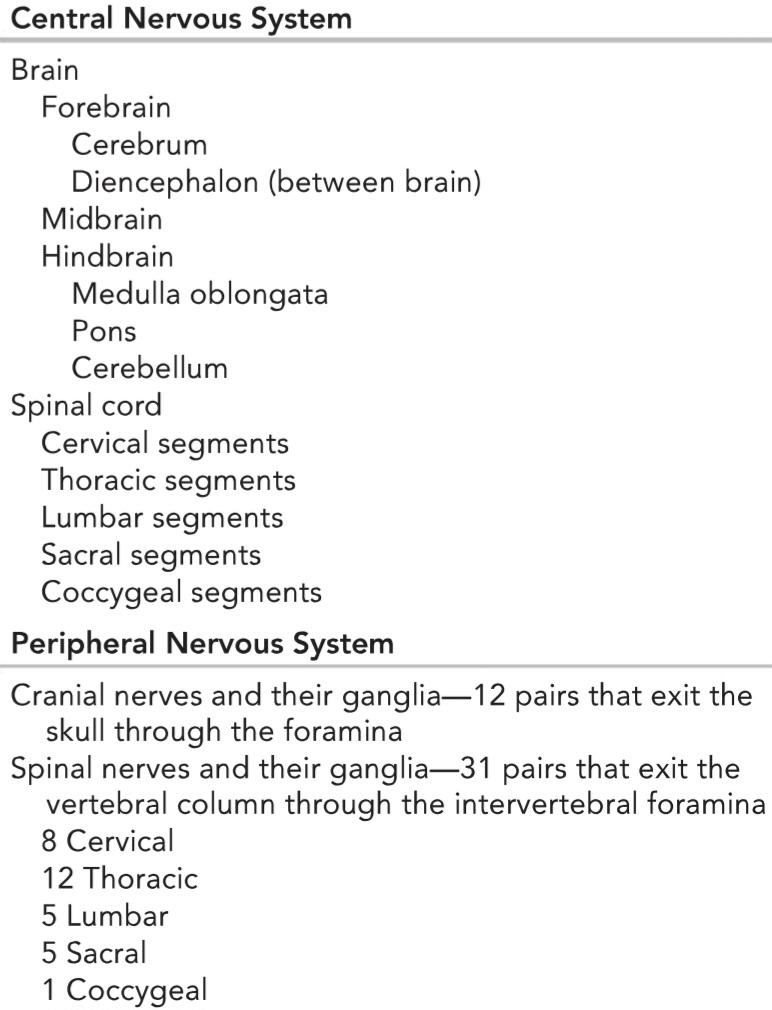

Figure 1-2 A: The protective covering of the spinal cord, the meninges, is formed by dura, arachnoid, and pia mater. The space between the arachnoid and pial membranes is called the subarachnoid space and contains cerebrospinal fluid (CSF). The subarachnoid space is enlarged at the cisterna magna and chiasmatic cistern. B: In the cranium, the dura consists of fused periosteal and meningeal layers that separate to form dural sinuses. Arachnoid mater projects into the dural venous sinuses to drain CSF from the subarachnoid space. (From Siegel, A., & Sapru, H. N. [2015]. Essential neuroscience [3rd ed.]. Baltimore, MD: Wolters Kluwer.)
شکل ۱-۲ الف: پوشش محافظ نخاع، مننژ، توسط سختشامه، عنکبوتیه و نرمشامه تشکیل شده است. فضای بین غشاهای عنکبوتیه و نرمشامه، فضای زیر عنکبوتیه نامیده میشود و حاوی مایع مغزی نخاعی (CSF) است. فضای زیر عنکبوتیه در سیسترنا ماگنا و سیسترن کیاسماتیک گسترش مییابد. ب: در جمجمه، سختشامه از لایههای پریوستئال و مننژیال به هم پیوسته تشکیل شده است که برای تشکیل سینوسهای سختشامه از هم جدا میشوند. ماده عنکبوتیه به سینوسهای وریدی سختشامه بیرون میزند تا CSF را از فضای زیر عنکبوتیه تخلیه کند. (از Siegel, A., & Sapru, H. N. [۲۰۱۵]. Essential neuroscience [ویرایش سوم]. Baltimore, MD: Wolters Kluwer.)

Figure 1-3 Photomicrograph of several large nerve cells with surrounding neuroglia. N, Neuron; n, nucleus; Ng, neuroglia; Np, neuropili; arrows, neurites. (From Gartner, L. P. [2017]. Color atlas and text of histology [7th ed.]. Baltimore, MD: Wolters Kluwer.)
شکل ۱-۳ عکس میکروسکوپی از چندین سلول عصبی بزرگ به همراه نوروگلیاهای اطراف. N، نورون؛ n، هسته؛ Ng، نوروگلیا؛ Np، نوروپیلی؛ فلشها، نوریتها. (از گارتنر، ال. پی. [۲۰۱۷]. اطلس رنگی و متن بافتشناسی [ویرایش هفتم]. بالتیمور، مریلند: ولترز کلوور.)
Spinal Cord
The spinal cord is situated within the vertebral canal of the vertebral column and is surrounded by three meninges (Figs. 1-4 and 1-5): the dura mater, the arachnoid mater, and the pia mater. Further protection is provided by the CSF, which surrounds the spinal cord in the subarachnoid space.
نخاع
نخاع در داخل کانال مهرهای ستون فقرات قرار دارد و توسط سه مننژ احاطه شده است (شکلهای ۱-۴ و ۱-۵): سختشامه، عنکبوتیه و نرمشامه. محافظت بیشتر توسط مایع مغزی نخاعی (CSF) که نخاع را در فضای زیر عنکبوتیه احاطه کرده است، انجام میشود.

Figure 1-4 A: Fetus with the brain and spinal cord exposed on the posterior surface. Note that the spinal cord extends the full length of the vertebral column. B: Sagittal section of the vertebral column in an adult showing the spinal cord terminating inferiorly at the level of the lower border of the 1st lumbar vertebra. C: Adult spinal cord and covering meninges showing the relationship to surrounding structures.
شکل ۱-۴ الف: جنینی که مغز و نخاع او در سطح خلفی نمایان است. توجه داشته باشید که نخاع در تمام طول ستون فقرات امتداد دارد. ب: برش ساژیتال ستون فقرات در یک فرد بالغ که نشان میدهد نخاع در سطح لبه پایینی اولین مهره کمری به سمت پایین ختم میشود. ج: نخاع بالغ و مننژهای پوشاننده آن که ارتباط آن با ساختارهای اطراف را نشان میدهد.
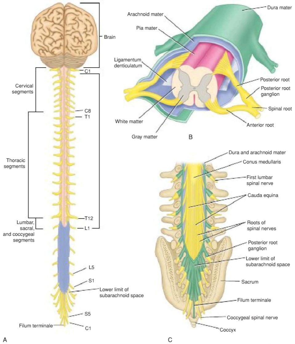
Figure 1-5 A: Brain, spinal cord, spinal nerve roots, and spinal nerves as seen on their posterior aspect. B: Transverse section through the thoracic region of the spinal cord showing the anterior and posterior roots of a spinal nerve and the meninges. C: Posterior view of the lower end of the spinal cord and cauda equina showing their relationship with the lumbar vertebrae, sacrum, and соссух.
شکل ۱-۵ الف: مغز، نخاع، ریشههای عصب نخاعی و اعصاب نخاعی از نمای خلفی. ب: برش عرضی از ناحیه سینهای نخاع که ریشههای قدامی و خلفی یک عصب نخاعی و مننژ را نشان میدهد. ج: نمای خلفی انتهای تحتانی نخاع و دم اسب که ارتباط آنها را با مهرههای کمری، خاجی و استخوان دنبالچه نشان میدهد.
The spinal cord is roughly cylindrical and begins superiorly at the foramen magnum in the skull, where it is continuous with the medulla oblongata of the brain. It terminates inferiorly in the lumbar region. Below, the spinal cord tapers off into the conus medullaris, from the apex of which the filum terminale (a prolongation of the pia mater) descends to attach to the back of the coccyx (see Fig. 1-4B).
نخاع تقریباً استوانهای شکل است و از بالا از سوراخ بزرگ جمجمه شروع میشود، جایی که با بصل النخاع مغز در امتداد است. در پایین در ناحیه کمری خاتمه مییابد. در پایین، نخاع به مخروط انتهایی باریک میشود، که از رأس آن فیلوم ترمینال (ادامه نرمشامه) پایین میآید تا به پشت دنبالچه متصل شود (شکل 1-4B را ببینید).
Along the entire length of the spinal cord, 31 pairs of spinal nerves are attached by the anterior or motor roots and the posterior or sensory roots (Fig. 1-6; also see Fig. 1-5). Each root is attached to the cord by a series of rootlets, which extend the whole length of the corresponding segment of the cord. Each posterior nerve root possesses a posterior root ganglion, the cells of which give rise to peripheral and central nerve fibers.
در تمام طول نخاع، ۳۱ جفت عصب نخاعی توسط ریشههای قدامی یا حرکتی و ریشههای خلفی یا حسی به هم متصل شدهاند (شکل ۱-۶؛ همچنین به شکل ۱-۵ مراجعه کنید). هر ریشه توسط مجموعهای از ریشههای کوچک به نخاع متصل میشود که در تمام طول قطعه مربوطه از نخاع امتداد دارند. هر ریشه عصبی خلفی دارای یک گانگلیون ریشه خلفی است که سلولهای آن باعث ایجاد فیبرهای عصبی محیطی و مرکزی میشوند.

Figure 1-6 A: Transverse section through the lumbar part of the spinal cord, oblique view. B: Transverse section through the lumbar part of the spinal cord, face view, showing the anterior and posterior roots of a spinal nerve.
شکل ۱-۶ الف: برش عرضی از طریق قسمت کمری نخاع، نمای مایل. ب: برش عرضی از طریق قسمت کمری نخاع، نمای روبرو، ریشههای قدامی و خلفی یک عصب نخاعی را نشان میدهد.
Spinal Cord Structure
The spinal cord is composed of an inner core of gray matter, which is surrounded by an outer covering of white matter. The gray matter is seen on cross section as an H-shaped pillar with anterior and posterior gray columns, or horns, united by a thin gray commissure containing the small central canal. The white matter, for purposes of description, is divided into anterior, lateral, and posterior white columns (see Fig. 1-6).
ساختار نخاع
نخاع از یک هسته داخلی ماده خاکستری تشکیل شده است که توسط یک پوشش خارجی از ماده سفید احاطه شده است. ماده خاکستری در مقطع عرضی به صورت یک ستون H شکل با ستونها یا شاخهای خاکستری قدامی و خلفی دیده میشود که توسط یک رابط خاکستری نازک حاوی کانال مرکزی کوچک به هم متصل شدهاند. ماده سفید، برای توصیف، به ستونهای سفید قدامی، جانبی و خلفی تقسیم میشود (شکل 1-6 را ببینید).
Brain
The brain (Fig. 1-7) lies in the cranial cavity and is continuous with the spinal cord through the foramen magnum (see Fig. 1-5A). As shown in Figure 1-2, it is surrounded by the dura mater, the arachnoid mater, and the pia mater. These three meninges are continuous with the corresponding meninges of the spinal cord. The CSF surrounds the brain in the subarachnoid space.
مغز
مغز (شکل ۱-۷) در حفره جمجمه قرار دارد و از طریق سوراخ بزرگ (فورامن مگنوم) در امتداد نخاع قرار دارد (شکل ۱-۵A را ببینید). همانطور که در شکل ۱-۲ نشان داده شده است، مغز توسط سخت شامه، عنکبوتیه و نرم شامه احاطه شده است. این سه مننژ در امتداد مننژهای مربوط به نخاع قرار دارند. مایع مغزی نخاعی (CSF) مغز را در فضای زیر عنکبوتیه احاطه کرده است.

Figure 1-7 Lateral view of the brain within the skull.
شکل ۱-۷ نمای جانبی مغز درون جمجمه.
The brain is conventionally divided into three major divisions: the hindbrain, the midbrain, and the forebrain in ascending order from the spinal cord (see Fig. 1-1A). The brainstem (a collective term for the medulla oblongata, pons, and midbrain) is what remains after the cerebral hemispheres and cerebellum (see below) are removed.
مغز به طور مرسوم به سه بخش اصلی تقسیم میشود: مغز پسین، مغز میانی و مغز پیشین به ترتیب صعودی از نخاع (شکل 1-1A را ببینید). ساقه مغز (اصطلاحی کلی برای بصل النخاع، پل مغزی و مغز میانی) چیزی است که پس از برداشتن نیمکرههای مغزی و مخچه (به پایین مراجعه کنید) باقی میماند.
Hindbrain
The hindbrain comprises the medulla oblongata, the pons, and the cerebellum.
مغز پسین
مغز پسین شامل بصل النخاع، پل مغزی و مخچه است.
Medulla Oblongata
The medulla oblongata is conical in shape and connects the pons superiorly to the spinal cord inferiorly (Fig. 1-8). It contains many collections of neurons, called nuclei, and serves as a conduit for ascending and descending nerve fibers.
بصل النخاع
بصل النخاع مخروطی شکل است و پل مغزی را از بالا به نخاع از پایین متصل میکند (شکل ۱-۸). این بخش شامل مجموعههای زیادی از نورونها به نام هسته است و به عنوان مجرایی برای فیبرهای عصبی صعودی و نزولی عمل میکند.

Figure 1-8 Inferior view of the brain.
شکل ۱-۸ نمای تحتانی مغز.
Pons
The pons is situated on the anterior surface of the cerebellum, inferior to the midbrain and superior to the medulla oblongata (Fig. 1-9; also see Fig. 1-8). The pons, or bridge, derives its name from the large number of transverse fibers on its anterior aspect connecting the two cerebellar hemispheres. It also contains many nuclei and ascending and descending nerve fibers.
پل مغزی
پل مغزی در سطح قدامی مخچه، پایینتر از مغز میانی و بالاتر از بصل النخاع قرار دارد (شکل 1-9؛ همچنین به شکل 1-8 مراجعه کنید). پل مغزی یا پل، نام خود را از تعداد زیادی فیبر عرضی در سطح قدامی خود که دو نیمکره مخچه را به هم متصل میکند، گرفته است. همچنین حاوی هستههای زیاد و فیبرهای عصبی صعودی و نزولی است.
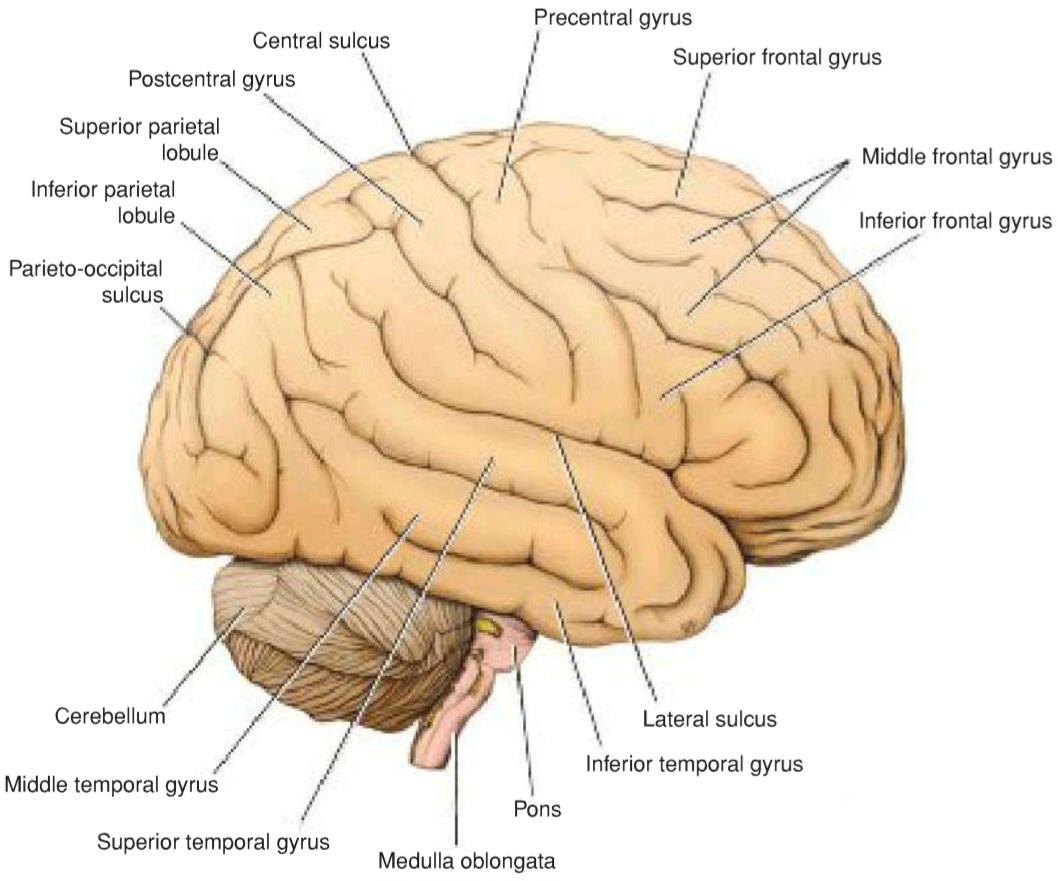
Figure 1-9 Brain viewed from its right lateral aspect.
شکل ۱-۹ مغز از نمای جانبی راست آن.
Cerebellum
The cerebellum lies within the posterior cranial fossa of the skull (see Figs. 1-7 to 1-9), posterior to the pons and the medulla oblongata. It consists of two laterally placed hemispheres connected by a median portion, the vermis. The cerebellum is connected to the midbrain by the superior cerebellar peduncles, to the pons by the middle cerebellar peduncles, and to the medulla by the inferior cerebellar peduncles (see Fig. 6-9). The peduncles are composed of large bundles of nerve fibers connecting the cerebellum to the remainder of the nervous system.
مخچه
مخچه در حفره جمجمهای خلفی جمجمه (شکلهای ۱-۷ تا ۱-۹)، در خلف پل مغزی و بصل النخاع قرار دارد. این مخچه از دو نیمکره جانبی تشکیل شده است که توسط یک قسمت میانی به نام ورمیس به هم متصل شدهاند. مخچه توسط پایههای مخچهای فوقانی به مغز میانی، توسط پایههای مخچهای میانی به پل مغزی و توسط پایههای مخچهای تحتانی به بصل النخاع متصل است (شکل ۶-۹ را ببینید). پایهها از دستههای بزرگی از الیاف عصبی تشکیل شدهاند که مخچه را به بقیه سیستم عصبی متصل میکنند.
The surface layer of each cerebellar hemisphere is called the cortex and is composed of gray matter (Fig. 1-10). The cerebellar cortex is thrown into folds, or folia, separated by closely set transverse fissures. Certain masses of gray matter are found in the interior of the cerebellum, embedded in the white matter; the largest of these is known as the dentate nucleus (see Fig. 6-7).
لایه سطحی هر نیمکره مخچه، قشر مخچه نامیده میشود و از ماده خاکستری تشکیل شده است (شکل 1-10). قشر مخچه به صورت چینها یا فولیاهایی (folia) قرار گرفته است که توسط شیارهای عرضی نزدیک به هم از هم جدا میشوند. تودههای خاصی از ماده خاکستری در داخل مخچه یافت میشوند که در ماده سفید قرار گرفتهاند. بزرگترین آنها به عنوان هسته دندانهدار شناخته میشود (شکل 6-7 را ببینید).

Figure 1-10 Median sagittal section of the brain to show the third ventricle, the cerebral aqueduct, and the fourth ventricle.
شکل ۱-۱۰ برش ساژیتال میانی مغز برای نشان دادن بطن سوم، مجرای مغزی و بطن چهارم.
The medulla oblongata, the pons, and the cerebellum surround a cavity filled with CSF, called the fourth ventricle. This is connected superiorly to the third ventricle by the cerebral aqueduct; inferiorly, it is continuous with the central canal of the spinal cord (Fig. 1-11). It communicates with the subarachnoid space through three openings in the inferior part of the roof. Through these openings, the CSF within the CNS can enter the subarachnoid space.
بصل النخاع، پل مغزی و مخچه حفرهای پر از مایع مغزی نخاعی (CSF) به نام بطن چهارم را احاطه کردهاند. این بطن از بالا توسط مجرای مغزی به بطن سوم متصل است؛ از پایین، با کانال مرکزی نخاع در ارتباط است (شکل 1-11). این بطن از طریق سه روزنه در قسمت تحتانی سقف با فضای زیر عنکبوتیه ارتباط برقرار میکند. از طریق این روزنهها، مایع مغزی نخاعی درون سیستم عصبی مرکزی میتواند وارد فضای زیر عنکبوتیه شود.
Midbrain
The midbrain is the narrow part of the brain that connects the forebrain to the hindbrain (see Figs. 1-1A and 1-10). The narrow cavity of the midbrain is the cerebral aqueduct, which connects the third and fourth ventricles. The midbrain contains many nuclei and bundles of ascending and descending nerve fibers.
مغز میانی
مغز میانی بخش باریکی از مغز است که مغز پیشین را به مغز پسین متصل میکند (شکلهای ۱-۱A و ۱-۱۰ را ببینید). حفره باریک مغز میانی، مجرای مغزی است که بطنهای سوم و چهارم را به هم متصل میکند. مغز میانی شامل هستههای زیاد و دستههایی از فیبرهای عصبی صعودی و نزولی است.
Forebrain
The forebrain comprises the diencephalon (between brain), which is the central part of the forebrain, and the cerebrum.
مغز پیشین
مغز پیشین شامل دیانسفالون (بین مغز)، که بخش مرکزی مغز پیشین است، و مخ میشود.
Diencephalon
The diencephalon is almost completely hidden from the surface of the brain. It consists of a dorsal thalamus and a ventral hypothalamus (see Fig. 1-10). The thalamus is a large, egg-shaped mass of gray matter that lies on either side of the third ventricle. The anterior end of the thalamus forms the posterior boundary of the interventricular foramen, the opening between the third and lateral ventricles. The hypothalamus forms the lower part of the lateral wall and floor of the third ventricle.
دیانسفالون
دیانسفالون تقریباً به طور کامل از سطح مغز پنهان است. این بخش از یک تالاموس پشتی و یک هیپوتالاموس شکمی تشکیل شده است (شکل 1-10 را ببینید). تالاموس یک توده بزرگ و تخم مرغی شکل از ماده خاکستری است که در دو طرف بطن سوم قرار دارد. انتهای قدامی تالاموس مرز خلفی سوراخ بین بطنی، دهانه بین بطن سوم و بطنهای جانبی، را تشکیل میدهد. هیپوتالاموس قسمت پایینی دیواره جانبی و کف بطن سوم را تشکیل میدهد.

Figure 1-11 Sagittal section through the brainstem and the cerebellum.
شکل ۱-۱۱ برش ساژیتال از ساقه مغز و مخچه.
Cerebrum
The cerebrum, the largest part of the brain, consists of two cerebral hemispheres, which are connected by a mass of white matter called the corpus callosum (see Figs. 1-9 and 1-10). Each hemisphere extends from the frontal to the occipital bones in the skull, superior to the anterior and middle cranial fossae; posteriorly, the cerebrum lies above the tentorium cerebelli (see Fig. 15-3). The hemispheres are separated by a deep cleft, the longitudinal fissure, into which projects the falx cerebri (see Fig. 15-1).
مخ
مخ، بزرگترین بخش مغز، از دو نیمکره مغزی تشکیل شده است که توسط تودهای از ماده سفید به نام جسم پینهای به هم متصل شدهاند (شکلهای ۱-۹ و ۱-۱۰ را ببینید). هر نیمکره از استخوانهای پیشانی تا استخوانهای پسسری در جمجمه، بالاتر از حفرههای جمجمهای قدامی و میانی امتداد دارد؛ در قسمت خلفی، مخ بالای چادرینه مخچه قرار دارد (شکل ۱۵-۳ را ببینید). نیمکرهها توسط یک شکاف عمیق، شکاف طولی، از هم جدا میشوند که داس مغزی به داخل آن بیرون زده است (شکل ۱۵-۱ را ببینید).
The surface layer of each hemisphere, the cortex, is composed of gray matter. The cerebral cortex is thrown into folds (gyri) separated by fissures, or sulci (see Fig. 1-9). This arrangement greatly increases the surface area of the cortex. A number of the large sulci are conveniently used to subdivide the surface of each hemisphere into lobes, which are named from the bones of the cranium they lie under.
لایه سطحی هر نیمکره، قشر مغز، از ماده خاکستری تشکیل شده است. قشر مغز به صورت چینهایی (gyri) قرار گرفته است که توسط شیارها یا شیارها از هم جدا شدهاند (شکل 1-9 را ببینید). این چیدمان، مساحت سطح قشر را به میزان قابل توجهی افزایش میدهد. تعدادی از شیارهای بزرگ به راحتی برای تقسیم سطح هر نیمکره به لوبها استفاده میشوند که از استخوانهای جمجمهای که در زیر آنها قرار دارند، نامگذاری شدهاند.
Within the hemisphere is a central core of white matter containing several large masses of gray matter, the basal nuclei or ganglia. A fan-shaped collection of nerve fibers, the corona radiata (Fig. 1-12), passes in the white matter to and from the cerebral cortex to the brainstem. The corona radiata converges on the basal nuclei and passes between them as the internal capsule. The tailed nucleus situated on the medial side of the internal capsule is the caudate nucleus (Fig. 1-13), and the lens-shaped nucleus on the lateral side of the internal capsule is the lentiform nucleus.
درون نیمکره، یک هسته مرکزی از ماده سفید وجود دارد که حاوی چندین توده بزرگ ماده خاکستری، هستههای پایه یا گانگلیونها، است. مجموعهای بادبزنی شکل از فیبرهای عصبی، تاج شعاعی (شکل 1-12)، در ماده سفید به قشر مغز و از آن به ساقه مغز میرود. تاج شعاعی روی هستههای پایه همگرا میشود و از بین آنها به عنوان کپسول داخلی عبور میکند. هسته دمدار که در سمت داخلی کپسول داخلی قرار دارد، هسته دمدار (شکل 1-13) است و هسته عدسی شکل در سمت جانبی کپسول داخلی، هسته عدسی شکل است.
Within each cerebral hemisphere is a cavity called the lateral ventricle (see Figs. 16-2 and 16-3). The lateral ventricles communicate with the third ventricle through the interventricular foramina.
درون هر نیمکره مغزی حفرهای به نام بطن جانبی وجود دارد (شکلهای 16-2 و 16-3 را ببینید). بطنهای جانبی از طریق سوراخهای بین بطنی با بطن سوم ارتباط برقرار میکنند.
During the process of development, the cerebrum becomes enormously enlarged and overhangs the diencephalon, the midbrain, and the hindbrain.
در طول فرآیند رشد، مخ به شدت بزرگ میشود و از دیانسفالون، مغز میانی و مغز خلفی بیرون میزند.

Figure 1-12 Right lateral view showing continuity of the corona radiata, the internal capsule, and the crus cerebri of the cerebral peduncles. Note the position of the lentiform nucleus lateral to the internal capsule.
شکل ۱-۱۲ نمای جانبی راست که پیوستگی تاج شعاعی، کپسول داخلی و ساق مغزی پایههای مغزی را نشان میدهد. به موقعیت هسته عدسی شکل در سمت جانبی کپسول داخلی توجه کنید.
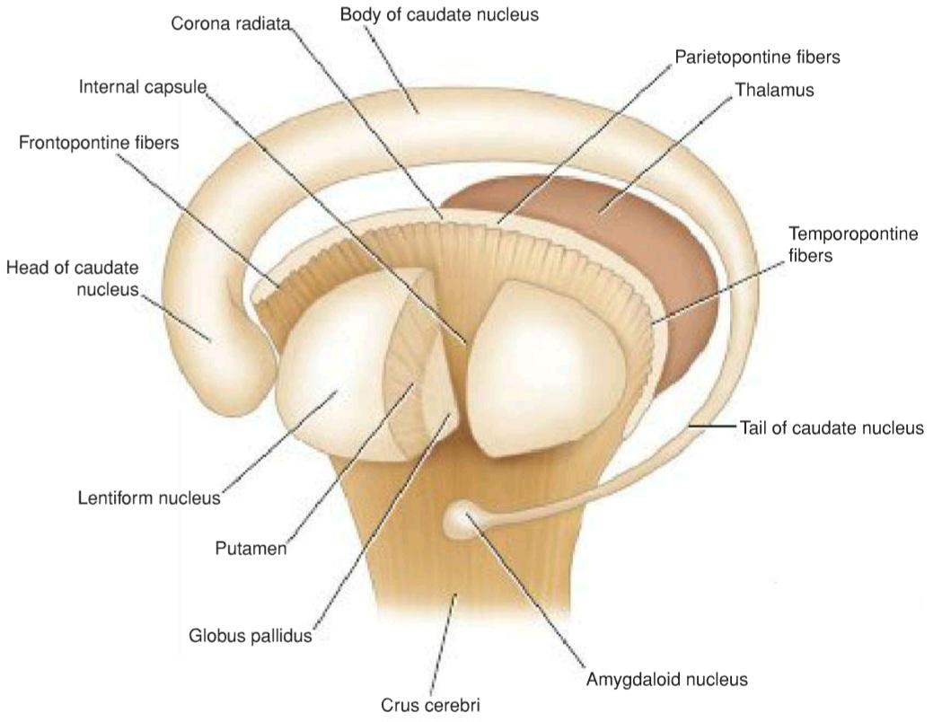
Figure 1-13 Diagram showing the relationship between the lentiform nucleus, the caudate nucleus, the Amygdaloid nucleus thalamus, and the internal capsule, as seen from the left lateral side.
شکل ۱-۱۳ نموداری که رابطه بین هسته عدسی شکل، هسته دمدار، هسته آمیگدالوئید تالاموس و کپسول داخلی را از نمای جانبی چپ نشان میدهد.
Brain Structure
Unlike the spinal cord, the brain is composed of an inner core of white matter, which is surrounded by an outer covering of gray matter. However, as mentioned previously, certain important masses of gray matter are situated deeply within the white matter: the gray cerebellar nuclei in the cerebellum and the gray thalamic, caudate, and lentiform nuclei in the cerebrum.
ساختار مغز
برخلاف نخاع، مغز از یک هسته داخلی ماده سفید تشکیل شده است که توسط یک پوشش بیرونی از ماده خاکستری احاطه شده است. با این حال، همانطور که قبلاً ذکر شد، تودههای مهم خاصی از ماده خاکستری در اعماق ماده سفید قرار دارند: هستههای خاکستری مخچه در مخچه و هستههای خاکستری تالاموس، دمی و عدسی شکل در مخ.
«فصل اول کتاب نورواناتومی بالینی اسنل»
🚀 با ما همراه شوید!
تازهترین مطالب و آموزشهای مغز و اعصاب را از دست ندهید. با فالو کردن کانال تلگرام آیندهنگاران مغز، از ما حمایت کنید!




عالی
🌹👌🏻👌🏻🌹