
علوم اعصاب شناختی؛ تشخیص ابژه؛ درک و کشف رمزگذاری ویژگی های چهره


دعای مطالعه [ نمایش ]
بِسْمِ الله الرَّحْمنِ الرَّحیمِ
اَللّهُمَّ اَخْرِجْنى مِنْ ظُلُماتِ الْوَهْمِ
خدايا مرا بيرون آور از تاريكىهاى وهم،
وَ اَكْرِمْنى بِنُورِ الْفَهْمِ
و به نور فهم گرامى ام بدار،
اَللّهُمَّ افْتَحْ عَلَيْنا اَبْوابَ رَحْمَتِكَ
خدايا درهاى رحمتت را به روى ما بگشا،
وَانْشُرْ عَلَيْنا خَزائِنَ عُلُومِكَ بِرَحْمَتِكَ يا اَرْحَمَ الرّاحِمينَ
و خزانههاى علومت را بر ما باز كن به امید رحمتت اى مهربانترين مهربانان.
» Cognitive Neuroscience: The Biology of the Mind
»» CHAPTER 6: Object Recognition; part three
6.4 Specificity of Object Recognition in Higher Visual Areas
6.4 ویژگی تشخیص اشیاء در مناطق بصری عالی
When we meet someone, we first look at the person’s face. In no culture do individuals look at thumbs, knees, or other body parts to recognize one another. The tendency to focus on faces reflects behavior that is deeply embedded in our evolutionary history. Facial features tell us about age, health, and gender, while facial expressions provide salient cues regarding emotional states, helping us to discriminate between pleasure and displeasure, friendship and antagonism, agreement and confusion.
وقتی با کسی ملاقات میکنیم، ابتدا به چهره آن شخص نگاه میکنیم. در هیچ فرهنگی افراد برای تشخیص یکدیگر به انگشتان شست، زانو یا سایر اعضای بدن نگاه نمیکنند. تمایل به تمرکز بر چهرهها نشان دهنده رفتاری است که عمیقاً در تاریخ تکاملی ما جا افتاده است. ویژگیهای چهره به ما درباره سن، سلامتی و جنسیت میگویند، در حالی که حالات چهره نشانههای برجسته ای را در مورد حالات عاطفی ارائه میدهد و به ما کمک میکند بین لذت و ناراحتی، دوستی و تضاد، توافق و سردرگمیتمایز قائل شویم.
The face, and particularly the eyes, of another person can provide significant clues about what is important in his environment. Looking at someone’s lips when she is speaking helps us to understand words more than we may realize. These observations, combined with neuropsychological reports of patients who had difficulty recognizing faces, led researchers to search for the neural mechanisms underlying face perception.
چهره و به خصوص چشمان شخص دیگری میتواند سرنخهای قابل توجهی در مورد آنچه در محیط او مهم است ارائه دهد. وقتی کسی در حال صحبت کردن است، نگاه کردن به لبها به ما کمک میکند تا کلمات را بیشتر از آنچه تصور میکنیم درک کنیم. این مشاهدات، همراه با گزارشهای عصبی روانشناختی بیمارانی که در تشخیص چهره مشکل داشتند، محققان را به جستجوی مکانیسمهای عصبی زیربنایی درک چهره سوق داد.
Is Face Processing Special?
آیا پردازش چهره خاص است؟
It seems reasonable to suppose that our brains have a general-purpose system for recognizing all sorts of visual inputs, with faces constituting just one important class of problems to solve. However, the evidence suggests otherwise. Multiple studies argue that face perception does not use the same processing mechanisms as those used in object recognition, but instead depends on a specialized network of brain regions.
منطقی به نظر میرسد که فرض کنیم مغز ما دارای یک سیستم همه منظوره برای تشخیص انواع ورودیهای بصری است که چهرهها فقط یک دسته مهم از مسائل را تشکیل میدهند که باید حل شوند. با این حال، شواهد خلاف این را نشان میدهد. مطالعات متعدد استدلال میکنند که ادراک چهره از مکانیزمهای پردازشی مشابهی که در تشخیص اشیا استفاده میشود استفاده نمیکند، بلکه به شبکهای تخصصی از مناطق مغز بستگی دارد.
To investigate whether face recognition and other forms of object perception use distinct processing systems, we should consider whether there are particular regions of the brain or specialized cells that respond to faces separately from those that respond to other types of stimuli. If so, we should ask whether the systems are functionally and operationally independent. The logic of this query is essentially the same as that underlying the idea of double dissociations (see Chapter 3). Finally, we should ask whether the two systems process information in a different way. Let’s see what evidence we have to answer these questions.
برای بررسی اینکه آیا تشخیص چهره و سایر اشکال ادراک شی از سیستمهای پردازش مجزا استفاده میکنند، باید در نظر بگیریم که آیا مناطق خاصی از مغز یا سلولهای تخصصی وجود دارد که به چهره جداگانه از آنهایی که به انواع دیگر محرکها پاسخ میدهند پاسخ میدهند. اگر چنین است، باید بپرسیم که آیا سیستمها از نظر عملکردی و عملیاتی مستقل هستند یا خیر. منطق این پرس و جو اساساً همان منطقی است که زیربنای ایده تفکیک دوگانه است (به فصل 3 مراجعه کنید). در نهایت، باید بپرسیم که آیا دو سیستم اطلاعات را به روشی متفاوت پردازش میکنند؟ بیایید ببینیم چه مدرکی برای پاسخ به این سوالات داریم.
Do the processes of face recognition and nonfacial object recognition involve physically distinct mechanisms? Although clinical evidence showed that people could have what appeared to be selective problems in face perception, more compelling evidence of specialized face perception mechanisms comes from neurophysiological studies with nonhuman primates. In one study (Baylis et al., 1985), recordings were made from cells in the superior temporal sulcus (STS) of a monkey while the animal was presented with stimuli like those in Figure 6.29a.
آیا فرآیندهای تشخیص چهره و تشخیص اشیاء غیر چهره شامل مکانیسمهای فیزیکی متمایز هستند؟ اگرچه شواهد بالینی نشان میدهد که افراد ممکن است مشکلاتی انتخابی در ادراک چهره داشته باشند، شواهد قانعکنندهتر از مکانیسمهای تخصصی ادراک چهره از مطالعات نوروفیزیولوژیکی با پستانداران غیرانسانی به دست آمده است. در یک مطالعه (Baylis و همکاران، 1985)، ضبطهایی از سلولهای شیار گیجگاهی فوقانی (STS) یک میمون انجام شد، در حالی که حیوان با محرکهایی مانند شکل 6.29a ارائه شد.
Five of these stimuli (A-E) were faces: four of other monkeys, and one of a human (one of the experimenters). The other five stimuli (F-J) ranged in complexity but included the most prominent features in the facial stimuli. For example, the grating (image G) reflected the symmetry of faces, and the circle (image I) was similar to eyes. Figure 6.29b shows two cells that responded selectively to the five facial stimuli. These cells did not change their firing rate to the nonfacial stimuli, and for two of the stimuli, the response of cell 1 actually decreased to some of the nonfacial stimuli.
پنج مورد از این محرکها (A-E) چهره بودند: چهار میمون دیگر و یکی از یک انسان (یکی از آزمایشکنندگان). پنج محرک دیگر (F-J) دارای پیچیدگی بودند اما برجستهترین ویژگیها را در محرکهای چهره شامل میشدند. به عنوان مثال، توری (تصویر G) تقارن چهرهها را منعکس میکند و دایره (تصویر I) شبیه چشمها بود. شکل 6.29b دو سلول را نشان میدهد که به طور انتخابی به پنج محرک چهره پاسخ دادند. این سلولها سرعت شلیک خود را به محرکهای غیرچهرهی تغییر ندادند و برای دو مورد از محرکها، پاسخ سلول 1 در واقع به برخی از محرکهای غیرچهرهی کاهش یافت.
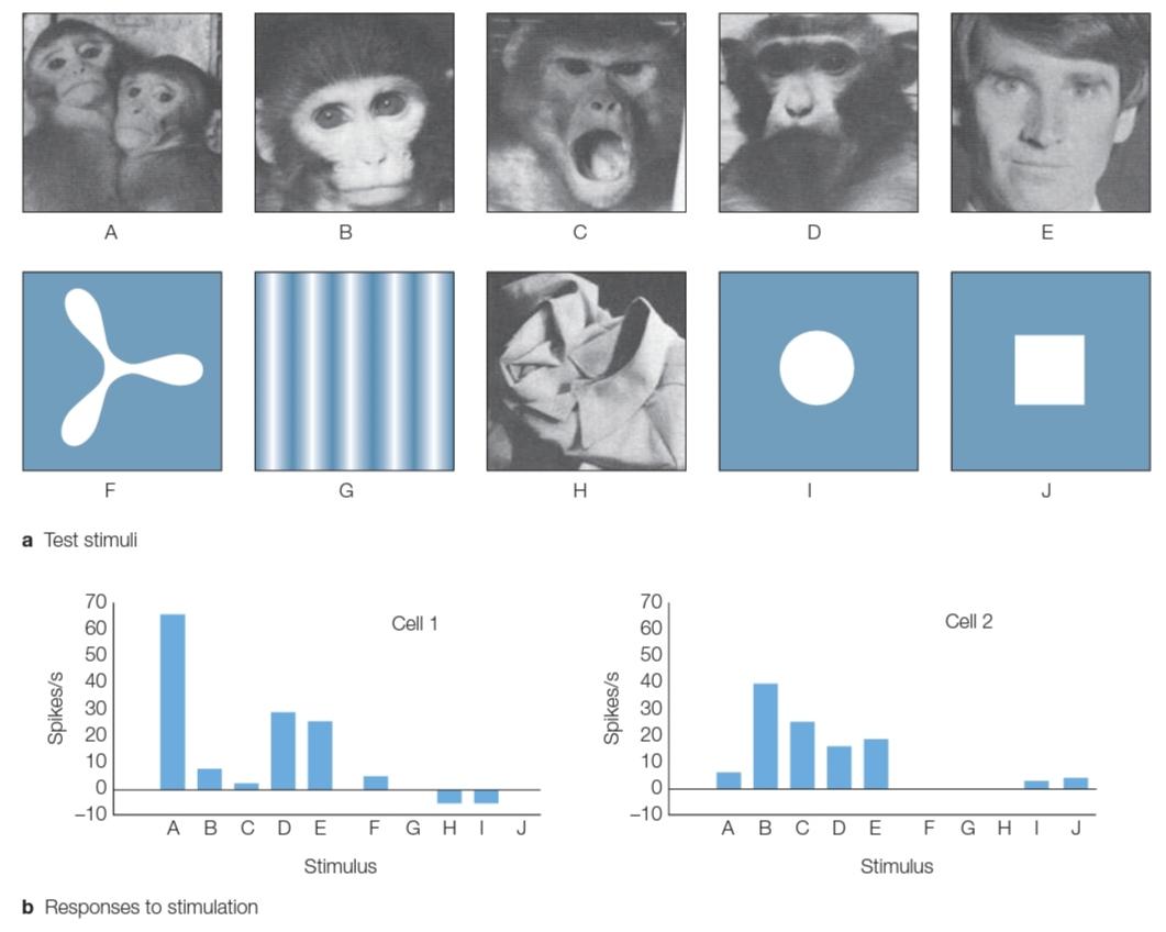
FIGURE 6.29 Identifying face cells in the superior temporal sulcus of the macaque monkey. The 10 stimuli (A-J) in part (a) produced the responses plotted in part (b) for two cells. Both cells responded vigorously to many of the facial stimuli. Either there was no change in activity when the animal looked at the objects, or, in some cases, the cells were actually inhibited relative to baseline. The firing-rate data are plotted as a change from baseline activity for that cell when no stimulus was presented.
شکل 6.29 شناسایی سلولهای چهره در شیار گیجگاهی فوقانی میمون ماکاک. 10 محرک (A-J) در قسمت (الف) پاسخهایی را که در قسمت (ب) برای دو سلول ترسیم شده است، ایجاد کردند. هر دو سلول به شدت به بسیاری از محرکهای چهره پاسخ دادند. یا وقتی حیوان به اشیاء نگاه میکرد، هیچ تغییری در فعالیت وجود نداشت، یا در برخی موارد، سلولها در واقع نسبت به خط پایه مهار میشدند. هنگامیکه هیچ محرکی ارائه نشده بود، دادههای سرعت شلیک بهعنوان تغییری از فعالیت پایه برای آن سلول ترسیم میشوند.
A subsequent study dramatically demonstrated the degree of this specificity by combining two neurophysiological methods in a novel manner. Monkeys were first placed in an fMRI scanner and shown pictures of faces or objects. As expected, sectors of the STS showed greater activation to the face stimuli (Tsao et al., 2006; Figure 6.30a).
مطالعه بعدی به طور چشمگیری درجه این ویژگی را با ترکیب دو روش عصبی فیزیولوژیکی به روشی جدید نشان داد. میمونها ابتدا در یک اسکنر fMRI قرار داده شدند و تصاویری از چهرهها یا اشیاء نشان داده شدند. همانطور که انتظار میرفت، بخشهای STS فعال شدن بیشتری را برای محرکهای چهره نشان دادند (تسائو و همکاران، 2006؛ شکل 6.30a).
The researchers then went on to record from individual neurons, using the imaging results to position the electrodes within one of the face-sensitive subregions. In that subregion, 97% of the neurons exhibited a strong preference for faces, showing strong responses to any face-containing stimulus and minimal responses to a wide range of other stimuli, such as body parts, food, or objects (Figure 6.30b, c). These regions in macaque monkeys, which have been shown to be specialized for processing faces and have a high concentration of face-selective cells, are called face patches. These data provide one of the most dramatic examples of stimulus specificity within a restricted part of the visual system.
سپس محققان با استفاده از نتایج تصویربرداری برای قرار دادن الکترودها در یکی از زیرمنطقههای حساس به چهره، از تک تک نورونها ضبط کردند. در آن منطقه فرعی، 97 درصد از نورونها ترجیح زیادی برای چهرهها نشان میدهند، که پاسخهای قوی به هر محرک حاوی چهره و کمترین پاسخ را به طیف وسیعی از محرکهای دیگر، مانند اعضای بدن، غذا، یا اشیا نشان میدهند (شکل 6.30b، c). این نواحی در میمونهای ماکاک که مشخص شده برای پردازش چهرهها تخصصی هستند و دارای غلظت بالایی از سلولهای انتخابکننده چهره هستند، تکههای چهره نامیده میشوند. این دادهها یکی از چشمگیرترین نمونههای اختصاصی بودن محرک را در بخش محدودی از سیستم بینایی ارائه میکنند.
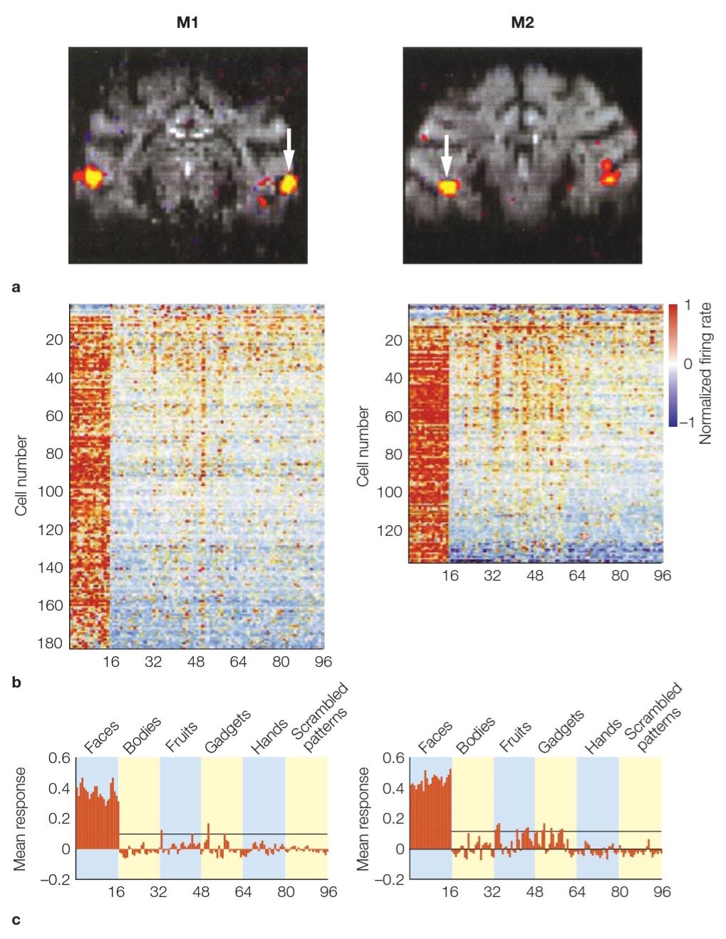
FIGURE 6.30 Superior temporal sulcus (STS) regions that respond to faces. (a) Functional MRI activations during face perception in two macaque monkeys (M1 and M2). The white arrows indicate where subsequent neurophysiological recording was done (left STS in M1 and right STS in M2). (b) The activity of each of the cells recorded in the STS of M1 (182 cells) and M2 (138 cells) that responded to visual stimuli (face, bodies, fruits, gadgets, hands, or scrambled patterns). In these graphs, each row corresponds to a different cell, and each column corresponds to a different image, grouped by categories. (c) The average response size for each of the image categories across all cells. These cells were highly selective for face stimuli.
شکل 6.30 نواحی شیار گیجگاهی فوقانی (STS) که به چهرهها پاسخ میدهند. (الف) فعال سازی عملکردی MRI در طول درک چهره در دو میمون ماکاک (M1 و M2). فلشهای سفید نشان میدهد که کجا ضبط نوروفیزیولوژیک بعدی انجام شده است (STS چپ در M1 و STS راست در M2). (ب) فعالیت هر یک از سلولهای ثبت شده در STS M1 (182 سلول) و M2 (138 سلول) که به محرکهای بینایی (چهره، بدن، میوهها، وسایل، دستها یا الگوهای درهم) پاسخ میدهند. در این نمودارها، هر سطر مربوط به یک سلول متفاوت است و هر ستون مربوط به یک تصویر متفاوت است که بر اساس دسته بندی گروه بندی شده است. (ج) اندازه متوسط پاسخ برای هر یک از دستههای تصویر در تمام سلولها. این سلولها برای محرکهای چهره بسیار انتخابی بودند.
Functional MRI has also been used in many studies to explore how faces are perceived in the human brain. As in the monkey study just described, we can compare conditions in which the participants view different classes of stimuli, using either block or event-related designs. These studies consistently reveal a network of areas in which the BOLD response is strongest to face stimuli. The most prominent of these is along the ventral surface of the temporal lobe in the fusiform gyrus (Figure 6.31), leading researchers to refer to this region as the fusiform face area or FFA, a term that combines anatomy and function.
MRI عملکردی همچنین در بسیاری از مطالعات برای بررسی نحوه درک چهره در مغز انسان استفاده شده است. همانطور که در مطالعه میمونی که توضیح داده شد، میتوانیم شرایطی را با هم مقایسه کنیم که در آن شرکتکنندگان کلاسهای مختلفی از محرکها را با استفاده از طرحهای بلوک یا مربوط به رویداد مشاهده میکنند. این مطالعات به طور مداوم شبکه ای از مناطق را نشان میدهد که در آنها واکنش BOLD قوی ترین واکنش به رویارویی با محرکها است. برجسته ترین آنها در امتداد سطح شکمیلوب گیجگاهی در شکنج دوکی شکل است (شکل 6.31)، که باعث شد محققان از این ناحیه به عنوان ناحیه دوکی شکل یا FFA نام ببرند، اصطلاحی که آناتومیو عملکرد را ترکیب میکند.
The FFA is not the only region in humans that shows a strong BOLD response to faces relative to other visual stimuli. Consistent with primate studies, face regions have been identified in other parts of the temporal lobe, including the STS. Furthermore, fMRI has shown that different face-sensitive regions are specialized for processing certain types of information that can be gleaned from faces.
FFA تنها منطقه ای در انسان نیست که نسبت به سایر محرکهای بصری به چهرهها واکنش BOLD قوی نشان میدهد. مطابق با مطالعات پستانداران، نواحی چهره در سایر قسمتهای لوب تمپورال از جمله STS شناسایی شدهاند. علاوه بر این، fMRI نشان داده است که مناطق مختلف حساس به چهره برای پردازش انواع خاصی از اطلاعات که میتوان از چهرهها به دست آورد، تخصصی شده است.
For example, the more ventral face pathway is sensitive to static, invariant features of facial structure (e.g., eye spacing), information important for facial identification. In contrast, face-selective regions in the right STS respond to movement information (Pitcher et al., 2011), such as the direction of people’s gaze, their emotional expressions, and their mouth movements. This distinction can be observed even when the faces are presented so quickly that people fail to perceive them consciously (Jiang & He, 2006). The BOLD response still shows an increase in FFA activity, independent of whether the faces depicted strong emotional expressions, while the response in the STS is observed only for the emotive faces.
برای مثال، مسیر شکمیتر چهره به ویژگیهای ثابت و ثابت ساختار چهره (به عنوان مثال فاصله چشم)، اطلاعات مهم برای شناسایی چهره حساس است. در مقابل، مناطق انتخابی چهره در STS مناسب به اطلاعات حرکتی پاسخ میدهند (پیچر و همکاران، 2011)، مانند جهت نگاه افراد، عبارات احساسی و حرکات دهانشان. این تمایز را میتوان حتی زمانی مشاهده کرد که چهرهها آنقدر سریع ارائه میشوند که مردم قادر به درک آگاهانه آنها نیستند (جیانگ و او، 2006). پاسخ BOLD همچنان افزایشی را در فعالیت FFA نشان میدهد، مستقل از اینکه آیا چهرهها عبارات احساسی قوی را نشان میدهند، در حالی که پاسخ در STS فقط برای چهرههای احساسی مشاهده میشود.
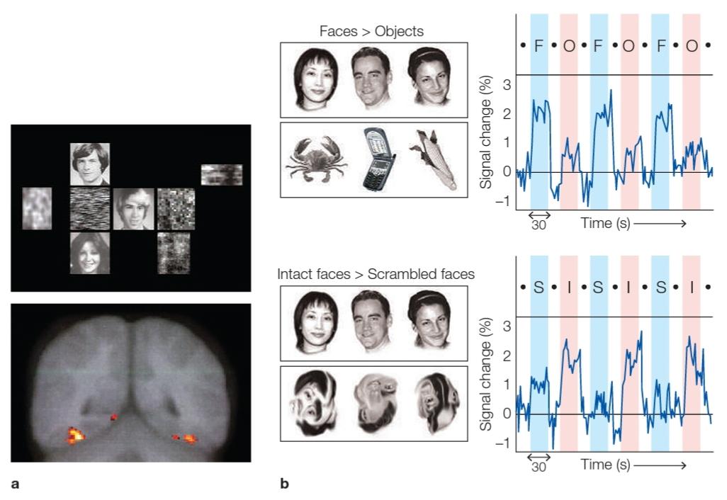
FIGURE 6.31 Isolating neural regions during face perception.
(a) Bilateral activation in the fusiform gyrus was observed with fMRI when participants viewed collages of faces and random patterns, as compared with collages of only random patterns. Note that, according to neuroradiological conventions, the right hemisphere is on the left. (b) In another fMRI study, participants viewed alternating blocks of stimuli. In one scanning run (top), the stimuli alternated between faces (F) and objects (0); in another run (bottom), they alternated between intact (I) and scrambled (S) faces. The right column shows the BOLD signal in the fusiform face area during the scanning run for the various stimuli. In each interval, the stimuli were drawn from the different sets, and these stimuli were separated by short intervals of fixation only. The BOLD signal was much stronger during intervals in which faces (versus objects) or intact faces (versus scrambled faces) were presented.
شکل 6.31 جداسازی نواحی عصبی در طول ادراک چهره.
(الف) زمانی که شرکتکنندگان کلاژ چهرهها و الگوهای تصادفی را مشاهده کردند، در مقایسه با کلاژهایی که فقط الگوهای تصادفی داشتند، فعالسازی دو طرفه در شکنج دوکی شکل با fMRI مشاهده شد. توجه داشته باشید که طبق قراردادهای نورورادیولوژیک، نیمکره راست در سمت چپ قرار دارد. (ب) در یک مطالعه fMRI دیگر، شرکت کنندگان بلوکهای متناوب محرک را مشاهده کردند. در یک مرحله اسکن (بالا)، محرکها بین چهرهها (F) و اشیاء (0) متناوب بودند. در یک اجرا دیگر (پایین)، آنها بین چهرههای دست نخورده (I) و درهم (S) متناوب شدند. ستون سمت راست سیگنال BOLD را در ناحیه چهره دوکی در طول اجرای اسکن برای محرکهای مختلف نشان میدهد. در هر بازه، محرکها از مجموعههای مختلف کشیده میشدند و این محرکها تنها با فواصل کوتاه تثبیت از هم جدا میشدند. سیگنال BOLD در فواصل زمانی که در آن چهرهها (در مقابل اشیاء) یا چهرههای دست نخورده (در مقابل چهرههای درهم) ارائه میشد، بسیار قوی تر بود.
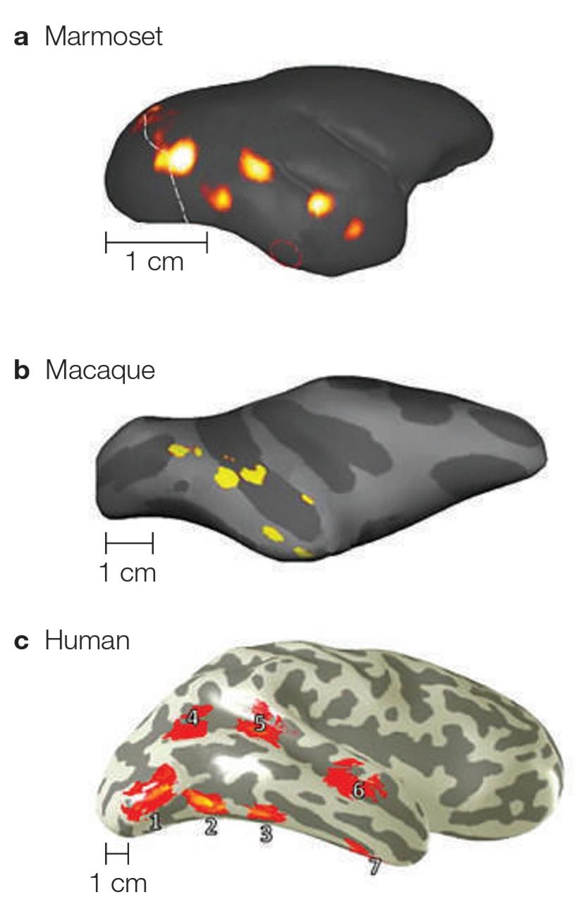
FIGURE 6.32 The evolution of face-processing networks. Cortical surface of a marmoset (a), a macaque (b), and a human (c). Yellow to red colors indicate homologous face-selective regions for each species. 1 = inferior occipital gyrus; 2 = posterior fusiform gyrus; 3 = medial fusiform gyrus; 4 = posterior superior temporal sulcus; 5 = middle superior temporal sulcus; 6 = anterior superior temporal sulcus; 7 = anterior temporal sulcus.
شکل 6.32 تکامل شبکههای پردازش چهره. سطح قشری یک مارموست (الف)، یک ماکاک (ب) و یک انسان (ج). رنگهای زرد تا قرمز، مناطق همولوگ انتخابی چهره را برای هر گونه نشان میدهد. 1 = شکنج اکسیپیتال تحتانی. 2 = شکنج دوکی شکل خلفی. 3 = شکنج دوکی شکل داخلی. 4 = شیار گیجگاهی فوقانی خلفی. 5 = شیار گیجگاهی فوقانی میانی. 6 = شیار گیجگاهی فوقانی قدامی. 7 = شیار گیجگاهی قدامی.
Recently, the marmoset, a monkey that is 10,000 years older evolutionarily than the macaque and 35 million years older than humans, was added to the list of primates with face recognition abilities (C. C. Hung et al., 2015). The marmoset brain is 12 times smaller than that of a macaque and 180 times smaller than a human brain. Even so, marmosets have a face recognition network that closely resembles the distribution of face patches observed in macaques and humans (Figure 6.32), and the homology also appears to hold at a functional level. For example, in the marmoset, neural activity in the more dorsal face patches is modulated by natural motions associated with faces, such as movements of the mouth or eyes, whereas the more ventral face regions are sensitive to static properties of faces (Weiner & Grill-Spector, 2015).
اخیراً مارموست، میمونی که از نظر تکاملی 10000 سال از ماکاک و 35 میلیون سال پیرتر از انسان است، به فهرست نخستیهای دارای توانایی تشخیص چهره اضافه شد (C. C. Hung et al., 2015). مغز مارموست 12 برابر کوچکتر از مغز ماکاک و 180 برابر کوچکتر از مغز انسان است. با این حال، مارموستها دارای یک شبکه تشخیص چهره هستند که شباهت زیادی به توزیع تکههای چهره مشاهده شده در ماکاکها و انسانها دارد (شکل 6.32)، و به نظر میرسد همسانی در سطح عملکردی باشد. به عنوان مثال، در مارموست، فعالیت عصبی در تکههای چهره پشتیتر توسط حرکات طبیعی مرتبط با چهرهها، مانند حرکات دهان یا چشمها تعدیل میشود، در حالی که نواحی شکمیتر چهره به خواص ساکن چهرهها حساس هستند (Weiner & Grill-Spector, 2015).
Electrophysiological methods also reveal a neural signature of face perception in humans. Faces elicit a large negative evoked response in the EEG signal approximately 170 ms after stimulus onset, or what is known as the N170 response. A similar negative deflection is found for other classes of objects, such as cars, birds, and furniture, but the magnitude of the response is much larger for human faces (Carmel & Bentin, 2002; Figure 6.33). Interestingly, the stimuli need not be pictures of real human faces. The N170 response is also elicited when people view faces of apes and when the facial stimuli are crude, schematic line drawings (Sagiv & Bentin, 2001).
روشهای الکتروفیزیولوژیک نیز نشانه عصبی ادراک چهره را در انسان نشان میدهد. چهرهها یک پاسخ برانگیخته منفی بزرگ را در سیگنال EEG تقریباً 170 میلی ثانیه پس از شروع محرک ایجاد میکنند، یا آنچه به عنوان پاسخ N170 شناخته میشود. انحراف منفی مشابهی برای سایر کلاسهای اشیاء، مانند ماشینها، پرندگان، و مبلمان مشاهده میشود، اما بزرگی پاسخ برای چهرههای انسان بسیار بزرگتر است (کارمل و بنتین، 2002؛ شکل 6.33). جالب اینجاست که محرکها نیازی به عکسهایی از چهرههای واقعی انسان ندارند. پاسخ N170 همچنین زمانی که افراد چهره میمونها را مشاهده میکنند و زمانی که محرکهای چهره ترسیمهای خطی شماتیک هستند، ایجاد میشود (Sagiv & Bentin، 2001).
Although face stimuli are very good at producing activation in the FFA in humans, it is important to consider alternative hypotheses. One idea is that this region is recruited when people have to make fine perceptual discriminations among highly familiar stimuli. Advocates of this hypothesis point out that imaging studies comparing face and object recognition usually entail an important, if underemphasized, confound: the level of expertise.
اگرچه محرکهای چهره در ایجاد فعال سازی در FFA در انسان بسیار خوب هستند، مهم است که فرضیههای جایگزین را در نظر بگیریم. یک ایده این است که این منطقه زمانی به کار گرفته میشود که افراد باید بین محرکهای بسیار آشنا تفاوتهای ادراکی خوبی ایجاد کنند. طرفداران این فرضیه خاطرنشان میکنند که مطالعات تصویربرداری که تشخیص چهره و شی را با هم مقایسه میکنند، معمولاً مستلزم یک آشفتگی مهم، اگر کم تأکید شود، هستند: سطح تخصص.
Consider a comparison of faces and flowers. Although neurologically healthy individuals are all experts in perceiving faces, the same is not true when it comes to perceiving flowers. Unless you are a botanist or an avid gardener, you are unlikely to be an expert in recognizing flowers. In addition, faces and flowers differ in terms of their social relevance: Face perception is essential to our social interactions. Whether or not we set out to remember someone’s face, we readily encode the features that distinguish one face from another. The same is probably not true for other classes of objects. Most of us are happy to recognize that a particular picture is of a pretty flower, perhaps even to note that it is a rose. But unless you are a rose enthusiast, you are not likely to recognize or encode the difference between a Dazzler and a Garibaldi, nor will you be able to recognize a particular individual rose that you have already seen.
مقایسه چهرهها و گلها را در نظر بگیرید. اگرچه افراد سالم از نظر عصبی همگی در درک چهره متخصص هستند، اما در مورد درک گلها این موضوع صادق نیست. اگر گیاه شناس یا باغبان مشتاق نباشید، بعید است در تشخیص گلها متخصص باشید. علاوه بر این، چهرهها و گلها از نظر ارتباط اجتماعی متفاوت هستند: درک چهره برای تعاملات اجتماعی ما ضروری است. چه بخواهیم چهره کسی را به خاطر بسپاریم یا نه، به راحتی ویژگیهایی را رمزگذاری میکنیم که یک چهره را از چهره دیگر متمایز میکند. این احتمالاً برای سایر کلاسهای اشیاء صادق نیست. بسیاری از ما خوشحالیم که میدانیم یک عکس خاص از یک گل زیبا است، شاید حتی اگر توجه داشته باشیم که یک گل رز است. اما تا زمانی که از علاقه مندان به گل رز نباشید، به احتمال زیاد تفاوت بین Dazzler و Garibaldi را نمیتوانید تشخیص دهید یا رمزگذاری کنید، و همچنین نمیتوانید یک گل رز خاص را که قبلاً دیده اید، تشخیص دهید.
To address this confound, researchers have used imaging studies to determine whether the FFA is activated in people who are experts at discriminating within specific classes of objects, such as cars or birds (Gauthier et al., 2000). The results are somewhat mixed. Activation in fusiform cortex, which is made up of more than just the FFA, is in fact greater when people view objects for which they have some expertise. For example, car aficionados show a stronger FFA response to cars than to birds.
برای رفع این آشفتگی، محققان از مطالعات تصویربرداری برای تعیین اینکه آیا FFA در افرادی که در تشخیص طبقههای خاصی از اشیاء، مانند ماشینها یا پرندگان خبره هستند، فعال میشود یا خیر، استفاده کردهاند (Gauthier et al., 2000). نتایج تا حدودی مختلط است. فعال شدن در قشر دوکی شکل، که از چیزی بیش از FFA تشکیل شده است، در واقع زمانی بیشتر میشود که افراد اشیایی را مشاهده کنند که در آن تخصص دارند. به عنوان مثال، علاقه مندان به اتومبیل، واکنش FFA قوی تری به اتومبیلها نسبت به پرندگان نشان میدهند.
What’s more, if participants are trained to make fine discriminations between novel objects, the fusiform response increases as expertise develops (Gauthier et al., 1999). However, the categorization of objects by experts activates a much broader region of ventral OTC, extending well beyond the FFA (Grill-Spector et al., 2004; Rhodes et al., 2004). It appears that, even when expertise effects are considered, there remains a strong bias for facial stimuli within more restricted regions such as the FFA.
علاوه بر این، اگر شرکت کنندگان برای ایجاد تمایز دقیق بین اشیاء جدید آموزش ببینند، پاسخ دوکی شکل با توسعه تخصص افزایش مییابد (Gauthier et al., 1999). با این حال، طبقهبندی اشیاء توسط متخصصان، ناحیه بسیار وسیعتری از OTC شکمیرا فعال میکند، که بسیار فراتر از FFA گسترش مییابد (گریل-اسپکتور و همکاران، 2004؛ رودز و همکاران، 2004). به نظر میرسد که حتی زمانی که اثرات تخصصی در نظر گرفته میشود، یک سوگیری قوی برای محرکهای چهره در مناطق محدودتر مانند FFA وجود دارد.
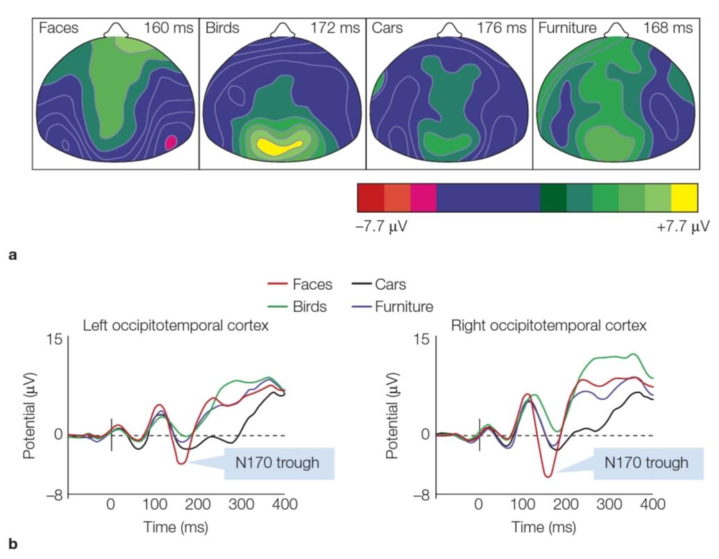
FIGURE 6.33 Electrophysiological response to faces: the N170 response. (a) Participants viewed pictures of faces, birds, cars, and furniture and were instructed to press a button whenever they saw a picture of a car. (b) Electrodes placed over occipitotemporal cortex produced these event-related potentials. Note that the negative-going deflection in the waveform around 170 ms is much larger for the face stimuli compared to the other categories.
شکل 6.33 پاسخ الکتروفیزیولوژیک به چهرهها: پاسخ N170. (الف) شرکتکنندگان تصاویری از چهرهها، پرندگان، ماشینها و مبلمان را مشاهده کردند و به آنها دستور داده شد که هر زمان که تصویری از یک ماشین دیدند، دکمهای را فشار دهند. (ب) الکترودهای قرار داده شده بر روی قشر occipitotemporal این پتانسیلهای مرتبط با رویداد را تولید میکنند. توجه داشته باشید که انحراف منفی در شکل موج در حدود 170 میلی ثانیه برای محرکهای چهره در مقایسه با دستههای دیگر بسیار بزرگتر است.
Diving Deeply Into Facial Perception
غواصی عمیق در درک چهره
In a landmark study, Le Chang and Doris Tsao (2017) at the California Institute of Technology claimed to have broken the code for face perception, at least in terms of how neurons represent information that supports our amazing ability to discriminate between faces and recognize individuals. To do so, they first took a set of 200 faces, chosen at random with the assumption that this selection process ensured a representative space that could characterize just about any face. (It is noteworthy that they used human faces here, rather than monkey faces.)
در یک مطالعه برجسته، لو چانگ و دوریس تسائو (2017) در مؤسسه فناوری کالیفرنیا ادعا کردند که کد ادراک چهره را شکسته اند، حداقل از نظر نحوه نمایش نورونها اطلاعاتی که از توانایی شگفت انگیز ما برای تمایز بین چهرهها و تشخیص افراد پشتیبانی میکند. برای انجام این کار، آنها ابتدا مجموعه ای از 200 چهره را انتخاب کردند که به چهره تصادفی انتخاب شدند با این فرض که این فرآیند انتخاب فضایی نماینده را تضمین میکند که میتواند تقریباً هر چهره ای را مشخص کند. (قابل توجه است که در اینجا به جای چهره میمونی از چهره انسان استفاده میکردند.)
By marking 58 key points on each face (Figure 6.34a), they could then use algorithms to generate the data set’s mathematically average face. They were also able to determine how different each face was from the average face and from other faces. The researchers used statistical measures to analyze this complex multidimensional space, asking whether certain dimensions best captured the similarities and differences between faces. Two prominent dimensions emerged: variation in shape (Figure 6.34b) and variation in appearance (Figure 6.34c). The re- searchers identified 25 unique parameters that captured variation on the shape dimension (e.g., width, height, roundness), and another 25 that captured variation on the appearance dimension (e.g., openness of eyes, hairstyle). With these tools, each of the 200 initial faces could be assigned a unique position in this 50-dimensional space.
با علامتگذاری 58 نقطه کلیدی در هر وجه (شکل 6.34a)، آنها میتوانند از الگوریتمهایی برای تولید صورت میانگین ریاضی مجموعه داده استفاده کنند. آنها همچنین توانستند تعیین کنند که هر چهره چقدر با چهره معمولی و سایر چهرهها متفاوت است. محققان از معیارهای آماری برای تجزیه و تحلیل این فضای چند بعدی پیچیده استفاده کردند و پرسیدند که آیا برخی از ابعاد به بهترین شکل شباهتها و تفاوتهای بین چهرهها را نشان میدهد یا خیر. دو بعد برجسته ظاهر شد: تنوع در شکل (شکل 6.34b) و تنوع در ظاهر (شکل 6.34c). محققان 25 پارامتر منحصر به فرد را شناسایی کردند که تغییرات ابعاد شکل (به عنوان مثال، عرض، ارتفاع، گردی) و 25 پارامتر دیگر را که تغییرات در بعد ظاهری (مانند باز بودن چشمها، مدل مو) را نشان میدادند، شناسایی کردند. با استفاده از این ابزارها میتوان به هر یک از 200 چهره اولیه موقعیت منحصر به فردی در این فضای 50 بعدی اختصاص داد.
The researchers then turned to neurophysiology to ask whether these statistically derived dimensions were biologically relevant. First they used fMRI to identify face-sensitive areas in the inferior temporal cortex of the macaque. Then they employed single-cell-recording methods to characterize the tuning profiles of neurons in lateral (LAT) and anterior (ANT) face areas within the IT cortex. Not surprisingly, the cells responded vigorously to the facial stimuli, with many showing little responsiveness to other classes of stimuli.
محققان سپس به فیزیولوژی عصبی روی آوردند تا بپرسند آیا این ابعاد به دست آمده از نظر آماری از نظر بیولوژیکی مرتبط هستند یا خیر. ابتدا از fMRI برای شناسایی نواحی حساس به چهره در قشر گیجگاهی تحتانی ماکاک استفاده کردند. سپس آنها از روشهای ضبط تک سلولی برای توصیف پروفایل تنظیم نورونها در نواحی چهره جانبی (LAT) و قدامی(ANT) در قشر IT استفاده کردند. جای تعجب نیست که سلولها به شدت به محرکهای چهره واکنش نشان میدهند و بسیاری از آنها نسبت به سایر کلاسهای محرک واکنش کمینشان میدهند.
The results showed not only that the shape and appearance dimensions were critical in describing the tuning properties of the cells, but also that the two areas had differential specializations. The activity level of cells in the LAT region varied strongly as a function of the shape; in contrast, the activity level of cells in the ANT region varied strongly as a function of appearance (Figure 6.34d). Even more striking, the cells’ activity level varied little when shown two faces that differed markedly on the irrelevant dimension. For example, a shape-responsive cell fired at a similar rate to faces that shared a similar shape, even if the stimuli had markedly different hairstyles.
نتایج نشان داد که نه تنها ابعاد شکل و ظاهر در توصیف خواص تنظیم سلولها حیاتی است، بلکه این دو ناحیه دارای تخصصهای متفاوتی هستند. سطح فعالیت سلولها در منطقه LAT به شدت به عنوان تابعی از شکل متفاوت است. در مقابل، سطح فعالیت سلولها در ناحیه ANT به شدت به عنوان تابعی از ظاهر متفاوت بود (شکل 6.34d). حتی قابل توجه تر، سطح فعالیت سلولها زمانی که دو چهره به طور قابل توجهی در ابعاد نامربوط متفاوت بودند، کمیمتفاوت بود. به عنوان مثال، یک سلول پاسخگو به شکل، با سرعتی مشابه با چهرههایی که شکل مشابهی داشتند، شلیک میکند، حتی اگر محرکها مدل موی متفاوتی داشته باشند.
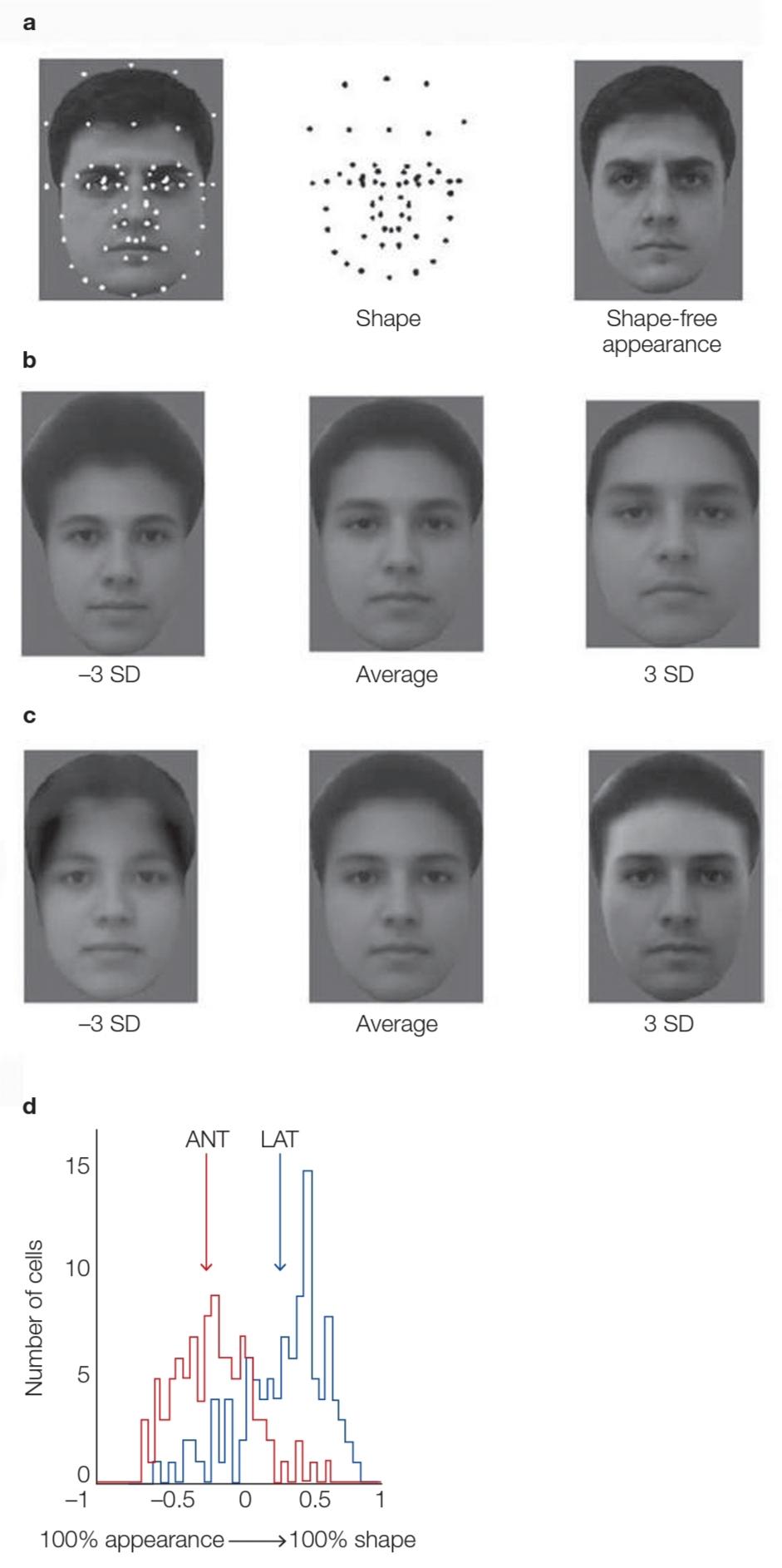
FIGURE 6.34 Discovering facial feature encoding.
(a) Dots were labeled on 200 facial images (left). The dot positions carried shape information about each facial image (middle). The landmarks were smoothly morphed to match the average landmark positions in the 200 faces, generating an image that carried shape-free appearance information about each face (right). (b) A face varying in shape across shape dimensions ranging from -3 standard deviations (SD) from the average to +3 standard deviations. The feature dimensions remain constant. (c) A face varying in the feature dimensions, with the shape remaining constant. (d) The number of cells in each area tested that were tuned to shape or appearance features. The lateral (LAT) face area cells (blue) within the IT cortex are tuned primarily to shape, whereas the anterior (ANT) face area cells (red) are tuned to appearance. The arrows indicate the population averages.
شکل 6.34 کشف رمزگذاری ویژگیهای چهره.
(الف) نقاط روی 200 تصویر چهره (سمت چپ) برچسب گذاری شدند. موقعیتهای نقطه حاوی اطلاعات شکل در مورد هر تصویر چهره (وسط). نشانهها به آرامیتغییر شکل دادند تا با میانگین موقعیتهای نقطه عطف در 200 چهره مطابقت داشته باشند، و تصویری ایجاد کردند که اطلاعات ظاهری بدون شکل را در مورد هر چهره (سمت راست) داشت. (ب) یک چهره با شکل متفاوت در ابعاد شکل از -3 انحراف استاندارد (SD) از میانگین تا +3 انحراف استاندارد. ابعاد ویژگی ثابت میماند. (ج) چهره ای که در ابعاد مشخصه متفاوت است و شکل آن ثابت میماند. (د) تعداد سلولها در هر ناحیه آزمایششده که با ویژگیهای شکل یا ظاهر تنظیم شدهاند. سلولهای ناحیه چهره جانبی (آبی) در قشر IT عمدتاً به شکل تنظیم میشوند، در حالی که سلولهای ناحیه چهره قدامی(ANT) (قرمز) به ظاهر تنظیم میشوند. فلشها میانگین جمعیت را نشان میدهند.
In effect, these researchers have created an encoding model for face perception-one that operates at the single-cell level. As described in Section 6.3, one test of an encoding model is how well the neural output of the model predicts the identity of a specific percept-in this case, how well the model decodes facial identity. Moreover, given this model’s 50-dimensional space, the researchers can generate a practically infinite set of faces to test with the decoder because each point in the space corresponds to a unique face.
در واقع، این محققان یک مدل رمزگذاری برای ادراک چهره ایجاد کرده اند که در سطح تک سلولی عمل میکند. همانطور که در بخش 6.3 توضیح داده شد، یک آزمون یک مدل رمزگذاری این است که خروجی عصبی مدل چقدر هویت یک ادراک خاص را پیشبینی میکند – در این مورد، مدل چقدر هویت چهره را رمزگشایی میکند. علاوه بر این، با توجه به فضای 50 بعدی این مدل، محققان میتوانند عملاً مجموعهای از چهرهها را برای آزمایش با رمزگشا ایجاد کنند، زیرا هر نقطه در فضا مربوط به یک چهره منحصر به فرد است.
After training a model with 2,000 faces, the researchers tested its decoding capability by showing it a novel set of 40 faces (Figure 6.35). The model was able to specify the correct face on about 50% of the trials using just the neural predictions from either face patch. Similar to what the physiological analysis had shown, accuracy remained high for LAT neurons if the model used only shape information and high for ANT neurons if the model used only appearance information. Other analyses showed that decoding accuracy remained high even if the stimuli varied in terms of viewing position or orientation, achieving a form of object constancy.
پس از آموزش مدلی با 2000 چهره، محققان قابلیت رمزگشایی آن را با نشان دادن مجموعه ای جدید از 40 چهره آزمایش کردند (شکل 6.35). این مدل تنها با استفاده از پیشبینیهای عصبی از هر کدام از وصلههای چهره، توانست چهره صحیح را در حدود 50 درصد از آزمایشها مشخص کند. مشابه آنچه که آنالیز فیزیولوژیکی نشان داده بود، اگر مدل فقط از اطلاعات شکل استفاده میکرد، دقت برای نورونهای LAT بالا باقی میماند و اگر مدل فقط از اطلاعات ظاهری استفاده میکرد برای نورونهای ANT بالا بود. تحلیلهای دیگر نشان داد که دقت رمزگشایی بالا باقی میماند، حتی اگر محرکها از نظر موقعیت یا جهت مشاهده متفاوت باشند، و به نوعی از ثبات جسم دست مییابند.
This pioneering study raises a number of interesting questions for future study. First, can this approach also be applied to understanding object recognition more generally, or are shape and appearance features specifically essential for face perception? Second, because activity was broadly distributed across the face-sensitive cells for all of the faces, it is more consistent with ensemble-coding models than with grandmother-cell models. Does this rule out the latter model? Or may the grandmother-cell model still apply further along the processing stream, perhaps when we have to give her a name?
این مطالعه پیشگام تعدادی سوال جالب را برای مطالعه آینده مطرح میکند. اولاً، آیا این رویکرد میتواند برای درک تشخیص شیء بهطور کلیتر نیز اعمال شود، یا ویژگیهای شکل و ظاهر بهطور خاص برای درک چهره ضروری هستند؟ دوم، از آنجا که فعالیت به طور گسترده در سلولهای حساس به چهره برای همه چهرهها توزیع شده است، با مدلهای کدگذاری گروهی سازگارتر است تا با مدلهای سلول مادربزرگ. آیا این مدل دوم را رد میکند؟ یا ممکن است مدل سلول مادربزرگ همچنان در طول جریان پردازش کاربرد بیشتری داشته باشد، شاید زمانی که باید نامیبرای او بگذاریم؟
Does the Visual System Contain Other Category-Specific Systems?
آیا سیستم بصری شامل سایر سیستمهای دستهبندی خاص است؟
The finding that marmosets have a face recognition network similar to that of humans pushes the evolutionary origins of such a network back at least to the common ancestor of New World and Old World primates—that is, about 50 million years ago. One hypothesis is that evolutionary pressures led to the development of a specialized system for face perception. If so, a natural question would be whether additional specialized systems exist for other biologically important classes of stimuli.
یافتهها مبنی بر اینکه مارموستها دارای یک شبکه تشخیص چهره مشابه با انسان هستند، منشأ تکاملی چنین شبکهای را حداقل به جد مشترک نخستیهای دنیای جدید و دنیای قدیم – یعنی حدود 50 میلیون سال پیش – برمیگرداند. یک فرضیه این است که فشارهای تکاملی منجر به توسعه یک سیستم تخصصی برای ادراک چهره شده است. اگر چنین باشد، یک سوال طبیعی این خواهد بود که آیا سیستمهای تخصصی اضافی برای سایر کلاسهای محرکهای مهم بیولوژیکی وجود دارد یا خیر.
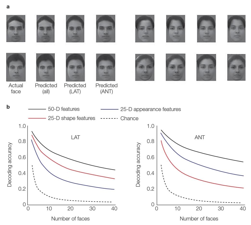
FIGURE 6.35 Decoding faces from neuronal output.
(a) Facial images reconstructed from the output of 200 cells. For comparison, the actual face is shown first, followed by what was predicted from combined data from both sites. predicted from just lateral (LAT) data, and predicted from just anterior (ANT) data. (b) Analysis showed the accuracy of the decoder using cells in LAT face area (left) or cells in ANT face area (right). Within each area, the decoder was based on activity related to shape features (red), appearance features (blue), or both (black), relative to chance (dotted). In LAT, decoding based on shape is more accurate; in ANT, decoding based on appearance is more accurate.
شکل 6.35 رمزگشایی چهرهها از خروجی عصبی.
(الف) تصاویر چهره بازسازی شده از خروجی 200 سلول. برای مقایسه، ابتدا چهره واقعی نشان داده میشود و سپس آنچه از دادههای ترکیبی از هر دو سایت پیش بینی شده بود، نشان داده میشود. پیشبینیشده از دادههای فقط جانبی (LAT) و پیشبینیشده از دادههای فقط قدامی(ANT). (ب) تجزیه و تحلیل دقت رمزگشا را با استفاده از سلولها در ناحیه چهره LAT (چپ) یا سلولها در ناحیه چهره ANT (راست) نشان داد. در هر ناحیه، رمزگشا بر اساس فعالیت مربوط به ویژگیهای شکل (قرمز)، ویژگیهای ظاهری (آبی)، یا هر دو (سیاه)، نسبت به شانس (نقطهدار) بود. در LAT، رمزگشایی بر اساس شکل دقیق تر است. در ANT رمزگشایی بر اساس ظاهر دقیق تر است.
In their investigations of the FFA, Russell Epstein and Nancy Kanwisher (1998) used a large set of control stimuli that were not faces. When they analyzed the results, they were struck by a serendipitous finding: One region of the ventral pathway, the parahippocampus, was consistently engaged when the control stimuli contained pictures of scenes such as land- scapes. This region was not activated by face stimuli or by pictures of individual objects. Subsequent experiments confirmed this pattern, leading to the name parahippocampal place area, or PPA. The BOLD response in this region was especially pronounced when people were required to make judgments about spatial properties or relations (e.g., “Is this an image of an outdoor or indoor scene?” or “Is the house at the base of the mountain?”).
راسل اپستین و نانسی کانویشر (1998) در تحقیقات خود در مورد FFA از مجموعه بزرگی از محرکهای کنترلی استفاده کردند که چهره نبودند. هنگامیکه آنها نتایج را تجزیه و تحلیل کردند، با یک یافته غیرمنتظره مواجه شدند: یک ناحیه از مسیر شکمی، پاراهیپوکامپ، زمانی که محرکهای کنترلی حاوی تصاویر صحنههایی مانند مناظر بود، به طور مداوم درگیر بود. این ناحیه توسط محرکهای چهره یا تصاویر اشیاء منفرد فعال نمیشد. آزمایشهای بعدی این الگو را تأیید کرد که منجر به نام منطقه مکان پاراهیپوکامپ یا PPA شد. واکنش جسورانه در این منطقه به ویژه زمانی که مردم ملزم به قضاوت در مورد ویژگیهای فضایی یا روابط بودند، آشکار میشد (به عنوان مثال، “این تصویری از یک صحنه بیرونی است یا داخلی؟” یا “آیا خانه در پایه کوه است؟”).
Reasonable evolutionary arguments can be made concerning why the brain might have dedicated regions devoted to recognizing faces or places, or to distinguishing animate objects (living things that move on their own) from inanimate objects (living things that don’t move on their own), or living things from nonliving things, but not to making other types of distinctions. Individuals who could distinguish one type of apple from another would be unlikely to have a strong adaptive advantage (although being able to perceive color differences that cue whether a particular piece of fruit is ripe would be important). Our ancestors who could remember where to find the ripe fruit, however, would have had a great advantage over their more forgetful peers. Clinical evidence supports the idea that there are regions dedicated to recognizing places. For example, patients with lesions to the parahippocampus become disoriented in new environments (Aguirre & D’Esposito, 1999; Habib & Sirigu, 1987).
استدلالهای تکاملی منطقی را میتوان در مورد اینکه چرا مغز ممکن است مناطقی را اختصاص داده باشد که به تشخیص چهرهها یا مکانها یا تمایز اجسام جاندار (جاندارانی که خود به خود حرکت میکنند) از اشیاء بیجان (جاندارانی که به خودی خود حرکت نمیکنند) یا موجودات زنده از موجودات غیرزنده، اما برای ایجاد انواع دیگر تمایزات اختصاص داده نشده است. افرادی که میتوانند یک نوع سیب را از دیگری تشخیص دهند، بعید است که مزیت تطبیقی قوی داشته باشند (اگرچه درک تفاوتهای رنگی که نشان دهنده رسیده بودن یک قطعه میوه خاص است مهم است). با این حال، اجداد ما که میتوانستند به خاطر بیاورند میوه رسیده را کجا پیدا کنند، نسبت به همسالان فراموشکارتر خود برتری زیادی داشتند. شواهد بالینی از این ایده حمایت میکند که مناطقی وجود دارند که به شناسایی مکانها اختصاص داده شده اند. به عنوان مثال، بیماران مبتلا به ضایعات پاراهیپوکامپ در محیطهای جدید دچار بی نظمیمیشوند (Aguirre & D’Esposito, 1999; Habib & Sirigu, 1987).
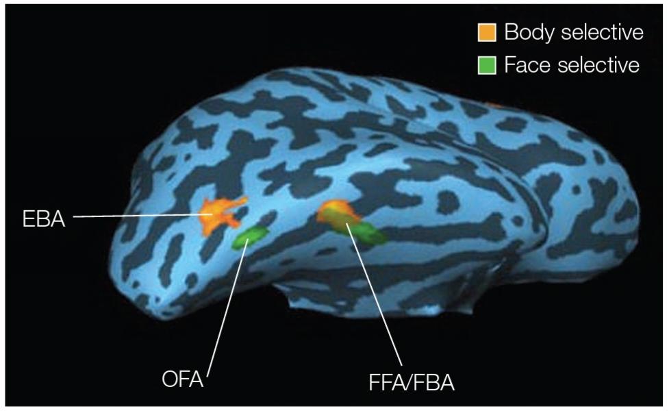
FIGURE 6.36 Locations of the EBA and FBA.
Right-hemisphere cortical surface of an “inflated brain” in one individual identifying the extrastriate body area (EBA), fusiform body area (FBA), and face-sensitive regions. Regions responded selectively to bodies or faces versus tools. Note that two regions responded to faces: the occipital face area (OFA) and the fusiform face area (FFA).
شکل 6.36 مکانهای EBA و FBA.
سطح قشر نیمکره راست یک “مغز متورم” در یک فرد که ناحیه خارج از بدن (EBA)، ناحیه بدن دوکی شکل (FBA) و مناطق حساس به چهره را شناسایی میکند. مناطق به طور انتخابی به بدن یا چهره در مقابل ابزار پاسخ دادند. توجه داشته باشید که دو ناحیه به چهرهها پاسخ دادند: ناحیه چهره پس سری (OFA) و ناحیه چهره دوکی (FFA).
Other studies suggest that the visual cortex may have a region that is especially important for recognizing parts of the body (Downing et al., 2001; Figure 6.36). This area, at the border of the occipital and temporal cortices, is referred to as the extrastriate body area (EBA). Another region, adjacent to and partially overlapping the FFA, shows a similar preference for body parts and has been called the fusiform body area (FBA) (Schwarzlose et al., 2005).
مطالعات دیگر نشان میدهد که قشر بینایی ممکن است دارای ناحیه ای باشد که به ویژه برای تشخیص بخشهای بدن مهم است (دانینگ و همکاران، 2001؛ شکل 6.36). این ناحیه در مرز قشر اکسیپیتال و گیجگاهی به عنوان ناحیه خارج از بدن (EBA) شناخته میشود. ناحیه دیگری که در مجاورت و تا حدی با FFA همپوشانی دارد، ترجیح مشابهی برای اعضای بدن نشان میدهد و ناحیه بدن دوکی شکل (FBA) نامیده میشود (Schwarzlose et al., 2005).
Testing Causality
آزمون علیت
Recording methods, either by single-cell physiology in the monkey or by fMRI and EEG recordings in people, are correlational in nature. Tests of causality generally require that the system be perturbed. For example, strokes can be considered a dramatic perturbation of normal brain function. More subtle methods involve transient perturbations. For example, when performing neurophysiological experiments in monkeys, the experimenter can make recordings to identify regions in which the cells show face-specific responses and then use the same electrodes to stimulate the brain (although the stimulation is generally at a level that activates many neurons).
روشهای ثبت، چه با فیزیولوژی تک سلولی در میمونها و چه با ثبت fMRI و EEG در افراد، ماهیت همبستگی دارند. آزمونهای علیت عموماً مستلزم این است که سیستم دچار اختلال شود. به عنوان مثال، سکته مغزی را میتوان یک اختلال چشمگیر در عملکرد طبیعی مغز در نظر گرفت. روشهای ظریف تر شامل آشفتگیهای گذرا هستند. به عنوان مثال، هنگام انجام آزمایشهای عصبی فیزیولوژیکی روی میمونها، آزمایشکننده میتواند برای شناسایی مناطقی که در آن سلولها پاسخهای خاص چهره را نشان میدهند، ضبط کند و سپس از همان الکترودها برای تحریک مغز استفاده کند (اگرچه تحریک عموماً در سطحی است که بسیاری از نورونها را فعال میکند).
Hossein Esteky and colleagues at the Shahid Beheshti University in Tehran used microstimulation to test the causal contribution of the IT cortex to face perception (Afraz et al., 2006). The monkeys were shown fuzzy images, and they had to judge whether the image included a flower or a face. When the IT cortex was stimulated, the monkeys showed a bias to report seeing a face. This effect was not seen when the microstimulation was targeted at nearby regions of the cortex.
حسین استکی و همکارانش در دانشگاه شهید بهشتی تهران از میکروتحریک برای آزمایش سهم علی قشر فناوری اطلاعات در ادراک چهره استفاده کردند (افراز و همکاران، 2006). به میمونها تصاویر مبهم نشان داده شد و آنها باید قضاوت میکردند که آیا این تصویر شامل یک گل است یا یک چهره. وقتی قشر IT تحریک شد، میمونها تمایلی به گزارش دیدن چهره نشان دادند. این اثر زمانی که ریزتحریک در نواحی مجاور قشر هدف قرار گرفت دیده نشد.
Although these results suggest that microstimulation of the IT cortex boosted activity in face-sensitive cells and resulted in a bias to see faces, we might expect that the random activation of these cells would be detrimental to identifying specific faces. A recent study (Moeller et al., 2017) showed just that. Monkeys were sequentially shown two faces and were required to move their eyes to indicate whether the faces were the same or different. In the absence of stimulation, the monkeys were excellent at judging the two faces to be the same (91% correct) and performed above chance when the faces were different (67% correct).
اگرچه این نتایج نشان میدهد که ریزتحریک قشر فناوری اطلاعات باعث افزایش فعالیت در سلولهای حساس به چهره شده و منجر به سوگیری برای دیدن چهرهها میشود، ممکن است انتظار داشته باشیم که فعالسازی تصادفی این سلولها برای شناسایی چهرههای خاص مضر باشد. مطالعه اخیر (Moeller et al., 2017) دقیقاً همین را نشان داد. به میمونها بهطور متوالی دو چهره نشان داده میشد و از آنها خواسته شد که چشمهای خود را حرکت دهند تا مشخص کنند که چهرهها یکسان یا متفاوت هستند. در غیاب تحریک، میمونها در قضاوت یکسان بودن دو چهره (91٪ درست) عالی بودند و زمانی که چهرهها متفاوت بودند (67٪ درست) بالاتر از حد ممکن عمل کردند.
A dramatic change in behavior, however, occurred when microstimulation was directed at face patches within the IT cortex. In Figure 6.37a we can see one of the face patch regions that was stimulated, which had previously been shown to contain a representation of individual identity independent of the face’s orientation. Under stimulation, the animals almost always judged these face pairs to be different (14% accurate on same-face trials; 95% correct on different-face trials; Figure 6.37b). It was as if the stimulation distorted the face representations.
با این حال، یک تغییر چشمگیر در رفتار زمانی رخ داد که ریزتحریک به تکههای چهره در قشر IT هدایت شد. در شکل 6.37a میتوانیم یکی از نواحی وصله چهره را ببینیم که تحریک شده است، که قبلاً نشان داده شده بود که نشاندهنده هویت فردی مستقل از جهتگیری چهره است. تحت تحریک، حیوانات تقریباً همیشه این جفتهای چهره را متفاوت ارزیابی میکردند (14٪ دقیق در آزمایشهای همچهره؛ 95٪ درست در آزمایشهای چهرههای مختلف؛ شکل 6.37b). انگار این تحریک، بازنماییهای چهره را مخدوش میکرد.
Stimulation methods have also been employed during neurosurgical procedures in which electrodes are implanted in a patient’s brain to determine the origin of epileptic activity. Josef Parvizi and his colleagues at Stanford University (2012) described one such patient who had electrodes in the vicinity of the fusiform gyrus. When the electrode was used to stimulate the brain, the patient reported, “You just turned into somebody else. Your face metamorphosed.” He went on to add, “You almost looked like somebody I’d seen before, but somebody different. That was a trip…. It’s almost like the shape of your face, your features, drooped.” (You can watch his reaction at http://www.popsci.com/science/article/2012-10 /targeted-brain-stimulation-warps-perception-faces -melting-them-someone-else-video.)
روشهای تحریک نیز در طی روشهای جراحی مغز و اعصاب که در آن الکترودهایی در مغز بیمار کاشته میشوند تا منشا فعالیت صرع را مشخص کنند، استفاده شده است. جوزف پرویزی و همکارانش در دانشگاه استنفورد (2012) یکی از این بیماران را توصیف کردند که الکترودهایی در مجاورت شکنج دوکی شکل داشت. هنگامیکه از الکترود برای تحریک مغز استفاده شد، بیمار گزارش داد: “تو به شخص دیگری تبدیل شدی. چهرهت دگرگون شد.” او در ادامه افزود: “تقریبا شبیه کسی بودی که من قبلا دیده بودم، اما فردی متفاوت. این یک سفر بود… تقریباً مانند شکل چهره، ویژگیهای شما، افتاده است.” (شما میتوانید واکنش او را در http://www.popsci.com/science/article/2012-10 /targeted-brain-stimulation-warps-perception-faces -melting-them-someone-else-video تماشا کنید.)
Oddly, these face illusions don’t require that the patient be looking at a face (Schalk et al., 2017). When shown a picture of a box while receiving stimulation in the fusiform area, one patient reported, “Just for the very first second… I saw an eye, an eye, and a mouth” (Figure 6.38). Such illusions were not observed when the stimulation was delivered through an electrode positioned in another part of the visual cortex.
به طرز عجیبی، این توهمات چهره نیازی به این ندارند که بیمار به چهره نگاه کند (شالک و همکاران، 2017). هنگامیکه تصویری از یک جعبه در حین دریافت تحریک در ناحیه دوکی شکل نشان داده شد، یک بیمار گزارش داد: “فقط برای اولین ثانیه… من یک چشم، یک چشم و یک دهان دیدم” (شکل 6.38). هنگامیکه تحریک از طریق الکترودی که در قسمت دیگری از قشر بینایی قرار گرفته بود، چنین توهماتی مشاهده نشد.
The preceding studies provide examples of single dissociations: Face perception was altered following stimulation in face-sensitive areas but not following stimulation in other areas. As discussed in Chapter 3, double dissociations provide much stronger evidence.
مطالعات قبلی نمونههایی از تفکیکهای منفرد را ارائه میکنند: ادراک چهره به دنبال تحریک در نواحی حساس به چهره تغییر میکند اما به دنبال تحریک در مناطق دیگر تغییر نمیکند. همانطور که در فصل 3 بحث شد، تفکیک دوگانه شواهد بسیار قوی تری ارائه میدهد.
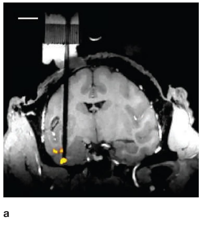
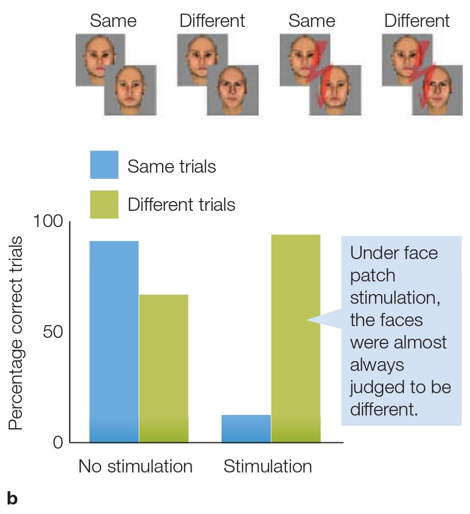 FIGURE 6.37 Perturbing the function of face patches in the IT cortex with microstimulation. (a) Coronal MRI slice showing one face patch region (yellow) that was stimulated in a monkey. The stimulating electrode is seen as the dark bar on the left. This face patch is the most anterior face patch and showed significant activation for faces versus objects. (b) Results for trials in which Cue 1 and Cue 2 were of the same identity (blue) or of different identities (light green), grouped according to whether stimulation was applied (right) or not applied (left). When the face patch was stimulated, the monkey’s performance decreased on same-identity trials and increased on different-identity trials.
شکل 6.37 برهم زدن عملکرد تکههای چهره در قشر IT با ریز تحریک. (الف) برش MRI تاج که یک ناحیه وصله چهره (زرد) را نشان میدهد که در یک میمون تحریک شده است. الکترود تحریک کننده به چهره نوار تیره در سمت چپ دیده میشود. این وصله چهره قدامیترین وصله چهره است و فعال سازی قابل توجهی را برای چهرهها در مقابل اشیا نشان میدهد. (ب) نتایج برای کارآزماییهایی که در آن Cue 1 و Cue 2 دارای هویت یکسان (آبی) یا هویتهای متفاوت (سبز روشن) بودند، بر اساس اینکه آیا تحریک (راست) اعمال شده است یا اعمال نشده است (چپ). هنگامیکه وصله چهره تحریک شد، عملکرد میمون در آزمایشهای با هویت مشابه کاهش یافت و در آزمایشهایی با هویت متفاوت افزایش یافت.
Brad Duchaine and his colleagues used transcranial magnetic stimulation (TMS) to provide one such test. They disrupted activity in three different regions that had been shown to exhibit category specificity (Pitcher et al., 2009). The study participants performed a series of discrimination tasks that involved judgments about faces, bodies, and objects. In separate blocks of trials, the TMS coil was positioned over the right occipital face area (rOFA), the right extrastriate body area (rEBA), and the right lateral occipital area (rLO; Figure 6.39a). (The FFA was not used, because it is inaccessible to TMS.)
براد دوچین و همکارانش از تحریک مغناطیسی ترانس کرانیال (TMS) برای ارائه چنین آزمایشی استفاده کردند. آنها فعالیت را در سه منطقه مختلف که مشخص شده بود ویژگی دسته بندی را نشان میدهند مختل کردند (Pitcher et al., 2009). شرکت کنندگان در مطالعه یک سری وظایف تبعیض آمیز را انجام دادند که شامل قضاوت در مورد چهرهها، بدنها و اشیاء بود. در بلوکهای جداگانه کارآزماییها، سیمپیچ TMS روی ناحیه چهره پس سری راست (rOFA)، ناحیه بدن بیرونی راست (rEBA) و ناحیه پس سری جانبی راست (rLO؛ شکل 6.39a) قرار گرفت. (FFA استفاده نشد، زیرا برای TMS غیرقابل دسترسی است.)
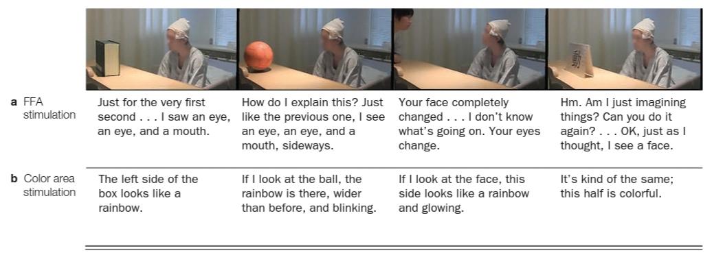
FIGURE 6.38 Patient’s descriptions of various objects during electrical stimulation of the FFA and the color area.
The patient was shown a box, a ball, the experimenter’s face, and a kanji character. In all cases, when the fusiform face area (FFA) was stimulated at the same time, the patient saw a face illusion (a), but when the color region (V4; see Chapter 5) was stimulated, he saw multiple colors (b).
شکل 6.38 توضیحات بیمار از اشیاء مختلف در طی تحریک الکتریکی FFA و ناحیه رنگی.
به بیمار یک جعبه، یک توپ، چهره آزمایشگر و یک شخصیت کانجی نشان داده شد. در تمام موارد، زمانی که ناحیه دوکی شکل (FFA) به طور همزمان تحریک شد، بیمار یک توهم چهره (a) را مشاهده کرد، اما وقتی ناحیه رنگی (V4؛ به فصل 5 مراجعه کنید) تحریک شد، چندین رنگ (b) را مشاهده کرد.
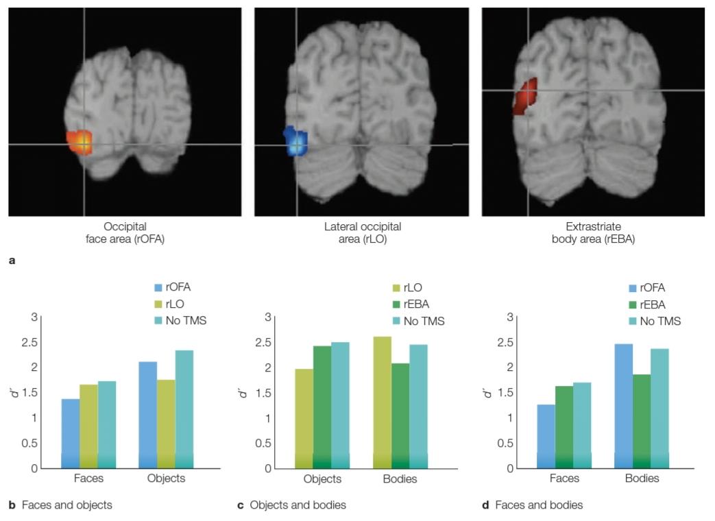
FIGURE 6.39 Triple dissociation of faces, bodies, and objects.
(a) TMS target sites based on fMRI studies identifying regions in the right hemisphere that are sensitive to faces (the right occipital face area, rOFA), objects (the right lateral occipital area, rLO), and bodies (the right extrastriate body area, rEBA). (b-d) In each panel, performance on two tasks was compared when TMS was applied in separate blocks to two of the stimulation sites, as well as in a control condition (no TMS). The dependent variable in each graph is d’, a measure of perceptual performance (high values = better performance). (b) Face performance was disrupted by TMS over the rOFA. (c) Object perception was disrupted by TMS over the rLO. (d) Body perception was disrupted by TMS over the rEBA.
شکل 6.39 تفکیک سه گانه چهره، بدن و اشیا.
(الف) مکانهای TMS بر اساس مطالعات fMRI که مناطقی را در نیمکره راست که به چهرهها (ناحیه پس سری سمت راست، rOFA)، اشیاء (ناحیه اکسیپیتال جانبی راست، rLO) و بدنها (ناحیه بدن خارج از لایه راست، rEBA) حساس هستند، هدف قرار میدهد. (ب-د) در هر پانل، عملکرد دو کار با استفاده از TMS در بلوکهای جداگانه به دو مکان تحریک، و همچنین در شرایط کنترل (بدون TMS) مقایسه شد. متغیر وابسته در هر نمودار d’ است، که معیاری برای عملکرد ادراکی است (مقادیر بالا = عملکرد بهتر). (ب) عملکرد چهره توسط TMS از طریق rOFA مختل شد. (ج) ادراک شی توسط TMS بر روی rLO مختل شد. (د) ادراک بدن توسط TMS بر روی rEBA مختل شد.
The results showed a neat triple dissociation (Figure 6.39b-d). When TMS was applied over the TOFA, participants had problems discriminating faces, but not objects or bodies. When it was applied over the rEBA, the result was impaired discrimination of bodies, but not faces or objects. Finally, as you have probably guessed, when TMS was applied over the rLO, the participants had difficulty picking out objects, but not faces or bodies (Pitcher et al., 2009). The last result is especially interesting because the perception of faces and bodies was not disrupted. Regions that are involved in category-independent object recognition processes must be downstream from the rLO. We will return to the topic of disrupted face recognition in Section 6.6, after we explore deficits in object recognition in the next section.
نتایج یک تفکیک سه گانه منظم را نشان داد (شکل 6.39b-d). هنگامیکه TMS بر روی TOFA اعمال شد، شرکتکنندگان در تشخیص چهرهها مشکل داشتند، اما نه اشیا یا بدن. هنگامیکه آن را روی rEBA اعمال میشد، نتیجه تشخیص اجسام مختل میشد، اما نه چهره یا اشیا. در نهایت، همانطور که احتمالاً حدس زده اید، زمانی که TMS روی rLO اعمال شد، شرکت کنندگان در انتخاب اشیا مشکل داشتند، اما نه چهره یا بدن (پیچر و همکاران، 2009). نتیجه آخر به ویژه جالب است زیرا درک چهره و بدن مختل نشد. مناطقی که در فرآیندهای تشخیص شی مستقل از دسته درگیر هستند باید از rLO پایین دست باشند. ما به موضوع تشخیص چهره مختل در بخش 6.6 باز میگردیم، پس از بررسی کاستیها در تشخیص اشیا در بخش بعدی.
TAKE-HOME MESSAGES
پیامهای کلیدی
▪️ Neurons in various areas of the monkey brain show selectivity for face stimuli.
▪️ نورونها در نواحی مختلف مغز میمون برای محرکهای چهره گزینش پذیری نشان میدهند.
▪️ Similar specificity for faces is observed in fMRI studies in humans, including an area in the right fusiform gyrus of the temporal lobe: the fusiform face area (FFA).
▪️ ویژگی مشابهی برای چهرهها در مطالعات fMRI در انسان مشاهده شده است، از جمله ناحیه ای در شکنج دوکی شکل سمت راست لوب تمپورال: ناحیه چهره دوکی شکل (FFA).
▪️ Just as the FFA is specialized for processing faces, the parahippocampal place area (PPA) is specialized for processing information about spatial relations or for classifying objects based on spatial properties (e.g., an indoor versus outdoor scene), and the extrastriate body area (EBA) and the fusiform body area (FBA) have been identified as more active when body parts are viewed.
▪️ همانطور که FFA برای پردازش چهرهها تخصصی است، ناحیه مکان پاراهیپوکامپ (PPA) برای پردازش اطلاعات در مورد روابط فضایی یا طبقهبندی اشیاء بر اساس ویژگیهای فضایی (به عنوان مثال، صحنههای داخلی در مقابل فضای باز)، و ناحیه بدن خارج از بدن (EBA) و ناحیه بدن دوکی شکل (FBA) زمانی که قسمتهای بدن فعالتر دیده میشوند، تخصصی شده است.
»» » تمامی کتاب