
علوم اعصاب شناختی؛ احساس و ادراک؛ ایمپلنت حلزون و کاشت شبکیه


دعای مطالعه [ نمایش ]
بِسْمِ الله الرَّحْمنِ الرَّحیمِ
اَللّهُمَّ اَخْرِجْنى مِنْ ظُلُماتِ الْوَهْمِ
خدايا مرا بيرون آور از تاريكىهاى وهم،
وَ اَكْرِمْنى بِنُورِ الْفَهْمِ
و به نور فهم گرامى ام بدار،
اَللّهُمَّ افْتَحْ عَلَيْنا اَبْوابَ رَحْمَتِكَ
خدايا درهاى رحمتت را به روى ما بگشا،
وَانْشُرْ عَلَيْنا خَزائِنَ عُلُومِكَ بِرَحْمَتِكَ يا اَرْحَمَ الرّاحِمينَ
و خزانههاى علومت را بر ما باز كن به امید رحمتت اى مهربانترين مهربانان.
5.9 Perceptual Reorganization
5.9 سازماندهی مجدد ادراکی
In 1949, Donald Hebb bucked the assumption that the brain was unchangeable after the early formative years. He suggested a theoretical framework for how functional reorganization, or what neuroscientists refer to as cortical plasticity, might occur in the brain through the remodeling of neuronal connections. Since then, more people have been looking for and observing brain plasticity in action. For example, Michael Merzenich (Mer- zenich & Jenkins, 1995; Merzenich et al., 1988) at UC San Francisco and Jon Kaas (1995) at Vanderbilt University discovered that experience could alter the size and shape of cortical sensory and motor maps in adult monkeys.
در سال 1949، دونالد هب این فرض را که مغز پس از سالهای اولیه شکلگیری غیرقابل تغییر است را رد کرد. او یک چارچوب نظری برای چگونگی سازماندهی مجدد عملکردی، یا آنچه دانشمندان علوم اعصاب به عنوان پلاستیسیته قشر مغز از آن یاد میکنند، در مغز از طریق بازسازی اتصالات عصبی رخ میدهد، پیشنهاد کرد. از آن زمان، افراد بیشتری به دنبال و مشاهده انعطاف پذیری مغز در عمل بوده اند. به عنوان مثال، مایکل مرزنیچ (Merzenich & Jenkins, 1995؛ Merzenich et al., 1988) در UC سانفرانسیسکو و Jon Kaas (1995) در دانشگاه Vanderbilt کشف کردند که تجربه میتواند اندازه و شکل نقشههای حسی و حرکتی قشر مغز را در میمونهای بالغ تغییر دهد.
After the researchers severed (deafferented) the nerve fibers from a monkey’s finger to its spinal cord, the relevant part of its cortex no longer responded at first when that finger was touched; however, that cortical area soon became active again: It began to respond to stimulation of the finger adjacent to the amputated finger. The sur-rounding cortical area had filled in and taken over the silent area. The researchers found similar changes when a particular finger was given extended sensory stimulation: It gained a little more acreage on the cortical map. This functional plasticity suggests that the adult cortex is a dynamic place where remarkable changes can still happen. In this section we take a look at changes in perceptual organization that can occur.
پس از اینکه محققان رشتههای عصبی انگشت میمون را به نخاع قطع کردند، قسمت مربوطه از قشر آن در ابتدا با لمس آن انگشت دیگر واکنش نشان نداد. با این حال، آن ناحیه قشر به زودی دوباره فعال شد: شروع به پاسخ دادن به تحریک انگشت مجاور انگشت قطع شده کرد. ناحیه قشری اطراف پر شده بود و ناحیه ساکت را گرفته بود. محققان تغییرات مشابهی را در زمانی که به انگشت خاصی تحریک حسی طولانی داده شد، یافتند: این انگشت در نقشه قشر مغز کمیبیشتر به دست آورد. این انعطاف پذیری عملکردی نشان میدهد که قشر بالغ یک مکان پویا است که هنوز تغییرات قابل توجهی میتواند رخ دهد. در این بخش نگاهی به تغییرات در سازمان ادراکی که ممکن است رخ دهد میاندازیم.
Development of Sensory Systems Just as the prenatal brain develops on a time clock, so do the sensory systems. Primary sensory areas exhibit experience-induced plasticity during defined windows of early life, known as critical periods. The brain requires external inputs during these periods to establish an optimal neural representation of the surrounding environment. For example, at birth the visual system needs input from the environment to properly develop the thalamic connections to cortical layer IV of the primary visual cortex.
توسعه سیستمهای حسی همانطور که مغز قبل از تولد در یک ساعت زمان رشد میکند، سیستمهای حسی نیز رشد میکنند. نواحی حسی اولیه، انعطافپذیری ناشی از تجربه را در طول پنجرههای مشخص اولیه زندگی، که به عنوان دورههای بحرانی شناخته میشوند، نشان میدهند. مغز در این دورهها به ورودیهای خارجی نیاز دارد تا یک نمایش عصبی بهینه از محیط اطراف ایجاد کند. به عنوان مثال، در بدو تولد، سیستم بینایی نیاز به ورودی از محیط دارد تا به درستی اتصالات تالاموس را به لایه IV قشر قشر بینایی اولیه توسعه دهد.
Again it was Hubel and Wiesel (1970, 1977) who first showed this in cats and monkeys. They sutured one eye shut during the first few weeks after a kitten was born (or the first few months after a monkey was born) and found that the cats and monkeys failed to develop normal ocular dominance columns in the primary visual cortex. Instead, the input to layer IV was limited to the seeing eye, and the cortical representation of this eye took over most of the sutured eye’s cortex. This change was permanent. When the sutures were removed after a few weeks, no cortical changes occurred, no rewiring took place, and the animals remained blind for life in that eye. When the researchers sutured one eye in an adult animal for even longer periods of time, however, the effect was minimal when the sutures were removed.
باز هم هوبل و ویزل (1970، 1977) بودند که اولین بار این را در گربهها و میمونها نشان دادند. آنها در چند هفته اول پس از تولد یک بچه گربه (یا چند ماه اول پس از تولد میمون) یک چشم را بخیه زدند و دریافتند که گربهها و میمونها نتوانستند ستونهای غالب چشمیطبیعی را در قشر بینایی اولیه ایجاد کنند. در عوض، ورودی لایه IV به چشم بینا محدود شد و نمایش قشر این چشم بیشتر قشر چشم بخیه شده را در بر گرفت. این تغییر دائمیبود. هنگامیکه بخیهها پس از چند هفته برداشته شد، هیچ تغییری در قشر مغز رخ نداد، سیم کشی مجدد صورت نگرفت و حیوانات برای زندگی در آن چشم نابینا ماندند. با این حال، زمانی که محققان یک چشم یک حیوان بالغ را برای مدت طولانیتری بخیه زدند، با برداشتن بخیهها، این اثر بسیار کم بود.
Human infants have also demonstrated the importance of critical time periods for the input of visual stimuli. For example, babies who have limited or no visual input from an eye because of a cataract must have the cataract removed in the first few weeks of life to gain vision in that eye. If the cataract is removed after the critical period, the child will not regain sight in that eye, whereas in an adult, a cataract formed later in life can be present for years and yet sight will be regained after its removal. Or consider infants born with strabismus (a misalignment of the eyes): If left unchecked beyond the critical period, this condition will result in abnormal development of the visual cortex and functional blindness in one eye. Recent research has found that cellular mechanisms can trigger, mediate, slow, and reopen critical windows (Werker & Hensch, 2015).
نوزادان انسان نیز اهمیت دورههای زمانی بحرانی را برای ورودی محرکهای بینایی نشان داده اند. به عنوان مثال، نوزادانی که به دلیل آب مروارید، ورودی بصری آنها از چشم محدود است یا اصلا وجود ندارد، باید در چند هفته اول زندگی، آب مروارید را حذف کنند تا در آن چشم بینایی داشته باشند. اگر آب مروارید بعد از دوره بحرانی برداشته شود، کودک بینایی را در آن چشم به دست نمیآورد، در حالی که در بزرگسالان، آب مروارید که در مراحل بعدی زندگی ایجاد میشود، میتواند سالها وجود داشته باشد و در عین حال بعد از برداشتن آن بینایی دوباره به دست میآید. یا نوزادانی را که با استرابیسم متولد شده اند در نظر بگیرید (انحراف چشمها): اگر این وضعیت بیش از دوره بحرانی کنترل نشود، منجر به رشد غیر طبیعی قشر بینایی و کوری عملکردی در یک چشم میشود. تحقیقات اخیر نشان داده است که مکانیسمهای سلولی میتوانند پنجرههای حیاتی را تحریک، واسطه، کند و دوباره باز کنند (Werker & Hensch, 2015).
Both circuit maturation and experience are also important in speech perception. Initially, development of the auditory cortex relies on genetically controlled intrinsic factors, but the refinement of cortical connections relies on feedback from auditory stimulation—that is, exposure to spoken language. Auditory stimulation or deprivation can significantly impact the development of cortical infrastructure and associated behavioral abilities. Congenitally deaf infants have a high risk for permanent language impairment, but hearing aids or cochlear implants implanted at an early enough age can enable many individuals to develop spoken language perception and production skills close to those of the hearing population.
بلوغ مدار و تجربه هم در ادراک گفتار مهم هستند. در ابتدا، توسعه قشر شنوایی به عوامل درونی کنترل شده ژنتیکی متکی است، اما اصلاح ارتباطات قشر مغز متکی به بازخورد از تحریک شنوایی – یعنی قرار گرفتن در معرض زبان گفتاری است. تحریک یا محرومیت شنوایی میتواند به طور قابل توجهی بر توسعه زیرساختهای قشر مغز و تواناییهای رفتاری مرتبط تأثیر بگذارد. نوزادان ناشنوای مادرزادی در معرض خطر بالایی برای اختلال زبان دائمیهستند، اما سمعکها یا کاشت حلزون در سنین پایین میتوانند بسیاری از افراد را قادر سازند تا درک زبان گفتاری و مهارتهای تولید نزدیک به افراد شنوایی را توسعه دهند.
Perceptual Reorganization Secondary to Early Sensory Loss
سازماندهی مجدد ادراکی ثانویه تا از دست دادن حسی اولیه
What happens in the brain when it doesn’t receive input from one sensory system? Is the brain hardwired, resulting in large swaths of the cerebral cortex remaining nonfunctional? Or does the brain reorganize-and if so, does this reorganization occur in a systematic manner?
وقتی مغز ورودی از یک سیستم حسی دریافت نمیکند چه اتفاقی در مغز میافتد؟ آیا مغز به سختی سیم کشی شده است و در نتیجه بخشهای بزرگی از قشر مغز غیرعملکردی باقی میماند؟ یا مغز سازماندهی مجدد میکند و اگر چنین است، آیا این سازماندهی مجدد به شیوهای سیستماتیک رخ میدهد؟
Blindness or highly impaired vision affects about 3% of the U.S. population. Some of these individuals are born without vision-a condition known as congenital blindness. Blindness at birth is generally caused by congenital defects of the eye, such as anophthalmia, microphthalmia, coloboma (a hole in one of the eye’s structures, such as the retina), cataract, retinal dystrophies, infantile glaucoma, or cloudy cornea. Blindness may also result from some perinatal insults, such as eye infections contracted from viral or bacterial infections of the birth canal (herpes, gonorrhea, or chlamydia), or cortical deficits from oxygen deprivation during birth.
نابینایی یا بینایی بسیار ضعیف حدود 3 درصد از جمعیت ایالات متحده را تحت تأثیر قرار میدهد. برخی از این افراد بدون بینایی به دنیا میآیند – وضعیتی که به عنوان نابینایی مادرزادی شناخته میشود. نابینایی در بدو تولد عموماً ناشی از نقایص مادرزادی چشم مانند آنوفتالمی، میکروفتالمی، کولوبوما (یک سوراخ در یکی از ساختارهای چشم مانند شبکیه)، آب مروارید، دیستروفی شبکیه، گلوکوم نوزادی یا قرنیه کدر است. کوری همچنین ممکن است ناشی از برخی توهینهای پری ناتال، مانند عفونتهای چشمیناشی از عفونتهای ویروسی یا باکتریایی مجرای زایمان (تبخال، سوزاک، یا کلامیدیا) یا کمبودهای قشر مغز ناشی از کمبود اکسیژن در هنگام تولد باشد.
Other individuals lose their sight at some point during life, usually from degenerative diseases such as macular degeneration, diabetic retinopathy, or cataracts. The great majority of these conditions are problems within the eye-that is, in the peripheral sense organ. Extensive experiments have been conducted with highly visually impaired individuals, as well as nonhuman species, to explore plasticity within the sensory systems and how it changes as a function of the time at which the sensory loss occurs.
افراد دیگر در طول زندگی بینایی خود را از دست میدهند، معمولاً به دلیل بیماریهای دژنراتیو مانند دژنراسیون ماکولا، رتینوپاتی دیابتی یا آب مروارید. اکثریت این بیماریها مشکلاتی در داخل چشم هستند، یعنی در اندام حس محیطی. آزمایشهای گستردهای با افراد بسیار کمبینا و همچنین گونههای غیرانسانی انجام شده است تا شکلپذیری در سیستمهای حسی و چگونگی تغییر آن بهعنوان تابعی از زمانی که در آن از دست دادن حسی رخ میدهد، کشف شود.
The results from an early PET study revealed a remarkable degree of functional reorganization in the visual cortex of congenitally blind individuals (Sadato et al., 1996). The participants in this study either had normal vision or were congenitally blind. They were scanned under two experimental conditions. In one condition, they were simply required to sweep their fingers back and forth over a rough surface covered with dots. In the second condition, they were given tactile discrimination tasks such as deciding whether two grooves in the surface were the same or different. Blood flow in the visual cortex during each of these tasks was compared to that during a control condition in which the participants were scanned while keeping their hands still.
نتایج یک مطالعه اولیه PET درجه قابل توجهی از سازماندهی مجدد عملکردی را در قشر بینایی افراد نابینای مادرزادی نشان داد (Sadato et al., 1996). شرکت کنندگان در این مطالعه یا بینایی طبیعی داشتند یا به طور مادرزادی نابینا بودند. آنها تحت دو شرایط آزمایشی اسکن شدند. در یک شرایط، آنها به سادگی مجبور بودند انگشتان خود را روی یک سطح ناهموار پوشیده از نقطه به جلو و عقب بکشند. در شرایط دوم، وظایف تمایز لمسی مانند تصمیم گیری در مورد یکسان یا متفاوت بودن دو شیار در سطح به آنها داده شد. جریان خون در قشر بینایی در طول هر یک از این کارها با جریان خون در یک وضعیت کنترلی مقایسه شد که در آن شرکت کنندگان در حالی که دستان خود را ثابت نگه داشتند، اسکن شدند.
Amazingly, the two groups experienced opposite changes in activation in the visual cortex. For the sighted participants, a significant drop in activation was found in the primary visual cortex during the tactile discrimination tasks. Visual tasks were accompanied by analogous decreases in the auditory or somatosensory cortex. Therefore, as attention was directed to one modality, activation (as measured by blood flow) decreased in other sensory systems. In blind participants, however, activation in the primary visual cortex increased during discrimination tasks, but only when they were actively using the tactile information. Interestingly, a second group of participants, who had become blind early in childhood (before their fifth year), also showed the same recruitment of visual cortex when performing the tactile discrimination task. Subsequent work has shown similar results in a task of great practical value to the blind: reading Braille (Sadato et al., 1998). Blind individuals show increased activation of the primary and secondary visual cortices during Braille reading.
به طور شگفت انگیزی، دو گروه تغییرات متضادی را در فعالیت در قشر بینایی تجربه کردند. برای شرکت کنندگان بینا، کاهش قابل توجهی در فعال سازی در قشر بینایی اولیه در طول وظایف تمایز لمسی مشاهده شد. وظایف بینایی با کاهش مشابه در قشر شنوایی یا حسی تنی همراه بود. بنابراین، همانطور که توجه به یک روش معطوف شد، فعال سازی (که با جریان خون اندازه گیری میشود) در سایر سیستمهای حسی کاهش یافت. در شرکتکنندگان نابینا، با این حال، فعالسازی در قشر بینایی اولیه در طول انجام وظایف تمایز افزایش مییابد، اما تنها زمانی که آنها فعالانه از اطلاعات لمسی استفاده میکردند. جالب توجه است، گروه دوم از شرکت کنندگان، که در اوایل کودکی (قبل از سال پنجم زندگی خود) نابینا شده بودند، همچنین در هنگام انجام تکلیف تمایز لمسی، به کارگیری قشر بینایی مشابهی را نشان دادند. کار بعدی نتایج مشابهی را در کار با ارزش عملی زیادی برای نابینایان نشان داده است: خواندن خط بریل (Sadato et al., 1998). افراد نابینا افزایش فعال شدن قشر بینایی اولیه و ثانویه را در طول خواندن بریل نشان میدهند.
The recruitment of “visual areas” in blind individuals is not limited to somatosensory stimuli. Using fMRI, Marina Bedny and her colleagues (2015) examined the hemodynamic response to a range of sounds both in blind individuals and in sighted individuals who were blindfolded. For the sighted individuals, these auditory stimuli resulted in a decrease in the BOLD response- an effect that was pronounced in different secondary visual areas (Figure 5.43). In contrast, the BOLD response tended to increase in the blind participants. Interestingly, the increase here was limited to lateral and medial occipital regions. Moreover, the magnitude of the BOLD response varied for different classes of stimuli, with the effect most pronounced when the blind participants were listening to stories and minimal when the participants listened to music. Further analysis of these data suggests that the response to spoken language was not related to the participant’s ability to use Braille.
استخدام “مناطق بینایی” در افراد نابینا به محرکهای حسی تنی محدود نمیشود. مارینا بدنی و همکارانش (2015) با استفاده از fMRI پاسخ همودینامیک به طیف وسیعی از صداها را هم در افراد نابینا و هم در افراد بینا که چشمبند داشتند بررسی کردند. برای افراد بینا، این محرکهای شنوایی منجر به کاهش پاسخ BOLD شد – اثری که در نواحی بینایی ثانویه مختلف تلفظ میشد (شکل 5.43). در مقابل، پاسخ BOLD تمایل به افزایش در شرکت کنندگان نابینا داشت. جالب توجه است، افزایش در اینجا محدود به مناطق اکسیپیتال جانبی و داخلی بود. علاوه بر این، میزان پاسخ BOLD برای کلاسهای مختلف محرکها متفاوت بود، با این تأثیر بیشتر زمانی که شرکتکنندگان نابینا به داستان گوش میدادند و زمانی که شرکتکنندگان به موسیقی گوش میدادند کمتر بود. تجزیه و تحلیل بیشتر این دادهها نشان میدهد که پاسخ به زبان گفتاری با توانایی شرکت کننده در استفاده از خط بریل ارتباطی ندارد.

FIGURE 5.43 BOLD response of left occipital regions to language and to nonlinguistic sounds. In secondary visual areas-lateral occipital (a) and medial occipital (b)-the BOLD responses to an English-language story, a foreign-language story, and music were higher in blind individuals compared to blindfolded sighted individuals, although the effect was modest for music. This difference was not seen in the more ventral fusiform gyrus (c). Asterisks indicate when a particular condition is significantly different from the rest.
شکل 5.43 پاسخ بولد نواحی اکسیپیتال چپ به زبان و صداهای غیرزبانی. در نواحی بینایی ثانویه – اکسیپیتال جانبی (الف) و اکسیپیتال میانی (ب) – پاسخهای جسورانه به یک داستان انگلیسی زبان، یک داستان به زبان خارجی و موسیقی در افراد نابینا در مقایسه با افراد بینا با چشمبند بیشتر بود، اگرچه این تأثیر برای موسیقی متوسط بود. این تفاوت در شکنج دوکی شکل شکمیتر (c) دیده نشد. ستارهها زمانی را نشان میدهند که یک وضعیت خاص به طور قابل توجهی با بقیه متفاوت است.
The same lab then asked a more subtle question: Does the auditory recruitment of “visual areas” in blind individuals have some similarity to how the area responded to visual information in those with sight (Bedny et al., 2010)? Focusing on the medial temporal area MT/MST, which shows high sensitivity to visual motion in sighted individuals, they found that blind individuals showed a strong response in this area to dynamic auditory stimuli (i.e., auditory motion), such as the sound of footsteps. This effect was not observed in the sighted individuals: Here the BOLD response remained flat or even dropped when the auditory stimuli were presented.
همان آزمایشگاه سپس سؤال ظریف تری را مطرح کرد: آیا استخدام شنوایی “مناطق بینایی” در افراد نابینا تا حدی شباهتی به نحوه پاسخ ناحیه به اطلاعات بصری در بیناییها دارد (بدنی و همکاران، 2010)؟ آنها با تمرکز بر ناحیه زمانی میانی MT/MST، که حساسیت بالایی به حرکت بینایی در افراد بینا نشان میدهد، دریافتند که افراد نابینا در این ناحیه به محرکهای شنیداری پویا (یعنی حرکت شنوایی)، مانند صدای پا، واکنش قوی نشان میدهند. این اثر در افراد بینا مشاهده نشد: در اینجا پاسخ BOLD ثابت میماند یا حتی با ارائه محرکهای شنیداری کاهش مییابد.
However, in subsequent work using more sophisticated fMRI analyses, Lukas Strnad and his colleagues (2013) found that, even in sighted individuals, MT/ MST contains information about dynamic auditory stimuli. These researchers performed multivoxel pat- tern analysis (MVPA), a method in which the pattern of activity across the voxels is used to classify the stimuli. As in the earlier study, only blind individuals showed an overall increase in the BOLD response in MT/MST to auditory motion. The interesting new finding was that with MVPA, information about the different auditory motion conditions could be observed in MT/MST (and not early visual cortex) in both blind and sighted individuals. These results suggest that neurons in MT/MST may be specialized in some way that makes them sensitive to dynamic information, regardless of whether that information is conveyed visually or through another modality.
با این حال، در کار بعدی با استفاده از تجزیه و تحلیلهای پیچیدهتر fMRI، لوکاس استرناد و همکارانش (2013) دریافتند که، حتی در افراد بینا، MT/MST حاوی اطلاعاتی درباره محرکهای شنیداری پویا است. این محققان تجزیه و تحلیل الگوی چندوکسلی (MVPA) را انجام دادند، روشی که در آن از الگوی فعالیت در سراسر وکسلها برای طبقه بندی محرکها استفاده میشود. همانطور که در مطالعه قبلی، تنها افراد نابینا افزایش کلی در پاسخ BOLD در MT/MST به حرکت شنوایی نشان دادند. یافته جدید جالب این بود که با MVPA، اطلاعات مربوط به شرایط مختلف حرکت شنوایی را میتوان در MT/MST (و نه قشر بینایی اولیه) در افراد نابینا و بینا مشاهده کرد. این نتایج نشان میدهد که نورونها در MT/MST ممکن است به گونهای تخصصی باشند که آنها را نسبت به اطلاعات پویا حساس کند، صرف نظر از اینکه آن اطلاعات به صورت بصری یا از طریق روش دیگری منتقل میشود.
Cortical reorganization appears to be a general phenomenon. Deaf individuals show activation to tactile and visual stimuli across Heschl’s gyrus (Karns et al., 2012). One account of these large-scale changes in cortical representation is called cross-modal plasticity. When a sensory system is absent, inputs from a different sensory system may expand in terms of their cortical recruitment. This reorganization may be facilitated by connectivity patterns that exist between different sensory areas-a hypothesis that could also account for why an area such as MT/MST is sensitive to auditory motion information even in sighted individuals.
به نظر میرسد سازماندهی مجدد قشر یک پدیده کلی است. افراد ناشنوا نسبت به محرکهای لمسی و بصری در سراسر شکنج هشل فعال میشوند (کارنز و همکاران، 2012). یکی از این تغییرات در مقیاس بزرگ در نمایش قشر مغز، انعطاف پذیری متقاطع نامیده میشود. هنگامیکه یک سیستم حسی وجود ندارد، ورودیهای یک سیستم حسی متفاوت ممکن است از نظر جذب قشر آنها گسترش یابد. این سازماندهی مجدد ممکن است توسط الگوهای ارتباطی که بین نواحی حسی مختلف وجود دارد تسهیل شود – فرضیه ای که میتواند دلیل حساس بودن ناحیه ای مانند MT/MST به اطلاعات حرکت شنوایی حتی در افراد بینا را نیز توضیح دهد.
Consistent with the idea that the cortical representation of intact sensory systems is enhanced because of cross-modal plasticity, deaf individuals have been shown to have enhanced visual perception. This effect was evident in one study in which the participants (deaf signers, deaf nonsigners, and hearing signers) had to make a difficult discrimination at the center of vision and then detect a peripheral target among distractors. Deaf adults were significantly better at this task than sighted adults (Dye, Hauser, et al., 2009). A similar heightened visual acuity has been reported in deaf individuals for detecting visual motion (Shiell et al., 2014). This behavioral advantage was correlated with an increase in the volume of the right planum temporale, the region typically associated with auditory perception (Shiell et al., 2016).
متناسب با ایدهای که میگوید نمایش قشری سیستمهای حسی سالم به خاطر پلاستیسیتهی چندمدالی تقویت میشود، نشان داده شده است که افراد ناشنوا دارای درک بصری بهتری هستند. این اثر در یک مطالعه مشخص بود که شرکتکنندگان (ناشنوایان زباندار، ناشنوایان غیرزباندار و شنوایان زباندار) باید یک تمایز دشوار را در مرکز دید انجام دهند و سپس یک هدف محیطی را در میان مزاحمها شناسایی کنند. بزرگسالان ناشنوا در این وظیفه به طور قابل توجهی بهتر از بزرگسالان بینا عمل کردند (Dye, Hauser, et al., 2009). یک تیزبینی بصری مشابه در افراد ناشنوا برای شناسایی حرکت بصری گزارش شده است (Shiell et al., 2014). این مزیت رفتاری با افزایش حجم ناحیهی راست پلانو تمپوراله، ناحیهای که معمولاً با درک شنیداری مرتبط است، همبستگی داشته است (Shiell et al., 2016).
Alternatively, the extensive plasticity observed when a sensory system is absent need not indicate that a region typically associated with one sensory system has now been taken over by a different sensory system. Instead, the recruitment of an area appears to be multimodal: In the congenitally blind, “visual areas” are recruited during tactile and auditory tasks; in the deaf, “auditory areas” are recruited during tactile and visual tasks. Bedny (2017) has raised the interesting hypothesis that reorganization within areas deprived of their natural input is best under- stood as an expansion of areas typically associated with more complex processing rather than linked to any particular sensory system.
از طرف دیگر، انعطاف پذیری گسترده مشاهده شده زمانی که یک سیستم حسی وجود ندارد، نیازی به نشان نمیدهد که منطقه ای که معمولاً با یک سیستم حسی مرتبط است، اکنون توسط یک سیستم حسی متفاوت تسخیر شده است. در عوض، استخدام یک منطقه به نظر میرسد چندوجهی است: در نابینایان مادرزادی، “مناطق بینایی” در طول وظایف لمسی و شنوایی به کار گرفته میشوند. در ناشنوایان، “مناطق شنوایی” در طول کارهای لمسی و بصری به کار گرفته میشوند. بدنی (2017) این فرضیه جالب را مطرح کرده است که سازماندهی مجدد در مناطق محروم از ورودی طبیعی خود به بهترین وجه به عنوان گسترش مناطقی که معمولاً با پردازش پیچیدهتر مرتبط هستند به جای مرتبط شدن با هر سیستم حسی خاص، درک میشود.
Cortical Reorganization Over Shorter Time Spans
سازماندهی مجدد قشر مغز در بازههای زمانی کوتاهتر
Much of the work on cortical reorganization has focused on individuals who have complete loss of one sensory system, usually audition or vision. The degree of reorganization in these populations is age dependent. For example, the BOLD response in MT/MST to auditory motion is much greater in individuals who were born without vision than in individuals who became blind during childhood. These studies are of great importance for understanding perception in individuals with sensory disabilities. A limitation here, at least for understanding the mechanisms and constraints on reorganization, is that the work is cross-sectional: The studies compare different groups of individuals at different time points, in terms either of age or of time elapsed since they incurred the sensory loss.
بسیاری از کارها در مورد سازماندهی مجدد قشر مغز بر روی افرادی متمرکز شده است که یک سیستم حسی را به طور کامل از دست داده اند، معمولاً گوش دادن یا بینایی. درجه سازماندهی مجدد در این جمعیتها به سن بستگی دارد. برای مثال، پاسخ BOLD در MT/MST به حرکت شنوایی در افرادی که بدون بینایی به دنیا آمده اند بسیار بیشتر از افرادی است که در دوران کودکی نابینا شده اند. این مطالعات برای درک ادراک در افراد دارای ناتوانی حسی از اهمیت بالایی برخوردار است. یک محدودیت در اینجا، حداقل برای درک مکانیسمها و محدودیتهای سازماندهی مجدد، این است که کار مقطعی است: مطالعات گروههای مختلف افراد را در مقاطع زمانی مختلف، از نظر سن یا زمان سپری شده از زمانی که دچار فقدان حسی شدهاند، مقایسه میکنند.
An alternative approach is to employ experimental manipulations that block or alter sensory information for a limited amount of time. Alvaro Pascual-Leone and his colleagues at Harvard Medical School (Merabet et al., 2008) studied cortical plasticity when sighted volunteers were deprived of visual information for 1 week. All of the study participants received intensive Braille training for 5 days (Figure 5.44). In one group, the participants were blindfolded for the entire week; participants in the control group did the same training but were not required to wear blindfolds.
یک رویکرد جایگزین استفاده از دستکاریهای تجربی است که اطلاعات حسی را برای مدت زمان محدودی مسدود یا تغییر میدهد. آلوارو پاسکوال-لئونه و همکارانش در دانشکده پزشکیهاروارد (Merabet و همکاران، 2008) در زمانی که داوطلبان بینا به مدت 1 هفته از اطلاعات بصری محروم شدند، پلاستیسیته قشر مغز را مطالعه کردند. همه شرکت کنندگان در مطالعه به مدت 5 روز آموزش فشرده بریل دریافت کردند (شکل 5.44). در یک گروه، شرکت کنندگان برای تمام هفته چشم بند بودند. شرکت کنندگان در گروه کنترل همان آموزش را انجام دادند اما ملزم به بستن چشم بند نبودند.
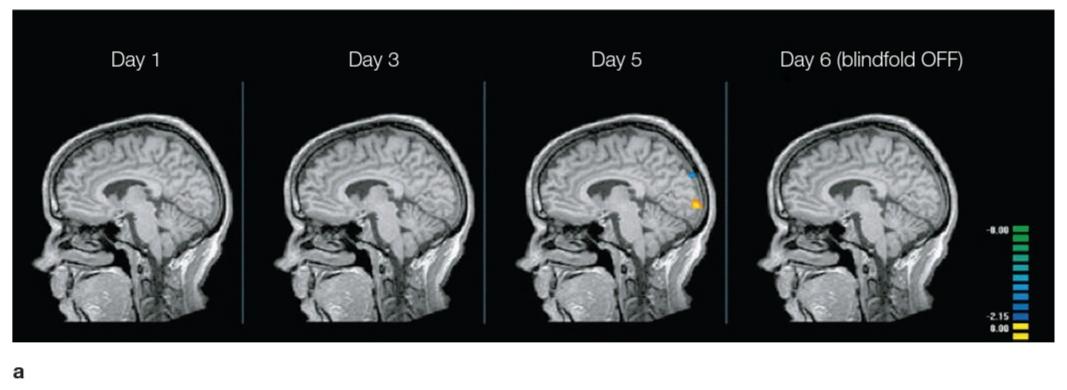
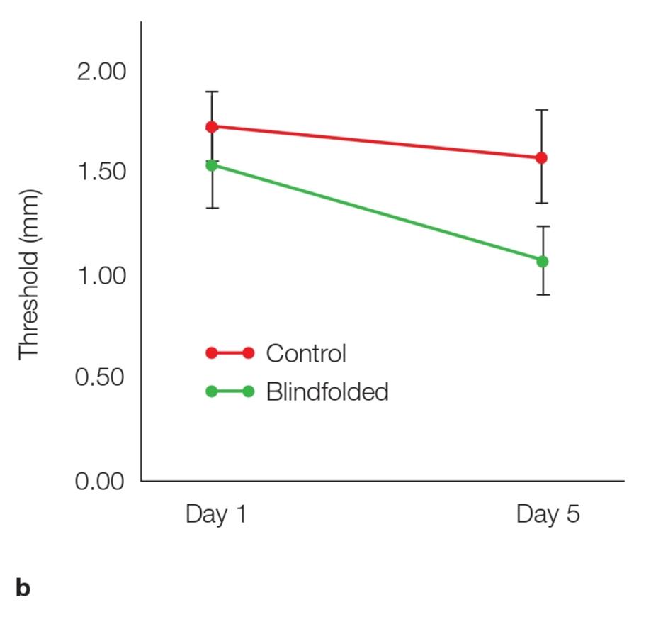

FIGURE 5.44 Perceptual and neural changes resulting from extended visual deprivation in sighted individuals.
(a) Functional MRI activation during tactile exploration. By Day 5, the blindfolded group showed greater activation than the controls in the occipital cortex. This effect disappeared after the blindfold was removed. (b) Performance on tactile acuity after 1 or 5 days of practice. Lower values correspond to better perceptual acuity. (c) Difference in occipital activation between blindfolded and control participants across days.
شکل 5.44 تغییرات ادراکی و عصبی ناشی از محرومیت طولانی بینایی در افراد بینا.
(الف) فعال سازی عملکردی MRI در طول اکتشاف لمسی. در روز 5، گروهی که چشم بسته بودند، فعالیت بیشتری نسبت به گروه کنترل در قشر اکسیپیتال نشان دادند. این اثر پس از برداشتن چشم بند ناپدید شد. (ب) عملکرد در حدت لمسی پس از 1 یا 5 روز تمرین. مقادیر پایین تر با دقت ادراکی بهتر مطابقت دارد. (ج) تفاوت در فعال سازی اکسیپیتال بین شرکت کنندگان چشم بسته و کنترل در طول روز.
At the end of training, the blindfolded participants were better at reading Braille, and their sensory advantage was also evident on other tactile discrimination tasks. Furthermore, at the end of training the blindfolded participants showed a BOLD response in visual cortex during tactile stimulation and disruptions in Braille reading when rTMS was applied over the visual cortex. Inter- estingly, these effects were relatively short-lived: Just one day after removing the blindfolds, the activation in visual cortex during tactile stimulation had disappeared (Figure 5.44a, c).
در پایان آموزش، شرکت کنندگان چشم بند در خواندن خط بریل بهتر بودند و مزیت حسی آنها در سایر کارهای تمایز لمسی نیز مشهود بود. علاوه بر این، در پایان آموزش، شرکتکنندگان با چشمبند پاسخ BOLD در قشر بینایی در طول تحریک لمسی و اختلال در خواندن خط بریل هنگام اعمال rTMS بر روی قشر بینایی نشان دادند. جالب توجه است که این اثرات نسبتاً کوتاه بودند: فقط یک روز پس از برداشتن چشمبندها، فعال شدن قشر بینایی در طی تحریک لمسی ناپدید شد (شکل 5.44a، c).
Paring down the time frame to an even shorter interval, another group of researchers examined the behavioral and neural consequences of altering somatosensory processing using a manipulation in which they glued together the index and middle fingers (Digits 2 and 3) of the participants’ dominant hand (Kolasinski et al., 2016). After just 24 hours, the cortical representation of the fingers had altered. The most striking change was a shift in the cortical position of the ring finger away from the middle finger (Figure 5.45).
با کاهش بازه زمانی حتی کوتاهتر، گروه دیگری از محققان پیامدهای رفتاری و عصبی تغییر پردازش حسی جسمیرا با استفاده از دستکاری که در آن انگشت اشاره و میانی (رقمهای 2 و 3) دست غالب شرکتکنندگان را به هم چسباندند، بررسی کردند (کولاسینسکی و همکاران، 2016). تنها پس از 24 ساعت، نمای قشر انگشتان تغییر کرده بود. بارزترین تغییر تغییر در موقعیت قشری انگشت حلقه از انگشت میانی بود (شکل 5.45).
Paralleling these neural changes, the participants” tactile acuity also changed. The participants now were more accurate in judging the order in which tactile stimuli were applied to the middle and ring fingers, consistent with the idea that a consequence of the gluing was to make the representation of these two fingers more distinct. On the other hand (no pun intended), the participants had more difficulty judging the order of stimuli applied to the ring and pinky finger. These studies show that plastic changes occur over relatively short time frames.
به موازات این تغییرات عصبی، دقت لامسه شرکتکنندگان نیز تغییر کرد. شرکتکنندگان اکنون در قضاوت در مورد ترتیب اعمال محرکهای لمسی به انگشتان حلقه و میانی دقیقتر بودند، مطابق با این ایده که نتیجه چسباندن نمایش این دو انگشت متمایزتر بود. تغییرات در بازههای زمانی نسبتاً کوتاه رخ میدهد.

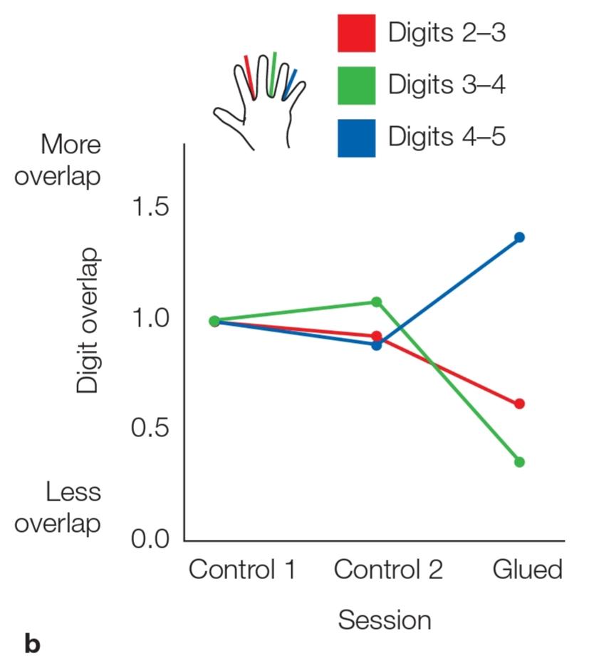
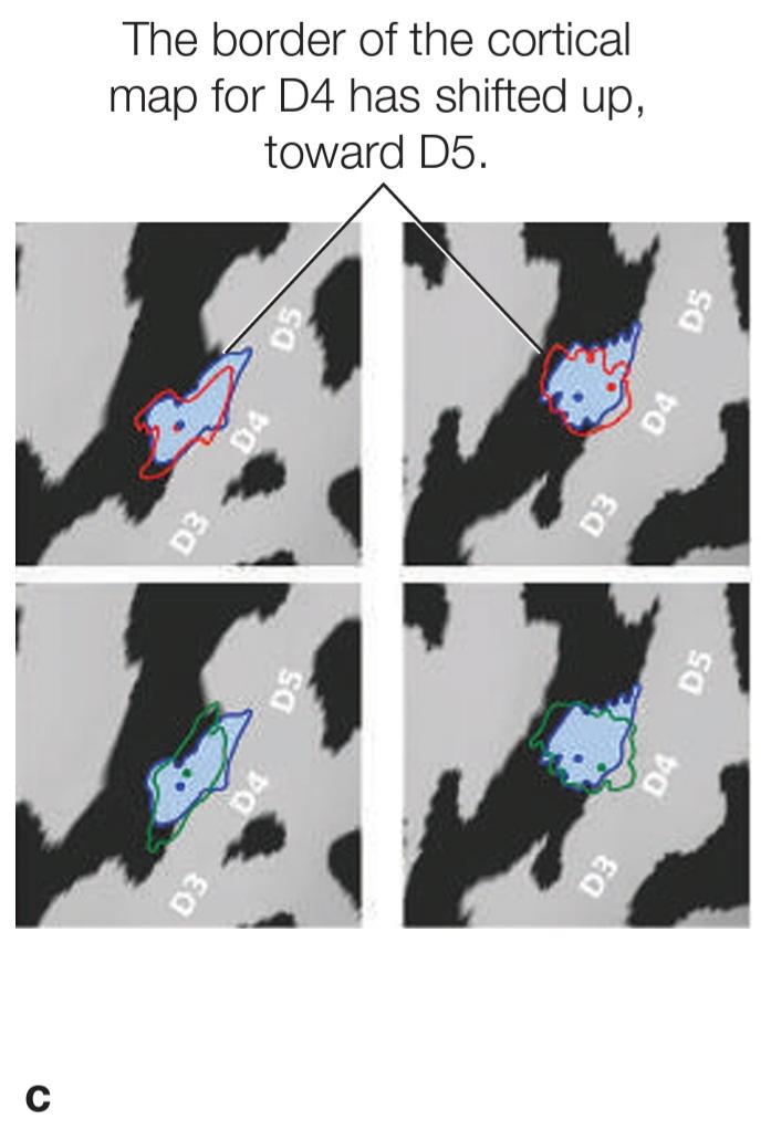
FIGURE 5.45 Rapid experience-dependent remapping in human S1.
(a) High-resolution 7-T fMRI shows digit representation in somatosensory cortex. Finger color on the hand corresponds to the color of the finger’s mapped representation. Mapping of the four digits in S1 is seen on the whole brain (bottom right). The larger panel is a zoomed view of S1, nicely showing the tidy organization of the finger’s mapping. (b) A summary of how cortical overlap shifted with gluing. The researchers normalized all values to 1 from an initial scan (Control 1). They then examined whether the cortical mapping changed after the Control 2 (unglued) or Glued conditions. After gluing, there was a significant decrease in cortical overlap of Digits 3 and 4 (D3 and D4) and an increase in over- lap between D4 and D5. That is, the cortical map for D4 moved away from that of D3 and toward D5. (c) Pattern of shift in the cortical representation of D4 in two participants. Contours show the extent of D4 representation, with the dark-blue line indicating the border during the post-gluing scan. The red and green lines indicate the border during two separate control trials.
شکل 5.45 نقشه برداری سریع وابسته به تجربه در S1 انسانی.
(الف) fMRI 7-T با وضوح بالا نمایش رقمیرا در قشر حسی تنی نشان میدهد. رنگ انگشت روی دست با رنگ نمایش نقشه شده انگشت مطابقت دارد. نقشه برداری از چهار رقم در S1 در کل مغز دیده میشود (پایین سمت راست). پانل بزرگتر نمای بزرگنمایی S1 است که سازماندهی مرتب نقشه برداری انگشت را به خوبی نشان میدهد. (ب) خلاصه ای از نحوه تغییر همپوشانی قشر مغز با چسباندن. محققان تمام مقادیر را از یک اسکن اولیه به 1 نرمال کردند (کنترل 1). آنها سپس بررسی کردند که آیا نقشه برداری قشری بعد از شرایط کنترل 2 (unglued) یا Glued تغییر کرده است یا خیر. پس از چسباندن، کاهش قابل توجهی در همپوشانی قشر ارقام 3 و 4 (D3 و D4) و افزایش همپوشانی بین D4 و D5 وجود داشت. یعنی نقشه قشری برای D4 از نقشه D3 دور شد و به سمت D5 رفت. (ج) الگوی تغییر در نمایش قشر D4 در دو شرکتکننده. خطوط وسعت نمایش D4 را نشان میدهد، با خط آبی تیره که در طول اسکن پس از چسباندن، مرز را نشان میدهد. خطوط قرمز و سبز مرز را در طول دو آزمایش کنترل جداگانه نشان میدهد.
Mechanisms of Cortical Reorganization
مکانیسمهای سازماندهی مجدد قشر مغز
The physiological mechanisms driving plasticity at the cellular level have been studied primarily in nonhuman animal models. The results suggest a cascade of effects operating across different timescales. In the short term, rapid changes probably reflect the unveiling of weak connections that already exist in the cortex, and immediate effects are likely to be the result of a sudden reduction in inhibition that normally suppresses inputs from neigh- boring regions.
مکانیسمهای فیزیولوژیکی محرک انعطافپذیری در سطح سلولی عمدتاً در مدلهای حیوانی غیرانسانی مورد مطالعه قرار گرفتهاند. نتایج نشان میدهد که مجموعهای از اثرات در بازههای زمانی مختلف عمل میکنند. در کوتاه مدت، تغییرات سریع احتمالاً منعکس کننده آشکار شدن اتصالات ضعیفی است که قبلاً در قشر مغز وجود دارد، و اثرات فوری احتمالاً نتیجه کاهش ناگهانی مهار است که معمولاً ورودیهای مناطق همسایه را سرکوب میکند.
Reorganization in the motor cortex has been found to depend on the level of gamma-aminobutyric acid (GABA), the principal inhibitory neurotransmitter (Ziemann et al., 2001). When GABA levels are high, activity in individual cortical neurons is relatively stable. If GABA levels are lower, the neurons respond to a wider range of stimuli. For example, a neuron that responds to the touch of one finger will respond to the touch of other fingers if GABA is blocked. Interestingly, temporary deafferentation of the hand (by blocking blood flow to it) leads to a lowering of GABA levels in the brain. These data suggest that short-term plasticity may be controlled by a release of tonic inhibition on synaptic input (thalamic or intracortical) from remote sources.
سازماندهی مجدد در قشر حرکتی یافت شده است که به سطح گاما آمینوبوتیریک اسید (GABA)، انتقال دهنده عصبی مهاری اصلی بستگی دارد (زیان و همکاران، 2001). هنگامیکه سطح GABA زیاد است، فعالیت در نورونهای قشر مغز نسبتاً پایدار است. اگر سطح GABA پایین تر باشد، نورونها به طیف وسیع تری از محرکها پاسخ میدهند. به عنوان مثال، یک نورون که به لمس یک انگشت پاسخ میدهد، در صورت مسدود شدن گابا، به لمس انگشتان دیگر پاسخ میدهد. جالب اینجاست که، دفع موقت دست (با مسدود کردن جریان خون به آن) منجر به کاهش سطح GABA در مغز میشود. این دادهها نشان میدهد که انعطاف پذیری کوتاه مدت ممکن است با آزاد کردن مهار تونیک بر روی ورودی سیناپسی (تالامیک یا داخل جمجمه) از منابع از راه دور کنترل شود.
Changes in cortical mapping over a period of days probably involve changes in the efficacy of existing circuitry. After loss of normal somatosensory input (e.g., through amputation or peripheral nerve section), cortical neurons that previously responded to that input might undergo “denervation hypersensitivity.” That is, the strength of the responses to any remaining weak excitatory input could be upregulated; remapping might well depend on such modulations of synaptic efficacy.
تغییر در نقشه برداری قشر در طی یک دوره از روزها احتمالاً شامل تغییر در اثربخشی مدار موجود است. پس از از دست دادن ورودی طبیعی حسی (به عنوان مثال، از طریق قطع عضو یا بخش عصبی محیطی)، نورونهای قشر مغز که قبلاً به آن ورودی پاسخ داده بودند، ممکن است تحت “حساسیت حساسیت” قرار بگیرند. یعنی قدرت پاسخ به هر ورودی تحریکی ضعیف باقی مانده میتواند تنظیم شود. بازسازی ممکن است به چنین مدولاسیون اثربخشی سیناپسی بستگی داشته باشد.
Strengthening of synapses is enhanced in the motor cortex by the neurotransmitters norepinephrine, dopamine, and acetylcholine; it is decreased in the presence of drugs that block the receptors for these transmitters (Meintzschel & Ziemann, 2005). These changes are similar to the forms of long-term potentiation and depression in the hippocampus that are thought to underlie the formation of spatial and episodic memories that we will discuss in Chapter 9. Finally, some evidence in animals suggests that the growth of intracortical axonal connections and even sprouting of new axons might contribute to very slow changes in cortical plasticity.
تقویت سیناپسها در قشر حرکتی توسط انتقال دهندههای عصبی نوراپی نفرین، دوپامین و استیل کولین افزایش مییابد. در حضور داروهایی که گیرندههای این فرستندهها را مسدود میکنند، کاهش مییابد (Meintzschel & Ziemann، 2005). این تغییرات شبیه به اشکال تقویت و افسردگی طولانی مدت در هیپوکامپ است که تصور میشود زیربنای تشکیل خاطرات مکانی و اپیزودیک است که در فصل 9 بحث خواهیم کرد. سرانجام، برخی از شواهد در حیوانات حاکی از آن است که رشد آکسون داخل بدن اتصالات و حتی جوانه زدن آکسونهای جدید ممکن است به تغییرات بسیار کند در انعطاف پذیری قشر مغز کمک کند.
At a functional level, one theory has been that cortical areas continue to follow basic principles after sensory deprivation. For example, the finding that visual cortex is recruited during Braille reading in people who are congenitally blind has been taken to suggest that this area continues to perform basic sensory functions such as fine-grained tactile discrimination. However, recent findings showing that the “visual cortex” in these individuals may participate in complex processes such as language (Bedny et al., 2011) or arithmetic (Kanjlia et al., 2016) suggest an alternative view.
در یک سطح عملکردی، یک تئوری این بوده است که مناطق قشر مغز پس از محرومیت حسی، از اصول اساسی پیروی میکنند. به عنوان مثال، این یافته که قشر بصری در هنگام خواندن بریل در افرادی که به طور مادرزادی نابینا هستند استخدام میشود، نشان میدهد که این منطقه به انجام عملکردهای حسی اساسی مانند تبعیض لمسی ریز دانه ادامه میدهد. با این حال، یافتههای اخیر نشان میدهد که “قشر بصری” در این افراد ممکن است در فرآیندهای پیچیده مانند زبان (Bedny و همکاران، 2011) یا حسابی شرکت کنند (Kanjlia et al.، 2016) یک دیدگاه جایگزین را نشان میدهد.
It may be more appropriate to consider cortical tis- sue as cognitively pluripotent, capable of assuming a wide range of cognitive functions (Bedny, 2017) where the input during development, constrained by connectivity and experience, determines the specialization. Interestingly, for Compensation resting-state fMRI studies (e.g., Sabbah et al., 2016) have shown that, relative to sighted individuals, visual regions in the blind show stronger connectivity with frontoparietal cortex, areas involved with language, numerical cognition, and executive control. It may be that when the normal input is absent, these “sensory areas” become part of a broader network for more general cognitive functions.
ممکن است مناسب تر باشد که قشر مغز را از نظر شناختی و ظریف در نظر بگیریم، قادر به فرض طیف گسترده ای از کارکردهای شناختی (Bedny، 2017) است که ورودی در طول توسعه، محدودیت با اتصال و تجربه، تخصص را تعیین میکند. جالب اینجاست که برای جبران خسارت مطالعات FMRI در حالت استراحت (به عنوان مثال، Sabbah و همکاران، 2016) نشان داده اند که، نسبت به افراد بینا، مناطق بینایی در کور ارتباطات قوی تری با قشر جلوی و جبهه، مناطق درگیر با زبان، شناخت عددی و اجرایی نشان میدهند. کنترل. این ممکن است که وقتی ورودی طبیعی وجود ندارد، این “مناطق حسی” بخشی از یک شبکه گسترده تر برای عملکردهای شناختی عمومیتر میشوند.
TAKE-HOME MESSAGES
پیامهای کلیدی
▪️ Brain regions typically associated with a particular sensory system become reorganized in individuals who lack that sensory system. For example, regions usually involved in visual processing become responsive to auditory and tactile stimuli in blind individuals.
▪️ مناطق مغزی که به طور معمول با یک سیستم حسی خاص در ارتباط هستند، در افرادی که فاقد آن سیستم حسی هستند، سازماندهی مجدد میشوند. به عنوان مثال، مناطقی که معمولاً در پردازش بصری درگیر هستند، به محرکهای شنوایی و لمسی در افراد نابینا پاسخگو میشوند.
▪️ These neural changes are frequently associated with behavioral changes. For example, deaf individuals show greater sensitivity than hearing individuals in detecting peripheral visual stimuli.
▪️ این تغییرات عصبی غالباً با تغییرات رفتاری همراه است. به عنوان مثال، افراد ناشنوا نسبت به شنوایی افراد در تشخیص محرکهای بصری محیطی حساسیت بیشتری نشان میدهند.
▪️ Cortical reorganization could reflect cross-area plasticity, a reorganization in the processing of information for spared sensory inputs, or recruitment of the deprived areas for more integrative processing.
▪️ بازسازی قشری میتواند نشاندهنده پلاستیسیته بین مناطقی باشد، بازسازی در پردازش اطلاعات برای دریافتهای حسی که حفظ شدهاند، یا استخدام نواحی محروم برای پردازش یکپارچهتر باشد.
▪️ Multiple mechanisms are thought to contribute to cortical plasticity, including changes in sensory inputs, neurotransmitters, the patterns of inhibition of circuits, and the structure of cortical layers and connections.
▪️ تصور میشود مکانیسمهای متعدد به انعطاف پذیری قشر مغز، از جمله تغییر در ورودیهای حسی، انتقال دهندههای عصبی، الگوهای مهار مدارها و ساختار لایهها و اتصالات قشر مغز کمک میکنند.
5.10 Engineering for Compensation
5.10 مهندسی برای جبران خسارت
Ever since Jose Delgado stopped a bull in its tracks with neural stimulation (see Chapter 2), neuroscientists have been intrigued by the idea that engineering solutions could modulate neural activity as a way to compensate for lost neural function. In later chapters we will look at a number of recent developments in the field of neuro- prosthetics, the use of invasive and noninvasive tools to exploit and control neural activity. Here we describe two procedures that have been developed to boost sensory processing. Knowing that sensation involves the translation of external signals into neuronal activity, neuroscientists and engineers have teamed up to create devices that can serve this function when our natural sensors have been lost.
از زمانی که خوزه دلگادو با تحریک عصبی یک گاو نر را در آهنگهای خود متوقف کرد (به فصل 2 مراجعه کنید)، دانشمندان علوم اعصاب از این ایده فریب خورده اند که راه حلهای مهندسی میتوانند فعالیت عصبی را به عنوان راهی برای جبران عملکرد عصبی از دست رفته تعدیل کنند. در فصلهای بعدی ما به تعدادی از تحولات اخیر در زمینه عصبی پروتز، استفاده از ابزارهای تهاجمیو غیر تهاجمیبرای بهره برداری و کنترل فعالیتهای عصبی خواهیم پرداخت. در اینجا ما دو روش را که برای تقویت پردازش حسی تهیه شده است، توصیف میکنیم. دانستن این احساس مستلزم ترجمه سیگنالهای خارجی به فعالیت عصبی، دانشمندان علوم اعصاب و مهندسان برای ایجاد دستگاههایی که میتوانند در هنگام از بین رفتن سنسورهای طبیعی ما بتوانند این عملکرد را ایجاد کنند، همکاری کرده اند.
Cochlear Implants
ایمپلنت حلزون
Cochlear implants are designed to help people with severe hearing problems for whom a typical hearing aid does not help. Permanent hearing loss is usually the result of damage or loss of the hair cells in the cochlea, often due to aging or frequent exposure to loud noise. Hearing aids facilitate hearing by amplifying the signals carried by sound waves and thus increasing the intensity. of the stimulus arriving at the sensory transducers in the ear. Such amplification works only if an adequate number of hair cells are still functioning. In contrast, cochlear implants bypass the damaged portions of these transducers, directly stimulating the auditory nerve.
ایمپلنت حلزون برای کمک به افراد دارای مشکلات شدید شنوایی که برای آنها سمعک معمولی کمکی نمیکند، طراحی شده است. کاهش شنوایی دائمیمعمولاً نتیجه آسیب یا ریزش سلولهای مو در حلزون است، که اغلب به دلیل پیری یا قرار گرفتن در معرض مکرر در برابر سر و صدای بلند است. سمعک با تقویت سیگنالهای حمل شده توسط امواج صوتی و در نتیجه افزایش شدت، شنوایی را تسهیل میکند. محرک میرسد که به مبدلهای حسی در گوش میرسد. چنین تقویت فقط در صورتی کار میکند که تعداد کافی از سلولهای مو هنوز در حال کار باشند. در مقابل، ایمپلنت حلزون قسمتهای آسیب دیده این مبدلها را دور میزند و به طور مستقیم عصب شنوایی را تحریک میکند.
The cochlear implant system begins with an external processor, usually worn behind the ear. This processor contains a tiny microphone that converts sound waves into electrical signals (Figure 5.46). By means of sophisticated algorithms based on years of research into the physics of sound and the physiology of the cochlea, these signals are converted into complex digital representations of the sound. The software can be tailored to filter out irrelevant sounds or adjusted to match the listener’s personal preferences. The digital representation is then transmitted as radio waves through the skin to an internal processor, where it is reconverted to an electrical output that can activate up to 22 electrodes, each designed to produce signals corresponding to different sound frequencies. These electrodes electrically stimulate the cochlear nerve. The resulting patterns lack, to some degree, the richness of natural sound. Nonetheless, given the plasticity of the brain, people with cochlear implants find tremendous improvements in their hearing. Voices may not have the richness we are accustomed to, but normal conversations are now possible.
سیستم ایمپلنت حلزون با یک پردازنده خارجی شروع میشود که معمولاً در پشت گوش پوشیده میشود. این پردازنده حاوی یک میکروفون ریز است که امواج صوتی را به سیگنالهای الکتریکی تبدیل میکند (شکل 5.46). این سیگنالها با استفاده از الگوریتمهای پیشرفته مبتنی بر سالها تحقیق در فیزیک صدا و فیزیولوژی حلزون، به بازنماییهای دیجیتالی پیچیده صدا تبدیل میشوند. این نرم افزار را میتوان برای فیلتر کردن صداهای نامربوط و یا تنظیم تنظیم شده برای مطابقت با ترجیحات شخصی شنونده تنظیم کرد. بازنمایی دیجیتال به عنوان امواج رادیویی از طریق پوست به یک پردازنده داخلی منتقل میشود، جایی که آن را به یک خروجی الکتریکی که میتواند تا 22 الکترود را فعال کند، بازسازی میشود، هر کدام برای تولید سیگنالهای مربوط به فرکانسهای مختلف صدا طراحی شده اند. این الکترودها به صورت الکتریکی عصب حلزون را تحریک میکنند. الگوهای حاصل، تا حدی غنای صدای طبیعی را ندارند. با این وجود، با توجه به انعطاف پذیری مغز، افراد مبتلا به کاشت حلزون حلزون در شنوایی خود پیشرفتهای چشمگیری پیدا میکنند. صداها ممکن است از غنای مورد نظر ما برخوردار نباشند، اما اکنون مکالمات عادی امکان پذیر است.

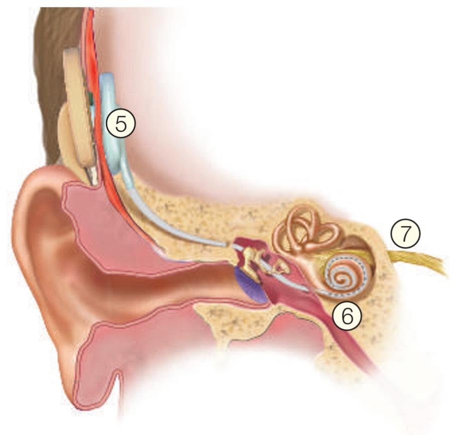
FIGURE 5.46 Cochlear implant.
شکل 5.46 ایمپلنت حلزون.
Sound is picked up by a small microphone (1) located behind the ear and converted into an electrical signal. An external processor (2) converts the signals into complex digital representations of the sound, which travel by wire (3) to an external transmitter (4), which transmits them as radio waves to the internal processor (5). Here they are reconverted to electrical signals that travel by wire (6) to the cochlea, where 22 electrodes are placed. The electrodes stimulate the auditory nerve (7).
صدا توسط یک میکروفون کوچک (1) که در پشت گوش قرار دارد، جمع میشود و به یک سیگنال الکتریکی تبدیل میشود. یک پردازنده خارجی (2) سیگنالها را به بازنماییهای پیچیده دیجیتالی صدا تبدیل میکند، که از طریق سیم (3) به یک فرستنده خارجی (4) سفر میکنند، که آنها را به عنوان امواج رادیویی به پردازنده داخلی منتقل میکند (5). در اینجا آنها به سیگنالهای الکتریکی که از طریق سیم (6) به حلزون حلزون میروند، که 22 الکترود در آن قرار دارند، بازسازی میشوند. الکترودها عصب شنوایی را تحریک میکنند (7).
BOX 5.2 \ HOT SCIENCE
Not-So-Blind Mice
باکس 5.2 \ علم داغ
موشهای نهچندان کور
A completely different approach to retinal implants, currently being developed in rodent models, uses optogenetic techniques. Recall that with optogenetics, cells are made to express light-sensitive receptors. Typically, these cells are located in the brain and thus require the implant of a light-emitting device. However, this same idea can be exploited to modify cells in the retina, using the natural light in the environment as the light source.
یک رویکرد کاملاً متفاوت به کاشت شبکیه، که در حال حاضر در مدلهای جوندگان توسعه یافته است، از تکنیکهای نوری استفاده میکند. به یاد بیاورید که با اپتوژنتیک، سلولها برای بیان گیرندههای حساس به نور ساخته شده اند. به طور معمول، این سلولها در مغز قرار دارند و بنابراین به کاشت یک دستگاه تابش نور نیاز دارند. با این حال، با استفاده از نور طبیعی در محیط به عنوان منبع نور، میتوان از همین ایده برای اصلاح سلولها در شبکیه استفاده کرد.
This approach has been tested in mice that have been genetically modified in two ways (Gaub et al., 2015). The first modification causes the mice to develop degeneration of the photoreceptors in the retina, similar to the effect of the disease retinitis pigmentosa. The second modification uses optogenetics to cause the mice to express light-sensitive receptors in bipolar cells. In normal animals, these cells integrate the output of the photoreceptors to drive ganglion cells; they are not directly responsive to light. The genetic mutants, when exposed to light, show typical mouse-like behavior, such as trying to move to darker places-a behavioral response that indicates they’re seeing light. Physiological recordings verify that the ganglion cells show a high sensitivity to light. Although it’s still in its infancy, this technique using the eye’s functioning hardware is extremely promising as a treatment for a host of diseases in which the transduction of light into nerve signals is compromised.
این رویکرد در موشهایی که از دو طریق از نظر ژنتیکی اصلاح شده اند، آزمایش شده است (گوب و همکاران، 2015). اصلاح اول باعث میشود موشها دژنراسیون گیرندههای گیرنده در شبکیه، مشابه اثر بیماری رتینیت Pingmentosa را ایجاد کنند. اصلاح دوم از اپتوژنتیک استفاده میکند تا موشها گیرندههای حساس به نور را در سلولهای دو قطبی بیان کنند. در حیوانات عادی، این سلولها خروجی گیرندههای گیرنده را برای هدایت سلولهای گانگلیونی ادغام میکنند. آنها مستقیماً به نور پاسخگو نیستند. جهشهای ژنتیکی، هنگامیکه در معرض نور قرار میگیرند، رفتار معمولی مانند موش را نشان میدهند، مانند تلاش برای حرکت به مکانهای تاریک تر-پاسخ رفتاری که نشان میدهد آنها در حال مشاهده نور هستند. ضبطهای فیزیولوژیکی تأیید میکنند که سلولهای گانگلیونی حساسیت بالایی به نور نشان میدهند. اگرچه هنوز در مراحل ابتدایی خود قرار دارد، این تکنیک با استفاده از سخت افزار عملکردی چشم به عنوان درمانی برای بسیاری از بیماریهایی که انتقال نور به سیگنالهای عصبی به خطر میافتد، بسیار امیدوار کننده است.
As of 2012 in the United States, where the Food and Drug Administration (FDA) has approved cochlear implants for people older than 12 months of age, the procedure had been done in roughly 58,000 adults and 38,000 children. Current efforts are aimed at determining whether this approach can also be used to treat children who are completely or severely deaf at birth because of malformations of middle-ear structures, prenatal infections (usually with cytomegalovirus), or genetically linked lack of hair cells in the cochlea. In Australia, cochlear implants have been used in infants younger than 12 months of age for several years.
از سال 2012 در ایالات متحده، جایی که سازمان غذا و داروی (FDA) کاشت حلزون حلزون را برای افراد مسن تر از 12 ماه تصویب کرده است، این عمل تقریباً در 58000 بزرگسال و 38000 کودک انجام شده است. تلاشهای فعلی با هدف تعیین اینکه آیا این روش نیز میتواند برای معالجه کودکانی که در بدو تولد کاملاً یا به شدت ناشنوا هستند به دلیل ناهنجاری در ساختارهای گوش متوسط ، عفونتهای قبل از تولد (معمولاً با سیتومگالوویروس) یا عدم وجود ژنتیکی مرتبط با سلولهای مو در سلول مو در این روش استفاده شود. حلزون در استرالیا، از کاشت حلزون در نوزادان جوانتر از 12 ماهگی برای چندین سال استفاده شده است.
A review of outcomes on speech perception, language, and speech production compared 403 children with congenital, bilateral, severe to profound hearing loss who had received cochlear implants at ages ranging from 6 months to 6 years. The findings demonstrated that those who received the implant before 12 months of age had superior communication outcomes, and that a greater percentage of children from this age group later demonstrated language outcomes within the normal range (Dettman et al., 2016).
مروری بر نتایج در مورد ادراک گفتار، زبان و تولید گفتار، 403 کودک را با کم شنوایی مادرزادی، دو طرفه، شدید تا عمیق که کاشت حلزون حلزون را در سنین 6 ماه تا 6 سال دریافت کرده بودند، مقایسه کرد. این یافتهها نشان میدهد که کسانی که قبل از 12 ماهگی کاشت را دریافت کرده اند، نتایج ارتباطی برتر دارند و درصد بیشتری از کودکان این گروه سنی بعداً نتایج زبان را در محدوده طبیعی نشان دادند (دتمن و همکاران، 2016).
Retinal Implants
کاشت شبکیه
Retinal implants are designed for patients who are blind because of degenerative diseases that affect the photoreceptors, resulting in progressive vision loss. Even when the visual loss is very advanced, many cells in the retina remain intact. In recognition of this fact, researchers have developed two basic types of retinal implants: the subretinal implant, which exploits the remaining photoreceptors, and the epiretinal implant, which bypasses the photoreceptor cells and directly stimulates the ganglion neurons of the retina. (See Box 5.2 for still another approach.) At present, both systems have produced, at best, modest improvements in function.
ایمپلنتهای شبکیه برای بیمارانی که به دلیل بیماریهای دژنراتیو که بر نور گیرندهها تأثیر میگذارند کور هستند و در نتیجه از بین رفتن چشم انداز پیشرونده طراحی شده است. حتی هنگامیکه از دست دادن بینایی بسیار پیشرفته است، بسیاری از سلولهای شبکیه دست نخورده باقی میمانند. به رسمیت شناختن این واقعیت، محققان دو نوع اصلی ایمپلنتهای شبکیه را ایجاد کرده اند: کاشت زیر مجموعه ای که از نور گیرندههای باقیمانده بهره میبرد، و کاشت اپیرتینال، که سلولهای نور گیرنده را دور میزند و به طور مستقیم نورونهای گانگلیون شبکیه را تحریک میکند. (به کادر 5.2 مراجعه کنید تا یک رویکرد دیگر.) در حال حاضر، هر دو سیستم، در بهترین حالت، پیشرفتهای متوسط در عملکرد را تولید کرده اند.
We will focus on the epiretinal implant because it has been approved by the FDA for clinical use. The implant, driven by an external video camera worn on a pair of glasses, sits on top of the retina above the layers of nerves (Figure 5.47). The output of the camera is digitized into electrical pulses that are wirelessly transmitted to the implant. The pulses activate a bank of tiny electrodes, each of which covers an area equivalent to hundreds of photoreceptors. The output of the electrodes directly stimulates the retinal ganglion cells, resulting in the transmission of signals along the optic nerve (see Figure 5.20). In this manner, the electrodes mimic the effect of how patterns of light activate different regions of the retina.
ما روی کاشت اپینال تمرکز خواهیم کرد زیرا توسط FDA برای استفاده بالینی تأیید شده است. ایمپلنت، توسط یک دوربین فیلمبرداری خارجی که روی یک جفت عینک پوشیده شده است، در بالای شبکیه بالای لایههای اعصاب قرار دارد (شکل 5.47). خروجی دوربین به پالسهای الکتریکی دیجیتالی میشود که به صورت بی سیم به کاشت منتقل میشوند. پالسها یک بانک الکترودهای کوچک را فعال میکنند که هر یک از آنها مساحت معادل صدها دستگاه گیرنده را پوشش میدهد. خروجی الکترودها به طور مستقیم سلولهای گانگلیون شبکیه را تحریک میکند و در نتیجه انتقال سیگنالها در طول عصب بینایی ایجاد میشود (شکل 5.20 را ببینید). به این ترتیب، الکترودها از تأثیر چگونگی فعال کردن الگوهای نور مناطق مختلف شبکیه تقلید میکنند.

FIGURE 5.47 The epiretinal implant system.
(a) Drawing of an epiretinal implant in place. (b) An Argos II subretinal implant placed epiretinally over the macular region. The round faint-white area on the right is the optic disk. (c) Implanted portion showing the electrode microarray, the band that encircles the orbit of the eye, the electronics case, and the internal (receiver) coil.
شکل 5.47 سیستم ایمپلنت اپیرتینال.
(الف) ترسیم یک کاشت اپیروین در محل. (ب) یک کاشت زیر مجموعه Argos II به صورت اپیراسیون بر فراز منطقه ماکولا قرار میگیرد. ناحیه ضعیف و سفید در سمت راست دیسک نوری است. (ج) بخش کاشته شده نشان دهنده ریزآرایی الکترود، باند که مدار چشم، مورد الکترونیک و سیم پیچ داخلی (گیرنده) را محاصره میکند.
The crude resolution of the video camera, at least in comparison to the human retina, accounts for the limited restorative effects of the epiretinal implant. To date, the system has been capable of restoring light sensitivity and low visual acuity (20/1262). With practice, users have been able to identify household objects, detect motion, and regain mobility-an early step in the right direction for those who have been blind for years.
وضوح خام دوربین فیلمبرداری، حداقل در مقایسه با شبکیه انسان، اثرات ترمیم کننده محدود کاشت اپینال را تشکیل میدهد. تا به امروز، این سیستم قادر به بازگرداندن حساسیت به نور و حدت بینایی کم (20/1262) است. با تمرین، کاربران توانسته اند اشیاء خانگی را شناسایی کنند، حرکت را تشخیص دهند و تحرک را دوباره در یک مرحله اولیه در جهت درست برای کسانی که سالها نابینا بوده اند، بازیابی کنند.
TAKE-HOME MESSAGES
پیامهای کلیدی
▪️ A common cause of vision and hearing loss is the loss of sensory receptors: photoreceptors in the retina, and hair cells in the cochlea.
▪️ علت اصلی بینایی و کاهش شنوایی، از بین رفتن گیرندههای حسی است: گیرندههای گیرنده در شبکیه و سلولهای مو در حلزون.
▪️ Cochlear implants bypass the transduction of sounds into electrical signals by producing an electrical output that stimulates the auditory nerve. The benefits of cochlear implants can take some time to become optimal, likely because the brain has to learn to interpret the modified auditory input.
▪️ کاشتهای حلزون شنوایی با تولید خروجی الکتریکی که عصب شنوایی را تحریک میکند، تبدیل صداها به سیگنالهای الکتریکی را دور میزنند. مزایای کاشتهای حلزون شنوایی ممکن است مدتی طول بکشد تا به حداکثر خود برسند، احتمالاً به این دلیل که مغز باید یاد بگیرد تا ورودی شنیداری تغییر یافته را تفسیر کند.
▪️A range of methods are being developed to create artificial systems that can replace the loss of photoreceptors. These may involve implants to serve as prosthetic photoreceptors or optogenetic manipulations to make spared neurons light sensitive.
▪️ طیف وسیعی از روشها برای ایجاد سیستمهای مصنوعی ایجاد میشود که میتواند جایگزین از بین رفتن نور گیرندهها شود. اینها ممکن است کاشت را به عنوان گیرنده پروتز پروتز یا دستکاریهای اپتوژنتیک برای ایجاد حساسیت به نور نورونها در نظر بگیرد.
Summary
خلاصه
Our senses translate information about the environment and our body into neuronal signals from which the brain generates perceptions that enable us to build a representation of the world. The five basic sensory systems are olfaction, gustation, somatosensation, audition, and vision. Each sense has specialized receptors that are activated by specific types of stimuli. Gustation and olfaction are known as the chemical senses because their initial response is to molecules (chemicals) in the environment. The somatosensory system has mechanical receptors to detect changes in touch, muscle length, and joint position, as well as nociceptors that respond to temperature and noxious stimuli. The hair cells in the cochlea act as mechanoreceptors in the auditory system. In the visual system, the photoreceptors in the retina (the rods and cones) contain pigments that are sensitive to light.
حواس ما اطلاعات مربوط به محیط و بدن خود را به سیگنالهای عصبی ترجمه میکنند که از آنها مغز ادراکاتی را تولید میکند که به ما امکان میدهد نمایشی از جهان بسازیم. پنج سیستم حسی اساسی شامل بویایی، چشایی، حس لامسه، شنوایی و بینایی هستند. هر حس دارای گیرندههای خاصی است که توسط انواع خاصی از محرکها فعال میشوند. چشایی و بویایی به عنوان حواس شیمیایی شناخته میشوند زیرا پاسخ اولیه آنها به مولکولها (مواد شیمیایی) در محیط است. سیستم حسی لمسی دارای گیرندههای مکانیکی برای تشخیص تغییرات در لمس، طول عضله و موقعیت مفصل است، همچنین نورسنجهای درد که به تغییرات دما و محرکهای زیانآور پاسخ میدهند. سلولهای مویی در حلزون گوش به عنوان مکانورسپتور در سیستم شنوایی عمل میکنند. در سیستم بینایی، گیرندههای نوری در شبکیه (میلهها و مخروطها) حاوی رنگدانههایی هستند که به نور حساس هستند.
Within each sense, specialized mechanisms have evolved to solve computational problems to facilitate our ability to perceive the world, recognizing important properties that are essential for our survival. Gustation enables us to recognize the content of food-or be wary of something that might make us sick. Somatosensation helps us recognize things that might be dangerous (e.g., heat from a fire) or movements that might be injurious. Audition and vision enable us to recognize things at a distance, greatly enhancing the range of our perceptual experience.
در هر حس، مکانیزمهای تخصصی برای حل مشکلات محاسباتی تکامل یافتهاند تا توانایی ما برای درک جهان را تسهیل کنند و ویژگیهای مهمیرا شناسایی کنند که برای بقای ما ضروری هستند. چشایی به ما این امکان را میدهد که محتوای غذا را تشخیص دهیم – یا از چیزی که ممکن است ما را بیمار کند مراقب باشیم. حواس جسمانی به ما کمک میکند چیزهایی را که ممکن است خطرناک باشند (مانند گرما از آتش) یا حرکات که ممکن است مضر باشند، تشخیص دهیم. شنوایی و بینایی به ما این امکان را میدهند که چیزهایی را در فاصله تشخیص دهیم و به طرز چشمگیری دامنه تجربه ادراکی ما را افزایش میدهند.
The gustatory, somatosensory, auditory, and visual cortices contain topographic maps, spatially organized representations of some property of the environment or body. These are known as gustotopic maps in the gustatory system, somatotopic maps in the somatosensory system, tonotopic maps in the auditory system, and retinotopic maps in the visual system. The body parts that have the largest cortical representation on somatotopic maps are those that are most important to that species. Within each sensory system, there is a considerable degree of specialization and elaboration. For example, even in the absence of color or motion perception, we may still be able to recognize shapes.
قشرهای چشایی، حسی حرکتی، شنوایی و بینایی نقشههای توپوگرافی دارند که نمایندگان سازمانیافته مکانی از برخی ویژگیهای محیط یا بدن هستند. اینها در سیستم چشایی به عنوان نقشههای چشایی، در سیستم حسی حرکتی به عنوان نقشههای حسی حرکتی، در سیستم شنوایی به عنوان نقشههای تونوتوپیک و در سیستم بینایی به عنوان نقشههای رتینوتوپیک شناخته میشوند. قسمتهای بدنی که بزرگترین نماینده قشری را در نقشههای حسی حرکتی دارند، همانهایی هستند که برای آن گونه بسیار مهماند. در هر سیستم حسی، درجهای از تخصص و توسعه قابل توجهی وجود دارد. برای مثال، حتی در غیاب ادراک رنگ یا حرکت، ممکن است همچنان قادر به شناسایی اشکال باشیم.
The five senses work not in isolation, but rather in con- cert to construct a rich interpretation of the world. It is this integration that underlies much of human cognition and enables us to survive, and indeed thrive, in a multisensory world.
این پنج حواس نه در انزوا کار میکنند، بلکه در ارتباط با تفسیر غنی از جهان هستند. این ادغام است که زیربنای بسیاری از شناخت انسان است و ما را قادر میسازد تا در دنیای چند حسی زنده بمانیم و در واقع رشد کنیم.
Key Terms
اصطلاحات کلیدی
achromatopsia (p. 203)
acuity (p. 172)
adaptation (p. 172)
akinetopsia (p. 205)
area MT (p. 198)
area V4 (p. 199)
chemical senses (p. 177)
cochlear nerve (p. 186)
cochlear nuclei (p. 187)
cones (p. 190)
cortical plasticity (p. 211)
cortical visual areas (p. 195)
extrastriate visual areas (p. 195)
fovea (p. 190)
ganglion cells (p. 190)
glomeruli (p. 174)
inferior colliculus (p. 187)
interaural time (p. 188)
lateral geniculate nucleus (LGN) (p. 192)
multisensory integration (p. 173)
nociceptors (p. 181)
odorant (p. 174)
olivary nucleus (p. 187)
photoreceptors (p. 190)
primary auditory cortex (A1) (p. 187)
primary gustatory cortex (p. 177)
primary olfactory cortex (p. 174)
primary somatosensory cortex (S1) (p. 182)
primary visual cortex (V1) (p. 192)
proprioception (p. 181)
receptive field (p. 193)
retina (p. 190)
retinotopic map (p. 193)
rods (p. 190)
saccades (p. 173)
secondary somatosensory cortex (S2)(p. 182)
superior colliculus (p. 192) synesthesia (p. 209)
tastant (p. 177)
tonotopic map (p. 186)
Think About It
به آن فکر کنید
1. Compare and contrast the functional organization of the visual and auditory systems. What computational problems must each system solve, and how are these solutions achieved in the nervous system?
1. سازماندهی عملکردی سیستمهای بصری و شنوایی را مقایسه و تضاد کنید. هر سیستم چه مشکلات محاسباتی را باید حل کند و چگونه این راه حلها در سیستم عصبی به دست میآیند؟
2. A person arrives at the hospital in a confused state and appears to have some impairment in visual perception. As the attending neurologist, you suspect that the person has had a stroke. How would you go about examining the patient to determine the level in the visual pathways where damage has occurred? Emphasize the behavioral tests you would administer, but feel free to make predictions about what you expect to see on MRI scans.
2. شخصی در وضعیت گیج کننده وارد بیمارستان میشود و به نظر میرسد در درک بینایی اختلال دارد. به عنوان متخصص مغز و اعصاب، شما گمان میکنید که این شخص سکته مغزی داشته است. چگونه میتوانید بیمار را برای تعیین سطح در مسیرهای بینایی که در آن آسیب دیده است، معاینه کنید؟ بر تستهای رفتاری که شما انجام میدهید تأکید کنید، اما در مورد آنچه انتظار دارید در اسکن MRI مشاهده کنید، پیش بینی کنید.
3. Define the physiological concepts of receptive field and visual area. How is the receptive field of a cell established? How are the boundaries between visual areas identified by researchers using either single-cell- recording methods or fMRI?
3. مفاهیم فیزیولوژیکی میدان گیرنده و ناحیه بصری را تعریف کنید. میدان گیرنده یک سلول چگونه است؟ چگونه مرزهای بین مناطق بصری توسط محققان با استفاده از روشهای ضبط تک سلولی یا FMRI مشخص میشود؟
4. This chapter focused mainly on salient visual properties such as color, shape, and motion. In looking around the environment, do you think these properties seem to reflect the most important cues for a highly skilled visual creature? What other sources of information might an adaptive visual system exploit?
4. این فصل عمدتاً بر خصوصیات بصری برجسته مانند رنگ، شکل و حرکت متمرکز شده است. به نظر شما به نظر میرسد که این خصوصیات مهمترین نشانهها را برای یک موجود بصری بسیار ماهر منعکس میکنند؟ چه منابع دیگر اطلاعات ممکن است یک سیستم بصری تطبیقی سوء استفاده کند؟
5. How might abnormalities in multisensory processing (e.g., synesthesia) be important for understanding how and why information becomes integrated across different sensory channels? Similarly, given the plasticity of the brain, does it even make sense to talk about a “visual system” or an “auditory system”?
5. چگونه ممکن است ناهنجاریها در پردازش چند حسی (به عنوان مثال، سینستزی) برای درک چگونگی و چرا اطلاعات در کانالهای حسی مختلف یکپارچه شود؟ به همین ترتیب، با توجه به انعطاف پذیری مغز، آیا صحبت در مورد “سیستم بصری” یا “سیستم شنوایی” حتی منطقی است؟
کلیک کنید تا Suggested Reading نمایش داده شود
Bedny, M. (2017). Evidence from blindness for a cognitively pluripotent cortex. Trends in Cognitive Sciences, 21(9), 637-648.
Driver, J., & Noesselt, T. (2008). Multisensory interplay reveals crossmodal influences on “sensory-specific” brain regions, neural responses, and judgments. Neuron, 57, 11-23.
Grill-Spector, K., & Malach, R. (2004). The human visual cortex. Annual Review of Neuroscience, 27, 649–677.
Palmer, S. E. (1999). Vision science: Photons to phenomenology. Cambridge, MA: MIT Press.
Ward, J. (2013). Synesthesia. Annual Review of Psychology, 64,49-75.
Yeshurun, Y., & Sobel, N. (2010). An odor is not worth a thousand words: From multidimensional odors to unidimensional odor objects. Annual Review of Psychology, 61, 219-241.



