
مبانی علوم اعصاب اریک کندل؛ ماهیت ساختاری پردازش بینایی؛ ادراک بصری


دعای مطالعه [ نمایش ]
بِسْمِ الله الرَّحْمنِ الرَّحیمِ
اَللّهُمَّ اَخْرِجْنى مِنْ ظُلُماتِ الْوَهْمِ
خدايا مرا بيرون آور از تاريكىهاى وهم،
وَ اَكْرِمْنى بِنُورِ الْفَهْمِ
و به نور فهم گرامى ام بدار،
اَللّهُمَّ افْتَحْ عَلَيْنا اَبْوابَ رَحْمَتِكَ
خدايا درهاى رحمتت را به روى ما بگشا،
وَانْشُرْ عَلَيْنا خَزائِنَ عُلُومِكَ بِرَحْمَتِكَ يا اَرْحَمَ الرّاحِمينَ
و خزانههاى علومت را بر ما باز كن به امید رحمتت اى مهربانترين مهربانان.
«مبانی علوم اعصاب» اثر دکتر اریک کندل و همکاران، یکی از جامعترین و معتبرترین منابع در حوزه علوم اعصاب محسوب میشود.
ترجمه دقیق این کتاب، با مدیریت داریوش طاهری و تحت نظارت برند علمی آیندهنگاران مغز، با الهام از تلاشهای علمی و تدریس اساتیدی همچون دکتر محمدتقی جغتایی و دکتر ثریا مهرابی، به فارسی برگردانده شده است.
این ترجمه، دسترسی به دانش عمیق علوم اعصاب را برای دانشجویان و پژوهشگران رشتههای پزشکی، علوم اعصاب، روانشناسی، علوم شناختی، هوش مصنوعی، گفتاردرمانی، مهندسی پزشکی و تمام علاقهمندان به درک عملکرد ذهن و مغز فراهم کرده و گامی در راستای ارتقای دانش و بهبود کیفیت زندگی است.
» کتاب مبانی علوم اعصاب اریک کندل
» » فصل ۲۱: فصل ماهیت ساختاری پردازش بینایی؛ قسمت اول
در حال ویرایش
Principles of Neural Science; Eric R. Kandel
»» Chapter 21: Sensory Coding
The Constructive Nature of Visual Processing
ماهیت سازنده پردازش بصری
Visual Perception Is a Constructive Process
ادراک بصری یک فرآیند سازنده است
Visual Processing Is Mediated by the Geniculostriate Pathway
پردازش بصری توسط مسیر ژنیکولواستریات انجام میشود
Form, Color, Motion, and Depth Are Processed in Discrete Areas of the Cerebral Cortex
فرم، رنگ، حرکت و عمق در نواحی مجزایی از قشر مغز پردازش میشوند
The Receptive Fields of Neurons at Successive Relays in the Visual Pathway Provide Clues to How the Brain Analyzes Visual Form
میدانهای گیرنده نورونها در رلههای متوالی در مسیر بینایی، سرنخهایی از نحوه تجزیه و تحلیل فرم بصری توسط مغز ارائه میدهند
The Visual Cortex Is Organized Into Columns of Specialized Neurons
قشر بینایی به صورت ستونهایی از نورونهای تخصصی سازماندهی شده است
Intrinsic Cortical Circuits Transform Neural Information
مدارهای قشری ذاتی، اطلاعات عصبی را تبدیل میکنند
Visual Information Is Represented by a Variety of Neural Codes
اطلاعات بصری توسط انواع کدهای عصبی نمایش داده میشود
Highlights
نکات برجسته
We are so familiar with seeing, that it takes a leap of imagination to realize that there are problems to be solved. But consider it. We are given tiny distorted upside-down images in the eyes and we see separate solid objects in surrounding space. From the patterns of stimulation on the retina we perceive the world of objects and this is nothing short of a miracle. -Richard L. Gregory, Eye and Brain, 1966
ما آنقدر با دیدن آشنا هستیم که درک این موضوع که مشکلاتی برای حل شدن وجود دارد، به یک جهش تخیل نیاز دارد. اما آن را در نظر بگیرید. تصاویر کوچک و وارونهای در چشمهای ما ایجاد میشود و ما اشیاء جامد جداگانهای را در فضای اطراف میبینیم. از الگوهای تحریک روی شبکیه، دنیای اشیاء را درک میکنیم و این چیزی جز یک معجزه نیست. – ریچارد ال. گرگوری، چشم و مغز، ۱۹۶۶
MOST OF OUR IMPRESSIONS of the world and our memories of it are based on sight. Yet the mechanisms that underlie vision are not at all obvious. How do we perceive form and movement? How do we distinguish colors? Identifying objects in complex visual environments is an extraordinary computational achievement that artificial vision systems have yet to duplicate. Vision is used not only for object recognition but also for guiding our movements, and these separate functions are mediated by at least two parallel and interacting pathways.
بیشتر برداشتهای ما از جهان و خاطرات ما از آن مبتنی بر بینایی است. با این حال، مکانیسمهایی که زیربنای بینایی هستند، به هیچ وجه آشکار نیستند. چگونه شکل و حرکت را درک میکنیم؟ چگونه رنگها را تشخیص میدهیم؟ شناسایی اشیاء در محیطهای بصری پیچیده، یک دستاورد محاسباتی خارقالعاده است که سیستمهای بینایی مصنوعی هنوز نتوانستهاند آن را تکرار کنند. بینایی نه تنها برای تشخیص اشیاء، بلکه برای هدایت حرکات ما نیز استفاده میشود و این عملکردهای جداگانه توسط حداقل دو مسیر موازی و متقابل انجام میشوند.
The existence of parallel pathways in the visual system raises one of the central questions of cognition, the binding problem: How are different types of information carried by discrete pathways brought together into a coherent visual image?
وجود مسیرهای موازی در سیستم بینایی، یکی از پرسشهای اصلی شناخت، یعنی مسئلهی اتصال، را مطرح میکند: چگونه انواع مختلف اطلاعات که توسط مسیرهای گسسته حمل میشوند، در یک تصویر بصری منسجم گرد هم میآیند؟
Visual Perception Is a Constructive Process
ادراک بصری یک فرآیند سازنده است
Vision is often incorrectly compared to the operation of a camera. A camera simply reproduces point-by-point the light intensities in one plane of the visual field. The visual system, in contrast, does something fundamentally different. It interprets the scene and parses it into distinct components, separating foreground from background. The visual system is less accurate than a camera at certain tasks, such as quantifying the absolute level of brightness or identifying spectral color. However, it excels at tasks such as recognizing a charging animal (or a speeding car) whether in bright sun- light or at dusk, in an open field or partly occluded by trees (or other cars). And it does so rapidly to let the viewer respond and, if necessary, escape.
بینایی اغلب به اشتباه با عملکرد دوربین مقایسه میشود. یک دوربین به سادگی شدت نور را در یک صفحه از میدان دید به صورت نقطه به نقطه بازتولید میکند. در مقابل، سیستم بینایی کاری اساساً متفاوت انجام میدهد. این سیستم صحنه را تفسیر کرده و آن را به اجزای مجزا تجزیه میکند و پیشزمینه را از پسزمینه جدا میکند. سیستم بینایی در برخی وظایف، مانند تعیین کمیت سطح مطلق روشنایی یا شناسایی رنگ طیفی، دقت کمتری نسبت به دوربین دارد. با این حال، در وظایفی مانند تشخیص یک حیوان در حال حمله (یا یک ماشین پرسرعت) چه در نور شدید خورشید یا هنگام غروب، در یک میدان باز یا تا حدی پوشیده از درختان (یا ماشینهای دیگر) عالی عمل میکند. و این کار را به سرعت انجام میدهد تا به بیننده اجازه دهد واکنش نشان دهد و در صورت لزوم فرار کند.
A potentially unifying insight reconciling the visual system’s remarkable ability to grasp the bigger picture with its inaccuracy regarding details of the input is that vision is a biological process that has evolved in step with our ecological needs. This insight helps explain why the visual system is so efficient at extracting useful information such as the identities of objects independent of lighting conditions, while giving less importance to aspects like the exact nature of the ambient light. Moreover, vision does so using previously learned rules about the structure of the world. Some of these rules appeared to have become wired into our neural circuits over the course of evolution. Others are more plastic and help the brain guess at the scene presented to the eyes based on the individual’s past experience. This complex, purposeful processing happens at all levels of the visual system. It starts even at the retina, which is specialized to pick out object boundaries rather than creating a point-by-point representation of uniform surfaces.
یک بینش بالقوه وحدتبخش که توانایی قابل توجه سیستم بینایی در درک تصویر بزرگتر را با عدم دقت آن در مورد جزئیات ورودی تطبیق میدهد، این است که بینایی یک فرآیند بیولوژیکی است که همگام با نیازهای اکولوژیکی ما تکامل یافته است. این بینش به توضیح این موضوع کمک میکند که چرا سیستم بینایی در استخراج اطلاعات مفید مانند هویت اشیاء مستقل از شرایط نوری بسیار کارآمد است، در حالی که به جنبههایی مانند ماهیت دقیق نور محیط اهمیت کمتری میدهد. علاوه بر این، بینایی این کار را با استفاده از قوانین آموخته شده قبلی در مورد ساختار جهان انجام میدهد. به نظر میرسد برخی از این قوانین در طول تکامل در مدارهای عصبی ما سیمکشی شدهاند. برخی دیگر انعطافپذیرتر هستند و به مغز کمک میکنند تا صحنهای را که به چشمها ارائه میشود بر اساس تجربه گذشته فرد حدس بزند. این پردازش پیچیده و هدفمند در تمام سطوح سیستم بینایی اتفاق میافتد. حتی از شبکیه شروع میشود، که برای انتخاب مرزهای اشیاء به جای ایجاد نمایش نقطه به نقطه از سطوح یکنواخت تخصص یافته است.
This constructive nature of visual perception has only recently been fully appreciated. Earlier thinking about sensory perception was greatly influenced by the British empiricist philosophers, notably John Locke, David Hume, and George Berkeley, who thought of perception as an atomistic process in which simple sensory elements, such as color, shape, and brightness, were assembled in an additive way, component by component. The modern view that perception is an active and creative process that involves more than just the information provided to the retina has its roots in the philosophy of Immanuel Kant and was developed in detail in the early 20th century by the German psychologists Max Wertheimer, Kurt Koffka, and Wolfgang Köhler, who founded the school of Gestalt psychology.
این ماهیت سازنده ادراک بصری اخیراً به طور کامل مورد توجه قرار گرفته است. تفکرات اولیه در مورد ادراک حسی تا حد زیادی تحت تأثیر فیلسوفان تجربهگرای بریتانیایی، به ویژه جان لاک، دیوید هیوم و جورج برکلی، بود که ادراک را به عنوان یک فرآیند اتمیستیک در نظر میگرفتند که در آن عناصر حسی ساده، مانند رنگ، شکل و روشنایی، به صورت افزایشی و جزء به جزء جمع میشدند. دیدگاه مدرن مبنی بر اینکه ادراک یک فرآیند فعال و خلاق است که شامل چیزی بیش از اطلاعات ارائه شده به شبکیه است، ریشه در فلسفه ایمانوئل کانت دارد و در اوایل قرن بیستم توسط روانشناسان آلمانی، مکس ورتهایمر، کورت کافکا و ولفگانگ کوهلر، که مکتب روانشناسی گشتالت را تأسیس کردند، به تفصیل توسعه یافت.
The German term Gestalt means configuration or form. The central idea of the Gestalt psychologists is that what we see about a stimulus-the perceptual interpretation we make of any visual object depends not just on the properties of the stimulus but also on its context, on other features in the visual field. The Gestalt psychologists argued that the visual system processes sensory information about the shape, color, distance, and movement of objects according to computational rules inherent in the system. The brain has a way of looking at the world, a set of expectations that derives in part from experience and in part from built- in neural wiring.
اصطلاح آلمانی گشتالت به معنای پیکربندی یا فرم است. ایده اصلی روانشناسان گشتالت این است که آنچه ما در مورد یک محرک میبینیم – تفسیر ادراکی که ما از هر شیء بصری انجام میدهیم، نه تنها به خواص محرک، بلکه به زمینه آن، به سایر ویژگیهای میدان بینایی نیز بستگی دارد. روانشناسان گشتالت استدلال میکردند که سیستم بینایی، اطلاعات حسی در مورد شکل، رنگ، فاصله و حرکت اشیاء را طبق قوانین محاسباتی ذاتی سیستم پردازش میکند. مغز روشی برای نگاه به جهان دارد، مجموعهای از انتظارات که بخشی از آن از تجربه و بخشی از آن از سیمکشی عصبی درونی ناشی میشود.
Max Wertheimer wrote: “There are entities where the behavior of the whole cannot be derived from its individual elements nor from the way these elements fit together; rather the opposite is true: the properties of any of the parts are determined by the intrinsic structural laws of the whole.” In the early part of the 20th century, the Gestalt psychologists worked out the laws of perception that determine how we group elements in the visual scene, including similarity, proximity, and good continuation.
ماکس ورتهایمر نوشت: «موجودیتهایی وجود دارند که رفتار کل آنها را نمیتوان از عناصر منفرد آن یا از نحوهی قرارگیری این عناصر در کنار هم استنباط کرد؛ بلکه عکس این قضیه صادق است: خواص هر یک از اجزا توسط قوانین ساختاری ذاتی کل تعیین میشود.» در اوایل قرن بیستم، روانشناسان گشتالت قوانین ادراک را که نحوهی گروهبندی عناصر در صحنهی بصری را تعیین میکنند، از جمله شباهت، نزدیکی و امتداد خوب، تدوین کردند.
We see a uniform six-by-six array of dots as either rows or columns because of the visual system’s tendency to impose a pattern. If the dots in each row are similar, we are more likely to see a pattern of alternating rows (Figure 21-1A). If the dots in each column are closer together than those in the rows, we are more disposed to see a pattern of columns (Figure 21-1B). The principle of good continuation is an important basis for linking line elements into unified shapes (Figure 21-1C). It is also seen in the phenomenon of contour saliency, whereby smooth contours tend to pop out from complex backgrounds (Figure 21-1D). The Gestalt features that we are disposed to pick out are also ones that characterize objects in natural scenes. Statistical studies of natural scenes show that object boundaries are likely to contain visual elements that lie in close proximity, are continuous across intersections, or form smooth contours. It is tempting to speculate that the formal features of objects in natural scenes created evolutionary pressure on our visual systems to develop neural circuits that have made us sensitive to those features.
ما به دلیل تمایل سیستم بینایی به ایجاد یک الگو، یک آرایه شش در شش از نقاط را به صورت ردیف یا ستون میبینیم. اگر نقاط در هر ردیف مشابه باشند، احتمال بیشتری وجود دارد که الگویی از ردیفهای متناوب را ببینیم (شکل 21-1A). اگر نقاط در هر ستون نسبت به نقاط موجود در ردیفها به هم نزدیکتر باشند، بیشتر احتمال دارد که الگویی از ستونها را ببینیم (شکل 21-1B). اصل تداوم خوب، مبنای مهمی برای پیوند عناصر خط به شکلهای یکپارچه است (شکل 21-1C). همچنین در پدیده برجستگی خطوط دیده میشود، که در آن خطوط صاف تمایل به بیرون زدن از پسزمینههای پیچیده دارند (شکل 21-1D). ویژگیهای گشتالتی که ما تمایل به انتخاب آنها داریم، همانهایی هستند که اشیاء را در صحنههای طبیعی مشخص میکنند. مطالعات آماری صحنههای طبیعی نشان میدهد که مرزهای اشیاء احتمالاً حاوی عناصر بصری هستند که در مجاورت نزدیک قرار دارند، در تقاطعها پیوسته هستند یا خطوط صاف را تشکیل میدهند. میتوان حدس زد که ویژگیهای ظاهری اشیاء در صحنههای طبیعی، فشار تکاملی بر سیستمهای بصری ما ایجاد کردهاند تا مدارهای عصبی را توسعه دهند که ما را نسبت به آن ویژگیها حساس کرده است.
Separating the figure and background in a visual scene is an important step in object recognition. At different moments, the same elements in the visual field can be organized into a recognizable figure or serve as part of the background for other figures (Figure 21-2). This process of segmentation relies not only on certain geometric principles, but also on cognitive influences such as attention and expectation. Thus, a priming stimulus or an internal representation of object shape can facilitate the association of visual elements into a unified percept (Figure 21-3). This internal representation can take many different forms reflecting the wide range of time scales and mechanisms of neural encoding. It could consist of transient reverberating spiking activity selective to a shape or a decision, lasting a fraction of a second, or the selective modulation of synaptic weights during a particular context of a task or an expected shape, or circuit changes that could comprise a long-term memory.
جداسازی شکل و پسزمینه در یک صحنه بصری، گامی مهم در تشخیص شیء است. در لحظات مختلف، عناصر یکسان در میدان بینایی میتوانند به صورت یک شکل قابل تشخیص سازماندهی شوند یا به عنوان بخشی از پسزمینه برای سایر اشکال عمل کنند (شکل 21-2). این فرآیند تقسیمبندی نه تنها به اصول هندسی خاصی، بلکه به تأثیرات شناختی مانند توجه و انتظار نیز متکی است. بنابراین، یک محرک اولیه یا یک نمایش داخلی از شکل شیء میتواند ارتباط عناصر بصری را در یک درک واحد تسهیل کند (شکل 21-3). این نمایش داخلی میتواند اشکال مختلفی داشته باشد که منعکس کننده طیف وسیعی از مقیاسهای زمانی و مکانیسمهای رمزگذاری عصبی است. این میتواند شامل فعالیت جرقهای طنینانداز گذرا باشد که به صورت انتخابی برای یک شکل یا یک تصمیم، که کسری از ثانیه طول میکشد، یا مدولاسیون انتخابی وزنهای سیناپسی در طول یک زمینه خاص از یک کار یا یک شکل مورد انتظار، یا تغییرات مدار که میتواند یک حافظه بلندمدت را تشکیل دهد.
The brain analyzes a visual scene at three levels: low, intermediate, and high (Figure 21-4). At the lowest level, which we consider in the next chapter (Chapter 22), visual attributes such as local contrast, orientation, color, and movement are discriminated. The intermediate level involves analysis of the layout of scenes and of surface properties, parsing the visual image into surfaces and global contours, and distinguishing foreground from background (Chapter 23). The highest level involves object recognition (Chapter 24). Once a scene has been parsed by the brain and objects recognized, the objects can be matched with memories of shapes and their associated meanings. Vision also has an important role in guiding body movement, particularly hand movement (Chapter 25).
مغز یک صحنه بصری را در سه سطح تجزیه و تحلیل میکند: پایین، متوسط و بالا (شکل 21-4). در پایینترین سطح، که در فصل بعدی (فصل 22) به آن میپردازیم، ویژگیهای بصری مانند کنتراست موضعی، جهتگیری، رنگ و حرکت تشخیص داده میشوند. سطح متوسط شامل تجزیه و تحلیل طرح صحنهها و ویژگیهای سطح، تجزیه تصویر بصری به سطوح و خطوط کلی و تمایز پیشزمینه از پسزمینه است (فصل 23). بالاترین سطح شامل تشخیص اشیاء است (فصل 24). هنگامی که یک صحنه توسط مغز تجزیه و اشیاء تشخیص داده شدند، میتوان اشیاء را با خاطرات مربوط به اشکال و معانی مرتبط با آنها تطبیق داد. بینایی همچنین نقش مهمی در هدایت حرکت بدن، به ویژه حرکت دست دارد (فصل 25).

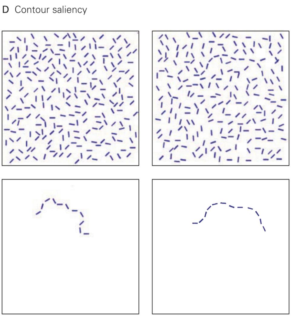
Figure 21-1 Organizational rules of visual perception. To link the elements of a visual scene into unified percepts, the visual system relies on organizational rules such as similarity, proximity, and good continuation.
شکل ۲۱-۱ قواعد سازماندهی ادراک بصری. برای پیوند دادن عناصر یک صحنه بصری به ادراکات یکپارچه، سیستم بصری به قواعد سازماندهی مانند شباهت، مجاورت و تداوم خوب متکی است.
A. Because the dots in alternating rows have the same color, an overall pattern of blue and white rows is perceived.
الف) از آنجا که نقاط در ردیفهای متناوب رنگ یکسانی دارند، الگویی کلی از ردیفهای آبی و سفید درک میشود.
B. The dots in the columns are closer together than those in the rows, leading to the perception of columns.
ب. نقاط ستونها نسبت به نقاط ردیفها به هم نزدیکتر هستند و این منجر به درک ستونها میشود.
C. Line segments are perceptually linked when they are collinear. In the top set of lines, one is more likely to see line segment a as belonging with c rather than d. In the bottom set, a and c are perceptually linked because they maintain the same curvature, whereas a and b appear to be discontinuous.
ج. پاره خطها وقتی همخط باشند، از نظر ادراکی به هم مرتبط هستند. در مجموعه بالایی خطوط، احتمال اینکه پاره خط a به c نسبت داده شود بیشتر از d است. در مجموعه پایینی، a و c از نظر ادراکی به هم مرتبط هستند زیرا انحنای یکسانی دارند، در حالی که a و b ناپیوسته به نظر میرسند.
D. The principle of good continuation is also seen in contour saliency. On the right, a smooth contour of line elements pops out from the background, whereas the jagged contour on the left is lost in the background. (Adapted, with permission, from Field, Hayes, and Hess 1993. Copyright © 1993 Elsevier Ltd.)
د. اصل تداوم خوب در برجستگی خطوط نیز دیده میشود. در سمت راست، یک خط صاف از عناصر خطی از پسزمینه بیرون زده است، در حالی که خط ناهموار سمت چپ در پسزمینه گم شده است. (اقتباس شده، با اجازه، از فیلد، هیز و هس ۱۹۹۳. حق چاپ © ۱۹۹۳ الزویر لیمیتد)

Figure 21-2 Object recognition depends on segmentation of a scene into foreground and background. Recognition of the white salamanders in this image depends on the brain “locating” the white salamanders in the foreground and the brown and black salamanders in the background. The image also illustrates the role of higher influences in segmentation: One can consciously select any of the three colors as the foreground. (Reproduced, with permission, from M.C. Escher’s “Symmetry Drawing E56” 2010 The M.C. Escher Company- Holland. All rights reserved. www.mcescher.com.)
شکل ۲۱-۲ تشخیص اشیا به تقسیمبندی یک صحنه به پیشزمینه و پسزمینه بستگی دارد. تشخیص سمندرهای سفید در این تصویر به «مکانیابی» سمندرهای سفید در پیشزمینه و سمندرهای قهوهای و سیاه در پسزمینه توسط مغز بستگی دارد. این تصویر همچنین نقش تأثیرات بالاتر را در تقسیمبندی نشان میدهد: میتوان آگاهانه هر یک از سه رنگ را به عنوان پیشزمینه انتخاب کرد. (با اجازه، از “طراحی تقارن E56” اثر ام.سی. اشر، ۲۰۱۰، شرکت ام.سی. اشر – هلند، بازنشر شده است. تمامی حقوق محفوظ است. www.mcescher.com.)

Figure 21-3 Expectation and perceptual task play a critical role in what is seen. It is difficult to separate the dark and white patches in this figure into foreground and background without additional information. This figure immediately becomes recognizable after viewing the priming image on page 501. In this example, higher-order representations of shape guide lower-order processes of surface segmentation. (Reproduced, with permission, from Porter 1954. Copyright 1954 by the Board of Trustees of the University of Illinois. Used with permission of the University of Illinois Press.)
شکل ۲۱-۳ انتظار و وظیفه ادراکی نقش مهمی در آنچه دیده میشود، ایفا میکنند. تفکیک تکههای تیره و سفید در این شکل به پیشزمینه و پسزمینه بدون اطلاعات اضافی دشوار است. این شکل بلافاصله پس از مشاهده تصویر اولیه در صفحه ۵۰۱ قابل تشخیص میشود. در این مثال، نمایشهای مرتبه بالاتر از شکل، فرآیندهای مرتبه پایینتر تقسیمبندی سطح را هدایت میکنند. (با اجازه، از Porter 1954 بازتولید شده است. حق چاپ ۱۹۵۴ توسط هیئت امنای دانشگاه ایلینوی. با اجازه انتشارات دانشگاه ایلینوی استفاده شده است.)

Priming image for Figure 21–3
تصویر اولیه برای شکل 21–3
In vision, as in other cognitive operations, various features-motion, depth, form, and color-occur together in a unified percept. This unity is achieved not by one hierarchical neural system but by multiple areas in the brain that are fed by parallel but interacting neural pathways. Because distributed processing is one of the main organizational principles in the neurobiology of vision, one must have a grasp of the anatomical pathways of the visual system to understand fully the physiological description of visual processing in later chapters.
در بینایی، مانند سایر عملیات شناختی، ویژگیهای مختلف – حرکت، عمق، شکل و رنگ – در یک ادراک واحد با هم رخ میدهند. این وحدت نه توسط یک سیستم عصبی سلسله مراتبی، بلکه توسط نواحی متعددی در مغز که توسط مسیرهای عصبی موازی اما در تعامل تغذیه میشوند، حاصل میشود. از آنجا که پردازش توزیعشده یکی از اصول اصلی سازماندهی در نوروبیولوژی بینایی است، برای درک کامل توصیف فیزیولوژیکی پردازش بصری در فصلهای بعدی، باید مسیرهای آناتومیکی سیستم بینایی را درک کرد.
In this chapter, we lay the foundation for understanding the neural circuitry and organizational principles of the visual pathways. These principles apply quite broadly and are relevant not only for the multiple areas of the brain concerned with vision but also for other types of sensory information processing by the brain.
در این فصل، ما پایه و اساس درک مدارهای عصبی و اصول سازمانی مسیرهای بینایی را بنا مینهیم. این اصول به طور گسترده اعمال میشوند و نه تنها برای نواحی متعدد مغز که با بینایی مرتبط هستند، بلکه برای سایر انواع پردازش اطلاعات حسی توسط مغز نیز مرتبط هستند.
Visual Processing Is Mediated by the Geniculostriate Pathway
پردازش بصری توسط مسیر ژنیکولواستریات انجام میشود
The brain’s analysis of visual scenes begins in the two retinas, which transform visual input using a strategy of parallel processing (Chapter 22). This important neural computation strategy is utilized at all stages of the visual pathway as well as in other sensory areas. The pixel-like bits of visual input falling on individual photoreceptors-rods and cones-are analyzed by retinal circuits to extract some 20 local features, such as the local contrasts of dark versus light, red versus green, and blue versus yellow. These features are computed by different populations of specialized neural circuits forming independent processing modules that separately cover the visual field. Thus, each point in the visual field is processed in multiple channels that extract distinct aspects of the visual input simultaneously and in parallel. These parallel streams are then sent out along the axons of the retinal ganglion cells, the projection neurons of the retina, which form the optic nerves.
تحلیل صحنههای بصری توسط مغز از دو شبکیه آغاز میشود که ورودی بصری را با استفاده از یک استراتژی پردازش موازی تبدیل میکنند (فصل ۲۲). این استراتژی مهم محاسبه عصبی در تمام مراحل مسیر بصری و همچنین در سایر نواحی حسی مورد استفاده قرار میگیرد. بیتهای پیکسلمانند ورودی بصری که بر روی گیرندههای نوری منفرد – میلهها و مخروطها – قرار میگیرند، توسط مدارهای شبکیه تجزیه و تحلیل میشوند تا حدود ۲۰ ویژگی محلی، مانند تضادهای محلی تاریکی در مقابل روشنایی، قرمز در مقابل سبز و آبی در مقابل زرد، استخراج شوند. این ویژگیها توسط جمعیتهای مختلفی از مدارهای عصبی تخصصی محاسبه میشوند که ماژولهای پردازش مستقلی را تشکیل میدهند که به طور جداگانه میدان بصری را پوشش میدهند. بنابراین، هر نقطه در میدان بصری در چندین کانال پردازش میشود که جنبههای متمایزی از ورودی بصری را به طور همزمان و موازی استخراج میکنند. سپس این جریانهای موازی در امتداد آکسونهای سلولهای گانگلیونی شبکیه، نورونهای پروژکتور شبکیه، که اعصاب بینایی را تشکیل میدهند، ارسال میشوند.
From the eye, the optic nerve extends to a midline crossing point, the optic chiasm. Beyond the chiasm, the fibers from each temporal hemiretina proceed to the ipsilateral hemisphere along the ipsilateral optic tract; fibers from the nasal hemiretinas cross to the contralateral hemisphere along the contralateral optic tract (Figure 21-5). Because the temporal hemiretina of one eye sees the same half of the visual field (hemifield) as the nasal hemiretina of the other, the partial decussation of fibers at the chiasm ensures that all the information about each hemifield is processed in the visual cortex of the contralateral hemisphere. The layout of the pathway also forms the basis for useful diagnostic information. As a consequence of the particular anatomy of this visual pathway, lesions at different points along the pathway lead to visual deficits with different geometric shapes (Figure 21-5) that can be distinguished reliably through clinical examination. The deficit could be entirely monocular; if present in both eyes, it could affect noncorresponding or corresponding parts of the visual field in the two eyes; it could be restricted to either the upper or the lower visual field or may extend into both, etc. Thus, the shape of the deficit could give valuable clues about type and location of the underlying nerve damage or occlusion (ranging from optic nerve degeneration, such as due to multiple sclerosis, to tumors, strokes, or physical trauma).
عصب بینایی از چشم تا نقطه تقاطع خط میانی، کیاسمای بینایی، امتداد مییابد. فراتر از کیاسم، فیبرهای هر نیمه شبکیه گیجگاهی در امتداد مسیر بینایی همان طرف به نیمکره همان طرف میروند؛ فیبرهای نیمه شبکیه بینی در امتداد مسیر بینایی طرف مقابل به نیمکره مقابل میرسند (شکل 21-5). از آنجا که نیمه شبکیه گیجگاهی یک چشم همان نیمه میدان بینایی (نیمه میدان) را میبیند که نیمه شبکیه بینی چشم دیگر میبیند، تقاطع جزئی فیبرها در کیاسم تضمین میکند که تمام اطلاعات مربوط به هر نیمه میدان در قشر بینایی نیمکره مقابل پردازش میشود. طرح مسیر همچنین اساس اطلاعات تشخیصی مفید را تشکیل میدهد. در نتیجه آناتومی خاص این مسیر بینایی، ضایعات در نقاط مختلف در طول مسیر منجر به نقص بینایی با اشکال هندسی مختلف میشود (شکل 21-5) که میتوان آنها را به طور قابل اعتمادی از طریق معاینه بالینی تشخیص داد. این نقص میتواند کاملاً تک چشمی باشد. اگر در هر دو چشم وجود داشته باشد، میتواند بخشهای غیرمتناظر یا متناظر میدان بینایی را در دو چشم تحت تأثیر قرار دهد. میتواند به میدان بینایی فوقانی یا تحتانی محدود شود یا به هر دو گسترش یابد و غیره. بنابراین، شکل نقص میتواند سرنخهای ارزشمندی در مورد نوع و محل آسیب یا انسداد عصبی زمینهای (از دژنراسیون عصب بینایی، مثلاً به دلیل بیماری اماس، گرفته تا تومورها، سکتههای مغزی یا آسیبهای جسمی) ارائه دهد.

Figure 21-4 A visual scene is analyzed at three levels. Simple attributes of the visual environment are analyzed (low-level processing), and these low-level features are then used to parse the visual scene (intermediate-level processing): Local visual features are assembled into surfaces, objects are segregated from background (surface segmentation), local orientation is integrated into global contours (contour integration), and surface shape is identified from shading and kinematic cues. Finally, surfaces and contours are used to identify the object (high-level processing). (M.C. Escher’s “Day and Night” © 2020 The M.C. Escher Company-The Netherlands. All rights reserved. www.mcescher.com)
شکل ۲۱-۴ یک صحنه بصری در سه سطح تجزیه و تحلیل میشود. ویژگیهای ساده محیط بصری تجزیه و تحلیل میشوند (پردازش سطح پایین) و سپس از این ویژگیهای سطح پایین برای تجزیه صحنه بصری استفاده میشود (پردازش سطح متوسط): ویژگیهای بصری محلی در سطوح مونتاژ میشوند، اشیاء از پسزمینه جدا میشوند (تقسیمبندی سطح)، جهتگیری محلی در خطوط کلی ادغام میشود (یکپارچهسازی خطوط) و شکل سطح از سایهزنی و نشانههای سینماتیکی شناسایی میشود. در نهایت، سطوح و خطوط برای شناسایی شیء استفاده میشوند (پردازش سطح بالا). (“روز و شب” اثر ام.سی. اشر © ۲۰۲۰ شرکت ام.سی. اشر – هلند. تمامی حقوق محفوظ است. www.mcescher.com)
Beyond the optic chiasm, the axons from nasal and temporal hemiretinas carrying input from one hemi- field join in the optic tract, which extends to the lateral geniculate nucleus (LGN) of the thalamus. The LGN in primates consists of six primary layers: four parvocellular (Latin Parvus, small) and two magnocellular, each paired with a thin but dense intercalated or koniocellular (Greek konio, dust) layer (see Figure 21-14). The term “koniocellular” refers to the substantially smaller cell bodies in these layers relative to those of magnocellular or parvocellular layers. The parallel channels established in the retinas remain anatomically segregated through the LGN. Parvocellular layers get input from the midget retinal ganglion cells, which are the most numerous in the primate retina (~70%) and carry red-green opponent information (Chapter 22). Magnocellular layers get achromatic contrast information from the parasol ganglion cells (~10%). Koniocellular layers get input from the small and large bistratified ganglion cells, carrying blue-yellow information, that together make up the third most populous set of retinal projections to the LGN (~8%). Koniocellular layers also get inputs from a number of other numerically much smaller classes of retinal ganglion cells.
فراتر از کیاسمای بینایی، آکسونهای همیرتینهای بینی و گیجگاهی که ورودی را از یک نیممیدان حمل میکنند، در مسیر بینایی به هم میپیوندند که تا هسته زانویی جانبی (LGN) تالاموس امتداد مییابد. LGN در نخستیسانان از شش لایه اصلی تشکیل شده است: چهار پاروسلولی (لاتین Parvus، کوچک) و دو مگنوسلولاری، که هر کدام با یک لایه نازک اما متراکم intercalated یا koniocellular (یونانی konio، گرد و غبار) جفت میشوند (شکل 21-14 را ببینید). اصطلاح “koniocellular” به اجسام سلولی اساساً کوچکتر در این لایهها نسبت به لایههای مگنوسلولاری یا پاروسلاری اشاره دارد. کانالهای موازی ایجاد شده در شبکیهها از طریق LGN از نظر آناتومیکی از هم جدا باقی میمانند. لایههای پاروسلاری ورودی را از سلولهای گانگلیونی شبکیه کوچک دریافت میکنند که بیشترین تعداد را در شبکیه نخستیسانان دارند (حدود 70٪) و اطلاعات حریف قرمز-سبز را حمل میکنند (فصل 22). لایههای مگنوسلولاری اطلاعات کنتراست بیرنگ را از سلولهای گانگلیونی پارازول (حدود 10٪) دریافت میکنند. لایههای کونیوسلولار از سلولهای گانگلیونی دوطبقهای کوچک و بزرگ که اطلاعات آبی-زرد را حمل میکنند، ورودی دریافت میکنند که در مجموع سومین مجموعه پرجمعیت از برآمدگیهای شبکیه به LGN (حدود ۸٪) را تشکیل میدهند. لایههای کونیوسلولار همچنین از تعدادی دیگر از دستههای عددی بسیار کوچکتر سلولهای گانگلیونی شبکیه ورودی دریافت میکنند.
Each geniculate layer receives input from either the ipsilateral or the contralateral eye (see Figure 21-12) but is aligned so as to come from a matching region of the contralateral hemifield. Thus, they form a set of concordant maps stacked atop one another. The thalamic neurons then relay retinal information to the primary visual cortex. But the LGN is not simply a relay; the retinal information it receives can be strongly modulated by attention and arousal through inhibitory connections to this brain region and by feedback from the visual cortex.
هر لایه زانویی ورودی را از چشم همان طرف یا چشم مقابل دریافت میکند (شکل 21-12 را ببینید) اما طوری تنظیم شده است که از ناحیهای منطبق از نیمه میدان مقابل بیاید. بنابراین، آنها مجموعهای از نقشههای هماهنگ را تشکیل میدهند که روی هم انباشته شدهاند. سپس نورونهای تالاموس اطلاعات شبکیه را به قشر بینایی اولیه منتقل میکنند. اما LGN صرفاً یک رله نیست؛ اطلاعات شبکیهای که دریافت میکند میتواند به شدت توسط توجه و برانگیختگی از طریق اتصالات مهاری به این ناحیه مغز و با بازخورد از قشر بینایی تعدیل شود.
The primary visual pathway is also called the geniculostriate pathway (Figure 21-6A) because it passes through the LGN on its way to the primary visual cortex (V1), also known as the striate cortex because of the myelin-rich stripe that runs through its middle layers. A second pathway extends from the retina to the pretectal area of the midbrain, where neurons mediate the pupillary reflexes that control the amount of light entering the eyes (Figure 21-6B). A third pathway from the retina runs to the superior colliculus and is important in controlling eye movements. This pathway continues to the pontine formation in the brain stem and then to the extraocular motor nuclei (Figure 21-6C).
مسیر بینایی اولیه، مسیر ژنیکولواستریات نیز نامیده میشود (شکل 21-6A) زیرا در مسیر خود به قشر بینایی اولیه (V1) از LGN عبور میکند، که به دلیل نوار غنی از میلین که از لایههای میانی آن عبور میکند، به عنوان قشر مخطط نیز شناخته میشود. مسیر دوم از شبکیه تا ناحیه پرهتکتال مغز میانی امتداد دارد، جایی که نورونها واسطه رفلکسهای مردمک هستند که میزان نور ورودی به چشمها را کنترل میکنند (شکل 21-6B). مسیر سوم از شبکیه به کولیکولوس فوقانی میرود و در کنترل حرکات چشم مهم است. این مسیر تا تشکیل پونتین در ساقه مغز و سپس به هستههای حرکتی خارج چشمی ادامه مییابد (شکل 21-6C).
Each LGN projects to the primary visual cortex through a pathway known as the optic radiation. These afferent fibers form a complete neural map of the contralateral visual field in the primary visual cortex. Beyond the striate cortex lie the extrastriate areas, a set of higher- order visual areas that are also organized as neural maps of the visual field. The preservation of the spatial arrangement of inputs from the retina is called retinotopy, and a neural map of the visual field is described as retinotopic or having a retinotopic frame of reference.
هر LGN از طریق مسیری که به عنوان تابش نوری شناخته میشود، به قشر بینایی اولیه منتقل میشود. این فیبرهای آوران، یک نقشه عصبی کامل از میدان بینایی طرف مقابل در قشر بینایی اولیه تشکیل میدهند. فراتر از قشر مخطط، نواحی خارج از مخطط قرار دارند، مجموعهای از نواحی بینایی مرتبه بالاتر که به عنوان نقشههای عصبی میدان بینایی نیز سازماندهی شدهاند. حفظ آرایش فضایی ورودیها از شبکیه، رتینوتوپی نامیده میشود و یک نقشه عصبی از میدان بینایی به عنوان رتینوتوپیک یا دارای چارچوب مرجع رتینوتوپیک توصیف میشود.
The primary visual cortex constitutes the first level of cortical processing of visual information. From there, information is transmitted over two major pathways. A ventral pathway into the temporal lobe carries information about what the stimulus is, and a dorsal pathway into the parietal lobe carries information about where the stimulus is, information that is critical for guiding movement.
قشر بینایی اولیه، اولین سطح پردازش قشری اطلاعات بینایی را تشکیل میدهد. از آنجا، اطلاعات از طریق دو مسیر اصلی منتقل میشوند. یک مسیر شکمی به لوب گیجگاهی، اطلاعاتی در مورد ماهیت محرک و یک مسیر پشتی به لوب آهیانه، اطلاعاتی در مورد محل محرک را حمل میکند، اطلاعاتی که برای هدایت حرکت حیاتی هستند.
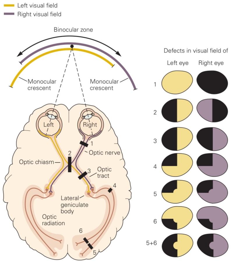
Figure 21-5 Representation of the visual field along the visual pathway. Each eye sees most of the visual field, with the exception of a portion of the peripheral visual field known as the monocular crescent. The axons of retinal neurons (ganglion cells) carry information from each visual hemifield along the optic nerve up to the optic chiasm, where fibers from the nasal hemiretina cross to the opposite hemisphere. Fibers from the temporal hemiretina stay on the same side, joining the fibers from the nasal hemiretina of the contralateral eye to form the optic tract. The optic tract carries information from the opposite visual hemifield originating in both eyes and projects into the lateral geniculate nucleus. Cells in this nucleus send their axons along the optic radiation to the primary visual cortex.
شکل 21-5 نمایش میدان بینایی در امتداد مسیر بینایی. هر چشم بیشتر میدان بینایی را میبیند، به استثنای بخشی از میدان بینایی محیطی که به عنوان هلال تک چشمی شناخته میشود. آکسونهای نورونهای شبکیه (سلولهای گانگلیون) اطلاعات را از هر نیمه میدان بینایی در امتداد عصب بینایی تا کیاسمای بینایی حمل میکنند، جایی که فیبرهای نیمه شبکیه بینی به نیمکره مقابل میرسند. فیبرهای نیمه شبکیه گیجگاهی در همان طرف باقی میمانند و به فیبرهای نیمه شبکیه بینی چشم مقابل میپیوندند تا راه بینایی را تشکیل دهند. راه بینایی اطلاعات را از نیمه میدان بینایی مقابل که از هر دو چشم سرچشمه میگیرد و به هسته زانویی جانبی منتهی میشود، حمل میکند. سلولهای این هسته آکسونهای خود را در امتداد تابش بینایی به قشر بینایی اولیه میفرستند.
Lesions along the visual pathway produce specific visual field deficits, as shown on the right.
ضایعات در امتداد مسیر بینایی، همانطور که در سمت راست نشان داده شده است، نقصهای خاصی در میدان بینایی ایجاد میکنند.
1. A lesion of an optic nerve causes a total loss of vision in one eye.
۱. ضایعه عصب بینایی باعث از بین رفتن کامل بینایی در یک چشم میشود.
2. A lesion of the optic chiasm causes a loss of vision in the temporal half of each visual hemifield (bitemporal hemianopsia).
۲. ضایعه کیاسمای بینایی باعث از بین رفتن بینایی در نیمه گیجگاهی هر یک از نیمکرههای بینایی میشود (همیآنوپسی دو گیجگاهی).
3. A lesion of the optic tract causes a loss of vision in the opposite half of the visual hemifield (contralateral hemianopsia).
۳. ضایعهی دستگاه بینایی باعث از بین رفتن بینایی در نیمهی مقابل میدان بینایی (همیآنوپسی طرف مقابل) میشود.
4. A lesion of the optic radiation fibers that curve into the temporal lobe (Meyer’s loop) causes loss of vision in the upper quadrant of the contralateral visual hemifield in both eyes (upper contralateral quadrantic anopsia).
۴. ضایعهای در فیبرهای تابشی بینایی که به لوب گیجگاهی انحنا مییابند (حلقه مایر) باعث از دست رفتن بینایی در ربع فوقانی نیمه میدان بینایی طرف مقابل در هر دو چشم میشود (آنوپسی ربع فوقانی طرف مقابل).
5, 6. Partial lesions of the visual cortex lead to deficits in portions of the contralateral visual hemifield. For example, a lesion in the upper bank of the calcarine sulcus (5) causes a partial deficit in the inferior quadrant, while a lesion in the lower bank (6) causes a partial deficit in the superior quadrant. The central area of the visual field tends to be unaffected by cortical lesions because of the extent of the representation of the fovea and the duplicate representation of the vertical meridian in the hemispheres.
5، 6. ضایعات جزئی قشر بینایی منجر به نقص در بخشهایی از نیمهی مقابل میدان بینایی میشود. به عنوان مثال، ضایعه در کناره بالایی شیار کالکارین (5) باعث نقص جزئی در ربع تحتانی میشود، در حالی که ضایعه در کناره پایینی (6) باعث نقص جزئی در ربع فوقانی میشود. ناحیه مرکزی میدان بینایی به دلیل وسعت نمایش فووآ و نمایش تکراری نصفالنهار عمودی در نیمکرهها، معمولاً تحت تأثیر ضایعات قشری قرار نمیگیرد.
A major fiber bundle called the corpus callosum connects the two hemispheres, transmitting information across the midline. The primary visual cortex in each hemisphere represents slightly more than half the visual field, with the two hemifield representations overlapping at the vertical meridian. One of the functions of the corpus callosum is to unify the perception of objects spanning the vertical meridian by linking the cortical areas that represent opposite hemifields.
یک دسته فیبر اصلی به نام جسم پینهای، دو نیمکره را به هم متصل میکند و اطلاعات را از خط میانی عبور میدهد. قشر بینایی اولیه در هر نیمکره کمی بیش از نیمی از میدان بینایی را نشان میدهد، و دو نمایش همیمیدانی در نصفالنهار عمودی همپوشانی دارند. یکی از کارکردهای جسم پینهای، یکپارچهسازی ادراک اشیاء در نصفالنهار عمودی با اتصال نواحی قشری است که نشان دهنده نیمههای مخالف هستند.
»» ادامه فصل ماهیت ساختاری پردازش بینایی
»» تمامی کتاب


