مبانی علوم اعصاب اریک کندل؛ ماهیت ساختاری پردازش بینایی؛ پردازش فرم، رنگ، حرکت و عمق


دعای مطالعه [ نمایش ]
بِسْمِ الله الرَّحْمنِ الرَّحیمِ
اَللّهُمَّ اَخْرِجْنى مِنْ ظُلُماتِ الْوَهْمِ
خدايا مرا بيرون آور از تاريكىهاى وهم،
وَ اَكْرِمْنى بِنُورِ الْفَهْمِ
و به نور فهم گرامى ام بدار،
اَللّهُمَّ افْتَحْ عَلَيْنا اَبْوابَ رَحْمَتِكَ
خدايا درهاى رحمتت را به روى ما بگشا،
وَانْشُرْ عَلَيْنا خَزائِنَ عُلُومِكَ بِرَحْمَتِكَ يا اَرْحَمَ الرّاحِمينَ
و خزانههاى علومت را بر ما باز كن به امید رحمتت اى مهربانترين مهربانان.
«مبانی علوم اعصاب» اثر دکتر اریک کندل و همکاران، یکی از جامعترین و معتبرترین منابع در حوزه علوم اعصاب محسوب میشود.
ترجمه دقیق این کتاب، با مدیریت داریوش طاهری و تحت نظارت برند علمی آیندهنگاران مغز، با الهام از تلاشهای علمی و تدریس اساتیدی همچون دکتر محمدتقی جغتایی و دکتر ثریا مهرابی، به فارسی برگردانده شده است.
این ترجمه، دسترسی به دانش عمیق علوم اعصاب را برای دانشجویان و پژوهشگران رشتههای پزشکی، علوم اعصاب، روانشناسی، علوم شناختی، هوش مصنوعی، گفتاردرمانی، مهندسی پزشکی و تمام علاقهمندان به درک عملکرد ذهن و مغز فراهم کرده و گامی در راستای ارتقای دانش و بهبود کیفیت زندگی است.
» کتاب مبانی علوم اعصاب اریک کندل
» » فصل ۲۱: فصل ماهیت ساختاری پردازش بینایی؛ قسمت دوم
در حال ویرایش
Principles of Neural Science; Eric R. Kandel
»» Chapter 21: Sensory Coding
The Constructive Nature of Visual Processing
ماهیت سازنده پردازش بصری
Visual Perception Is a Constructive Process
ادراک بصری یک فرآیند سازنده است
Visual Processing Is Mediated by the Geniculostriate Pathway
پردازش بصری توسط مسیر ژنیکولواستریات انجام میشود
Form, Color, Motion, and Depth Are Processed in Discrete Areas of the Cerebral Cortex
فرم، رنگ، حرکت و عمق در نواحی مجزایی از قشر مغز پردازش میشوند
The Receptive Fields of Neurons at Successive Relays in the Visual Pathway Provide Clues to How the Brain Analyzes Visual Form
میدانهای گیرنده نورونها در رلههای متوالی در مسیر بینایی، سرنخهایی از نحوه تجزیه و تحلیل فرم بصری توسط مغز ارائه میدهند
The Visual Cortex Is Organized Into Columns of Specialized Neurons
قشر بینایی به صورت ستونهایی از نورونهای تخصصی سازماندهی شده است
Intrinsic Cortical Circuits Transform Neural Information
مدارهای قشری ذاتی، اطلاعات عصبی را تبدیل میکنند
Visual Information Is Represented by a Variety of Neural Codes
اطلاعات بصری توسط انواع کدهای عصبی نمایش داده میشود
Highlights
نکات برجسته
Form, Color, Motion, and Depth Are Processed in Discrete Areas of the Cerebral Cortex
فرم، رنگ، حرکت و عمق در نواحی مجزایی از قشر مغز پردازش میشوند
In the late 19th and early 20th centuries, the cerebral cortex was differentiated into discrete regions by the anatomist Korbinian Brodmann and others using anatomical criteria. The criteria included the size, shape, and packing density of neurons in the cortical layers and the thickness and density of myelin. The functionally distinct cortical areas we have considered heretofore correspond only loosely to Brodmann’s classification. The primary visual cortex (V1) is identical to Brodmann’s area 17. In the extrastriate cortex, the secondary visual area (V2) corresponds to area 18. Beyond that, however, area 19 contains several functionally distinct areas that generally cannot be defined by anatomical criteria.
در اواخر قرن نوزدهم و اوایل قرن بیستم، قشر مغز توسط آناتومیست کوربینیان برودمن و دیگران با استفاده از معیارهای آناتومیکی به مناطق مجزا تفکیک شد. این معیارها شامل اندازه، شکل و تراکم نورونها در لایههای قشری و ضخامت و تراکم میلین بود. نواحی قشری از نظر عملکردی متمایز که تاکنون در نظر گرفتهایم، تنها تا حدودی با طبقهبندی برودمن مطابقت دارند. قشر بینایی اولیه (V1) با ناحیه 17 برودمن یکسان است. در قشر خارج از مخطط، ناحیه بینایی ثانویه (V2) با ناحیه 18 مطابقت دارد. با این حال، فراتر از آن، ناحیه 19 شامل چندین ناحیه از نظر عملکردی متمایز است که عموماً نمیتوان آنها را با معیارهای آناتومیکی تعریف کرد.
The number of functionally discrete areas of visual cortex varies between species. Macaque monkeys have more than 30 areas. Although not all visual areas in humans have yet been identified, the number is likely to be at least as great as in the macaque. If one includes oculomotor areas and prefrontal areas contributing to visual memory, almost half of the cerebral cortex is involved with vision. Functional magnetic resonance imaging (fMRI) has made it possible to establish homologies between the visual areas of the macaque and human brains (Figure 21-7). Based on pathway tracing studies in monkeys, we now appreciate that these areas are organized in functional streams (Figure 21-7B).
تعداد نواحی عملکردی مجزای قشر بینایی بین گونهها متفاوت است. میمونهای ماکاک بیش از 30 ناحیه دارند. اگرچه هنوز همه نواحی بینایی در انسان شناسایی نشدهاند، اما احتمالاً تعداد آنها حداقل به اندازه ماکاک است. اگر نواحی حرکتی چشم و نواحی پیشپیشانی که در حافظه بصری نقش دارند را نیز در نظر بگیریم، تقریباً نیمی از قشر مغز درگیر بینایی است. تصویربرداری تشدید مغناطیسی عملکردی (fMRI) امکان ایجاد همولوژی بین نواحی بینایی مغز ماکاک و انسان را فراهم کرده است (شکل 21-7). بر اساس مطالعات ردیابی مسیر در میمونها، اکنون میدانیم که این نواحی در جریانهای عملکردی سازماندهی شدهاند (شکل 21-7B).
The visual areas of cortex can be differentiated by the functional properties of their neurons. Studies of such functional properties have revealed that the visual areas are organized in two hierarchical pathways, a ventral pathway involved in object recognition and a dorsal pathway dedicated to the use of visual information for guiding movements. The ventral or object- recognition pathway extends from the primary visual cortex to the temporal lobe; it is described in detail in Chapter 24. The dorsal or movement-guidance pathway connects the primary visual cortex with the parietal lobe and then with the frontal lobes.
نواحی بینایی قشر مغز را میتوان با ویژگیهای عملکردی نورونهایشان از هم متمایز کرد. مطالعات انجام شده روی چنین ویژگیهای عملکردی نشان داده است که نواحی بینایی در دو مسیر سلسله مراتبی سازماندهی شدهاند، یک مسیر شکمی که در تشخیص اشیا نقش دارد و یک مسیر پشتی که به استفاده از اطلاعات بینایی برای هدایت حرکات اختصاص دارد. مسیر شکمی یا تشخیص اشیا از قشر بینایی اولیه تا لوب گیجگاهی امتداد دارد؛ این مسیر به تفصیل در فصل ۲۴ شرح داده شده است. مسیر پشتی یا هدایت حرکت، قشر بینایی اولیه را به لوب آهیانه و سپس به لوبهای پیشانی متصل میکند.

Figure 21-6 Pathways for visual processing, pupillary reflex and accommodation, and control of eye position.
شکل 21-6 مسیرهای پردازش بینایی، رفلکس مردمک و تطابق، و کنترل موقعیت چشم.
A. Visual processing. The eye sends information first to thalamic nuclei, including the lateral geniculate nucleus and pulvinar, and from there to cortical areas. Cortical projections go forward from the primary visual cortex to areas in the parietal lobe (the dorsal pathway, which is concerned with visually guided movement) and areas in the temporal lobe (the ventral pathway, which is concerned with object recognition). The pulvinar also serves as a relay between cortical areas to supplement their direct connections. (Abbreviation: SC, superior colliculus).
الف) پردازش بینایی. چشم ابتدا اطلاعات را به هستههای تالاموس، شامل هسته زانویی جانبی و پولوینار، و از آنجا به نواحی قشری ارسال میکند. تصاویر قشری از قشر بینایی اولیه به سمت نواحی در لوب آهیانه (مسیر پشتی، که مربوط به حرکت هدایتشده بصری است) و نواحی در لوب گیجگاهی (مسیر شکمی، که مربوط به تشخیص اشیاء است) میروند. پولوینار همچنین به عنوان یک رله بین نواحی قشری برای تکمیل ارتباطات مستقیم آنها عمل میکند. (مخفف: SC، کولیکولوس فوقانی).
B. Pupillary reflex and accommodation. Light signals are relayed through the midbrain pretectum, to preganglionic parasympathetic neurons in the accessory oculomotor (Edinger-Westphal) nucleus, and out through the parasympathetic outflow of the oculomotor nerve to the ciliary ganglion. Postganglionic neurons innervate the smooth muscle of the pupillary sphincter, as well as the muscles controlling the lens.
ب. رفلکس مردمک و تطابق. سیگنالهای نوری از طریق پرهتکتوم مغز میانی، به نورونهای پاراسمپاتیک پیشگانگلیونی در هسته حرکتی جانبی چشم (ادینگر-وستفال) منتقل میشوند و از طریق جریان پاراسمپاتیک عصب حرکتی چشم به گانگلیون مژگانی میروند. نورونهای پسگانگلیونی، عضله صاف اسفنکتر مردمک و همچنین عضلات کنترلکننده عدسی را عصبدهی میکنند.
C. Eye movement. Information from the retina is sent to the superior colliculus (SC) directly along the optic nerve and indirectly through the geniculostriate pathway to cortical areas (primary visual cortex, posterior parietal cortex, and frontal eye fields) that project back to the superior colliculus. The colliculus projects to the pons (PPRF), which then sends control signals to oculomotor nuclei, including the abducens nucleus, which controls lateral movement of the eyes. (Abbreviations: FEF, frontal eye field; LGN, lateral geniculate nucleus; PPRF, paramedian pontine reticular formation.)
ج. حرکت چشم. اطلاعات از شبکیه مستقیماً در امتداد عصب بینایی به کولیکولوس فوقانی (SC) و به طور غیرمستقیم از طریق مسیر ژنیکولواستریات به نواحی قشری (قشر بینایی اولیه، قشر جداری خلفی و میدانهای چشمی پیشانی) ارسال میشود که به کولیکولوس فوقانی برمیگردند. کولیکولوس به پل مغزی (PPRF) امتداد مییابد، که سپس سیگنالهای کنترلی را به هستههای حرکتی چشم، از جمله هسته ابدوسنس، که حرکت جانبی چشمها را کنترل میکند، میفرستد. (اختصارات: FEF، میدان چشمی پیشانی؛ LGN، هسته ژنیکولیت جانبی؛ PPRF، تشکیلات مشبک پل مغزی پارامدین.)
The pathways are interconnected so that information is shared. For example, movement information in the dorsal pathway can contribute to object recognition through kinematic cues. Information about movements in space derived from areas in the dorsal pathway is therefore important for the perception of object shape and is fed into the ventral pathway.
این مسیرها به هم متصل هستند به طوری که اطلاعات به اشتراک گذاشته میشوند. به عنوان مثال، اطلاعات حرکتی در مسیر پشتی میتواند از طریق نشانههای سینماتیکی به تشخیص اشیا کمک کند. بنابراین، اطلاعات مربوط به حرکات در فضا که از نواحی مسیر پشتی به دست میآید، برای درک شکل اشیا مهم است و به مسیر شکمی منتقل میشود.
All connections between cortical areas are reciprocal each area sends information back to the areas from which it receives input. These feedback connections provide information about cognitive functions, including spatial attention, stimulus expectation, and emotional content, to earlier levels of visual processing. The pulvinar in the thalamus serves as a relay between cortical areas (see Figure 21-7B).
تمام ارتباطات بین نواحی قشری متقابل است. هر ناحیه اطلاعات را به نواحی که از آنها ورودی دریافت میکند، برمیگرداند. این ارتباطات بازخوردی، اطلاعاتی در مورد عملکردهای شناختی، از جمله توجه فضایی، انتظار محرک و محتوای عاطفی، به سطوح اولیه پردازش بصری ارائه میدهند. پولوینار در تالاموس به عنوان یک رله بین نواحی قشری عمل میکند (شکل 21-7B را ببینید).
The dorsal pathway courses through the parietal cortex, a region that uses visual information to direct the movement of the eyes and limbs, that is, for visuomotor integration. The lateral intraparietal area, named for its location in the intraparietal sulcus, is involved in representing points in space that are the targets of eye movements or reaching. Patients with lesions of parietal areas fail to attend to objects on one side of the body, a syndrome called unilateral neglect (see Figure 59-1 in Chapter 59).
مسیر پشتی از طریق قشر آهیانه، ناحیهای که از اطلاعات بصری برای هدایت حرکت چشمها و اندامها، یعنی برای ادغام بینایی-حرکتی استفاده میکند، امتداد مییابد. ناحیه داخل آهیانه جانبی، که به دلیل موقعیت آن در شیار داخل آهیانه نامگذاری شده است، در بازنمایی نقاطی در فضا که هدف حرکات چشم یا رسیدن هستند، نقش دارد. بیمارانی که ضایعات نواحی آهیانه دارند، نمیتوانند به اشیاء یک طرف بدن توجه کنند، سندرمی که غفلت یک طرفه نامیده میشود (به شکل 59-1 در فصل 59 مراجعه کنید).
The ventral pathway extends into the temporal lobe. The inferior temporal cortex stores information about the shapes and identities of objects; one portion represents faces, for damage to that region results in the inability to recognize faces (prosopagnosia).
مسیر شکمی به لوب گیجگاهی امتداد مییابد. قشر گیجگاهی تحتانی اطلاعات مربوط به شکل و هویت اشیاء را ذخیره میکند؛ یک بخش از آن چهرهها را نشان میدهد، زیرا آسیب به آن ناحیه منجر به ناتوانی در تشخیص چهرهها (پروزوپاگنوزیا) میشود.
The dorsal and ventral pathways each comprise a hierarchical series of areas that can be delineated by several criteria. First, at many relays, the array of inputs forms a map of the visual hemifield. The boundaries of these maps can be used to demarcate the boundaries of visual areas. This is particularly useful at early levels of the pathway where the receptive fields of neurons are small and visuotopic maps are precisely organized (see the next section for the definition of receptive field). At higher levels, however, the receptive fields become larger, the maps less precise, and visuotopic organization is therefore a less reliable basis to delineate the boundaries of an area.
مسیرهای پشتی و شکمی هر کدام شامل یک سری سلسله مراتبی از نواحی هستند که میتوانند با چندین معیار مشخص شوند. اول، در بسیاری از رلهها، آرایه ورودیها نقشهای از نیممیدان بینایی را تشکیل میدهند. مرزهای این نقشهها میتوانند برای مشخص کردن مرزهای نواحی بینایی استفاده شوند. این امر به ویژه در سطوح اولیه مسیر که در آن میدانهای گیرنده نورونها کوچک هستند و نقشههای ویزوتوپیک دقیقاً سازماندهی شدهاند، مفید است (برای تعریف میدان گیرنده به بخش بعدی مراجعه کنید). با این حال، در سطوح بالاتر، میدانهای گیرنده بزرگتر میشوند، نقشهها دقت کمتری دارند و بنابراین سازماندهی ویزوتوپیک مبنای کمتری برای تعیین مرزهای یک ناحیه است.
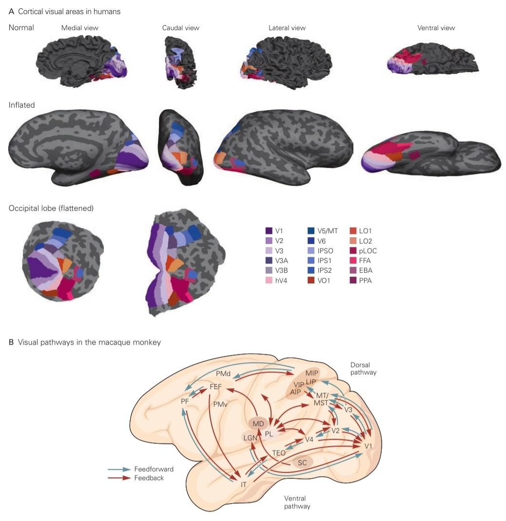
Figure 21-7 Visual pathways in the cerebral cortex.
شکل ۲۱-۷ مسیرهای بینایی در قشر مغز.
A. Functional magnetic resonance imaging shows areas of the human cerebral cortex involved in visual processing. The top row shows areas on the gyri and sulci of a normal view of a brain; the middle row shows “inflated” views of the brain following a computational process that simulates inflating the brain like a balloon so as to stretch out the “wrinkles” of gyri and sulci into a smooth surface while minimizing local distortions. Light and dark gray regions identify gyri and sulci, respectively; the bottom row shows a two-dimensional representation of the occipital lobe (left) and a representation with less distortion by making a cut along the calcarine fissure. Different approaches are required for demarcating different functional areas. Retinotopic areas, by definition, contain continuous maps of visual space and are identified using stimuli such as rotating spirals or expanding circles that sweep through visual space. Maps in adjacent cortical areas run in opposite directions on the cortical surface and meet along boundaries of local mirror reversals. These mirror reversals can be used to identify area boundaries and thus demarcate each area. These retinotopic areas, including early visual areas V1, V2, and V3, and areas V3A, V3B, V6, hV4, VO1, LO1, LO2, and V5/ MT, share boundaries in pairs; these boundaries converge (at the representation of the fovea) at the occipital pole. A different approach, identifying loci of attention, is used to map areas IPS1 and IPS2. Yet further sets of approaches or responsive- ness to specific attributes or classes of objects (such as faces) are used for less strictly retinotopic areas. Functional specificity has been demonstrated for a number of visual areas: VO1 is implicated in color processing, the lateral occipital complex (LO2, PLOC) codes object shape, fusiform face area (FFA) codes faces, the parahippocampal place area (PPA) responds more strongly to places than to objects, the extrastriate body area (EBA) responds more strongly to body parts than objects, and V5/MT is involved in motion processing. Areas in the intra- parietal sulcus (IPS1 and IPS2) are involved in control of spatial attention and saccadic eye movements. (Images courtesy of V. Piech, reproduced with permission.)
الف) تصویربرداری تشدید مغناطیسی عملکردی، نواحی قشر مغز انسان را که در پردازش بصری دخیل هستند، نشان میدهد. ردیف بالا نواحی روی چین و شکنجها و شیارهای یک نمای طبیعی از مغز را نشان میدهد؛ ردیف وسط، نماهای “باد شده” مغز را پس از یک فرآیند محاسباتی نشان میدهد که باد کردن مغز مانند یک بادکنک را شبیهسازی میکند تا “چین و چروکهای” چین و شکنجها را به یک سطح صاف تبدیل کند و در عین حال اعوجاجهای موضعی را به حداقل برساند. نواحی خاکستری روشن و تیره به ترتیب چین و چروکها و شیارها را مشخص میکنند؛ ردیف پایین، نمایش دو بعدی لوب پس سری (چپ) و نمایشی با اعوجاج کمتر را با ایجاد برش در امتداد شکاف کالکارین نشان میدهد. رویکردهای مختلفی برای مشخص کردن نواحی عملکردی مختلف مورد نیاز است. نواحی رتینوتوپیک، طبق تعریف، حاوی نقشههای پیوسته از فضای بصری هستند و با استفاده از محرکهایی مانند مارپیچهای چرخان یا دایرههای در حال گسترش که در فضای بصری حرکت میکنند، شناسایی میشوند. نقشهها در نواحی قشری مجاور در جهتهای مخالف روی سطح قشری حرکت میکنند و در امتداد مرزهای معکوسهای آینهای موضعی به هم میرسند. این معکوسسازیهای آینهای میتوانند برای شناسایی مرزهای ناحیه و در نتیجه مشخص کردن هر ناحیه استفاده شوند. این نواحی رتینوتوپیک، شامل نواحی بینایی اولیه V1، V2 و V3، و نواحی V3A، V3B، V6، hV4، VO1، LO1، LO2 و V5/MT، مرزهای مشترکی دارند؛ این مرزها (در نمایش فووآ) در قطب پسسری همگرا میشوند. رویکرد متفاوتی، یعنی شناسایی جایگاههای توجه، برای نقشهبرداری از نواحی IPS1 و IPS2 استفاده میشود. با این حال، مجموعههای بیشتری از رویکردها یا پاسخدهی به ویژگیها یا طبقات خاص اشیاء (مانند چهرهها) برای نواحی با رتینوتوپیک کمتر مورد استفاده قرار میگیرند. ویژگی عملکردی برای تعدادی از نواحی بینایی نشان داده شده است: VO1 در پردازش رنگ نقش دارد، کمپلکس اکسیپیتال جانبی (LO2، PLOC) شکل شیء را کدگذاری میکند، ناحیه دوکیشکل صورت (FFA) چهرهها را کدگذاری میکند، ناحیه مکانی پاراهیپوکامپ (PPA) به مکانها بیشتر از اشیاء پاسخ میدهد، ناحیه بدن برونمخطط (EBA) به قسمتهای بدن بیشتر از اشیاء پاسخ میدهد، و V5/MT در پردازش حرکت نقش دارد. نواحی شیار درون آهیانه (IPS1 و IPS2) در کنترل توجه فضایی و حرکات جهشی چشم نقش دارند. (تصاویر با اجازه V. Piech، بازنشر شده است.)
B. In the macaque monkey, V1 is located on the surface of the occipital lobe and sends axons in two pathways. A dorsal pathway courses through a number of areas in the parietal lobe and into the frontal lobe and mediates attentional control and visually guided movements. A ventral pathway projects through V4 into areas of the inferior temporal cortex and mediates object recognition. In addition to feedforward pathways extending from primary visual cortex into the temporal, parietal, and frontal lobes (blue arrows), reciprocal or feedback pathways run in the opposite direction (red arrows). Feed- forward and feedback can operate directly, between cortical areas, or indirectly, via the thalamus, in particular the pulvinar, which acts as a relay between cortical areas. The subcortical pathways involved include thalamic nuclei-the lateral geniculate nucleus (LGN), pulvinar nucleus (PL), and mediodorsal nucleus (MD)—and the superior colliculus (SC). (Abbreviations: AIP, anterior intraparietal area; FEF, frontal eye field; IT, inferior temporal cortex; LIP, lateral intraparietal area; MIP, medial intraparietal area; MT, middle temporal area; PF, prefrontal cortex; PMd, dorsal premotor cortex; PMv, ventral premotor cortex; TEO, posterior division of area IT; V1, primary visual cortex, Brodmann’s area 17; V2, secondary visual area, Brodmann’s area 18; V3, V4, third and fourth visual areas; VIP, ventral intraparietal area.)
ب. در میمون ماکاک، V1 در سطح لوب پسسری قرار دارد و آکسونها را از دو مسیر ارسال میکند. یک مسیر پشتی از تعدادی ناحیه در لوب آهیانه و به لوب پیشانی عبور میکند و کنترل توجه و حرکات هدایتشده بصری را واسطهگری میکند. یک مسیر شکمی از طریق V4 به نواحی قشر گیجگاهی تحتانی امتداد مییابد و تشخیص اشیا را واسطهگری میکند. علاوه بر مسیرهای فیدفوروارد که از قشر بینایی اولیه به لوبهای گیجگاهی، آهیانه و پیشانی امتداد مییابند (فلشهای آبی)، مسیرهای متقابل یا بازخورد در جهت مخالف (فلشهای قرمز) اجرا میشوند. فیدفوروارد و بازخورد میتوانند به طور مستقیم، بین نواحی قشری، یا به طور غیرمستقیم، از طریق تالاموس، به ویژه پولوینار، که به عنوان یک رله بین نواحی قشری عمل میکند، عمل کنند. مسیرهای زیرقشری درگیر شامل هستههای تالاموس – هسته زانویی جانبی (LGN)، هسته پولوینار (PL) و هسته مدیودورسال (MD) – و کولیکولوس فوقانی (SC) هستند. (اختصارات: AIP، ناحیه داخل آهیانه ای قدامی؛ FEF، میدان دید فرونتال؛ IT، قشر گیجگاهی تحتانی؛ LIP، ناحیه داخل آهیانه ای جانبی؛ MIP، ناحیه داخل آهیانه ای میانی؛ MT، ناحیه گیجگاهی میانی؛ PF، قشر پیش پیشانی؛ PMd، قشر پیش حرکتی پشتی؛ PMv، قشر پیش حرکتی شکمی؛ TEO، تقسیم خلفی ناحیه IT؛ V1، قشر بینایی اولیه، ناحیه ۱۷ برودمن؛ V2، ناحیه بینایی ثانویه، ناحیه ۱۸ برودمن؛ V3، V4، نواحی بینایی سوم و چهارم؛ VIP، ناحیه داخل آهیانه ای شکمی.)
Another means to differentiate one area from another, as shown by experiments in monkeys, depends upon the distinctive functional properties exhibited by the neurons in each area. The clearest example of this is an area in the dorsal pathway, the middle temporal area (MT or V5), which contains neurons with a strong selectivity for the direction of movement across their receptive fields. Consistent with the idea that the middle temporal area is involved in the analysis of motion, lesions of this area produce deficits in the ability to track moving objects.
روش دیگر برای تمایز یک ناحیه از ناحیه دیگر، همانطور که در آزمایشها روی میمونها نشان داده شده است، به ویژگیهای عملکردی متمایزی که توسط نورونها در هر ناحیه نشان داده میشود، بستگی دارد. واضحترین مثال این، ناحیهای در مسیر پشتی، ناحیه گیجگاهی میانی (MT یا V5) است که شامل نورونهایی با گزینشپذیری قوی برای جهت حرکت در میدانهای گیرنده خود است. مطابق با این ایده که ناحیه گیجگاهی میانی در تجزیه و تحلیل حرکت نقش دارد، ضایعات این ناحیه باعث نقص در توانایی ردیابی اشیاء متحرک میشود.
A classical view of the organization of visual cortical areas is a hierarchical one, where the areas at the bottom of the hierarchy, such as V1 and V2, represent the visual primitives of orientation, direction of movement, depth, and color. In this view, the top of the ventral pathway’s hierarchy would represent whole objects, with the areas in between representing inter- mediate level vision. This idea of “complexification” along the hierarchy suggests a mapping between the levels of visual perception and stages in the sequence of cortical areas. But more recent findings indicate a more complex story, where even the primary visual cortex plays a role in intermediate-level vision, and neurons in the higher areas may process information on components of objects. Moreover, as shown in Figure 21-7, one also has to take into account the fact that there is a powerful reverse flow of information, or feedback, from the “higher” to the “lower” cortical areas. As will be described in Chapter 23, this reverse direction of information contains higher order “top- down” cognitive influences including attention, object expectation, perceptual task, perceptual learning, and efference copy. Top-down influences may play a role in scene segmentation, object relationships, and perception of object details, as well as object recognition itself.
یک دیدگاه کلاسیک از سازماندهی نواحی قشر بینایی، یک دیدگاه سلسله مراتبی است که در آن نواحی پایین سلسله مراتب، مانند V1 و V2، نمایانگر اصول اولیه بصری جهت گیری، جهت حرکت، عمق و رنگ هستند. در این دیدگاه، بالای سلسله مراتب مسیر شکمی نمایانگر کل اشیاء است و نواحی بین آنها نمایانگر بینایی سطح میانی هستند. این ایده “پیچیدگی” در امتداد سلسله مراتب، نقشه برداری بین سطوح ادراک بصری و مراحل توالی نواحی قشری را نشان میدهد. اما یافتههای جدیدتر، داستان پیچیدهتری را نشان میدهند، که در آن حتی قشر بینایی اولیه نیز در بینایی سطح میانی نقش دارد و نورونهای نواحی بالاتر ممکن است اطلاعات مربوط به اجزای اشیاء را پردازش کنند. علاوه بر این، همانطور که در شکل 21-7 نشان داده شده است، باید این واقعیت را نیز در نظر گرفت که یک جریان معکوس قدرتمند از اطلاعات یا بازخورد از نواحی قشری “بالاتر” به “پایینتر” وجود دارد. همانطور که در فصل ۲۳ توضیح داده خواهد شد، این جهت معکوس اطلاعات شامل تأثیرات شناختی «از بالا به پایین» مرتبه بالاتر از جمله توجه، انتظار شیء، وظیفه ادراکی، یادگیری ادراکی و کپی وابران است. تأثیرات از بالا به پایین ممکن است در تقسیمبندی صحنه، روابط شیء و درک جزئیات شیء و همچنین خود تشخیص شیء نقش داشته باشند.
The Receptive Fields of Neurons at Successive Relays in the Visual Pathway Provide Clues to How the Brain Analyzes Visual Form
میدانهای گیرنده نورونها در رلههای متوالی در مسیر بینایی، سرنخهایی از نحوه تجزیه و تحلیل فرم بصری توسط مغز ارائه میدهند
In 1906, Charles Sherrington coined the term receptive field in his analysis of the scratch withdrawal reflex: “The whole collection of points of skin surface from which the scratch-reflex can be elicited is termed the receptive field of that reflex.” When it became possible to record from single neurons in the eye, H. Keffer Hartline applied the concept of the receptive field in his study of the retina of the horseshoe crab, Limulus: “The region of the retina which must be illuminated in order to obtain a response in any given fiber … is termed the receptive field of that fiber.” In the visual system, a neuron’s receptive field represents a small window on the visual field (Figure 21-8).
در سال ۱۹۰۶، چارلز شرینگتون اصطلاح میدان گیرنده را در تحلیل خود از رفلکس عقبنشینی ناشی از خراش ابداع کرد: «کل مجموعه نقاط سطح پوست که از آنها میتوان رفلکس خراش را ایجاد کرد، میدان گیرنده آن رفلکس نامیده میشود.» هنگامی که ثبت از نورونهای منفرد در چشم امکانپذیر شد، اچ. کفر هارتلاین مفهوم میدان گیرنده را در مطالعه خود بر روی شبکیه خرچنگ نعل اسبی، لیمولوس، به کار برد: «ناحیهای از شبکیه که باید روشن شود تا پاسخی در هر فیبر داده شده ایجاد شود… میدان گیرنده آن فیبر نامیده میشود.» در سیستم بینایی، میدان گیرنده یک نورون، پنجره کوچکی در میدان بینایی است (شکل ۲۱-۸).
But responses to only one spot of light yielded a limited understanding of a cell’s receptive field. Using two small spots of light, both Hartline and Stephen Kuffler, who studied the mammalian retina, found an inhibitory surround or lateral inhibitory region in the receptive field. In 1953, Kuffler observed that “not only the areas from which responses can actually be set up by retinal illumination may be included in a definition of the receptive field but also all areas which show a functional connection, by an inhibitory or excitatory effect on a ganglion cell.” Kuffler thus demonstrated that the receptive fields of retinal ganglion cells have functionally distinct subareas. These receptive fields have a center-surround organization and fall into one of two categories: on-center and off-center. Later work demonstrated that neurons in the LGN have similar receptive fields.
اما پاسخ به تنها یک نقطه نور، درک محدودی از میدان گیرنده سلول به همراه داشت. هم هارتلین و هم استفن کافلر، که شبکیه پستانداران را مطالعه میکردند، با استفاده از دو نقطه کوچک نور، یک ناحیه مهاری احاطهکننده یا ناحیه مهاری جانبی در میدان گیرنده یافتند. در سال ۱۹۵۳، کافلر مشاهده کرد که “نه تنها مناطقی که پاسخها میتوانند توسط روشنایی شبکیه تنظیم شوند، ممکن است در تعریف میدان گیرنده گنجانده شوند، بلکه تمام مناطقی که یک اتصال عملکردی را نشان میدهند، توسط یک اثر مهاری یا تحریکی بر روی یک سلول گانگلیونی نیز میتوانند در تعریف میدان گیرنده گنجانده شوند.” بنابراین کافلر نشان داد که میدانهای گیرنده سلولهای گانگلیونی شبکیه دارای زیرناحیههای عملکردی متمایزی هستند. این میدانهای گیرنده دارای یک سازماندهی مرکز-محاصره هستند و در یکی از دو دسته قرار میگیرند: مرکز-مرکز و خارج از مرکز. کارهای بعدی نشان داد که نورونها در LGN میدانهای گیرنده مشابهی دارند.
The on-center cells fire when a spot of light is turned on within a circular central region. Off-center cells fire when a spot of light in the center of their receptive field is turned off. The surrounding annular region has the opposite sign. For on-center cells, a light stimulus any- where in the annulus surrounding the center produces a response when the light is turned off, a response termed on-center, off-surround. The center and surround areas are mutually inhibitory (Figure 21-9). When both center and surround are illuminated with diffuse light, there is little or no response. Conversely, a light-dark boundary across the receptive field produces a brisk response. Because these neurons are most sensitive to borders and contours-to differences in illumination as opposed to uniform surfaces-they encode information about contrast in the visual field.
سلولهای مرکز روشن زمانی فعال میشوند که یک نقطه نور در یک ناحیه مرکزی دایرهای روشن شود. سلولهای خارج از مرکز زمانی فعال میشوند که یک نقطه نور در مرکز میدان گیرنده آنها خاموش شود. ناحیه حلقوی اطراف علامت مخالف دارد. برای سلولهای مرکز روشن، یک محرک نوری در هر جایی از حلقه اطراف مرکز، زمانی که نور خاموش شود، پاسخی ایجاد میکند، پاسخی که مرکز روشن، خارج از محیط نامیده میشود. نواحی مرکز و محیط به طور متقابل مهاری هستند (شکل 21-9). هنگامی که هر دو مرکز و محیط با نور پراکنده روشن میشوند، پاسخ کمی وجود دارد یا هیچ پاسخی وجود ندارد. برعکس، یک مرز روشن-تاریک در سراسر میدان گیرنده، پاسخ سریعی ایجاد میکند. از آنجا که این نورونها بیشترین حساسیت را به مرزها و خطوط کانتور – به تفاوتها در روشنایی در مقایسه با سطوح یکنواخت – دارند، اطلاعات مربوط به کنتراست در میدان بینایی را رمزگذاری میکنند.
The size on the retina of a receptive field varies both according to the field’s eccentricity-its position relative to the fovea, the central part of the retina where visual acuity is highest-and the position of neurons along the visual pathway. Receptive fields with the same eccentricity are relatively small at early levels in visual processing and become progressively larger at later levels. The size of the receptive field is expressed in terms of degrees of visual angle; the entire visual field covers nearly 180° (Figure 21-10A). In early relays of visual processing, the receptive fields near the fovea are the smallest. The receptive fields for retinal ganglion cells that monitor portions of the fovea subtend approximately 0.1°, whereas those in the visual periphery can be a couple of orders of magnitude larger.
اندازه میدان دریافتی روی شبکیه، هم بر اساس خروج از مرکز میدان – موقعیت آن نسبت به گودی چشم، قسمت مرکزی شبکیه که در آن حدت بینایی بیشترین مقدار را دارد – و هم بر اساس موقعیت نورونها در امتداد مسیر بینایی تغییر میکند. میدانهای دریافتی با خروج از مرکز یکسان، در سطوح اولیه پردازش بینایی نسبتاً کوچک هستند و در سطوح بعدی به تدریج بزرگتر میشوند. اندازه میدان دریافتی بر حسب درجه زاویه بینایی بیان میشود؛ کل میدان بینایی تقریباً ۱۸۰ درجه را پوشش میدهد (شکل ۲۱-۱۰A). در رلههای اولیه پردازش بینایی، میدانهای دریافتی نزدیک گودی چشم کوچکترین هستند. میدانهای دریافتی برای سلولهای گانگلیونی شبکیه که بخشهایی از گودی چشم را رصد میکنند، تقریباً ۰.۱ درجه هستند، در حالی که میدانهای دریافتی در حاشیه بینایی میتوانند چند برابر بزرگتر باشند.

Figure 21-8 Receptive fields of retinal ganglion cells in relation to photoreceptors.
شکل 21-8 میدانهای گیرنده سلولهای گانگلیونی شبکیه در ارتباط با گیرندههای نوری.
A. The number of photoreceptors contributing to the receptive field of a retinal ganglion cell varies depending on the location of the receptive field on the retina. A cell near the fovea receives input from fewer receptors covering a smaller area, whereas a cell farther from the fovea receives input from many more receptors covering a larger area (see Figure 21-10).
الف) تعداد گیرندههای نوری که در میدان گیرنده یک سلول گانگلیون شبکیه نقش دارند، بسته به محل میدان گیرنده روی شبکیه متفاوت است. سلولی که در نزدیکی گودی مرکزی (fovea) قرار دارد، از گیرندههای کمتری که ناحیه کوچکتری را پوشش میدهند، ورودی دریافت میکند، در حالی که سلولی که از گودی مرکزی دورتر است، از گیرندههای بسیار بیشتری که ناحیه بزرگتری را پوشش میدهند، ورودی دریافت میکند (شکل 21-10 را ببینید).
B. Light passes through nerve cell layers to reach the photoreceptors at the back of the retina. Signals from the photoreceptors are then transmitted by neurons in the outer and inner nuclear layers to a retinal ganglion cell.
ب. نور از لایههای سلولهای عصبی عبور میکند تا به گیرندههای نوری در پشت شبکیه برسد. سپس سیگنالهای گیرندههای نوری توسط نورونهای لایههای هستهای بیرونی و درونی به یک سلول گانگلیون شبکیه منتقل میشوند.
The amount of cortex dedicated to a degree of visual space changes with eccentricity. More area of cortex is dedicated to the central part of the visual field, where the receptive fields are smallest and the visual system has the greatest spatial resolution (Figure 21-10C).
میزان قشر اختصاص داده شده به درجهای از فضای بصری با خروج از مرکز تغییر میکند. ناحیه بیشتری از قشر به بخش مرکزی میدان بینایی اختصاص داده شده است، جایی که میدانهای گیرنده کوچکترین هستند و سیستم بصری بیشترین وضوح فضایی را دارد (شکل 21-10C).
Receptive-field properties change from relay to relay along a visual pathway. By determining these properties, one can assay the function of each relay nucleus and how visual information is progressively analyzed by the brain. For example, the change in receptive-field structure that occurs between the LGN and cerebral cortex reveals an important mechanism in the brain’s analysis of visual form. The key property of the form pathway is selectivity for the orientation of contours in the visual field. This is an emergent property of signal processing in primary visual cortex; it is not a property of the cortical input but is generated within the cortex itself.
ویژگیهای میدان دریافتی در طول یک مسیر بینایی از رلهای به رله دیگر تغییر میکنند. با تعیین این ویژگیها، میتوان عملکرد هر هسته رله و نحوه تجزیه و تحلیل تدریجی اطلاعات بصری توسط مغز را ارزیابی کرد. به عنوان مثال، تغییر در ساختار میدان دریافتی که بین LGN و قشر مغز رخ میدهد، مکانیسم مهمی را در تجزیه و تحلیل مغز از فرم بصری آشکار میکند. ویژگی کلیدی مسیر فرم، گزینشپذیری برای جهتگیری خطوط در میدان بینایی است. این یک ویژگی نوظهور پردازش سیگنال در قشر بینایی اولیه است؛ این ویژگی، ویژگی ورودی قشری نیست، بلکه در خود قشر تولید میشود.
Whereas retinal ganglion cells and neurons in the LGN have concentric center-surround receptive fields, those in the cortex, although equally sensitive to contrast, also analyze contours. David Hubel and Torsten Wiesel discovered this characteristic in 1958 while studying what visual stimuli provoked activity in neurons in the primary visual cortex. While showing an anesthetized animal slides containing a variety of images, they recorded extracellularly from individual neurons in the visual cortex. As they switched from one slide to another, they found a neuron that produced a brisk train of action potentials. The cell was responding not to the image on the slide but to the edge of the slide as it was moved into position.
در حالی که سلولها و نورونهای گانگلیونی شبکیه در LGN دارای میدانهای گیرندهی مرکز-محیط متحدالمرکز هستند، سلولهای موجود در قشر مغز، اگرچه به همان اندازه به کنتراست حساس هستند، اما خطوط مرزی را نیز تجزیه و تحلیل میکنند. دیوید هوبل و تورستن ویزل این ویژگی را در سال ۱۹۵۸ هنگام مطالعهی اینکه چه محرکهای بصری باعث فعالیت در نورونها در قشر بینایی اولیه میشوند، کشف کردند. آنها هنگام نشان دادن اسلایدهای حیوانی بیهوش حاوی تصاویر متنوع، به صورت خارج سلولی از نورونهای منفرد در قشر بینایی ثبت کردند. همانطور که از یک اسلاید به اسلاید دیگر تغییر مکان میدادند، نورونی را یافتند که رشتهای سریع از پتانسیلهای عمل تولید میکرد. سلول نه به تصویر روی اسلاید، بلکه به لبهی اسلاید هنگام جابجایی به موقعیت مناسب پاسخ میداد.
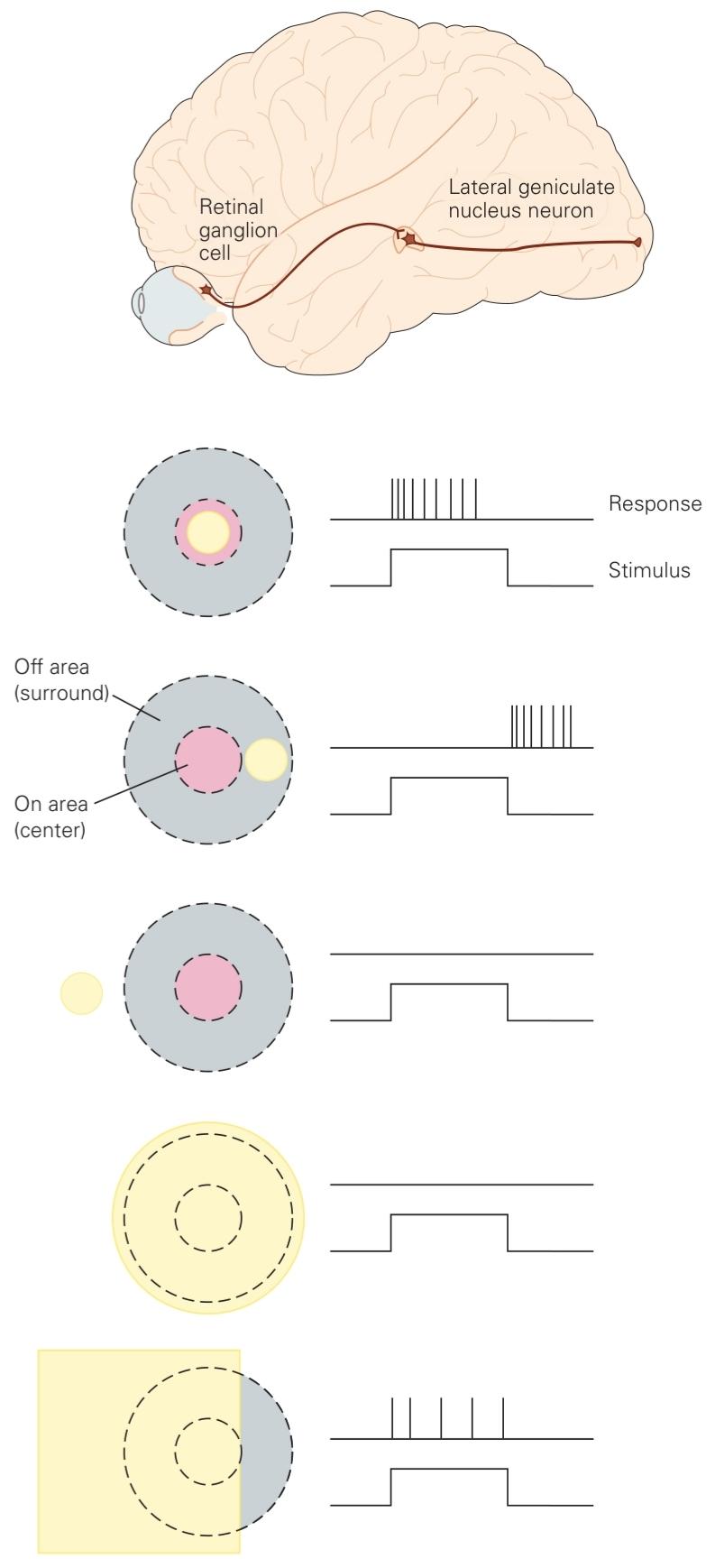
Figure 21-9 Receptive fields of neurons at early relays of visual pathways. A circular symmetric receptive field with mutually antagonistic center and surround is characteristic of retinal ganglion cells and neurons in the lateral geniculate nucleus of the thalamus. The center can respond to the turning on or turning off of a spot of light (yellow) depending on whether the receptived field belongs to an “on-center” or “off-center” class, respectively. The surround has the opposite response. Outside the surround, there is no response to light, thus defining the receptive field boundary. The response is weak when light covers both the center and surround, so these neurons respond optimally to contrast (a light-dark boundary) in the visual field.
شکل ۲۱-۹ میدانهای گیرنده نورونها در رلههای اولیه مسیرهای بینایی. یک میدان گیرنده متقارن دایرهای با مرکز و محیط متضاد، مشخصه سلولهای گانگلیونی شبکیه و نورونها در هسته زانویی جانبی تالاموس است. مرکز میتواند به روشن یا خاموش شدن یک نقطه نور (زرد) پاسخ دهد، بسته به اینکه میدان گیرنده به ترتیب به کلاس “روی مرکز” یا “خارج از مرکز” تعلق داشته باشد. محیط اطراف پاسخ متضادی دارد. در خارج از محیط اطراف، هیچ پاسخی به نور وجود ندارد، بنابراین مرز میدان گیرنده را تعریف میکند. پاسخ زمانی ضعیف است که نور هم مرکز و هم محیط اطراف را بپوشاند، بنابراین این نورونها به طور بهینه به کنتراست (مرز نور-تاریکی) در میدان بینایی پاسخ میدهند.
»» قسمت اول فصل ماهیت ساختاری پردازش بینایی
»» تمامی کتاب