مبانی علوم اعصاب اریک کندل؛ بینایی رنگی و مدارهای گیرندههای مخروطی


دعای مطالعه [ نمایش ]
بِسْمِ الله الرَّحْمنِ الرَّحیمِ
اَللّهُمَّ اَخْرِجْنى مِنْ ظُلُماتِ الْوَهْمِ
خدايا مرا بيرون آور از تاريكىهاى وهم،
وَ اَكْرِمْنى بِنُورِ الْفَهْمِ
و به نور فهم گرامى ام بدار،
اَللّهُمَّ افْتَحْ عَلَيْنا اَبْوابَ رَحْمَتِكَ
خدايا درهاى رحمتت را به روى ما بگشا،
وَانْشُرْ عَلَيْنا خَزائِنَ عُلُومِكَ بِرَحْمَتِكَ يا اَرْحَمَ الرّاحِمينَ
و خزانههاى علومت را بر ما باز كن به امید رحمتت اى مهربانترين مهربانان.
«مبانی علوم اعصاب» اثر دکتر اریک کندل و همکاران، یکی از جامعترین و معتبرترین منابع در حوزه علوم اعصاب محسوب میشود.
ترجمه دقیق این کتاب، با مدیریت داریوش طاهری و تحت نظارت برند علمی آیندهنگاران مغز، با الهام از تلاشهای علمی و تدریس اساتیدی همچون دکتر محمدتقی جغتایی و دکتر ثریا مهرابی، به فارسی برگردانده شده است.
این ترجمه، دسترسی به دانش عمیق علوم اعصاب را برای دانشجویان و پژوهشگران رشتههای پزشکی، علوم اعصاب، روانشناسی، علوم شناختی، هوش مصنوعی، گفتاردرمانی، مهندسی پزشکی و تمام علاقهمندان به درک عملکرد ذهن و مغز فراهم کرده و گامی در راستای ارتقای دانش و بهبود کیفیت زندگی است.
» کتاب مبانی علوم اعصاب اریک کندل
» » فصل ۲۲: فصل پردازش بصری سطح پایین: شبکیه؛ قسمت سوم
در حال ویرایش
Principles of Neural Science; Eric R. Kandel
»» Chapter 22: Low-Level Visual Processing: The Retina
The Photoreceptor Layer Samples the Visual Image
لایه گیرنده نور از تصویر بصری نمونهبرداری میکند
Ocular Optics Limit the Quality of the Retinal Image
اپتیک چشم، کیفیت تصویر شبکیه را محدود میکند
There Are Two Types of Photoreceptors: Rods and Cones
دو نوع گیرنده نوری وجود دارد: میلهایها و مخروطیها
Phototransduction Links the Absorption of a Photon to a Change in Membrane Conductance
انتقال نوری، جذب فوتون را به تغییر در رسانایی غشاء مرتبط میکند
Light Activates Pigment Molecules in the Photoreceptors
نور مولکولهای رنگدانه را در گیرندههای نوری فعال میکند
Excited Rhodopsin Activates a Phosphodiesterase Through the G Protein Transducin
رودوپسین برانگیخته شده از طریق پروتئین G ترانسدوسین، یک فسفودی استراز را فعال میکند.
Multiple Mechanisms Shut Off the Cascade
چندین مکانیسم، آبشار را خاموش میکنند
Defects in Phototransduction Cause Disease
نقص در انتقال نوری باعث بیماری میشود
Ganglion Cells Transmit Neural Images to the Brain
سلولهای گانگلیون تصاویر عصبی را به مغز منتقل میکنند
The Two Major Types of Ganglion Cells Are ON Cells and OFF Cells
دو نوع اصلی سلولهای گانگلیون، سلولهای روشن (ON) و سلولهای خاموش (OFF) هستند.
Many Ganglion Cells Respond Strongly to Edges in the Image
بسیاری از سلولهای گانگلیون به شدت به لبههای تصویر پاسخ میدهند
The Output of Ganglion Cells Emphasizes Temporal Changes in Stimuli
خروجی سلولهای گانگلیون بر تغییرات زمانی در محرکها تأکید دارد
Retinal Output Emphasizes Moving Objects
خروجی شبکیه، اشیاء متحرک را برجسته میکند
Several Ganglion Cell Types Project to the Brain Through Parallel Pathways
چندین نوع سلول گانگلیون از طریق مسیرهای موازی به مغز میرسند
A Network of Interneurons Shapes the Retinal Output
شبکهای از نورونهای رابط، خروجی شبکیه را شکل میدهد
Parallel Pathways Originate in Bipolar Cells
مسیرهای موازی از سلولهای دوقطبی سرچشمه میگیرند
Spatial Filtering Is Accomplished by Lateral Inhibition
فیلترینگ مکانی با مهار جانبی انجام میشود
Temporal Filtering Occurs in Synapses and Feedback Circuits
فیلترینگ زمانی در سیناپسها و مدارهای بازخورد رخ میدهد
Color Vision Begins in Cone-Selective Circuits
بینایی رنگی از مدارهای انتخابی مخروطی آغاز میشود
Congenital Color Blindness Takes Several Forms
کوررنگی مادرزادی اشکال مختلفی دارد
Rod and Cone Circuits Merge in the Inner Retina
مدارهای میلهای و مخروطی در شبکیه داخلی ادغام میشوند
The Retina’s Sensitivity Adapts to Changes in Illumination
حساسیت شبکیه با تغییرات روشنایی سازگار میشود
Light Adaptation Is Apparent in Retinal Processing and Visual Perception
سازگاری با نور در پردازش شبکیه و ادراک بصری آشکار است
Multiple Gain Controls Occur within the Retina
کنترلهای چندگانهی بهره در شبکیه رخ میدهند
Light Adaptation Alters Spatial Processing
سازگاری با نور، پردازش فضایی را تغییر میدهد
Highlights
نکات برجسته
Box 22-1 Spatiotemporal Sensitivity of Human Perception
کادر ۲۲-۱ حساسیت مکانی-زمانی ادراک انسان
Although small spots of light are useful for probing the receptive fields of single neurons in visual pathways, different stimuli are needed to learn about human visual perception. Grating stimuli are commonly used to probe how our visual system deals with spatial and temporal patterns.
اگرچه لکههای کوچک نور برای بررسی میدانهای گیرنده نورونهای منفرد در مسیرهای بینایی مفید هستند، اما برای یادگیری در مورد ادراک بصری انسان به محرکهای مختلفی نیاز است. محرکهای شبکهای معمولاً برای بررسی چگونگی برخورد سیستم بینایی ما با الگوهای مکانی و زمانی استفاده میشوند.
The subject views a display in which the intensity varies about the mean as a sinusoidal function of space (Figure 22-12). Then the contrast of the display-defined as the peak-to-peak amplitude of the sinusoid divided by the mean-is reduced to a threshold at which the grating is barely visible. This measurement is repeated for gratings of different spatial frequencies.
فرد نمایشگری را مشاهده میکند که در آن شدت به صورت تابعی سینوسی از فضا، حول میانگین تغییر میکند (شکل 22-12). سپس کنتراست نمایشگر – که به صورت دامنه قله تا قله سینوسی تقسیم بر میانگین تعریف میشود – به آستانهای کاهش مییابد که در آن توری به سختی قابل مشاهده است. این اندازهگیری برای توریهایی با فرکانسهای فضایی مختلف تکرار میشود.
When the inverse of this threshold is plotted against the spatial frequency, the resulting contrast sensitivity curve provides a measure of sensitivity of visual perception to patterns of different scales (Figure 22-13A). When measured at high light intensity, sensitivity declines sharply at high spatial frequencies, with an absolute threshold at approximately 50 cycles per degree. This sensitivity is limited fundamentally by the quality of the optical image and the spacing of cone cells in the fovea (see Figure 22-1C).
وقتی معکوس این آستانه در برابر فرکانس فضایی رسم شود، منحنی حساسیت کنتراست حاصل، معیاری از حساسیت ادراک بصری به الگوهایی با مقیاسهای مختلف را ارائه میدهد (شکل 22-13A). هنگامی که در شدت نور بالا اندازهگیری میشود، حساسیت در فرکانسهای فضایی بالا به شدت کاهش مییابد، با آستانه مطلق تقریباً 50 سیکل در هر درجه. این حساسیت اساساً توسط کیفیت تصویر نوری و فاصله سلولهای مخروطی در گودی چشم محدود میشود (شکل 22-1C را ببینید).
Interestingly, sensitivity also declines at low spatial frequencies. Patterns with a frequency of approximately 5 cycles per degree are most visible. The visual system is said to have band-pass behavior because it rejects all but a band of spatial frequencies.
جالب توجه است که حساسیت در فرکانسهای مکانی پایین نیز کاهش مییابد. الگوهایی با فرکانس تقریباً ۵ سیکل در هر درجه بیشترین قابلیت مشاهده را دارند. گفته میشود که سیستم بینایی رفتار میانگذر دارد زیرا همه فرکانسهای مکانی به جز یک باند را رد میکند.
One can use the same techniques to measure the sensitivity of individual retinal ganglion cells in primates. The results resemble those for human subjects (Figure 22-13), suggesting that these basic features of visual perception are determined by the retina.
میتوان از همین تکنیکها برای اندازهگیری حساسیت سلولهای گانگلیون شبکیه در نخستیسانان استفاده کرد. نتایج مشابه نتایج مربوط به انسان است (شکل ۲۲-۱۳)، که نشان میدهد این ویژگیهای اساسی ادراک بصری توسط شبکیه تعیین میشوند.
The band-pass behavior can be understood on the basis of spatial antagonism in center-surround receptive fields. A very fine grating presents many dark and bright stripes within the receptive-field center; their effects cancel one another and thus provide no net excitation. A very coarse grating presents a single stripe to both the center and surround of the receptive field, and their antagonism again provides the ganglion cell little net excitation. The strongest response is produced by a grating of intermediate spatial frequency that just covers the center with one stripe and most of the surround with stripes of the opposite polarity (Figure 22-13B).
رفتار عبور باند را میتوان بر اساس تضاد فضایی در میدانهای گیرنده مرکز-پیرامون درک کرد. یک شبکه بسیار ریز، نوارهای تاریک و روشن زیادی را در مرکز میدان گیرنده ایجاد میکند؛ اثرات آنها یکدیگر را خنثی میکند و بنابراین هیچ تحریک خالصی ایجاد نمیکند. یک شبکه بسیار درشت، یک نوار واحد را هم به مرکز و هم به اطراف میدان گیرنده ارائه میدهد و تضاد آنها دوباره تحریک خالص کمی برای سلول گانگلیونی ایجاد میکند. قویترین پاسخ توسط یک شبکه با فرکانس فضایی متوسط ایجاد میشود که فقط مرکز را با یک نوار و بیشتر اطراف را با نوارهایی با قطب مخالف میپوشاند (شکل 22-13B).
In dim light, the visual system’s contrast sensitivity declines, but more so at high than at low spatial frequencies (Figure 22-13A). Thus, the peak sensitivity shifts to lower spatial frequencies, and eventually the curve loses its peak altogether. In this state, the visual system has so-called low-pass behavior, for it preferentially encodes stimuli of low spatial frequency. The fact that in dim light the receptive fields of ganglion cells lose their antagonistic surrounds explains the transition from band-pass to low-pass spatial filtering (Figure 22-13B).
در نور کم، حساسیت کنتراست سیستم بینایی کاهش مییابد، اما این کاهش در فرکانسهای فضایی بالا بیشتر از فرکانسهای فضایی پایین است (شکل 22-13A). بنابراین، اوج حساسیت به فرکانسهای فضایی پایینتر تغییر میکند و در نهایت منحنی به طور کلی اوج خود را از دست میدهد. در این حالت، سیستم بینایی رفتاری به اصطلاح پایینگذر دارد، زیرا ترجیحاً محرکهای با فرکانس فضایی پایین را رمزگذاری میکند. این واقعیت که در نور کم، میدانهای گیرنده سلولهای گانگلیونی، محیطهای متضاد خود را از دست میدهند، گذار از فیلترینگ فضایی میانگذر به پایینگذر را توضیح میدهد (شکل 22-13B).
Similar experiments can be done to test visual sensitivity to temporal patterns. Here, the intensity of a test stimulus flickers sinusoidally in time, while the contrast is gradually brought to the threshold level of detection. For humans, contrast sensitivity declines sharply at very high flicker frequencies, but declines also at very low frequencies (Figure 22-14A). Flicker at approximately 10 Hz is the most effective stimulus. One finds similar band-pass behavior in the flicker sensitivity of macaque retinal ganglion cells (Figure 22-14B).
آزمایشهای مشابهی را میتوان برای آزمایش حساسیت بینایی به الگوهای زمانی انجام داد. در اینجا، شدت یک محرک آزمایشی به صورت سینوسی در طول زمان سوسو میزند، در حالی که کنتراست به تدریج به سطح آستانه تشخیص میرسد. برای انسان، حساسیت کنتراست در فرکانسهای سوسو زدن بسیار بالا به شدت کاهش مییابد، اما در فرکانسهای بسیار پایین نیز کاهش مییابد (شکل 22-14A). سوسو زدن در فرکانس تقریباً 10 هرتز مؤثرترین محرک است. میتوان رفتار عبور باند مشابهی را در حساسیت سوسو زدن سلولهای گانگلیونی شبکیه میمون ماکاک مشاهده کرد (شکل 22-14B).
Sensitivity to temporal contrast also depends on the mean light level. For human subjects, the optimum flicker frequency shifts downward at lower stimulus intensities and the peak in the curve becomes less and less prominent (Figure 22-14). The fact that primate retinal ganglion cells duplicate this behavior suggests that retinal processing limits the performance of the entire visual system in these simple tasks.
حساسیت به کنتراست زمانی همچنین به میانگین سطح نور بستگی دارد. برای افراد انسانی، فرکانس بهینه سوسو زدن در شدتهای محرک پایینتر به سمت پایین تغییر میکند و قله در منحنی کمرنگتر و کمرنگتر میشود (شکل 22-14). این واقعیت که سلولهای گانگلیونی شبکیه نخستیسانان این رفتار را تقلید میکنند، نشان میدهد که پردازش شبکیه، عملکرد کل سیستم بینایی را در این وظایف ساده محدود میکند.
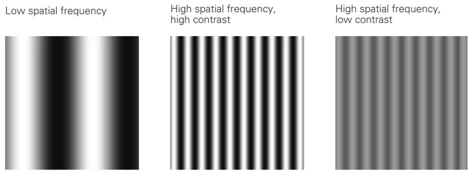
Figure 22-12 Sinusoid grating displays used in psychophysical experiments with human subjects. Such stimuli were used in the experiments discussed in Figure 22-13.
شکل 22-12 نمایشگرهای شبکه سینوسی مورد استفاده در آزمایشهای روانفیزیکی با سوژههای انسانی. چنین محرکهایی در آزمایشهای مورد بحث در شکل 22-13 استفاده شدهاند.
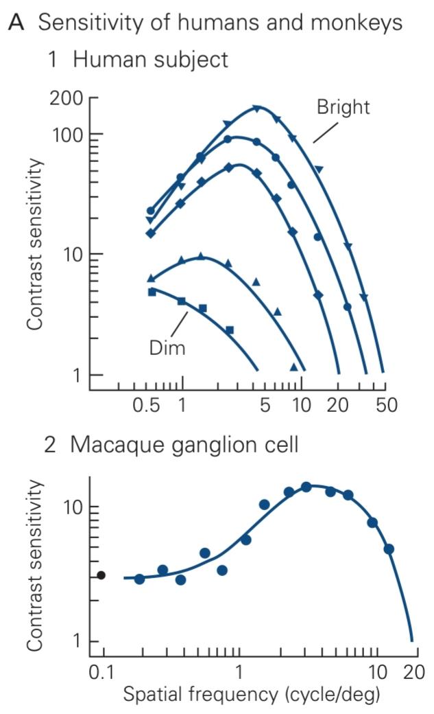
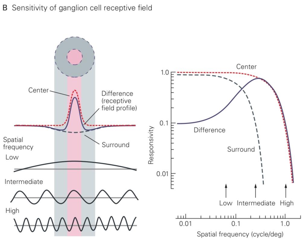
Figure 22–13 Spatial contrast sensitivity.
شکل ۲۲-۱۳ حساسیت کنتراست فضایی.
A. 1. The contrast sensitivity of human subjects was measured using gratings with different spatial frequencies (see Figure 22-12). At each frequency, the contrast was increased to the threshold for detection, and the inverse of that contrast value was plotted against spatial frequency, as shown here. The curves were obtained at different mean intensities, decreasing by factors of 10 from the top to the bottom curve. (Reproduced, with permission, from De Valois, Morgan, and Snodderly 1974.) 2. Contrast sensitivity of a P-type ganglion cell in the macaque retina measured at high intensity. At each spatial frequency, the contrast was gradually increased until it produced a detectable change in the neuron’s firing rate. The inverse of that threshold contrast was plotted as in part A-1. The isolated dot at left marks the sensitivity at zero spatial frequency, a spatially uniform field. (Reproduced, with permission, from Derrington and Lennie 1984.)
الف. ۱. حساسیت کنتراست افراد انسانی با استفاده از شبکههایی با فرکانسهای فضایی مختلف اندازهگیری شد (شکل ۲۲-۱۲ را ببینید). در هر فرکانس، کنتراست تا آستانه تشخیص افزایش یافت و معکوس آن مقدار کنتراست در مقابل فرکانس فضایی رسم شد، همانطور که در اینجا نشان داده شده است. منحنیها با شدتهای میانگین مختلف به دست آمدند و از بالا به پایین منحنی با ضریب ۱۰ کاهش یافتند. (با اجازه، از De Valois، Morgan و Snodderly 1974 بازتولید شده است.) ۲. حساسیت کنتراست یک سلول گانگلیونی نوع P در شبکیه میمون ماکاک که با شدت بالا اندازهگیری شده است. در هر فرکانس فضایی، کنتراست به تدریج افزایش یافت تا زمانی که تغییر قابل تشخیصی در نرخ شلیک نورون ایجاد کند. معکوس آن کنتراست آستانه همانطور که در قسمت A-1 رسم شد. نقطه جدا شده در سمت چپ، حساسیت را در فرکانس فضایی صفر، یک میدان فضایی یکنواخت، نشان میدهد. (با اجازه، از Derrington و Lennie 1984 بازتولید شده است.)
B. Stimulation of a center-surround receptive field with sinusoid gratings. The neuron’s sensitivity to light at different points on the retina is modeled as a “difference-of- Gaussians” receptive field, with a narrow positive Gaussian for the excitatory center and a broad negative Gaussian for the inhibitory surround. Multiplying the profile of the grating stimulus (intensity vs position) with the profile of the receptive field (sensitivity vs position) and integrating over all space calculates the stimulus strength delivered by a particular grating. The resulting sensitivity of the receptive field to gratings of different frequency is shown in the plot on the right. At low spatial frequencies, the negative contribution from the surround cancels the contribution from the center, leading to a drop in the difference curve. (Reproduced, with permission, from Enroth-Cugell and Robson 1984.)
ب. تحریک میدان گیرنده مرکز-محیط با توریهای سینوسی. حساسیت نورون به نور در نقاط مختلف شبکیه به عنوان یک میدان گیرنده “اختلاف گاوسی” مدلسازی میشود، با یک گاوسی مثبت باریک برای مرکز تحریکی و یک گاوسی منفی پهن برای احاطه بازدارنده. ضرب کردن پروفیل محرک توری (شدت در مقابل موقعیت) در پروفیل میدان گیرنده (حساسیت در مقابل موقعیت) و انتگرالگیری در کل فضا، قدرت محرک ارائه شده توسط یک توری خاص را محاسبه میکند. حساسیت حاصل از میدان گیرنده به توریهای فرکانس مختلف در نمودار سمت راست نشان داده شده است. در فرکانسهای مکانی پایین، سهم منفی از محیط، سهم از مرکز را خنثی میکند و منجر به افت منحنی اختلاف میشود. (با اجازه، از Enroth-Cugell و Robson 1984 بازتولید شده است.)
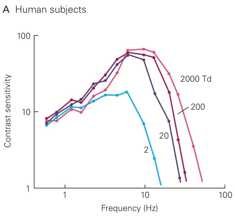
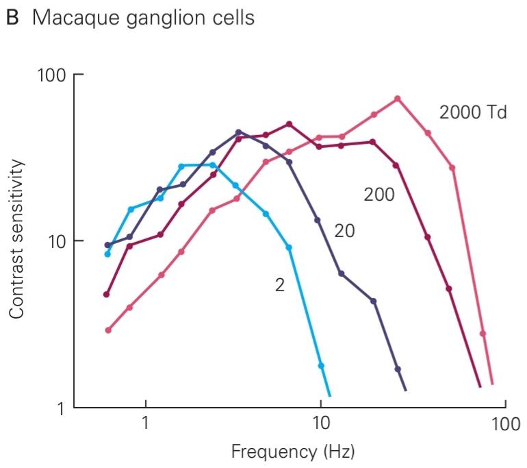
Figure 22-14 Temporal contrast sensitivity. (Reproduced, with permission, from Lee et al. 1990.)
شکل 22-14 حساسیت کنتراست زمانی. (با اجازه، از Lee و همکاران، 1990، بازنشر شده است.)
A. The sensitivity of human subjects to temporal flicker was measured by methods similar to those in Figure 22-13A, but the stimulus was a large spot whose intensity varied sinusoidally in time rather than in space. The inverse of the threshold contrast required for detection is plotted against the frequency of the sinusoidal flicker. Sensitivity declines at both high and low frequencies. The mean light level varied, decreasing by factors of 10 from the top to the bottom trace.
الف. حساسیت افراد به سوسو زدن زمانی با روشهایی مشابه روشهای موجود در شکل ۲۲-۱۳A اندازهگیری شد، اما محرک یک نقطه بزرگ بود که شدت آن به صورت سینوسی در زمان و نه در مکان تغییر میکرد. معکوس کنتراست آستانه مورد نیاز برای تشخیص در برابر فرکانس سوسو زدن سینوسی رسم شده است. حساسیت در هر دو فرکانس بالا و پایین کاهش مییابد. میانگین سطح نور متغیر بود و از بالا به پایین نمودار با ضریب ۱۰ کاهش مییافت.
B. The flicker sensitivity of M-type ganglion cells in the macaque retina was measured by the same method applied to human subjects in part A. The detection threshold for the neural response was defined as a variation of 20 spikes per second in the cell’s firing rate in phase with the flicker.
ب. حساسیت به سوسو زدن سلولهای گانگلیونی نوع M در شبکیه میمون ماکاک با همان روشی که در بخش الف برای نمونههای انسانی اعمال شد، اندازهگیری شد. آستانه تشخیص برای پاسخ عصبی به صورت تغییر 20 اسپایک در ثانیه در نرخ شلیک سلول همفاز با سوسو زدن تعریف شد.
These neural representations are directed to various visual centers in the brain, including the lateral geniculate nucleus of the thalamus, a relay to the visual cortex; the superior colliculus, a midbrain region involved in spatial attention and orienting movements; the pretectum, involved in control of the pupil; the accessory optic system, which analyzes self-motion to stabilize gaze; and the suprachiasmatic nucleus, a central clock that directs circadian rhythm and whose phase can be set by light cues (Chapter 44). In many cases, the axons of one type of ganglion cell extend collaterals to multiple areas of the central nervous system. M-cells, for example, project to the thalamus and the superior colliculus.
این بازنماییهای عصبی به مراکز مختلف بینایی در مغز هدایت میشوند، از جمله هسته زانویی جانبی تالاموس، که به قشر بینایی متصل است؛ کولیکولوس فوقانی، ناحیهای از مغز میانی که در توجه فضایی و حرکات جهتیابی نقش دارد؛ پرهتکتوم، که در کنترل مردمک چشم نقش دارد؛ سیستم نوری جانبی که حرکت خود را برای تثبیت نگاه تجزیه و تحلیل میکند؛ و هسته سوپراکیاسماتیک، یک ساعت مرکزی که ریتم شبانهروزی را هدایت میکند و فاز آن را میتوان با نشانههای نوری تنظیم کرد (فصل ۴۴). در بسیاری از موارد، آکسونهای یک نوع سلول گانگلیونی، رشتههای جانبی را به نواحی متعدد سیستم عصبی مرکزی گسترش میدهند. به عنوان مثال، سلولهای M به تالاموس و کولیکولوس فوقانی امتداد مییابند.
A Network of Interneurons Shapes the Retinal Output
شبکهای از نورونهای رابط، خروجی شبکیه را شکل میدهد
We now consider in more detail the retinal circuit and how it accounts for the intricate response properties of retinal ganglion cells.
اکنون با جزئیات بیشتری مدار شبکیه و چگونگی تأثیر آن بر خواص پاسخ پیچیده سلولهای گانگلیونی شبکیه را بررسی میکنیم.
Parallel Pathways Originate in Bipolar Cells
مسیرهای موازی از سلولهای دوقطبی سرچشمه میگیرند
The photoreceptor forms synapses with bipolar cells and horizontal cells (see Figure 22-3A). In the dark, the photoreceptor’s synaptic terminal releases glutamate continuously. When stimulated by light, the photoreceptor hyperpolarizes, less calcium enters the terminal, and the terminal releases less glutamate. Photoreceptors do not fire action potentials; like bipolar cells, they release neurotransmitter in a graded fashion using a specialized structure, the ribbon synapse. In fact, most retinal processing is accomplished with graded membrane potentials: Action potentials occur primarily in certain amacrine cells and in the retinal ganglion cells.
گیرنده نوری با سلولهای دوقطبی و سلولهای افقی سیناپس تشکیل میدهد (شکل 22-3A را ببینید). در تاریکی، پایانه سیناپسی گیرنده نوری به طور مداوم گلوتامات آزاد میکند. هنگامی که توسط نور تحریک میشود، گیرنده نوری بیش از حد قطبی میشود، کلسیم کمتری وارد پایانه میشود و پایانه گلوتامات کمتری آزاد میکند. گیرندههای نوری پتانسیل عمل ایجاد نمیکنند؛ مانند سلولهای دوقطبی، آنها انتقالدهنده عصبی را به صورت درجهبندی شده با استفاده از یک ساختار تخصصی، سیناپس روبانی، آزاد میکنند. در واقع، بیشتر پردازشهای شبکیه با پتانسیلهای غشایی درجهبندی شده انجام میشود: پتانسیلهای عمل در درجه اول در سلولهای آماکرین خاص و در سلولهای گانگلیونی شبکیه رخ میدهند.
The two principal varieties of bipolar cells, ON and OFF cells, respond to glutamate at the synapse through distinct mechanisms. The OFF cells use ionotropic receptors, namely glutamate-gated cation channels of the AMPA-kainate variety (AMPA = α-amino-3- hydroxy-5-methylisoxazole-4-propionate). The gluta- mate released in darkness depolarizes these cells. The ON cells use metabotropic receptors that are linked to a G protein whose action ultimately closes cation channels. Glutamate activation of these receptors thus hyperpolarizes the cells in the dark.
دو نوع اصلی سلولهای دوقطبی، سلولهای روشن و خاموش، از طریق مکانیسمهای متمایز به گلوتامات در سیناپس پاسخ میدهند. سلولهای خاموش از گیرندههای یونوتروپیک، یعنی کانالهای کاتیونی دریچهدار گلوتامات از نوع AMPA-کائینات (AMPA = α-آمینو-۳-هیدروکسی-۵-متیل ایزوکسازول-۴-پروپیونات) استفاده میکنند. گلوتامات آزاد شده در تاریکی، این سلولها را دپلاریزه میکند. سلولهای روشن از گیرندههای متابوتروپیک استفاده میکنند که به یک پروتئین G متصل هستند که عملکرد آن در نهایت کانالهای کاتیونی را میبندد. بنابراین فعال شدن گلوتامات این گیرندهها، سلولها را در تاریکی بیش از حد قطبی میکند.
Bipolar ON and OFF cells differ in shape and especially in the levels within the inner plexiform layer where their axons terminate. The axons of ON cells end in the proximal (lower) half, while those of OFF cells end in the distal (upper) half (Figure 22–15). There, they form specific synaptic connections on the dendrites of amacrine and ganglion cells. The ON bipolar cells excite ON ganglion cells, while OFF bipolar cells excite OFF ganglion cells (see Figure 22-3A). Thus, the two principal subdivisions of retinal output, the ON and OFF pathways, are already established at the level of bipolar cells.
سلولهای دوقطبی روشن و خاموش از نظر شکل و به ویژه در سطوح درون لایه شبکهای داخلی که آکسونهای آنها در آن خاتمه مییابند، متفاوت هستند. آکسونهای سلولهای ON در نیمه پروگزیمال (پایینی) و آکسونهای سلولهای OFF در نیمه دیستال (بالایی) پایان مییابند (شکل 22-15). در آنجا، آنها اتصالات سیناپسی خاصی را روی دندریتهای سلولهای آماکرین و گانگلیونی تشکیل میدهند. سلولهای دوقطبی روشن، سلولهای گانگلیونی روشن را تحریک میکنند، در حالی که سلولهای دوقطبی خاموش، سلولهای گانگلیونی خاموش را تحریک میکنند (شکل 22-3A را ببینید). بنابراین، دو زیربخش اصلی خروجی شبکیه، مسیرهای روشن و خاموش، از قبل در سطح سلولهای دوقطبی ایجاد شدهاند.
Bipolar cells can also be distinguished by the morphology of their dendrites (Figure 22-15). In the central region of the primate retina, the midget bipolar cell receives input from a single cone and excites a P-type ganglion cell. This explains why the centers of P-cell receptive fields are so small. The diffuse bipolar cell receives input from many cones and excites an M-type ganglion cell. Accordingly, the receptive-field centers of M-cells are much larger. Thus, stimulus representations in the ganglion cell population originate in dedicated bipolar cell pathways that are differentiated by their selective connections to photoreceptors and post- synaptic targets.
سلولهای دوقطبی را میتوان از روی مورفولوژی دندریتهایشان نیز تشخیص داد (شکل 22-15). در ناحیه مرکزی شبکیه چشم نخستیسانان، سلول دوقطبی کوتوله از یک مخروط واحد ورودی دریافت میکند و یک سلول گانگلیونی نوع P را تحریک میکند. این توضیح میدهد که چرا مراکز میدانهای گیرنده سلول P بسیار کوچک هستند. سلول دوقطبی پراکنده از مخروطهای زیادی ورودی دریافت میکند و یک سلول گانگلیونی نوع M را تحریک میکند. بر این اساس، مراکز میدان گیرنده سلولهای M بسیار بزرگتر هستند. بنابراین، بازنماییهای محرک در جمعیت سلولهای گانگلیونی از مسیرهای سلولی دوقطبی اختصاصی سرچشمه میگیرند که با اتصالات انتخابی آنها به گیرندههای نوری و اهداف پس سیناپسی متمایز میشوند.
Spatial Filtering Is Accomplished by Lateral Inhibition
Signals in the parallel on and off pathways are modified by interactions with horizontal and amacrine cells (see Figure 22-3A). Horizontal cells have broadly arborizing dendrites that spread laterally in the outer plexiform layer. Photoreceptors contact the tips of these arbors at glutamatergic terminals shared with bipolar cells. In addition, horizontal cells are electrically coupled to each other through gap junctions.
سیگنالها در مسیرهای موازی روشن و خاموش توسط برهمکنش با سلولهای افقی و آماکرین اصلاح میشوند (شکل 22-3A را ببینید). سلولهای افقی دارای دندریتهای شاخهای گسترده هستند که به صورت جانبی در لایه بیرونی شبکهای شکل پخش میشوند. گیرندههای نوری با نوک این شاخهها در پایانههای گلوتاماترژیک مشترک با سلولهای دوقطبی تماس میگیرند. علاوه بر این، سلولهای افقی از طریق اتصالات شکافی به صورت الکتریکی به یکدیگر متصل میشوند.
A horizontal cell effectively measures the aver- age level of excitation of the photoreceptor population over a broad region. This signal is fed back to the photoreceptor terminal through an inhibitory synapse. Thus, the photoreceptor terminal is under two opposing influences: light falling on the receptor hyperpolarizes it, but light falling on the surrounding region depolarizes it through the sign-inverting synapses from horizontal cells. As a result, the bipolar cell has an antagonistic receptive-field structure.
یک سلول افقی به طور مؤثر میانگین سطح تحریک جمعیت گیرندههای نوری را در یک ناحیه وسیع اندازهگیری میکند. این سیگنال از طریق یک سیناپس مهاری به ترمینال گیرنده نوری بازخورد داده میشود. بنابراین، ترمینال گیرنده نوری تحت دو تأثیر متضاد قرار دارد: نوری که بر گیرنده میافتد آن را بیشقطبی میکند، اما نوری که بر ناحیه اطراف آن میافتد از طریق سیناپسهای معکوسکننده علامت از سلولهای افقی، آن را دپلاریزه میکند. در نتیجه، سلول دوقطبی دارای ساختار میدان دریافتی متضاد است.
This spatial antagonism in the receptive field is enhanced by lateral inhibition from amacrine cells in the inner retina. Amacrine cells are neurons whose processes ramify only in the inner plexiform layer. Approximately 30 types of amacrine cells are known, some with small arbors only tens of micrometers across, and others with processes that extend across the entire retina. Amacrine cells generally receive excitatory signals from bipolar cells at glutamatergic synapses. Some amacrine cells feed back directly to the presynaptic bipolar cell at a reciprocal inhibitory synapse. Some amacrine cells are electrically coupled to others of the same type, forming an electrical network much like that of the horizontal cells.
این تضاد فضایی در میدان گیرنده توسط مهار جانبی از سلولهای آماکرین در شبکیه داخلی افزایش مییابد. سلولهای آماکرین نورونهایی هستند که زوائد آنها فقط در لایه شبکهای داخلی منشعب میشوند. تقریباً 30 نوع سلول آماکرین شناخته شده است، برخی با شاخههای کوچک تنها دهها میکرومتر عرض، و برخی دیگر با زوائدی که در کل شبکیه امتداد دارند. سلولهای آماکرین عموماً سیگنالهای تحریکی را از سلولهای دوقطبی در سیناپسهای گلوتاماترژیک دریافت میکنند. برخی از سلولهای آماکرین مستقیماً در یک سیناپس مهاری متقابل به سلول دوقطبی پیشسیناپسی بازخورد میدهند. برخی از سلولهای آماکرین از نظر الکتریکی به سلولهای دیگر از همان نوع متصل میشوند و یک شبکه الکتریکی بسیار شبیه به سلولهای افقی تشکیل میدهند.
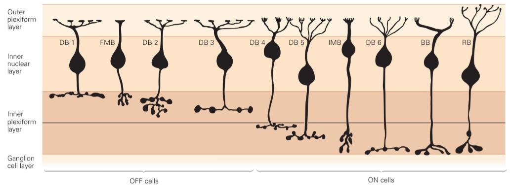
Figure 22-15 Bipolar cells in the macaque retina. The cells are arranged according to the depth of their terminal arbors in the inner plexiform layer. The horizontal line dividing the distal (upper) and proximal (lower) levels of this layer represents the border between the axonal terminals of OFF and ON type cells. Terminals in the upper half are presumed to be those of OFF cells, and those in the lower half ON cells. Cell types are diffuse bipolar cells (DB), ON and OFF midget bipolars (IMB, FMB), S-cone ON bipolar (BB), and rod bipolar (RB). (Reproduced, with permission, from Boycott and Wässle 1999.)
شکل 22-15 سلولهای دوقطبی در شبکیه میمون ماکاک. سلولها بر اساس عمق آربورهای انتهایی خود در لایه شبکهای داخلی مرتب شدهاند. خط افقی که سطوح دیستال (فوقانی) و پروگزیمال (پایینی) این لایه را از هم جدا میکند، مرز بین پایانههای آکسونی سلولهای نوع OFF و ON را نشان میدهد. پایانههای نیمه بالایی به عنوان سلولهای OFF و پایانههای نیمه پایینی به عنوان سلولهای ON در نظر گرفته میشوند. انواع سلولها عبارتند از سلولهای دوقطبی منتشر (DB)، دوقطبیهای کوچک روشن و خاموش (IMB، FMB)، دوقطبی ON مخروطی S (BB) و دوقطبی میلهای (RB). (با اجازه، از Boycott و Wässle 1999 کپی شده است.)
Through this inhibitory network, a bipolar cell terminal can receive inhibition from distant bipolar cells, in a manner closely analogous to the lateral inhibition of photoreceptor terminals (see Figure 22-3A). Amacrine cells also inhibit retinal ganglion cells directly. These lateral inhibitory connections contribute substantially to the antagonistic receptive field component of retinal ganglion cells.
از طریق این شبکه مهاری، یک پایانه سلول دوقطبی میتواند مهار را از سلولهای دوقطبی دور دریافت کند، به شیوهای بسیار مشابه مهار جانبی پایانههای گیرنده نوری (شکل 22-3A را ببینید). سلولهای آماکرین همچنین سلولهای گانگلیونی شبکیه را مستقیماً مهار میکنند. این اتصالات مهاری جانبی به طور قابل توجهی در مؤلفه میدان گیرنده آنتاگونیستی سلولهای گانگلیونی شبکیه نقش دارند.
Temporal Filtering Occurs in Synapses and Feedback Circuits
فیلترینگ زمانی در سیناپسها و مدارهای بازخورد رخ میدهد
For many ganglion cells, a step change in light intensity produces a transient response, an initial peak in firing that declines to a smaller steady rate (see Figure 22-10). Part of this sensitivity originates in the negative- feedback circuits involving horizontal and amacrine cells. For example, a sudden decrease in light intensity depolarizes the cone terminal, which excites the horizontal cell, which in turn repolarizes the cone terminal (see Figure 22-3A). Because this feedback loop involves a brief delay, the voltage response of the cone peaks abruptly and then settles to a smaller steady level. Similar processing occurs at the reciprocal synapses between bipolar and amacrine cells in the inner retina.
برای بسیاری از سلولهای گانگلیونی، تغییر پلهای در شدت نور، یک پاسخ گذرا ایجاد میکند، یک اوج اولیه در شلیک که به یک نرخ ثابت کوچکتر کاهش مییابد (شکل 22-10 را ببینید). بخشی از این حساسیت از مدارهای بازخورد منفی مربوط به سلولهای افقی و آماکرین سرچشمه میگیرد. به عنوان مثال، کاهش ناگهانی شدت نور، پایانه مخروطی را دپلاریزه میکند، که سلول افقی را تحریک میکند، که به نوبه خود پایانه مخروطی را دوباره پلاریزه میکند (شکل 22-3A را ببینید). از آنجا که این حلقه بازخورد شامل یک تأخیر کوتاه است، پاسخ ولتاژ مخروط به طور ناگهانی به اوج میرسد و سپس به یک سطح ثابت کوچکتر میرسد. پردازش مشابهی در سیناپسهای متقابل بین سلولهای دوقطبی و آماکرین در شبکیه داخلی رخ میدهد.
In both cases, the delayed-inhibition circuit favors rapidly changing inputs over slowly changing inputs. The effects of this filtering, which can be observed in visual perception, are most pronounced for large stimuli that drive the horizontal and amacrine cell networks most effectively. For example, a large spot can be seen easily when it flickers at a rate of 10 Hz but not at a low rate (see Figure 22–14).
در هر دو مورد، مدار مهار تأخیری، ورودیهای با تغییر سریع را به ورودیهای با تغییر آهسته ترجیح میدهد. اثرات این فیلترینگ، که میتوان آن را در ادراک بصری مشاهده کرد، برای محرکهای بزرگی که شبکههای سلولی افقی و آماکرین را به طور مؤثر هدایت میکنند، برجستهتر است. به عنوان مثال، یک لکه بزرگ را میتوان به راحتی دید وقتی که با سرعت 10 هرتز سوسو میزند، اما نه با سرعت کم (شکل 22-14 را ببینید).
In addition to these circuit properties, certain cellular processes contribute to shaping the temporal response. For example, the AMPA-kainate type of glutamate receptor undergoes strong desensitization. A step increase in the concentration of glutamate at the dendrite of a bipolar or ganglion cell leads to an immediate opening of additional glutamate receptors. As these receptors desensitize, the postsynaptic conductance decreases again. The effect is to render a step response more transient.
علاوه بر این خواص مدار، فرآیندهای سلولی خاصی در شکلدهی پاسخ زمانی نقش دارند. به عنوان مثال، گیرنده گلوتامات از نوع AMPA-kainate دچار حساسیتزدایی شدید میشود. افزایش مرحلهای غلظت گلوتامات در دندریت یک سلول دوقطبی یا گانگلیونی منجر به باز شدن فوری گیرندههای گلوتامات اضافی میشود. با بیحس شدن این گیرندهها، رسانایی پسسیناپسی دوباره کاهش مییابد. اثر این امر، گذراتر کردن پاسخ مرحلهای است.
Retinal circuits seem to go to great lengths to speed up their responses and emphasize temporal changes. One likely reason is that the very first cell in the retinal circuit, the photoreceptor, is exceptionally slow (see Figure 22-7C). Following a flash of light, a cone takes about 40 ms to reach the peak response, an intolerable delay for proper visual function. Through the various filtering mechanisms in retinal circuitry, subsequent neurons respond most vigorously during the rising phase of the cone’s response. Indeed, some ganglion cells have a response peak only 20 ms after the flash. Temporal processing in the retina clearly helps to reduce visual reaction times, a life-extending trait as important in highway traffic as on the savannas of our ancestors.
به نظر میرسد مدارهای شبکیه برای سرعت بخشیدن به پاسخهای خود و تأکید بر تغییرات زمانی، تلاش زیادی میکنند. یکی از دلایل احتمالی این است که اولین سلول در مدار شبکیه، یعنی گیرنده نوری، فوقالعاده کند است (شکل 22-7C را ببینید). پس از یک تابش نور، یک مخروط حدود 40 میلیثانیه طول میکشد تا به اوج پاسخ برسد، که تأخیری غیرقابل تحمل برای عملکرد صحیح بینایی است. از طریق مکانیسمهای مختلف فیلترینگ در مدار شبکیه، نورونهای بعدی در طول مرحله افزایش پاسخ مخروط، با بیشترین شدت پاسخ میدهند. در واقع، برخی از سلولهای گانگلیونی تنها 20 میلیثانیه پس از تابش نور، اوج پاسخ دارند. پردازش زمانی در شبکیه به وضوح به کاهش زمان واکنش بصری کمک میکند، یک ویژگی افزایشدهنده طول عمر که در ترافیک بزرگراه به همان اندازه در دشتهای اجداد ما اهمیت دارد.
Color Vision Begins in Cone-Selective Circuits
بینایی رنگی از مدارهای انتخابی مخروطی آغاز میشود
Throughout recorded history, philosophers and scientists have been fascinated by color perception. This interest was originally driven by the relevance of color to art, later by its relation to the physical properties of light, and finally by commercial interests in television and photography. The 19th century witnessed a profusion of theories to explain color perception, of which two have survived modern scrutiny. They are based on careful psychophysics that placed strong constraints on the underlying neural mechanisms.
در طول تاریخ ثبتشده، فیلسوفان و دانشمندان مجذوب ادراک رنگ بودهاند. این علاقه در ابتدا به دلیل ارتباط رنگ با هنر، بعدها به دلیل ارتباط آن با خواص فیزیکی نور و در نهایت به دلیل منافع تجاری در تلویزیون و عکاسی ایجاد شد. قرن نوزدهم شاهد انبوهی از نظریهها برای توضیح ادراک رنگ بود که دو مورد از آنها از بررسیهای مدرن جان سالم به در بردهاند. این نظریهها مبتنی بر روانفیزیک دقیقی هستند که محدودیتهای شدیدی را بر مکانیسمهای عصبی زیربنایی اعمال میکنند.
Early experiments demonstrated that any given natural light could be color-matched by mixing together appropriate amounts of three primary lights. This led to the trichromatic theory of color perception based on absorption of light by three mechanisms, each with a different sensitivity spectrum. These correspond to the three cone types (see Figure 22-6), whose measured absorption spectra fully explain the color- matching results both in normal individuals and those with genetic anomalies in the pigment genes.
آزمایشهای اولیه نشان داد که هر نور طبیعی معینی میتواند با ترکیب مقادیر مناسبی از سه نور اولیه، از نظر رنگ تطبیق داده شود. این امر منجر به نظریه سهرنگی ادراک رنگ بر اساس جذب نور توسط سه مکانیسم شد که هر کدام طیف حساسیت متفاوتی دارند. این مکانیسمها مربوط به سه نوع مخروط هستند (شکل 22-6 را ببینید)، که طیفهای جذب اندازهگیری شده آنها به طور کامل نتایج تطبیق رنگ را هم در افراد عادی و هم در افراد دارای ناهنجاریهای ژنتیکی در ژنهای رنگدانه توضیح میدهد.
The so-called opponent-process theory was pro- posed to explain our perception of different hues. According to this theory, color vision involves three processes that respond in opposite ways to light of different colors: (y-b) would be stimulated by yellow and inhibited by blue light; (r-g) stimulated by red and inhibited by green; and (w-bk) stimulated by white and inhibited by black. We recognize some of these 19th century postulates in the postreceptor circuitry of the retina.
نظریه موسوم به «فرآیند متضاد» برای توضیح درک ما از رنگهای مختلف ارائه شد. بر اساس این نظریه، بینایی رنگی شامل سه فرآیند است که به روشهای متضاد به نور رنگهای مختلف پاسخ میدهند: (y-b) توسط نور زرد تحریک و توسط نور آبی مهار میشود؛ (r-g) توسط نور قرمز تحریک و توسط نور سبز مهار میشود؛ و (w-bk) توسط نور سفید تحریک و توسط نور سیاه مهار میشود. ما برخی از این فرضیات قرن نوزدهم را در مدار پسگیرندهای شبکیه میشناسیم.
In the central 10° of the human retina, a single midget bipolar cell that receives input from a single cone excites each P-type ganglion cell. An L-ON ganglion cell, for example, has a receptive field center consisting of a single L cone and an antagonistic surround involving a mixture of L and M cones. When this neuron’s receptive field is stimulated with a large uniform spot of light that extends over both the center and the surround, this neuron is depolarized by red light and hyperpolarized by green light. Similar antagonism holds for the three other P-cells: L-OFF, M-ON, and M-OFF. These P-cells send their signals to the parvo- cellular layers of the lateral geniculate nucleus.
در 10 درجه مرکزی شبکیه انسان، یک سلول دوقطبی بسیار کوچک که از یک مخروط واحد ورودی دریافت میکند، هر سلول گانگلیونی نوع P را تحریک میکند. به عنوان مثال، یک سلول گانگلیونی L-ON دارای یک مرکز میدان گیرنده متشکل از یک مخروط L و یک محیط آنتاگونیستی شامل ترکیبی از مخروطهای L و M است. هنگامی که میدان گیرنده این نورون با یک نقطه نور یکنواخت بزرگ که هم در مرکز و هم در محیط اطراف امتداد دارد، تحریک میشود، این نورون توسط نور قرمز دپلاریزه و توسط نور سبز هایپرپلاریزه میشود. آنتاگونیسم مشابهی برای سه سلول P دیگر نیز وجود دارد: L-OFF، M-ON و M-OFF. این سلولهای P سیگنالهای خود را به لایههای پاروسلولی هسته زانویی جانبی ارسال میکنند.
A dedicated type of S-ON bipolar cell collects the signals of S-cones selectively and transmits them to ganglion cells of the small bistratified type. Because this ganglion cell also receives excitation from L-OFF and M-OFF bipolar cells, it is depolarized by blue light and hyperpolarized by yellow light. Another type of ganglion cell shows the opposite signature: S-OFF and (L+M)-ON. These signals are transmitted to the koniocellular layers of the lateral geniculate nucleus.
یک نوع اختصاصی از سلولهای دوقطبی S-ON، سیگنالهای مخروطهای S را به صورت انتخابی جمعآوری کرده و آنها را به سلولهای گانگلیونی از نوع دوطبقهای کوچک منتقل میکند. از آنجا که این سلول گانگلیونی نیز از سلولهای دوقطبی L-OFF و M-OFF تحریک دریافت میکند، توسط نور آبی دپلاریزه و توسط نور زرد هایپرپلاریزه میشود. نوع دیگری از سلولهای گانگلیونی، امضای متضادی را نشان میدهد: S-OFF و (L+M)-ON. این سیگنالها به لایههای کونیوسلولار هسته زانویی جانبی منتقل میشوند.
The M-cells are excited by diffuse bipolar cells, which in turn collect inputs from many cones regard- less of pigment type. These ganglion cells therefore have large receptive fields with broad spectral sensitivity. Their axons project to the magnocellular layers of the lateral geniculate nucleus.
سلولهای M توسط سلولهای دوقطبی منتشر تحریک میشوند که به نوبه خود ورودیها را از بسیاری از مخروطها صرف نظر از نوع رنگدانه جمعآوری میکنند. بنابراین، این سلولهای گانگلیونی میدانهای گیرنده بزرگی با حساسیت طیفی وسیع دارند. آکسونهای آنها به لایههای مگنوسلولار هسته زانویی جانبی امتداد مییابند.
In this way, chromatic signals are combined and encoded by the retina for transmission to the thalamus and cortex. In circuits of the primary visual cortex, these signals are recombined in different ways, leading to a great variety of receptive field layouts. Only about 10% of cortical neurons are preferentially driven by color contrast rather than luminance contrast. This likely reflects the fact that color vision-despite its great aesthetic appeal-makes only a small contribution to our overall fitness. As an illustration of this, recall that colorblind individuals, who in a sense have lost half of their color space, can grow up without ever noticing that defect.
به این ترتیب، سیگنالهای رنگی توسط شبکیه برای انتقال به تالاموس و قشر مغز ترکیب و کدگذاری میشوند. در مدارهای قشر بینایی اولیه، این سیگنالها به روشهای مختلف بازترکیب میشوند و منجر به تنوع زیادی در طرحبندی میدانهای دریافتی میشوند. تنها حدود 10٪ از نورونهای قشر مغز ترجیحاً توسط کنتراست رنگ هدایت میشوند تا کنتراست روشنایی. این احتمالاً نشان دهنده این واقعیت است که بینایی رنگی – علیرغم جذابیت زیباییشناختی عالی آن – تنها سهم کمی در تناسب اندام کلی ما دارد. به عنوان نمونهای از این موضوع، به یاد داشته باشید که افراد کوررنگ، که به نوعی نیمی از فضای رنگی خود را از دست دادهاند، میتوانند بدون اینکه هرگز متوجه آن نقص شوند، بزرگ شوند.
»» فصل بعد: فصل پردازش بینایی سطح متوسط و اولیههای بینایی
»» تمامی کتاب