مبانی علوم اعصاب اریک کندل؛ انواع کوررنگی مادرزادی؛ سازش با نور


دعای مطالعه [ نمایش ]
بِسْمِ الله الرَّحْمنِ الرَّحیمِ
اَللّهُمَّ اَخْرِجْنى مِنْ ظُلُماتِ الْوَهْمِ
خدايا مرا بيرون آور از تاريكىهاى وهم،
وَ اَكْرِمْنى بِنُورِ الْفَهْمِ
و به نور فهم گرامى ام بدار،
اَللّهُمَّ افْتَحْ عَلَيْنا اَبْوابَ رَحْمَتِكَ
خدايا درهاى رحمتت را به روى ما بگشا،
وَانْشُرْ عَلَيْنا خَزائِنَ عُلُومِكَ بِرَحْمَتِكَ يا اَرْحَمَ الرّاحِمينَ
و خزانههاى علومت را بر ما باز كن به امید رحمتت اى مهربانترين مهربانان.
«مبانی علوم اعصاب» اثر دکتر اریک کندل و همکاران، یکی از جامعترین و معتبرترین منابع در حوزه علوم اعصاب محسوب میشود.
ترجمه دقیق این کتاب، با مدیریت داریوش طاهری و تحت نظارت برند علمی آیندهنگاران مغز، با الهام از تلاشهای علمی و تدریس اساتیدی همچون دکتر محمدتقی جغتایی و دکتر ثریا مهرابی، به فارسی برگردانده شده است.
این ترجمه، دسترسی به دانش عمیق علوم اعصاب را برای دانشجویان و پژوهشگران رشتههای پزشکی، علوم اعصاب، روانشناسی، علوم شناختی، هوش مصنوعی، گفتاردرمانی، مهندسی پزشکی و تمام علاقهمندان به درک عملکرد ذهن و مغز فراهم کرده و گامی در راستای ارتقای دانش و بهبود کیفیت زندگی است.
» کتاب مبانی علوم اعصاب اریک کندل
» » فصل ۲۲: فصل پردازش بصری سطح پایین: شبکیه؛ قسمت آخر
در حال ویرایش
Principles of Neural Science; Eric R. Kandel
»» Chapter 22: Low-Level Visual Processing: The Retina
The Photoreceptor Layer Samples the Visual Image
لایه گیرنده نور از تصویر بصری نمونهبرداری میکند
Ocular Optics Limit the Quality of the Retinal Image
اپتیک چشم، کیفیت تصویر شبکیه را محدود میکند
There Are Two Types of Photoreceptors: Rods and Cones
دو نوع گیرنده نوری وجود دارد: استوانهایها و مخروطیها
Phototransduction Links the Absorption of a Photon to a Change in Membrane Conductance
انتقال نوری، جذب فوتون را به تغییر در رسانایی غشاء مرتبط میکند
Light Activates Pigment Molecules in the Photoreceptors
نور مولکولهای رنگدانه را در گیرندههای نوری فعال میکند
Excited Rhodopsin Activates a Phosphodiesterase Through the G Protein Transducin
رودوپسین برانگیخته شده از طریق پروتئین G ترانسدوسین، یک فسفودی استراز را فعال میکند.
Multiple Mechanisms Shut Off the Cascade
چندین مکانیسم، آبشار را خاموش میکنند
Defects in Phototransduction Cause Disease
نقص در انتقال نوری باعث بیماری میشود
Ganglion Cells Transmit Neural Images to the Brain
سلولهای گانگلیون تصاویر عصبی را به مغز منتقل میکنند
The Two Major Types of Ganglion Cells Are ON Cells and OFF Cells
دو نوع اصلی سلولهای گانگلیون، سلولهای روشن (ON) و سلولهای خاموش (OFF) هستند.
Many Ganglion Cells Respond Strongly to Edges in the Image
بسیاری از سلولهای گانگلیون به شدت به لبههای تصویر پاسخ میدهند
The Output of Ganglion Cells Emphasizes Temporal Changes in Stimuli
خروجی سلولهای گانگلیون بر تغییرات زمانی در محرکها تأکید دارد
Retinal Output Emphasizes Moving Objects
خروجی شبکیه، اشیاء متحرک را برجسته میکند
Several Ganglion Cell Types Project to the Brain Through Parallel Pathways
چندین نوع سلول گانگلیون از طریق مسیرهای موازی به مغز میرسند
A Network of Interneurons Shapes the Retinal Output
شبکهای از نورونهای رابط، خروجی شبکیه را شکل میدهد
Parallel Pathways Originate in Bipolar Cells
مسیرهای موازی از سلولهای دوقطبی سرچشمه میگیرند
Spatial Filtering Is Accomplished by Lateral Inhibition
فیلترینگ مکانی با مهار جانبی انجام میشود
Temporal Filtering Occurs in Synapses and Feedback Circuits
فیلترینگ زمانی در سیناپسها و مدارهای بازخورد رخ میدهد
Color Vision Begins in Cone-Selective Circuits
بینایی رنگی از مدارهای انتخابی مخروطی آغاز میشود
Congenital Color Blindness Takes Several Forms
کوررنگی مادرزادی اشکال مختلفی دارد
Rod and Cone Circuits Merge in the Inner Retina
مدارهای استوانهای و مخروطی در شبکیه داخلی ادغام میشوند
The Retina’s Sensitivity Adapts to Changes in Illumination
حساسیت شبکیه با تغییرات روشنایی سازگار میشود
Light Adaptation Is Apparent in Retinal Processing and Visual Perception
سازگاری با نور در پردازش شبکیه و ادراک بصری آشکار است
Multiple Gain Controls Occur within the Retina
کنترلهای چندگانهی بهره در شبکیه رخ میدهند
Light Adaptation Alters Spatial Processing
سازگاری با نور، پردازش فضایی را تغییر میدهد
Highlights
نکات برجسته
Congenital Color Blindness Takes Several Forms
کوررنگی مادرزادی اشکال مختلفی دارد
Few people are truly colorblind in the sense of being wholly unable to distinguish a change in color from a change in the intensity of light, but many individuals have impaired color vision and experience difficulties in making distinctions that for most of us are trivial, for example between red and green. Most such abnormalities of color vision are congenital and have been characterized in detail; some other abnormalities result from injury or disease of the visual pathway.
تعداد کمی از افراد واقعاً کوررنگ هستند، به این معنا که کاملاً قادر به تشخیص تغییر رنگ از تغییر شدت نور نیستند، اما بسیاری از افراد اختلال در بینایی رنگ دارند و در تشخیص رنگهایی که برای اکثر ما بیاهمیت هستند، مثلاً بین قرمز و سبز، دچار مشکل میشوند. اکثر این ناهنجاریهای بینایی رنگ مادرزادی هستند و به تفصیل شرح داده شدهاند. برخی دیگر از ناهنجاریها ناشی از آسیب یا بیماری مسیر بینایی هستند.

Figure 22-16 A test for some forms of color blindness. The numerals embedded in this color pattern can be distinguished by people with trichromatic vision but not by dichromats who are weak in red-green discrimination. If you don’t see any numbers please have your vision tested. (Reproduced, with permission, from Ishihara 1993.)
شکل ۲۲-۱۶ آزمایشی برای برخی از انواع کوررنگی. اعداد تعبیه شده در این الگوی رنگی را افرادی که دید سه رنگی دارند میتوانند تشخیص دهند، اما افراد دو رنگ که در تشخیص قرمز-سبز ضعیف هستند، نمیتوانند. اگر هیچ عددی را نمیبینید، لطفاً بینایی خود را آزمایش کنید. (با اجازه، از Ishihara 1993 کپی شده است.)
Some people have only two classes of cones instead of three. These dichromats find it difficult or impossible to distinguish some surfaces whose colors appear distinct to trichromats. The dichromat’s problem is that every surface reflectance function is represented by a two-value description rather than a three-value one, and this reduced description causes dichromats to confuse many more surfaces than do trichromats. Simple tests for color blindness exploit this fact (Figure 22–16).
بعضی افراد به جای سه دسته مخروط، فقط دو دسته دارند. این دو رنگپرانها تشخیص برخی سطوح را که رنگهایشان برای سه رنگپرانها متمایز به نظر میرسد، دشوار یا غیرممکن میدانند. مشکل دو رنگپرانها این است که هر تابع بازتاب سطح با یک توصیف دو مقداری به جای یک توصیف سه مقداری نمایش داده میشود و این توصیف کاهشیافته باعث میشود که دو رنگپرانها سطوح بسیار بیشتری را نسبت به سه رنگپرانها اشتباه بگیرند. آزمایشهای ساده برای کوررنگی از این واقعیت بهره میبرند (شکل ۲۲-۱۶).
Although there are three forms of dichromacy, corresponding to the loss of each of the three types of cones, two kinds are much more common than the third. The common forms correspond to the loss of the L cones or M cones and are called protanopia and deuteranopia, respectively. Protanopia and deuteranopia almost always occur in males, each with a frequency of about 1%. The conditions are transmitted by women who are not themselves affected, and so implicate genes on the X chromosome. A third form of dichromacy, tritanopia, involves loss or dysfunction of the S cone. It affects only about 1 in 10,000 people, afflicts women and men with equal frequency, and involves a gene on chromosome 7.
اگرچه سه نوع دورنگبینی وجود دارد که مربوط به از دست دادن هر یک از سه نوع مخروط است، دو نوع بسیار شایعتر از نوع سوم هستند. اشکال رایج مربوط به از دست دادن مخروطهای L یا مخروطهای M هستند و به ترتیب پروتانوپیا و دوترانوپیا نامیده میشوند. پروتانوپیا و دوترانوپیا تقریباً همیشه در مردان رخ میدهند و هر کدام با فراوانی حدود ۱٪. این شرایط توسط زنانی که خودشان تحت تأثیر قرار نمیگیرند منتقل میشود و بنابراین ژنهای روی کروموزوم X را درگیر میکند. نوع سوم دورنگبینی، تریتانوپیا، شامل از دست دادن یا اختلال عملکرد مخروط S است. این بیماری فقط حدود ۱ نفر از هر ۱۰۰۰۰ نفر را تحت تأثیر قرار میدهد، زنان و مردان را با فراوانی برابر مبتلا میکند و ژنی را روی کروموزوم ۷ درگیر میکند.
Because the L and M cones exist in large numbers, one might think that the loss of one or the other type would impair vision more broadly than just weakening color vision. In fact, this does not happen because the total number of L and M cones in the dichromat retina is not altered. All cells destined to become L or M cones are probably converted to L cones in deuteranopes and to M cones in protanopes.
از آنجا که مخروطهای L و M به تعداد زیادی وجود دارند، ممکن است تصور شود که از دست دادن یکی از این دو نوع، بینایی را به طور گستردهتری مختل میکند تا فقط بینایی رنگی را تضعیف کند. در واقع، این اتفاق نمیافتد زیرا تعداد کل مخروطهای L و M در شبکیهی دو رنگپریده تغییر نمیکند. تمام سلولهایی که قرار است به مخروطهای L یا M تبدیل شوند، احتمالاً در دوترانوپها به مخروطهای L و در پروتانوپها به مخروطهای M تبدیل میشوند.
In addition to the relatively severe forms of color- blindness represented by dichromacy, there are milder forms, again affecting mostly males. These so-called anomalous trichromats have cones whose spectral sensitivities differ from those in normal trichromats. Anomalous trichromacy results from the replacement of one of the normal cone pigments by an altered protein with a different spectral sensitivity. Two common forms, protanomaly and deuteranomaly, together affect about 7% of males and represent, respectively, the replacement of the L or M cones by a pigment with some intermediate spectral sensitivity.
علاوه بر اشکال نسبتاً شدید کوررنگی که با دورنگبینی نشان داده میشود، اشکال خفیفتری نیز وجود دارد که باز هم بیشتر مردان را تحت تأثیر قرار میدهد. این افراد که به اصطلاح سهرنگبینهای غیرعادی نامیده میشوند، مخروطهایی دارند که حساسیت طیفی آنها با سهرنگبینهای عادی متفاوت است. سهرنگبینی غیرعادی ناشی از جایگزینی یکی از رنگدانههای مخروطی عادی با یک پروتئین تغییر یافته با حساسیت طیفی متفاوت است. دو شکل رایج، پروتانومالی و دوترانومالی، با هم حدود 7٪ از مردان را تحت تأثیر قرار میدهند و به ترتیب نشاندهنده جایگزینی مخروطهای L یا M با رنگدانهای با حساسیت طیفی متوسط هستند.
The genetics of color vision defects are well understood. The genes for the L and M pigments reside on the X chromosome in a head-to-tail arrangement (Figure 22-17A). The pigment proteins have very similar structures, differing in only 4% of their amino acids. People with normal color vision possess a single copy of the gene for the L pigment and from one to three- occasionally as many as five-nearly identical copies of the gene for the M pigment.
ژنتیک نقصهای دید رنگ به خوبی شناخته شده است. ژنهای مربوط به رنگدانههای L و M به صورت سر به دم روی کروموزوم X قرار دارند (شکل 22-17A). پروتئینهای رنگدانه ساختارهای بسیار مشابهی دارند و تنها در 4٪ از اسیدهای آمینه خود متفاوت هستند. افرادی که دید رنگی طبیعی دارند، یک کپی از ژن مربوط به رنگدانه L و از یک تا سه – گاهی اوقات تا پنج – کپی تقریباً یکسان از ژن مربوط به رنگدانه M دارند.
The proximity and similarity of these genes predisposes them to varied forms of recombination, leading either to the loss of a gene or to the formation of hybrid genes that account for the common forms of red-green defect (Figure 22-17B). Examination of these genes in dichromats reveals a loss of the L-pigment gene in protanopes and a loss of one or more M-pigment genes in deuteranopes. Anomalous trichromats have L-M or M-L hybrid genes that code for visual pigments with shifted spectral sensitivity; the extent of the shift depends on the point of recombination. In tritanopes, the loss of S-cone function arises from mutations in the S-pigment gene.
نزدیکی و شباهت این ژنها، آنها را مستعد اشکال متنوع نوترکیبی میکند که یا منجر به از دست دادن یک ژن یا تشکیل ژنهای هیبریدی میشود که اشکال رایج نقص قرمز-سبز را تشکیل میدهند (شکل 22-17B). بررسی این ژنها در دیکروماتها، از دست دادن ژن رنگدانه L در پروتانوپها و از دست دادن یک یا چند ژن رنگدانه M در دوترانوپها را نشان میدهد. تریکروماتهای غیرعادی دارای ژنهای هیبریدی L-M یا M-L هستند که رنگدانههای بینایی را با حساسیت طیفی تغییر یافته کدگذاری میکنند. میزان تغییر به نقطه نوترکیبی بستگی دارد. در تریتانوپها، از دست دادن عملکرد مخروط S ناشی از جهش در ژن رنگدانه S است.
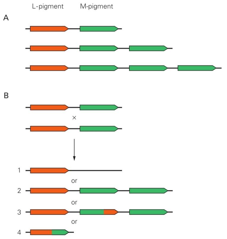
Figure 22-17 L- and M-pigment genes on the X chromosome.
شکل 22-17 ژنهای رنگدانه L- و M- روی کروموزوم X.
A. The L- and M-pigment genes normally lie next to each other on the chromosome. The base of each arrow corresponds to the 5′ end of the gene, and the tip corresponds to the 3′ end. Males with normal color vision can have one, two, or three copies of the gene for the M pigment on each X chromosome. (Adapted, with permission, from Nathans, Thomas, and Hogness 1986. Copyright © 1986 AAAS.)
الف) ژنهای رنگدانه L و M معمولاً روی کروموزوم در کنار یکدیگر قرار دارند. پایه هر فلش مربوط به انتهای 5′ ژن و نوک آن مربوط به انتهای 3′ است. مردانی که دید رنگی طبیعی دارند میتوانند یک، دو یا سه نسخه از ژن رنگدانه M را روی هر کروموزوم X داشته باشند. (اقتباس شده، با اجازه، از ناتانز، توماس و هاگنس 1986. حق چاپ © 1986 AAAS.)
B. Recombinations of the L- and M-pigment genes can lead to the generation of a hybrid gene (3 and 4) or the loss of a gene (1), the patterns observed in colorblind men. Spurious recombination can also cause gene duplication (2), a pattern observed in some people with normal color vision. (Adapted from Streyer 1988. Used with permission from J. Nathans.)
ب. نوترکیبی ژنهای رنگدانه L و M میتواند منجر به تولید یک ژن ترکیبی (3 و 4) یا از بین رفتن یک ژن (1) شود، الگوهایی که در مردان کوررنگ مشاهده میشود. نوترکیبی کاذب همچنین میتواند باعث تکثیر ژن (2) شود، الگویی که در برخی از افراد با دید رنگی طبیعی مشاهده میشود. (اقتباس از Streyer 1988. با اجازه J. Nathans استفاده شده است.)
Rod and Cone Circuits Merge in the Inner Retina
مدارهای استوانهای و مخروطی در شبکیه داخلی ادغام میشوند
For vision under low-light conditions, the mammalian retina has an ON bipolar cell that is exclusively connected to rods (see Figure 22-3B). By collecting inputs from up to 50 rods, this rod bipolar cell can pool the effects of dispersed single-photon absorptions in a small patch of retina. There is no corresponding OFF bipolar cell dedicated to rods.
برای بینایی در شرایط کم نور، شبکیه پستانداران دارای یک سلول دو قطبی روشن است که منحصراً به سلولهای استوانهای متصل است (شکل 22-3B را ببینید). با جمعآوری ورودیها از حداکثر 50 سلول استوانهای، این سلول دو قطبی استوانهای میتواند اثرات جذبهای تک فوتونی پراکنده را در یک بخش کوچک از شبکیه جمعآوری کند. هیچ سلول دو قطبی خاموش متناظری که به سلولهای استوانهای اختصاص داده شده باشد، وجود ندارد.
Unlike all other bipolar cells, the rod bipolar cell does not contact ganglion cells directly but instead excites a dedicated neuron, the All amacrine cell. This amacrine cell receives inputs from several rod bipolar cells and conveys its output to cone bipolar cells. It provides excitatory signals to ON bipolar cells through gap junctions as well as glycinergic inhibitory signals to OFF bipolar cells. These cone bipolar cells in turn excite ON and OFF ganglion cells, as described earlier. Thus, the rod signal is fed into the cone system after a detour that produces the appropriate signal polarities for the ON and OFF pathways. The purpose of the added interneurons may be to allow greater pooling of rod signals than of cone signals.
برخلاف سایر سلولهای دوقطبی، سلول دوقطبی استوانهای مستقیماً با سلولهای گانگلیونی تماس نمیگیرد، بلکه در عوض یک نورون اختصاصی، یعنی سلول تمام آماکرین، را تحریک میکند. این سلول آماکرین ورودیهایی را از چندین سلول دوقطبی استوانهای دریافت میکند و خروجی خود را به سلولهای دوقطبی مخروطی منتقل میکند. این سلول از طریق اتصالات شکافی، سیگنالهای تحریکی را برای سلولهای دوقطبی روشن و همچنین سیگنالهای مهاری گلیسینرژیک را برای سلولهای دوقطبی خاموش فراهم میکند. این سلولهای دوقطبی مخروطی به نوبه خود، سلولهای گانگلیونی روشن و خاموش را، همانطور که قبلاً توضیح داده شد، تحریک میکنند. بنابراین، سیگنال استوانهای پس از یک مسیر انحرافی که قطبیتهای سیگنال مناسب را برای مسیرهای روشن و خاموش تولید میکند، به سیستم مخروطی وارد میشود. هدف از نورونهای رابط اضافه شده ممکن است فراهم کردن امکان تجمع بیشتر سیگنالهای استوانهای نسبت به سیگنالهای مخروطی باشد.
Rod signals also enter the cone system through two other pathways. Rods can drive neighboring cones directly through electrical junctions, and they make connections with an OFF bipolar cell that services primarily cones. Once the rod signal has reached the cone bipolars through these pathways, it can take advantage of the same intricate circuitry of the inner retina. Thus, the rod system of the mammalian retina may have been an evolutionary afterthought added to the cone circuits.
سیگنالهای استوانهای همچنین از طریق دو مسیر دیگر وارد سیستم مخروطی میشوند. استوانهایها میتوانند مخروطهای همسایه را مستقیماً از طریق اتصالات الکتریکی هدایت کنند و با یک سلول دوقطبی خاموش که عمدتاً به مخروطها سرویس میدهد، ارتباط برقرار کنند. هنگامی که سیگنال استوانهای از طریق این مسیرها به دوقطبیهای مخروطی رسید، میتواند از همان مدار پیچیده شبکیه داخلی بهره ببرد. بنابراین، سیستم استوانهای شبکیه پستانداران ممکن است یک فکر ثانویه تکاملی باشد که به مدارهای مخروطی اضافه شده است.
The Retina’s Sensitivity Adapts to Changes in Illumination
حساسیت شبکیه با تغییرات روشنایی سازگار میشود
Vision operates under many different lighting conditions. The intensity of the light coming from an object depends on the intensity of the ambient illumination and the fraction of this light reflected by the object’s surface, called the reflectance. The range of intensities encountered in a day is enormous, with variation spanning 10 orders of magnitude, but most of this variation is useless for the purpose of guiding behavior.
بینایی تحت شرایط نوری بسیار متفاوتی عمل میکند. شدت نور ساطع شده از یک جسم به شدت روشنایی محیط و کسری از این نور که توسط سطح جسم منعکس میشود، که ضریب بازتاب نامیده میشود، بستگی دارد. دامنه شدتهای نوری که در یک روز با آن مواجه میشویم بسیار زیاد است و تغییرات آن به 10 برابر میرسد، اما بیشتر این تغییرات برای هدایت رفتار بیفایده است.
The illumination intensity varies by about nine orders of magnitude, mostly because our planet turns about its axis once a day, while the object reflectance varies much less, by about one order of magnitude in a typical scene. But this reflectance is the interesting quantity for vision, for it characterizes objects and distinguishes them from the background. In fact, our visual system is remarkably good at calculating surface reflectances independently of ambient illumination (Figure 22–18).
شدت روشنایی حدود نه مرتبه بزرگی تغییر میکند، عمدتاً به این دلیل که سیاره ما روزی یک بار به دور محور خود میچرخد، در حالی که بازتاب اجسام بسیار کمتر، حدود یک مرتبه بزرگی در یک صحنه معمولی، تغییر میکند. اما این بازتاب، کمیت جالبی برای بینایی است، زیرا اشیاء را مشخص میکند و آنها را از پسزمینه متمایز میکند. در واقع، سیستم بینایی ما در محاسبه بازتابهای سطحی مستقل از روشنایی محیط، به طرز چشمگیری خوب است (شکل ۲۲-۱۸).
With an overall increase in ambient illumination, all points in the visual scene become brighter by the same factor. If the eye could simply reduce its sensitivity by that same factor, the neural representation of the image would remain unchanged at the level of the ganglion cells and could be processed by the rest of the brain in the same way as before the change in illumination. Moreover, the retinal ganglion cells would only need to encode the 10-fold range of image intensities owing to the different object reflectances, instead of the 10-billion-fold range that includes variations in ambient illumination. Some of this adjustment in sensitivity is performed by the pupil, which contracts in bright light, reducing retinal illumination by up to a factor of 10. In addition, the retina itself performs an automatic gain control, called light adaptation, that approaches the ideal normalization we have imagined here.
با افزایش کلی روشنایی محیط، تمام نقاط در صحنه بصری به همان اندازه روشنتر میشوند. اگر چشم میتوانست به سادگی حساسیت خود را به همان اندازه کاهش دهد، نمایش عصبی تصویر در سطح سلولهای گانگلیونی بدون تغییر باقی میماند و میتوانست توسط بقیه مغز به همان روشی که قبل از تغییر روشنایی پردازش میشد، پردازش شود. علاوه بر این، سلولهای گانگلیونی شبکیه فقط باید محدوده 10 برابری شدت تصویر را به دلیل بازتابهای مختلف اشیاء رمزگذاری کنند، به جای محدوده 10 میلیارد برابری که شامل تغییرات در روشنایی محیط است. بخشی از این تنظیم حساسیت توسط مردمک چشم انجام میشود که در نور شدید منقبض میشود و روشنایی شبکیه را تا 10 برابر کاهش میدهد. علاوه بر این، خود شبکیه یک کنترل خودکار افزایش، به نام انطباق نور، انجام میدهد که به نرمالسازی ایدهآلی که در اینجا تصور کردهایم، نزدیک میشود.
Light Adaptation Is Apparent in Retinal Processing and Visual Perception
سازگاری با نور در پردازش شبکیه و ادراک بصری آشکار است
When flashes of light of different intensity are presented with a constant background illumination, the responses of a retinal ganglion cell fit a sigmoidal curve (Figure 22-19A). The weakest flashes elicit no response, a graded increase in flash intensity elicits graded responses, and the brightest flashes elicit saturation. When the background illumination is increased, the response curve maintains the same shape but is shifted to higher flash intensities. Compensating for the increase in background illumination, the ganglion cell is now less sensitive to light variations: In the presence of a higher background, a larger change is needed to cause the same response. This lateral shifting of the stimulus-response relationship is a hallmark of light adaptation in the retina.
وقتی فلاشهای نور با شدتهای مختلف با روشنایی پسزمینه ثابت ارائه میشوند، پاسخهای یک سلول گانگلیونی شبکیه با یک منحنی سیگموئیدی مطابقت دارند (شکل 22-19A). ضعیفترین فلاشها هیچ پاسخی ایجاد نمیکنند، افزایش تدریجی شدت فلاش، پاسخهای تدریجی ایجاد میکند و درخشانترین فلاشها اشباع را ایجاد میکنند. هنگامی که روشنایی پسزمینه افزایش مییابد، منحنی پاسخ همان شکل را حفظ میکند اما به شدتهای فلاش بالاتر منتقل میشود. برای جبران افزایش روشنایی پسزمینه، سلول گانگلیونی اکنون نسبت به تغییرات نور حساسیت کمتری دارد: در حضور پسزمینه بالاتر، تغییر بزرگتری برای ایجاد همان پاسخ لازم است. این تغییر جانبی رابطه محرک-پاسخ، مشخصه سازگاری نور در شبکیه است.
The consequences of this gain change for human visual perception are readily apparent in psychophysical experiments. When human subjects are asked to detect a flash in a background field of constant illumination, detection on a brighter background necessitates a brighter flash (Figure 22–19B). Under the ideal gain-control mechanism discussed earlier, two stimuli would produce the same response if they caused the same fractional change from the background intensity. In that case, the threshold flash intensity should be proportional to the background intensity, a relationship known as Weber’s law of adaptation, which we encountered in considering somatic receptor sensitivity (Chapter 17). The visual system follows Weber’s law approximately: Over the entire range of vision, sensitivity decreases somewhat less steeply with increasing background intensity (Figure 22-19B).
پیامدهای این تغییر بهره برای ادراک بصری انسان به راحتی در آزمایشهای روانفیزیکی آشکار میشود. وقتی از افراد خواسته میشود که در یک میدان پسزمینه با روشنایی ثابت، یک فلاش را تشخیص دهند، تشخیص در یک پسزمینه روشنتر، فلاش روشنتری را ایجاب میکند (شکل 22-19B). تحت مکانیسم کنترل بهره ایدهآل که قبلاً مورد بحث قرار گرفت، دو محرک اگر باعث تغییر کسری یکسانی از شدت پسزمینه شوند، پاسخ یکسانی ایجاد میکنند. در این صورت، شدت آستانه فلاش باید متناسب با شدت پسزمینه باشد، رابطهای که به عنوان قانون سازگاری وبر شناخته میشود، که در بررسی حساسیت گیرندههای سوماتیک (فصل 17) با آن مواجه شدیم. سیستم بینایی تقریباً از قانون وبر پیروی میکند: در کل دامنه بینایی، حساسیت با افزایش شدت پسزمینه تا حدودی با شیب کمتری کاهش مییابد (شکل 22-19B).


Figure 22-18 A brightness illusion.
شکل ۲۲-۱۸ یک خطای دید ناشی از روشنایی.
A. The two tiles marked with small dots appear to have different color but actually reflect the same light intensity. (To see this, fold the page so they touch.) The trace underneath plots a profile of light intensity at the level of the arrowheads. Your visual system interprets this retinal image as a regular tile pat- tern under spatially varying illumination with a diffuse shadow in the right half. Under that interpretation, the right tile must have a lighter color than the left, which is what you perceive. This process is automatic and requires no conscious analysis.
الف) دو کاشی که با نقاط کوچک مشخص شدهاند، به نظر میرسد رنگ متفاوتی دارند، اما در واقع شدت نور یکسانی را منعکس میکنند. (برای دیدن این، صفحه را تا کنید تا به هم برسند.) رد زیر، نمایی از شدت نور را در سطح نوک پیکانها رسم میکند. سیستم بینایی شما این تصویر شبکیهای را به عنوان یک الگوی کاشی منظم تحت روشنایی متغیر مکانی با سایهای پراکنده در نیمه سمت راست تفسیر میکند. تحت این تفسیر، کاشی سمت راست باید رنگ روشنتری نسبت به کاشی سمت چپ داشته باشد، که همان چیزی است که شما درک میکنید. این فرآیند خودکار است و نیازی به تجزیه و تحلیل آگاهانه ندارد.
B. Retinal processing contributes to the perception of “lightness” by discounting the shadow’s smooth gradients of illumination and accentuating the sharp edges between checkerboard fields. The receptive field for a visual neuron with an excitatory center and inhibitory surround is shown at the top. As shown in a hundredfold magnification at the bottom, the surround is weak but extends over a much larger area than the center.
ب. پردازش شبکیه با نادیده گرفتن شیبهای ملایم روشنایی سایه و برجسته کردن لبههای تیز بین میدانهای شطرنجی، به درک «روشنایی» کمک میکند. میدان گیرنده برای یک نورون بینایی با یک مرکز تحریکی و یک محیط اطراف مهاری در بالا نشان داده شده است. همانطور که در بزرگنمایی صد برابر در پایین نشان داده شده است، محیط اطراف ضعیف است اما در ناحیهای بسیار بزرگتر از مرکز گسترش یافته است.
C. The result when a population of visual neurons with receptive fields as in B processes the image in A. This operation-the convolution of the image in A with the profile in B-subtracts from each point in the visual field the average intensity in a large surrounding region. The neural representation of the object has largely lost the effects of shading, and the two tiles in question do indeed have different brightness values in this representation.
ج. نتیجه زمانی که جمعیتی از نورونهای بینایی با میدانهای دریافتکننده مانند تصویر B، تصویر A را پردازش میکنند. این عملیات – کانولوشن تصویر A با نیمرخ تصویر B – از هر نقطه در میدان بینایی، میانگین شدت در یک ناحیه بزرگ اطراف را کم میکند. نمایش عصبی جسم تا حد زیادی اثرات سایهزنی را از دست داده است و دو کاشی مورد نظر در واقع مقادیر روشنایی متفاوتی در این نمایش دارند.
Multiple Gain Controls Occur Within the Retina
چندین کنترل بهره در شبکیه چشم وجود دارد
The enormous change in gain required for light adaptation arises at multiple sites within the retina. In star- light, a single rod cell is stimulated by a photon only every few seconds, a rate insufficient to alter the cell’s adaptation status. However, a retinal ganglion cell combines signals from many rods, thus receiving a steady stream of photon signals that can elicit a light- dependent gain change in the cell.
تغییر عظیم در بهره مورد نیاز برای سازگاری با نور، در چندین نقطه از شبکیه رخ میدهد. در نور ستارگان، یک سلول استوانهای تنها هر چند ثانیه توسط یک فوتون تحریک میشود، نرخی که برای تغییر وضعیت سازگاری سلول کافی نیست. با این حال، یک سلول گانگلیونی شبکیه سیگنالهای بسیاری از سلولهای استوانهای را ترکیب میکند و بنابراین جریان ثابتی از سیگنالهای فوتونی را دریافت میکند که میتواند تغییر بهره وابسته به نور را در سلول ایجاد کند.
At somewhat higher light intensities, a rod bipolar cell begins to adapt, changing its responsiveness depending on the average light level. Next, we reach a light intensity at which the gain of individual rod cells gradually decreases. Beyond that, the rods saturate: All their cGMP-dependent channels are closed, and the membrane potential no longer responds to the light stimulus. By this time, around dawn, the much less sensitive cone cells are being stimulated effectively and gradually take over from the rods. As the ambient light increases further, toward noon, light adaptation results principally from gain changes within the cones.
در شدتهای نور نسبتاً بالاتر، یک سلول دوقطبی استوانهای شروع به سازگاری میکند و بسته به میانگین سطح نور، میزان پاسخگویی خود را تغییر میدهد. سپس، به شدت نوری میرسیم که در آن بهره سلولهای استوانهای به تدریج کاهش مییابد. فراتر از آن، استوانهها اشباع میشوند: تمام کانالهای وابسته به cGMP آنها بسته میشوند و پتانسیل غشایی دیگر به محرک نوری پاسخ نمیدهد. در این زمان، حدود سپیده دم، سلولهای مخروطی که حساسیت بسیار کمتری دارند، به طور مؤثر تحریک میشوند و به تدریج جای استوانهها را میگیرند. با افزایش بیشتر نور محیط، به سمت ظهر، سازگاری نوری عمدتاً ناشی از تغییرات بهره در مخروطها است.
The cellular mechanisms of light adaptation are best understood in the photoreceptors. The calcium- dependent feedback pathways discussed earlier have a prominent role. Recall that when a light flash closes the cGMP-gated channels, the resulting decrease in intracellular Ca2+ accelerates several biochemical reactions that terminate the response to the flash (see Figure 22-7B). When illumination is continuous, however, the Ca2+ concentration remains low, and all these reactions are therefore in a steady state that both lowers the gain and accelerates the time course of the receptor’s response to light (Figure 22-19C). As a result, the light-adapted photoreceptor can respond to rapid changes in intensity much more quickly. This has important consequences for human visual perception; the contrast sensitivity to high-frequency flicker increases with intensity, an effect observed in primate retinal ganglion cells as well (see Figure 22–14).
مکانیسمهای سلولی سازگاری با نور به بهترین شکل در گیرندههای نوری قابل درک هستند. مسیرهای بازخورد وابسته به کلسیم که قبلاً مورد بحث قرار گرفتند، نقش برجستهای دارند. به یاد بیاورید که وقتی یک فلاش نوری کانالهای دریچهدار cGMP را میبندد، کاهش حاصل از +Ca2 درون سلولی، چندین واکنش بیوشیمیایی را که پاسخ به فلاش را خاتمه میدهند، تسریع میکند (شکل 22-7B را ببینید). با این حال، هنگامی که روشنایی مداوم است، غلظت +Ca2 پایین میماند و بنابراین همه این واکنشها در حالت پایداری قرار دارند که هم بهره را کاهش میدهد و هم روند زمانی پاسخ گیرنده به نور را تسریع میکند (شکل 22-19C). در نتیجه، گیرنده نوری سازگار با نور میتواند به تغییرات سریع شدت بسیار سریعتر پاسخ دهد. این امر پیامدهای مهمی برای ادراک بصری انسان دارد. حساسیت کنتراست به سوسو زدن فرکانس بالا با افزایش شدت افزایش مییابد، اثری که در سلولهای گانگلیونی شبکیه پستانداران نیز مشاهده میشود (شکل 22-14 را ببینید).

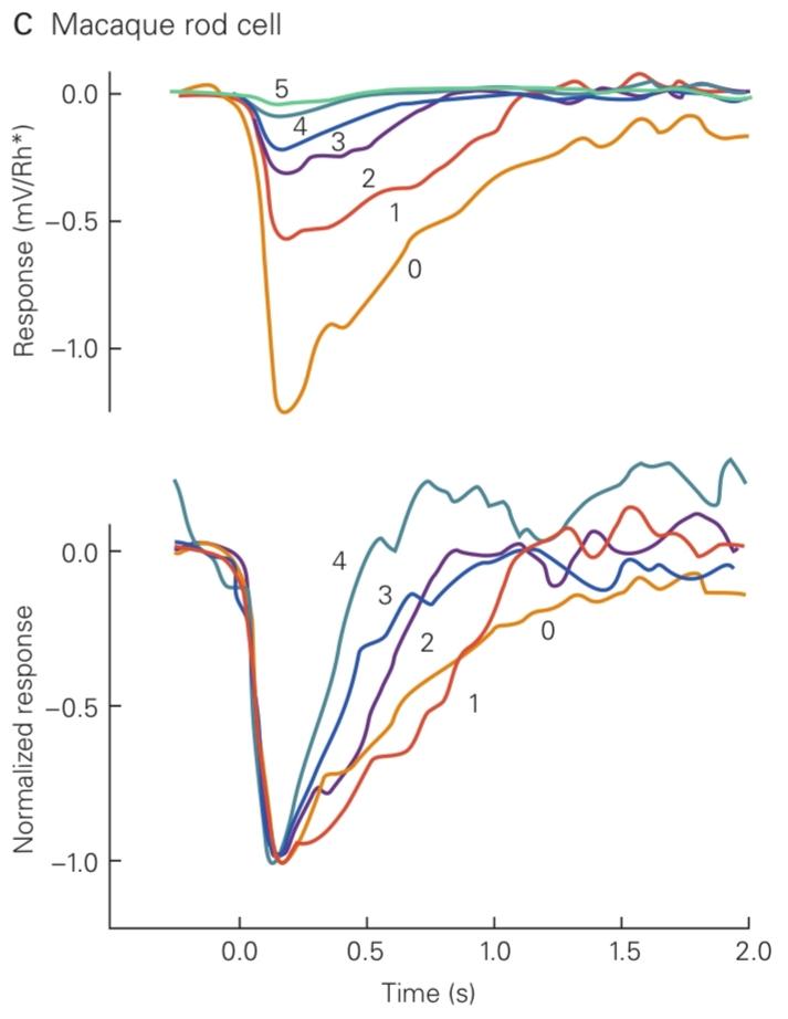
Figure 22-19 Light adaptation.
شکل ۲۲-۱۹ سازگاری با نور.
A. The receptive field of a cat retinal ganglion cell was illuminated uniformly at a steady background intensity, and a test spot was flashed briefly on the receptive field center. The peak firing rate following the flash was measured and plotted against the logarithm of the flash intensity. Each curve corresponds to a different background intensity, increasing by factors of 10 from left to right. (Reproduced, with permission, from Sakmann and Creutzfeldt 1969. Copyright © 1969 Springer.)
الف) میدان گیرندهی سلول گانگلیون شبکیهی یک گربه به طور یکنواخت با شدت پسزمینهی ثابت روشن شد و یک نقطهی آزمایشی به طور خلاصه در مرکز میدان گیرنده تابانده شد. حداکثر نرخ شلیک پس از تابش اندازهگیری و در برابر لگاریتم شدت تابش رسم شد. هر منحنی مربوط به شدت پسزمینهی متفاوتی است که از چپ به راست با ضریب ۱۰ افزایش مییابد. (با اجازه، از Sakmann and Creutzfeldt 1969 کپیرایت © 1969 Springer.)
B. A small test spot was flashed briefly on a steadily illuminated background, and the flash intensity gradually increased to where a human subject could just detect it. The procedure was repeated at different background intensities. Here, the threshold flash intensity is plotted against the background intensity. The curve has two branches connected by a distinct kink: These correspond to the regimes of rod and cone vision. The slope of Weber’s law represents the idealization when the threshold intensity is proportional to the background intensity. (Adapted from Wyszecki and Stiles 1982.)
ب. یک نقطه آزمایشی کوچک به طور خلاصه روی یک پسزمینه روشن ثابت تابانده شد و شدت فلاش به تدریج افزایش یافت تا جایی که یک فرد میتوانست آن را تشخیص دهد. این روش با شدتهای مختلف پسزمینه تکرار شد. در اینجا، شدت آستانه فلاش در برابر شدت پسزمینه رسم شده است. منحنی دارای دو شاخه است که توسط یک پیچ خوردگی مشخص به هم متصل شدهاند: اینها مربوط به رژیمهای بینایی استوانهای و مخروطی هستند. شیب قانون وبر نشان دهنده ایدهآلسازی زمانی است که شدت آستانه متناسب با شدت پسزمینه باشد. (اقتباس از Wyszecki و Stiles 1982.)
C. The top plot shows the responses of a macaque monkey’s rod cell to flashes presented at varying background intensities. The cell’s single-photon response was calculated from the recorded membrane potential divided by the number of rhodopsins (Rh) activated by the flash. The gain of the single- photon response decreases substantially with increasing back ground intensity. The background intensity, in photon/um2/s, is 0 for trace 0, 3.1 for trace 1, 12 for trace 2, 41 for trace 3, 84 for trace 4, and 162 for trace 5. In the bottom plot, the same data (except for the smallest response) are normalized to the same amplitude, showing that the time course of the single-photon response accelerates at high intensity. (Reproduced, with permission, from Schneeweis and Schnapf 2000.)
ج. نمودار بالا پاسخهای سلول استوانهای یک میمون ماکاک را به فلاشهای ارائه شده در شدتهای مختلف پسزمینه نشان میدهد. پاسخ تک فوتونی سلول از پتانسیل غشایی ثبت شده تقسیم بر تعداد رودوپسینها (Rh) فعال شده توسط فلاش محاسبه شد. بهره پاسخ تک فوتونی با افزایش شدت پسزمینه به طور قابل توجهی کاهش مییابد. شدت پسزمینه، بر حسب فوتون بر مجذور ثانیه، برای رد ۰، ۰، برای رد ۱، ۳.۱، برای رد ۲، ۱۲، برای رد ۳، ۴۱، برای رد ۴، ۸۴ و برای رد ۵، ۱۶۲ است. در نمودار پایین، همان دادهها (به جز کوچکترین پاسخ) به همان دامنه نرمال شدهاند که نشان میدهد سیر زمانی پاسخ تک فوتونی در شدت بالا شتاب میگیرد. (با اجازه، از Schneeweis و Schnapf 2000 بازتولید شده است.)
Light Adaptation Alters Spatial Processing
سازگاری با نور، پردازش فضایی را تغییر میدهد
In addition to the sensitivity and speed of the retinal response, light adaptation also changes the rules of spatial processing. In bright light, many ganglion cells have a sharp center-surround structure in their receptive fields (see Figure 22-10). As the light dims, the antagonistic surround becomes broad and weak and eventually disappears. Under these conditions, the circuits of the retina function to simply accumulate the rare photons rather than computing local intensity gradients. These changes in receptive-field proper ties occur because of changes in the lateral inhibition produced by the networks of horizontal and amacrine cells (see Figure 22–3). An important regulator of these processes is dopamine, released in a light-dependent manner by specialized amacrine cells.
علاوه بر حساسیت و سرعت پاسخ شبکیه، سازگاری با نور، قوانین پردازش فضایی را نیز تغییر میدهد. در نور شدید، بسیاری از سلولهای گانگلیونی در میدانهای گیرنده خود ساختار تیز مرکزی-محیطی دارند (شکل 22-10 را ببینید). با کم شدن نور، محیط متضاد پهن و ضعیف میشود و در نهایت ناپدید میشود. در این شرایط، مدارهای شبکیه به جای محاسبه گرادیانهای شدت محلی، به سادگی فوتونهای نادر را جمع میکنند. این تغییرات در روابط خاص میدان گیرنده به دلیل تغییرات در مهار جانبی تولید شده توسط شبکههای سلولهای افقی و آماکرین رخ میدهد (شکل 22-3 را ببینید). یک تنظیمکننده مهم این فرآیندها دوپامین است که به صورت وابسته به نور توسط سلولهای آماکرین تخصصی آزاد میشود.
These retinal effects leave their signature on human perception. In bright light, our visual system prefers fine gratings to coarse gratings. But in dim light, we are most sensitive to coarse gratings: With the loss of center- surround antagonism, the low spatial frequencies are no longer attenuated (see Box 22-1 and Figure 22–13).
این اثرات شبکیهای، ردپای خود را بر ادراک انسان باقی میگذارند. در نور روشن، سیستم بینایی ما شبکههای ریز را به شبکههای درشت ترجیح میدهد. اما در نور کم، ما بیشترین حساسیت را به شبکههای درشت داریم: با از بین رفتن تضاد مرکز-اطراف، فرکانسهای فضایی پایین دیگر تضعیف نمیشوند (به کادر 22-1 و شکل 22-13 مراجعه کنید).
In conclusion, light adaptation has two important roles. One is to discard information about the intensity of ambient light while retaining information about object reflectances. The other is to match the small dynamic range of firing in retinal ganglion cells to the large range of light intensities in the environment. These large gain changes must be accomplished with graded neuronal signals before action potentials are produced in optic nerve fibers, because the firing rates of these fibers can vary effectively over only two orders of magnitude. In fact, the crucial need for light adaptation may be why this neural circuitry resides in the eye and not in the brain at the other end of the optic nerve.
در نتیجه، سازگاری با نور دو نقش مهم دارد. یکی کنار گذاشتن اطلاعات مربوط به شدت نور محیط و در عین حال حفظ اطلاعات مربوط به بازتابهای اشیاء است. دیگری تطبیق محدوده دینامیکی کوچک شلیک در سلولهای گانگلیونی شبکیه با محدوده وسیع شدت نور در محیط است. این تغییرات بزرگ در بهره باید قبل از تولید پتانسیلهای عمل در فیبرهای عصب بینایی با سیگنالهای نورونی درجهبندی شده انجام شود، زیرا نرخ شلیک این فیبرها میتواند به طور مؤثر تنها در دو مرتبه بزرگی تغییر کند. در واقع، نیاز حیاتی به سازگاری با نور ممکن است دلیل وجود این مدار عصبی در چشم و نه در مغز در انتهای دیگر عصب بینایی باشد.
Highlights
نکات برجسته
1. The retina transforms light patterns projected onto photoreceptors into neural signals that are conveyed through the optic nerve to specialized visual centers in the brain. Different populations of ganglion cells transmit multiple neural representations of the retinal image along parallel pathways.
۱. شبکیه الگوهای نوری تابیده شده بر روی گیرندههای نوری را به سیگنالهای عصبی تبدیل میکند که از طریق عصب بینایی به مراکز بینایی تخصصی در مغز منتقل میشوند. جمعیتهای مختلف سلولهای گانگلیونی، بازنماییهای عصبی متعددی از تصویر شبکیه را در امتداد مسیرهای موازی منتقل میکنند.
2. The retina discards much of the stimulus information available at the receptor level and extracts certain low-level features of the visual field useful to the central visual system. Fine spatial resolution is maintained only in a narrow region at the center of gaze. Intensity gradients in the image, such as object edges, are emphasized over spatially uniform portions; temporal changes are enhanced over unchanging parts of the scene.
۲. شبکیه بخش زیادی از اطلاعات محرک موجود در سطح گیرنده را کنار میگذارد و برخی از ویژگیهای سطح پایین میدان بینایی را که برای سیستم بینایی مرکزی مفید هستند، استخراج میکند. وضوح مکانی دقیق فقط در یک ناحیه باریک در مرکز نگاه حفظ میشود. گرادیانهای شدت در تصویر، مانند لبههای اشیاء، بر بخشهای یکنواخت مکانی تأکید میشوند؛ تغییرات زمانی بر بخشهای بدون تغییر صحنه تقویت میشوند.
3. The retina adapts flexibly to the changing conditions for vision, especially the large diurnal changes in illumination. Information about the absolute light level is largely discarded, favoring the subsequent analysis of object reflectances within the scene.
۳. شبکیه به طور انعطافپذیری با شرایط متغیر بینایی، به ویژه تغییرات زیاد روزانه در روشنایی، سازگار میشود. اطلاعات مربوط به سطح نور مطلق تا حد زیادی کنار گذاشته میشود و تجزیه و تحلیل بعدی بازتابهای اشیاء در صحنه ترجیح داده میشود.
4. The transduction of light stimuli begins in the outer segment of the photoreceptor cell when a pigment molecule absorbs a photon. This sets in motion an amplifying G protein cascade that ultimately reduces the membrane conductance, hyperpolarizes the photoreceptor, and decreases glutamate release at the synapse. Multiple feed- back mechanisms, in which intracellular Ca2+ has an important role, serve to turn off the enzymes in the cascade and terminate the light response.
۴. انتقال محرکهای نوری در بخش بیرونی سلول گیرنده نوری آغاز میشود، زمانی که یک مولکول رنگدانه، فوتونی را جذب میکند. این امر باعث به حرکت درآمدن آبشاری از پروتئین G تقویتکننده میشود که در نهایت رسانایی غشا را کاهش میدهد، گیرنده نوری را بیشقطبی میکند و آزادسازی گلوتامات را در سیناپس کاهش میدهد. مکانیسمهای بازخوردی متعدد، که در آنها Ca2+ درون سلولی نقش مهمی دارد، آنزیمهای موجود در آبشار را خاموش کرده و پاسخ نوری را خاتمه میدهند.
5. Rod photoreceptors are efficient collectors of light and serve nocturnal vision. Cones are much less sensitive and function throughout the day. Cones synapse onto bipolar cells that in turn excite ganglion cells. Rods connect to specialized rod bipolar cells whose signals are conveyed through amacrine cells to the cone bipolar cells.
۵. گیرندههای نوری استوانهای، جمعآوریکنندههای کارآمد نور هستند و به بینایی شبانه کمک میکنند. مخروطها حساسیت بسیار کمتری دارند و در طول روز نیز فعالیت میکنند. مخروطها با سلولهای دوقطبی سیناپس برقرار میکنند که به نوبه خود سلولهای گانگلیونی را تحریک میکنند. استوانهها به سلولهای دوقطبی استوانهای تخصصی متصل میشوند که سیگنالهای آنها از طریق سلولهای آماکرین به سلولهای دوقطبی مخروطی منتقل میشود.
6. The vertical excitatory pathways are modulated by horizontal connections that are primarily inhibitory. Through these lateral networks, light in the receptive-field surround of a ganglion cell counteracts the effect of light in the center. The same negative-feedback circuits also sharpen the transient response of ganglion cells.
۶. مسیرهای تحریکی عمودی توسط اتصالات افقی که عمدتاً مهاری هستند، تعدیل میشوند. از طریق این شبکههای جانبی، نور در میدان گیرندهی اطراف یک سلول گانگلیون، اثر نور در مرکز را خنثی میکند. همین مدارهای بازخورد منفی، پاسخ گذرای سلولهای گانگلیون را نیز تشدید میکنند.
7. The segregation of information into parallel path- ways and the shaping of response properties by inhibitory lateral connections are pervasive organizational principles in the visual system.
۷. تفکیک اطلاعات به مسیرهای موازی و شکلدهی ویژگیهای پاسخ توسط ارتباطات جانبی مهاری، اصول سازمانی فراگیر در سیستم بینایی هستند.
Markus Meister Marc Tessier-Lavigne
کلیک کنید تا Selected Reading نمایش داده شود
Dowling JE. 2012. The Retina: An Approachable Part of the Brain. Cambridge, MA: Harvard Univ. Press.
Fain GL, Matthews HR, Cornwall MC, Koutalos Y. 2001. Adaptation in vertebrate photoreceptors. Physiol Rev 81:117–151.
Field GD, Chichilnisky EJ. 2007. Information processing in the primate retina: circuitry and coding. Ann Rev Neurosci 30:1-30.
Gollisch T, Meister M. 2010. Eye smarter than scientists believed: neural computations in circuits of the retina. Neuron 65:150-164.
Lamb TD. 2016. Why rods and cones? Eye (Lond) 30:179–185. Masland RH. 2012. The tasks of amacrine cells. Vis Neurosci 29:3-9.
Meister M, Berry MJ. 1999. The neural code of the retina. Neuron 22:435–450.
Oyster CW. 1999. The Human Eye: Structure and Function. Sunderland, MA: Sinauer.
Roof DJ, Makino CL. 2000. The structure and function of retinal photoreceptors. In: DM Albert, FA Jakobiec (eds). Principles and Practice of Ophthalmology, pp. 1624-1673. Philadelphia: Saunders.
Shapley R, Enroth-Cugell C. 1984. Visual adaptation and retinal gain controls. Prog Retin Eye Res 3:223–346.
Wandell BA. 1995. Foundations of Vision. Sunderland, MA: Sinauer.
Wässle H. 2004. Parallel processing in the mammalian retina. Nat Rev Neurosci 5:747-757.
Williams DR. 2011. Imaging single cells in the living retina. Vision Res 51:1379-1396.
کلیک کنید تا References نمایش داده شود
Alberts B, Johnson A, Lewis J, Raff M, Roberts K, Walter P. 2008. Molecular Biology of the Cell, 5th ed. New York: Garland Science.
Boycott BB, Dowling JE. 1969. Organization of the primate retina: light microscopy. Philos Trans R Soc Lond B Biol Sci 255:109-184.
Boycott B, Wässle H. 1999. Parallel processing in the mammalian retina: the Proctor Lecture. Invest Ophthalmol Vis Sci 40:1313-1327.
Curcio CA, Hendrickson A. 1991. Organization and development of the primate photoreceptor mosaic. Prog Retinal Res 10:89-120.
Derrington AM, Lennie P. 1984. Spatial and temporal contrast sensitivities of neurones in lateral geniculate nucleus of macaque. J Physiol 357:219-240.
De Valois RL, Morgan H, Snodderly DM. 1974. Psychophysical studies of monkey vision. 3. Spatial luminance contrast sensitivity tests of macaque and human observers. Vision Res 14:75-81.
Enroth-Cugell C, Robson JG. 1984. Functional characteristics and diversity of cat retinal ganglion cells. Basic characteristics and quantitative description. Invest Ophthalmol Vis Sci 25:250-227.
Hurvich LM. 1981. Color Vision. Sunderland, MA: Sinauer.
Ishihara S. 1993. Ishihara’s Tests for Colour-Blindness. Tokyo: Kanehara.
Lee BB, Pokorny J, Smith VC, Martin PR, Valberg A. 1990. Luminance and chromatic modulation sensitivity of macaque ganglion cells and human observers. J Opt Soc Am A 7:2223-2236.
Nathans J, Hogness DS. 1984. Isolation and nucleotide sequence of the gene encoding human rhodopsin. Proc Natl Acad Sci USA 81:4851-4855.
Nathans J, Thomas D, Hogness DS. 1986. Molecular genetics of human color vision: the genes encoding blue, green, and red pigments. Science 232:193-202.
O’Brien DF. 1982. The chemistry of vision. Science 218:961-966.
Polyak SL. 1941. The Retina. Chicago: Univ. Chicago Press. Rodieck RW. 1965. Quantitative analysis of cat retinal ganglion cell response to visual stimuli. Vision Res 5:583-601.
Sakmann B, Creutzfeldt OD. 1969. Scotopic and mesopic light adaptation in the cat’s retina. Pflügers Arch 313: 168-185.
Schnapf JL, Kraft TW, Nunn BJ, Baylor DA. 1988. Spectral sensitivity of primate photoreceptors. Vis Neurosci 1:255-221.
Schneeweis DM, Schnapf JL. 1995. Photovoltage of rods and cones in the macaque retina. Science 228:1053-1056.
Schneeweis DM, Schnapf JL. 2000. Noise and light adaptation in rods of the macaque monkey. Vis Neurosci 17:659–666.
Solomon GS, Lennie P. 2007. The machinery of color vision. Nat Rev Neurosci 8:276-286.
Stryer L. 1988. Biochemistry, 3rd ed. New York: Freeman. Wade NJ. 1998. A Natural History of Vision. Cambridge: MIT Press.
Wald G, Brown PK. 1956. Synthesis and bleaching of rhodopsin. Nature 177:174-176.
Wyszecki G, Stiles WS. 1982. Color Science: Concepts and Methods, Quantitative Data and Formulas, Chapter 7 “Visual Thresholds.” 2nd ed. New York: Wiley.
Young RW. 1970. Visual cells. Sci Am 223:80-91.
»» فصل بعد: فصل پردازش بینایی سطح متوسط و اولیههای بینایی
»» تمامی کتاب