مبانی علوم اعصاب اریک کندل؛ پردازش بصری سطح پایین: شبکیه


دعای مطالعه [ نمایش ]
بِسْمِ الله الرَّحْمنِ الرَّحیمِ
اَللّهُمَّ اَخْرِجْنى مِنْ ظُلُماتِ الْوَهْمِ
خدايا مرا بيرون آور از تاريكىهاى وهم،
وَ اَكْرِمْنى بِنُورِ الْفَهْمِ
و به نور فهم گرامى ام بدار،
اَللّهُمَّ افْتَحْ عَلَيْنا اَبْوابَ رَحْمَتِكَ
خدايا درهاى رحمتت را به روى ما بگشا،
وَانْشُرْ عَلَيْنا خَزائِنَ عُلُومِكَ بِرَحْمَتِكَ يا اَرْحَمَ الرّاحِمينَ
و خزانههاى علومت را بر ما باز كن به امید رحمتت اى مهربانترين مهربانان.
«مبانی علوم اعصاب» اثر دکتر اریک کندل و همکاران، یکی از جامعترین و معتبرترین منابع در حوزه علوم اعصاب محسوب میشود.
ترجمه دقیق این کتاب، با مدیریت داریوش طاهری و تحت نظارت برند علمی آیندهنگاران مغز، با الهام از تلاشهای علمی و تدریس اساتیدی همچون دکتر محمدتقی جغتایی و دکتر ثریا مهرابی، به فارسی برگردانده شده است.
این ترجمه، دسترسی به دانش عمیق علوم اعصاب را برای دانشجویان و پژوهشگران رشتههای پزشکی، علوم اعصاب، روانشناسی، علوم شناختی، هوش مصنوعی، گفتاردرمانی، مهندسی پزشکی و تمام علاقهمندان به درک عملکرد ذهن و مغز فراهم کرده و گامی در راستای ارتقای دانش و بهبود کیفیت زندگی است.
» کتاب مبانی علوم اعصاب اریک کندل
» » فصل ۲۲: فصل پردازش بصری سطح پایین: شبکیه؛ قسمت اول
در حال ویرایش
Principles of Neural Science; Eric R. Kandel
»» Chapter 22: Low-Level Visual Processing: The Retina
The Photoreceptor Layer Samples the Visual Image
لایه گیرنده نور از تصویر بصری نمونهبرداری میکند
Ocular Optics Limit the Quality of the Retinal Image
اپتیک چشم، کیفیت تصویر شبکیه را محدود میکند
There Are Two Types of Photoreceptors: Rods and Cones
دو نوع گیرنده نوری وجود دارد: استوانهایها و مخروطیها
Phototransduction Links the Absorption of a Photon to a Change in Membrane Conductance
انتقال نوری، جذب فوتون را به تغییر در رسانایی غشاء مرتبط میکند
Light Activates Pigment Molecules in the Photoreceptors
نور مولکولهای رنگدانه را در گیرندههای نوری فعال میکند
Excited Rhodopsin Activates a Phosphodiesterase Through the G Protein Transducin
رودوپسین برانگیخته شده از طریق پروتئین G ترانسدوسین، یک فسفودی استراز را فعال میکند.
Multiple Mechanisms Shut Off the Cascade
چندین مکانیسم، آبشار را خاموش میکنند
Defects in Phototransduction Cause Disease
نقص در انتقال نوری باعث بیماری میشود
Ganglion Cells Transmit Neural Images to the Brain
سلولهای گانگلیون تصاویر عصبی را به مغز منتقل میکنند
The Two Major Types of Ganglion Cells Are ON Cells and OFF Cells
دو نوع اصلی سلولهای گانگلیون، سلولهای روشن (ON) و سلولهای خاموش (OFF) هستند.
Many Ganglion Cells Respond Strongly to Edges in the Image
بسیاری از سلولهای گانگلیون به شدت به لبههای تصویر پاسخ میدهند
The Output of Ganglion Cells Emphasizes Temporal Changes in Stimuli
خروجی سلولهای گانگلیون بر تغییرات زمانی در محرکها تأکید دارد
Retinal Output Emphasizes Moving Objects
خروجی شبکیه، اشیاء متحرک را برجسته میکند
Several Ganglion Cell Types Project to the Brain Through Parallel Pathways
چندین نوع سلول گانگلیون از طریق مسیرهای موازی به مغز میرسند
A Network of Interneurons Shapes the Retinal Output
شبکهای از نورونهای رابط، خروجی شبکیه را شکل میدهد
Parallel Pathways Originate in Bipolar Cells
مسیرهای موازی از سلولهای دوقطبی سرچشمه میگیرند
Spatial Filtering Is Accomplished by Lateral Inhibition
فیلترینگ مکانی با مهار جانبی انجام میشود
Temporal Filtering Occurs in Synapses and Feedback Circuits
فیلترینگ زمانی در سیناپسها و مدارهای بازخورد رخ میدهد
Color Vision Begins in Cone-Selective Circuits
بینایی رنگی از مدارهای انتخابی مخروطی آغاز میشود
Congenital Color Blindness Takes Several Forms
کوررنگی مادرزادی اشکال مختلفی دارد
Rod and Cone Circuits Merge in the Inner Retina
مدارهای استوانهای و مخروطی در شبکیه داخلی ادغام میشوند
The Retina’s Sensitivity Adapts to Changes in Illumination
حساسیت شبکیه با تغییرات روشنایی سازگار میشود
Light Adaptation Is Apparent in Retinal Processing and Visual Perception
سازگاری با نور در پردازش شبکیه و ادراک بصری آشکار است
Multiple Gain Controls Occur within the Retina
کنترلهای چندگانهی بهره در شبکیه رخ میدهند
Light Adaptation Alters Spatial Processing
سازگاری با نور، پردازش فضایی را تغییر میدهد
Highlights
نکات برجسته
THE RETINA IS THE BRAIN’S WINDOW on the world. All visual experience is based on information processed by this neural circuit in the eye. The retina’s output is conveyed to the brain by just one million optic nerve fibers, and yet almost half of the cerebral cortex is used to process these signals. Visual information lost in the retina-by design or deficiency—can never be recovered. Because retinal processing sets fundamental limits on what can be seen, there is great interest in understanding how the retina functions.
شبکیه، پنجره مغز به جهان است. تمام تجربیات بصری مبتنی بر اطلاعاتی است که توسط این مدار عصبی در چشم پردازش میشود. خروجی شبکیه تنها توسط یک میلیون فیبر عصبی بینایی به مغز منتقل میشود، و با این حال تقریباً نیمی از قشر مغز برای پردازش این سیگنالها استفاده میشود. اطلاعات بصری از دست رفته در شبکیه – به دلیل طراحی یا نقص – هرگز قابل بازیابی نیستند. از آنجا که پردازش شبکیه محدودیتهای اساسی بر آنچه میتوان دید، ایجاد میکند، علاقه زیادی به درک نحوه عملکرد شبکیه وجود دارد.
On the surface, the vertebrate eye appears to act much like a camera. The pupil forms a variable aperture, and the cornea and lens provide the refractive optics that project a small image of the outside world onto the light-sensitive retina lining the back of the eye- ball (Figure 22-1). But this is where the analogy ends. The retina is a thin sheet of neurons, a few hundred micrometers thick, composed of five major cell types that are arranged in three cellular layers separated by two synaptic layers (Figure 22-2).
در ظاهر، به نظر میرسد چشم مهرهداران بسیار شبیه یک دوربین عمل میکند. مردمک چشم یک روزنه متغیر را تشکیل میدهد و قرنیه و عدسی، اپتیک انکساری را فراهم میکنند که تصویر کوچکی از دنیای بیرون را بر روی شبکیه حساس به نور که پشت کره چشم قرار دارد، میاندازد (شکل 22-1). اما این تشبیه همین جا به پایان میرسد. شبکیه یک صفحه نازک از نورونها با ضخامت چند صد میکرومتر است که از پنج نوع سلول اصلی تشکیل شده است که در سه لایه سلولی قرار گرفتهاند و توسط دو لایه سیناپسی از هم جدا شدهاند (شکل 22-2).
The photoreceptor cells, in the outermost layer, absorb light and convert it into a neural signal, a process known as phototransduction. These signals are passed synaptically to bipolar cells, which in turn connect to retinal ganglion cells in the innermost layer. Retinal ganglion cells are the output neurons of the retina, and their axons form the optic nerve. In addition to this direct pathway from sensory to out- put neurons, the retinal circuit includes many lateral connections provided by horizontal cells in the outer synaptic layer and amacrine cells in the inner synaptic layer (Figure 22–3).
سلولهای گیرنده نوری، در بیرونیترین لایه، نور را جذب کرده و آن را به یک سیگنال عصبی تبدیل میکنند، فرآیندی که به عنوان انتقال نوری شناخته میشود. این سیگنالها به صورت سیناپسی به سلولهای دوقطبی منتقل میشوند که به نوبه خود به سلولهای گانگلیونی شبکیه در درونیترین لایه متصل میشوند. سلولهای گانگلیونی شبکیه، نورونهای خروجی شبکیه هستند و آکسونهای آنها عصب بینایی را تشکیل میدهند. علاوه بر این مسیر مستقیم از نورونهای حسی به خروجی، مدار شبکیه شامل اتصالات جانبی زیادی است که توسط سلولهای افقی در لایه سیناپسی بیرونی و سلولهای آماکرین در لایه سیناپسی داخلی فراهم میشود (شکل 22-3).
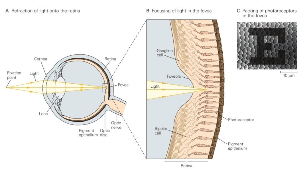
Figure 22-1 The eye projects the visual scene onto the retina’s photoreceptors.
شکل ۲۲-۱ چشم، صحنهی بصری را روی گیرندههای نوری شبکیه منعکس میکند.
A. Light from an object in the visual field is refracted by the cornea and lens and focused onto the retina.
الف) نور از جسمی که در میدان بینایی قرار دارد، توسط قرنیه و عدسی شکسته شده و روی شبکیه متمرکز میشود.
B. In the foveola, corresponding to the very center of gaze, the proximal neurons of the retina are shifted aside so light has direct access to the photoreceptors.
ب. در فووئولا، که مربوط به مرکز نگاه است، نورونهای پروگزیمال شبکیه به کناری منتقل میشوند، بنابراین نور مستقیماً به گیرندههای نوری دسترسی دارد.
C. A letter from the eye chart used to assess normal visual acuity is projected onto the densely packed photoreceptors in the fovea. Although less sharply focused than shown here as a result of diffraction by the eye’s optics, the smallest discernible strokes of the letter are approximately one cone diameter in width. (Adapted, with permission, from Curcio and Hendrickson 1991. Copyright © 1991 Elsevier Ltd.)
ج. یک حرف از نمودار چشم که برای ارزیابی حدت بینایی طبیعی استفاده میشود، بر روی گیرندههای نوری متراکم در گودی چشم (fovea) تابانده میشود. اگرچه به دلیل پراش نور توسط اپتیک چشم، وضوح کمتری نسبت به آنچه در اینجا نشان داده شده است، دارد، اما کوچکترین خطوط قابل تشخیص این حرف تقریباً به اندازه قطر مخروط در عرض هستند. (اقتباس شده، با اجازه، از Curcio و Hendrickson 1991. حق چاپ © 1991 Elsevier Ltd.)
The retinal circuit performs low-level visual processing, the initial stage in the analysis of visual images. It extracts from the raw images in the eyes certain spatial and temporal features and conveys them to higher visual centers. The rules of this processing are adapted to changes in environmental conditions. In particular, the retina must adjust its sensitivity to ever- changing conditions of illumination. This adaptation allows our vision to remain more or less stable despite the vast range of light intensities encountered during the course of each day.
مدار شبکیه پردازش بصری سطح پایین، مرحله اولیه در تجزیه و تحلیل تصاویر بصری، را انجام میدهد. این مدار ویژگیهای مکانی و زمانی خاصی را از تصاویر خام در چشم استخراج کرده و آنها را به مراکز بصری بالاتر منتقل میکند. قوانین این پردازش با تغییرات در شرایط محیطی سازگار است. به طور خاص، شبکیه باید حساسیت خود را با شرایط دائماً در حال تغییر روشنایی تنظیم کند. این سازگاری به بینایی ما اجازه میدهد تا علیرغم طیف وسیعی از شدتهای نوری که در طول هر روز با آن مواجه میشویم، کم و بیش پایدار بماند.
In this chapter, we discuss in turn the three important aspects of retinal function: phototransduction, preprocessing, and adaptation. We will illustrate both the neural mechanisms by which they are achieved and their consequences for visual perception.
در این فصل، به ترتیب سه جنبه مهم عملکرد شبکیه را مورد بحث قرار میدهیم: انتقال نوری، پیشپردازش و سازگاری. ما هم مکانیسمهای عصبی که از طریق آنها این موارد حاصل میشوند و هم پیامدهای آنها را برای ادراک بصری نشان خواهیم داد.
The Photoreceptor Layer Samples the Visual Image
لایه گیرنده نور از تصویر بصری نمونهبرداری میکند
Ocular Optics Limit the Quality of the Retinal Image
اپتیک چشم، کیفیت تصویر شبکیه را محدود میکند
The sharpness of the retinal image is determined by several factors: diffraction at the pupil’s aperture, refractive errors in the cornea and lens, and scattering due to material in the light path. A point in the outside world is generally focused into a small blurred circle on the retina. As in other optical devices, this blur is smallest near the optical axis, where the image quality approaches the limit imposed by diffraction at the pupil. Away from the axis, the image is degraded significantly owing to aberrations in the cornea and lens and may be degraded further by abnormal conditions such as light-scattering cataracts or refractive errors such as myopia.
وضوح تصویر شبکیه توسط چندین عامل تعیین میشود: پراش در روزنه مردمک، خطاهای انکساری در قرنیه و عدسی، و پراکندگی ناشی از مواد موجود در مسیر نور. یک نقطه در دنیای بیرون عموماً به صورت یک دایره کوچک تار روی شبکیه متمرکز میشود. همانند سایر دستگاههای نوری، این تاری در نزدیکی محور نوری، جایی که کیفیت تصویر به حد مجاز ناشی از پراش در مردمک نزدیک میشود، کمترین مقدار را دارد. دور از محور، تصویر به دلیل انحرافات در قرنیه و عدسی به طور قابل توجهی تخریب میشود و ممکن است در اثر شرایط غیرطبیعی مانند آب مروارید پراکنده کننده نور یا خطاهای انکساری مانند نزدیکبینی، بیشتر تخریب شود.

Figure 22-2 The retina comprises five distinct layers of neurons and synapses.
شکل 22-2 شبکیه از پنج لایه مجزا از نورونها و سیناپسها تشکیل شده است.
A. A perpendicular section of the human retina seen through the light microscope. Three layers of cell bodies are evident. The outer nuclear layer contains cell bodies of photoreceptors; the inner nuclear layer includes horizontal, bipolar, and amacrine cells; and the ganglion cell layer contains ganglion cells and some displaced amacrine cells. Two layers of fibers and synapses separate these: the outer plexiform layer and the inner plexiform layer. (Reproduced, with permission, from Boycott and Dowling 1969. Permission conveyed through Copyright Clearance Center.)
الف. برشی عمودی از شبکیه چشم انسان که از طریق میکروسکوپ نوری دیده میشود. سه لایه از اجسام سلولی قابل مشاهده است. لایه هستهای بیرونی شامل اجسام سلولی گیرندههای نوری است؛ لایه هستهای داخلی شامل سلولهای افقی، دوقطبی و آماکرین است؛ و لایه سلولهای گانگلیونی شامل سلولهای گانگلیونی و برخی از سلولهای آماکرین جابجا شده است. دو لایه از الیاف و سیناپسها این لایهها را از هم جدا میکنند: لایه شبکهای بیرونی و لایه شبکهای داخلی. (با اجازه، از Boycott and Dowling 1969 بازتولید شده است. اجازه از طریق مرکز صدور مجوز حق چاپ منتقل شده است.)
B. Neurons in the retina of the macaque monkey based on Golgi staining. The cellular and synaptic layers are aligned with the image in part A. (Abbreviations: M ganglion, magnocellular ganglion cell; P ganglion, parvocellular ganglion cell.) (Reproduced, with permission, from Polyak 1941.)
ب. نورونهای شبکیه میمون ماکاک بر اساس رنگآمیزی گلژی. لایههای سلولی و سیناپسی با تصویر در بخش الف همتراز شدهاند. (اختصارات: گانگلیون M، سلول گانگلیون مگنوسلولار؛ گانگلیون P، سلول گانگلیون پاروسلولار.) (با اجازه، از Polyak 1941 بازتولید شده است.)
The area of retina near the optical axis, the fovea, is where vision is sharpest and corresponds to the center of gaze that we direct toward the objects of our attention. The density of photoreceptors, bipolar cells, and ganglion cells is highest at the fovea (Figure 22-1B). The spacing between photoreceptors there is well matched to the size of the optical blur circle, and thus the image is sampled in an ideal fashion. Light must generally traverse several layers of cells before reaching the photoreceptors, but in the center of the fovea, called the foveola, the other cellular layers are pushed aside to reduce additional blur from light scattering (Figure 22-1B). Finally, the back of the eye is lined by a black pigment epithelium that absorbs light and keeps it from scattering back into the eye.
ناحیه شبکیه نزدیک محور نوری، گودی چشم (fovea)، جایی است که بینایی واضحترین حالت خود را دارد و مطابق با مرکز نگاه ما به سمت اشیاء مورد توجه ماست. تراکم گیرندههای نوری، سلولهای دوقطبی و سلولهای گانگلیونی در گودی چشم بیشترین مقدار را دارد (شکل 22-1B). فاصله بین گیرندههای نوری در آنجا به خوبی با اندازه دایره تاری نوری مطابقت دارد و بنابراین تصویر به روشی ایدهآل نمونهبرداری میشود. نور معمولاً باید قبل از رسیدن به گیرندههای نوری از چندین لایه سلول عبور کند، اما در مرکز گودی چشم، که foveola نامیده میشود، سایر لایههای سلولی کنار زده میشوند تا تاری اضافی ناشی از پراکندگی نور کاهش یابد (شکل 22-1B). در نهایت، پشت چشم توسط یک اپیتلیوم رنگدانهای سیاه پوشیده شده است که نور را جذب میکند و از پراکندگی مجدد آن به داخل چشم جلوگیری میکند.
The retina contains another special site, the optic disc, where the axons of retinal ganglion cells converge and extend through the retina to emerge from the back of the eye as the optic nerve (Figure 22-1A). By necessity, this area is devoid of photoreceptors and thus corresponds to a blind spot in the visual field of each eye. Because the disc lies nasal to the fovea of each eye, light coming from a single point never falls on both blind spots simultaneously, so that normally we are unaware of them. We can experience the blind spot by using only one eye (Figure 22-4). The blind spot demonstrates what blind people experience-not black- ness, but simply nothing. This explains why damage to the peripheral retina often goes unnoticed. It is usually through accidents, such as bumping into an unnoticed object, or through clinical testing that a deficit of sight is revealed.
شبکیه دارای یک محل ویژه دیگر به نام دیسک بینایی است که در آن آکسونهای سلولهای گانگلیونی شبکیه به هم نزدیک میشوند و از طریق شبکیه امتداد مییابند تا از پشت چشم به عنوان عصب بینایی خارج شوند (شکل 22-1A). این ناحیه لزوماً فاقد گیرندههای نوری است و بنابراین با یک نقطه کور در میدان بینایی هر چشم مطابقت دارد. از آنجا که دیسک در نزدیکی گودی هر چشم قرار دارد، نوری که از یک نقطه میآید هرگز به طور همزمان بر روی هر دو نقطه کور نمیافتد، به طوری که معمولاً ما از آنها بیخبر هستیم. ما میتوانیم نقطه کور را تنها با استفاده از یک چشم تجربه کنیم (شکل 22-4). نقطه کور نشان میدهد که افراد نابینا چه چیزی را تجربه میکنند – نه سیاهی، بلکه به سادگی هیچ چیز. این توضیح میدهد که چرا آسیب به شبکیه محیطی اغلب مورد توجه قرار نمیگیرد. معمولاً از طریق حوادثی مانند برخورد با یک جسم ناشناخته یا از طریق آزمایشهای بالینی است که نقص بینایی آشکار میشود.

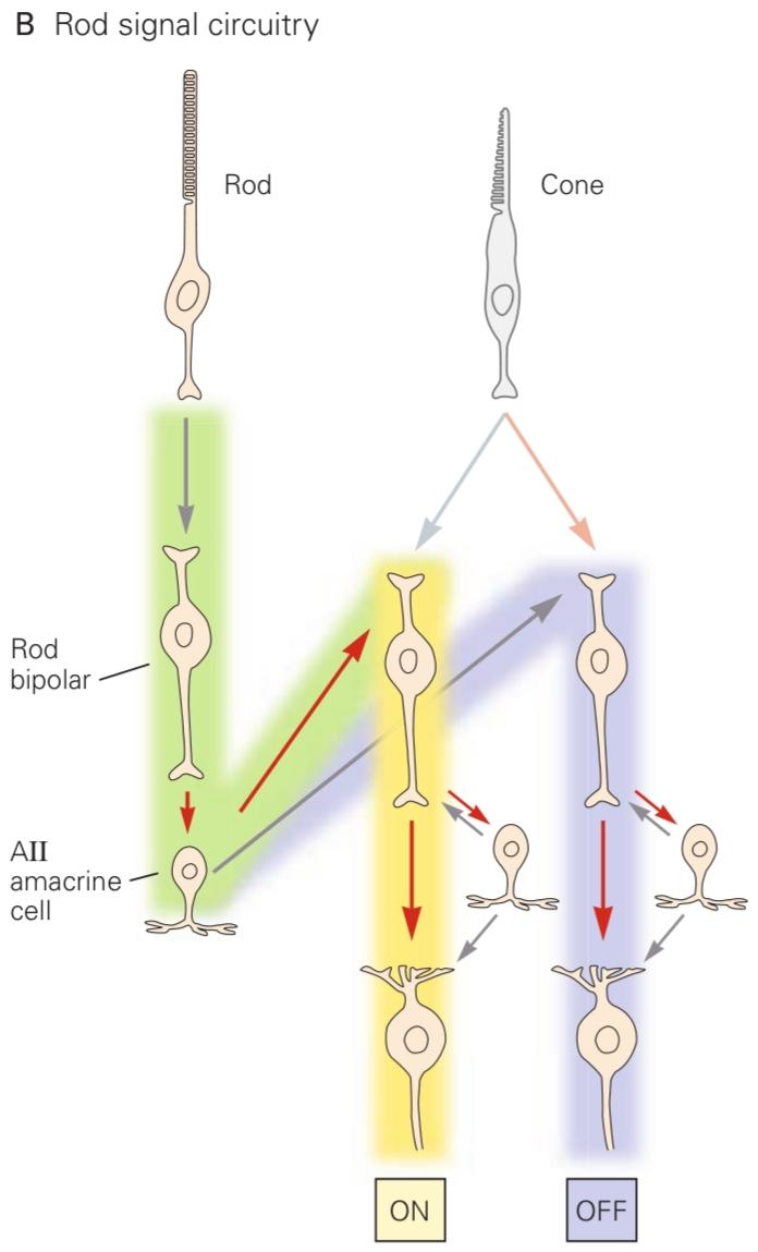
Figure 22-3 The retinal circuitry.
شکل 22-3 مدار شبکیه.
A. The circuitry for cone signals, showing the split into ON cell and OFF cell pathways (see Figure 22-10) as well as the pathway for lateral inhibition in the outer layer. Red arrows indicate sign-preserving connections through electrical or glutamatergic synapses. Gray arrows represent sign-inverting connections through GABAergic, glycinergic, or glutamatergic synapses.
الف) مدار سیگنالهای مخروطی، که تقسیم به مسیرهای سلول روشن و خاموش (شکل 22-10 را ببینید) و همچنین مسیر مهار جانبی در لایه بیرونی را نشان میدهد. فلشهای قرمز نشان دهنده اتصالات حفظ کننده علامت از طریق سیناپسهای الکتریکی یا گلوتاماترژیک هستند. فلشهای خاکستری نشان دهنده اتصالات معکوس کننده علامت از طریق سیناپسهای گاباارژیک، گلیسینرژیک یا گلوتاماترژیک هستند.
B. Rod signals feed into the cone circuitry through All amacrine cells, where the ON and OFF cell pathways diverge.
ب. سیگنالهای استوانهای از طریق سلولهای همه آماکرین، جایی که مسیرهای سلولهای روشن و خاموش از هم جدا میشوند، به مدار مخروطی وارد میشوند.
The blind spot is a necessary consequence of the inside-out design of the retina, which has puzzled and amused biologists for generations. The purpose of this organization may be to enable the tight apposition of photoreceptors with the retinal pigment epithelium, which plays an essential role in the turnover of retinal pigment and recycles photoreceptor membranes by phagocytosis.
نقطه کور نتیجه ضروری طراحی از درون به بیرون شبکیه است که زیستشناسان را برای نسلها متحیر و سرگرم کرده است. هدف از این سازماندهی ممکن است فراهم کردن امکان قرارگیری محکم گیرندههای نوری با اپیتلیوم رنگدانه شبکیه باشد که نقش اساسی در گردش رنگدانه شبکیه دارد و غشاهای گیرنده نوری را با فاگوسیتوز بازیافت میکند.
There Are Two Types of Photoreceptors: Rods and Cones
دو نوع گیرنده نوری وجود دارد: استوانهایها و مخروطیها
All photoreceptor cells have a common structure with four functional regions: the outer segment, located at the distal surface of the neural retina; the inner segment, located more proximally; the cell body; and the synaptic terminal (Figure 22-5A).
تمام سلولهای گیرنده نوری ساختار مشترکی با چهار ناحیه عملکردی دارند: بخش بیرونی، واقع در سطح دیستال شبکیه عصبی؛ بخش داخلی، واقع در نزدیکیتر؛ جسم سلولی؛ و پایانه سیناپسی (شکل 22-5A).
Most vertebrates have two types of photoreceptors, rods and cones, distinguished by their morphology. A rod has a long, cylindrical outer segment within which the stacks of discs are separated from the plasma membrane, whereas a cone often has a shorter, tapered outer segment, and the discs are continuous with the outer membrane (Figure 22-5B).
بیشتر مهرهداران دو نوع گیرنده نوری دارند، استوانهایها و مخروطیها، که از نظر مورفولوژی از هم متمایز میشوند. استوانهایها دارای یک بخش بیرونی بلند و استوانهای هستند که در آن تودههای دیسک از غشای پلاسمایی جدا شدهاند، در حالی که مخروطها اغلب دارای یک بخش بیرونی کوتاهتر و مخروطی هستند و دیسکها در امتداد غشای خارجی قرار دارند (شکل 22-5B).

Figure 22-4 The blind spot of the human retina. Locate the blind spot in your left eye by shutting the right eye and fixating the cross with the left eye. Hold the book about 12 inches from your eye and move it slightly nearer or farther until the circle on the left disappears. Now place a pencil vertically on the page and sweep it sideways over the circle. Note the pencil appears unbroken, even though no light can reach your retina from the region of the circle. Next, move the pencil lengthwise and observe what happens when its tip enters the circle. (Adapted, with permission, from Hurvich 1981.)
شکل ۲۲-۴ نقطه کور شبکیه چشم انسان. با بستن چشم راست و ثابت کردن ضربدر با چشم چپ، نقطه کور چشم چپ خود را پیدا کنید. کتاب را در فاصله حدود ۱۲ اینچی از چشم خود نگه دارید و آن را کمی نزدیکتر یا دورتر حرکت دهید تا دایره سمت چپ ناپدید شود. حالا یک مداد را به صورت عمودی روی صفحه قرار دهید و آن را به صورت جانبی روی دایره بکشید. توجه داشته باشید که مداد بدون شکستگی به نظر میرسد، حتی اگر هیچ نوری از ناحیه دایره به شبکیه شما نرسد. سپس، مداد را در امتداد طول حرکت دهید و مشاهده کنید که وقتی نوک آن وارد دایره میشود چه اتفاقی میافتد. (اقتباس شده، با اجازه، از Hurvich 1981.)
Rods and cones also differ in function, most importantly in their sensitivity to light. Rods can signal the absorption of a single photon and are responsible for vision under dim illumination such as moonlight. But as the light level increases toward dawn, the electrical response of rods becomes saturated and the cells cease to respond to variations in intensity. Cones are much less sensitive to light; they make no contribution to night vision but are solely responsible for vision in daylight. Their response is considerably faster than that of rods. Primates have only one type of rod but three kinds of cone photoreceptors, distinguished by the range of wavelengths to which they respond: the L (long-wave), M (medium-wave), and S (short-wave) cones (Figure 22-6).
سلولهای استوانهای و مخروطی از نظر عملکرد نیز با هم متفاوتند، که مهمترین آنها حساسیت آنها به نور است. سلولهای استوانهای میتوانند جذب یک فوتون واحد را نشان دهند و مسئول بینایی در نور کم مانند نور ماه هستند. اما با افزایش سطح نور به سمت طلوع آفتاب، پاسخ الکتریکی سلولهای استوانهای اشباع میشود و سلولها دیگر به تغییرات شدت پاسخ نمیدهند. سلولهای مخروطی حساسیت بسیار کمتری به نور دارند. آنها هیچ کمکی به دید در شب نمیکنند، بلکه صرفاً مسئول بینایی در نور روز هستند. پاسخ آنها به طور قابل توجهی سریعتر از سلولهای استوانهای است. نخستیسانان فقط یک نوع سلول استوانهای دارند، اما سه نوع گیرنده نوری مخروطی دارند که با طیف طول موجهایی که به آنها پاسخ میدهند، متمایز میشوند: مخروطهای L (موج بلند)، M (موج متوسط) و S (موج کوتاه) (شکل 22-6).
The human retina contains approximately 100 million rods and 5 million cones, but the two cell types are differently distributed. The central fovea contains no rods but is densely packed with small cones. A few millimeters outside the fovea, rods greatly outnumber cones. All photoreceptors become larger and more widely spaced toward the periphery of the retina.
شبکیه انسان تقریباً حاوی ۱۰۰ میلیون سلول استوانهای و ۵ میلیون سلول مخروطی است، اما این دو نوع سلول به طور متفاوتی توزیع شدهاند. گودی مرکزی هیچ سلول استوانهای ندارد، اما به طور متراکم با مخروطهای کوچک پر شده است. چند میلیمتر خارج از گودی، تعداد سلولهای استوانهای بسیار بیشتر از مخروطها است. همه گیرندههای نوری بزرگتر میشوند و به سمت حاشیه شبکیه فاصله بیشتری از هم دارند.

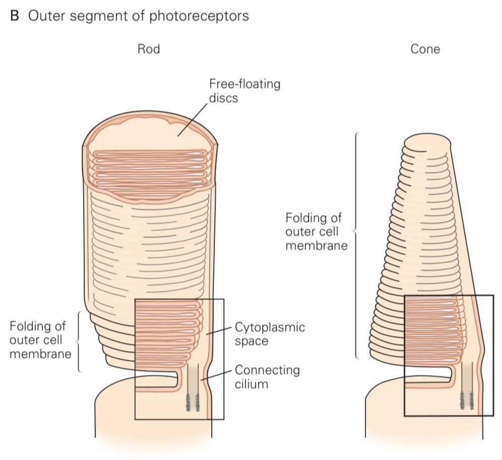
Figure 22-5 Rod and cone photoreceptors have similar structures.
شکل 22-5 گیرندههای نوری استوانهای و مخروطی ساختارهای مشابهی دارند.
A. Both rod and cone cells have specialized regions called the outer and inner segments. The outer segment is attached to the inner segment by a cilium and contains the light-transducing apparatus. The inner segment holds mitochondria and much of the machinery for protein synthesis.
الف) هر دو سلول استوانهای و مخروطی دارای نواحی تخصصی به نام بخشهای بیرونی و درونی هستند. بخش بیرونی توسط یک مژک به بخش داخلی متصل شده و حاوی دستگاه انتقال نور است. بخش داخلی میتوکندری و بخش زیادی از ماشینآلات سنتز پروتئین را در خود جای داده است.
B. The outer segment consists of a stack of membranous discs that contain the light-absorbing photopigments. In both types of cells, these discs are formed by infolding of the plasma mem- brane. In rods, however, the folds pinch off from the membrane so that the discs are free-floating within the outer segment, whereas in cones, the discs remain part of the plasma membrane. (Adapted, with permission, from O’Brien 1982. Copyright © 1982 AAAS; Young 1970.)
ب. بخش بیرونی از مجموعهای از دیسکهای غشایی تشکیل شده است که حاوی رنگدانههای نوری جاذب نور هستند. در هر دو نوع سلول، این دیسکها با چینخوردگی غشای پلاسمایی تشکیل میشوند. با این حال، در سلولهای استوانهای، چینخوردگیها از غشا جدا میشوند به طوری که دیسکها آزادانه در بخش بیرونی شناور هستند، در حالی که در سلولهای مخروطی، دیسکها بخشی از غشای پلاسمایی باقی میمانند. (اقتباس شده با اجازه، از O’Brien 1982. حق چاپ © 1982 AAAS; Young 1970.)

Figure 22-6 Sensitivity spectra for the three types of cones and the rod. At each wavelength, the sensitivity is inversely proportional to the intensity of light required to elicit a criterion response in the sensory neuron. Sensitivity varies over a large range and thus is shown on a logarithmic scale. The different classes of photoreceptors are sensitive to broad and overlapping ranges of wavelengths. (Reproduced, with permission, from Schnapf et al. 1988.)
شکل 22-6 طیف حساسیت برای سه نوع مخروط و استوانه. در هر طول موج، حساسیت با شدت نور مورد نیاز برای ایجاد پاسخ معیار در نورون حسی نسبت معکوس دارد. حساسیت در محدوده وسیعی تغییر میکند و بنابراین در مقیاس لگاریتمی نشان داده شده است. طبقات مختلف گیرندههای نوری به محدودههای وسیع و همپوشانی از طول موجها حساس هستند. (با اجازه، از Schnapf و همکاران، 1988، بازنشر شده است.)
The S cones make up only 10% of all cones and are absent from the central fovea.
مخروطهای S تنها 10٪ از کل مخروطها را تشکیل میدهند و در حفره مرکزی وجود ندارند.
The retinal center of gaze is clearly specialized for daytime vision. The dense packing of cone photoreceptors in the fovea sets the limits of our visual acuity. In fact, the smallest letters we can read on a doctor’s eye chart have strokes whose images are just one to two cone diameters wide on the retina, a visual angle of about 1 minute of arc (Figure 22-1C). At night, the central fovea is blind owing to the absence of rods. Astronomers know that one must look just to the side of a dim star to see it at all. During nighttime walks in the forest, we nonastronomers tend to follow our daytime reflex of looking straight at the source of a suspicious sound. Mysteriously, the object disappears, only to jump back into our peripheral field of view as we avert our gaze.
مرکز نگاه شبکیه به وضوح برای دید در روز تخصص یافته است. تراکم زیاد گیرندههای نوری مخروطی در گودی، محدودیتهای حدت بینایی ما را تعیین میکند. در واقع، کوچکترین حروفی که میتوانیم روی نمودار چشم پزشک بخوانیم، خطوطی دارند که تصاویر آنها فقط یک تا دو برابر قطر مخروط روی شبکیه است، زاویه دیدی حدود ۱ دقیقه قوسی (شکل ۲۲-۱C). در شب، گودی مرکزی به دلیل عدم وجود سلولهای استوانهای کور است. ستارهشناسان میدانند که برای دیدن یک ستاره کمنور، باید فقط به کنار آن نگاه کرد. در طول پیادهرویهای شبانه در جنگل، ما غیرستارهشناسان تمایل داریم از رفلکس روزانه خود که نگاه مستقیم به منبع صدای مشکوک است، پیروی کنیم. به طرز مرموزی، جسم ناپدید میشود، و فقط وقتی نگاهمان را برمیگردانیم، دوباره به میدان دید محیطی ما برمیگردد.
»» ادامه فصل پردازش بصری سطح پایین: شبکیه
»» فصل بعد: فصل پردازش بینایی سطح متوسط و اولیههای بینایی
»» تمامی کتاب