
نوروآناتومی بالینی اسنل | ساقه مغز؛ بصلالنخاع – مرور مبانی و اصول


دعای مطالعه [ نمایش ]
بِسْمِ الله الرَّحْمنِ الرَّحیمِ
اَللّهُمَّ اَخْرِجْنى مِنْ ظُلُماتِ الْوَهْمِ
خدايا مرا بيرون آور از تاريكىهاى وهم،
وَ اَكْرِمْنى بِنُورِ الْفَهْمِ
و به نور فهم گرامى ام بدار،
اَللّهُمَّ افْتَحْ عَلَيْنا اَبْوابَ رَحْمَتِكَ
خدايا درهاى رحمتت را به روى ما بگشا،
وَانْشُرْ عَلَيْنا خَزائِنَ عُلُومِكَ بِرَحْمَتِكَ يا اَرْحَمَ الرّاحِمينَ
و خزانههاى علومت را بر ما باز كن به امید رحمتت اى مهربانترين مهربانان.
آیندهنگاران مغز: «ما مغز را میشناسیم، تا آینده را بسازیم.»
کتاب «نوروآناتومی بالینی اسنل» اثر دکتر ریچارد اس. اسنل، با ویرایش جدید دکتر رایان اسپلیتگربر، همچنان بهعنوان مرجع طلایی این حوزه شناخته میشود. این اثر با تلفیق تازهترین پژوهشها و کاربردهای بالینی، پلی استوار میان دانش پایه و عمل پزشکی بنا میکند.
تلاشهای علمی و سالها تدریس ارزشمند استاد گرانقدر دکتر محمدتقی جغتایی الهامبخش ترجمه و گسترش این دانش در ایران بوده است.
ترجمه دقیق این کتاب توسط برند علمی آیندهنگاران مغز به مدیریت داریوش طاهری، امکان دسترسی فارسیزبانان به مرزهای نوین علوم اعصاب را فراهم کرده و رسالتی علمی در جهت شناخت عمیقتر مغز و ترسیم آیندهای روشنتر بر عهده گرفته است.
»» فصل ۵: ساقه مغز؛ قسمت سوم
»» Chapter 5: Brainstem
INTRODUCTION TO THE BRAINSTEM
The brainstem is made up of the medulla oblongata, the pons, and the midbrain and occupies the posterior cranial fossa of the skull (Fig. 5-8). It is stalklike in shape and connects the narrow spinal cord with the expanded forebrain (see Atlas Plates 1-8).
مقدمهای بر ساقه مغز
ساقه مغز از بصل النخاع، پل مغزی و مغز میانی تشکیل شده و حفره خلفی جمجمه را اشغال میکند (شکل 5-8). ساقه مغز شکلی ساقه مانند دارد و نخاع باریک را به مغز قدامی منبسط شده متصل میکند (به صفحات اطلس 1-8 مراجعه کنید).
The brainstem has three broad functions: (1) It serves as a conduit for the ascending tracts and descending tracts connecting the spinal cord to the different parts of the higher centers in the forebrain; (2) it contains important reflex centers associated with the control of respiration and the cardiovascular system and with the control of consciousness; and (3) it contains the important nuclei of CNs III through XII.
ساقه مغز سه عملکرد کلی دارد: (1) به عنوان مجرایی برای مسیرهای صعودی و نزولی که نخاع را به قسمتهای مختلف مراکز بالاتر در مغز قدامی متصل میکنند، عمل میکند. (2) شامل مراکز رفلکس مهمی است که با کنترل تنفس و سیستم قلبی عروقی و کنترل هوشیاری مرتبط هستند. و (3) حاوی هستههای مهم CN های III تا XII است.

Figure 5-8 Posterior view of the brainstem after removal of the occipital and parietal bones and the cerebrum, the cerebellum, and the roof of the fourth ventricle. Laminae of the upper cervical vertebrae have also been removed.
شکل ۵-۸ نمای خلفی ساقه مغز پس از برداشتن استخوانهای پسسری و آهیانه و مخ، مخچه و سقف بطن چهارم. لامینای مهرههای گردنی فوقانی نیز برداشته شدهاند.
MEDULLA OBLONGATA
The medulla oblongata connects the pons superiorly with the spinal cord inferiorly (see Fig. 5-8). The junction of the medulla and spinal cord is at the origin of the anterior and posterior roots of the first cervical spinal nerve, which corresponds approximately to the level of the foramen magnum. The medulla oblongata is conical in shape, its broad extremity being directed superiorly (Fig. 5-9). The central canal of the spinal cord continues upward into the lower half of the medulla; in the upper half of the medulla, it expands as the cavity of the fourth ventricle.
بصل النخاع
بصل النخاع، پل مغزی را از بالا به نخاع از پایین متصل میکند (شکل ۵-۸ را ببینید). محل اتصال بصل النخاع و نخاع در مبدا ریشههای قدامی و خلفی اولین عصب نخاعی گردنی است که تقریباً با سطح سوراخ بزرگ مطابقت دارد. بصل النخاع مخروطی شکل است و انتهای پهن آن به سمت بالا قرار دارد (شکل ۵-۹). کانال مرکزی نخاع به سمت بالا تا نیمه پایینی بصل النخاع ادامه مییابد. در نیمه بالایی بصل النخاع، به عنوان حفره بطن چهارم گسترش مییابد.
On the anterior surface of the medulla is the anterior median fissure, which is continuous inferiorly with the anterior median fissure of the spinal cord (see Fig. 5-9A). Swellings on each side of the median fissure are called the pyramids. The pyramids are composed of bundles of nerve fibers, called corticospinal fibers, which originate in large nerve cells in the precentral gyrus of the cerebral cortex. The pyramids taper inferiorly, and here the majority of the descending fibers cross over to the opposite side, forming the decussation of the pyramids. The anterior external arcuate fibers emerge from the anterior median fissure above the decussation and pass laterally over the surface of the medulla oblongata to enter the cerebellum. Posterolateral to the pyramids are the olives, which are oval elevations produced by the underlying inferior olivary nuclei. In the groove between the pyramid and the olive emerge the rootlets of the hypoglossal nerve. Posterior to the olives are the inferior cerebellar peduncles, which connect the medulla to the cerebellum. In the groove between the olive and the inferior cerebellar peduncle emerge the roots of the glossopharyngeal and vagus nerves and the cranial roots of the accessory nerve.
در سطح قدامی بصل النخاع، شیار میانی قدامی قرار دارد که در امتداد تحتانی شیار میانی قدامی نخاع قرار دارد (شکل 5-9A را ببینید). برآمدگیهای هر طرف شیار میانی، هرم نامیده میشوند. هرمها از دستههایی از الیاف عصبی به نام الیاف قشری-نخاعی تشکیل شدهاند که از سلولهای عصبی بزرگ در شکنج پیشمرکزی قشر مغز منشأ میگیرند. هرمها به سمت پایین باریک میشوند و در اینجا اکثر الیاف نزولی به سمت مقابل عبور میکنند و تقاطع هرمها را تشکیل میدهند. الیاف قوسی خارجی قدامی از شیار میانی قدامی بالای تقاطع خارج میشوند و به صورت جانبی از روی سطح بصل النخاع عبور میکنند تا وارد مخچه شوند. در سمت خلفی-جانبی هرمها، زیتونها قرار دارند که برآمدگیهای بیضی شکلی هستند که توسط هستههای زیتونی تحتانی زیرین تولید میشوند. در شیار بین هرم و زیتون، ریشههای کوچک عصب زیرزبانی ظاهر میشوند. در خلف زیتونها، پایههای مخچهای تحتانی قرار دارند که بصلالنخاع را به مخچه متصل میکنند. در شیار بین زیتون و پایه مخچهای تحتانی، ریشههای اعصاب زبانی-حلقی و واگ و ریشههای جمجمهای عصب فرعی (اکسسوری) خارج میشوند.
The posterior surface of the superior half of the medulla oblongata forms the lower part of the floor of the fourth ventricle (see Fig. 5-9B). The posterior surface of the inferior half of the medulla is continuous with the posterior aspect of the spinal cord and possesses a posterior median sulcus. On each side of the median sulcus, an elongated swelling, the gracile tubercle, is produced by the underlying gracile nucleus. Lateral to the gracile tubercle is a similar swelling, the cuneate tubercle, produced by the underlying cuneate nucleus.
سطح خلفی نیمه فوقانی بصل النخاع، قسمت پایینی کف بطن چهارم را تشکیل میدهد (شکل 5-9B را ببینید). سطح خلفی نیمه تحتانی بصل النخاع در امتداد سطح خلفی نخاع قرار دارد و دارای یک شیار میانی خلفی است. در هر طرف شیار میانی، یک برآمدگی کشیده به نام برجستگی ظریف توسط هسته ظریف زیرین ایجاد میشود. در کنار برجستگی ظریف، برآمدگی مشابهی به نام برجستگی کونئات وجود دارد که توسط هسته کونئات زیرین ایجاد میشود.

Figure 5-9 Medulla oblongata. A: Anterior view. B: Posterior view. Note that the roof of the fourth ventricle and the cerebellum have been removed.
شکل ۵-۹ بصل النخاع. الف: نمای قدامی. ب: نمای خلفی. توجه داشته باشید که سقف بطن چهارم و مخچه برداشته شده است.
Internal Structure
As in the spinal cord, the medulla oblongata consists of white matter and gray matter, but a study of transverse sections of this region shows that they have been extensively rearranged. This rearrangement can be explained embryologically by the expansion of the neural tube to form the hindbrain vesicle, which becomes the fourth ventricle (Fig. 5-10). The extensive lateral spread of the fourth ventricle results in an alteration in the position of the derivatives of the alar and basal plates of the embryo. To assist in understanding this concept, remember that, in the spinal cord, the derivatives of the alar and basal plates are situated posterior and anterior to the sulcus limitans, respectively; in the case of the medulla oblongata, they are situated lateral and medial to the sulcus limitans, respectively.
ساختار داخلی
همانند نخاع، بصل النخاع از ماده سفید و ماده خاکستری تشکیل شده است، اما مطالعه مقاطع عرضی این ناحیه نشان میدهد که آنها به طور گسترده بازآرایی شدهاند. این بازآرایی را میتوان از نظر جنینشناسی با گسترش لوله عصبی برای تشکیل وزیکول مغز خلفی توضیح داد که به بطن چهارم تبدیل میشود (شکل 5-10). گسترش جانبی گسترده بطن چهارم منجر به تغییر در موقعیت مشتقات صفحات آلار و قاعده جنین میشود. برای کمک به درک این مفهوم، به یاد داشته باشید که در نخاع، مشتقات صفحات آلار و قاعده به ترتیب در خلف و قدام شیار محدودکننده قرار دارند. در مورد بصل النخاع، آنها به ترتیب در طرفین و داخل شیار محدودکننده قرار دارند.

Figure 5-10 Stages in the development of the spinal cord (A-D) and the medulla oblongata (E, F). The neural crest cells will form the first afferent sensory neurons in the posterior root ganglia of the spinal nerves and the sensory ganglia of the cranial nerves.
شکل ۵-۱۰ مراحل رشد نخاع (A-D) و بصل النخاع (E، F). سلولهای تاج عصبی، اولین نورونهای حسی آوران را در گانگلیونهای ریشه خلفی اعصاب نخاعی و گانگلیونهای حسی اعصاب جمجمهای تشکیل میدهند.
The internal structure of the medulla oblongata is considered at four levels: (1) level of decussation of pyramids, (2) level of decussation of lemnisci, (3) level of the olives, and (4) level just inferior to the pons. Table 5-2 compares the different levels of the medulla oblongata and the major structures present at each level.
ساختار داخلی بصل النخاع در چهار سطح در نظر گرفته شده است: (1) سطح تقاطع هرمها، (2) سطح تقاطع لمنیسها، (3) سطح زیتونها، و (4) سطح درست پایینتر از پل مغزی. جدول 5-2 سطوح مختلف بصل النخاع و ساختارهای اصلی موجود در هر سطح را با هم مقایسه میکند.
Table 5-2 Levels of the Medulla Oblongata and Their Major Structures a
جدول ۵-۲ سطوح بصل النخاع و ساختارهای اصلی آنهاa
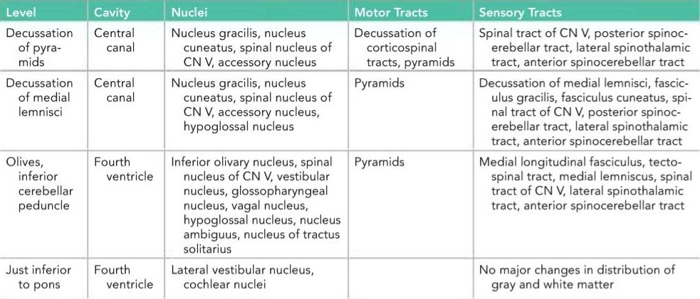
a Note that the reticular formation is present at all levels. CN, cranial nerve.
aتوجه داشته باشید که تشکیلات مشبک در تمام سطوح وجود دارد. CN، عصب جمجمهای.
Level of Pyramid Decussation
A transverse section through the inferior half of the medulla oblongata (Figs. 5-11A and 5-12) passes through the decussation of the pyramids, the great motor decussation. In the superior part of the medulla, the corticospinal fibers occupy and form the pyramid, but inferiorly, about three-fourths of the fibers cross the median plane and continue down the spinal cord in the lateral white column as the lateral corticospinal tract. As these fibers cross the midline, they sever the continuity between the anterior column of the gray matter of the spinal cord and the gray matter that surrounds the central canal.
سطح تقاطع هرمی
یک برش عرضی از نیمه تحتانی بصل النخاع (شکلهای 5-11A و 5-12) از تقاطع هرمها، تقاطع حرکتی بزرگ، عبور میکند. در قسمت فوقانی بصل النخاع، الیاف قشری-نخاعی هرم را اشغال کرده و تشکیل میدهند، اما در قسمت تحتانی، حدود سه چهارم الیاف از صفحه میانی عبور کرده و در ستون سفید جانبی به عنوان راه قشری-نخاعی جانبی به سمت پایین نخاع ادامه مییابند. با عبور این الیاف از خط میانی، پیوستگی بین ستون قدامی ماده خاکستری نخاع و ماده خاکستری که کانال مرکزی را احاطه کرده است، قطع میشود.
The fasciculus gracilis and the fasciculus cuneatus continue to ascend superiorly posterior to the central gray matter. The nucleus gracilis and the nucleus cuneatus appear as posterior extensions of the central gray matter.
فاسیکولوس گراسیلیس و فاسیکولوس کونئاتوس همچنان به سمت بالا و خلف ماده خاکستری مرکزی صعود میکنند. هسته گراسیلیس و هسته کونئاتوس به عنوان امتدادهای خلفی ماده خاکستری مرکزی ظاهر میشوند.
The substantia gelatinosa in the posterior gray column of the spinal cord becomes continuous with the inferior end of the nucleus of the spinal tract of the trigeminal nerve. The fibers of the tract of the nucleus are situated between the nucleus and the surface of the medulla oblongata.
ماده ژلاتینی در ستون خاکستری خلفی نخاع با انتهای تحتانی هسته دستگاه نخاعی عصب سه قلو پیوسته میشود. الیاف دستگاه هسته بین هسته و سطح بصل النخاع قرار دارند.
The lateral and anterior white columns of the spinal cord are easily identified in these sections, and their fiber arrangement is unchanged.
ستونهای سفید جانبی و قدامی نخاع به راحتی در این بخشها قابل تشخیص هستند و ترتیب الیاف آنها بدون تغییر است.

Figure 5-11 Transverse sections of the medulla oblongata. A: Level of decussation of the pyramids. B: Level of decussation of the medial lemnisci.
شکل ۵-۱۱ مقاطع عرضی بصل النخاع. الف: سطح تقاطع هرمها. ب: سطح تقاطع لمنیسهای داخلی.

Figure 5-12 Transverse section of the medulla oblongata at the level of decussation of the pyramids. (Weigert stain.)
شکل ۵-۱۲ برش عرضی بصل النخاع در سطح تقاطع هرمها. (رنگآمیزی وایگرت.)
Level of Lemnisci Decussation
A transverse section through the inferior half of the medulla oblongata, a short distance above the level of the decussation of the pyramids, passes through the decussation of lemnisci, the great sensory decussation (Fig. 5-13; also see Fig. 5-11B). The decussation of the lemnisci takes place anterior to the central gray matter and posterior to the pyramids. It should be understood that the lemnisci have been formed from the internal arcuate fibers, which have emerged from the anterior aspects of the nucleus gracilis and nucleus cuneatus. The internal arcuate fibers first travel anteriorly and laterally around the central gray matter. They then curve medially toward the midline, where they decussate with the corresponding fibers of the opposite side.
سطح تقاطع لمنیسیکها
یک برش عرضی از نیمه تحتانی بصل النخاع، کمی بالاتر از سطح تقاطع هرمها، از تقاطع لمنیسیکها، تقاطع حسی بزرگ، عبور میکند (شکل 5-13؛ همچنین به شکل 5-11B مراجعه کنید). تقاطع لمنیسیکها در قدام ماده خاکستری مرکزی و خلف هرمها انجام میشود. باید توجه داشت که لمنیسیکها از الیاف قوسی داخلی تشکیل شدهاند که از وجوه قدامی هستههای گراسیلیس و کونئاتوس خارج شدهاند. الیاف قوسی داخلی ابتدا به صورت قدامی و جانبی در اطراف ماده خاکستری مرکزی حرکت میکنند. سپس به سمت داخل به سمت خط میانی انحنا مییابند، جایی که با الیاف مربوطه در طرف مقابل تقاطع میکنند.
The nucleus of the spinal tract of the trigeminal nerve lies lateral to the internal arcuate fibers. The spinal tract of the trigeminal nerve lies lateral to the nucleus.
هستهی راه نخاعی عصب سهقلو در سمت خارجی الیاف قوسی داخلی قرار دارد. راه نخاعی عصب سهقلو در سمت خارجی هسته قرار دارد.
The lateral and anterior spinothalamic tracts and the spinotectal tracts occupy an area lateral to the decussation of the lemnisci. They are very close to one another and collectively are known as the spinal lemniscus. The spinocerebellar, vestibulospinal, and the rubrospinal tracts are situated in the anterolateral region of the medulla oblongata.
راههای اسپینوتالامیک جانبی و قدامی و راههای اسپینوتکتال ناحیهای را در سمت خارجی محل تقاطع لمنیسها اشغال میکنند. آنها بسیار به یکدیگر نزدیک هستند و در مجموع به عنوان لمنیس نخاعی شناخته میشوند. راههای اسپینوسربلار، وستیبولواسپینال و روبرواسپینال در ناحیهی قدامی-جانبی بصلالنخاع واقع شدهاند.

Figure 5-13 Transverse section of the medulla oblongata at the level of decussation of the medial lemnisci. (Weigert stain.)
شکل ۵-۱۳ برش عرضی بصل النخاع در سطح تقاطع لبه داخلی (رنگآمیزی وایگرت).
Level of the Olives
A transverse section through the olives passes across the inferior part of the fourth ventricle (Figs. 5-14 and 5-15). The amount of gray matter has increased at this level owing to the presence of the olivary nuclear complex; the nuclei of the vestibulocochlear, glossopharyngeal, vagus, accessory, and hypoglossal nerves; and the arcuate nuclei.
سطح زیتونها
یک برش عرضی از زیتونها از قسمت تحتانی بطن چهارم عبور میکند (شکلهای ۵-۱۴ و ۵-۱۵). مقدار ماده خاکستری در این سطح به دلیل وجود کمپلکس هستهای زیتونی؛ هستههای اعصاب دهلیزی-حلقی، زبانی-حلقی، واگ، فرعی و زیرزبانی؛ و هستههای قوسی افزایش یافته است.
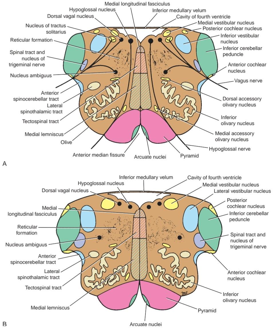
Figure 5-14 Transverse sections of the medulla oblongata at the level of the middle of the olivary nuclei (A) and the superior part of the olivary nuclei just inferior to the pons (B).
شکل ۵-۱۴ برشهای عرضی بصل النخاع در سطح وسط هستههای زیتونی (A) و قسمت فوقانی هستههای زیتونی درست پایینتر از پل مغزی (B).

Figure 5-15 Transverse section of the medulla oblongata at the level of the middle of the olivary nuclei. (Weigert stain.)
شکل ۵-۱۵ برش عرضی بصل النخاع در سطح وسط هستههای زیتونی. (رنگآمیزی وایگرت.)
Olivary Nuclear Complex
The largest nucleus of this complex is the inferior olivary nucleus. The gray matter is shaped like a crumpled bag with its mouth directed medially; it is responsible for the elevation on the surface of the medulla called the olive. Smaller dorsal and medial accessory olivary nuclei also are present. The cells of the inferior olivary nucleus send fibers medially across the midline to enter the cerebellum through the inferior cerebellar peduncle. Afferent fibers reach the inferior olivary nuclei from the spinal cord (the spino-olivary tracts) and from the cerebellum and cerebral cortex. The function of the olivary nuclei is associated with voluntary muscle movement.
مجتمع هستهای زیتونی
بزرگترین هسته این مجتمع، هسته زیتونی تحتانی است. ماده خاکستری به شکل یک کیسه مچاله شده با دهانهای به سمت داخل است؛ این ماده مسئول بالا رفتن سطح بصل النخاع به نام زیتون است. هستههای زیتونی فرعی پشتی و میانی کوچکتر نیز وجود دارند. سلولهای هسته زیتونی تحتانی، فیبرهایی را به سمت داخل از خط وسط میفرستند تا از طریق پایه مخچه تحتانی وارد مخچه شوند. فیبرهای آوران از نخاع (راههای نخاعی-زیتونی) و از مخچه و قشر مغز به هستههای زیتونی تحتانی میرسند. عملکرد هستههای زیتونی با حرکت ارادی عضلات مرتبط است.
Vestibulocochlear Nuclei
The vestibular nuclear complex is made up of the following nuclei: (1) medial vestibular nucleus, (2) inferior vestibular nucleus, (3) lateral vestibular nucleus, and (4) superior vestibular nucleus. The details of these nuclei and their connections are discussed later.
هستههای دهلیزی-حلزونی
مجموعه هستهای دهلیزی از هستههای زیر تشکیل شده است: (1) هسته دهلیزی میانی، (2) هسته دهلیزی تحتانی، (3) هسته دهلیزی جانبی و (4) هسته دهلیزی فوقانی. جزئیات این هستهها و اتصالات آنها بعداً مورد بحث قرار میگیرد.
The medial and inferior vestibular nuclei can be seen on section at this level.
هستههای دهلیزی میانی و تحتانی را میتوان در این سطح در مقطع مشاهده کرد.
The two cochlear nuclei are the anterior cochlear nucleus, situated on the anterolateral aspect of the inferior cerebellar peduncle, and the posterior cochlear nucleus, situated on the posterior aspect of the peduncle lateral to the floor of the fourth ventricle. The connections of these nuclei are described later (see pp. 204-207).
دو هسته حلزونی عبارتند از هسته حلزونی قدامی که در وجه قدامی-جانبی پایه مخچه تحتانی قرار دارد و هسته حلزونی خلفی که در وجه خلفی پایه مخچه جانبی کف بطن چهارم واقع شده است. اتصالات این هستهها بعداً شرح داده شده است (به صفحات 204-207 مراجعه کنید).
Nucleus Ambiguus
The nucleus ambiguus consists of large motor neurons and is situated deep within the reticular formation (Fig. 5-16; also see Fig. 5-14). The emerging nerve fibers join the glossopharyngeal, vagus, and cranial part of the accessory nerve and are distributed to voluntary skeletal muscle.
هسته مبهم
هسته مبهم از نورونهای حرکتی بزرگی تشکیل شده است و در اعماق تشکیل شبکهای قرار دارد (شکل 5-16؛ همچنین به شکل 5-14 مراجعه کنید). فیبرهای عصبی بیرون آمده به بخش زبانی-حلقی، واگ و جمجمهای عصب فرعی میپیوندند و در عضله اسکلتی ارادی توزیع میشوند.

Figure 5-16 Position of the cranial nerve nuclei within the brainstem. The hatched area indicates the position of the vestibular nuclei.
شکل ۵-۱۶ موقعیت هستههای اعصاب جمجمهای در ساقه مغز. ناحیه هاشور خورده موقعیت هستههای دهلیزی را نشان میدهد.
Central Gray Matter
The central gray matter lies beneath the floor of the fourth ventricle at this level (see Figs. 5-14 and 5-15). Passing from medial to lateral (see Fig. 5-16), the following important structures may be recognized: (1) the hypoglossal nucleus, (2) the dorsal nucleus of the vagus, (3) the nucleus of the tractus solitarius, and (4) the medial and inferior vestibular nuclei. The nucleus ambiguus has become deeply placed within the reticular formation (see Fig. 5-14). The connections and functional significance of these nuclei are described in Chapter 11.
ماده خاکستری مرکزی
ماده خاکستری مرکزی در این سطح، زیر کف بطن چهارم قرار دارد (به شکلهای 5-14 و 5-15 مراجعه کنید). با عبور از قسمت میانی به جانبی (به شکل 5-16 مراجعه کنید)، ساختارهای مهم زیر قابل تشخیص هستند: (1) هسته زیرزبانی، (2) هسته پشتی عصب واگ، (3) هسته عضله منفرد، و (4) هستههای دهلیزی میانی و تحتانی. هسته مبهم در عمق تشکیل شبکهای قرار گرفته است (به شکل 5-14 مراجعه کنید). ارتباطات و اهمیت عملکردی این هستهها در فصل 11 شرح داده شده است.
The arcuate nuclei are thought to be inferiorly displaced pontine nuclei (see pp. 206-207) and are situated on the anterior surface of the pyramids. They receive nerve fibers from the cerebral cortex and send efferent fibers to the cerebellum through the anterior external arcuate fibers.
تصور میشود هستههای قوسی، هستههای پونتینِ جابهجا شده به سمت پایین هستند (به صفحات 206-207 مراجعه کنید) و در سطح قدامی هرمها قرار دارند. آنها فیبرهای عصبی را از قشر مغز دریافت میکنند و فیبرهای وابران را از طریق فیبرهای قوسی خارجی قدامی به مخچه میفرستند.
The pyramids containing the corticospinal and some corticonuclear fibers are situated in the anterior part of the medulla separated by the anterior median fissure (see Figs. 5-14 and 5-15); the corticospinal fibers descend to the spinal cord, and the corticonuclear fibers are distributed to the motor nuclei of the CNS situated within the medulla.
هرمهای حاوی فیبرهای قشری-نخاعی و برخی از فیبرهای قشری-هستهای در قسمت قدامی بصل النخاع قرار دارند که توسط شکاف میانی قدامی از هم جدا شدهاند (به شکلهای 5-14 و 5-15 مراجعه کنید)؛ فیبرهای قشری-نخاعی به نخاع میرسند و فیبرهای قشری-هستهای در هستههای حرکتی سیستم عصبی مرکزی واقع در بصل النخاع توزیع میشوند.
The medial lemniscus forms a flattened tract on each side of the midline posterior to the pyramid (see Figs. 5-7 and 5-15). These fibers emerge from the decus- sation of the lemnisci and convey sensory information to the thalamus.
لنیسک داخلی یک مسیر مسطح در هر طرف خط میانی خلفی هرم تشکیل میدهد (به شکلهای 5-7 و 5-15 مراجعه کنید). این فیبرها از محل تقاطع لمنیسها خارج میشوند و اطلاعات حسی را به تالاموس منتقل میکنند.
The medial longitudinal fasciculus forms a small tract of nerve fibers situated on each side of the midline posterior to the medial lemniscus and anterior to the hypoglossal nucleus (see Figs. 5-14 and 5-15). It consists of ascending and descending fibers, the connections of which are described on page 205.
فاسیکول طولی داخلی، یک مسیر کوچک از الیاف عصبی را تشکیل میدهد که در هر طرف خط میانی، خلفی به لمنیسکوس داخلی و قدامی به هسته هیپوگلوسال قرار دارد (شکلهای 5-14 و 5-15 را ببینید). این مسیر از الیاف صعودی و نزولی تشکیل شده است که اتصالات آنها در صفحه 205 شرح داده شده است.
The inferior cerebellar peduncle is situated in the posterolateral corner of the section on the lateral side of the fourth ventricle.
پایه مخچه تحتانی در گوشه خلفی-جانبی برش در سمت جانبی بطن چهارم قرار دارد.
The spinal tract of the trigeminal nerve and its nucleus are situated on the anteromedial aspect of the inferior cerebellar peduncle.
راه نخاعی عصب سه قلو و هسته آن در قسمت قدامی-میانی پایه مخچه تحتانی قرار دارند.
The anterior spinocerebellar tract is situated near the surface in the interval between the inferior olivary nucleus and the nucleus of the spinal tract of the trigeminal nerve. The spinal lemniscus, consisting of the anterior spinothalamic, the lateral spinothalamic, and spinotectal tracts, is deeply placed.
راه نخاعی-مخچهای قدامی در نزدیکی سطح و در فاصله بین هسته زیتونی تحتانی و هسته راه نخاعی عصب سهقلو قرار دارد. لمنیسکوس نخاعی، متشکل از راههای نخاعی-تالامیک قدامی، نخاعی-تالامیک جانبی و نخاعی-تکتال، در عمق قرار دارد.
The reticular formation, consisting of a diffuse mixture of nerve fibers and small groups of nerve cells, is deeply placed posterior to the olivary nucleus. The reticular formation represents, at this level, only a small part of this system, which is also present in the pons and midbrain.
تشکیلات مشبک، متشکل از مخلوطی پراکنده از الیاف عصبی و گروههای کوچکی از سلولهای عصبی، در عمق خلفی هسته زیتونی قرار دارد. تشکیلات مشبک، در این سطح، تنها بخش کوچکی از این سیستم را نشان میدهد که در پل مغزی و مغز میانی نیز وجود دارد.
The glossopharyngeal, vagus, and cranial part of the accessory nerves can be seen running forward and laterally through the reticular formation (see Fig. 5-14). The nerve fibers emerge between the olives and the inferior cerebellar peduncles. The hypoglossal nerves also run anteriorly and laterally through the reticular formation and emerge between the pyramids and the olives.
بخش زبانی-حلقی، واگ و جمجمهای اعصاب فرعی را میتوان در حال عبور به جلو و جانبی از طریق تشکیلات مشبک مشاهده کرد (شکل 5-14 را ببینید). الیاف عصبی بین زیتونها و پایههای مخچهای تحتانی ظاهر میشوند. اعصاب زیرزبانی نیز به صورت قدامی و جانبی از طریق تشکیلات مشبک عبور میکنند و بین هرمها و زیتونها ظاهر میشوند.
Level Just Inferior to the Pons
In comparison to the previous level, little changes in the distribution of the gray and white matter (see Figs. 5-14
and 5-16). The lateral vestibular nucleus has replaced the inferior vestibular nucleus, and the cochlear nuclei now are visible on the anterior and posterior surfaces of the inferior cerebellar peduncle.
سطح کمی پایینتر از پل مغزی
در مقایسه با سطح قبلی، تغییرات کمی در توزیع ماده خاکستری و سفید وجود دارد (به شکلهای ۵-۱۴ و ۵-۱۶ مراجعه کنید). هسته دهلیزی جانبی جایگزین هسته دهلیزی تحتانی شده است و هستههای حلزونی اکنون در سطوح قدامی و خلفی پایه مخچه تحتانی قابل مشاهده هستند.
«فصل پنجم کتاب نورواناتومی بالینی اسنل»
🚀 با ما همراه شوید!
تازهترین مطالب و آموزشهای مغز و اعصاب را از دست ندهید. با فالو کردن کانال تلگرام آیندهنگاران مغز، از ما حمایت کنید!



