نخاع و مسیرهای صعودی، نزولی و بین قطعهای؛ فصل چهارم نوروآناتومی بالینی


دعای مطالعه [ نمایش ]
بِسْمِ الله الرَّحْمنِ الرَّحیمِ
اَللّهُمَّ اَخْرِجْنى مِنْ ظُلُماتِ الْوَهْمِ
خدايا مرا بيرون آور از تاريكىهاى وهم،
وَ اَكْرِمْنى بِنُورِ الْفَهْمِ
و به نور فهم گرامى ام بدار،
اَللّهُمَّ افْتَحْ عَلَيْنا اَبْوابَ رَحْمَتِكَ
خدايا درهاى رحمتت را به روى ما بگشا،
وَانْشُرْ عَلَيْنا خَزائِنَ عُلُومِكَ بِرَحْمَتِكَ يا اَرْحَمَ الرّاحِمينَ
و خزانههاى علومت را بر ما باز كن به امید رحمتت اى مهربانترين مهربانان.
آیندهنگاران مغز: «ما مغز را میشناسیم، تا آینده را بسازیم.»
کتاب «نوروآناتومی بالینی اسنل» اثر دکتر ریچارد اس. اسنل، با ویرایش جدید دکتر رایان اسپلیتگربر، همچنان بهعنوان مرجع طلایی این حوزه شناخته میشود. این اثر با تلفیق تازهترین پژوهشها و کاربردهای بالینی، پلی استوار میان دانش پایه و عمل پزشکی بنا میکند.
تلاشهای علمی و سالها تدریس ارزشمند استاد گرانقدر دکتر محمدتقی جغتایی الهامبخش ترجمه و گسترش این دانش در ایران بوده است.
ترجمه دقیق این کتاب توسط برند علمی آیندهنگاران مغز به مدیریت داریوش طاهری، امکان دسترسی فارسیزبانان به مرزهای نوین علوم اعصاب را فراهم کرده و رسالتی علمی در جهت شناخت عمیقتر مغز و ترسیم آیندهای روشنتر بر عهده گرفته است.
»» فصل ۴: نخاع و مسیرهای صعودی، نزولی و بین قطعهای
»» Chapter 4: Spinal Cord and Ascending, Descending, and Intersegmental Tracts
CHAPTER OBJECTIVES
• To learn how injuries to the spinal cord can occur
• To understand the position of the main nervous pathways and nerve cell groups in the spinal cord as well as be able to correlate radiologic evidence of bone injury with segmental levels of the spinal cord and neurologic deficits
• To review the basic structure of the delicate spinal cord and the positions and functions of the various ascending and descending tracts that lie within it
• To make simple line drawings of each of the ascending and descending tracts, showing their cells of origin, their course through the central nervous system, and their destination
اهداف فصل
• یادگیری چگونگی وقوع آسیبهای نخاعی
• درک موقعیت مسیرهای عصبی اصلی و گروههای سلولی عصبی در نخاع و همچنین توانایی مرتبط کردن شواهد رادیولوژیک آسیب استخوانی با سطوح قطعهای نخاع و نقصهای عصبی
• بررسی ساختار اساسی نخاع ظریف و موقعیتها و عملکردهای مسیرهای صعودی و نزولی مختلف که در آن قرار دارند
• ترسیم خطوط ساده از هر یک از مسیرهای صعودی و نزولی، نشان دادن سلولهای مبدا، مسیر آنها در سیستم عصبی مرکزی و مقصد آنها
A 35-year-old man is galloping his horse when he attempts to jump over a farm gate. The horse refuses to jump, and he is thrown to the ground. His head strikes a log, and his head and neck are excessively flexed. On initial evaluation in the emergency department after he regains consciousness, he is found to have signs and symptoms of severe neurologic deficits in the upper and lower extremities. A lateral radiograph of the cervical region of the spine shows fragmentation of the body of the fourth cervical vertebra with backward displacement of a large bony fragment on the left side.
مردی ۳۵ ساله در حال تاخت و تاز با اسب خود است که سعی میکند از روی دروازه مزرعه بپرد. اسب از پریدن امتناع میکند و او به زمین پرتاب میشود. سر او به یک کنده برخورد میکند و سر و گردنش بیش از حد خم میشوند. در ارزیابی اولیه در بخش اورژانس پس از به هوش آمدن، مشخص میشود که علائم و نشانههای نقص شدید عصبی در اندامهای فوقانی و تحتانی دارد. رادیوگرافی جانبی از ناحیه گردنی ستون فقرات، تکهتکه شدن تنه مهره چهارم گردنی را با جابجایی یک قطعه استخوانی بزرگ به عقب در سمت چپ نشان میدهد.
After stabilization of the vertebral column by using skeletal traction to prevent further neurologic damage, a complete examination reveals that the patient has signs and symptoms indicating incomplete hemisection of the spinal cord on the left side.
پس از تثبیت ستون فقرات با استفاده از کشش اسکلتی برای جلوگیری از آسیب عصبی بیشتر، معاینه کامل نشان میدهد که بیمار علائم و نشانههایی دارد که نشان دهنده نیمهقطعه شدن ناقص نخاع در سمت چپ است.
Any medical personnel involved in the evaluation and treatment of a patient with spinal cord injuries must know the structure of the spinal cord and the arrangement and functions of the various nerve tracts passing up and down this vital conduit in the central nervous system.
هر پرسنل پزشکی که در ارزیابی و درمان بیمار مبتلا به آسیبهای نخاعی مشارکت دارد، باید ساختار نخاع و ترتیب و عملکرد رشتههای عصبی مختلفی که از این مجرای حیاتی در سیستم عصبی مرکزی عبور میکنند را بداند.
Because of the devastating nature of spinal cord injuries and the prolonged disability that results, all concerned with the care of such patients must be trained to prevent any additional cord injury and provide the best chance for recovery. All medical personnel must have a clear picture of the extent of the cord lesion and the possible expectations for the return of function.
به دلیل ماهیت ویرانگر آسیبهای نخاعی و ناتوانی طولانیمدت ناشی از آن، تمام افرادی که در مراقبت از چنین بیمارانی نقش دارند باید آموزش ببینند تا از هرگونه آسیب بیشتر به نخاع جلوگیری کرده و بهترین شانس را برای بهبودی فراهم کنند. تمام پرسنل پزشکی باید تصویر روشنی از میزان ضایعه نخاعی و انتظارات احتمالی برای بازگشت عملکرد داشته باشند.
Spinal cord injuries are common and can occur as a result of automobile and motorcycle accidents, falls, sports injuries, and gunshot wounds. Spinal cord and spinal nerve damage may also be associated with vertebral fractures; vertebral infections; vertebral tumors, both primary and secondary; and herniated intervertebral discs. The student must learn the course and connections of the various tracts within the spinal cord in order to be able to diagnose and understand the treatment of cord injuries. Particular attention should be paid as to whether a specific tract crosses the midline to the opposite side of the central nervous system (CNS) or remains on the same side. If the tract does cross the midline, the level of the crossover is important.
آسیبهای نخاعی شایع هستند و میتوانند در نتیجه تصادفات اتومبیل و موتورسیکلت، سقوط، آسیبهای ورزشی و زخمهای ناشی از گلوله رخ دهند. آسیب نخاع و اعصاب نخاعی همچنین ممکن است با شکستگیهای مهرهای؛ عفونتهای مهرهای؛ تومورهای مهرهای، چه اولیه و چه ثانویه؛ و فتق دیسکهای بین مهرهای مرتبط باشند. دانشجو باید مسیر و اتصالات مسیرهای مختلف درون نخاع را بیاموزد تا بتواند آسیبهای نخاعی را تشخیص داده و درمان کند. باید توجه ویژهای به این نکته شود که آیا یک مسیر خاص از خط وسط به طرف مقابل سیستم عصبی مرکزی (CNS) عبور میکند یا در همان طرف باقی میماند. اگر مسیر از خط وسط عبور کند، سطح تقاطع مهم است.
The assessment of neurologic damage requires not only an understanding of the main nervous pathways within the spinal cord but an ability to correlate radiologic evidence of bone injury with segmental levels of the spinal cord. The close relationship of the spinal cord to the bony vertebral column necessitates a brief review of the vertebral column before the spinal cord is considered.
ارزیابی آسیب عصبی نه تنها نیازمند درک مسیرهای عصبی اصلی درون نخاع است، بلکه توانایی مرتبط کردن شواهد رادیولوژیک آسیب استخوانی با سطوح قطعهای نخاع را نیز میطلبد. ارتباط نزدیک نخاع با ستون فقرات استخوانی، بررسی مختصری از ستون فقرات را قبل از بررسی نخاع ضروری میسازد.
BRIEF REVIEW OF THE VERTEBRAL COLUMN
The vertebral column is the central bony pillar of the body. It supports the skull, pectoral girdle, upper limbs, and thoracic cage and, by way of the pelvic girdle, transmits body weight to the lower limbs. Within its cavity lie the spinal cord, spinal nerve roots, and the covering meninges, to which the vertebral column gives great protection.
مروری کوتاه بر ستون فقرات
ستون مهرهها ستون استخوانی مرکزی بدن است. این ستون از جمجمه، کمربند سینهای، اندامهای فوقانی و قفسه سینه پشتیبانی میکند و از طریق کمربند لگنی، وزن بدن را به اندامهای تحتانی منتقل میکند. در حفره آن، نخاع، ریشههای عصبی نخاعی و مننژهای پوشاننده قرار دارند که ستون مهرهها از آنها محافظت زیادی میکند.
Vertebral Column Composition
The vertebral column (Figs. 4-1 and 4-2) is composed of 33 vertebrae-7 cervical, 12 thoracic, 5 lumbar, 5 sacral (fused to form the sacrum), and 4 coccygeal (the lower 3 are commonly fused). Because it is segmented and made up of vertebrae, joints, and pads of fibrocartilage called intervertebral discs, it is a flexible structure. The intervertebral discs form about a quarter of the length of the column.
ترکیب ستون مهرهها
ستون مهرهها (شکلهای ۴-۱ و ۴-۲) از ۳۳ مهره تشکیل شده است – ۷ مهره گردنی، ۱۲ مهره سینهای، ۵ مهره کمری، ۵ مهره خاجی (که به هم جوش خورده و استخوان خاجی را تشکیل میدهند) و ۴ مهره دنبالچهای (که ۳ مهره پایینی معمولاً به هم جوش خوردهاند). از آنجایی که این ستون قطعه قطعه است و از مهرهها، مفاصل و بالشتکهایی از جنس غضروف فیبری به نام دیسکهای بین مهرهای تشکیل شده است، ساختاری انعطافپذیر دارد. دیسکهای بین مهرهای حدود یک چهارم طول ستون را تشکیل میدهند.

Figure 4-1 Posterior view of the skeleton showing the vertebral column. The surface marking of the external occipital protuberance of the skull, the ligamentum nuchae (solid black line) and some important palpable spines (solid dots) are also shown.
شکل ۴-۱ نمای خلفی اسکلت که ستون مهرهها را نشان میدهد. علامتگذاری سطحی برآمدگی پسسری خارجی جمجمه، رباط پشت سر (خط سیاه ممتد) و برخی از خارهای قابل لمس مهم (نقاط ممتد) نیز نشان داده شده است.

Figure 4-2 A: Lateral view of the vertebral column. B: General features of different kinds of vertebrae.
شکل ۴-۲ الف: نمای جانبی ستون مهرهها. ب: ویژگیهای کلی انواع مختلف مهرهها.
Vertebra General Characteristics
Although vertebrae show regional differences, they all possess a common pattern (Fig. 4-2B). A typical vertebra consists of a rounded body anteriorly and a vertebral arch posteriorly. These enclose a space called the vertebral foramen, through which run the spinal cord and its coverings. The vertebral arch consists of a pair of cylindrical pedicles, which form the sides of the arch, and a pair of flattened laminae, which complete the arch posteriorly. The vertebral arch gives rise to seven processes: one spinous, two transverse, and four articular.
ویژگیهای کلی مهرهها
اگرچه مهرهها تفاوتهای منطقهای نشان میدهند، اما همه آنها دارای یک الگوی مشترک هستند (شکل 4-2B). یک مهره معمولی از یک بدنه گرد در جلو و یک قوس مهرهای در عقب تشکیل شده است. این دو، فضایی به نام سوراخ مهرهای را احاطه میکنند که از طریق آن نخاع و پوششهای آن عبور میکنند. قوس مهرهای از یک جفت پایه استوانهای تشکیل شده است که طرفین قوس را تشکیل میدهند و یک جفت تیغه صاف که قوس را در عقب کامل میکنند. قوس مهرهای هفت زائده ایجاد میکند: یک خاردار، دو عرضی و چهار مفصلی.
The spinous process, or spine, is directed posteriorly from the junction of the two laminae. The transverse processes are directed laterally from the junction of the laminae and the pedicles. Both the spinous and transverse processes serve as levers and receive attachments of muscles and ligaments.
زائده خاری یا ستون فقرات، از محل اتصال دو لامینا به سمت خلف هدایت میشود. زائدههای عرضی از محل اتصال لامیناها و پدیکولها به سمت طرفین هدایت میشوند. هر دو زائده خاری و عرضی به عنوان اهرم عمل میکنند و اتصالات عضلات و رباطها را دریافت میکنند.
The articular processes are vertically arranged and consist of two superior and two inferior processes. They arise from the junction of the laminae and the pedicles. The two superior articular processes of one vertebral arch articulate with the two inferior articular processes of the arch above, forming two synovial joints.
زوائد مفصلی به صورت عمودی قرار گرفتهاند و از دو زائده فوقانی و دو زائده تحتانی تشکیل شدهاند. آنها از محل اتصال لامیناها و پدیکولها منشأ میگیرند. دو زائده مفصلی فوقانی یک قوس مهرهای با دو زائده مفصلی تحتانی قوس بالایی مفصل میشوند و دو مفصل سینوویال را تشکیل میدهند.
The pedicles are notched on their upper and lower borders, forming the superior and inferior vertebral notches. On each side, the superior notch of one vertebra and the inferior notch of an adjacent vertebra together form an intervertebral foramen. These foramina, in an articulated skeleton, serve to transmit the spinal nerves and blood vessels. The anterior and posterior nerve roots of a spinal nerve unite within these foramina with their coverings of dura to form the segmental spinal nerves.
دندیکولها در لبههای بالایی و پایینی خود دارای بریدگی هستند و بریدگیهای مهرهای فوقانی و تحتانی را تشکیل میدهند. در هر طرف، بریدگی فوقانی یک مهره و بریدگی تحتانی مهره مجاور با هم یک سوراخ بین مهرهای را تشکیل میدهند. این سوراخها، در یک اسکلت مفصلی، برای انتقال اعصاب نخاعی و رگهای خونی عمل میکنند. ریشههای عصبی قدامی و خلفی یک عصب نخاعی در داخل این سوراخها با پوشش سختشامه خود به هم متصل میشوند و اعصاب نخاعی سگمنتال را تشکیل میدهند.
Vertebral Column Joints
Below the axis, the vertebrae articulate with each other by means of cartilaginous joints between their bodies and by synovial joints between their articular processes.
مفاصل ستون فقرات
در زیر محور، مهرهها از طریق مفاصل غضروفی بین بدنههایشان و از طریق مفاصل سینوویال بین زوائد مفصلیشان با یکدیگر مفصل میشوند.
Joints Between Two Vertebral Bodies
Sandwiched between the vertebral bodies is an intervertebral disc of fibrocartilage (Fig. 4-3).
مفاصل بین دو جسم مهرهای
بین جسم مهرهها یک دیسک بین مهرهای از جنس غضروف لیفی قرار دارد (شکل ۴-۳).

Figure 4-3 A: Joints in the cervical, thoracic, and lumbar regions of the vertebral column. B: Third lumbar vertebra seen from above showing the relationship between intervertebral disc and cauda equina. C: Lumbar vertebrae in sagittal section showing vertebral ligament relationships.
شکل ۴-۳ الف: مفاصل در نواحی گردنی، سینهای و کمری ستون مهرهها. ب: مهره سوم کمری از بالا دیده میشود که رابطه بین دیسک بین مهرهای و دم اسب را نشان میدهد. ج: مهرههای کمری در مقطع ساژیتال که روابط رباطهای مهرهای را نشان میدهد.
Intervertebral Discs
The intervertebral discs are thickest in the cervical and lumbar regions, where the movements of the vertebral column are greatest. They serve as shock absorbers when the load on the vertebral column is suddenly increased. Unfortunately, their resilience is gradually lost with advancing age.
دیسکهای بین مهرهای
دیسکهای بین مهرهای در نواحی گردنی و کمری، جایی که حرکات ستون فقرات بیشترین است، ضخیمترین ضخامت را دارند. آنها به عنوان ضربهگیر در هنگام افزایش ناگهانی بار روی ستون فقرات عمل میکنند. متأسفانه، با افزایش سن، خاصیت ارتجاعی آنها به تدریج از بین میرود.
Each disc consists of a peripheral part, the annulus fibrosus, and a central part, the nucleus pulposus (Fig. 4-3B,C). The annulus fibrosus is composed of fibrocartilage, which is strongly attached to the vertebral bodies and the anterior and posterior longitudinal ligaments of the vertebral column.
هر دیسک از یک بخش محیطی به نام حلقه فیبروزوس و یک بخش مرکزی به نام هسته پولپوزوس تشکیل شده است (شکل 4-3B,C). حلقه فیبروزوس از غضروف فیبری تشکیل شده است که به شدت به جسم مهرهها و رباطهای طولی قدامی و خلفی ستون مهرهها متصل است.
The nucleus pulposus in the young is an ovoid mass of gelatinous material. It is normally under pressure and situated slightly nearer to the posterior than to the anterior margin of the disc. The upper and lower surfaces of the bodies of adjacent vertebrae that abut onto the disc are covered with thin plates of hyaline cartilage.
هسته پولپوزوس در دوران جوانی یک توده تخممرغی شکل از جنس ژلاتین است. این ماده معمولاً تحت فشار است و کمی نزدیکتر به لبه خلفی دیسک قرار دارد تا لبه قدامی آن. سطوح بالایی و پایینی جسم مهرههای مجاور که به دیسک متصل هستند با صفحات نازکی از غضروف هیالین پوشیده شدهاند.
The semifluid nature of the nucleus pulposus allows it to change shape and permits one vertebra to rock forward or backward on another. A sudden increase in the compression load on the vertebral column flattens the nucleus pulposus, which is accommodated by the resilience of the surrounding annulus fibrosus. Sometimes, the outward thrust is too great for the annulus fibrosus and it ruptures, allowing the nucleus pulposus to herniate and protrude into the vertebral canal, where it may press on the spinal nerve roots, the spinal nerve, or even the spinal cord.
ماهیت نیمه سیال هسته پولپوزوس به آن اجازه میدهد تا شکل خود را تغییر دهد و به یک مهره اجازه میدهد تا به جلو یا عقب روی مهره دیگر حرکت کند. افزایش ناگهانی بار فشاری روی ستون مهرهها، هسته پولپوزوس را مسطح میکند که توسط خاصیت ارتجاعی حلقه فیبروزوس اطراف آن، تحمل میشود. گاهی اوقات، فشار رو به بیرون برای آنولوس فیبروزوس بیش از حد زیاد است و باعث پارگی آن میشود و به نوکلئوس پولپوزوس اجازه میدهد تا فتق پیدا کند و به داخل کانال مهرهای بیرون بزند، جایی که ممکن است به ریشههای عصب نخاعی، عصب نخاعی یا حتی نخاع فشار وارد کند.
With advancing age, the nucleus pulposus becomes smaller and is replaced by fibrocartilage. The collagen fibers of the annulus degenerate, and, as a result, the annulus cannot always contain the nucleus pulposus under stress. In old age, the discs are thin and less elastic, and distinguishing the nucleus from the annulus is no longer possible.
با افزایش سن، هسته دیسک (نوکلئوس پولپوزوس) کوچکتر شده و با غضروف فیبری جایگزین میشود. الیاف کلاژن حلقه دیسک تحلیل میروند و در نتیجه، حلقه دیسک نمیتواند همیشه هسته دیسک را تحت فشار در خود جای دهد. در سنین بالا، دیسکها نازک و خاصیت ارتجاعی کمتری دارند و تشخیص هسته دیسک از حلقه دیسک دیگر امکانپذیر نیست.
Ligaments
The anterior and posterior longitudinal ligaments run as continuous bands down the anterior and posterior surfaces of the vertebral column from the skull to the sacrum (Fig. 4-3C). The anterior ligament is wide and is strongly attached to the front and sides of the vertebral bodies and to the intervertebral discs. The posterior ligament is weak and narrow and is attached to the posterior borders of the discs.
رباطها
رباطهای طولی قدامی و خلفی به صورت نوارهای پیوسته در سطوح قدامی و خلفی ستون مهرهها از جمجمه تا استخوان خاجی امتداد دارند (شکل 4-3C). رباط قدامی پهن است و محکم به جلو و کنارههای جسم مهرهها و دیسکهای بین مهرهای متصل است. رباط خلفی ضعیف و باریک است و به لبههای خلفی دیسکها متصل است.
Joints Between Two Vertebral Arches
The joints between two vertebral arches consist of synovial joints between the superior and inferior artic- ular processes of adjacent vertebrae (Fig. 4-3A).
مفاصل بین دو قوس مهرهای
مفاصل بین دو قوس مهرهای شامل مفاصل سینوویال بین زوائد مفصلی فوقانی و تحتانی مهرههای مجاور است (شکل 4-3A).
Ligaments
Refer to Figure 4-3C.
رباطها
به شکل۴-۳c مراجعه کنید.
• The supraspinous ligament runs between the tips of adjacent spines.
• The interspinous ligament connects adjacent spines.
• The intertransverse ligaments run between adjacent transverse processes.
• The ligamentum flavum connects the laminae of adjacent vertebrae.
• رباط فوق خاری بین نوک خارهای مجاور قرار دارد.
• رباط بین خاری، خارهای مجاور را به هم متصل میکند.
• رباطهای عرضی بین زوائد عرضی مجاور قرار دارند.
• رباط زرد، تیغههای مهرههای مجاور را به هم متصل میکند.
In the cervical region, the supraspinous and interspinous ligaments are greatly thickened to form the strong ligamentum nuchae.
در ناحیه گردنی، رباطهای فوق خاری و بین خاری به شدت ضخیم میشوند و رباط قوی پشت گردن (لیگامان گردن) را تشکیل میدهند.
Vertebral Joint Nerve Supply
The joints between the vertebral bodies are innervated by the small meningeal branches of each spinal nerve (Fig. 4-4). The joints between the articular processes are innervated by branches from the posterior rami of the spinal nerves; the joints of any particular level receive nerve fibers from two adjacent spinal nerves.
عصب رسانی به مفاصل مهره ای
مفاصل بین جسم مهره ها توسط شاخه های کوچک مننژی هر عصب نخاعی عصب دهی می شوند (شکل 4-4). مفاصل بین زوائد مفصلی توسط شاخه هایی از شاخه های خلفی اعصاب نخاعی عصب دهی می شوند. مفاصل هر سطح خاص، فیبرهای عصبی را از دو عصب نخاعی مجاور دریافت می کنند.
The atlanto-occipital joints and the atlanto-axial joints should be reviewed in a textbook of gross anatomy.
مفاصل آتلانتو-اکسیپیتال و مفاصل آتلانتو-اکسیپیتال باید در یک کتاب درسی آناتومی کلی بررسی شوند.

Figure 4-4 The innervation of vertebral joints. At any particular vertebral level, the joints receive nerve fibers from two adjacent spinal nerves.
شکل ۴-۴ عصبدهی مفاصل مهرهای. در هر سطح مهرهای خاص، مفاصل از دو عصب نخاعی مجاور، فیبرهای عصبی دریافت میکنند.
SPINAL CORD
The spinal cord is roughly cylindrical in shape. It begins superiorly at the foramen magnum in the skull, where it is continuous with the medulla oblongata of the brain, and it terminates inferiorly in the adult at the level of the lower border of the first lumbar vertebra. In the young child, it is relatively longer and usually ends at the upper border of the third lumbar vertebra. Thus, it occupies the upper two thirds of the vertebral canal of the vertebral column and is surrounded by the three meninges, the dura mater, the arachnoid mater, and the pia mater. Further protection is provided by the cerebrospinal fluid (CSF), which surrounds the spinal cord in the subarachnoid space.
نخاع
نخاع تقریباً استوانهای شکل است. از بالا از سوراخ بزرگ جمجمه شروع میشود، جایی که با بصل النخاع مغز در امتداد است و در بزرگسالان از پایین در سطح لبه پایینی اولین مهره کمری خاتمه مییابد. در کودک خردسال، نسبتاً بلندتر است و معمولاً در لبه بالایی سومین مهره کمری خاتمه مییابد. بنابراین، دو سوم بالایی کانال مهرهای ستون فقرات را اشغال میکند و توسط سه مننژ، سختشامه، عنکبوتیه و نرمشامه احاطه شده است. مایع مغزی نخاعی (CSF) که نخاع را در فضای زیر عنکبوتیه احاطه میکند، محافظت بیشتری را فراهم میکند.
In the cervical region, where it gives origin to the brachial plexus, and in the lower thoracic and lumbar regions, where it gives origin to the lumbosacral plexus, the spinal cord is fusiformly enlarged; the enlargements are referred to as the cervical and lumbar enlargements (Fig. 4-5). Inferiorly, the spinal cord tapers off into the conus medullaris, from the apex of which a prolongation of the pia mater, the filum terminale, descends to attach to the posterior surface of the coccyx. The cord possesses a deep longitudinal fissure called the anterior median fissure in the midline anteriorly and a shallow furrow called the posterior median sulcus on the posterior surface (Fig. 4-5B).
در ناحیه گردنی، جایی که منشأ شبکه بازویی است، و در نواحی تحتانی قفسه سینه و کمری، جایی که منشأ شبکه کمری-خاجی است، نخاع به صورت دوکی شکل بزرگ شده است؛ این بزرگ شدنها به عنوان بزرگ شدنهای گردنی و کمری شناخته میشوند (شکل 4-5). در قسمت پایین، نخاع به مخروط انتهایی باریک میشود، که از رأس آن امتدادی از نرمشامه، فیلوم ترمینال، به سطح خلفی دنبالچه متصل میشود. نخاع دارای یک شکاف طولی عمیق به نام شکاف میانی قدامی در خط وسط قدامی و یک شیار کمعمق به نام شیار میانی خلفی در سطح خلفی است (شکل 4-5B).
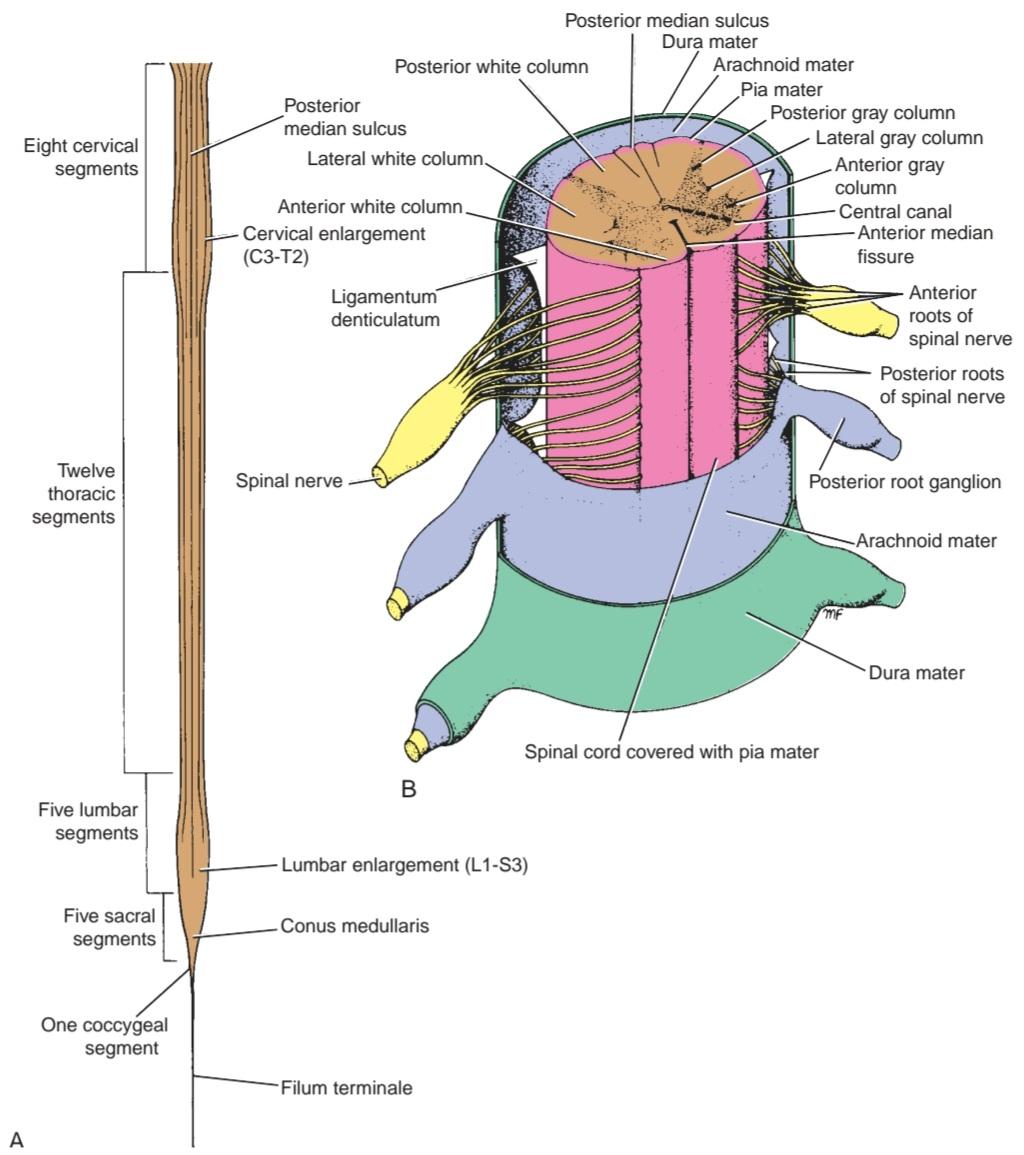
Figure 4-5 Spinal cord. A: Posterior view, showing cervical and lumbar enlargements. B: Three segments of the spinal cord showing the coverings of dura mater, arachnoid mater, and pia mater.
شکل ۴-۵ نخاع. الف: نمای خلفی، بزرگ شدنهای گردنی و کمری را نشان میدهد. ب: سه بخش از نخاع که پوششهای سختشامه، عنکبوتیه و نرمشامه را نشان میدهد.
Along the entire length of the spinal cord, 31 pairs of spinal nerves are attached by the anterior or motor roots and the posterior or sensory roots (Fig. 4-5B). Each root is attached to the cord by a series of rootlets, which extend the whole length of the corresponding segment of the cord. Each posterior nerve root possesses a posterior root ganglion, the cells of which give rise to peripheral and central nerve fibers.
در تمام طول نخاع، ۳۱ جفت عصب نخاعی توسط ریشههای قدامی یا حرکتی و ریشههای خلفی یا حسی به نخاع متصل شدهاند (شکل ۴-۵B). هر ریشه توسط مجموعهای از ریشههای کوچک به نخاع متصل میشود که در تمام طول قطعه مربوطه از نخاع امتداد دارند. هر ریشه عصبی خلفی دارای یک گانگلیون ریشه خلفی است که سلولهای آن باعث ایجاد فیبرهای عصبی محیطی و مرکزی میشوند.
Spinal Cord Structure
The spinal cord is composed of an inner core of gray matter, which is surrounded by an outer covering of white matter (Fig. 4-6); there is no indication that the cord is segmented.
ساختار نخاع
نخاع از یک هسته داخلی از ماده خاکستری تشکیل شده است که توسط یک پوشش خارجی از ماده سفید احاطه شده است (شکل 4-6)؛ هیچ نشانهای مبنی بر قطعه قطعه بودن نخاع وجود ندارد.

Figure 4-6 Transverse sections of the spinal cord at different levels showing the arrangement of the gray matter and white matter.
شکل ۴-۶ برشهای عرضی نخاع در سطوح مختلف که نحوه قرارگیری ماده خاکستری و ماده سفید را نشان میدهد.
For a comparison of the structural details in different regions of the spinal cord, see Table 4-1.
برای مقایسه جزئیات ساختاری در نواحی مختلف نخاع، به جدول 4-1 مراجعه کنید.
Table 4-1 Comparison of Structural Details in Different Regions of the Spinal Cord a
جدول۴-۱ مقایسه جزئیات ساختاری در نواحی مختلف نخاع a

a The information in this table is useful for identifying the specific level of the spinal cord from which a section has been taken.
a اطلاعات موجود در این جدول برای شناسایی سطح خاصی از نخاع که از آن برش گرفته شده است، مفید است.
Gray Matter
On cross section, the gray matter is seen as an H-shaped pillar with anterior and posterior gray columns, or horns, united by a thin gray commissure containing the small central canal. A small lateral gray column or horn is present in the thoracic and upper lumbar segments of the cord. The amount of gray matter present at any given level of the spinal cord is related to the amount of muscle innervated at that level. Thus, its size is greatest within the cervical and lumbosacral enlargements of the cord, which innervate the muscles of the upper and lower limbs, respectively (Figs. 4-7 to 4-10; also see Fig. 4-6).
ماده خاکستری
در برش عرضی، ماده خاکستری به صورت یک ستون H شکل با ستونها یا شاخهای خاکستری قدامی و خلفی دیده میشود که توسط یک رابط خاکستری نازک حاوی کانال مرکزی کوچک به هم متصل شدهاند. یک ستون یا شاخ خاکستری جانبی کوچک در بخشهای سینهای و کمری فوقانی نخاع وجود دارد. مقدار ماده خاکستری موجود در هر سطح معین از نخاع به مقدار عضلهای که در آن سطح عصبدهی میشود، مربوط میشود. بنابراین، اندازه آن در بزرگشدگیهای گردنی و کمری-خاجی نخاع که به ترتیب عضلات اندام فوقانی و تحتانی را عصبدهی میکنند، بیشترین مقدار را دارد (شکلهای 4-7 تا 4-10؛ همچنین به شکل 4-6 مراجعه کنید).
Structure
As in other CNS regions, spinal cord gray matter consists of a mixture of nerve cells and their processes, neuroglia, and blood vessels. The nerve cells are multipolar, and the neuroglia forms an intricate network around the nerve cell bodies and their neurites.
ساختار
مانند سایر نواحی سیستم عصبی مرکزی، ماده خاکستری نخاع از ترکیبی از سلولهای عصبی و زوائد آنها، نوروگلیا و رگهای خونی تشکیل شده است. سلولهای عصبی چند قطبی هستند و نوروگلیا یک شبکه پیچیده در اطراف اجسام سلولهای عصبی و نوریتهای آنها تشکیل میدهد.
ANTERIOR GRAY COLUMN NERVE CELL GROUPS
Most nerve cells are large and multipolar, and their axons pass out in the anterior roots of the spinal nerves as a efferents, which innervate skeletal muscles. The smaller nerve cells are also multipolar, and the axons of many of these pass out in the anterior roots of the spinal nerves as y efferents, which innervate the intrafusal muscle fibers of neuromuscular spindles.
گروههای سلولهای عصبی ستون خاکستری قدامی
بیشتر سلولهای عصبی بزرگ و چند قطبی هستند و آکسونهای آنها در ریشههای قدامی اعصاب نخاعی به صورت یک رشته وابران خارج میشوند که عضلات اسکلتی را عصبدهی میکنند. سلولهای عصبی کوچکتر نیز چند قطبی هستند و آکسونهای بسیاری از آنها در ریشههای قدامی اعصاب نخاعی به صورت یک رشته وابران خارج میشوند که فیبرهای عضلانی داخل دوکی دوکهای عصبی-عضلانی را عصبدهی میکنند.
For practical purposes, the nerve cells of the anterior gray column can be divided into three basic groups or columns: medial, central, and lateral (Fig. 4-6).
برای اهداف عملی، سلولهای عصبی ستون خاکستری قدامی را میتوان به سه گروه یا ستون اصلی تقسیم کرد: داخلی، مرکزی و جانبی (شکل ۴-۶).
The medial group is present in most segments of the spinal cord and is responsible for innervating the skeletal muscles of the neck and trunk, including the intercostal and abdominal musculature.
گروه داخلی در بیشتر بخشهای نخاع وجود دارد و مسئول عصبدهی عضلات اسکلتی گردن و تنه، از جمله عضلات بین دندهای و شکمی است.
The central group is the smallest and is present in some cervical and lumbosacral segments. In the cervical part of the cord, some of these nerve cells (segments C3-C5) specifically innervate the diaphragm and are collectively referred to as the phrenic nucleus (Fig. 4-7). In the upper five or six cervical segments, some of the nerve cells innervate the sternocleidomastoid and trapezius muscles and are referred to as the accessory nucleus. The axons of these cells form the spinal part of the accessory nerve. The lumbosacral nucleus present in the second lumbar down to the first sacral segment of the cord is made up of nerve cells whose axons have an unknown distribution.
گروه مرکزی کوچکترین گروه است و در برخی از بخشهای گردنی و کمری-خاجی وجود دارد. در بخش گردنی نخاع، برخی از این سلولهای عصبی (بخشهای C3-C5) به طور خاص دیافراگم را عصبدهی میکنند و در مجموع به عنوان هسته فرنیک شناخته میشوند (شکل ۴-۷). در پنج یا شش بخش فوقانی گردن، برخی از سلولهای عصبی عضلات جناغی-چانهای و ذوزنقهای را عصبدهی میکنند و به عنوان هسته فرعی شناخته میشوند. آکسونهای این سلولها بخش نخاعی عصب فرعی را تشکیل میدهند. هسته کمری-خاجی موجود در دومین بخش کمری تا اولین بخش خاجی نخاع، از سلولهای عصبی تشکیل شده است که آکسونهای آنها توزیع ناشناختهای دارند.
The lateral group is present in the cervical and lumbosacral segments of the cord and is responsible for innervating the skeletal muscles of the limbs (Figs. 4-6, 47, 4-9, and 4-10).
گروه جانبی در بخشهای گردنی و کمری-خاجی نخاع وجود دارد و مسئول عصبدهی عضلات اسکلتی اندامها است (شکلهای ۴-۶، ۴۷، ۴-۹ و ۴-۱۰).

Figure 4-7 Transverse section of the spinal cord at the level of the fifth cervical segment. (Weigert stain.)
شکل ۴-۷ برش عرضی نخاع در سطح پنجمین قطعه گردنی. (رنگآمیزی وایگرت.)
POSTERIOR GRAY COLUMN NERVE CELL GROUPS
Two of the four nerve cell groups of the posterior gray column extend throughout the length of the cord; the other two are restricted to the thoracic and lumbar segments.
گروههای سلولهای عصبی ستون خاکستری خلفی
دو گروه از چهار گروه سلولهای عصبی ستون خاکستری خلفی در سراسر طول نخاع امتداد دارند؛ دو گروه دیگر به بخشهای سینهای و کمری محدود میشوند.
The substantia gelatinosa group is situated at the apex of the posterior gray column throughout the length of the spinal cord (Figs. 4-6 to 4-10). It is largely Posterior gray horn.
گروه ماده ژلاتینی در راس ستون خاکستری خلفی در سراسر طول نخاع قرار دارد (شکلهای ۴-۶ تا ۴-۱۰). این بخش عمدتاً شاخ خاکستری خلفی است.
composed of Golgi type Il neurons and receives afferent fibers concerned with pain, temperature, and touch from the posterior root. Furthermore, it receives input from descending fibers from supraspinal levels. Pain and temperature inputs are thought to be modified by excitatory or inhibitory information from other sensory inputs and by information from the cerebral cortex.
از نورونهای گلژی نوع II تشکیل شده و فیبرهای آوران مربوط به درد، دما و لمس را از ریشه خلفی دریافت میکند. علاوه بر این، از فیبرهای نزولی از سطوح فوق نخاعی ورودی دریافت میکند. تصور میشود ورودیهای درد و دما توسط اطلاعات تحریکی یا مهاری از سایر ورودیهای حسی و توسط اطلاعات قشر مغز اصلاح میشوند.
The nucleus proprius is a group of large nerve cells situated anterior to the substantia gelatinosa throughout the spinal cord (Figs. 4-6 to 4-10). This nucleus constitutes the main bulk of cells present in the posterior gray column and receives fibers from the posterior white column that are associated with the senses of position and movement (proprioception), two-point discrimination, and vibration.
هسته پروپریوس گروهی از سلولهای عصبی بزرگ است که در جلوی ماده ژلاتینی در سراسر نخاع قرار دارند (شکلهای ۴-۶ تا ۴-۱۰). این هسته بخش عمده سلولهای موجود در ستون خاکستری خلفی را تشکیل میدهد و فیبرهایی را از ستون سفید خلفی دریافت میکند که با حس موقعیت و حرکت (حس عمقی)، تمایز دو نقطهای و ارتعاش مرتبط هستند.

Figure 4-8 Transverse section of the spinal cord at the level of the second thoracic segment. (Weigert stain.)
شکل ۴-۸ برش عرضی نخاع در سطح دومین قطعه سینهای. (رنگآمیزی وایگرت.)
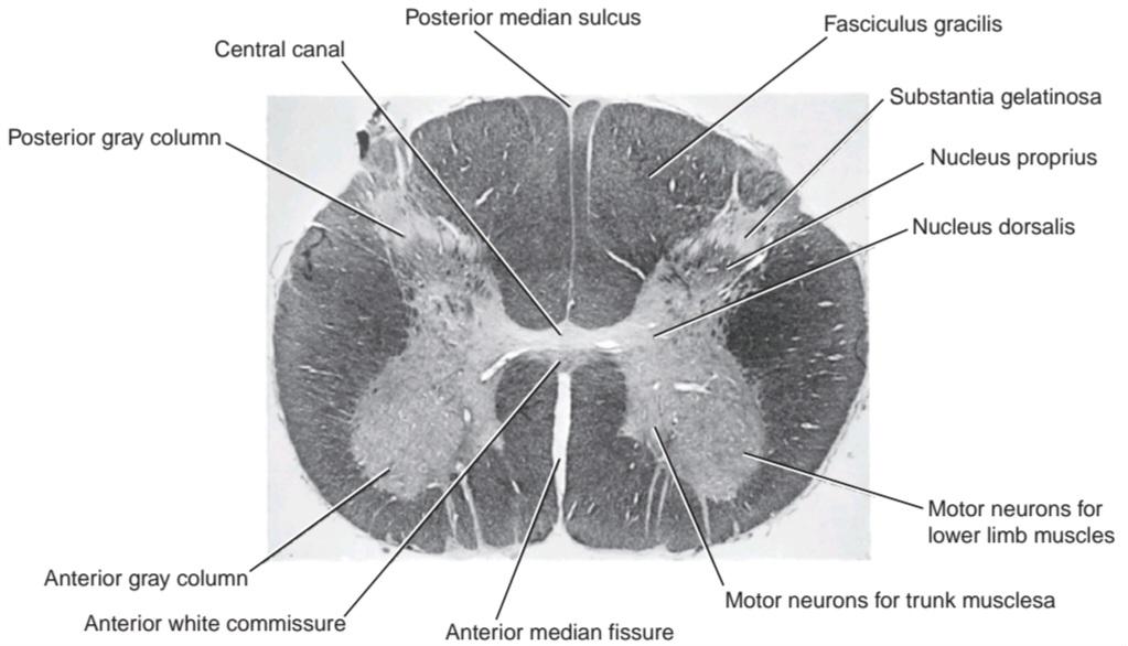
Figure 4-9 Transverse section of the spinal cord at the level of the fourth lumbar segment. (Weigert stain.)
شکل ۴-۹ برش عرضی نخاع در سطح قطعه چهارم کمری. (رنگآمیزی وایگرت.)
The nucleus dorsalis (Clarke column) is a group of nerve cells situated at the base of the posterior gray column and extending from the eighth cervical segment caudally to the third or fourth lumbar segment (Figs. 4-6 to 4-9). Most of the cells are comparatively large and are associated with proprioceptive endings (neuromuscular spindles and tendon spindles).
هسته پشتی (ستون کلارک) گروهی از سلولهای عصبی است که در پایه ستون خاکستری خلفی قرار دارد و از هشتمین قطعه گردنی به سمت خلف تا سومین یا چهارمین قطعه کمری امتداد مییابد (شکلهای ۴-۶ تا ۴-۹). اکثر سلولها نسبتاً بزرگ هستند و با انتهای حس عمقی (دوکهای عصبی-عضلانی و دوکهای تاندونی) مرتبط هستند.
The visceral afferent nucleus is a group of nerve cells of medium size situated lateral to the nucleus dorsalis; it extends from the first thoracic to the third lumbar segment of the spinal cord. It is believed to be associated with receiving visceral afferent information.
هسته آوران احشایی گروهی از سلولهای عصبی با اندازه متوسط است که در سمت جانبی هسته پشتی قرار دارد. این هسته از اولین قطعه سینهای تا سومین قطعه کمری نخاع امتداد دارد. اعتقاد بر این است که با دریافت اطلاعات آوران احشایی مرتبط است.
LATERAL GRAY COLUMN NERVE CELL GROUPS
The intermediolateral group of cells form the small lateral gray column, which extends from the first thoracic to the second or third lumbar segment of the spinal cord (Figs. 4-6 and 4-8). The cells are relatively small and give rise to preganglionic sympathetic fibers.
گروههای سلولهای عصبی ستون خاکستری جانبی
گروه سلولهای بین میانی-جانبی، ستون خاکستری جانبی کوچک را تشکیل میدهند که از اولین قطعه سینهای تا دومین یا سومین قطعه کمری نخاع امتداد دارد (شکلهای ۴-۶ و ۴-۸). این سلولها نسبتاً کوچک هستند و باعث ایجاد فیبرهای سمپاتیک پیش عقدهای میشوند.
A similar group of cells found in the second, third, and fourth sacral segments of the spinal cord give rise to preganglionic parasympathetic fibers (Figs. 46 and 4-10).
گروه مشابهی از سلولها که در بخشهای دوم، سوم و چهارم خاجی نخاع یافت میشوند، باعث ایجاد فیبرهای پاراسمپاتیک پیش عقدهای میشوند (شکلهای ۴۶ و ۴-۱۰).
GRAY COMMISSURE AND CENTRAL CANAL
In transverse sections of the spinal cord, the anterior and posterior gray columns on each side are connected by a transverse gray commissure; the gray matter resembles the letter H (Figs. 4-6 to 4-10). The central canal is situated in the center of the gray commissure. The part of the gray commissure that is situated posterior to the central canal is the posterior gray commissure; the anterior part is the anterior gray commissure.
رابط خاکستری و کانال مرکزی
در مقاطع عرضی نخاع، ستونهای خاکستری قدامی و خلفی در هر طرف توسط یک رابط خاکستری عرضی به هم متصل میشوند؛ ماده خاکستری شبیه حرف H است (شکلهای ۴-۶ تا ۴-۱۰). کانال مرکزی در مرکز رابط خاکستری قرار دارد. بخشی از رابط خاکستری که در خلف کانال مرکزی قرار دارد، رابط خاکستری خلفی است؛ بخش قدامی، رابط خاکستری قدامی است.
The central canal is present throughout the spinal cord. Superiorly, it is continuous with the central canal of the caudal half of the medulla oblongata; above this, it opens into the cavity of the fourth ventricle. Inferiorly in the conus medullaris, it expands into the fusiform terminal ventricle and terminates below within the root of the filum terminale. It is filled with CSF and is lined with ciliated columnar epithelium, the ependyma.
کانال مرکزی در سراسر نخاع وجود دارد. از بالا، با کانال مرکزی نیمه خلفی بصل النخاع در امتداد است؛ از بالا به حفره بطن چهارم باز میشود. از پایین در مخروط انتهایی، به بطن انتهایی دوکیشکل گسترش مییابد و در پایین در ریشه فیلوم ترمینال خاتمه مییابد. این کانال با مایع مغزی نخاعی پر شده و با اپیتلیوم ستونی مژکدار، اپاندیم، پوشیده شده است.
Thus, the central canal is closed inferiorly and opens superiorly into the fourth ventricle.
بنابراین، کانال مرکزی از پایین بسته و از بالا به بطن چهارم باز میشود.

Figure 4-10 Transverse section of the spinal cord at the level of the second sacral segment. (Weigert stain.)
شکل ۴-۱۰ برش عرضی نخاع در سطح دومین قطعه خاجی. (رنگآمیزی وایگرت.)
White Matter
The white matter, for purposes of description, may be divided into anterior, lateral, and posterior white columns or funiculi (Figs. 4-5 to 4-10). The anterior column on each side lies between the midline and the point of emergence of the anterior nerve roots; the lateral column lies between the emergence of the anterior nerve roots and the entry of the posterior nerve roots; the posterior column lies between the entry of the posterior nerve roots and the midline.
ماده سفید
ماده سفید، برای توصیف، ممکن است به ستونهای سفید یا طنابهای عصبی قدامی، جانبی و خلفی تقسیم شود (شکلهای ۴-۵ تا ۴-۱۰). ستون قدامی در هر طرف بین خط میانی و نقطه خروج ریشههای عصبی قدامی قرار دارد؛ ستون جانبی بین خروج ریشههای عصبی قدامی و محل ورود ریشههای عصبی خلفی قرار دارد؛ ستون خلفی بین محل ورود ریشههای عصبی خلفی و خط میانی قرار دارد.
Structure
As in other CNS regions, the spinal cord white matter consists of a mixture of nerve fibers, neuroglia, and blood vessels. It surrounds the gray matter, and its white color is due to the high proportion of myelinated nerve fibers.
ساختار
همانند سایر نواحی سیستم عصبی مرکزی، ماده سفید نخاع از ترکیبی از رشتههای عصبی، نوروگلیا و رگهای خونی تشکیل شده است. این ماده، ماده خاکستری را احاطه کرده و رنگ سفید آن به دلیل نسبت بالای رشتههای عصبی میلیندار است.
NERVE FIBER TRACT ARRANGEMENT
The arrangement of the nerve fiber tracts within the spinal cord has been deduced as the result of animal experimentation and study of the human spinal cord for degenerative nerve fibers resulting from injury or disease. Although some nerve tracts are concentrated in certain areas of the white matter, considerable over- lap is present. For purposes of description, the spinal tracts are divided into ascending, descending, and intersegmental tracts, and their relative positions in the white matter are described below. A simplified diagram, showing the general arrangement of the major tracts, is shown in Figure 4-11.
ترتیب قرارگیری رشتههای عصبی
ترتیب قرارگیری رشتههای عصبی در نخاع در نتیجه آزمایشهای حیوانی و مطالعه نخاع انسان برای رشتههای عصبی دژنراتیو ناشی از آسیب یا بیماری استنباط شده است. اگرچه برخی از رشتههای عصبی در نواحی خاصی از ماده سفید متمرکز شدهاند، اما همپوشانی قابل توجهی وجود دارد. برای اهداف توصیفی، رشتههای نخاعی به رشتههای صعودی، نزولی و بین قطعهای تقسیم میشوند و موقعیت نسبی آنها در ماده سفید در زیر شرح داده شده است. یک نمودار ساده که ترتیب کلی رشتههای اصلی را نشان میدهد، در شکل 4-11 نشان داده شده است.

Figure 4-11 Transverse section of the spinal cord at the midcervical level showing the general arrangement of the ascending tracts on the right and the descending tracts on the left.
شکل ۴-۱۱ برش عرضی نخاع در سطح میانی گردن که ترتیب کلی مسیرهای صعودی در سمت راست و مسیرهای نزولی در سمت چپ را نشان میدهد.
ASCENDING TRACTS
On entering the spinal cord, the sensory nerve fibers of different sizes and functions are sorted out and segregated into nerve bundles or tracts in the white matter (Fig. 4-12; also see Fig. 4-11). Some of the nerve fibers serve to link different segments of the spinal cord, while others ascend from the spinal cord to higher centers and thus connect the spinal cord with the brain. These bundles of the ascending fibers are referred to as the ascending tracts.
مسیرهای صعودی
با ورود به نخاع، فیبرهای عصبی حسی با اندازهها و عملکردهای مختلف، دستهبندی شده و در ماده سفید به دستههای عصبی یا مسیرها تقسیم میشوند (شکل ۴-۱۲؛ همچنین به شکل ۴-۱۱ مراجعه کنید). برخی از فیبرهای عصبی برای اتصال بخشهای مختلف نخاع عمل میکنند، در حالی که برخی دیگر از نخاع به مراکز بالاتر صعود میکنند و بنابراین نخاع را به مغز متصل میکنند. این دستههای فیبرهای صعودی، مسیرهای صعودی نامیده میشوند.
The ascending tracts conduct two types of afferent information, which may or may not reach consciousness. Exteroceptive information originates from outside the body, such as pain, temperature, and touch. Proprioceptive information originates from inside the body, for example, from muscles and joints.
مسیرهای صعودی دو نوع اطلاعات آوران را هدایت میکنند که ممکن است به هوشیاری برسند یا نرسند. اطلاعات حس بیرونی از خارج از بدن سرچشمه میگیرند، مانند درد، دما و لمس. اطلاعات حس عمقی از داخل بدن، به عنوان مثال، از عضلات و مفاصل سرچشمه میگیرند.

Figure 4-12 Simplest form of the ascending sensory pathway from the sensory nerve ending to the cerebral cortex. Note the three neurons involved.
شکل ۴-۱۲ سادهترین شکل مسیر حسی صعودی از انتهای عصب حسی به قشر مغز. به سه نورون درگیر توجه کنید.
Anatomical Organization
General information from the peripheral sensory endings is conducted through the nervous system by a series of neurons. In its simplest form, the ascending pathway to consciousness consists of three neurons (Fig. 4-12). The first neuron, the first-order neuron, has its cell body in the posterior root ganglion of the spinal nerve. A peripheral process connects with a sensory receptor ending, whereas a central process enters the spinal cord through the posterior root to synapse on the second-order neuron. The second-order neuron gives rise to an axon that decussates (crosses to the opposite side) and ascends to a higher level of the CNS, where it synapses with the third-order neuron. The third-order neuron is usually in the thalamus and gives rise to a projection fiber that passes to a sensory region of the cerebral cortex. This three-neuron chain is the most common arrangement, but some afferent pathways use more or fewer neurons. Many of the neurons in the ascending pathways branch and give a major input into the reticular formation, which, in turn, activates the cerebral cortex, maintaining wakefulness. Other branches pass to motor neurons and participate in reflex muscular activity.
سازمان آناتومیک
اطلاعات عمومی از انتهای حسی محیطی توسط مجموعهای از نورونها از طریق سیستم عصبی منتقل میشود. در سادهترین شکل، مسیر صعودی به سمت هوشیاری شامل سه نورون است (شکل ۴-۱۲). جسم سلولی نورون اول، نورون درجه یک، در گانگلیون ریشه خلفی عصب نخاعی قرار دارد. یک زائده محیطی با انتهای گیرنده حسی متصل میشود، در حالی که یک زائده مرکزی از طریق ریشه خلفی وارد نخاع میشود تا روی نورون درجه دو سیناپس کند. نورون درجه دو باعث ایجاد آکسونی میشود که به صورت متقاطع (از طرف مقابل عبور میکند) حرکت میکند و به سطح بالاتری از سیستم عصبی مرکزی صعود میکند، جایی که با نورون درجه سه سیناپس میکند. نورون درجه سه معمولاً در تالاموس است و باعث ایجاد یک فیبر خروجی میشود که به ناحیه حسی قشر مغز میرود. این زنجیره سه نورونی رایجترین آرایش است، اما برخی از مسیرهای آوران از نورونهای کمتر یا بیشتری استفاده میکنند. بسیاری از نورونهای مسیرهای صعودی منشعب میشوند و ورودی اصلی را به تشکیلات مشبک میدهند که به نوبه خود قشر مغز را فعال کرده و بیداری را حفظ میکند. شاخههای دیگر به نورونهای حرکتی منتقل میشوند و در فعالیت رفلکسی عضلات شرکت میکنند.
Functions
Painful and thermal sensations ascend in the lateral spinothalamic tract; light (crude) touch and pressure ascend in the anterior spinothalamic tract (Fig. 4-13). Discriminative touch-that is, the ability to localize accurately the area of the body touched and also to be aware that two points are touched simultaneously, even though they are close together (two-point discrimination)-ascends in the posterior white columns. Also ascending in the posterior white columns is information from muscles and joints pertaining to movement and position of different parts of the body. In addition, vibratory sensations ascend in the posterior white column. Unconscious information from muscles, joints, the skin, and subcutaneous tissue reaches the cerebellum by way of the anterior and posterior spinocerebellar tracts and by the cuneocerebellar tract. Pain, thermal, and tactile information are passed to the superior colliculus of the midbrain through the spinotectal tract for the purpose of spinovisual reflexes. The spinoreticular tract provides a pathway from the muscles, joints, and skin to the reticular formation, while the spino-olivary tract provides an indirect pathway for further afferent information to reach the cerebellum.
عملکردها
حسهای دردناک و حرارتی در مسیر نخاعی-تالامیک جانبی بالا میروند؛ لمس سبک (خام) و فشار در مسیر نخاعی-تالامیک قدامی بالا میروند (شکل ۴-۱۳). لمس تشخیصی – یعنی توانایی تعیین دقیق محل ناحیه لمس شده بدن و همچنین آگاهی از اینکه دو نقطه به طور همزمان لمس میشوند، حتی اگر نزدیک به هم باشند (تمایز دو نقطه) – در ستونهای سفید خلفی بالا میرود. همچنین در ستونهای سفید خلفی، اطلاعاتی از عضلات و مفاصل مربوط به حرکت و موقعیت قسمتهای مختلف بدن بالا میرود. علاوه بر این، احساسات ارتعاشی در ستون سفید خلفی بالا میروند. اطلاعات ناخودآگاه از عضلات، مفاصل، پوست و بافت زیر جلدی از طریق مسیرهای نخاعی-مخچهای قدامی و خلفی و از طریق مسیر کونئوسربلار به مخچه میرسند. اطلاعات درد، حرارتی و لمسی از طریق مسیر نخاعی-تالامیک به کولیکولوس فوقانی مغز میانی برای ایجاد رفلکسهای نخاعی-بینایی منتقل میشوند. راه نخاعی-شبکهای مسیری از عضلات، مفاصل و پوست به تشکیلات مشبک فراهم میکند، در حالی که راه نخاعی-زیتونی مسیری غیرمستقیم برای رسیدن اطلاعات آوران بیشتر به مخچه فراهم میکند.

Figure 4-13 Transverse section of the spinal cord showing the origin of the main ascending sensory tracts. Note that the sensations of pain and temperature ascend in the lateral spinothalamic tract, and light touch and pressure ascend in the anterior spinothalamic tract.
شکل ۴-۱۳ برش عرضی نخاع که مبدا مسیرهای حسی صعودی اصلی را نشان میدهد. توجه داشته باشید که حس درد و دما در مسیر نخاعی-تالاموسی جانبی و لمس سبک و فشار در مسیر نخاعی-تالاموسی قدامی بالا میروند.
Pain and Temperature Pathways
The pain and thermal receptors in the skin and other tissues are free nerve endings. The pain impulses are transmitted to the spinal cord in fast-conducting 8 A-type fibers and slow-conducting C-type fibers. The fast-conducting fibers alert the individual to initial sharp pain, and the slow-conducting fibers are responsible for prolonged burning, aching pain. The sensations of heat and cold also travel by 8 A and C fibers.
مسیرهای درد و دما
گیرندههای درد و گرما در پوست و سایر بافتها، پایانههای عصبی آزاد هستند. تکانههای درد از طریق فیبرهای نوع A با هدایت سریع ۸ و فیبرهای نوع C با هدایت آهسته به نخاع منتقل میشوند. فیبرهای با هدایت سریع، فرد را از درد شدید اولیه آگاه میکنند و فیبرهای با هدایت آهسته مسئول درد سوزشی و طولانی مدت هستند. حس گرما و سرما نیز توسط فیبرهای A و C منتقل میشوند.
The main somatosensory pathways are summarized in Table 4-2.
مسیرهای اصلی حسی-پیکری در جدول 4-2 خلاصه شدهاند.
Table 4-2 Main Somatosensory Pathways to Consciousness a
جدول ۴-۲ مسیرهای اصلی حسی-پیکری به هوشیاری a
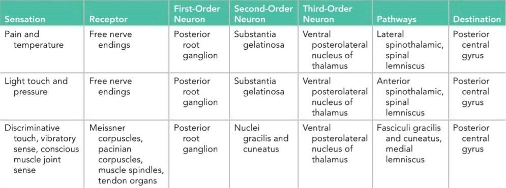
a Note that all ascending pathways send branches to the reticular activating system.
aتوجه داشته باشید که تمام مسیرهای صعودی شاخههایی به سیستم فعالکننده مشبک میفرستند.
Lateral Spinothalamic Tract
Axons entering the spinal cord from the posterior root ganglion proceed to the tip of the posterior gray column and divide into ascending and descending branches (Fig. 4-14). These branches travel for a distance of one or two segments of the spinal cord and form the posterolateral tract of Lissauer. These fibers of the first-order neuron terminate by synapsing with cells in the posterior gray column, including cells in the substantia gelatinosa. Substance P, a peptide, is thought to be the neurotransmitter at these synapses.
راه نخاعی-تالاموسی جانبی
آکسونهایی که از گانگلیون ریشه خلفی وارد نخاع میشوند، به سمت نوک ستون خاکستری خلفی پیش میروند و به شاخههای صعودی و نزولی تقسیم میشوند (شکل ۴-۱۴). این شاخهها مسافتی به اندازه یک یا دو قطعه از نخاع را طی میکنند و راه خلفی-جانبی لیساور را تشکیل میدهند. این فیبرهای نورون درجه اول با سیناپس زدن با سلولهای ستون خاکستری خلفی، از جمله سلولهای جسم ژلاتینی، خاتمه مییابند. تصور میشود ماده P، یک پپتید، انتقالدهنده عصبی در این سیناپسها باشد.
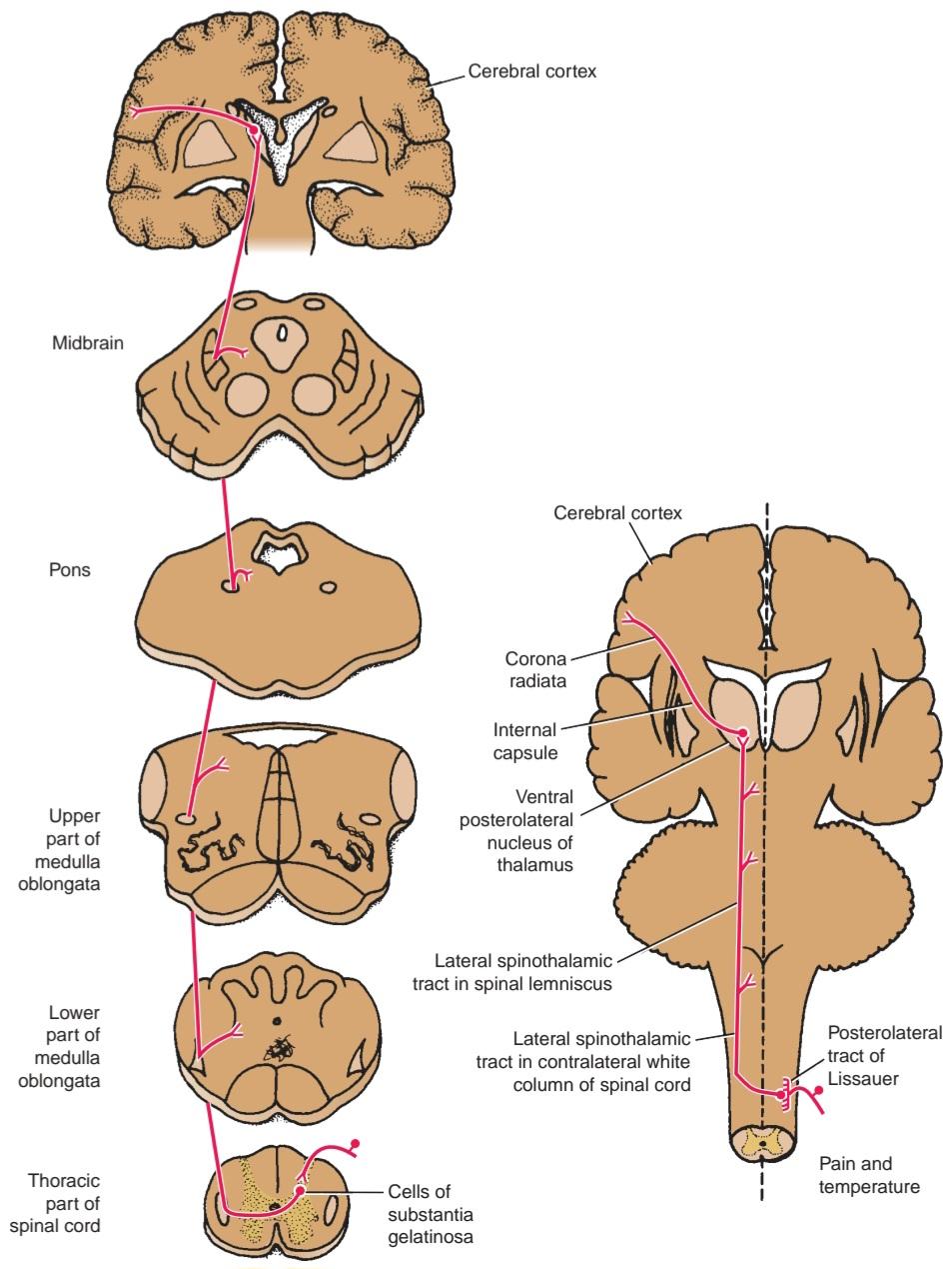
Figure 4-14 Pain and temperature pathways.
شکل ۴-۱۴ مسیرهای درد و دما.
Axons of the second-order neurons now cross obliquely to the opposite side in the anterior gray and white commissures within one spinal segment of the cord, ascending in the contralateral white column as the lateral spinothalamic tract. The lateral spinothalamic tract lies medial to the anterior spinocerebellar tract. As the lateral spinothalamic tract ascends through the spinal cord, new fibers are added to the anteromedial aspect of the tract. Thus, in the upper cervical segments of the cord, the sacral fibers are lateral and the cervical segments are medial. The fibers carrying pain are situated slightly anterior to those conducting temperature.
آکسونهای نورونهای رده دوم اکنون به صورت مایل به سمت مقابل در رابطهای خاکستری و سفید قدامی در یک قطعه نخاعی نخاع، عبور میکنند و در ستون سفید مقابل به عنوان راه نخاعی-تالامیک جانبی صعود میکنند. راه نخاعی-تالامیک جانبی در سمت داخلی راه نخاعی-مخچهای قدامی قرار دارد. با صعود راه نخاعی-تالامیک جانبی از طریق نخاع، الیاف جدیدی به جنبه قدامی-داخلی راه اضافه میشوند. بنابراین، در بخشهای گردنی فوقانی نخاع، الیاف خاجی جانبی و بخشهای گردنی داخلی هستند. الیافی که درد را منتقل میکنند کمی جلوتر از الیافی که دما را منتقل میکنند، قرار دارند.
As the lateral spinothalamic tract ascends through the medulla oblongata, it lies near the lateral surface and between the inferior olivary nucleus and the nucleus of the spinal tract of the trigeminal nerve. It is now accompanied by the anterior spinothalamic tract and the spinotectal tract; together they form the spinal lemniscus.
هنگامی که راه نخاعی-تالامیک جانبی از طریق بصل النخاع صعود میکند، در نزدیکی سطح جانبی و بین هسته زیتونی تحتانی و هسته راه نخاعی عصب سه قلو قرار میگیرد. اکنون با راه نخاعی-تالامیک قدامی و راه نخاعی-تکتال همراه است. آنها با هم لمنیسکوس نخاعی را تشکیل میدهند.
The spinal lemniscus continues to ascend through the posterior part of the pons. In the midbrain, it lies in the tegmentum lateral to the medial lemniscus. Many of the fibers of the lateral spinothalamic tract end by synapsing with the third-order neuron in the ventral posterolateral nucleus of the thalamus. Crude pain and temperature sensations are probably appreciated and emotional reactions initiated here.
لمنیسکوس نخاعی همچنان از قسمت خلفی پل مغزی به سمت بالا میرود. در مغز میانی، در تگمنتوم جانبی لمنیسکوس میانی قرار دارد. بسیاری از فیبرهای راه نخاعی-تالاموسی جانبی با سیناپس با نورون رده سوم در هسته خلفی-جانبی شکمی تالاموس به پایان میرسند. احتمالاً احساسات درد و دما در اینجا درک و واکنشهای عاطفی آغاز میشوند.
Axons of the third-order neurons in the ventral posterolateral nucleus of the thalamus now pass through the posterior limb of the internal capsule and the corona radiata to reach the somesthetic area in the postcentral gyrus of the cerebral cortex. The contralateral half of the body is represented as inverted, with the hand and mouth situated inferiorly and the leg situated superiorly, and with the foot and anogenital region on the medial surface of the hemisphere. (For details, see Chapter 7.) From here, the information is transmitted to other regions of the cerebral cortex to be used by motor areas and the parietal association area. The role of the cerebral cortex is interpreting the quality of the sensory information at the level of consciousness.
آکسونهای نورونهای رده سوم در هسته خلفی-جانبی شکمی تالاموس اکنون از اندام خلفی کپسول داخلی و تاج شعاعی عبور میکنند تا به ناحیه سمستتیک در شکنج پس مرکزی قشر مغز برسند. نیمه مقابل بدن به صورت معکوس نشان داده شده است، به طوری که دست و دهان در پایین و پا در بالا قرار دارند و پا و ناحیه آنوژنیتال در سطح داخلی نیمکره قرار دارند. (برای جزئیات، به فصل ۷ مراجعه کنید.) از اینجا، اطلاعات به سایر نواحی قشر مغز منتقل میشود تا توسط نواحی حرکتی و ناحیهی ارتباط آهیانهای مورد استفاده قرار گیرد. نقش قشر مغز تفسیر کیفیت اطلاعات حسی در سطح هوشیاری است.
Pain Reception
The perception of pain is a complex phenomenon that is influenced by the emotional state and past experiences of the individual. Pain is a sensation that warns of potential injury and alerts the person to avoid or treat it.
پذیرش درد
درک درد پدیدهای پیچیده است که تحت تأثیر وضعیت عاطفی و تجربیات گذشته فرد قرار میگیرد. درد حسی است که فرد را از آسیب احتمالی آگاه میکند و به او هشدار میدهد که از آن اجتناب کند یا آن را درمان کند.
Pain can be divided into two main types: fast pain and slow pain. Fast pain is experienced within about 0.1 second after the pain stimulus is applied; slow pain is felt 1.0 second or later after the stimulation. Fast pain is described by the patient as sharp pain, acute pain, or pricking pain and is the type of pain felt after pricking the finger with a needle. Fast pain is almost confined to the skin.
درد را میتوان به دو نوع اصلی تقسیم کرد: درد سریع و درد آهسته. درد سریع در حدود ۰.۱ ثانیه پس از اعمال محرک درد تجربه میشود؛ درد آهسته ۱.۰ ثانیه یا دیرتر پس از تحریک احساس میشود. درد سریع توسط بیمار به عنوان درد تیز، درد حاد یا درد سوزن سوزن شدن توصیف میشود و نوعی درد است که پس از سوزن زدن انگشت احساس میشود. درد سریع تقریباً محدود به پوست است.
Slow pain is described as burning pain, aching pain, and throbbing pain and is produced on tissue destruction, as for example, in the development of an abscess or in severe arthritis. Slow pain can occur in any tissue of the body.
درد آهسته به عنوان درد سوزشی، درد تیرکشنده و درد ضرباندار توصیف میشود و در اثر تخریب بافت، مثلاً در ایجاد آبسه یا در آرتریت شدید، ایجاد میشود. درد آهسته میتواند در هر بافتی از بدن رخ دهد.
All types of pain reception take place in free nerve endings. Fast pain is experienced by mechanical or thermal types of stimuli, and slow pain may be elicited by mechanical, thermal, and chemical stimuli.
همه انواع دریافت درد در انتهای عصب آزاد انجام میشود. درد سریع توسط انواع محرکهای مکانیکی یا حرارتی تجربه میشود و درد آهسته ممکن است توسط محرکهای مکانیکی، حرارتی و شیمیایی ایجاد شود.
Many chemical substances have been found in extracts from damaged tissue that will excite free nerve endings. These include serotonin; histamine; bradykinin; acids, such as lactic acid; and K+ ions. The threshold for pain endings can be lowered by prostaglandins and substance P, but they cannot stimulate the endings directly by themselves.
بسیاری از مواد شیمیایی در عصارههای بافت آسیبدیده یافت شدهاند که انتهای عصب آزاد را تحریک میکنند. این مواد شامل سروتونین؛ هیستامین؛ برادیکینین؛ اسیدها، مانند اسید لاکتیک؛ و یونهای +K هستند. آستانه درد انتهای عصب میتواند توسط پروستاگلاندینها و ماده P کاهش یابد، اما آنها نمیتوانند انتهای عصب را مستقیماً تحریک کنند.
The individual should be aware of the existence of stimuli that, if allowed to persist, will cause tissue destruction; pain receptors have little or no adaptation.
فرد باید از وجود محرکهایی آگاه باشد که در صورت تداوم، باعث تخریب بافت میشوند. گیرندههای درد سازگاری کمی دارند یا اصلاً سازگاری ندارند.
Pain Conduction to the Central Nervous System
Fast pain travels in peripheral nerves in large diameter A 8 axons at velocities between 6 and 30 msec. Slow pain travels in the small-diameter C fibers at velocities between 0.5 and 2.0 msec. The fast pain impulses reach consciousness first to alert the individual to danger so that a suitable protective response may take place. Slow pain is appreciated later and lasts much longer.
انتقال درد به سیستم عصبی مرکزی
درد سریع در اعصاب محیطی در آکسونهای A8 با قطر بزرگ با سرعت بین ۶ تا ۳۰ میلیثانیه حرکت میکند. درد آهسته در فیبرهای C با قطر کوچک با سرعت بین ۰.۵ تا ۲.۰ میلیثانیه حرکت میکند. تکانههای سریع درد ابتدا به هوشیاری میرسند تا فرد را از خطر آگاه کنند تا یک پاسخ محافظتی مناسب ایجاد شود. درد آهسته دیرتر احساس میشود و مدت زمان بیشتری طول میکشد.
Pain Conduction in the Central Nervous System
Afferent pain fibers enter the spinal cord, for example, in the posterior roots of a spinal nerve and terminate predominantly in the superficial layers of the posterior gray horn. The main excitatory neurotransmitter released by A 8 fibers and C fibers is the amino acid glutamate. Substance P, a neuropeptide, is also released from C fibers. Whereas glutamate is a fast-acting localized neurotransmitter, substance P has a slow release and diffuses widely in the posterior horn and can influence many neurons.
هدایت درد در سیستم عصبی مرکزی
فیبرهای آوران درد، به عنوان مثال، در ریشههای خلفی یک عصب نخاعی وارد نخاع میشوند و عمدتاً در لایههای سطحی شاخ خاکستری خلفی خاتمه مییابند. انتقالدهنده عصبی تحریکی اصلی که توسط فیبرهای A8 و فیبرهای C آزاد میشود، اسید آمینه گلوتامات است. ماده P، یک نوروپپتید، نیز از فیبرهای C آزاد میشود. در حالی که گلوتامات یک انتقالدهنده عصبی موضعی سریع الاثر است، ماده P آزادسازی آهستهای دارد و به طور گسترده در شاخ خلفی منتشر میشود و میتواند بر بسیاری از نورونها تأثیر بگذارد.
The initial sharp, pricking, fast-acting pain fibers stimulate the second-order neurons of the lateral spinothalamic tract. The axons immediately cross to the opposite side of the spinal cord and ascend to the thalamus where they are relayed to the sensory postcentral gyrus. The burning, aching, slow-acting pain fibers also stimulate the second-order neurons of the lateral spinal thalamic tract in the posterior gray horn and ascend with the axons of the fast-acting pain fibers. However, most of the incoming slow fibers to the spinal cord prob- ably take part in additional relays involving several neurons in the posterior horn before ascending in the spinal cord. The repeated arrival of noxious stimuli through C fibers in the posterior gray horn during severe injury results in an increased response of the second-order neurons. This winding up phenomenon is attributed to the release of the neurotransmitter glutamate from C fibers.
فیبرهای درد تیز، سوزنزننده و سریعالاثر اولیه، نورونهای رده دوم دستگاه اسپینوتالامیک جانبی را تحریک میکنند. آکسونها بلافاصله از طرف مقابل نخاع عبور کرده و به تالاموس صعود میکنند و در آنجا به شکنج حسی پسمرکزی منتقل میشوند. فیبرهای درد سوزشی، دردناک و کنداثر نیز نورونهای رده دوم دستگاه تالاموس نخاعی جانبی را در شاخ خاکستری خلفی تحریک کرده و همراه با آکسونهای فیبرهای درد سریعالاثر بالا میروند. با این حال، بیشتر فیبرهای کند ورودی به نخاع احتمالاً قبل از بالا آمدن در نخاع، در رلههای اضافی شامل چندین نورون در شاخ خلفی شرکت میکنند. ورود مکرر محرکهای مضر از طریق فیبرهای C در شاخ خاکستری خلفی در طول آسیب شدید منجر به افزایش پاسخ نورونهای رده دوم میشود. این پدیده پیچشی به آزاد شدن انتقالدهنده عصبی گلوتامات از فیبرهای C نسبت داده میشود.
The fast type of pain is precisely localized. For example, if someone hits a thumb with a hammer, where the injury has occurred is clear. The slow type of pain is only poorly localized. For example, in a patient with osteoarthritis of the hip joint, the individual can only vaguely localize the pain to the hip area and not to the specific site of the disease. This may be explained by the fact that fast pain fibers directly ascend the spinal cord in the lateral spinothalamic tract, whereas the slow pain fibers take part in multiple relays in the posterior gray horn before ascending to higher centers.
نوع سریع درد دقیقاً موضعی است. به عنوان مثال، اگر کسی با چکش به انگشت شست خود ضربه بزند، محل آسیب مشخص است. نوع آهسته درد، فقط به طور ضعیفی موضعی است. به عنوان مثال، در بیماری که مبتلا به آرتروز مفصل ران است، فرد فقط میتواند به طور مبهم درد را در ناحیه ران و نه در محل خاص بیماری، موضعی کند. این امر را میتوان با این واقعیت توضیح داد که فیبرهای درد سریع مستقیماً از نخاع در مسیر اسپینوتالامیک جانبی بالا میروند، در حالی که فیبرهای درد آهسته قبل از صعود به مراکز بالاتر، در چندین رله در شاخ خاکستری خلفی شرکت میکنند.
Other Terminations of the Lateral Spinothalamic Tract
As is now generally agreed, fast pain impulses travel directly up to the ventral posterolateral nucleus of the thalamus and are then relayed to the cerebral cortex.
سایر پایانههای دستگاه اسپینوتالامیک جانبی
همانطور که اکنون به طور کلی پذیرفته شده است، تکانههای سریع درد مستقیماً به هسته خلفی-جانبی شکمی تالاموس میروند و سپس به قشر مغز منتقل میشوند.
The majority of the slow pain fibers in the lateral spinothalamic tract terminate in the reticular formation, which then activates the entire nervous system. In the lower areas of the brain, the individual becomes aware of the chronic, nauseous, suffering type of pain.
اکثر فیبرهای درد آهسته در مسیر اسپینوتالامیک جانبی به تشکیلات مشبک ختم میشوند که سپس کل سیستم عصبی را فعال میکند. در نواحی پایینتر مغز، فرد از نوع درد مزمن، تهوعآور و رنجآور آگاه میشود.
As the result of research using the positron emission tomography scan, the postcentral gyrus, the cingulate gyrus of the limbic system, and the insular gyrus are sites concerned with the reception and interpretation of nociceptor information. The postcentral gyrus is responsible for the interpretation of pain in relation to past experiences. The cingulate gyrus is involved with the interpretation of the emotional aspect of pain, whereas the insular gyrus is concerned with the interpretation of pain stimuli from the internal organs of the body and brings about an autonomic response.
در نتیجه تحقیقات با استفاده از اسکن توموگرافی انتشار پوزیترون، شکنج پس مرکزی، شکنج سینگولیت سیستم لیمبیک و شکنج اینسولار مکانهایی هستند که با دریافت و تفسیر اطلاعات گیرنده درد مرتبط هستند. شکنج پس مرکزی مسئول تفسیر درد در رابطه با تجربیات گذشته است. شکنج سینگولیت در تفسیر جنبه عاطفی درد نقش دارد، در حالی که شکنج اینسولار با تفسیر محرکهای درد از اندامهای داخلی بدن مرتبط است و یک پاسخ خودکار ایجاد میکند.
Reception of pain information by the CNS is modulated first in the posterior gray horns of the spinal cord and at other sites at higher levels.
دریافت اطلاعات درد توسط سیستم عصبی مرکزی ابتدا در شاخهای خاکستری خلفی نخاع و در سایر مکانها در سطوح بالاتر تعدیل میشود.
Pain Control in the Central Nervous System
Massage and the application of liniments to painful areas in the body can relieve pain. The technique of acupuncture, which was discovered several thousand years ago in China, is also beneficial in relieving pain. Low-frequency electrical stimulation of the skin also relieves pain in certain cases.
کنترل درد در سیستم عصبی مرکزی
ماساژ و استفاده از پمادهای موضعی در نواحی دردناک بدن میتواند درد را تسکین دهد. تکنیک طب سوزنی که چند هزار سال پیش در چین کشف شد، نیز در تسکین درد مفید است. تحریک الکتریکی با فرکانس پایین پوست نیز در موارد خاص درد را تسکین میدهد.
Gating Theory
Although the precise mechanism for the phenomena described above is not understood, the gating theory suggested that, at the site where the pain fiber enters the CNS, inhibition could occur by means of connector neurons excited by large, myelinated afferent fibers carrying information of nonpainful touch and pressure. The excess tactile stimulation produced by massage, for example, “closed the gate” for pain. Once the nonpainful tactile stimulation ceased, however, “the gate was opened,” and information on the painful stimuli ascended the lateral spinothalamic tract. Although the gate theory may partially explain the phenomena, the analgesia system is probably involved with the liberation of enkephalins and endorphins in the posterior gray columns.
نظریه دروازهای
اگرچه مکانیسم دقیق پدیدههای شرح داده شده در بالا مشخص نیست، اما نظریه دروازهای پیشنهاد میکند که در محلی که فیبر درد وارد سیستم عصبی مرکزی میشود، مهار میتواند توسط نورونهای رابط که توسط فیبرهای آوران بزرگ و میلیندار حامل اطلاعات لمس و فشار غیردردناک تحریک میشوند، رخ دهد. به عنوان مثال، تحریک لمسی اضافی تولید شده توسط ماساژ، “دروازه درد را میبندد”. با این حال، هنگامی که تحریک لمسی غیردردناک متوقف شد، “دروازه باز شد” و اطلاعات مربوط به محرکهای دردناک از دستگاه اسپینوتالامیک جانبی بالا رفت. اگرچه نظریه دروازه ممکن است تا حدی این پدیدهها را توضیح دهد، اما سیستم ضد درد احتمالاً در آزادسازی انکفالینها و اندورفینها در ستونهای خاکستری خلفی نقش دارد.
Analgesia System
Stimulation of certain areas of the brainstem can reduce or block sensations of pain. These areas include the periventricular area of the diencephalon, the periaqueductal gray matter of the midbrain, and midline nuclei of the brainstem. Fibers of the reticulospinal tract are believed to pass down to the spinal cord and synapse on cells concerned with pain sensation in the posterior gray column. The analgesic system can suppress both sharp pricking pain and burning pain sensations.
سیستم ضد درد
تحریک نواحی خاصی از ساقه مغز میتواند احساس درد را کاهش یا مسدود کند. این نواحی شامل ناحیه اطراف بطنی دیانسفالون، ماده خاکستری اطراف مجرا در مغز میانی و هستههای خط میانی ساقه مغز است. اعتقاد بر این است که فیبرهای دستگاه رتیکولواسپینال به نخاع منتقل میشوند و روی سلولهایی که در ستون خاکستری خلفی مسئول احساس درد هستند، سیناپس برقرار میکنند. سیستم ضد درد میتواند هم درد تیز و هم درد سوزشی را سرکوب کند.
Recently, two compounds with morphinelike actions, enkephalins and endorphins, have been isolated in the CNS. These compounds and serotonin serve as neurotransmitter substances in the analgesic system of the brain, and they may inhibit the release of substance P in the posterior gray column.
اخیراً، دو ترکیب با اثرات شبه مورفینی، انکفالینها و اندورفینها، در سیستم عصبی مرکزی (CNS) جداسازی شدهاند. این ترکیبات و سروتونین به عنوان مواد انتقالدهنده عصبی در سیستم ضد درد مغز عمل میکنند و ممکن است آزادسازی ماده P را در ستون خاکستری خلفی مهار کنند.
Light (Crude) Touch and Pressure Pathways
These pathwaysare summarized in Table 4-2.
مسیرهای لمس و فشار سبک (خام)
این مسیرها در جدول 4-2 خلاصه شدهاند.
Anterior Spinothalamic Tract
Axons enter the spinal cord from the posterior root ganglion and proceed to the tip of the posterior gray column, where they divide into ascending and descending branches (Fig. 4-15). These branches travel for a distance of one or two segments of the spinal cord, contributing to the posterolateral tract of Lissauer. These fibers of the first-order neuron are believed to terminate by synapsing with cells in the substantia gelatinosa group in the posterior gray column.
راه نخاعی-تالاموسی قدامی
آکسونها از گانگلیون ریشه خلفی وارد نخاع میشوند و به نوک ستون خاکستری خلفی میروند، جایی که به شاخههای صعودی و نزولی تقسیم میشوند (شکل ۴-۱۵). این شاخهها به اندازه یک یا دو بخش از نخاع حرکت میکنند و به راه خلفی-جانبی لیساور میپیوندند. اعتقاد بر این است که این فیبرهای نورون درجه اول با سیناپس با سلولهای گروه ماده ژلاتینی در ستون خاکستری خلفی خاتمه مییابند.
Axons of the second-order neuron now cross very obliquely to the opposite side in the anterior gray and white commissures within several spinal segments and ascend in the opposite anterolateral white column as the anterior spinothalamic tract. As the anterior spinothalamic tract ascends through the spinal cord, new fibers are added to the medial aspect of the tract. Thus, in the upper cervical segments of the cord, the sacral fibers are mostly lateral and the cervical segments are mostly medial.
آکسونهای نورون رده دوم اکنون به صورت بسیار مایل به سمت مقابل در رابطهای خاکستری و سفید قدامی در چندین قطعه نخاعی عبور میکنند و در ستون سفید قدامی-جانبی مقابل به عنوان راه نخاعی-تالاموسی قدامی صعود میکنند. همانطور که راه نخاعی-تالاموسی قدامی از طریق نخاع صعود میکند، الیاف جدیدی به جنبه داخلی راه اضافه میشوند. بنابراین، در بخشهای بالای گردنی نخاع، الیاف خاجی عمدتاً جانبی و بخشهای گردنی عمدتاً داخلی هستند.
As the anterior spinothalamic tract ascends through the medulla oblongata, it accompanies the lateral spinothalamic tract and the spinotectal tract, all of which form the spinal lemniscus.
هنگامی که راه نخاعی-تالاموسی قدامی از طریق بصل النخاع صعود میکند، راه نخاعی-تالاموسی جانبی و راه نخاعی-تکتال را همراهی میکند که همگی لمنیسکوس نخاعی را تشکیل میدهند.
The spinal lemniscus continues to ascend through the posterior part of the pons, and the tegmentum of the midbrain and the fibers of the anterior spinothalamic tract terminate by synapsing with the third-order neuron in the ventral posterolateral nucleus of the thalamus. Crude awareness of touch and pressure is believed to be appreciated here.
لمنیسکوس نخاعی همچنان از طریق قسمت خلفی پل مغزی صعود میکند و تگمنتوم مغز میانی و الیاف راه نخاعی-تالاموسی قدامی با سیناپس زدن با نورون رده سوم در هسته خلفی-جانبی شکمی تالاموس خاتمه مییابند. اعتقاد بر این است که آگاهی خام از لمس و فشار در اینجا مورد قدردانی قرار میگیرد.
Axons of the third-order neurons in the ventral posterolateral nucleus of the thalamus pass through the posterior limb of the internal capsule and the corona radiata to reach the somesthetic area in the postcentral gyrus of the cerebral cortex. The contralateral half of the body is represented inverted, with the hand and mouth situated inferiorly, as described previously. (For details, see Chapter 7.) The conscious appreciation of touch and pressure depends on the activity of the cerebral cortex. The sensations can be only crudely localized, and very little discrimination of intensity is possible.
آکسونهای نورونهای رده سوم در هسته خلفی-جانبی شکمی تالاموس از طریق بازوی خلفی کپسول داخلی و تاج شعاعی عبور میکنند تا به ناحیه حسی-حرکتی در شکنج پسمرکزی قشر مغز برسند. نیمه مقابل بدن به صورت معکوس نمایش داده میشود و دست و دهان در پایین قرار دارند، همانطور که قبلاً توضیح داده شد. (برای جزئیات، به فصل 7 مراجعه کنید.) درک آگاهانه لمس و فشار به فعالیت قشر مغز بستگی دارد. احساسات فقط میتوانند به طور خام محلیسازی شوند و تشخیص شدت بسیار کمی امکانپذیر است.
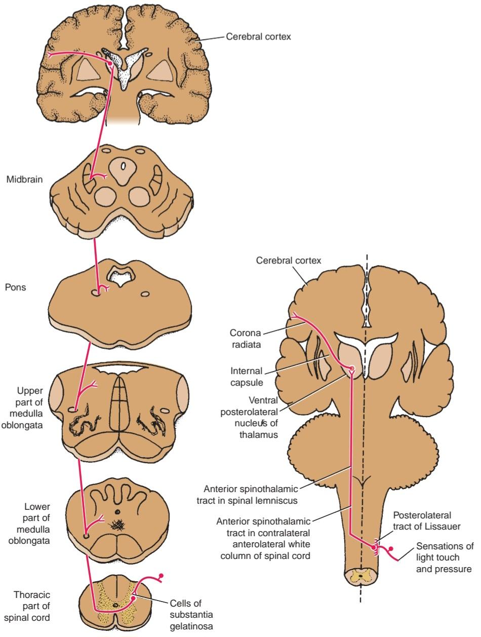
Figure 4-15 Light touch and pressure pathways.
شکل ۴-۱۵ مسیرهای لمس و فشار سبک.
Discriminative Touch, Vibratory Sense, and Conscious Muscle Joint Sense
These pathways are summarized in Table 4-2.
لمس تشخیصی، حس ارتعاشی و حس مشترک عضله آگاهانه
این مسیرها در جدول 4-2 خلاصه شدهاند.
Posterior White Column: Fasciculus Gracilis and Fasciculus Cuneatus
Axons enter the spinal cord from the posterior root ganglion and pass directly to the posterior white column of the same side (Fig. 4-16). Here, the fibers divide into long ascending and short descending branches. The descending branches pass down a variable number of segments, giving off collateral branches that synapse with cells in the posterior gray horn, with internuncial neurons, and with anterior horn cells. These short descending fibers are clearly involved with intersegmental reflexes.
ستون سفید خلفی: فاسیکولوس گراسیلیس و فاسیکولوس کونئاتوس
آکسونها از گانگلیون ریشه خلفی وارد نخاع میشوند و مستقیماً به ستون سفید خلفی همان طرف میروند (شکل ۴-۱۶). در اینجا، فیبرها به شاخههای بلند صعودی و کوتاه نزولی تقسیم میشوند. شاخههای نزولی از تعداد متغیری قطعه عبور میکنند و شاخههای جانبی را ایجاد میکنند که با سلولهای شاخ خاکستری خلفی، با نورونهای بین حلقی و با سلولهای شاخ قدامی سیناپس برقرار میکنند. این فیبرهای کوتاه نزولی به وضوح در رفلکسهای بین قطعهای دخیل هستند.
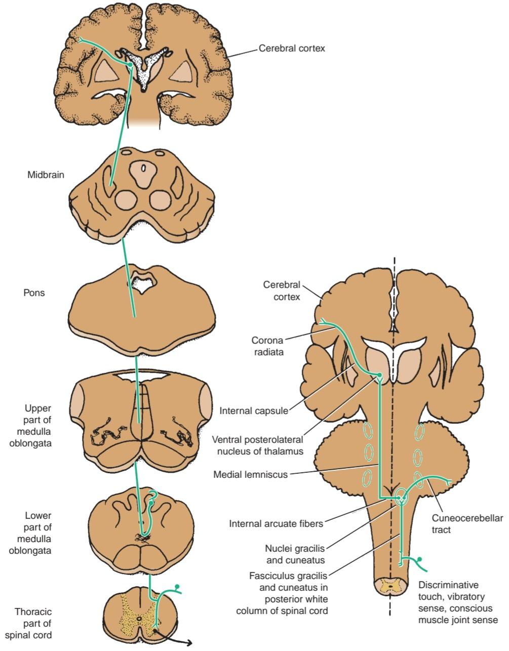
Figure 4-16 Discriminative touch, vibratory sense, and conscious muscle joint sense pathways.
شکل ۴-۱۶ لامسه تشخیصی، حس ارتعاشی و مسیرهای حسی عضله-مفصل آگاهانه.
Long ascending fibers may also end by synapsing with cells in the posterior gray horn, with internuncial neurons, and with anterior horn cells. This distribution may extend over numerous segments of the spinal cord. As in the case of the short descending fibers, they are involved with intersegmental reflexes.
فیبرهای صعودی بلند همچنین ممکن است با سلولهای شاخ خاکستری خلفی، با نورونهای بینحلقهای و با سلولهای شاخ قدامی سیناپس برقرار کنند. این توزیع ممکن است در بخشهای متعددی از نخاع گسترش یابد. همانند فیبرهای نزولی کوتاه، آنها در رفلکسهای بینقطعهای دخیل هستند.
Many of the long ascending fibers travel upward in the posterior white column as the fasciculus gracilis and fasciculus cuneatus. The fasciculus gracilis is present throughout the length of the spinal cord and contains the long ascending fibers from the sacral, lumbar, and lower six thoracic spinal nerves. The fasciculus cuneatus is situated laterally in the upper thoracic and cervical segments of the spinal cord and is separated from the fasciculus gracilis by a septum. The fasciculus cuneatus contains the long ascending fibers from the upper six thoracic and all the cervical spinal nerves.
بسیاری از فیبرهای صعودی بلند در ستون سفید خلفی به عنوان فاسیکولوس گراسیلیس و فاسیکولوس کونئاتوس به سمت بالا حرکت میکنند. فاسیکولوس گراسیلیس در سراسر طول نخاع وجود دارد و شامل فیبرهای صعودی بلند از اعصاب نخاعی خاجی، کمری و شش عصب نخاعی سینهای پایینی است. فاسیکولوس کونئاتوس به صورت جانبی در بخشهای فوقانی سینهای و گردنی نخاع قرار دارد و توسط یک دیواره از فاسیکولوس گراسیلیس جدا میشود. فاسیکولوس کونئاتوس شامل فیبرهای صعودی بلند از شش عصب نخاعی سینهای بالایی و تمام اعصاب نخاعی گردنی است.
Fibers of the fasciculus gracilis and fasciculus cuneatus ascend ipsilaterally and terminate by synapsing on the second-order neurons in the nuclei gracilis and cuneatus of the medulla oblongata. The axons of the second-order neurons, called the internal arcuate fibers, sweep anteromedially around the central gray matter and cross the median plane, decussating with the corresponding fibers of the opposite side in the sensory decussation. The fibers then ascend as a single compact bundle, the medial lemniscus, through the medulla oblongata, the pons, and the midbrain. The fibers terminate by synapsing on the third-order neurons in the ventral posterolateral nucleus of the thalamus.
فیبرهای فاسیکولوس گراسیلیس و فاسیکولوس کونئاتوس به صورت همسو صعود میکنند و با سیناپس روی نورونهای رده دوم در هستههای گراسیلیس و کونئاتوس بصل النخاع خاتمه مییابند. آکسونهای نورونهای رده دوم، که فیبرهای قوسی داخلی نامیده میشوند، به صورت قدامی-داخلی در اطراف ماده خاکستری مرکزی حرکت میکنند و از صفحه میانی عبور میکنند و با فیبرهای مربوطه در سمت مقابل در تقاطع حسی، متقاطع میشوند. سپس فیبرها به صورت یک دسته فشرده واحد، لمنیسکوس داخلی، از طریق بصل النخاع، پل مغزی و مغز میانی صعود میکنند. فیبرها با سیناپس روی نورونهای رده سوم در هسته خلفی-جانبی شکمی تالاموس خاتمه مییابند.
Axons of the third-order neuron leave and pass through the posterior limb of the internal capsule and corona radiata to reach the somesthetic area in the postcentral gyrus of the cerebral cortex. The contralateral half of the body is represented inverted, with the hand and mouth situated inferiorly, as described previously. (For details, see Chapter 7.) In this manner, the impressions of touch with fine gradations of intensity, exact localization, and two-point discrimination can be appreciated. Vibratory sense and the position of the different parts of the body can be consciously recognized.
آکسونهای نورون رده سوم از اندام خلفی کپسول داخلی و تاج شعاعی خارج میشوند و از طریق آن به ناحیه حسی-حرکتی در شکنج پس مرکزی قشر مغز میرسند. نیمهی مقابل بدن به صورت وارونه نمایش داده میشود، به طوری که دست و دهان در پایین قرار دارند، همانطور که قبلاً توضیح داده شد. (برای جزئیات، به فصل ۷ مراجعه کنید.) به این ترتیب، میتوان تأثیرات لمس را با درجهبندیهای ظریف شدت، مکانیابی دقیق و تمایز دو نقطهای درک کرد. حس ارتعاش و موقعیت قسمتهای مختلف بدن را میتوان آگاهانه تشخیص داد.
Many fibers in the fasciculus cuneatus from the cervical and upper thoracic segments, having terminated on the second-order neuron of the nucleus cuneatus, are relayed and travel as the axons of the second-order neurons to enter the cerebellum through the inferior cerebellar peduncle of the same side. This pathway is the cuneocerebellar tract, and the fibers are known as the posterior external arcuate fibers. The function of these fibers is to convey information of muscle joint sense to the cerebellum.
بسیاری از فیبرهای فاسیکولوس کونئاتوس از بخشهای گردنی و سینهای فوقانی، که به نورون درجه دوم هسته کونئاتوس ختم میشوند، رله شده و به عنوان آکسونهای نورونهای درجه دوم از طریق پایه مخچهای تحتانی همان طرف وارد مخچه میشوند. این مسیر، راه کونئوسربلار است و فیبرها به عنوان فیبرهای قوسی خارجی خلفی شناخته میشوند. وظیفه این فیبرها انتقال اطلاعات حس مفصل عضله به مخچه است.
Muscle Joint Sense Pathways to the Cerebellum
Muscle joint sense pathways to the cerebellum are summarized in Table 4-3.
مسیرهای حس عضلات و مفاصل به مخچه
مسیرهای حس عضلات و مفاصل به مخچه در جدول 4-3 خلاصه شده است.
Table 4-3 Muscle Joint Sense Pathways to the Cerebellum
جدول ۴-۳ مسیرهای حس مشترک عضله به مخچه

Posterior Spinocerebellar Tract
Axons entering the spinal cord from the posterior root ganglion enter the posterior gray column and terminate by synapsing on the second-order neurons at the base of the posterior gray column (Fig. 4-17). These neurons are known collectively as the nucleus dorsalis (Clarke column). Axons of the second-order neurons enter the posterolateral part of the lateral white column on the same side and ascend as the posterior spinocerebellar tract to the medulla oblongata. Here, the tract joins the inferior cerebellar peduncle and terminates in the cerebellar cortex. Note that it does not ascend to the cerebral cortex. Because the nucleus dorsalis (Clarke column) extends only from the eighth cervical segment caudally to the third or fourth lumbar segment, axons entering the spinal cord from the posterior roots of the lower lumbar and sacral segments ascend in the posterior white column until they reach the third or fourth lumbar segment, where they enter the nucleus dorsalis.
راه نخاعی-مخچهای خلفی
آکسونهایی که از گانگلیون ریشه خلفی وارد نخاع میشوند، وارد ستون خاکستری خلفی میشوند و با سیناپس زدن روی نورونهای رده دوم در پایه ستون خاکستری خلفی خاتمه مییابند (شکل 4-17). این نورونها در مجموع به عنوان هسته پشتی (ستون کلارک) شناخته میشوند. آکسونهای نورونهای رده دوم از همان طرف وارد قسمت خلفی-جانبی ستون سفید جانبی میشوند و به عنوان راه نخاعی-مخچهای خلفی به بصل النخاع صعود میکنند. در اینجا، این راه به پایه مخچهای تحتانی میپیوندد و در قشر مخچه خاتمه مییابد. توجه داشته باشید که به قشر مغز صعود نمیکند. از آنجا که هسته پشتی (ستون کلارک) فقط از هشتمین قطعه گردنی به صورت خلفی تا قطعه کمری سوم یا چهارم امتداد دارد، آکسونهایی که از ریشههای خلفی قطعات کمری تحتانی و خاجی وارد نخاع میشوند، در ستون سفید خلفی صعود میکنند تا به قطعه کمری سوم یا چهارم برسند، جایی که وارد هسته پشتی میشوند.
The posterior spinocerebellar fibers receive muscle joint information from the muscle spindles, tendon organs, and joint receptors of the trunk and lower limbs. This information concerning tension of muscle tendons and the movements of muscles and joints is used by the cerebellum in the coordination of limb movements and the maintenance of posture.
فیبرهای نخاعی-مخچهای خلفی، اطلاعات مفصل عضلانی را از دوکهای عضلانی، اندامهای تاندونی و گیرندههای مفصلی تنه و اندامهای تحتانی دریافت میکنند. این اطلاعات مربوط به تنش تاندونهای عضلات و حرکات عضلات و مفاصل توسط مخچه در هماهنگی حرکات اندامها و حفظ وضعیت بدن استفاده میشود.
Anterior Spinocerebellar Tract
Axons entering the spinal cord from the posterior root ganglion terminate by synapsing with the second- order neurons in the nucleus dorsalis at the base of the posterior gray column (Fig. 4-17). Most axons of the second-order neurons cross to the opposite side and ascend as the anterior spinocerebellar tract in the contralateral white column; a minority ascend as the anterior spinocerebellar tract in the lateral white column of the same side. The fibers, having ascended through the medulla oblongata and pons, enter the cerebellum through the superior cerebellar peduncle and terminate in the cerebellar cortex. Those fibers that crossed over to the opposite side in the spinal cord are believed to cross back within the cerebellum. The anterior spinocerebellar tract conveys muscle joint information from the muscle spindles, tendon organs, and joint receptors of the trunk and the upper and lower limbs. It is also believed that the cerebellum receives information from the skin and superficial fascia by this tract.
راه نخاعی-مخچهای قدامی
آکسونهایی که از گانگلیون ریشه خلفی وارد نخاع میشوند، با سیناپس با نورونهای رده دوم در هسته پشتی در پایه ستون خاکستری خلفی خاتمه مییابند (شکل 4-17). اکثر آکسونهای نورونهای رده دوم به سمت مقابل عبور میکنند و به عنوان راه نخاعی-مخچهای قدامی در ستون سفید طرف مقابل صعود میکنند؛ تعداد کمی از آنها به عنوان راه نخاعی-مخچهای قدامی در ستون سفید جانبی همان طرف صعود میکنند. فیبرها، پس از صعود از طریق بصل النخاع و پل مغزی، از طریق پایه مخچهای فوقانی وارد مخچه میشوند و در قشر مخچه خاتمه مییابند. اعتقاد بر این است که آن دسته از فیبرهایی که از سمت مقابل در نخاع عبور کردهاند، در داخل مخچه به عقب برمیگردند. راه نخاعی-مخچهای قدامی اطلاعات مفصل عضلانی را از دوکهای عضلانی، اندامهای تاندونی و گیرندههای مفصلی تنه و اندامهای فوقانی و تحتانی منتقل میکند. همچنین اعتقاد بر این است که مخچه از طریق این مسیر، اطلاعات را از پوست و فاسیای سطحی دریافت میکند.
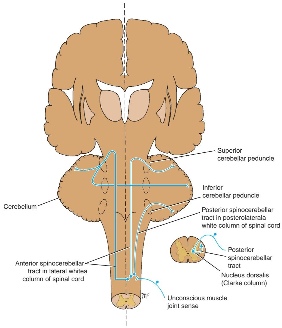
Figure 4-17 Unconscious muscle joint sense pathways to the cerebellum.
شکل ۴-۱۷ مسیرهای حسی ناخودآگاه عضله و مفصل به مخچه.
Cuneocerebellar Tract
These fibers are described on page 142. They originate in the nucleus cuneatus and enter the cerebellum through the inferior cerebellar peduncle of the same side (Fig. 4-16). They are known as the posterior external arcuate fibers, and their function is to convey information of muscle joint sense to the cerebellum.
مسیر کونئوسربلار
این فیبرها در صفحه ۱۴۲ شرح داده شدهاند. آنها از هسته کونئاتوس منشأ میگیرند و از طریق پایه مخچهای تحتانی همان طرف وارد مخچه میشوند (شکل ۴-۱۶). آنها به عنوان فیبرهای قوسی خارجی خلفی شناخته میشوند و عملکرد آنها انتقال اطلاعات حس مفصل عضله به مخچه است.
Other Ascending Pathways
The spinotectal, spinoreticular, and spino-olivary tracts are shown in Figure 4-18.
سایر مسیرهای صعودی
راههای اسپینوتکتال، اسپینورتیکولار و اسپینو-زیسترودی در شکل 4-18 نشان داده شدهاند.

Figure 4-18 Spinotectal, spinoreticular, and spino-olivary tracts.
شکل ۴-۱۸ مسیرهای اسپینوتکتال، اسپینورتیکولار و اسپینو-زیستروده.
Spinotectal Tract
Axons enter the spinal cord from the posterior root ganglion and travel to the gray matter where they synapse on unknown second-order neurons. Axons of the second-order neurons cross the median plane and ascend as the spinotectal tract in the anterolateral white column lying close to the lateral spinothalamic tract. After passing through the medulla oblongata and pons, they terminate by synapsing with neurons in the superior colliculus of the midbrain. This pathway provides afferent information for spinovisual reflexes and brings about movements of the eyes and head toward the source of the stimulation.
مسیر اسپینوتکتال
آکسونها از گانگلیون ریشه خلفی وارد نخاع میشوند و به ماده خاکستری میروند، جایی که با نورونهای ناشناخته رده دوم سیناپس میکنند. آکسونهای نورونهای رده دوم از صفحه میانی عبور میکنند و به عنوان مسیر اسپینوتکتال در ستون سفید قدامی-جانبی که نزدیک به مسیر اسپینوتکتال جانبی قرار دارد، بالا میروند. پس از عبور از بصل النخاع و پل مغزی، با سیناپس با نورونهای موجود در کولیکولوس فوقانی مغز میانی خاتمه مییابند. این مسیر اطلاعات آوران را برای رفلکسهای اسپینو-بینایی فراهم میکند و حرکات چشمها و سر را به سمت منبع تحریک ایجاد میکند.
Spinoreticular Tract
Axons enter the spinal cord from the posterior root ganglion and terminate on unknown second-order neurons in the gray matter. Axons from these second-order neurons ascend the spinal cord as the spinoreticular tract in the lateral white column mixed with the lateral spinothalamic tract. Most of the fibers are uncrossed and terminate by synapsing with neurons of the reticular formation in the medulla oblongata, pons, and midbrain. The spinoreticular tract provides an afferent pathway for the reticular formation, which plays an important role in influencing levels of consciousness. (For details, see p. 204.)
راه نخاعی-شبکهای
آکسونها از گانگلیون ریشه خلفی وارد نخاع میشوند و به نورونهای ناشناخته درجه دو در ماده خاکستری ختم میشوند. آکسونهای این نورونهای درجه دو به عنوان راه نخاعی-شبکهای در ستون سفید جانبی، در ترکیب با راه نخاعی-تالاموسی جانبی، از نخاع بالا میروند. اکثر فیبرها متقاطع نیستند و با سیناپس با نورونهای تشکیلات مشبک در بصل النخاع، پل مغزی و مغز میانی خاتمه مییابند. راه نخاعی-شبکهای یک مسیر آوران برای تشکیلات مشبک فراهم میکند که نقش مهمی در تأثیرگذاری بر سطوح هوشیاری دارد. (برای جزئیات، به صفحه 204 مراجعه کنید.)
Spino-olivary Tract
Axons enter the spinal cord from the posterior root ganglion and terminate on unknown second-order neurons in the posterior gray column. Axons from the second-order neurons cross the midline and ascend as the spinoolivary tract in the white matter at the junction of the anterior and lateral columns. The axons end by synapsing on third-order neurons in the inferior olivary nuclei in the medulla oblongata. Axons of the third-order neurons cross the midline and enter the cerebellum through the inferior cerebellar peduncle. The spino-olivary tract conveys information to the cerebellum from cutaneous and proprioceptive organs.
راه نخاعی-زیستروزی
آکسونها از گانگلیون ریشه خلفی وارد نخاع میشوند و در ستون خاکستری خلفی به نورونهای ناشناخته رده دوم ختم میشوند. آکسونهای نورونهای رده دوم از خط میانی عبور میکنند و به عنوان راه نخاعی-زیستروزی در ماده سفید در محل اتصال ستونهای قدامی و جانبی بالا میروند. آکسونها با سیناپس روی نورونهای رده سوم در هستههای زیتونی تحتانی در بصلالنخاع به پایان میرسند. آکسونهای نورونهای رده سوم از خط میانی عبور میکنند و از طریق پایه مخچه تحتانی وارد مخچه میشوند. راه نخاعی-زیستروزی اطلاعات را از اندامهای پوستی و حس عمقی به مخچه منتقل میکند.
Visceral Sensory Tracts
Sensations that arise in viscera located in the thorax and abdomen enter the spinal cord through the posterior roots. The cell bodies of the first-order neuron are situated in the posterior root ganglia. The peripheral processes of these cells receive nerve impulses from pain and stretch receptor endings in the viscera. The causes of visceral pain include ischemia, chemical damage, spasm of smooth muscle, and distention. The central processes, having entered the spinal cord, synapse with second-order neurons in the gray matter, probably in the posterior or lateral gray columns.
مسیرهای حسی احشایی
حسهایی که در احشای واقع در قفسه سینه و شکم ایجاد میشوند، از طریق ریشههای خلفی وارد نخاع میشوند. اجسام سلولی نورونهای رده اول در گانگلیونهای ریشه خلفی قرار دارند. زوائد محیطی این سلولها، تکانههای عصبی را از انتهای گیرندههای درد و کشش در احشای دریافت میکنند. علل درد احشایی شامل ایسکمی، آسیب شیمیایی، اسپاسم عضلات صاف و اتساع است. زوائد مرکزی، پس از ورود به نخاع، با نورونهای رده دوم در ماده خاکستری، احتمالاً در ستونهای خاکستری خلفی یا جانبی، سیناپس برقرار میکنند.
Axons of the second-order neurons are believed to join the spinothalamic tracts and ascend and terminate on third-order neurons in the ventral posterolateral nucleus of the thalamus. The final destination of these axons is probably in the postcentral gyrus of the cerebral cortex.
اعتقاد بر این است که آکسونهای نورونهای رده دوم به مسیرهای اسپینوتالامیک میپیوندند و به نورونهای رده سوم در هسته خلفی-جانبی شکمی تالاموس صعود کرده و در آنجا خاتمه مییابند. مقصد نهایی این آکسونها احتمالاً در شکنج پس مرکزی قشر مغز است.
Many of the visceral afferent fibers that enter the spinal cord branch participate in reflex activity.
بسیاری از فیبرهای آوران احشایی که وارد شاخه نخاع میشوند، در فعالیت رفلکسی شرکت میکنند.
DESCENDING TRACTS
Motor neurons situated in the anterior gray columns of the spinal cord send axons to innervate skeletal muscle through the anterior roots of the spinal nerves. These motor neurons are referred to as the lower motor neurons and constitute the final common pathway to the muscles (Fig. 4-19).
مسیرهای نزولی
نورونهای حرکتی واقع در ستونهای خاکستری قدامی نخاع، آکسونهایی را برای عصبدهی به عضله اسکلتی از طریق ریشههای قدامی اعصاب نخاعی ارسال میکنند. این نورونهای حرکتی به عنوان نورونهای حرکتی تحتانی شناخته میشوند و مسیر مشترک نهایی به عضلات را تشکیل میدهند (شکل 4-19).
Lower motor neurons are constantly bombarded by nervous impulses that descend from the medulla, pons, midbrain, and cerebral cortex as well as those that enter along sensory fibers from the posterior roots. Nerve fibers that descend in the white matter from different supraspinal nerve centers are segregated into nerve bundles called the descending tracts. These supraspinal neurons and their tracts are referred to as the upper motor neurons, and they provide numerous separate pathways that can influence motor activity.
نورونهای حرکتی تحتانی دائماً توسط تکانههای عصبی که از بصل النخاع، پل مغزی، مغز میانی و قشر مغز و همچنین آنهایی که از ریشههای خلفی در امتداد فیبرهای حسی وارد میشوند، بمباران میشوند. فیبرهای عصبی که از مراکز عصبی فوق نخاعی مختلف در ماده سفید فرود میآیند، به دستههای عصبی به نام مسیرهای نزولی تقسیم میشوند. این نورونهای فوق نخاعی و مسیرهای آنها به عنوان نورونهای حرکتی فوقانی شناخته میشوند و مسیرهای جداگانه متعددی را فراهم میکنند که میتوانند بر فعالیت حرکتی تأثیر بگذارند.
A summary of the main descending pathways in the spinal cord is shown in Table 4-4.
خلاصهای از مسیرهای نزولی اصلی در نخاع در جدول 4-4 نشان داده شده است.

Figure 4-19 Simple form of the descending motor pathway from the cerebral cortex to the skeletal muscle. Note the three neurons involved.
شکل ۴-۱۹ شکل سادهای از مسیر حرکتی نزولی از قشر مغز به عضله اسکلتی. به سه نورون درگیر توجه کنید.
Table 4-4 Main Descending Pathways to the Spinal Cord a
جدول ۴-۴ مسیرهای نزولی اصلی به نخاع a

a Note that the corticospinal tracts are believed to control the prime mover muscles (especially the highly skilled movements), whereas the other descending tracts are important in controlling the simple basic movements. For simplicity, the internuncial neurons are omitted from this table.
aتوجه داشته باشید که اعتقاد بر این است که مسیرهای قشری-نخاعی عضلات محرک اصلی (به ویژه حرکات بسیار ماهرانه) را کنترل میکنند، در حالی که سایر مسیرهای نزولی در کنترل حرکات ساده و اساسی اهمیت دارند. برای سادگی، نورونهای بینزبانی از این جدول حذف شدهاند.
Anatomical Organization
Control of skeletal muscle activity from the cerebral cortex and other higher centers is conducted through the nervous system by a series of neurons (Fig. 4-19). The descending pathway from the cerebral cortex is often made up of three neurons. The first neuron, the first-order neuron, has its cell body in the cerebral cortex. Its axon descends to synapse on the second-order neuron, an internuncial neuron, situated in the anterior gray column of the spinal cord. The axon of the second-order neuron is short and synapses with the third-order neuron, the lower motor neuron, in the anterior gray column. The axon of the third-order neuron innervates the skeletal muscle through the anterior root and spinal nerve. In some instances, the axon of the first-order neuron terminates directly on the third-order neuron (as in reflex arcs).
سازمان آناتومیکی
کنترل فعالیت عضله اسکلتی از قشر مغز و سایر مراکز بالاتر از طریق سیستم عصبی توسط مجموعهای از نورونها انجام میشود (شکل ۴-۱۹). مسیر نزولی از قشر مغز اغلب از سه نورون تشکیل شده است. جسم سلولی نورون اول، نورون درجه یک، در قشر مغز قرار دارد. آکسون آن به سمت پایین و روی نورون درجه دو، یک نورون بینراهی، که در ستون خاکستری قدامی نخاع قرار دارد، سیناپس میکند. آکسون نورون درجه دو کوتاه است و با نورون درجه سه، نورون حرکتی تحتانی، در ستون خاکستری قدامی سیناپس میکند. آکسون نورون درجه سه از طریق ریشه قدامی و عصب نخاعی به عضله اسکلتی عصب میدهد. در برخی موارد، آکسون نورون درجه یک مستقیماً روی نورون درجه سه خاتمه مییابد (مانند قوسهای رفلکسی).
Functions
Corticospinal tracts (Fig. 4-20) are the pathways concerned with voluntary, discrete, skilled movements, especially those of the distal parts of the limbs. Reticulospinal tracts may facilitate or inhibit the activity of the a and y motor neurons in the anterior gray columns and may, therefore, facilitate or inhibit voluntary movement or reflex activity. The tectospinal tract is concerned with reflex postural movements in response to visual stimuli. Those fibers that are associated with the sympathetic neurons in the lateral gray column are concerned with the pupillodilation reflex in response to darkness. The rubrospinal tract acts on both the a and y motor neurons in the anterior gray columns and facilitates the activity of flexor muscles and inhibits the activity of extensor or antigravity muscles. The vestibulospinal tract, by acting on the motor neurons in the anterior gray columns, facilitates the activity of the extensor muscles, inhibits the activity of the flexor muscles, and is concerned with the postural activity associated with balance. The olivospinal tract may play a role in muscular activity, but some doubt its existence. The descending autonomic fibers are concerned with the control of visceral activity.
عملکردها
راههای قشری-نخاعی (شکل 4-20) مسیرهایی هستند که با حرکات ارادی، مجزا و ماهرانه، به ویژه حرکات قسمتهای انتهایی اندامها، مرتبط هستند. راههای رتیکولواسپینال ممکن است فعالیت نورونهای حرکتی a و y را در ستونهای خاکستری قدامی تسهیل یا مهار کنند و بنابراین، ممکن است حرکت ارادی یا فعالیت رفلکسی را تسهیل یا مهار کنند. راه تکتواسپینال مربوط به حرکات رفلکسی وضعیتی در پاسخ به محرکهای بینایی است. آن دسته از فیبرهایی که با نورونهای سمپاتیک در ستون خاکستری جانبی مرتبط هستند، مربوط به رفلکس گشاد شدن مردمک در پاسخ به تاریکی هستند. راه روبرواسپینال بر روی هر دو نورون حرکتی a و y در ستونهای خاکستری قدامی عمل میکند و فعالیت عضلات خمکننده را تسهیل و فعالیت عضلات بازکننده یا ضد جاذبه را مهار میکند. راه دهلیزی-نخاعی، با عمل بر روی نورونهای حرکتی در ستونهای خاکستری قدامی، فعالیت عضلات بازکننده را تسهیل، فعالیت عضلات خمکننده را مهار و با فعالیت وضعیتی مرتبط با تعادل مرتبط است. ممکن است راه الیواسپینال در فعالیت عضلانی نقش داشته باشد، اما برخی در وجود آن تردید دارند. فیبرهای عصبی خودکار نزولی با کنترل فعالیت احشایی مرتبط هستند.

Figure 4-20 Transverse section of the spinal cord showing the termination of the descending motor tracts. Note that the existence of the olivospinal tract as a separate pathway is now in considerably doubt.
شکل ۴-۲۰ برش عرضی نخاع که انتهای مسیرهای حرکتی نزولی را نشان میدهد. توجه داشته باشید که وجود مسیر اولیواسپینال به عنوان یک مسیر جداگانه اکنون به طور قابل توجهی مورد تردید است.
Corticospinal Tracts
Fibers of the corticospinal tract arise as axons of pyramidal cells situated in the fifth layer of the cerebral cortex (Fig. 4-21). About a third of the fibers originate from the primary motor cortex (area 4), another third from the secondary motor cortex (area 6), and the final third from the parietal lobe (areas 3, 1, and 2); thus, two thirds of the fibers arise from the precentral gyrus, and one third of the fibers arise from the postcentral gyrus. These fibers do not control motor activity but influence sensory input to the nervous system. Because electrical stimulation of different parts of the precentral gyrus produces movements of different parts of the opposite side of the body, we can represent the parts of the body in this area of the cortex. Such a homunculus is shown in Figure 4-21. Note that the region controlling the face is situated inferiorly, and the region controlling the lower limb is situated superiorly and on the medial surface of the hemisphere. The homunculus is a distorted picture of the body, with the various parts having a size proportional to the area of the cerebral cortex devoted to their control. Interestingly, most corticospinal fibers are myelinated and are relatively slow-conducting, small fibers.
مسیرهای قشری-نخاعی
الیاف مسیر قشری-نخاعی به صورت آکسونهای سلولهای هرمی واقع در لایه پنجم قشر مغز (شکل ۴-۲۱) ایجاد میشوند. حدود یک سوم از الیاف از قشر حرکتی اولیه (ناحیه ۴)، یک سوم دیگر از قشر حرکتی ثانویه (ناحیه ۶) و یک سوم آخر از لوب آهیانه (نواحی ۳، ۱ و ۲) منشأ میگیرند. بنابراین، دو سوم الیاف از شکنج پیشمرکزی و یک سوم الیاف از شکنج پسمرکزی ناشی میشوند. این الیاف فعالیت حرکتی را کنترل نمیکنند، اما بر ورودی حسی به سیستم عصبی تأثیر میگذارند. از آنجا که تحریک الکتریکی قسمتهای مختلف شکنج پیشمرکزی حرکات قسمتهای مختلف طرف مقابل بدن را ایجاد میکند، میتوانیم قسمتهای بدن را در این ناحیه از قشر مغز نشان دهیم. چنین هومونکولوسی در شکل ۴-۲۱ نشان داده شده است. توجه داشته باشید که ناحیه کنترل کننده صورت در پایین قرار دارد و ناحیه کنترل کننده اندام تحتانی در بالا و روی سطح داخلی نیمکره قرار دارد. هومونکولوس تصویر تحریفشدهای از بدن است که اندازه قسمتهای مختلف آن متناسب با ناحیهای از قشر مغز است که به کنترل آنها اختصاص داده شده است. جالب توجه است که اکثر فیبرهای قشری-نخاعی میلیندار هستند و فیبرهای کوچکی با رسانایی نسبتاً کند میباشند.
The descending fibers converge in the corona radiata and then pass through the posterior limb of the internal capsule. Here, the fibers are organized so that those closest to the genu are concerned with cervical portions of the body, while those situated more posteriorly are concerned with the lower extremity. The tract then continues through the middle three fifths of the basis pedunculi of the midbrain. Here, the fibers concerned with cervical portions of the body are situated medially, while those concerned with the leg are placed laterally.
الیاف نزولی در تاج شعاعی همگرا میشوند و سپس از اندام خلفی کپسول داخلی عبور میکنند. در اینجا، الیاف به گونهای سازماندهی شدهاند که آنهایی که به زانو نزدیکتر هستند، مربوط به قسمتهای گردنی بدن هستند، در حالی که آنهایی که در عقبتر قرار دارند، مربوط به اندام تحتانی هستند. سپس این مسیر از طریق سه پنجم میانی پایههای ساقه مغز میانی ادامه مییابد. در اینجا، الیاف مربوط به قسمتهای گردنی بدن در قسمت میانی قرار دارند، در حالی که الیاف مربوط به پا در قسمت جانبی قرار دارند.
On entering the pons, the tract is broken into many bundles by the transverse pontocerebellar fibers (see Figs. 5-19 to 5-22). In the medulla oblongata, the bundles become grouped together along the anterior border to form a swelling known as the pyramid (hence the alternative name, pyramidal tract) (see Figs. 5-11 and 5-12). At the junction of the medulla oblongata and the spinal cord, most of the fibers cross the midline at the decussation of the pyramids (Fig. 4-21) and enter the lateral white column of the spinal cord to form the lateral corticospinal tract (Fig. 4-20). The remaining fibers do not cross in the decussation but descend in the anterior white column of the spinal cord as the anterior corticospinal tract (Figs. 4-20 and 4-21). These fibers eventually cross the midline and terminate in the anterior gray column of the spinal cord segments in the cervical and upper thoracic regions.
با ورود به پل مغزی، این مسیر توسط الیاف عرضی پل مغزی به دستههای زیادی تقسیم میشود (شکلهای 5-19 تا 5-22 را ببینید). در بصل النخاع، دستهها در امتداد لبه قدامی به هم متصل میشوند تا برآمدگیای به نام هرم (از این رو نام جایگزین، مسیر هرمی) تشکیل دهند (شکلهای 5-11 و 5-12 را ببینید). در محل اتصال بصل النخاع و نخاع، بیشتر فیبرها در محل تقاطع هرمها از خط میانی عبور میکنند (شکل 4-21) و وارد ستون سفید جانبی نخاع میشوند تا راه قشری-نخاعی جانبی را تشکیل دهند (شکل 4-20). فیبرهای باقیمانده در محل تقاطع از هم عبور نمیکنند، بلکه در ستون سفید قدامی نخاع به عنوان راه قشری-نخاعی قدامی پایین میآیند (شکلهای 4-20 و 4-21). این فیبرها در نهایت از خط میانی عبور کرده و در ستون خاکستری قدامی قطعات نخاع در نواحی گردنی و فوقانی قفسه سینه خاتمه مییابند.

Figure 4-21 Corticospinal tracts.
شکل ۴-۲۱ مسیرهای قشری-نخاعی.
The lateral corticospinal tract descends the length of the spinal cord; its fibers terminate in the anterior gray column of all the spinal cord segments.
راه قشری-نخاعی جانبی در طول نخاع به سمت پایین میرود؛ فیبرهای آن در ستون خاکستری قدامی تمام بخشهای نخاع خاتمه مییابند.
Most corticospinal fibers synapse with internuncial neurons, which, in turn, synapse with a motor neurons and some y motor neurons. Only the largest corticospinal fibers synapse directly with the motor neurons.
بیشتر فیبرهای قشری-نخاعی با نورونهای بینزبانی سیناپس میکنند که به نوبه خود با نورونهای حرکتی A و برخی از نورونهای حرکتی Y سیناپس میکنند. تنها بزرگترین فیبرهای قشری-نخاعی مستقیماً با نورونهای حرکتی سیناپس میکنند.
The corticospinal tracts are not the sole pathway for serving voluntary movement. Rather, they form the pathway that confers speed and agility to voluntary movements and is thus used in performing rapid skilled movements. Many of the simple, basic voluntary movements are mediated by other descending tracts.
راههای قشری-نخاعی تنها مسیر برای ارائه حرکت ارادی نیستند. بلکه، آنها مسیری را تشکیل میدهند که سرعت و چابکی را به حرکات ارادی میدهد و بنابراین در انجام حرکات سریع و ماهرانه استفاده میشود. بسیاری از حرکات ارادی ساده و اساسی توسط سایر مسیرهای نزولی انجام میشوند.
Branches
1. Branches are given off early in their descent and return to the cerebral cortex to inhibit activity in adjacent regions of the cortex.
شاخهها
۱. شاخهها در اوایل نزول خود جدا میشوند و برای مهار فعالیت در مناطق مجاور قشر مغز به قشر مغز باز میگردند.
2. Branches pass to the caudate and lentiform nuclei, the red nuclei, and the olivary nuclei and the reticular formation. These branches keep the subcortical regions informed about the cortical motor activity. Once alerted, the subcortical regions may react and send their own nervous impulses to the a and y motor neurons by other descending pathways.
۲. شاخهها به هستههای دمدار و عدسی شکل، هستههای قرمز و هستههای زیتونی و تشکیلات مشبک میروند. این شاخهها نواحی زیرقشری را در مورد فعالیت حرکتی قشر مغز مطلع نگه میدارند. پس از هشدار، نواحی زیرقشری ممکن است واکنش نشان دهند و تکانههای عصبی خود را از طریق مسیرهای نزولی دیگر به نورونهای حرکتی a و y ارسال کنند.
Reticulospinal Tracts
Throughout the midbrain, pons, and medulla oblongata, groups of scattered nerve cells and nerve fibers exist that are collectively known as the reticular formation. From the pons, these neurons send axons, which are mostly uncrossed, down into the spinal cord and form the pontine reticulospinal tract (Fig. 4-22). From the medulla, similar neurons send axons, which are crossed and uncrossed, to the spinal cord and form the medullary reticulospinal tract.
راههای مشبک نخاعی
در سراسر مغز میانی، پل مغزی و بصل النخاع، گروههایی از سلولهای عصبی و فیبرهای عصبی پراکنده وجود دارند که در مجموع به عنوان تشکیلات مشبک شناخته میشوند. این نورونها از پل مغزی، آکسونهایی را که عمدتاً متقاطع نیستند، به داخل نخاع میفرستند و راه مشبک نخاعی پل مغزی را تشکیل میدهند (شکل 4-22). از بصل النخاع، نورونهای مشابه، آکسونهایی را که متقاطع و متقاطع نیستند، به نخاع میفرستند و راه مشبک نخاعی مدولاری را تشکیل میدهند.

Figure 4-22 Reticulospinal tracts.
شکل ۴-۲۲ راههای رتیکولواسپینال.
Reticulospinal fibers from the pons descend through the anterior white column, while those from the medulla oblongata descend in the lateral white column. Both sets of fibers enter the anterior gray columns of the spinal cord and may facilitate or inhibit a and y motor neuron activity. By these means, the reticulospinal tracts influence voluntary movements and reflex activity. The reticulospinal fibers are also now thought to include the descending autonomic fibers. The reticulospinal tracts thus provide a pathway by which the hypothalamus can control the sympathetic outflow and the sacral parasympathetic outflow.
فیبرهای رتیکولواسپینال از پل مغزی از طریق ستون سفید قدامی پایین میآیند، در حالی که فیبرهای بصل النخاع در ستون سفید جانبی پایین میآیند. هر دو مجموعه فیبر وارد ستونهای خاکستری قدامی نخاع میشوند و ممکن است فعالیت نورونهای حرکتی a و y را تسهیل یا مهار کنند. به این ترتیب، راههای رتیکولواسپینال بر حرکات ارادی و فعالیت رفلکسی تأثیر میگذارند. اکنون تصور میشود که فیبرهای رتیکولواسپینال شامل فیبرهای اتونوم نزولی نیز هستند. بنابراین، راههای رتیکولواسپینال مسیری را فراهم میکنند که از طریق آن هیپوتالاموس میتواند جریان خروجی سمپاتیک و جریان خروجی پاراسمپاتیک خاجی را کنترل کند.
Tectospinal Tract
Fibers of this tract arise from nerve cells in the superior colliculus of the midbrain (Fig. 4-23). Most of the fibers cross the midline soon after their origin and descend through the brainstem close to the medial longitudinal fasciculus. The tectospinal tract descends through the anterior white column of the spinal cord close to the anterior median fissure (Fig. 4-20). The majority of the fibers terminate in the anterior gray column in the upper cervical segments of the spinal cord by synapsing with internuncial neurons. These fibers are believed to be concerned with reflex postural movements in response to visual stimuli.
راه تکتوسپینال
الیاف این راه از سلولهای عصبی در کولیکولوس فوقانی مغز میانی منشأ میگیرند (شکل ۴-۲۳). اکثر الیاف کمی پس از منشأ خود از خط میانی عبور میکنند و از ساقه مغز نزدیک به فاسیکولوس طولی داخلی پایین میروند. راه تکتوسپینال از طریق ستون سفید قدامی نخاع نزدیک به شکاف میانی قدامی پایین میرود (شکل ۴-۲۰). اکثر الیاف با سیناپس با نورونهای بینحلقهای در ستون خاکستری قدامی در بخشهای گردنی فوقانی نخاع خاتمه مییابند. اعتقاد بر این است که این الیاف در پاسخ به محرکهای بینایی با حرکات رفلکسی وضعیتی مرتبط هستند.
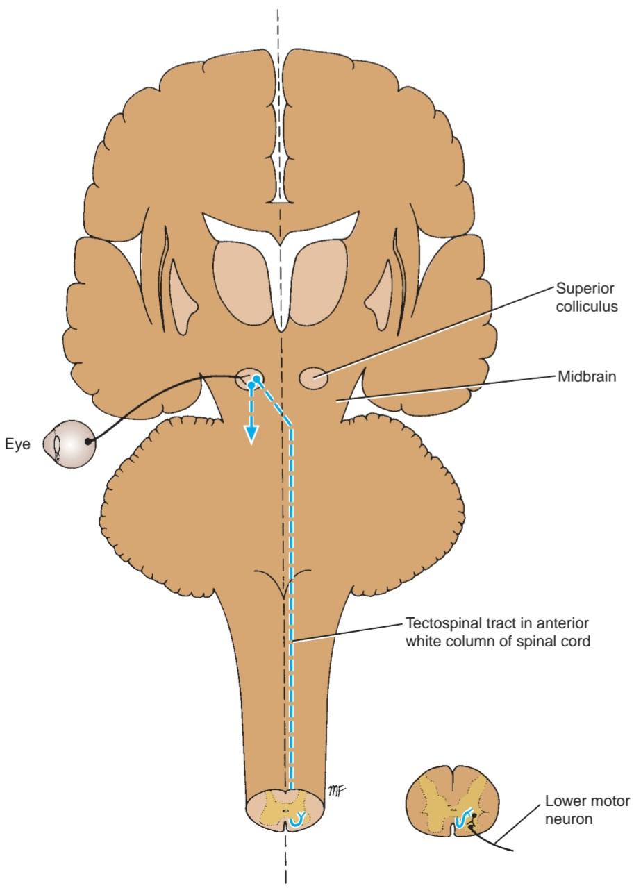
Figure 4-23 Tectospinal tract.
شکل ۴-۲۳ مسیر تکتوسپینال.
Rubrospinal Tract
The red nucleus is situated in the tegmentum of the midbrain at the level of the superior colliculus (Fig. 4-24). The axons of neurons in this nucleus cross the midline at the level of the nucleus and descend as the rubrospinal tract through the pons and medulla oblongata to enter the lateral white column of the spinal cord (Fig. 4-20). The fibers terminate by synapsing with internuncial neurons in the anterior gray column of the cord.
مسیر روبرواسپینال
هسته قرمز در تگمنتوم مغز میانی در سطح کولیکولوس فوقانی قرار دارد (شکل ۴-۲۴). آکسونهای نورونهای این هسته از خط میانی در سطح هسته عبور میکنند و به عنوان مسیر روبرواسپینال از طریق پل مغزی و بصل النخاع پایین میآیند تا وارد ستون سفید جانبی نخاع شوند (شکل ۴-۲۰). فیبرها با سیناپس با نورونهای بینحلقهای در ستون خاکستری قدامی نخاع خاتمه مییابند.
Neurons of the red nucleus receive afferent impulses through connections with the cerebral cortex and the cerebellum. This is believed to be an important indirect pathway by which the cerebral cortex and the cerebellum can influence a and y motor neuron activity. The tract facilitates the activity of the flexor muscles and inhibits the activity of the extensor or antigravity muscles.
نورونهای هسته قرمز از طریق ارتباط با قشر مغز و مخچه، تکانههای آوران را دریافت میکنند. اعتقاد بر این است که این یک مسیر غیرمستقیم مهم است که از طریق آن قشر مغز و مخچه میتوانند بر فعالیت نورون حرکتی a و y تأثیر بگذارند. این مسیر فعالیت عضلات خمکننده را تسهیل میکند و فعالیت عضلات بازکننده یا ضد جاذبه را مهار میکند.
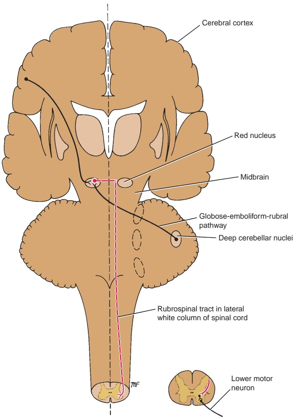
Figure 4-24 Rubrospinal tract.
شکل ۴-۲۴ دستگاه روبرواسپینال.
Vestibulospinal Tract
Vestibular nuclei are situated in the pons and medulla oblongata beneath the floor of the fourth ventricle (Fig. 4-25). The vestibular nuclei receive afferent fibers from the inner ear through the vestibular nerve and from the cerebellum. Neurons of the lateral vestibular nucleus give rise to the axons that form the vestibulospinal tract. The tract descends uncrossed through the medulla and through the length of the spinal cord in the anterior white column (Fig. 4-20). The fibers terminate by synapsing with internuncial neurons of the anterior gray column of the spinal cord.
مسیر دهلیزی-نخاعی
هستههای دهلیزی در پل مغزی و بصل النخاع در زیر کف بطن چهارم قرار دارند (شکل 4-25). هستههای دهلیزی از طریق عصب دهلیزی، فیبرهای آوران را از گوش داخلی و از مخچه دریافت میکنند. نورونهای هسته دهلیزی جانبی، آکسونهایی را ایجاد میکنند که مسیر دهلیزی-نخاعی را تشکیل میدهند. این مسیر به صورت غیر متقاطع از بصل النخاع و در طول نخاع در ستون سفید قدامی پایین میرود (شکل 4-20). فیبرها با سیناپس با نورونهای بین حلقی ستون خاکستری قدامی نخاع خاتمه مییابند.
The inner ear and the cerebellum, by means of this tract, facilitate the activity of the extensor muscles and inhibit the activity of the flexor muscles in association with the maintenance of balance.
گوش داخلی و مخچه، از طریق این مسیر، فعالیت عضلات بازکننده را تسهیل و فعالیت عضلات خمکننده را در ارتباط با حفظ تعادل مهار میکنند.
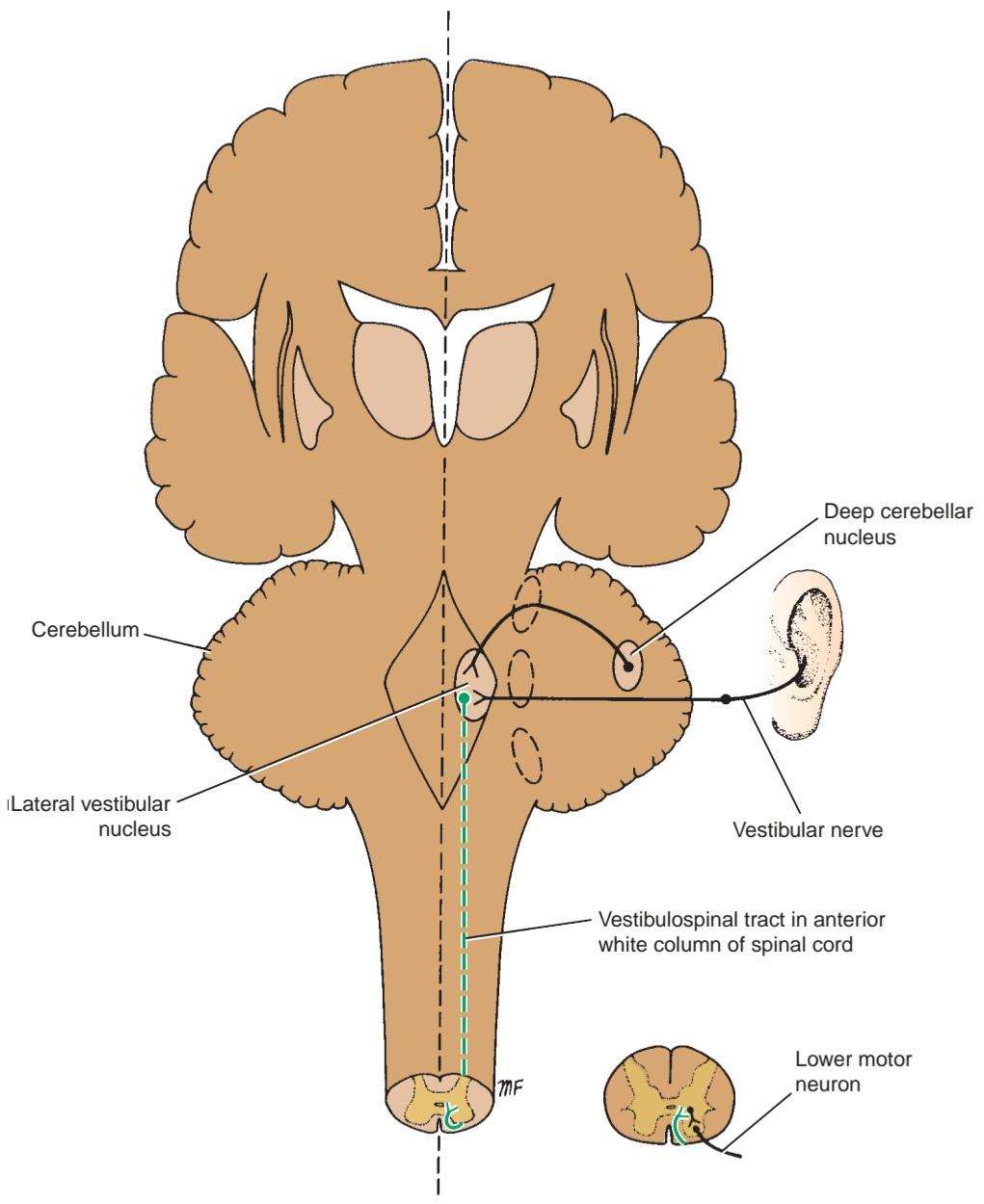
Figure 4-25 Vestibulospinal tract.
شکل ۴-۲۵ راه دهلیزی-نخاعی.
Olivospinal Tract
The olivospinal tract was thought to arise from the inferior olivary nucleus and to descend in the lateral white column of the spinal cord (Fig. 4-26), to influence the activity of the motor neurons in the anterior gray column. Many now doubt that it exists.
راه زیتونی-نخاعی
تصور میشد که راه زیتونی-نخاعی از هسته زیتونی تحتانی منشأ میگیرد و در ستون سفید جانبی نخاع پایین میرود (شکل ۴-۲۶)، تا بر فعالیت نورونهای حرکتی در ستون خاکستری قدامی تأثیر بگذارد. اکنون بسیاری در مورد وجود آن شک دارند.
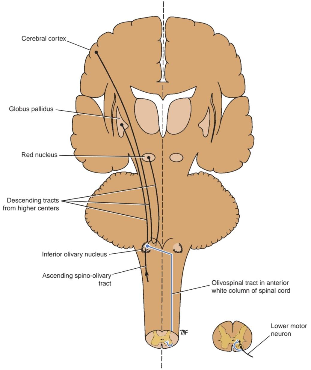
Figure 4-26 Olivospinal tract. The existence of this tract as a separate pathway is doubtful.
شکل ۴-۲۶ مسیر زیتونی-نخاعی. وجود این مسیر به عنوان یک مسیر جداگانه مورد تردید است.
Descending Autonomic Fibers
The higher centers of the CNS associated with the control of autonomic activity are situated in the cere- bral cortex, hypothalamus, amygdaloid complex, and reticular formation. Although distinct tracts have not been recognized, investigation of spinal cord lesions has demonstrated that descending autonomic tracts do exist and probably form part of the reticulospinal tract.
فیبرهای خودکار نزولی
مراکز بالاتر سیستم عصبی مرکزی مرتبط با کنترل فعالیت خودکار در قشر مغز، هیپوتالاموس، کمپلکس آمیگدالوئید و تشکیلات مشبک قرار دارند. اگرچه مسیرهای مجزایی شناسایی نشدهاند، بررسی ضایعات نخاع نشان داده است که مسیرهای خودکار نزولی وجود دارند و احتمالاً بخشی از مسیر شبکهای-نخاعی را تشکیل میدهند.
The fibers arise from neurons in the higher centers and cross the midline in the brainstem. They are believed to descend in the lateral white column of the spinal cord and to terminate by synapsing on the autonomic motor cells in the lateral gray columns in the thoracic and upper lumbar (sympathetic outflow) and midsacral (parasympathetic) levels of the spinal cord.
این فیبرها از نورونهای مراکز بالاتر منشأ میگیرند و از خط میانی در ساقه مغز عبور میکنند. اعتقاد بر این است که آنها در ستون سفید جانبی نخاع نزول میکنند و با سیناپس زدن روی سلولهای حرکتی خودکار در ستونهای خاکستری جانبی در سطوح سینهای و کمری فوقانی (خروجی سمپاتیک) و میانی-خاجی (پاراسمپاتیک) نخاع خاتمه مییابند.
INTERSEGMENTAL TRACTS
Short ascending and descending tracts that originate and end within the spinal cord exist in the anterior, lateral, and posterior white columns. The function of these pathways is to interconnect the neurons of different segmental levels, and the pathways are particularly important in intersegmental spinal reflexes.
مسیرهای بینقطعهای
مسیرهای کوتاه صعودی و نزولی که از نخاع سرچشمه گرفته و در آن پایان مییابند، در ستونهای سفید قدامی، جانبی و خلفی وجود دارند. عملکرد این مسیرها اتصال نورونهای سطوح مختلف قطعهای است و این مسیرها به ویژه در رفلکسهای نخاعی بینقطعهای اهمیت دارند.
Reflex Arc
A reflex may be defined as an involuntary response to a stimulus. It depends on the integrity of the reflex arc (Fig. 4-27). In its simplest form, a reflex arc consists of the following anatomical structures: (1) a receptor organ, (2) an afferent neuron, (3) an effector neuron, and (4) an effector organ. A reflex arc involving only one synapse is referred to as a monosynaptic reflex arc. Interruption of the reflex arc at any point along its course would abolish the response.
قوس رفلکس
رفلکس را میتوان به عنوان یک پاسخ غیرارادی به یک محرک تعریف کرد. این امر به یکپارچگی قوس رفلکس بستگی دارد (شکل 4-27). در سادهترین شکل، یک قوس رفلکس از ساختارهای آناتومیکی زیر تشکیل شده است: (1) یک اندام گیرنده، (2) یک نورون آوران، (3) یک نورون مؤثر و (4) یک اندام مؤثر. قوس رفلکسی که تنها شامل یک سیناپس باشد، قوس رفلکس تک سیناپسی نامیده میشود. قطع قوس رفلکس در هر نقطهای در طول مسیر آن، پاسخ را از بین میبرد.
In the spinal cord, reflex arcs play an important role in maintaining muscle tone, which is the basis for body posture. The receptor organ is situated in the skin, muscle, or tendon. The cell body of the afferent neuron is located in the posterior root ganglion, and the central axon of this first-order neuron terminates by synapsing on the effector neuron. Since the afferent fibers are of large diameter and are rapidly conducting and because of the presence of only one synapse, a very quick response is possible.
در نخاع، قوسهای رفلکس نقش مهمی در حفظ تون عضلانی دارند که اساس وضعیت بدن است. اندام گیرنده در پوست، عضله یا تاندون قرار دارد. جسم سلولی نورون آوران در گانگلیون ریشه خلفی قرار دارد و آکسون مرکزی این نورون درجه اول با سیناپس زدن روی نورون مؤثر خاتمه مییابد. از آنجایی که فیبرهای آوران قطر زیادی دارند و به سرعت هدایت میشوند و به دلیل وجود تنها یک سیناپس، پاسخ بسیار سریعی امکانپذیر است.
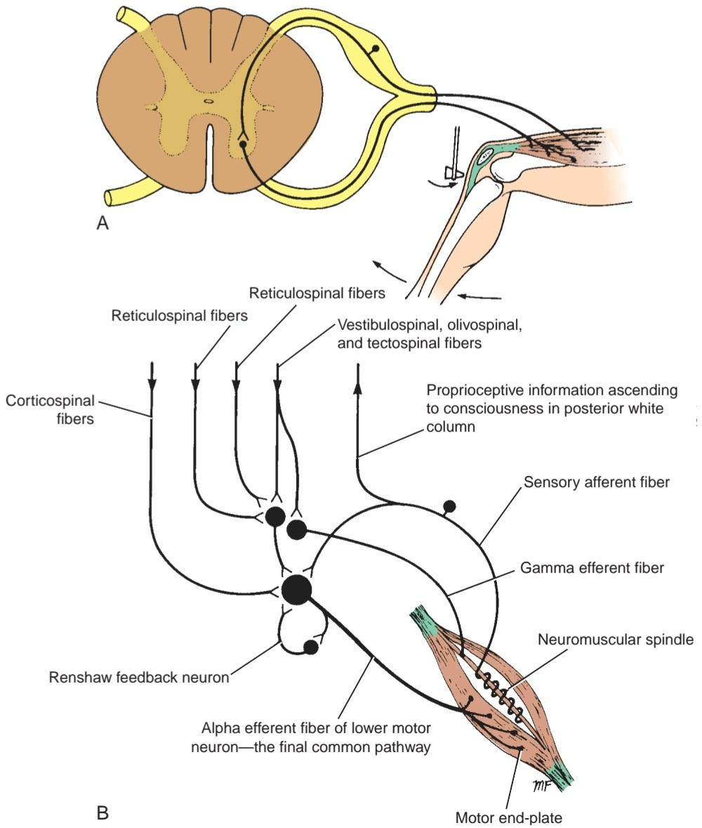
Figure 4-27 A: A monosynaptic reflex arc. B: Multiple neurons synapsing with the lower motor neuron. Note the presence of the Renshaw feedback neuron.
شکل ۴-۲۷ الف: یک قوس رفلکس تک سیناپسی. ب: چندین نورون در حال سیناپس با نورون حرکتی تحتانی. به وجود نورون بازخورد رنشاو توجه کنید.
Physiologic study of the electrical activity of the effector neuron shows that a prolonged asynchronous discharge follows the very quick monosynaptic discharge. The reason for this later discharge is that the afferent fibers entering the spinal cord frequently branch, and the branches synapse with many internuncial neurons, which ultimately synapse with the effector neuron (Fig. 4-28). These additional neuronal circuits prolong the bombardment of the effector neurons after the initial stimulation by the afferent neuron has ceased. The presence of internuncial neurons also results in the spread of the afferent stimulus to neurons at different segmental levels of the spinal cord.
مطالعه فیزیولوژیکی فعالیت الکتریکی نورون مؤثر نشان میدهد که یک تخلیه ناهمزمان طولانی پس از تخلیه بسیار سریع تک سیناپسی رخ میدهد. دلیل این تخلیه دیرهنگام این است که فیبرهای آوران که وارد نخاع میشوند، مرتباً شاخه شاخه میشوند و شاخهها با بسیاری از نورونهای بینرشتهای سیناپس میدهند که در نهایت با نورون مؤثر سیناپس میکنند (شکل ۴-۲۸). این مدارهای عصبی اضافی، بمباران نورونهای مؤثر را پس از توقف تحریک اولیه توسط نورون آوران، طولانیتر میکنند. وجود نورونهای بینرشتهای همچنین منجر به گسترش محرک آوران به نورونها در سطوح مختلف سگمنتال نخاع میشود.
The law of reciprocal innervationis important to understand in considering reflex skeletal muscle activity. Simply stated, it means that the flexor and extensor reflexes of the same limb cannot be made to contract simultaneously. For this law to work, the afferent nerve fibers responsible for flexor reflex muscle action must have branches that synapse with the extensor motor neurons of the same limb, causing them to be inhibited.
درک قانون عصبدهی متقابل در بررسی فعالیت رفلکسی عضلات اسکلتی مهم است. به عبارت ساده، این قانون به این معنی است که رفلکسهای خمکننده و بازکننده یک اندام نمیتوانند همزمان منقبض شوند. برای اینکه این قانون کار کند، فیبرهای عصبی آوران مسئول عملکرد رفلکس خمکننده عضلات باید شاخههایی داشته باشند که با نورونهای حرکتی بازکننده همان اندام سیناپس برقرار کنند و باعث مهار آنها شوند.
Another interesting property of spinal reflexes is that the evocation of a reflex on one side of the body causes opposite effects on the limb of the other side of the body. This crossed extensor reflex is demonstrated when afferent stimulation of the reflex arc that causes the ipsilateral limb to flex results in the contralateral limb being extended.
یکی دیگر از ویژگیهای جالب رفلکسهای نخاعی این است که برانگیختن یک رفلکس در یک طرف بدن باعث ایجاد اثرات متضاد بر اندام طرف دیگر بدن میشود. این رفلکس بازکننده متقاطع زمانی نشان داده میشود که تحریک آوران قوس رفلکس که باعث خم شدن اندام همان طرف میشود، منجر به باز شدن اندام طرف مقابل شود.

Figure 4-28 A: Multiple branching of afferent fibers entering the spinal cord and the presence of many internuncial neurons that synapse with the effector neuron. B: Law of reciprocal innervation and the crossed extensor reflex.
شکل ۴-۲۸ الف: انشعابات متعدد فیبرهای آوران که وارد نخاع میشوند و وجود نورونهای بینحلقهای زیادی که با نورون مؤثر سیناپس میدهند. ب: قانون عصبدهی متقابل و رفلکس اکستانسور متقاطع.
Influence of Higher Neuronal Centers on Spinal Reflex Activity
The spinal segmental reflex arc involving motor activity is greatly influenced by higher centers in the brain. These influences are mediated through the corticospinal, reticulospinal, tectospinal, rubrospinal, and vestibulospinal tracts. In the clinical condition known as spinal shock (see p. 167), which follows the sudden removal of these influences by spinal cord severance, the segmental spinal reflexes are depressed. When the spinal shock disappears in a few weeks, the segmental spinal reflexes return, and the muscle tone is increased. This decerebrate rigidity is due to the overactivity of the y efferent nerve fibers to the muscle spindles, which results from the release of these neurons from the higher centers. The next stage may be paraplegia in extension with domination of the increased tone of the extensor muscles over the flexor muscles. Some neurologists believe that this condition is due to incomplete severance of all the descending tracts with persistence of the vestibulospinal tract. Should all the descending tracts be severed, the condition of paraplegia in flexion occurs. In this condition, the reflex responses are flexor in nature, and the tone of the extensor muscles is diminished.
تأثیر مراکز عصبی بالاتر بر فعالیت رفلکس نخاعی
قوس رفلکس سگمنتال نخاعی که شامل فعالیت حرکتی است، به شدت تحت تأثیر مراکز بالاتر در مغز قرار دارد. این تأثیرات از طریق مسیرهای قشری-نخاعی، رتیکولو-نخاعی، تکتو-نخاعی، روبرو-نخاعی و دهلیزی-نخاعی اعمال میشوند. در وضعیت بالینی که به عنوان شوک نخاعی شناخته میشود (به صفحه ۱۶۷ مراجعه کنید)، که به دنبال حذف ناگهانی این تأثیرات توسط قطع نخاع رخ میدهد، رفلکسهای نخاعی سگمنتال سرکوب میشوند. هنگامی که شوک نخاعی در عرض چند هفته از بین میرود، رفلکسهای نخاعی سگمنتال بازمیگردند و تون عضلانی افزایش مییابد. این سفتی غیرمغزی به دلیل فعالیت بیش از حد فیبرهای عصبی وابران y به دوکهای عضلانی است که در نتیجه آزاد شدن این نورونها از مراکز بالاتر ایجاد میشود. مرحله بعدی ممکن است پاراپلژی در اکستانسیون با غلبه تون افزایش یافته عضلات اکستانسور بر عضلات فلکسور باشد. برخی از متخصصان مغز و اعصاب معتقدند که این وضعیت به دلیل قطع ناقص تمام مسیرهای نزولی با باقی ماندن مسیر دهلیزی-نخاعی است. در صورت قطع شدن تمام مسیرهای نزولی، وضعیت پاراپلژی در خم شدن رخ میدهد. در این وضعیت، پاسخهای رفلکسی ماهیت خم شدنی دارند و تون عضلات بازکننده کاهش مییابد.
RENSHAW CELLS AND LOWER MOTOR NEURON INHIBITION
Lower motor neuron axons give off collateral branches as they pass through the white matter to reach the anterior roots of the spinal nerve. These collaterals synapse on neurons described by Renshaw, which, in turn, synapse on the lower motor neurons (Fig. 4-27). These internuncial neurons are believed to provide feedback on the lower motor neurons, inhibiting their activity.
سلولهای رنشاو و مهار نورون حرکتی تحتانی
آکسونهای نورون حرکتی تحتانی هنگام عبور از ماده سفید برای رسیدن به ریشههای قدامی عصب نخاعی، شاخههای جانبی ایجاد میکنند. این نورونهای جانبی با نورونهای توصیفشده توسط رنشاو سیناپس برقرار میکنند که به نوبه خود با نورونهای حرکتی تحتانی سیناپس برقرار میکنند (شکل 4-27). اعتقاد بر این است که این نورونهای بینرشتهای، بازخوردی بر نورونهای حرکتی تحتانی ارائه میدهند و فعالیت آنها را مهار میکنند.
«فصل چهارم کتاب نورواناتومی بالینی اسنل»
🚀 با ما همراه شوید!
تازهترین مطالب و آموزشهای مغز و اعصاب را از دست ندهید. با فالو کردن کانال تلگرام آیندهنگاران مغز، از ما حمایت کنید!