
رشتههای عصبی و عصبدهی محیطی؛ فصل سوم نوروآناتومی بالینی اسنل


دعای مطالعه [ نمایش ]
بِسْمِ الله الرَّحْمنِ الرَّحیمِ
اَللّهُمَّ اَخْرِجْنى مِنْ ظُلُماتِ الْوَهْمِ
خدايا مرا بيرون آور از تاريكىهاى وهم،
وَ اَكْرِمْنى بِنُورِ الْفَهْمِ
و به نور فهم گرامى ام بدار،
اَللّهُمَّ افْتَحْ عَلَيْنا اَبْوابَ رَحْمَتِكَ
خدايا درهاى رحمتت را به روى ما بگشا،
وَانْشُرْ عَلَيْنا خَزائِنَ عُلُومِكَ بِرَحْمَتِكَ يا اَرْحَمَ الرّاحِمينَ
و خزانههاى علومت را بر ما باز كن به امید رحمتت اى مهربانترين مهربانان.
آیندهنگاران مغز: «ما مغز را میشناسیم، تا آینده را بسازیم.»
کتاب «نوروآناتومی بالینی اسنل» اثر دکتر ریچارد اس. اسنل، با ویرایش جدید دکتر رایان اسپلیتگربر، همچنان بهعنوان مرجع طلایی این حوزه شناخته میشود. این اثر با تلفیق تازهترین پژوهشها و کاربردهای بالینی، پلی استوار میان دانش پایه و عمل پزشکی بنا میکند.
تلاشهای علمی و سالها تدریس ارزشمند استاد گرانقدر دکتر محمدتقی جغتایی الهامبخش ترجمه و گسترش این دانش در ایران بوده است.
ترجمه دقیق این کتاب توسط برند علمی آیندهنگاران مغز به مدیریت داریوش طاهری، امکان دسترسی فارسیزبانان به مرزهای نوین علوم اعصاب را فراهم کرده و رسالتی علمی در جهت شناخت عمیقتر مغز و ترسیم آیندهای روشنتر بر عهده گرفته است.
»» فصل ۳: رشتههای عصبی و عصبدهی محیطی؛ مرور و مبانی
»» Chapter 3: Nerve Fibers and Peripheral Innervation
CHAPTER OBJECTIVES
• To consider the basic structure and function of nerve fibers
• To understand the process of nerve degeneration and regeneration
• To review the special organs that lie at the ends of sensory and motor nerves
• To examine the different sensory modalities
• To learn the terms used in assessing skin sensory loss and abnormal muscle activity
اهداف فصل
• بررسی ساختار و عملکرد اساسی رشتههای عصبی
• درک فرآیند تخریب و بازسازی عصب
• بررسی اندامهای ویژهای که در انتهای اعصاب حسی و حرکتی قرار دارند
• بررسی روشهای مختلف حسی
• یادگیری اصطلاحات مورد استفاده در ارزیابی از دست دادن حس پوست و فعالیت غیرطبیعی عضلات
A 45-year-old man is recovering from a mild upper respiratory tract infection when he suddenly notices weakness in both legs while walking upstairs. He also develops a numb sensation over the lower part of both legs and feet. Two days later, while shaving, he notices weakness of the muscles on the right side of his face.
مردی ۴۵ ساله که در حال بهبودی از عفونت خفیف دستگاه تنفسی فوقانی است، ناگهان هنگام بالا رفتن از پلهها متوجه ضعف در هر دو پایش میشود. او همچنین در قسمت پایینی هر دو پا و کف پا احساس بیحسی میکند. دو روز بعد، هنگام اصلاح صورت، متوجه ضعف عضلات سمت راست صورتش میشود.
On physical examination, the patient does not appear to be ill. He has no pyrexia. Examination of his leg muscles shows obvious signs of muscle weakness involving both legs, especially below the knees. Both ankle reflexes are absent, and the right knee jerk is diminished. He has sensory deficits for touch and pain sensations in the distribution of the stocking area of both feet and lower legs and a mild form of facial nerve palsy involving the right side of the face. Neurologic evidence of loss of function of the brain or spinal cord is not present.
در معاینه فیزیکی، بیمار به نظر بیمار نمیرسد. تب ندارد. معاینه عضلات پای او علائم واضحی از ضعف عضلانی در هر دو پا، به ویژه زیر زانوها را نشان میدهد. رفلکسهای مچ پا در هر دو پا وجود ندارد و تکانهای زانوی راست کاهش یافته است. او دارای نقص حسی در حس لامسه و درد در ناحیه جوراب هر دو پا و ساق پا و نوع خفیفی از فلج عصب صورت در سمت راست صورت است. شواهد عصبی مبنی بر از دست دادن عملکرد مغز یا نخاع وجود ندارد.
The patient is suspected of having Guillain-Barré syndrome and is admitted to the hospital for observation. The cause of this disease is unknown, although it is believed to be viral and involves the immune system. Histologically, the peripheral nerves show focal scattered areas of demyelination with an accumulation of lymphocytes and macrophages. As the myelin is lost, the axons are left naked, and the Schwann cell bodies remain intact. In the majority of patients, recovery occurs in 2 to 4 weeks with nerve remyelination. Hospitalization is necessary in the early stages because the disease can spread rapidly to involve the intercostal and phrenic nerves, resulting in paralysis of the intercostal muscles and diaphragm. For the same reason, the coughing and swallowing reflexes should be watched carefully. A clinician would find this disease impossible to understand without knowledge of peripheral nerve structure.
بیمار مشکوک به سندرم گیلن باره است و برای معاینه در بیمارستان بستری میشود. علت این بیماری ناشناخته است، اگرچه اعتقاد بر این است که ویروسی است و سیستم ایمنی را درگیر میکند. از نظر بافتشناسی، اعصاب محیطی نواحی پراکنده و کانونی از دمیلیناسیون را با تجمع لنفوسیتها و ماکروفاژها نشان میدهند. با از بین رفتن میلین، آکسونها بدون پوشش باقی میمانند و اجسام سلولی شوان دست نخورده باقی میمانند. در اکثر بیماران، بهبودی در عرض 2 تا 4 هفته با میلینسازی مجدد عصب رخ میدهد. بستری شدن در مراحل اولیه ضروری است زیرا بیماری میتواند به سرعت گسترش یابد و اعصاب بین دندهای و فرنیک را درگیر کند و منجر به فلج عضلات بین دندهای و دیافراگم شود. به همین دلیل، رفلکسهای سرفه و بلع باید به دقت تحت نظر قرار گیرند. یک پزشک بدون آگاهی از ساختار اعصاب محیطی، درک این بیماری را غیرممکن خواهد یافت.
In this chapter, the process of nerve degeneration and regeneration is described in detail because nerve lesions are very common in clinical practice and can be caused by a wide variety of diseases, including trauma, neoplasms, infection, metabolic dysfunction (diabetes), and chemical toxins such as lead. The process of nerve degeneration is fast and can take place in nerves in the central and peripheral nervous systems. The regeneration of nerves is slow and confined to the peripheral nervous system (PNS). Because so much research today is being devoted to investigating why regeneration in the central nervous system (CNS) ceases within 2 weeks, the histologic changes that occur must be learned.
در این فصل، فرآیند تخریب و بازسازی عصب به تفصیل شرح داده شده است زیرا ضایعات عصبی در عمل بالینی بسیار رایج هستند و میتوانند در اثر طیف گستردهای از بیماریها، از جمله تروما، نئوپلاسمها، عفونت، اختلال عملکرد متابولیک (دیابت) و سموم شیمیایی مانند سرب ایجاد شوند. فرآیند تخریب عصب سریع است و میتواند در اعصاب سیستم عصبی مرکزی و محیطی رخ دهد. بازسازی اعصاب کند است و محدود به سیستم عصبی محیطی (PNS) است. از آنجایی که امروزه تحقیقات زیادی به بررسی این موضوع اختصاص داده شده است که چرا بازسازی در سیستم عصبی مرکزی (CNS) ظرف 2 هفته متوقف میشود، باید تغییرات بافتشناسی که رخ میدهد، آموخته شود.
The material in this chapter commonly forms the basis for examination questions.
مطالب این فصل معمولاً مبنای سؤالات امتحانی را تشکیل میدهد.

NERVE FIBERS
Nerve fiber is the name given to an axon (or a dendrite) of a nerve cell. The structure of axons and dendrites is described on page 33-34. Bundles of nerve fibers found in the CNS are referred to as nerve tracts (Fig. 3-1); bundles of nerve fibers found in the PNS are called peripheral nerves (Fig. 3-2).
رشتههای عصبی
رشته عصبی نامی است که به آکسون (یا دندریت) یک سلول عصبی داده میشود. ساختار آکسونها و دندریتها در صفحات ۳۳-۳۴ شرح داده شده است. دستههای رشتههای عصبی موجود در سیستم عصبی مرکزی (CNS) به عنوان مسیرهای عصبی شناخته میشوند (شکل ۳-۱)؛ دستههای رشتههای عصبی موجود در سیستم عصبی محیطی (PNS) اعصاب محیطی نامیده میشوند (شکل ۳-۲).
Two types of nerve fibers are present in the CNS and PNS: myelinated and nonmyelinated.
دو نوع فیبر عصبی در سیستم عصبی مرکزی (CNS) و سیستم عصبی محیطی (PNS) وجود دارد: میلیندار و بدون میلین.

Figure 3-1 Sections through the thoracic region of the spinal cord showing examples of nerve fibers entering or leaving the central nervous system; ascending and descending nerve fibers (tracts or pathways) are also shown.
شکل ۳-۱ برشهایی از ناحیه قفسه سینه نخاع که نمونههایی از فیبرهای عصبی ورودی یا خروجی به سیستم عصبی مرکزی را نشان میدهند؛ فیبرهای عصبی صعودی و نزولی (مسیرها یا راهها) نیز نشان داده شدهاند.

Figure 3-2 Exploded view of a peripheral nerve showing the connective tissue sheaths and the structure of myelinated and nonmyelinated nerve fibers.
شکل ۳-۲ نمای باز شده از یک عصب محیطی که غلافهای بافت همبند و ساختار فیبرهای عصبی میلیندار و بدون میلین را نشان میدهد.
Myelinated Nerve Fibers
A myelinated nerve fiber is one that is surrounded by a myelin sheath. The myelin sheath is not part of the neuron but is formed by a supporting cell (Fig. 3-3; also see Fig. 3-2). In the CNS, the supporting cell is called the oligodendrocyte; in the PNS, it is called the Schwann cell.
فیبرهای عصبی میلیندار
یک فیبر عصبی میلیندار، فیبری است که توسط غلاف میلین احاطه شده است. غلاف میلین بخشی از نورون نیست، بلکه توسط یک سلول پشتیبان تشکیل شده است (شکل 3-3؛ همچنین به شکل 3-2 مراجعه کنید). در سیستم عصبی مرکزی، سلول پشتیبان، الیگودندروسیت نامیده میشود؛ در سیستم عصبی محیطی، سلول شوان نامیده میشود.
Figure 3-3 The relationship between an oligodendrocyte and myelinated nerve fibers in the central nervous system. Note the absence of a basement membrane.
شکل ۳-۳ رابطه بین یک الیگودندروسیت و فیبرهای عصبی میلیندار در سیستم عصبی مرکزی. به عدم وجود غشای پایه توجه کنید.

The myelin sheath is a segmented, discontinuous layer interrupted at regular intervals by the nodes of Ranvier (Figs. 3-4 and 3-5). Each segment of the myelin sheath measures approximately 0.5 to 1.0 mm in length. In the CNS, each oligodendrocyte may form and maintain myelin sheaths for as many as 60 nerve fibers (axons). In the PNS, only one Schwann cell maintains each segment of one nerve fiber.
غلاف میلین یک لایه قطعه قطعه و ناپیوسته است که در فواصل منظم توسط گرههای رانویه قطع میشود (شکلهای 3-4 و 3-5). هر قطعه از غلاف میلین تقریباً 0.5 تا 1.0 میلیمتر طول دارد. در سیستم عصبی مرکزی، هر الیگودندروسیت میتواند غلافهای میلین را برای حداکثر 60 فیبر عصبی (آکسون) تشکیل داده و حفظ کند. در سیستم عصبی محیطی، تنها یک سلول شوان هر قطعه از یک فیبر عصبی را حفظ میکند.

Figure 3-4 A myelinated nerve fiber in the peripheral nervous system. A-D: Cross sections showing the stages in the formation of the myelin sheath. E: A longitudinal section of a mature myelinated nerve fiber showing a node of Ranvier. Note the presence of a basement membrane.
شکل ۳-۴ الف یک فیبر عصبی میلیندار در سیستم عصبی محیطی. الف-د: مقاطع عرضی که مراحل تشکیل غلاف میلین را نشان میدهند. ه: برش طولی از یک فیبر عصبی میلیندار بالغ که گره رانویه را نشان میدهد. به وجود غشای پایه توجه کنید.

Figure 3-5 Electron micrograph of a longitudinal section of several myelinated axons showing the structure of a node of Ranvier (arrow). At the node, two adjacent Schwann cells terminate, and the myelin sheaths become thinner by the turning off of the lamellae. Note the many microtubules and microfilaments within the axons (x12,220). (Courtesy Dr. H. de F. Webster.)
شکل ۳-۵ تصویر میکروسکوپ الکترونی از برش طولی چندین آکسون میلیندار که ساختار یک گره رانویه را نشان میدهد (فلش). در گره، دو سلول شوان مجاور به هم متصل میشوند و غلافهای میلین با خاموش شدن تیغهها نازکتر میشوند. به میکروتوبولها و میکروفیلامنتهای فراوان درون آکسونها توجه کنید (12220x). (با احترام از دکتر اچ. دی. اف. وبستر.)
Myelin Formation
Myelin sheaths begin to form before birth and during the first year postnatally. The process has been studied with the electron microscope (Fig. 3-6).
تشکیل میلین
غلافهای میلین قبل از تولد و در طول سال اول پس از تولد شروع به تشکیل میکنند. این فرآیند با میکروسکوپ الکترونی مورد مطالعه قرار گرفته است (شکل 3-6).
In the peripheral nervous system (PNS), the nerve fiber or axon first indents the side of a Schwann cell (Fig. 3-4). Later, as the axon sinks farther into the Schwann cell, the external plasma membrane of the Schwann cell forms a mesaxon, which suspends the axon within the Schwann cell (Fig. 3-6A). Subsequently, the Schwann cell is believed to rotate on the axon so that the plasma membrane becomes wrapped around the axon in a spiral. The direction of the spiral is clockwise in some segments and counterclockwise in others. To begin with, the wrappings are loose, but gradually the cytoplasm between the layers of the cell membrane disappears, leaving cytoplasm near the surface and in the region of the nucleus. The wrappings become tight with maturation of the nerve fiber. The thickness of the myelin depends on the number of spirals of Schwann cell membrane. Some nerve fibers are surrounded by only a few turns of the membrane, while others have as many as 50 turns. In electron micrographs of cross sections of mature myelinated nerve fibers, the myelin is seen to be laminated. Each lamella measures 13 to 18 nm thick. The dark major dense line, about 2.5 nm thick, consists of two inner protein layers of the plasma membrane that are fused together. The lighter minor dense line, about 10 nm thick, is formed by the approximation of the outer surfaces of adjacent plasma membranes and is made up of lipid. The fused outer protein layers of the plasma membranes are very thin and form a thin intraperiod line situated in the center of the lighter lipid layer. At the node of Ranvier, two adjacent Schwann cells terminate, and the myelin sheaths become thinner by the turning off of the lamellae (Fig. 3-5). At these regions, the plasma membrane of the axon, the axolemma, is exposed.
در سیستم عصبی محیطی (PNS)، فیبر عصبی یا آکسون ابتدا در کنار سلول شوان فرو میرود (شکل 3-4). بعداً، با فرو رفتن بیشتر آکسون در سلول شوان، غشای پلاسمایی خارجی سلول شوان یک مزاکسون تشکیل میدهد که آکسون را درون سلول شوان معلق نگه میدارد (شکل 3-6A). متعاقباً، اعتقاد بر این است که سلول شوان روی آکسون میچرخد به طوری که غشای پلاسمایی به صورت مارپیچی به دور آکسون پیچیده میشود. جهت مارپیچ در برخی بخشها در جهت عقربههای ساعت و در برخی دیگر خلاف جهت عقربههای ساعت است. در ابتدا، پوششها شل هستند، اما به تدریج سیتوپلاسم بین لایههای غشای سلولی ناپدید میشود و سیتوپلاسم در نزدیکی سطح و در ناحیه هسته باقی میماند. پوششها با بلوغ فیبر عصبی محکم میشوند. ضخامت میلین به تعداد مارپیچهای غشای سلول شوان بستگی دارد. برخی از فیبرهای عصبی تنها با چند چرخش غشاء احاطه شدهاند، در حالی که برخی دیگر تا 50 چرخش دارند. در تصاویر میکروسکوپ الکترونی از مقاطع عرضی فیبرهای عصبی میلیندار بالغ، میلین به صورت لایه لایه دیده میشود. ضخامت هر تیغه ۱۳ تا ۱۸ نانومتر است. خط متراکم اصلی تیره، با ضخامت حدود ۲.۵ نانومتر، از دو لایه پروتئینی داخلی غشای پلاسمایی تشکیل شده است که به هم جوش خوردهاند. خط متراکم فرعی روشنتر، با ضخامت حدود ۱۰ نانومتر، با نزدیک شدن سطوح خارجی غشاهای پلاسمایی مجاور تشکیل شده و از لیپید ساخته شده است. لایههای پروتئینی خارجی جوش خورده غشاهای پلاسمایی بسیار نازک هستند و یک خط درون دورهای نازک واقع در مرکز لایه لیپیدی روشنتر تشکیل میدهند. در گره رانویه، دو سلول شوان مجاور به هم ختم میشوند و غلافهای میلین با خاموش شدن تیغهها نازکتر میشوند (شکل ۳-۵). در این مناطق، غشای پلاسمایی آکسون، آکسولما، نمایان میشود.

Figure 3-6 Electron micrograph of a transverse section of a peripheral nerve showing a myelinated axon with spiral myelin lamellae (center). Note the mesaxon (arrow). Parts of two other myelinated fibers are also shown. A number of nonmyelinated axons are enclosed in the peripheral cytoplasm of a Schwann cell (top). The mesaxons are indicated by arrows (x28,000). (Courtesy Dr. H. de F. Webster.)
شکل ۳-۶ تصویر میکروسکوپ الکترونی از برش عرضی یک عصب محیطی که یک آکسون میلیندار با تیغههای میلین مارپیچی را نشان میدهد (مرکز). به مزاکسون (فلش) توجه کنید. بخشهایی از دو فیبر میلیندار دیگر نیز نشان داده شده است. تعدادی آکسون بدون میلین در سیتوپلاسم محیطی یک سلول شوان (بالا) محصور شدهاند. مزاکسونها با فلش (28000x) نشان داده شدهاند. (با تشکر از دکتر اچ. دی. اف. وبستر.)
The incisures of Schmidt-Lanterman are seen on longitudinal sections of myelinated nerve fibers. They represent areas where the dark major dense line is not formed as a result of the localized persistence of Schwann cell cytoplasm (Fig. 3-7). This persistence of cytoplasm involves all the layers of the myelin, and thus, there is a continuous spiral of cytoplasm from the outermost region of the Schwann cell to the region of the axon. This spiral of cytoplasm may provide a pathway for the conduction of metabolites from the surface region of the Schwann cell to the axon.
شیارهای اشمیت-لانترمن روی مقاطع طولی فیبرهای عصبی میلیندار دیده میشوند. آنها نواحیای را نشان میدهند که در آنها خط متراکم اصلی تیره در نتیجهی پایداری موضعی سیتوپلاسم سلول شوان تشکیل نمیشود (شکل 3-7). این پایداری سیتوپلاسم شامل تمام لایههای میلین میشود و بنابراین، یک مارپیچ پیوسته از سیتوپلاسم از بیرونیترین ناحیهی سلول شوان تا ناحیهی آکسون وجود دارد. این مارپیچ سیتوپلاسم ممکن است مسیری را برای هدایت متابولیتها از ناحیهی سطحی سلول شوان به آکسون فراهم کند.

Figure 3-7 Schmidt-Lanterman incisures in the myelin sheath of a peripheral nerve. A: Transverse section of a myelinated nerve fiber. B: Schematic diagram of a myelinated nerve fiber in which the myelin sheath has been unrolled.
شکل ۳-۷ برشهای اشمیت-لانترمن در غلاف میلین یک عصب محیطی. الف: برش عرضی یک فیبر عصبی میلیندار. ب: نمودار شماتیک یک فیبر عصبی میلیندار که در آن غلاف میلین از هم باز شده است.
In the central nervous system (CNS), oligodendrocytes are responsible for the formation of the myelin sheaths. The plasma membrane of the oligodendrocyte becomes wrapped around the axon, and the number of layers will determine the thickness of the myelin sheath (Fig. 3-3). The nodes of Ranvier are situated in the intervals between adjacent oligodendrocytes. A single oligodendrocyte may be connected to the myelin sheaths of as many as 60 nerve fibers. For this reason, the process of myelination in the CNS cannot take place by rotation of the oligodendrocyte on the axon, as did the Schwann cell in the PNS. Myelination in the CNS possibly occurs by the growth in length of the process of the oligodendrocyte, the process wrapping itself around the axon. Incisures of Schmidt-Lanterman exist in CNS nerve fibers. Table 3-1 provides a summary of facts concerning CNS and PNS myelination.
در سیستم عصبی مرکزی (CNS)، الیگودندروسیتها مسئول تشکیل غلافهای میلین هستند. غشای پلاسمایی الیگودندروسیت به دور آکسون پیچیده میشود و تعداد لایهها، ضخامت غلاف میلین را تعیین میکند (شکل 3-3). گرههای رانویه در فواصل بین الیگودندروسیتهای مجاور قرار دارند. یک الیگودندروسیت واحد ممکن است به غلافهای میلین تا 60 فیبر عصبی متصل باشد. به همین دلیل، فرآیند میلینسازی در CNS نمیتواند با چرخش الیگودندروسیت روی آکسون، مانند سلول شوان در PNS، انجام شود. میلینسازی در CNS احتمالاً با افزایش طول زائده الیگودندروسیت، یعنی زائدهای که خود را به دور آکسون میپیچد، رخ میدهد. شیارهای اشمیت-لانترمن در فیبرهای عصبی CNS وجود دارند. جدول 3-1 خلاصهای از حقایق مربوط به میلینسازی CNS و PNS را ارائه میدهد.
Table 3-1 Classification of Neurons
جدول ۳-۱ طبقه بندی نورون ها

CNS, central nervous system.
سیستم عصبی مرکزی، سیستم عصبی مرکزی.
Nonmyelinated Nerve Fibers
The smaller axons of the CNS, the postganglionic axons of the autonomic part of the nervous system, and some fine sensory axons associated with the reception of pain are nonmyelinated.
فیبرهای عصبی بدون میلین
آکسونهای کوچکتر سیستم عصبی مرکزی (CNS)، آکسونهای پسگانگلیونی بخش خود مختار سیستم عصبی و برخی از آکسونهای حسی ظریف مرتبط با دریافت درد، بدون میلین هستند.
In the PNS, each axon, which is usually less than 1 um in diameter, indents the surface of the Schwann cell so that it lies within a trough (Fig. 3-2). As many as 15 or more axons may share a single Schwann cell, each lying within its own trough or sometimes sharing a trough. In some situations, the troughs are deep and the axons are embedded deep in the Schwann cells, forming a mesaxon from the Schwann cell plasma membrane (Fig. 3-8; also see Fig. 3-6). The Schwann cells lie close to one another along the length of the axons, and nodes of Ranvier do not exist.
در سیستم عصبی محیطی (PNS)، هر آکسون که معمولاً قطری کمتر از ۱ میکرومتر دارد، سطح سلول شوان را فرو میبرد به طوری که درون یک ناودان قرار میگیرد (شکل ۳-۲). ممکن است ۱۵ آکسون یا بیشتر، یک سلول شوان را به اشتراک بگذارند که هر کدام درون ناودان خود قرار دارند یا گاهی اوقات یک ناودان را به اشتراک میگذارند. در برخی موارد، ناودانها عمیق هستند و آکسونها در اعماق سلولهای شوان فرو رفتهاند و یک مزاکسون از غشای پلاسمایی سلول شوان تشکیل میدهند (شکل ۳-۸؛ همچنین به شکل ۳-۶ مراجعه کنید). سلولهای شوان در امتداد آکسونها نزدیک به یکدیگر قرار دارند و گرههای رانویه وجود ندارند.

Figure 3-8 Electron micrograph of a transverse section of a myelinated nerve fiber and sev- eral nonmyelinated nerve fibers. (Courtesy Dr. J. M. Kerns.)
شکل ۳-۸ تصویر میکروسکوپ الکترونی از برش عرضی یک فیبر عصبی میلیندار و چندین فیبر عصبی بدون میلین. (با تشکر از دکتر جی. ام. کرنز.)
In areas with synapses or where motor transmission occurs, the axon emerges from the trough of the Schwann cell for a short distance, thus exposing the active region of the axon (Fig. 3-9).
در مناطقی که سیناپس وجود دارد یا جایی که انتقال حرکتی رخ میدهد، آکسون از ناودان سلول شوان برای فاصله کوتاهی خارج میشود و بنابراین ناحیه فعال آکسون را در معرض دید قرار میدهد (شکل 3-9).
In the CNS, nonmyelinated nerve fibers run in small groups and are not particularly related to the oligoden- drocytes.
در سیستم عصبی مرکزی، فیبرهای عصبی بدون میلین در گروههای کوچکی قرار دارند و ارتباط خاصی با الیگودندروسیتها ندارند.
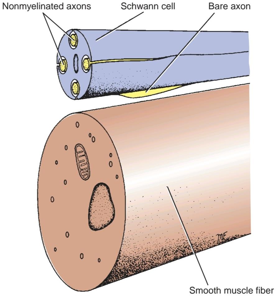
Figure 3-9 Autonomic neuromuscular junction between a nonmyelinated axon and a smooth muscle fiber.
شکل ۳-۹ اتصال عصبی-عضلانی خودکار بین یک آکسون بدون میلین و یک فیبر عضله صاف.
PERIPHERAL NERVES
Peripheral nerves is a collective term for the cranial and spinal nerves. Each peripheral nerve consists of parallel bundles of nerve fibers, which may be efferent or afferent axons, may be myelinated or nonmyelinated, and are surrounded by connective tissue sheaths (Figs. 3-10 and 3-11).
اعصاب محیطی
اعصاب محیطی یک اصطلاح کلی برای اعصاب جمجمهای و نخاعی است. هر عصب محیطی از دستههای موازی از الیاف عصبی تشکیل شده است که ممکن است آکسونهای آوران یا وابران باشند، ممکن است میلیندار یا بدون میلین باشند و توسط غلافهای بافت همبند احاطه شدهاند (شکلهای 3-10 و 3-11).

Figure 3-10 Peripheral nerve. I.s. Paraffin section. x270. This is a higher magnification of a region similar to the boxed area of Figure 1a. A distinguishing characteristic of longitudinal sections of peripheral nerves is that they appear to follow a zigzag course, particularly evident in this photomicrograph. The sinuous course of these fibers is accentuated by the presence of nuclei of Schwann cells (SCC), fibroblasts (F), and endothelial cells of capillaries belonging to the endoneurium. Many of these nerve fibers are myelinated (M) as corroborated by the presence of the nodes of Ranvier (NR) and myelin proteins around the axons (Ax). (From Gartner, L. P. Color Atlas and Text of Histology, 7e. Baltimore: Wolters Kluwer, 2018.)
شکل ۳-۱۰ عصب محیطی. برش پارافین I.s. 270x. این بزرگنمایی بالاتر از ناحیهای مشابه ناحیه کادربندی شده شکل 1a است. یکی از ویژگیهای متمایز برشهای طولی اعصاب محیطی این است که به نظر میرسد مسیر زیگزاگی را دنبال میکنند، که به ویژه در این فتومیکروگراف مشهود است. مسیر سینوسی این فیبرها با حضور هستههای سلولهای شوان (SCC)، فیبروبلاستها (F) و سلولهای اندوتلیال مویرگهای متعلق به اندونوریوم برجسته شده است. بسیاری از این فیبرهای عصبی میلیندار (M) هستند که با حضور گرههای رانویه (NR) و پروتئینهای میلین در اطراف آکسونها (Ax) تأیید میشود. (از Gartner, L. P. Color Atlas and Text of Histology, 7e. Baltimore: Wolters Kluwer, 2018.)
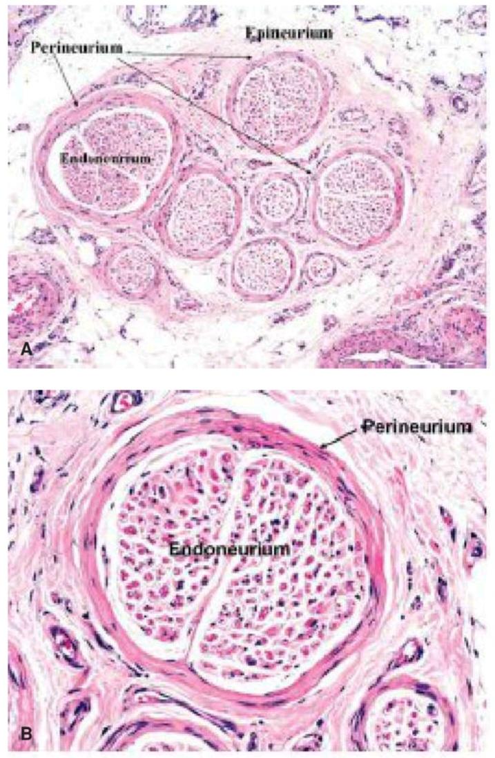
Figure 3-11 Peripheral nerve sheaths and compartments. A: A low-power view of a transverse section of a normal sural nerve. The nerve fascicles with roughly circular outlines are surrounded by perineurium and embedded in the connective tissue of the epineurium. Epineurial blood vessels (A) are also cut in cross section and there is adherent adipose tissue. The 1 μm sections are stained with hematoxylin and eosin (x16). B: The endoneurial compartment containing myelinated and non-myelinated nerve fibers and their accompanying Schwann cells is surrounded by perineurium. Paraffin section stained with hematoxylin and eosin (x45). (From Mills, S. E. Histology for Pathologists, 4e. Philadelphia, PA: Wolters Kluwer, 2013.)
شکل ۳-۱۱ غلافها و محفظههای عصبی محیطی. الف: نمای کمتوان از برش عرضی یک عصب سورال طبیعی. دستههای عصبی با خطوط تقریباً دایرهای توسط پرینوریوم احاطه شده و در بافت همبند اپینوریوم فرو رفتهاند. رگهای خونی اپینوریوم (A) نیز در مقطع عرضی برش داده شدهاند و بافت چربی چسبنده وجود دارد. برشهای 1 میکرومتری با هماتوکسیلین و ائوزین رنگآمیزی شدهاند (x16). ب: محفظه اندونوریال حاوی فیبرهای عصبی میلیندار و غیر میلیندار و سلولهای شوان همراه آنها توسط پرینوریوم احاطه شده است. برش پارافینی با هماتوکسیلین و ائوزین رنگآمیزی شده است (x45). (از Mills, S. E. Histology for Pathologists, 4e. Philadelphia, PA: Wolters Kluwer, 2013.)
The nerve trunk is surrounded by a dense connective tissue sheath called the epineurium (Fig. 3-12). Within the sheath are bundles of nerve fibers, each of which is surrounded by a connective tissue sheath called the perineurium. Between the individual nerve fibers is a loose, delicate connective tissue referred to as the endoneurium. The connective tissue sheaths serve to support the nerve fibers and their associated blood vessels and lymph vessels. Peripheral nerve fibers can be classified according to their speed of conduction and size (see Table 2-3).
تنه عصبی توسط یک غلاف بافت همبند متراکم به نام اپینوریوم احاطه شده است (شکل 3-12). درون غلاف، دستههایی از فیبرهای عصبی وجود دارند که هر کدام توسط یک غلاف بافت همبند به نام پرینوریوم احاطه شدهاند. بین فیبرهای عصبی منفرد، یک بافت همبند شل و ظریف به نام اندونوریوم وجود دارد. غلافهای بافت همبند، فیبرهای عصبی و رگهای خونی و لنفاوی مرتبط با آنها را پشتیبانی میکنند. فیبرهای عصبی محیطی را میتوان بر اساس سرعت هدایت و اندازه آنها طبقهبندی کرد (به جدول 2-3 مراجعه کنید).
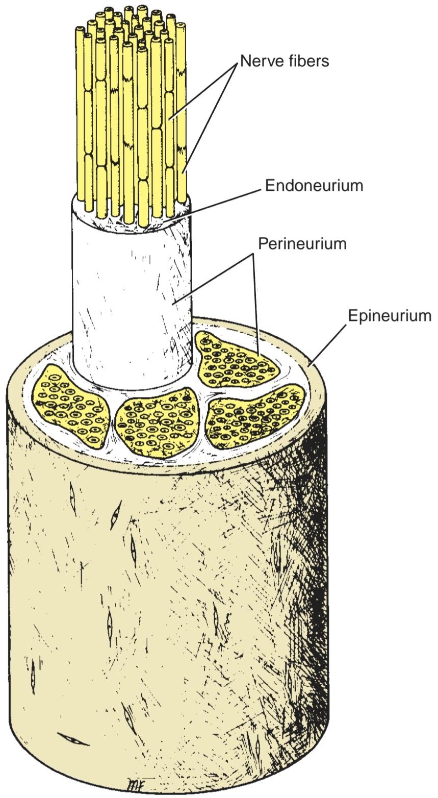
Figure 3-12 Structure of a peripheral nerve.
شکل ۳-۱۲ ساختار یک عصب محیطی.
Spinal Nerves and Spinal Nerve Roots
The 31 pairs of spinal nerves leave the spinal cord and pass through intervertebral foramina in the vertebral column. (For details, see Fig. 1-5.) Each spinal nerve is connected to the spinal cord by two roots: the anterior root and the posterior root (Fig. 3-13). The anterior root consists of bundles of nerve fibers carrying nerve impulses away from the CNS, the efferent fibers. The posterior root consists of bundles of nerve fibers carrying nerve impulses to the CNS, the afferent fibers. Because these fibers are concerned with conveying information to the CNS, they are called sensory fibers. The cell bodies of these nerve fibers are situated in a swelling on the posterior root called the posterior root ganglion.
اعصاب نخاعی و ریشههای عصب نخاعی
۳۱ جفت عصب نخاعی از نخاع خارج شده و از سوراخهای بین مهرهای در ستون فقرات عبور میکنند. (برای جزئیات، به شکل ۱-۵ مراجعه کنید.) هر عصب نخاعی توسط دو ریشه به نخاع متصل است: ریشه قدامی و ریشه خلفی (شکل ۳-۱۳). ریشه قدامی شامل دستههایی از الیاف عصبی است که تکانههای عصبی را از سیستم عصبی مرکزی (CNS) دور میکنند، یعنی الیاف وابران. ریشه خلفی شامل دستههایی از الیاف عصبی است که تکانههای عصبی را به سیستم عصبی مرکزی (CNS) حمل میکنند، یعنی الیاف آوران. از آنجایی که این الیاف مربوط به انتقال اطلاعات به سیستم عصبی مرکزی هستند، به آنها الیاف حسی گفته میشود. اجسام سلولی این الیاف عصبی در یک برآمدگی روی ریشه خلفی به نام گانگلیون ریشه خلفی قرار دارند.

Figure 3-13 A: Transverse section of the thoracic region of the spinal cord showing the formation of a spinal nerve from the union of an anterior and a posterior nerve root. B: Transverse section of the pons showing the sensory and motor roots of the trigeminal nerve.
شکل ۳-۱۳الف: برش عرضی ناحیه سینهای نخاع که تشکیل عصب نخاعی را از اتصال یک ریشه عصبی قدامی و یک ریشه عصبی خلفی نشان میدهد. ب: برش عرضی پل مغزی که ریشههای حسی و حرکتی عصب سهقلو را نشان میدهد.
Cranial Nerves
The 12 pairs of cranial nerves (see Fig. 3-13) leave the brain and pass through foramina in the skull. Some of these nerves are composed entirely of afferent nerve fibers bringing sensations to the brain (olfactory, optic, and vestibulocochlear), others are composed entirely of efferent fibers (oculomotor, trochlear, abducent, accessory, and hypoglossal), while the remainder possess both afferent and efferent fibers (trigeminal, facial, glossopharyngeal, and vagus). The cranial nerves are described in detail in Chapter 11.
اعصاب جمجمهای
۱۲ جفت عصب جمجمهای (شکل ۳-۱۳ را ببینید) از مغز خارج شده و از سوراخهای جمجمه عبور میکنند. برخی از این اعصاب کاملاً از الیاف عصبی آوران تشکیل شدهاند که حس را به مغز منتقل میکنند (بویایی، بینایی و دهلیزی-حلقی)، برخی دیگر کاملاً از الیاف وابران تشکیل شدهاند (حرکتی چشمی، قرقرهای، دورکننده، فرعی و زیرزبانی)، در حالی که بقیه دارای الیاف آوران و وابران هستند (سه قلو، صورتی، زبانی-حلقی و واگ). اعصاب جمجمهای به تفصیل در فصل ۱۱ توضیح داده شدهاند.
Sensory Ganglia
The sensory ganglia of the posterior spinal nerve roots and of the trunks of the trigeminal, facial, glossopharyn- geal, and vagal cranial nerves have the same structure.
گانگلیونهای حسی
گانگلیونهای حسی ریشههای خلفی عصب نخاعی و تنههای اعصاب جمجمهای سه قلو، صورت، زبانی-حلقی و واگ ساختار یکسانی دارند.
Each ganglion is surrounded by a layer of connective tissue that is continuous with the epineurium and perineurium of the peripheral nerve. The neurons are unipolar, possessing cell bodies that are rounded or oval in shape (Fig. 3-14). The cell bodies tend to be aggregated and separated by bundles of nerve fibers. A single nonmyelinated process leaves each cell body and, after a convoluted course, bifurcates at a T junction into peripheral and central branches. The former axon terminates in a series of dendrites in a peripheral sensory ending, and the latter axon enters the CNS. The nerve impulse, on reaching the T junction, passes directly from the peripheral axon to the central axon, thus bypassing the nerve cell body.
هر گانگلیون توسط لایهای از بافت همبند احاطه شده است که در امتداد اپینوریوم و پرینوریوم عصب محیطی قرار دارد. نورونها تکقطبی هستند و دارای اجسام سلولی گرد یا بیضی شکل میباشند (شکل 3-14). اجسام سلولی تمایل دارند که توسط دستههایی از الیاف عصبی جمع شده و از هم جدا شوند. یک زائده بدون میلین از هر جسم سلولی خارج میشود و پس از طی مسیری پیچیده، در محل اتصال T به شاخههای محیطی و مرکزی تقسیم میشود. آکسون اول در یک سری دندریت در یک انتهای حسی محیطی خاتمه مییابد و آکسون دوم وارد سیستم عصبی مرکزی میشود. تکانه عصبی، با رسیدن به محل اتصال T، مستقیماً از آکسون محیطی به آکسون مرکزی منتقل میشود و بنابراین جسم سلولی عصبی را دور میزند.
Each nerve cell body is closely surrounded by a layer of flattened cells called capsular cells or satellite cells. The capsular cells are similar in structure to Schwann cells and are continuous with these cells as they envelop the peripheral and central processes of each neuron.
هر جسم سلولی عصبی توسط لایهای از سلولهای مسطح به نام سلولهای کپسولی یا سلولهای ماهوارهای احاطه شده است. سلولهای کپسولی از نظر ساختاری مشابه سلولهای شوان هستند و در امتداد این سلولها قرار دارند، زیرا زوائد محیطی و مرکزی هر نورون را میپوشانند.

Figure 3-14 Sensory ganglion. I.s. Human. Paraffin section. ×132. The dorsal root ganglion provides a good representative example of a sensory ganglion. It possesses a vascular (BV) connective tissue capsule (C), which also envelops its sensory root. The neurons of the dorsal root ganglion are pseudounipolar in morphology; therefore, their somata (So) appear spherical in shape. The fibers (f), many of which are myelinated, alternate with rows of cell bodies. Note that some somata are large (arrow), whereas others are small (arrowhead). Each soma is surrounded by neuroectodermally derived capsule cells (Cc). A region similar to the boxed area is presented at a high magnification in Figure 4. (From Gartner, L. P. Color Atlas and Text of Histology, 7e. Baltimore: Wolters Kluwer, 2018.)
شکل ۳-۱۴ گانگلیون حسی. I.s. انسان. برش پارافین. ×132. گانگلیون ریشه پشتی نمونه خوبی از یک گانگلیون حسی را ارائه میدهد. این گانگلیون دارای یک کپسول (C) بافت همبند عروقی (BV) است که ریشه حسی آن را نیز احاطه کرده است. نورونهای گانگلیون ریشه پشتی از نظر مورفولوژی شبه تک قطبی هستند. بنابراین، سوماتا (So) آنها به شکل کروی به نظر میرسد. الیاف (f)، که بسیاری از آنها میلیندار هستند، به طور متناوب با ردیفهایی از اجسام سلولی قرار دارند. توجه داشته باشید که برخی از سوماتاها بزرگ هستند (فلش)، در حالی که برخی دیگر کوچک هستند (نوک فلش). هر سوما توسط سلولهای کپسولی مشتق شده از نورواکتودرم (Cc) احاطه شده است. ناحیهای مشابه ناحیه کادربندی شده با بزرگنمایی بالا در شکل 4 ارائه شده است. (از Gartner, L. P. Color Atlas and Text of Histology, 7e. Baltimore: Wolters Kluwer, 2018.)
Autonomic Ganglia
The autonomic ganglia (sympathetic and parasympa- thetic ganglia) are situated at a distance from the brain and spinal cord. They are found in the sympathetic trunks, in prevertebral autonomic plexuses (e.g., in the cardiac, celiac, and mesenteric plexuses), and as ganglia in or close to viscera. Each ganglion is surrounded by a layer of connective tissue that is continuous with the epineurium and perineurium of the peripheral nerve. The neurons are multipolar and possess cell bodies that are irregular in shape (Fig. 3-15). The dendrites of the neurons make synaptic connections with the myelinated axons of preganglionic neurons. The axons of the neurons are of small diameter (C fibers) and unmyelinated, and they pass to viscera, blood vessels, and sweat glands.
گانگلیونهای اتونومیک
گانگلیونهای اتونومیک (گانگلیونهای سمپاتیک و پاراسمپاتیک) در فاصلهای از مغز و نخاع قرار دارند. آنها در تنههای سمپاتیک، در شبکههای اتونومیک پیش مهرهای (مثلاً در شبکههای قلبی، سلیاک و مزانتریک) و به صورت گانگلیون در داخل یا نزدیک احشاء یافت میشوند. هر گانگلیون توسط لایهای از بافت همبند احاطه شده است که با اپینوریوم و پرینوریوم عصب محیطی در امتداد است. نورونها چند قطبی هستند و دارای اجسام سلولی با شکل نامنظم هستند (شکل 3-15). دندریتهای نورونها با آکسونهای میلیندار نورونهای پیشگانگلیونی، اتصالات سیناپسی برقرار میکنند. آکسونهای نورونها قطر کمی (فیبرهای C) و بدون میلین دارند و به احشاء، رگهای خونی و غدد عرق منتقل میشوند.
Just as with sensory ganglia, each nerve cell body is closely surrounded by capsular cells (or satellite cells). Unlike those of sensory ganglia, however, these capsular cells are similar in structure to Schwann cells and are continuous with them as they envelop the peripheral and central processes of each neuron.
درست مانند گانگلیونهای حسی، هر جسم سلولی عصبی توسط سلولهای کپسولی (یا سلولهای ماهوارهای) احاطه شده است. با این حال، برخلاف گانگلیونهای حسی، این سلولهای کپسولی از نظر ساختار مشابه سلولهای شوان هستند و در امتداد آنها قرار دارند، زیرا زوائد محیطی و مرکزی هر نورون را میپوشانند.

Figure 3-15 Photomicrograph of a longitudinal section of a ganglion of the sympathetic trunk stained with hematoxylin and eosin (x300).
شکل ۳-۱۵ عکس میکروسکوپی از برش طولی گانگلیون تنه سمپاتیک رنگآمیزی شده با هماتوکسیلین و ائوزین (300x).
Peripheral Nerve Plexuses
Peripheral nerves are composed of bundles of nerve fibers. In their course, peripheral nerves sometimes divide into branches that join neighboring peripheral nerves. If this occurs frequently, a network of nerves, called a nerve plexus, forms. Note that the formation of a nerve plexus allows individual nerve fibers to pass from one peripheral nerve to another, and in most instances, branching of nerve fibers does not take place. A plexus thus permits a redistribution of the nerve fibers within the different peripheral nerves.
شبکههای عصبی محیطی
اعصاب محیطی از دستههایی از فیبرهای عصبی تشکیل شدهاند. در مسیر خود، اعصاب محیطی گاهی به شاخههایی تقسیم میشوند که به اعصاب محیطی مجاور میپیوندند. اگر این اتفاق به طور مکرر رخ دهد، شبکهای از اعصاب به نام شبکه عصبی تشکیل میشود. توجه داشته باشید که تشکیل شبکه عصبی به فیبرهای عصبی منفرد اجازه میدهد تا از یک عصب محیطی به عصب محیطی دیگر منتقل شوند و در بیشتر موارد، شاخهبندی فیبرهای عصبی اتفاق نمیافتد. بنابراین، یک شبکه عصبی امکان توزیع مجدد فیبرهای عصبی را در اعصاب محیطی مختلف فراهم میکند.
At the root of the limbs, the anterior rami of the spinal nerves form complicated plexuses. The cervical and brachial plexuses are at the root of the upper limbs (Fig. 3-16), and the lumbar and sacral plexuses are at the root of the lower limbs. This allows the nerve fibers derived from different segments of the spinal cord to be arranged and distributed efficiently in different nerve trunks to the various parts of the upper and lower limbs.
در ریشه اندامها، شاخههای قدامی اعصاب نخاعی، شبکههای عصبی پیچیدهای را تشکیل میدهند. شبکههای عصبی گردنی و بازویی در ریشه اندامهای فوقانی قرار دارند (شکل 3-16) و شبکههای عصبی کمری و خاجی در ریشه اندامهای تحتانی قرار دارند. این امر به فیبرهای عصبی مشتق شده از بخشهای مختلف نخاع اجازه میدهد تا به طور مؤثر در تنههای عصبی مختلف به قسمتهای مختلف اندامهای فوقانی و تحتانی مرتب و توزیع شوند.
Cutaneous nerves, as they approach their final destination, commonly form fine plexuses that again permit a rearrangement of nerve fibers before they reach their terminal sensory endings.
اعصاب پوستی، هنگامی که به مقصد نهایی خود نزدیک میشوند، معمولاً شبکههای عصبی ظریفی را تشکیل میدهند که مجدداً امکان بازآرایی فیبرهای عصبی را قبل از رسیدن به انتهای حسی انتهایی خود فراهم میکنند.
The autonomic nervous system also possesses numerous nerve plexuses that consist of preganglionic and postganglionic nerve fibers and ganglia.
سیستم عصبی خودکار همچنین دارای شبکههای عصبی متعددی است که از فیبرها و گانگلیونهای عصبی پیش عقدهای و پس عقدهای تشکیل شدهاند.

Figure 3-16 Brachial plexus.
شکل ۳-۱۶ شبکه عصبی بازویی.
RECEPTOR ENDINGS
An individual receives impressions from the outside world and from within the body by special sensory nerve endings or receptors.
پایانههای گیرنده
یک فرد از طریق پایانههای عصبی حسی یا گیرندههای خاص، تأثیرات را از دنیای بیرون و از درون بدن دریافت میکند.
Sensory receptors can be classified into five basic functional types:
گیرندههای حسی را میتوان به پنج نوع عملکردی اساسی طبقهبندی کرد:
Mechanoreceptors respond to mechanical deformation.
گیرندههای مکانیکی به تغییر شکل مکانیکی پاسخ میدهند.
Thermoreceptors respond to changes in temperature; some receptors respond to cold and others to heat. Nociceptors respond to any stimuli that bring about damage to the tissue.
گیرندههای حرارتی به تغییرات دما پاسخ میدهند؛ برخی از گیرندهها به سرما و برخی دیگر به گرما. گیرندههای درد به هر محرکی که باعث آسیب به بافت شود، پاسخ میدهند.
Electromagnetic receptors, the rods and cones of the eyes, are sensitive to changes in light intensity and wavelength.
گیرندههای الکترومغناطیسی، میلهها و مخروطهای چشم، به تغییرات شدت و طول موج نور حساس هستند.
Chemoreceptors respond to chemical changes associated with taste and smell and oxygen and carbon dioxide concentrations in the blood.
گیرندههای شیمیایی به تغییرات شیمیایی مرتبط با طعم و بو و غلظت اکسیژن و دی اکسید کربن در خون پاسخ میدهند.
Anatomical Types of Receptors
For convenience, the sensory endings can be classified, on a structural basis, into nonencapsulated and encap- sulated receptors. Table 3-2 classifies and compares the receptor types.
انواع آناتومیکی گیرندهها
برای راحتی، انتهای حسی را میتوان بر اساس ساختار، به گیرندههای بدون کپسول و کپسولدار طبقهبندی کرد. جدول 3-2 انواع گیرندهها را طبقهبندی و مقایسه میکند.
Table 3-2 Classification and Comparison of Receptor Types
جدول ۳-۲ طبقهبندی و مقایسه انواع گیرندهها

Nonencapsulated Receptors
Free Nerve Endings
Free nerve endings are widely distributed throughout the body (Fig. 3-17). They are found between the epithelial cells of the skin, the cornea, and the alimentary tract, and in connective tissues, including the dermis, fascia, ligaments, joint capsules, tendons, periosteum, perichondrium, haversian systems of bone, tympanic membrane, and dental pulp; they are also present in muscle.
گیرندههای بدون کپسول
پایانههای عصبی آزاد
پایانههای عصبی آزاد به طور گسترده در سراسر بدن پراکنده شدهاند (شکل 3-17). آنها بین سلولهای اپیتلیال پوست، قرنیه و دستگاه گوارش و در بافتهای همبند، از جمله درم، فاسیا، رباطها، کپسولهای مفصلی، تاندونها، پریوستئوم، پریکندریوم، سیستمهای هاورس استخوان، پرده صماخ و پالپ دندان یافت میشوند. آنها همچنین در عضله وجود دارند.
The afferent nerve fibers from the free nerve endings are either myelinated or nonmyelinated. The terminal endings are devoid of a myelin sheath as well as Schwann cells covering their tips.
فیبرهای عصبی آوران از انتهای آزاد عصب، یا میلیندار هستند یا بدون میلین. انتهای انتهایی فاقد غلاف میلین و همچنین سلولهای شوان پوشاننده نوک آنها میباشند.
Most of these endings detect pain, while others detect crude touch, pressure, and tickle sensations, and possibly cold and heat.
بیشتر این انتهاییها درد را تشخیص میدهند، در حالی که برخی دیگر حسهای لمس، فشار و قلقلک خام و احتمالاً سرما و گرما را تشخیص میدهند.

Figure 3-17 Free nerve endings in the skin. The nerve fibers in the epidermis are naked.
شکل ۳-۱۷ پایانههای عصبی آزاد در پوست. رشتههای عصبی در اپیدرم برهنه هستند.
Merkel Discs
Merkel discs are found in hairless skin, for example, the fingertips (Figs. 3-18 and 3-19), and in hair follicles. The nerve fiber passes into the epidermis and terminates as a disc-shaped expansion that is applied closely to a dark-staining epithelial cell in the deeper part of the epidermis, called the Merkel cell. In hairy skin, clusters of Merkel discs, known as tactile domes, are found in the epidermis between the hair follicles.
دیسکهای مرکل
دیسکهای مرکل در پوستهای بدون مو، مثلاً نوک انگشتان (شکلهای 3-18 و 3-19) و در فولیکولهای مو یافت میشوند. فیبر عصبی به اپیدرم وارد میشود و به صورت یک گسترش دیسکی شکل خاتمه مییابد که نزدیک به یک سلول اپیتلیال تیره رنگ در قسمت عمیقتر اپیدرم، به نام سلول مرکل، قرار دارد. در پوست مودار، خوشههایی از دیسکهای مرکل، که به عنوان گنبدهای لمسی شناخته میشوند، در اپیدرم بین فولیکولهای مو یافت میشوند.
Merkel discs are slowly adapting touch receptors that transmit information about the degree of pressure exerted on the skin, such as from holding a pen.
دیسکهای مرکل به آرامی در حال تطبیق گیرندههای لمسی هستند که اطلاعاتی در مورد میزان فشار وارد شده بر پوست، مانند فشار ناشی از نگه داشتن یک خودکار، منتقل میکنند.

Figure 3-18 Merkel discs in the skin. A: Low magnification. B: Merkel disc showing the expanded ending of an axon with a stippled tactile cell.
شکل ۳-۱۸ دیسکهای مرکل در پوست. الف: بزرگنمایی کم. ب: دیسک مرکل انتهای منبسط شده یک آکسون را با یک سلول لمسی نقطه نقطه نشان میدهد.

Figure 3-19 Photomicrograph of digital skin showing fine nerve terminals ending in Merkel discs, stained by the silver method. (Courtesy Dr. N. Cauna.)
شکل ۳-۱۹ عکس میکروسکوپی از پوست دیجیتال که پایانههای عصبی ظریف منتهی به دیسکهای مرکل را نشان میدهد، که با روش نقره رنگآمیزی شده است. (با احترام دکتر ن. کاونا.)
Hair Follicle Receptors
Nerve fibers wind around the follicle in its outer connective tissue sheath below the sebaceous gland. Some branches surround the follicle, while others run parallel to its long axis (Figs. 3-20 and 3-21). Many naked axon filaments terminate among the cells of the outer root sheath.
گیرندههای فولیکول مو
الیاف عصبی در غلاف بافت همبند بیرونی فولیکول در زیر غده چربی به دور آن میپیچند. برخی از شاخهها فولیکول را احاطه کردهاند، در حالی که برخی دیگر به موازات محور طولی آن امتداد دارند (شکلهای 3-20 و 3-21). بسیاری از رشتههای آکسون برهنه در میان سلولهای غلاف ریشه بیرونی خاتمه مییابند.
Bending of the hair stimulates the follicle receptor, which belongs to the rapidly adapting group of mechanoreceptors. While the hair remains bent, the receptor is silent, but when the hair is released, a further burst of nerve impulses is initiated.
خم شدن مو، گیرنده فولیکول را که متعلق به گروه گیرندههای مکانیکی با سازگاری سریع است، تحریک میکند. در حالی که مو خم شده است، گیرنده خاموش است، اما هنگامی که مو آزاد میشود، موج دیگری از تکانههای عصبی آغاز میشود.

Figure 3-20 Nerve endings around a hair follicle.
شکل ۳-۲۰ انتهای عصبی اطراف فولیکول مو.

Figure 3-21 Photomicrograph of nerve endings around a hair follicle stained by the silver method. (Courtesy Dr. M. J. T. Fitzgerald.)
شکل ۳-۲۱ عکس میکروسکوپی از انتهای اعصاب اطراف فولیکول مو که با روش نقره رنگآمیزی شده است. (با تشکر از دکتر ام. جی. تی. فیتزجرالد.)
Encapsulated Receptors
Encapsulated receptors show wide variations in size and shape, and the termination of the nerve is covered by a capsule.
گیرندههای کپسولدار
گیرندههای کپسولدار از نظر اندازه و شکل تغییرات گستردهای نشان میدهند و انتهای عصب توسط یک کپسول پوشانده شده است.
Meissner Corpuscles
Meissner corpuscles are located in the dermal papillae of the skin (Figs. 3-22 and 3-23), especially that of the palm of the hand and the sole of the foot. Many also are present in the skin of the nipple and the external genitalia. Each corpuscle is ovoid in shape and consists of a stack of modified flattened Schwann cells arranged transversely across the long axis of the corpuscle. The corpuscle is enclosed by a capsule of connective tissue that is continuous with the endoneurium of the nerves that enter it. A few myelinated nerve fibers enter the deep end of the corpuscle; myelinated and unmyelinated branches decrease in size and ramify among the Schwann cells. The number of Meissner corpuscles considerably diminishes between birth and old age.
اجسام مایسنر
اجسام مایسنر در پاپیلای پوستی پوست (شکلهای 3-22 و 3-23)، به ویژه در کف دست و کف پا قرار دارند. بسیاری از آنها نیز در پوست نوک پستان و دستگاه تناسلی خارجی وجود دارند. هر جسم به شکل تخممرغی است و از مجموعهای از سلولهای شوان مسطح و تغییر شکلیافته تشکیل شده است که به صورت عرضی در امتداد محور طولی جسم قرار گرفتهاند. جسم توسط کپسولی از بافت همبند احاطه شده است که با اندونوریوم اعصابی که وارد آن میشوند، پیوسته است. تعداد کمی از فیبرهای عصبی میلیندار وارد انتهای عمقی جسم میشوند. شاخههای میلیندار و بدون میلین از نظر اندازه کاهش مییابند و در بین سلولهای شوان منشعب میشوند. تعداد اجسام مایسنر بین تولد و پیری به طور قابل توجهی کاهش مییابد.

Figure 3-22 Detailed structure of a Meissner corpuscle in the skin.
شکل ۳-۲۲ ساختار دقیق یک جسمک مایسنر در پوست.

Figure 3-23 Photomicrograph of a Meissner corpuscle of the skin. (Courtesy Dr. N. Cauna.)
شکل ۳-۲۳ عکس میکروسکوپی از یک جسم مایسنر پوست. (با تشکر از دکتر ن. کاونا.)
Meissner corpuscles are very sensitive to touch and are rapidly adapting mechanoreceptors. They enable an individual to distinguish between two pointed structures when they are placed close together on the skin (two-point tactile discrimination).
اجسام مایسنر به لمس بسیار حساس هستند و به سرعت در حال تطبیق با گیرندههای مکانیکی هستند. آنها به فرد این امکان را میدهند که وقتی دو ساختار نوکتیز روی پوست نزدیک به هم قرار میگیرند، آنها را از هم تشخیص دهد (تمایز لمسی دو نقطهای).
Pacinian Corpuscles
Pacinian corpuscles (Figs. 3-24 and 3-25) are widely distributed throughout the body and are abundant in the dermis, subcutaneous tissue, ligaments, joint capsules, pleura, peritoneum, nipples, and external genitalia. Each corpuscle is ovoid in shape, measuring about 2 mm long and about 100 to 500 um across. It consists of a capsule and a central core containing the nerve ending. The capsule consists of numerous concentric lamellae of flattened cells. A large myelinated nerve fiber enters the corpuscle and loses its myelin sheath and then its Schwann cell covering. The naked axon, surrounded by lamellae formed of flattened cells, passes through the center of the core and terminates in an expanded end.
اجسام پاچینی
اجسام پاچینی (شکلهای ۳-۲۴ و ۳-۲۵) به طور گسترده در سراسر بدن پراکنده شدهاند و در درم، بافت زیر جلدی، رباطها، کپسولهای مفصلی، پلور، صفاق، نوک سینهها و دستگاه تناسلی خارجی به وفور یافت میشوند. هر جسم به شکل تخممرغی است و حدود ۲ میلیمتر طول و حدود ۱۰۰ تا ۵۰۰ میکرومتر عرض دارد. این جسم از یک کپسول و یک هسته مرکزی حاوی انتهای عصبی تشکیل شده است. کپسول از تیغههای متحدالمرکز متعددی از سلولهای مسطح تشکیل شده است. یک فیبر عصبی میلیندار بزرگ وارد جسم میشود و غلاف میلین و سپس پوشش سلول شوان خود را از دست میدهد. آکسون برهنه، که توسط تیغههایی از سلولهای مسطح احاطه شده است، از مرکز هسته عبور میکند و به یک انتهای گشاد ختم میشود.
The Pacinian corpuscle is a rapidly adapting mechanoreceptor that is particularly sensitive to vibration. It can respond to up to 600 stimuli per second.
جسم پاچینی یک گیرنده مکانیکی به سرعت سازگار است که به ویژه به ارتعاش حساس است. میتواند تا ۶۰۰ محرک در ثانیه پاسخ دهد.

Figure 3-24 Detailed structure of a Pacinian corpuscle in the skin.
شکل ۳-۲۴ ساختار دقیق یک جسمک پاچینی در پوست.

Figure 3-25 Pacinian corpuscle. Paraffin section. 132. Pacinian corpuscles, located in the dermis and hypodermis, are mechanoreceptors. They are composed of a core with an inner (IC) and an outer (OC) region, as well as a capsule (Ca) that surrounds the core. The inner core invests the afferent nerve fiber (NF), which loses its myelin sheath soon after entering the corpuscle. The core cells are modified Schwann cells, whereas the components of the capsule are continuous with the endoneurium of the afferent nerve fiber. Pacinian corpuscles are readily recognizable in section since they resemble the cut surface of an onion. Observe the presence of an arrector pili muscle (AP) and profiles of ducts (d) of a sweat gland in the vicinity of, but not associated with, the pacinian corpuscle. (From Gartner, L. P. Color Atlas and Text of Histology, 7e. Baltimore: Wolters Kluwer, 2018.)
شکل 3-25 جسمک پاچینی. برش پارافین. 132. جسمکهای پاچینی، که در درم و هیپودرم قرار دارند، گیرندههای مکانیکی هستند. آنها از یک هسته با یک ناحیه داخلی (IC) و یک ناحیه خارجی (OC) و همچنین یک کپسول (Ca) که هسته را احاطه کرده است، تشکیل شدهاند. هسته داخلی، فیبر عصبی آوران (NF) را در خود جای میدهد که غلاف میلین خود را به زودی پس از ورود به جسمک از دست میدهد. سلولهای هسته، سلولهای شوان اصلاحشده هستند، در حالی که اجزای کپسول در امتداد اندونوریوم فیبر عصبی آوران قرار دارند. جسمکهای پاچینی در برش به راحتی قابل تشخیص هستند زیرا شبیه سطح برش پیاز هستند. وجود عضله راستکننده مو (AP) و پروفایل مجاری (d) یک غده عرق را در مجاورت جسمک پاچینی، اما نه مرتبط با آن، مشاهده کنید. (از گارتنر، ال. پی. اطلس رنگی و متن بافتشناسی، جلد هفتم. بالتیمور: ولترز کلوور، ۲۰۱۸.)
Ruffini Corpuscles
Ruffini corpuscles are located in the dermis of hairy skin. Each corpuscle consists of several large unmyelinated nerve fibers ending within a bundle of collagen fibers and surrounded by a cellular capsule. These slowly adapting mechanoreceptors are stretch receptors, which respond when the skin is stretched.
اجسام رافینی
اجسام رافینی در درم پوست مودار قرار دارند. هر جسم از چندین فیبر عصبی بزرگ بدون میلین تشکیل شده است که به یک دسته از الیاف کلاژن ختم میشوند و توسط یک کپسول سلولی احاطه شدهاند. این گیرندههای مکانیکی که به آرامی سازگار میشوند، گیرندههای کششی هستند که هنگام کشش پوست پاسخ میدهند.
Cutaneous Receptor Function
The different histologic types of receptors were previously thought to correspond to specific types of sensation, until scientists pointed out that some areas of the body have only one or two histologic types of receptors and yet are sensitive to a variety of different stimuli. Moreover, although the body has these different receptors, all nerves only transmit nerve impulses. Now scientists generally agree that the type of sensation felt is determined by the specific area of the CNS to which the afferent nerve fiber passes. For example, if a pain nerve fiber is stimulated by heat, cold, touch, or pressure, the individual will experience only pain.
عملکرد گیرندههای پوستی
قبلاً تصور میشد که انواع مختلف بافتشناسی گیرندهها با انواع خاصی از حس مطابقت دارند، تا اینکه دانشمندان خاطرنشان کردند که برخی از نواحی بدن فقط یک یا دو نوع بافتشناسی گیرنده دارند و با این حال به انواع محرکهای مختلف حساس هستند. علاوه بر این، اگرچه بدن این گیرندههای مختلف را دارد، اما همه اعصاب فقط تکانههای عصبی را منتقل میکنند. اکنون دانشمندان عموماً موافقند که نوع حس احساس شده توسط ناحیه خاصی از سیستم عصبی مرکزی که فیبر عصبی آوران به آن منتقل میشود، تعیین میشود. به عنوان مثال، اگر یک فیبر عصبی درد توسط گرما، سرما، لمس یا فشار تحریک شود، فرد فقط درد را تجربه خواهد کرد.
Sensory Stimulus Transduction into Nerve Impulse
Transduction is the process by which one form of energy (the stimulus) is changed into another form of energy (electrochemical energy of the nerve impulse). The stimulus, when applied to the receptor, brings about a change in potential of the plasma membrane of the nerve ending. Because this process takes place in the receptor, it is referred to as the receptor potential. The amplitude of the receptor potential is proportional to the intensity of the stimulus. By opening more ion channels for a longer time, a stronger mechanical pressure, for example, can produce a greater depolarization than does weak pressure. With chemoreceptors and photoreceptors, the receptor potential is produced by second messengers activated when the stimulus agent binds to the membrane receptors coupled to G proteins. If large enough, the receptor potential will generate an AP that will travel along the afferent nerve fiber to the CNS.
انتقال محرک حسی به تکانه عصبی
انتقال فرآیندی است که طی آن یک شکل از انرژی (محرک) به شکل دیگری از انرژی (انرژی الکتروشیمیایی تکانه عصبی) تبدیل میشود. محرک، هنگامی که به گیرنده اعمال میشود، باعث تغییر در پتانسیل غشای پلاسمایی انتهای عصب میشود. از آنجا که این فرآیند در گیرنده اتفاق میافتد، به آن پتانسیل گیرنده میگویند. دامنه پتانسیل گیرنده متناسب با شدت محرک است. با باز کردن کانالهای یونی بیشتر برای مدت زمان طولانیتر، به عنوان مثال، یک فشار مکانیکی قویتر میتواند دپلاریزاسیون بیشتری نسبت به فشار ضعیف ایجاد کند. در گیرندههای شیمیایی و گیرندههای نوری، پتانسیل گیرنده توسط پیامرسانهای ثانویه فعال میشود که هنگام اتصال عامل محرک به گیرندههای غشایی متصل به پروتئینهای G فعال میشوند. اگر پتانسیل گیرنده به اندازه کافی بزرگ باشد، یک پتانسیل گیرنده یک پتانسیل فعال (AP) ایجاد میکند که در امتداد فیبر عصبی آوران به سیستم عصبی مرکزی (CNS) منتقل میشود.
Joint Receptors
Four types of sensory endings can be located in the capsule and ligaments of synovial joints. Three of these endings are encapsulated and resemble Pacinian, Ruffini, and tendon stretch receptors. They provide the CNS with information regarding the position and movements of the joint. A fourth type of ending is nonencapsulated and is thought to be sensitive to excessive movements and to transmit pain sensations.
گیرندههای مفصلی
چهار نوع انتهای حسی میتوانند در کپسول و رباطهای مفاصل سینوویال قرار داشته باشند. سه مورد از این انتهاها کپسولدار هستند و شبیه گیرندههای پاچینی، رافینی و کششی تاندون هستند. آنها اطلاعاتی در مورد موقعیت و حرکات مفصل به سیستم عصبی مرکزی ارائه میدهند. نوع چهارم انتها کپسولدار نیست و تصور میشود که به حرکات بیش از حد حساس است و حس درد را منتقل میکند.
Neuromuscular Spindles
Neuromuscular spindles, or muscular spindles (Figs. 3-26 and 3-27), are found in skeletal muscle and are most numerous toward the tendinous attachment of the muscle. They provide the CNS with sensory information regarding the muscle length and the rate of change in the muscle length. The CNS uses this information to control muscle activity.
دوکهای عصبی-عضلانی
دوکهای عصبی-عضلانی یا دوکهای عضلانی (شکلهای 3-26 و 3-27)، در عضله اسکلتی یافت میشوند و بیشترین تعداد آنها در جهت اتصال تاندونی عضله قرار دارد. آنها اطلاعات حسی در مورد طول عضله و میزان تغییر در طول عضله را برای سیستم عصبی مرکزی فراهم میکنند. سیستم عصبی مرکزی از این اطلاعات برای کنترل فعالیت عضله استفاده میکند.

Figure 3-26 Neuromuscular spindle showing two types of intrafusal fibers: the nuclear bag and nuclear chain fibers.
شکل ۳-۲۶ دوک عصبی-عضلانی دو نوع فیبر داخل دوکی را نشان میدهد: کیسه هستهای و فیبرهای زنجیره هستهای.

Figure 3-27 Photomicrograph of a neuromuscular spindle.
شکل ۳-۲۷ عکس میکروسکوپی از دوک عصبی-عضلانی.
Each spindle measures about 1 to 4 mm in length and is surrounded by a fusiform capsule of connective tissue (Fig. 3-28). Within the capsule are 6 to 14 slender intrafusal muscle fibers; the ordinary muscle fibers situated outside the spindles are referred to as extrafusal fibers. The intrafusal fibers of the spindles are of two types: the nuclear bag and nuclear chain fibers. The nuclear bag fibers are recognized by the presence of numerous nuclei in the equatorial region, which consequently is expanded; also, cross striations are absent in this region. In the nuclear chain fibers, the nuclei form a single longitudinal row or chain in the center of each fiber at the equatorial region. The nuclear bag fibers are larger in diameter than the nuclear chain fibers, and they extend beyond the capsule at each end to be attached to the endomysium of the extrafusal fibers.
هر دوک حدود ۱ تا ۴ میلیمتر طول دارد و توسط یک کپسول دوکیشکل از بافت همبند احاطه شده است (شکل ۳-۲۸). درون کپسول ۶ تا ۱۴ فیبر عضلانی باریک درون دوکی وجود دارد؛ فیبرهای عضلانی معمولی که در خارج از دوکها قرار دارند، فیبرهای خارج دوکی نامیده میشوند. فیبرهای داخل دوکی دوکها دو نوع هستند: کیسه هستهای و فیبرهای زنجیره هستهای. فیبرهای کیسه هستهای با وجود هستههای متعدد در ناحیه استوایی شناخته میشوند که در نتیجه گسترش مییابند. همچنین، خطوط متقاطع در این ناحیه وجود ندارند. در فیبرهای زنجیره هستهای، هستهها یک ردیف یا زنجیره طولی واحد را در مرکز هر فیبر در ناحیه استوایی تشکیل میدهند. فیبرهای کیسه هستهای قطر بزرگتری نسبت به فیبرهای زنجیره هستهای دارند و در هر انتها از کپسول فراتر میروند تا به اندومیزیم فیبرهای خارج دوکی متصل شوند.

Figure 3-28 Muscle spindle. Mouse. Electron microscopy. x6,300. Parts of the outer capsule (oC) may be observed at the corners of this electron micrograph. The periaxial space (PS) surrounds the slender inner capsule (iC), whose component cells form attenuated branches, subdividing the axial space (AS) into several compartments for the nuclear chain (NC) and nuclear bag (NB) intrafusal fibers and their corresponding sensory terminals (ST). Note that the attenuated processes of the inner capsule cells establish contact with each other (arrows). (From Ovalle, W., Dow, P. (1983). Comparative ultrastructure of the inner capsule of the muscle spindle and the tendon organ. American Journal of Anatomy, 166, 343-357.)
شکل ۳-۲۸ دوک عضلانی. موش. میکروسکوپ الکترونی. x6,300. بخشهایی از کپسول خارجی (oC) را میتوان در گوشههای این میکروگراف الکترونی مشاهده کرد. فضای پیرامونی (PS) کپسول داخلی باریک (iC) را احاطه کرده است که سلولهای تشکیلدهنده آن شاخههای ضعیفی را تشکیل میدهند و فضای محوری (AS) را به چندین محفظه برای فیبرهای داخل دوکی زنجیره هستهای (NC) و کیسه هستهای (NB) و پایانههای حسی مربوطه (ST) آنها تقسیم میکنند. توجه داشته باشید که زوائد ضعیفشده سلولهای کپسول داخلی با یکدیگر تماس برقرار میکنند (فلشها). (از Ovalle, W., Dow, P. (1983). فراساختار مقایسهای کپسول داخلی دوک عضلانی و اندام تاندونی. مجله آناتومی آمریکا، 166، 343-357.)
The two types of sensory innervation of muscle spindles are annulospiral and flower spray. The annulospiralendings are situated at the equator of the intrafusal fibers. As the large myelinated nerve fiber pierces the capsule, it loses its myelin sheath, and the naked axon winds spirally around the nuclear bag or chain portions of the intrafusal fibers.
دو نوع عصبدهی حسی دوکهای عضلانی عبارتند از: آنولواسپیرال و فلاور اسپری. انتهای آنولواسپیرال در خط استوای فیبرهای داخل دوکی قرار دارند. هنگامی که فیبر عصبی بزرگ میلیندار کپسول را سوراخ میکند، غلاف میلین خود را از دست میدهد و آکسون برهنه به صورت مارپیچی به دور کیسه هستهای یا بخشهای زنجیرهای فیبرهای داخل دوکی میپیچد.
The flower-spray endings are situated mainly on the nuclear chain fibers some distance away from the equatorial region. A myelinated nerve fiber slightly smaller than that for the annulospiral ending pierces the capsule and loses its myelin sheath, and the naked axon branches terminally and ends as varicosities; it resembles a spray of flowers.
انتهای افشانههای گل عمدتاً روی فیبرهای زنجیره هستهای کمی دورتر از ناحیه استوایی قرار دارند. یک فیبر عصبی میلیندار که کمی کوچکتر از انتهای آنولوسپیرال است، کپسول را سوراخ میکند و غلاف میلین خود را از دست میدهد و آکسون برهنه به صورت انتهایی منشعب میشود و به صورت واریس پایان مییابد؛ شبیه افشانه گل است.
Stretching (elongation) of the intrafusal fibers results in stimulation of the annulospiral and flower-spray endings, and nerve impulses pass to the spinal cord in the afferent neurons.
کشش (طویل شدن) فیبرهای داخل دوکی منجر به تحریک انتهای حلقهای-مارپیچی و انتهای گل میشود و تکانههای عصبی از طریق نورونهای آوران به نخاع منتقل میشوند.
Motor innervation of the intrafusal fibers is provided by fine y motor fibers. The nerves terminate in small motor endplates situated at both ends of the intrafusal fibers. Stimulation of the motor nerves causes both ends of the intrafusal fibers to contract and activate the sensory endings. The equatorial region, which lacks cross striations, is noncontractile. The extrafusal fibers of the remainder of the muscle receive their innervation in the usual way from large a-size axons.
عصبدهی حرکتی فیبرهای داخل دوکی توسط فیبرهای حرکتی ظریف y انجام میشود. اعصاب به صفحات انتهایی حرکتی کوچکی که در دو انتهای فیبرهای داخل دوکی قرار دارند، ختم میشوند. تحریک اعصاب حرکتی باعث میشود که هر دو انتهای فیبرهای داخل دوکی منقبض شده و انتهای حسی را فعال کنند. ناحیه استوایی که فاقد خطوط متقاطع است، غیرانقباضی است. فیبرهای خارج دوکی بقیه عضله، عصبدهی خود را به روش معمول از آکسونهای بزرگ با اندازه a دریافت میکنند.
Neuromuscular Spindle Function
Under resting conditions, the muscle spindles give rise to afferent nerve impulses all the time, and most of this information is not consciously perceived. When muscle activity occurs, either actively or passively, the intrafusal fibers are stretched, and the rate of passage of nerve impulses to the spinal cord or brain increases in the afferent neurons. Similarly, if the intrafusal fibers are now relaxed due to the cessation of muscle activity, the result is a decrease in the rate of passage of nerve impulses. The neuromuscular spindle thus plays a very important role in keeping the CNS informed about the length of a muscle and the rate of change of its length, thereby indirectly influencing the control of voluntary muscle.
عملکرد دوک عصبی-عضلانی
در شرایط استراحت، دوکهای عضلانی دائماً باعث ایجاد تکانههای عصبی آوران میشوند و بیشتر این اطلاعات به صورت آگاهانه درک نمیشوند. هنگامی که فعالیت عضلانی رخ میدهد، چه به صورت فعال و چه غیرفعال، فیبرهای داخل دوکی کشیده میشوند و سرعت عبور تکانههای عصبی به نخاع یا مغز در نورونهای آوران افزایش مییابد. به طور مشابه، اگر فیبرهای داخل دوکی به دلیل توقف فعالیت عضلانی شل شوند، نتیجه کاهش سرعت عبور تکانههای عصبی است. بنابراین، دوک عصبی-عضلانی نقش بسیار مهمی در آگاه نگه داشتن سیستم عصبی مرکزی در مورد طول عضله و سرعت تغییر طول آن ایفا میکند و از این طریق به طور غیرمستقیم بر کنترل عضلات ارادی تأثیر میگذارد.
Stretch Reflex
The neurons of the spinal cord involved in the simple stretch reflex are as follows. Stretching a muscle results in elongation of the intrafusal fibers of the muscle spindle and stimulation of the annulospiral and flower-spray endings. The nerve impulses reach the spinal cord in the afferent neurons and synapse with the large a motor neurons situated in the anterior gray horns of the spinal cord. Nerve impulses now pass via the efferent motor nerves and stimulate the extrafusal muscle fibers, and the muscle contracts. This simple stretch reflex depends on a two-neuron arc consisting of an afferent neuron and an efferent neuron. Interestingly, the muscle spindle afferent impulses inhibit the a motor neurons supplying the antagonist muscles. This effect is called reciprocal inhibition.
رفلکس کششی
نورونهای نخاع که در رفلکس کششی ساده دخیل هستند به شرح زیر هستند. کشش یک عضله منجر به طویل شدن فیبرهای داخل دوکی دوک عضلانی و تحریک انتهای حلقوی-اسپری و انتهای گل میشود. تکانههای عصبی از طریق نورونهای آوران به نخاع میرسند و با نورونهای حرکتی بزرگ a واقع در شاخهای خاکستری قدامی نخاع سیناپس برقرار میکنند. تکانههای عصبی اکنون از طریق اعصاب حرکتی وابران عبور میکنند و فیبرهای عضلانی خارج دوکی را تحریک میکنند و عضله منقبض میشود. این رفلکس کششی ساده به یک قوس دو نورونی متشکل از یک نورون آوران و یک نورون وابران بستگی دارد. جالب توجه است که تکانههای آوران دوک عضلانی، نورونهای حرکتی a را که عضلات آنتاگونیست را تغذیه میکنند، مهار میکنند. این اثر مهار متقابل نامیده میشود.
Intrafusal Fiber Control
In the brain and spinal cord, certain centers give rise to tracts that synapse with y motor neurons in the spinal cord. The reticular formation, the basal ganglia, and the cerebellum are examples of such centers. By these means, these centers can greatly influence voluntary muscle activity. The y efferent motor fibers cause shortening of the intrafusal fibers, stretching the equatorial regions and stimulating the annulospiral and flower- spray endings. This, in turn, initiates the reflex contraction of the extrafusal fibers described previously.
کنترل فیبرهای درون دوکی
در مغز و نخاع، مراکز خاصی باعث ایجاد مسیرهایی میشوند که با نورونهای حرکتی y در نخاع سیناپس برقرار میکنند. تشکیلات مشبک، عقدههای قاعدهای و مخچه نمونههایی از چنین مراکزی هستند. به این ترتیب، این مراکز میتوانند فعالیت عضلانی ارادی را تا حد زیادی تحت تأثیر قرار دهند. فیبرهای حرکتی وابران y باعث کوتاه شدن فیبرهای درون دوکی، کشش نواحی استوایی و تحریک انتهای حلقوی-مارپیچی و انتهای گل-اسپری میشوند. این به نوبه خود، انقباض رفلکسی فیبرهای خارج دوکی را که قبلاً توضیح داده شد، آغاز میکند.
Approximately one-third of all the motor fibers passing to a muscle are y efferents; the remaining two-thirds are the large a motor fibers. The nuclear bag fibers are believed to be concerned with dynamic responses and are associated more with position and velocity of contraction, whereas the nuclear chain fibers are associated with slow static contractions of voluntary muscle.
تقریباً یک سوم از کل فیبرهای حرکتی که به عضله منتقل میشوند، وابران y هستند؛ دو سوم باقی مانده، فیبرهای حرکتی بزرگ a هستند. اعتقاد بر این است که فیبرهای کیسه هستهای با پاسخهای دینامیکی مرتبط هستند و بیشتر با موقعیت و سرعت انقباض مرتبط هستند، در حالی که فیبرهای زنجیره هستهای با انقباضات استاتیک آهسته عضله ارادی مرتبط هستند.
Neurotendinous Spindles
Neurotendinous spindles (Golgi tendon organs) are present in tendons and are located near the junctions of tendons with muscles (Fig. 3-29). They provide the CNS with sensory information regarding the tension of muscles.
دوکهای عصبی-تاندونی
دوکهای عصبی-تاندونی (اندامهای تاندونی گلژی) در تاندونها وجود دارند و در نزدیکی محل اتصال تاندونها به عضلات قرار دارند (شکل 3-29). آنها اطلاعات حسی مربوط به کشش عضلات را در اختیار سیستم عصبی مرکزی قرار میدهند.
Each spindle consists of a fibrous capsule that surrounds a small bundle of loosely arranged tendon (collagen) fibers (intrafusal fibers). The tendon cells are larger and more numerous than those found elsewhere in the tendon. One or more myelinated sensory nerve fibers pierce the capsule, lose their myelin sheath, branch, and terminate in club-shaped endings.
هر دوک از یک کپسول فیبری تشکیل شده است که دسته کوچکی از الیاف تاندون (کلاژن) با آرایش شل (الیاف داخل دوکی) را احاطه کرده است. سلولهای تاندون بزرگتر و بیشتر از سلولهایی هستند که در سایر نقاط تاندون یافت میشوند. یک یا چند فیبر عصبی حسی میلیندار کپسول را سوراخ میکنند، غلاف میلین خود را از دست میدهند، شاخه میشوند و به انتهای چماقی شکل ختم میشوند.

Figure 3-29 A neurotendinous spindle.
شکل 3-29 یک دوک عصبی-تاندونی.
The nerve endings are activated by being squeezed by the adjacent tendon fibers within the spindle when tension develops in the tendon. Unlike the neuromuscular spindle, which is sensitive to changes in muscle length, the neurotendinous organ detects changes in muscle tension.
انتهای عصب با فشرده شدن توسط فیبرهای تاندون مجاور درون دوک عصبی، هنگام ایجاد تنش در تاندون، فعال میشوند. برخلاف دوک عصبی-عضلانی که به تغییرات طول عضله حساس است، اندام عصبی-تاندونی تغییرات در تنش عضلانی را تشخیص میدهد.
Neurotendinous Spindle Function
Increased muscle tension stimulates the neurotendinous spindles, and an increased number of nerve impulses reach the spinal cord through the afferent nerve fibers. These fibers synapse with the large o motor neurons situated in the anterior gray horns of the spinal cord. Unlike the muscle spindle reflex, this reflex is inhibitory and inhibits muscle contraction. In this manner, the tendon reflex prevents the development of too much tension in the muscle. Although this function is probably important as a protective mechanism, its main function is to provide the CNS with information that can influence voluntary muscle activity.
عملکرد دوک عصبی-تاندونی
افزایش تنش عضلانی، دوکهای عصبی-تاندونی را تحریک میکند و تعداد بیشتری از تکانههای عصبی از طریق فیبرهای عصبی آوران به نخاع میرسند. این فیبرها با نورونهای حرکتی بزرگ o واقع در شاخهای خاکستری قدامی نخاع سیناپس برقرار میکنند. برخلاف رفلکس دوک عضلانی، این رفلکس مهاری است و انقباض عضله را مهار میکند. به این ترتیب، رفلکس تاندونی از ایجاد تنش بیش از حد در عضله جلوگیری میکند. اگرچه این عملکرد احتمالاً به عنوان یک مکانیسم محافظتی مهم است، اما عملکرد اصلی آن ارائه اطلاعاتی به سیستم عصبی مرکزی است که میتواند بر فعالیت ارادی عضلات تأثیر بگذارد.
EFFECTOR ENDINGS
The PNS controls muscle and secretory cell functions. These axonal endings establish chemical synapses on nonneuronal cells such as skeletal, smooth, and cardiac muscle cells and to the cells of glandular tissue.
پایانههای مؤثر
سیستم عصبی محیطی (PNS) عملکردهای عضلات و سلولهای ترشحی را کنترل میکند. این پایانههای آکسونی، سیناپسهای شیمیایی را روی سلولهای غیرعصبی مانند سلولهای ماهیچه اسکلتی، صاف و قلبی و سلولهای بافت غدهای برقرار میکنند.
Skeletal Muscle Innervation
Skeletal muscle is innervated by one or more nerves. In the limbs and head and neck, the innervation is usually single, but, in the large muscles of the abdominal wall, the innervation is multiple, the latter muscles having retained their embryonic segmental nerve supply.
عصبدهی عضله اسکلتی
عصبرسانی عضله اسکلتی توسط یک یا چند عصب انجام میشود. در اندامها و سر و گردن، عصبدهی معمولاً تکی است، اما در عضلات بزرگ دیواره شکم، عصبدهی چندگانه است و عضلات بزرگ دیواره شکم، عصبدهی قطعهای جنینی خود را حفظ کردهاند.
The nerve supply and blood supply to a muscle enter it at a more or less constant position called the neurovascular hilus. The nerve to a muscle contains motor and sensory fibers. The motor fibers are of three types: (1) large a myelinated fibers, (2) small y myelinated fibers, and (3) fine unmyelinated C fibers. The large myelinated axons of the o anterior horn cells supply the extrafusal fibers that form the main mass of the muscle. The small y myelinated fibers supply the intrafusal fibers of the neuromuscular spindles. The fine unmyelinated fibers are postganglionic autonomic efferents that supply the smooth muscle in the walls of blood vessels.
عصبرسانی و خونرسانی به عضله در یک موقعیت کم و بیش ثابت به نام ناف عصبی-عروقی وارد آن میشوند. عصب عضله شامل فیبرهای حرکتی و حسی است. فیبرهای حرکتی سه نوع هستند: (1) فیبرهای بزرگ میلیندار a، (2) فیبرهای کوچک میلیندار y و (3) فیبرهای ظریف بدون میلین C. آکسونهای بزرگ میلیندار سلولهای شاخ قدامی o، فیبرهای خارج دوکی را که توده اصلی عضله را تشکیل میدهند، تغذیه میکنند. فیبرهای کوچک میلیندار y، فیبرهای داخل دوکی دوکهای عصبی-عضلانی را تغذیه میکنند. فیبرهای ظریف بدون میلین، وابرانهای اتونوم پسگانگلیونی هستند که عضله صاف را در دیواره رگهای خونی تغذیه میکنند.
The sensory fibers are of three main types: (1) myelinated fibers that originate in annulospiral and flower-spray endings of the neuromuscular spindles, (2) myelinated fibers that originate in neurotendinous spindles; and (3) myelinated and nonmyelinated fibers that originate from a variety of sensory endings in muscle connective tissue.
فیبرهای حسی سه نوع اصلی دارند: (1) فیبرهای میلیندار که از انتهای حلقوی-اسپری و انتهای گل-اسپری دوکهای عصبی-عضلانی منشأ میگیرند، (2) فیبرهای میلیندار که از دوکهای عصبی-تاندونی منشأ میگیرند؛ و (3) فیبرهای میلیندار و بدون میلین که از انتهای حسی متنوعی در بافت همبند ماهیچهای منشأ میگیرند.
Motor Unit
The motor unit may be defined as the single a motor neuron and the muscle fibers that it innervates (Fig. 3-30). The muscle fibers of a single motor unit are widely scattered throughout the muscle. Where fine, precise muscle control is required, such as in the extraocular muscles or the small muscles of the hand, the motor units possess only a few muscle fibers. Where precise control is not necessary, however, as in a large limb muscle such as the gluteus maximus, a single motor nerve may innervate many hundreds of muscle fibers.
واحد حرکتی
واحد حرکتی را میتوان به عنوان یک نورون حرکتی و فیبرهای عضلانی که عصبدهی میکند تعریف کرد (شکل 3-30). فیبرهای عضلانی یک واحد حرکتی به طور گسترده در سراسر عضله پراکنده شدهاند. در جایی که کنترل دقیق و ظریف عضلات مورد نیاز است، مانند عضلات خارج چشمی یا عضلات کوچک دست، واحدهای حرکتی تنها دارای تعداد کمی فیبر عضلانی هستند. با این حال، در جایی که کنترل دقیق لازم نیست، مانند یک عضله بزرگ اندام مانند عضله سرینی بزرگ، یک عصب حرکتی واحد ممکن است صدها فیبر عضلانی را عصبدهی کند.
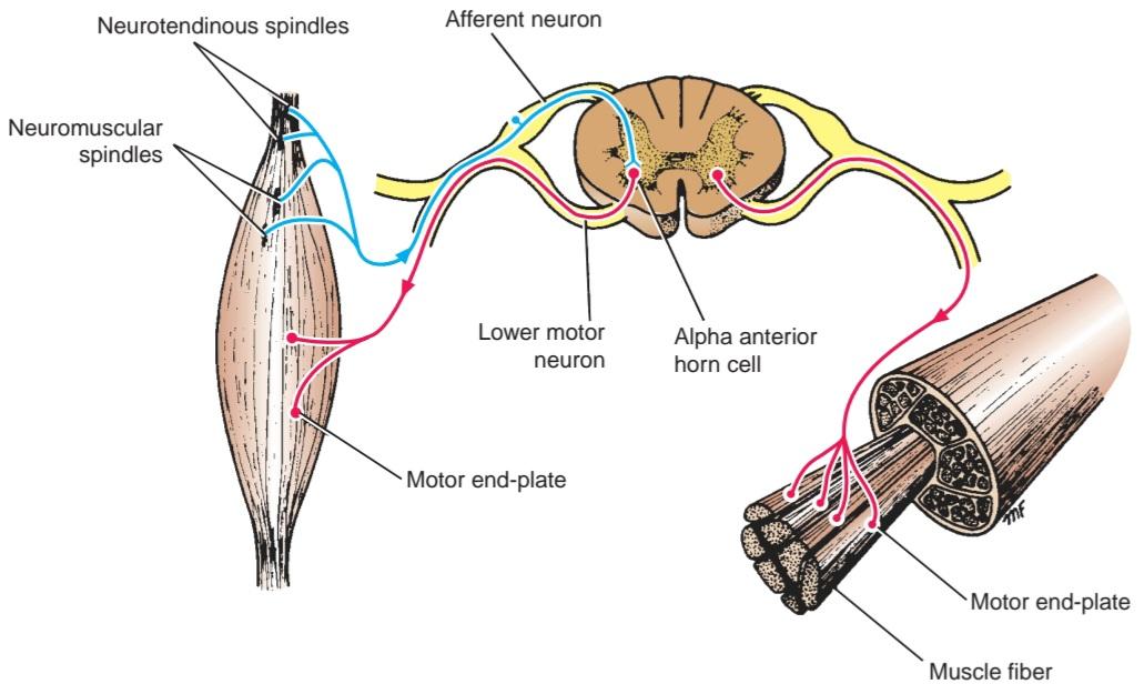
Figure 3-30 Simple reflex arc consisting of an afferent neuron arising from neuromuscular spindles and neurotendinous spindles and an efferent lower motor neuron whose cell body is an a anterior horn cell within the spinal cord. Note that the efferent neuron terminates on muscle fibers at motor endplates.
شکل ۳-۳۰ قوس رفلکس ساده شامل یک نورون آوران که از دوکهای عصبی-عضلانی و دوکهای عصبی-تاندونی منشأ میگیرد و یک نورون حرکتی تحتانی وابران که جسم سلولی آن یک سلول شاخ قدامی در نخاع است. توجه داشته باشید که نورون وابران در صفحات انتهایی حرکتی به فیبرهای عضلانی ختم میشود.
Skeletal Muscle Neuromuscular Junctions
Skeletal muscle fibers are innervated by large, a myelinated nerve fibers derived from large motor neurons in the anterior gray columns (horns) of the spinal cord or from cranial nerve motor nuclei. As each myelinated fiber enters a skeletal muscle, it branches many times. The number of branches depends on the size of the motor unit. A single branch then terminates on a muscle fiber at the neuromuscular junction or motor endplate (Figs. 3-31 and 3-32).
اتصالات عصبی-عضلانی عضله اسکلتی
فیبرهای عضله اسکلتی توسط فیبرهای عصبی بزرگ و میلیندار مشتق شده از نورونهای حرکتی بزرگ در ستونهای خاکستری قدامی (شاخکهای) نخاع یا از هستههای حرکتی عصب جمجمهای عصبدهی میشوند. هر فیبر میلیندار با ورود به عضله اسکلتی، چندین شاخه میشود. تعداد شاخهها به اندازه واحد حرکتی بستگی دارد. سپس یک شاخه واحد در محل اتصال عصبی-عضلانی یا صفحه انتهایی حرکتی به یک فیبر عضلانی ختم میشود (شکلهای 3-31 و 3-32).

Figure 3-31 A: A skeletal neuromuscular junction. B: Enlarged view of a muscle fiber showing the terminal naked axon lying in the surface groove of the muscle fiber.
شکل 3-31 الف: یک محل اتصال عصبی-عضلانی اسکلتی. ب: نمای بزرگ شده از یک فیبر عضلانی که آکسون برهنه انتهایی را که در شیار سطحی فیبر عضلانی قرار دارد، نشان میدهد.
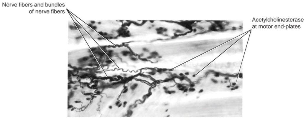
Figure 3-32 Photomicrograph showing nerve fibers terminating on skeletal muscle fibers at motor endplates, stained histochemically for acetylcholinesterase and counterstained with silver. (Courtesy Dr. M. J. T. Fitzgerald.)
شکل 3-32 عکس میکروسکوپی که فیبرهای عصبی منتهی به فیبرهای عضله اسکلتی در صفحات انتهایی حرکتی را نشان میدهد، که به روش هیستوشیمیایی برای استیل کولین استراز رنگآمیزی شده و با نقره رنگآمیزی متقابل شده است. (با احترام از دکتر ام. جی. تی. فیتزجرالد.)
The great majority of muscle fibers are innervated by just one motor endplate. On reaching the muscle fiber, the nerve loses its myelin sheath and breaks up into a number of fine branches. Each branch ends as a naked axon and forms the neural element of the motor endplate (Fig. 3-33). The axon is expanded slightly and contains many mitochondria and vesicles (approximately 45 nm in diameter). At the site of the motor endplate, the surface of the muscle fiber is elevated slightly to form the muscular element of the plate, often referred to as the sole plate (see Fig. 3-31A). The elevation is due to the local accumulation of granular sarcoplasm beneath the sarcolemma and the presence of numerous nuclei and mitochondria, the latter providing the ATP, which is the energy source for acetylcholine (ACh) synthesis.
اکثریت قریب به اتفاق فیبرهای عضلانی تنها توسط یک صفحه انتهایی حرکتی عصبدهی میشوند. عصب با رسیدن به فیبر عضلانی، غلاف میلین خود را از دست میدهد و به تعدادی شاخه ظریف تقسیم میشود. هر شاخه به عنوان یک آکسون برهنه به پایان میرسد و عنصر عصبی صفحه انتهایی حرکتی را تشکیل میدهد (شکل 3-33). آکسون کمی منبسط شده و حاوی میتوکندریها و وزیکولهای زیادی (با قطر تقریباً 45 نانومتر) است. در محل صفحه انتهایی حرکتی، سطح فیبر عضلانی کمی بالا میرود تا عنصر عضلانی صفحه را تشکیل دهد که اغلب به عنوان صفحه کف شناخته میشود (شکل 3-31A را ببینید). این بالا رفتن به دلیل تجمع موضعی سارکوپلاسم دانهای در زیر سارکولم و وجود هستهها و میتوکندریهای متعدد است که دومی ATP را فراهم میکند که منبع انرژی برای سنتز استیل کولین (ACh) است.
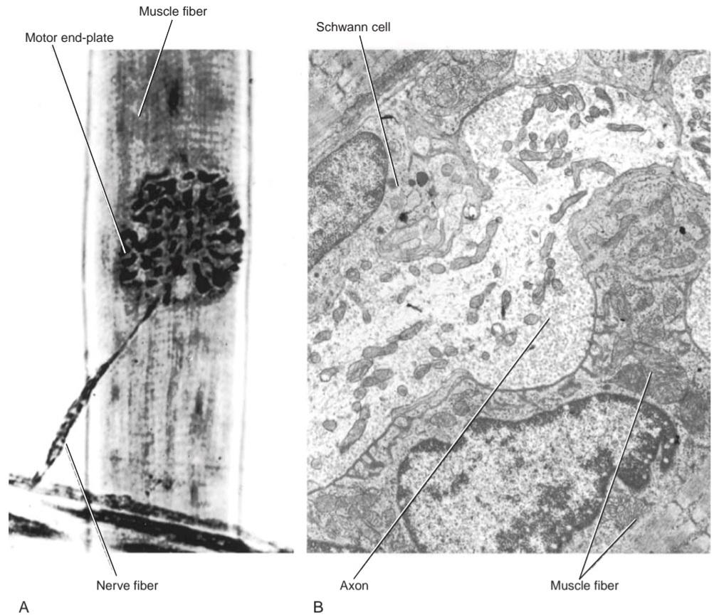
Figure 3-33 A: Photomicrograph of a motor endplate showing terminal branching of a nerve fiber. B: Electron micrograph of a terminal axon at a motor endplate showing the axon lying in a groove on the surface of the muscle fiber. (Courtesy Dr. J. M. Kerns.)
شکل ۳-۳۳ الف: عکس میکروسکوپی از صفحه انتهایی حرکتی که انشعاب انتهایی یک فیبر عصبی را نشان میدهد. ب: عکس میکروسکوپی الکترونی از آکسون انتهایی در صفحه انتهایی حرکتی که آکسون قرار گرفته در شیاری روی سطح فیبر عضلانی را نشان میدهد. (با تشکر از دکتر جی. ام. کرنز.)
The expanded naked axon lies in a groove on the surface of the muscle fiber outside the plasma membrane (sarcolemma). Each groove is formed by the infolding of the plasma membrane. The groove may branch many times, with each branch containing a division of the axon. Importantly, the axons are truly naked; the Schwann cells merely serve as a cap or roof to the groove and never project into it. The floor of the groove is formed of the plasma membrane, which is thrown into numerous small folds, called junctional folds, which increase the surface area of the plasma membrane that lies close to the naked axon (Fig. 3-34).
آکسون برهنهی منبسطشده در شیاری روی سطح فیبر عضلانی خارج از غشای پلاسمایی (سارکولما) قرار دارد. هر شیار با چینخوردگی غشای پلاسمایی تشکیل میشود. این شیار ممکن است بارها شاخه شاخه شود و هر شاخه حاوی یک شاخه از آکسون باشد. نکتهی مهم این است که آکسونها واقعاً برهنه هستند؛ سلولهای شوان صرفاً به عنوان یک کلاهک یا سقف برای شیار عمل میکنند و هرگز به داخل آن نمیروند. کف شیار از غشای پلاسمایی تشکیل شده است که به صورت چینهای کوچک متعددی به نام چینهای اتصالی درآمده است که سطح غشای پلاسمایی نزدیک به آکسون برهنه را افزایش میدهد (شکل 3-34).
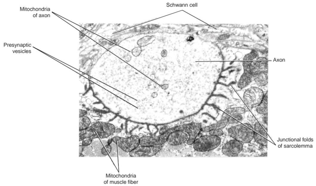
Figure 3-34 Electron micrograph of a cross section of an axon at a motor endplate showing the axon lying in a groove of folded sarcolemma. (Courtesy Dr. J. M. Kerns.)
شکل 3-34 تصویر میکروسکوپ الکترونی از مقطع عرضی آکسون در صفحه انتهایی حرکتی که آکسون را در شیاری از سارکولمای چینخورده نشان میدهد. (با احترام از دکتر جی. ام. کرنز.)
The plasma membrane of the axon (the axolemma or presynaptic membrane) is separated, by a space about 30 to 50 nm wide, from the plasma membrane of the muscle fiber (the sarcolemma or postsynaptic membrane). This space constitutes the synaptic cleft. The synaptic cleft is filled with the basement membranes of the axon and the muscle fiber (see Fig. 3-31B). The motor endplate is strengthened by the connective tissue sheath of the nerve fiber (endoneurium), which becomes continuous with the connective tissue sheath of the muscle fiber (endomysium).
غشای پلاسمایی آکسون (آکسولما یا غشای پیشسیناپسی) با فضایی به عرض حدود 30 تا 50 نانومتر، از غشای پلاسمایی فیبر عضلانی (سارکولما یا غشای پسسیناپسی) جدا میشود. این فضا شکاف سیناپسی را تشکیل میدهد. شکاف سیناپسی با غشاهای پایه آکسون و فیبر عضلانی پر شده است (شکل 3-31B را ببینید). صفحه انتهایی حرکتی توسط غلاف بافت همبند فیبر عصبی (اندونوریوم) تقویت میشود که با غلاف بافت همبند فیبر عضلانی (اندومیزیوم) پیوسته میشود.
A nerve impulse (AP), on reaching the presynaptic membrane of the motor endplate, causes the opening of voltage-gated Ca2+ channels that allow Ca2+ ions to enter the axon. This stimulates the fusion of some of the synaptic vesicles with the presynaptic membrane and causes the release of ACh into the synaptic cleft. The ACh is thus discharged into the cleft by a process of exocytosis and diffuses rapidly across the cleft to reach the nicotinic type of ACh receptors on the postsynaptic membrane of the junctional folds. The postsynaptic membrane possesses large numbers of ACh-gated channels.
یک تکانه عصبی (AP)، با رسیدن به غشای پیشسیناپسی صفحه انتهایی حرکتی، باعث باز شدن کانالهای +Ca2 وابسته به ولتاژ میشود که به یونهای +Ca2 اجازه ورود به آکسون را میدهد. این امر باعث تحریک ادغام برخی از وزیکولهای سیناپسی با غشای پیشسیناپسی و آزاد شدن استیلکولین به شکاف سیناپسی میشود. بنابراین، استیلکولین (Ach) از طریق فرآیند اگزوسیتوز به داخل شکاف تخلیه میشود و به سرعت در سراسر شکاف پخش میشود تا به گیرندههای استیلکولین نوع نیکوتینی روی غشای پسسیناپسی چینهای اتصالی برسد. غشای پسسیناپسی دارای تعداد زیادی کانال وابسته به استیلکولین است.
Once the ACh-gated channels are opened, the postsynaptic membrane becomes more permeable to Na+ ions, which flow into the muscle cell, and a local potential called the endplate potential is created. (The ACh-gated channels are also permeable to K+ ions, which flow out of the cell but to a lesser extent.) If the endplate potential is large enough, the voltage-gated channels for Na+ ions are opened, and an AP will be initiated and spread along the surface of the plasma membrane (sarcolemma). The wave of depolarization is carried into the muscle fiber to the contractile myofibrils through the system of T tubules. This leads to the release of Ca2+ ions from the sarcoplasmic reticulum, which, in turn, causes the muscle to contract.
هنگامی که کانالهای دریچهدار ACh باز میشوند، غشای پسسیناپسی نسبت به یونهای +Na که به داخل سلول ماهیچهای جریان مییابند، نفوذپذیرتر میشود و یک پتانسیل موضعی به نام پتانسیل صفحه انتهایی ایجاد میشود. (کانالهای دریچهدار ACh نسبت به یونهای +K نیز نفوذپذیر هستند که از سلول خارج میشوند، اما به میزان کمتری.) اگر پتانسیل صفحه انتهایی به اندازه کافی بزرگ باشد، کانالهای دریچهدار ولتاژی برای یونهای +Na باز میشوند و یک AP آغاز شده و در امتداد سطح غشای پلاسمایی (سارکولم) پخش میشود. موج دپلاریزاسیون از طریق سیستم توبولهای T به داخل فیبر ماهیچهای و به میوفیبریلهای انقباضی منتقل میشود. این امر منجر به آزاد شدن یونهای +Ca2 از شبکه سارکوپلاسمی میشود که به نوبه خود باعث انقباض ماهیچه میشود.
The amount of ACh released at the motor endplate will depend on the number of nerve impulses arriving at the nerve terminal. Once the ACh crosses the synaptic cleft and triggers the ionic channels on the postsynaptic membrane, it immediately undergoes hydrolysis due to the presence of the enzyme acetylcholinesterase (AChE) (see Fig. 3-32). The enzyme adheres to the fine collagen fibrils of the basement membranes in the cleft; some of the ACh diffuses away from the cleft. The ACh remains in contact with the postsynaptic membrane for about 1 msec, and it is rapidly destroyed to prevent re-excitation of the muscle fiber. After the fall in concentration of ACh in the cleft, the ionic channels close and remain closed until more ACh arrives.
مقدار ACh آزاد شده در صفحه انتهایی حرکتی به تعداد تکانههای عصبی که به پایانه عصبی میرسند بستگی دارد. به محض اینکه ACh از شکاف سیناپسی عبور میکند و کانالهای یونی روی غشای پس سیناپسی را تحریک میکند، به دلیل وجود آنزیم استیل کولین استراز (AChE) بلافاصله هیدرولیز میشود (شکل 3-32 را ببینید). آنزیم به فیبریلهای کلاژن ظریف غشاهای پایه در شکاف میچسبد. مقداری از ACh از شکاف پخش میشود. ACh حدود 1 میلیثانیه در تماس با غشای پس سیناپسی باقی میماند و به سرعت تخریب میشود تا از تحریک مجدد فیبر عضلانی جلوگیری شود. پس از کاهش غلظت ACh در شکاف، کانالهای یونی بسته میشوند و تا رسیدن ACh بیشتر بسته میمانند.
Skeletal muscle fiber contraction is thus controlled by the frequency of the nerve impulses that arrive at the motor nerve terminal. A resting muscle fiber shows small occasional depolarizations (endplate potentials) at the motor endplate, which are insufficient to cause an AP and make the fiber contract. These are believed to be due to the sporadic release of ACh into the synaptic cleft from a single presynaptic vesicle.
بنابراین انقباض فیبر عضله اسکلتی توسط فرکانس تکانههای عصبی که به پایانه عصبی حرکتی میرسند، کنترل میشود. یک فیبر عضلانی در حال استراحت، دپلاریزاسیونهای گاهبهگاه کوچکی (پتانسیلهای صفحه انتهایی) را در صفحه انتهایی حرکتی نشان میدهد که برای ایجاد AP و انقباض فیبر کافی نیستند. اعتقاد بر این است که این موارد به دلیل آزادسازی پراکنده استیلکولین (ACh) به داخل شکاف سیناپسی از یک وزیکول پیشسیناپسی واحد است.
The sequence of events that takes place at a motor endplate on stimulation of a motor nerve can be briefly summarized as follows:
توالی وقایعی که در صفحه انتهایی حرکتی هنگام تحریک عصب حرکتی رخ میدهد را میتوان به طور خلاصه به شرح زیر خلاصه کرد:
ACH → Nicotinic type of ACh receptor, ACh-gated
channels opened → Na+ influx → Endplate potential created
Endplate potential (if large enough) → Na-gated chan- nels opened → Nat influx→ AP created AP→ Increased release of Ca2+ → Muscle fiber contraction Immediate hydrolysis of ACh by AChE → ACh-gated
channels closed→ Muscle fiber repolarization
ACH ← گیرنده ACh از نوع نیکوتینی، کانالهای دریچهدار ACh باز میشوند ← ورود +Na ← پتانسیل صفحه انتهایی ایجاد میشود
پتانسیل صفحه انتهایی (اگر به اندازه کافی بزرگ باشد) ← کانالهای دریچهدار Na باز میشوند ← ورود +Na ← ایجاد AP AP ← افزایش آزادسازی +Ca2 ← انقباض فیبر عضلانی هیدرولیز فوری ACh توسط AChE ← کانالهای دریچهدار ACh بسته میشوند ← رپلاریزاسیون فیبر عضلانی
If drugs having a similar chemical structure to ACh were to arrive at the receptor site of a motor endplate, they might bring about the same changes as ACh and mimic its action. Two examples of such drugs are nicotine and carbamylcholine. If, on the other hand, drugs having a similar chemical structure to ACh were to arrive at the receptor site of a motor endplate and were unable to bring about the sequence of changes normally induced by ACh, they would occupy the receptor site and block access of ACh. Such drugs would be competing with ACh and are called competitive blocking agents. An example of such a drug is d-tubocurarine, which causes skeletal muscle to relax and not contract by preventing the action of locally produced ACh (see p. 114).
اگر داروهایی که ساختار شیمیایی مشابهی با ACh دارند به محل گیرنده صفحه انتهایی حرکتی برسند، ممکن است همان تغییرات ACh را ایجاد کرده و عملکرد آن را تقلید کنند. دو نمونه از این داروها نیکوتین و کاربامیل کولین هستند. از سوی دیگر، اگر داروهایی که ساختار شیمیایی مشابهی با ACh دارند به محل گیرنده صفحه انتهایی حرکتی برسند و نتوانند توالی تغییراتی را که معمولاً توسط ACh ایجاد میشود، ایجاد کنند، محل گیرنده را اشغال کرده و دسترسی ACh را مسدود میکنند. چنین داروهایی با ACh رقابت میکنند و عوامل مسدودکننده رقابتی نامیده میشوند. نمونهای از چنین دارویی d-توبوکورارین است که با جلوگیری از عملکرد ACh تولید شده موضعی، باعث شل شدن و عدم انقباض عضله اسکلتی میشود (به صفحه 114 مراجعه کنید).
Smooth Muscle Neuromuscular Junctions
In smooth muscle, where the action is slow and widespread, such as within the wall of the intestine, the autonomic nerve fibers branch extensively; thus, a single neuron exerts control over a large number of muscle fibers. In some areas (e.g., the longitudinal layer of smooth muscle in the intestine), only a few muscle fibers are associated with autonomic endings, the wave of contraction passing from one muscle cell to another by means of gap junctions (Fig. 3-35).
اتصالات عصبی-عضلانی عضله صاف
در عضله صاف، جایی که عمل کند و گسترده است، مانند درون دیواره روده، فیبرهای عصبی خودکار به طور گسترده شاخه شاخه میشوند؛ بنابراین، یک نورون واحد بر تعداد زیادی از فیبرهای عضلانی کنترل اعمال میکند. در برخی نواحی (به عنوان مثال، لایه طولی عضله صاف در روده)، تنها تعداد کمی از فیبرهای عضلانی با انتهای خودکار مرتبط هستند، موج انقباض از طریق اتصالات شکافی از یک سلول عضلانی به سلول دیگر منتقل میشود (شکل 3-35).
In smooth muscle, in which the action is fast and precision is required, such as in the iris, the branching of the nerve fibers is less extensive; thus, a single neuron exerts control over only a few muscle fibers.
در عضله صاف، که در آن عمل سریع و دقت لازم است، مانند عنبیه، انشعاب رشتههای عصبی کمتر گسترده است؛ بنابراین، یک نورون واحد تنها بر تعداد کمی از رشتههای عضلانی کنترل دارد.

Figure 3-35 Autonomic neuromuscular junction. The exposed axons are close to the smooth muscle fibers.
شکل ۳-۳۵ اتصال عصبی-عضلانی خودکار. آکسونهای نمایان نزدیک به فیبرهای عضله صاف هستند.
The autonomic nerve fibers, which are postganglionic, are nonmyelinated and terminate as a series of varicosed branches. An interval of 10 to 100 nm may exist between the axon and the muscle fiber. At the site where transmission is to occur, the Schwann cell is retracted so that the axon lies within a shallow groove on its surface. Therefore, part of the axon is naked, permitting free diffusion of the transmitter substance from the axon to the muscle cell. Here, the axoplasm contains numerous vesicles similar to those seen at the motor endplate of skeletal muscle.
فیبرهای عصبی اتونوم که پسگانگلیونی هستند، بدون میلین بوده و به صورت مجموعهای از شاخههای واریسی خاتمه مییابند. ممکن است فاصلهای بین 10 تا 100 نانومتر بین آکسون و فیبر عضلانی وجود داشته باشد. در محلی که قرار است انتقال رخ دهد، سلول شوان به عقب کشیده میشود به طوری که آکسون در یک شیار کمعمق روی سطح آن قرار میگیرد. بنابراین، بخشی از آکسون برهنه است و امکان انتشار آزاد ماده انتقالدهنده از آکسون به سلول عضلانی را فراهم میکند. در اینجا، آکوپلاسم حاوی وزیکولهای متعددی مشابه آنچه در صفحه انتهایی حرکتی عضله اسکلتی دیده میشود، است.
Smooth muscle is innervated by sympathetic and parasympathetic parts of the autonomic system. Those nerves that are cholinergic liberate ACh at their endings by a process of exocytosis, with the ACh being present in the vesicles at the nerve ending. Those nerves that are noradrenergic liberate norepinephrine (NE) at their endings by a process of exocytosis, with the NE being present in dark-cored vesicles at the nerve endings. Both ACh and NE bring about depolarization of the muscle fibers innervated, which thereupon contract. The fate of these neurotransmitter substances differs. ACh is hydrolyzed in the presence of AChE in the synaptic cleft of the muscle fiber, and NE is taken up by the nerve endings. Notably, in some areas of the body (e.g., bronchial muscle), the NE liberated from postganglionic sympathetic fibers causes smooth muscle to relax and not contract.
عضله صاف توسط بخشهای سمپاتیک و پاراسمپاتیک سیستم اتونوم عصبدهی میشود. اعصابی که کولینرژیک هستند، در انتهای خود از طریق فرآیند اگزوسیتوز، استیلکولین (ACh) آزاد میکنند که استیلکولین در وزیکولهای انتهای عصب وجود دارد. اعصابی که نورآدرنرژیک هستند، در انتهای خود از طریق فرآیند اگزوسیتوز، نوراپی نفرین (NE) آزاد میکنند که NE در وزیکولهای هسته تیره در انتهای عصب وجود دارد. هم استیلکولین و هم نوراپی نفرین باعث دپلاریزاسیون فیبرهای عضلانی عصبدهی شده میشوند که در نتیجه منقبض میشوند. سرنوشت این مواد انتقالدهنده عصبی متفاوت است. استیلکولین در حضور استیلکولین E در شکاف سیناپسی فیبر عضلانی هیدرولیز میشود و NE توسط انتهای عصب جذب میشود. به ویژه، در برخی از نواحی بدن (به عنوان مثال، عضله برونش)، NE آزاد شده از فیبرهای سمپاتیک پسگانگلیونی باعث شل شدن و عدم انقباض عضله صاف میشود.
Cardiac Muscle Neuromuscular Junctions
Nonmyelinated postganglionic sympathetic and parasympathetic autonomic nerves extend into the connective tissue between the muscle fibers and terminate in close proximity to the individual cardiac muscle fibers. At the site where transmission takes place, the axon becomes naked because of the retraction of the Schwann cell. This permits free diffusion of the neurotransmitter substance from the axon to the muscle fiber. Because of the presence of intermittent desmosomes and gap junctions between abutting muscle fibers, excitation and contraction of one muscle fiber rapidly spread from fiber to fiber.
اتصالات عصبی-عضلانی عضله قلبی
اعصاب اتونوم سمپاتیک و پاراسمپاتیک بدون میلین پسگانگلیونی به بافت همبند بین فیبرهای عضلانی امتداد یافته و در نزدیکی فیبرهای عضلانی قلب خاتمه مییابند. در محلی که انتقال انجام میشود، آکسون به دلیل انقباض سلول شوان برهنه میشود. این امر امکان انتشار آزاد ماده انتقالدهنده عصبی از آکسون به فیبر عضلانی را فراهم میکند. به دلیل وجود دسموزومهای متناوب و اتصالات شکافدار بین فیبرهای عضلانی مجاور، تحریک و انقباض یک فیبر عضلانی به سرعت از فیبری به فیبر دیگر گسترش مییابد.
Nerve Endings on Secretory Cells of Glands
Nonmyelinated postganglionic autonomic nerves extend into the connective tissue of glands and branch close to the secretory cells (Fig. 3-36). In many glands, the nerve fibers have been found to innervate only the blood vessels.
انتهای عصبی روی سلولهای ترشحی غدد
اعصاب اتونوم پسگانگلیونی بدون میلین به بافت همبند غدد امتداد یافته و نزدیک به سلولهای ترشحی شاخه شاخه میشوند (شکل 3-36). در بسیاری از غدد، مشخص شده است که فیبرهای عصبی فقط رگهای خونی را عصبدهی میکنند.

Figure 3-36 Nerve fibers ending around glandular acini.
شکل ۳-۳۶ رشتههای عصبی که در اطراف آسینیهای غدهای ختم میشوند.
SEGMENTAL INNERVATION OF SKIN
The area of skin supplied by a single spinal nerve and, therefore, a single segment of the spinal cord is called a dermatome. On the trunk, dermatomes extend round the body from the posterior to the anterior median plane. Adjacent dermatomes overlap considerably, so to produce a region of complete anesthesia, at least three contiguous spinal nerves have to be sectioned. Note that the area of tactile loss is always greater than the area of loss of painful and thermal sensations. The reason for this difference is that the degree of overlap of fibers carrying pain and thermal sensations is much more extensive than the overlap of fibers carrying tactile sensations. Dermatomal charts for the anterior and posterior surfaces of the body are shown in Figures 3-37 and 3-38.
عصبدهی قطعهای پوست
ناحیهای از پوست که توسط یک عصب نخاعی واحد و بنابراین، یک قطعه از نخاع تغذیه میشود، درماتوم نامیده میشود. در تنه، درماتومها از سطح خلفی تا صفحه میانی قدامی در اطراف بدن امتداد دارند. درماتومهای مجاور به طور قابل توجهی همپوشانی دارند، بنابراین برای ایجاد یک ناحیه بیحسی کامل، حداقل سه عصب نخاعی مجاور باید برش داده شوند. توجه داشته باشید که ناحیه از دست دادن حس لامسه همیشه بزرگتر از ناحیه از دست دادن حسهای دردناک و حرارتی است. دلیل این تفاوت این است که میزان همپوشانی فیبرهای حامل حس درد و حرارت بسیار گستردهتر از همپوشانی فیبرهای حامل حس لامسه است. نمودارهای درماتوم برای سطوح قدامی و خلفی بدن در شکلهای 3-37 و 3-38 نشان داده شده است.
In the limbs, the arrangement of the dermatomes is more complicated because of the embryologic rotation of the limbs as they grow out from the trunk.
در اندامها، چیدمان درماتومها به دلیل چرخش جنینی اندامها هنگام رشد از تنه پیچیدهتر است.
In the face, the divisions of the trigeminal nerve supply a precise area of skin, with little or no overlap to the cutaneous area of another division.
در صورت، انشعابات عصب سهقلو ناحیه دقیقی از پوست را تغذیه میکنند که همپوشانی کمی با ناحیه پوستی انشعاب دیگر دارد یا اصلاً همپوشانی ندارد.

Figure 3-37 Anterior aspect of the body showing the distribution of cutaneous nerves on the left side and dermatomes on the right side.
شکل ۳-۳۷ نمای قدامی بدن که توزیع اعصاب پوستی را در سمت چپ و درماتومها را در سمت راست نشان میدهد.
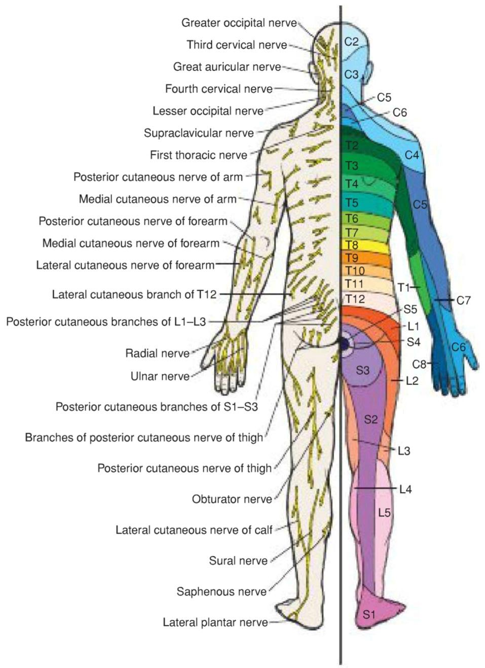
Figure 3-38 Posterior aspect of the body showing the distribution of cutaneous nerves on the left side and dermatomes on the right side.
شکل 3-38 نمای خلفی بدن که توزیع اعصاب پوستی در سمت چپ و درماتومها در سمت راست را نشان میدهد.
SEGMENTAL INNERVATION OF MUSCLES
Skeletal muscle also receives a segmental innervation. Most of these muscles are innervated by more than one spinal nerve and, therefore, by the same number of segments of the spinal cord. Thus, to paralyze a muscle completely, sectioning several spinal nerves or destroying several segments of the spinal cord would be necessary.
عصبدهی قطعهای عضلات
عصبرسانی قطعهای عضلات اسکلتی نیز به صورت قطعهای انجام میشود. اکثر این عضلات توسط بیش از یک عصب نخاعی عصبدهی میشوند و بنابراین، به همان تعداد از بخشهای نخاع نیز عصبدهی میشوند. بنابراین، برای فلج کردن کامل یک عضله، قطع چندین عصب نخاعی یا از بین بردن چندین بخش از نخاع ضروری است.
Learning the segmental innervation of all the muscles of the body is an impossible task. Nevertheless, the segmental innervation of the following muscles should be known because testing them by eliciting simple muscle reflexes in the patient is possible (Fig. 3-39):
یادگیری عصبدهی سگمنتال تمام عضلات بدن کاری غیرممکن است. با این وجود، عصبدهی سگمنتال عضلات زیر باید شناخته شده باشد زیرا آزمایش آنها با ایجاد رفلکسهای عضلانی ساده در بیمار امکانپذیر است (شکل 3-39):
Biceps brachii tendon reflex C5-C6: Flexion of the elbow joint by tapping the biceps tendon
رفلکس تاندون عضله دوسر بازویی C5-C6: خم شدن مفصل آرنج با ضربه زدن به تاندون عضله دوسر
Triceps tendon reflex C6-C7 and C8: Extension of the elbow joint by tapping the triceps tendon
رفلکس تاندون عضله سه سر بازویی C6-C7 و C8: باز شدن مفصل آرنج با ضربه زدن به تاندون عضله سه سر
Brachioradialis tendon reflex C5-C6 and C7: Supination of the radioulnar joints by tapping the insertion of the brachioradialis tendon
رفلکس تاندون عضله براکیورادیال C5-C6 و C7: سوپیناسیون مفاصل رادیواولنار با ضربه زدن به محل اتصال تاندون عضله براکیورادیال
Abdominal superficial reflexes: Contraction of underlying abdominal muscles by stroking the skin: upper abdominal skin T6-T7, middle abdominal skin T8-T9, lower abdominal skin T10-T12
رفلکسهای سطحی شکم: انقباض عضلات زیرین شکم با لمس پوست: پوست بالای شکم T6-T7، پوست وسط شکم T8-T9، پوست پایین شکم T10-T12
Patellar tendon reflex (knee jerk) L2, L3, and L4: Extension of knee joint on tapping the patellar tendon
رفلکس تاندون کشکک (حرکت ناگهانی زانو) L2، L3 و L4: باز شدن مفصل زانو با ضربه زدن به تاندون کشکک
Achilles tendon reflex (ankle jerk) S1 and S2: Plantar flexion of ankle joint on tapping the Achilles tendon (calcaneus tendon)
رفلکس تاندون آشیل (حرکت ناگهانی مچ پا) S1 و S2: خم شدن کف پا مفصل مچ پا با ضربه زدن به تاندون آشیل (تاندون پاشنه)

Figure 3-39 Biceps brachii tendon reflex. Note that the reflex arc passes through the fifth and sixth cervical segments of the spinal cord. This is usually monosynaptic, and the internuncial neuron (black) is absent.
شکل ۳-۳۹ رفلکس تاندون عضله دوسر بازویی. توجه داشته باشید که قوس رفلکس از قطعات پنجم و ششم گردنی نخاع عبور میکند. این معمولاً تک سیناپسی است و نورون بین حلقی (سیاه) وجود ندارد.
MUSCLE TONE AND MUSCLE ACTION
A motor unit consists of a motor neuron in the anterior gray column (horn) of the spinal cord and all the muscle fibers it supplies (Fig. 3-40). In a large buttock muscle, such as the gluteus maximus, where fine control is unnecessary, a given motor neuron may supply as many as 200 muscle fibers. In contrast, in the small muscles of the hand or the extrinsic muscles of the eyeball, where fine control is required, one nerve fiber supplies only a few muscle fibers.
تون عضلانی و عملکرد عضله
یک واحد حرکتی شامل یک نورون حرکتی در ستون خاکستری قدامی (شاخ) نخاع و تمام فیبرهای عضلانی است که آن را تغذیه میکند (شکل 3-40). در یک عضله بزرگ باسن، مانند عضله سرینی بزرگ، که کنترل دقیق ضروری نیست، یک نورون حرکتی ممکن است تا 200 فیبر عضلانی را تغذیه کند. در مقابل، در عضلات کوچک دست یا عضلات خارجی کره چشم، که کنترل دقیق مورد نیاز است، یک فیبر عصبی تنها تعداد کمی از فیبرهای عضلانی را تغذیه میکند.
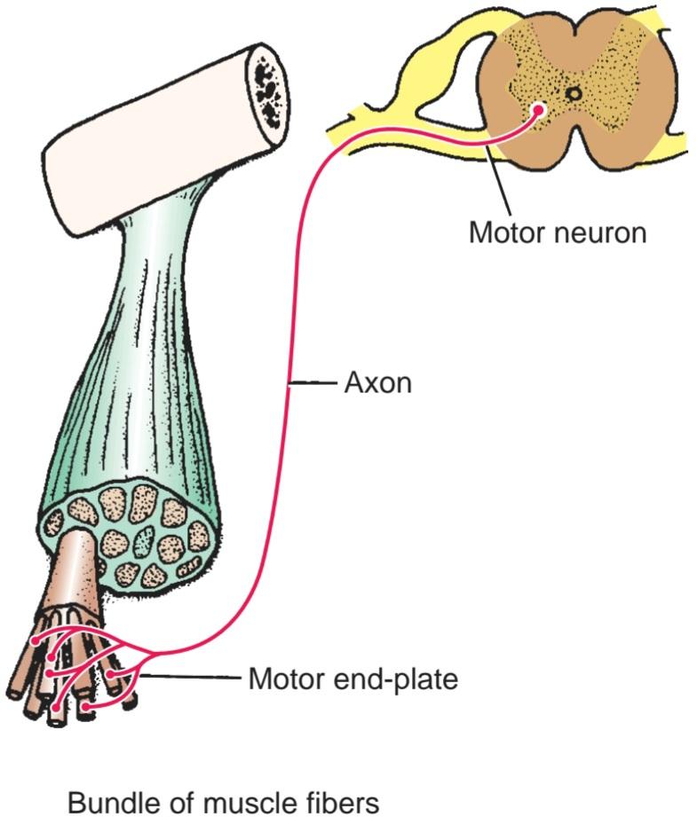
Figure 3-40 Components of a motor unit.
شکل ۳-۴۰ اجزای یک واحد حرکتی.
Every skeletal muscle, while resting, is in a partial state of contraction, a condition referred to as muscle tone. With no intermediate stage, muscle fibers are either fully contracted or relaxed; therefore, a few muscle fibers within a muscle are fully contracted all the time. To bring about this state and to avoid fatigue, different groups of motor units and, thus, different groups of muscle fibers are activated at different times. This is accomplished by the asynchronous discharge of nervous impulses in the motor neurons in the anterior gray horn of the spinal cord.
هر عضله اسکلتی، در حالت استراحت، در حالت انقباض جزئی قرار دارد، وضعیتی که به آن تون عضلانی میگویند. بدون مرحله میانی، فیبرهای عضلانی یا کاملاً منقبض یا شل هستند؛ بنابراین، تعداد کمی از فیبرهای عضلانی در یک عضله همیشه کاملاً منقبض هستند. برای ایجاد این حالت و جلوگیری از خستگی، گروههای مختلفی از واحدهای حرکتی و در نتیجه گروههای مختلفی از فیبرهای عضلانی در زمانهای مختلف فعال میشوند. این امر با تخلیه غیرهمزمان تکانههای عصبی در نورونهای حرکتی در شاخ خاکستری قدامی نخاع انجام میشود.
Basically, muscle tone is dependent on the integrity of a simple monosynaptic reflex arc composed of two neurons in the nervous system (Fig. 3-41). The lengthening and shortening in a muscle are detected by sensitive sensory endings called muscle spindles (see p. 90), and the tension is detected by tendon spindles (see p. 92). The nervous impulses travel in the large afferent fibers to the spinal cord. There, they synapse with the motor neurons situated in the anterior gray column, which, in turn, send impulses down their axons to the muscle fibers. The muscle spindles themselves are innervated by small y efferent fibers that regulate the response of the muscle spindles, acting synergically with external stretch. In this manner, muscle tone is maintained reflexly and adjusted to the needs of posture and movement.
اساساً، تون عضلانی به یکپارچگی یک قوس رفلکس تک سیناپسی ساده متشکل از دو نورون در سیستم عصبی وابسته است (شکل 3-41). بلند شدن و کوتاه شدن در یک عضله توسط انتهای حسی حساس به نام دوکهای عضلانی تشخیص داده میشود (به صفحه 90 مراجعه کنید) و تنش توسط دوکهای تاندون تشخیص داده میشود (به صفحه 92 مراجعه کنید). تکانههای عصبی در فیبرهای بزرگ آوران به نخاع منتقل میشوند. در آنجا، آنها با نورونهای حرکتی واقع در ستون خاکستری قدامی سیناپس برقرار میکنند که به نوبه خود، تکانههایی را از طریق آکسونهای خود به فیبرهای عضلانی میفرستند. خود دوکهای عضلانی توسط فیبرهای کوچک وابران y عصبدهی میشوند که پاسخ دوکهای عضلانی را تنظیم میکنند و با کشش خارجی به صورت سینرژیک عمل میکنند. به این ترتیب، تون عضلانی به صورت رفلکسی حفظ شده و با نیازهای وضعیت بدن و حرکت تنظیم میشود.
Should the afferent or efferent pathways of the reflex arc be cut, the muscle would lose its tone immediately and become flaccid. A flaccid muscle, on palpation, feels like a mass of dough that has completely lost its resilience. It quickly atrophies and becomes reduced in volume. The degree of activity of the motor anterior column cells and, therefore, the degree of muscle tone depends on the summation of the nerve impulses received by these cells from other neurons of the nervous system.
اگر مسیرهای آوران یا وابران قوس رفلکسی قطع شوند، عضله بلافاصله تون خود را از دست میدهد و شل میشود. یک عضله شل، در لمس، مانند تودهای خمیری احساس میشود که کاملاً خاصیت ارتجاعی خود را از دست داده است. این عضله به سرعت آتروفی شده و حجم آن کاهش مییابد. میزان فعالیت سلولهای ستون قدامی حرکتی و بنابراین، میزان تون عضلانی به مجموع تکانههای عصبی دریافتی توسط این سلولها از سایر نورونهای سیستم عصبی بستگی دارد.
Muscle movement is accomplished by activating increasing numbers of motor units and, at the same time, reducing the activity of the motor units of muscles that will oppose or antagonize the movement. When the maximum effort is required, all the motor units of a muscle are active.
حرکت عضله با فعال کردن تعداد فزایندهای از واحدهای حرکتی و همزمان با آن، کاهش فعالیت واحدهای حرکتی عضلاتی که با حرکت مخالفت یا آنتاگونیسم میکنند، انجام میشود. هنگامی که حداکثر تلاش مورد نیاز است، تمام واحدهای حرکتی یک عضله فعال هستند.
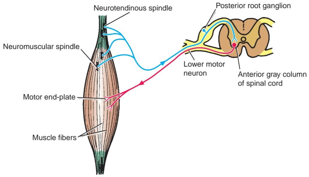
Figure 3-41 Simple reflex arc consisting of an afferent neuron arising from neuromuscular spindles and neurotendinous spindles and an efferent neuron whose cell body lies in the anterior gray column (horn) of the spinal cord. Note that for simplicity, the afferent fibers from the neurotendinous spindle and the neuromuscular spindle are shown as one pathway; in fact, the neurotendinous receptor is inhibitory and reduces tone, whereas the neuromus- cular spindle is excitatory and increases tone.
شکل ۳-۴۱ قوس رفلکس ساده شامل یک نورون آوران ناشی از دوکهای عصبی-عضلانی و دوکهای عصبی-تاندونی و یک نورون وابران که جسم سلولی آن در ستون خاکستری قدامی (شاخ) نخاع قرار دارد. توجه داشته باشید که برای سادگی، فیبرهای آوران از دوک عصبی-تاندونی و دوک عصبی-عضلانی به عنوان یک مسیر نشان داده شدهاند؛ در واقع، گیرنده عصبی-تاندونی مهاری است و تون را کاهش میدهد، در حالی که دوک عصبی-عضلانی تحریکی است و تون را افزایش میدهد.
MOTOR UNIT SUMMATION
When a muscle begins to contract, the smaller motor units are stimulated first. This is because the smaller motor units are innervated by smaller neurons in the spinal cord and brainstem, and they have a lower threshold of excitability. As the contraction increases, progressively larger motor units are recruited. This phenomenon causes a gradual increase in muscle strength as the muscle contracts.
جمعبندی واحدهای حرکتی
وقتی یک عضله شروع به انقباض میکند، واحدهای حرکتی کوچکتر ابتدا تحریک میشوند. دلیل این امر این است که واحدهای حرکتی کوچکتر توسط نورونهای کوچکتر در نخاع و ساقه مغز عصبدهی میشوند و آستانه تحریکپذیری پایینتری دارند. با افزایش انقباض، واحدهای حرکتی بزرگتر به تدریج به کار گرفته میشوند. این پدیده باعث افزایش تدریجی قدرت عضله با انقباض عضله میشود.
MUSCLE FATIGUE
The progressive loss of strength of a muscle with prolonged strong contraction is due to the reduction in the amounts of ATP within the muscle fibers. Nerve impulses continue to arrive at the neuromuscular junction, and normal depolarization of the plasma membrane of the muscle fiber occurs.
خستگی عضلانی
کاهش تدریجی قدرت عضله با انقباض قوی و طولانی مدت به دلیل کاهش مقدار ATP در فیبرهای عضلانی است. تکانههای عصبی همچنان به محل اتصال عصبی-عضلانی میرسند و دپلاریزاسیون طبیعی غشای پلاسمایی فیبر عضلانی رخ میدهد.
POSTURE
Posture is the position adopted by the individual within his or her environment. In the standing position, the line of gravity passes through the odontoid process of the axis, behind the centers of the hip joints, and in front of the knee and ankle joints. In order to stabilize the body and prevent it from collapsing, humans have well-developed antigravity muscles that exhibit the greatest degree of tone. Therefore, posture depends on the degree and distribution of muscle tone, which, in turn, depends on the normal integrity of simple reflex arcs centered in the spinal cord.
وضعیت بدن
وضعیت بدن، وضعیتی است که فرد در محیط خود اتخاذ میکند. در حالت ایستاده، خط ثقل از زائده دندانی محور، پشت مراکز مفاصل ران و جلوی مفاصل زانو و مچ پا عبور میکند. برای تثبیت بدن و جلوگیری از فروپاشی آن، انسانها دارای عضلات ضد جاذبهای هستند که به خوبی توسعه یافتهاند و بیشترین میزان تون را نشان میدهند. بنابراین، وضعیت بدن به میزان و توزیع تون عضلانی بستگی دارد که به نوبه خود به یکپارچگی طبیعی قوسهای رفلکس ساده متمرکز در نخاع بستگی دارد.
An individual may assume a particular posture (sitting or standing) over long periods of time with little evidence of fatigue. The reason for this is that muscle tone is maintained through different groups of muscle fibers contracting in relays, with only a small number of muscle fibers within a muscle being in a state of contraction at any one time. The active muscle fiber groups are scattered throughout the muscle.
یک فرد ممکن است برای مدت طولانی یک وضعیت خاص (نشسته یا ایستاده) را بدون هیچ گونه نشانه خستگی به خود بگیرد. دلیل این امر این است که تون عضلانی از طریق گروههای مختلف فیبرهای عضلانی که به صورت رله منقبض میشوند، حفظ میشود و تنها تعداد کمی از فیبرهای عضلانی در یک عضله در هر زمان در حالت انقباض هستند. گروههای فعال فیبرهای عضلانی در سراسر عضله پراکنده هستند.
In order to maintain posture, the simple muscle reflex, on which muscle tone is dependent, must receive adequate nervous input from higher levels of the nervous system (Fig. 3-42). For example, impulses arising from the labyrinths and neck muscles, information arising from the cerebellum, midbrain, and cerebral centers, and general information arising from other muscle groups, joints, and even skin receptors will result in nervous impulses impinging on the large anterior gray column cells (i.e., the final common pathway) controlling the muscle fibers.
برای حفظ وضعیت بدن، رفلکس ساده عضلانی که تون عضلانی به آن وابسته است، باید ورودی عصبی کافی از سطوح بالاتر سیستم عصبی دریافت کند (شکل 3-42). به عنوان مثال، تکانههای ناشی از لابیرنتها و عضلات گردن، اطلاعات ناشی از مخچه، مغز میانی و مراکز مغزی و اطلاعات کلی ناشی از سایر گروههای عضلانی، مفاصل و حتی گیرندههای پوستی منجر به تکانههای عصبی میشوند که به سلولهای ستون خاکستری قدامی بزرگ (یعنی مسیر مشترک نهایی) که فیبرهای عضلانی را کنترل میکنند، برخورد میکنند.
When an individual assumes a given posture, the tone of the muscles controlling that posture is constantly undergoing fine adjustments so that the posture is maintained. Normal posture thus depends not only on the integrity of the reflex arc but also on the summation of the nervous impulses received by the motor anterior gray column cells from other neurons of the nervous system (Fig. 3-43). The detail of the different nervous pathways involved in bringing the information to the anterior gray column cells is dealt with in Chapter 4.
وقتی فردی وضعیت خاصی را در نظر میگیرد، تون عضلات کنترلکننده آن وضعیت دائماً در حال تنظیم دقیق است تا وضعیت حفظ شود. بنابراین، وضعیت طبیعی بدن نه تنها به یکپارچگی قوس رفلکسی، بلکه به مجموع تکانههای عصبی دریافتی توسط سلولهای حرکتی ستون خاکستری قدامی از سایر نورونهای سیستم عصبی نیز بستگی دارد (شکل 3-43). جزئیات مسیرهای عصبی مختلف دخیل در رساندن اطلاعات به سلولهای ستون خاکستری قدامی در فصل 4 بررسی شده است.

Figure 3-42 Nervous input from higher levels of the central nervous system, which can influence the activity of the anterior gray column (horn) cells of the spinal cord.
شکل ۳-۴۲ ورودی عصبی از سطوح بالاتر سیستم عصبی مرکزی، که میتواند بر فعالیت سلولهای ستون خاکستری قدامی (شاخک) نخاع تأثیر بگذارد.
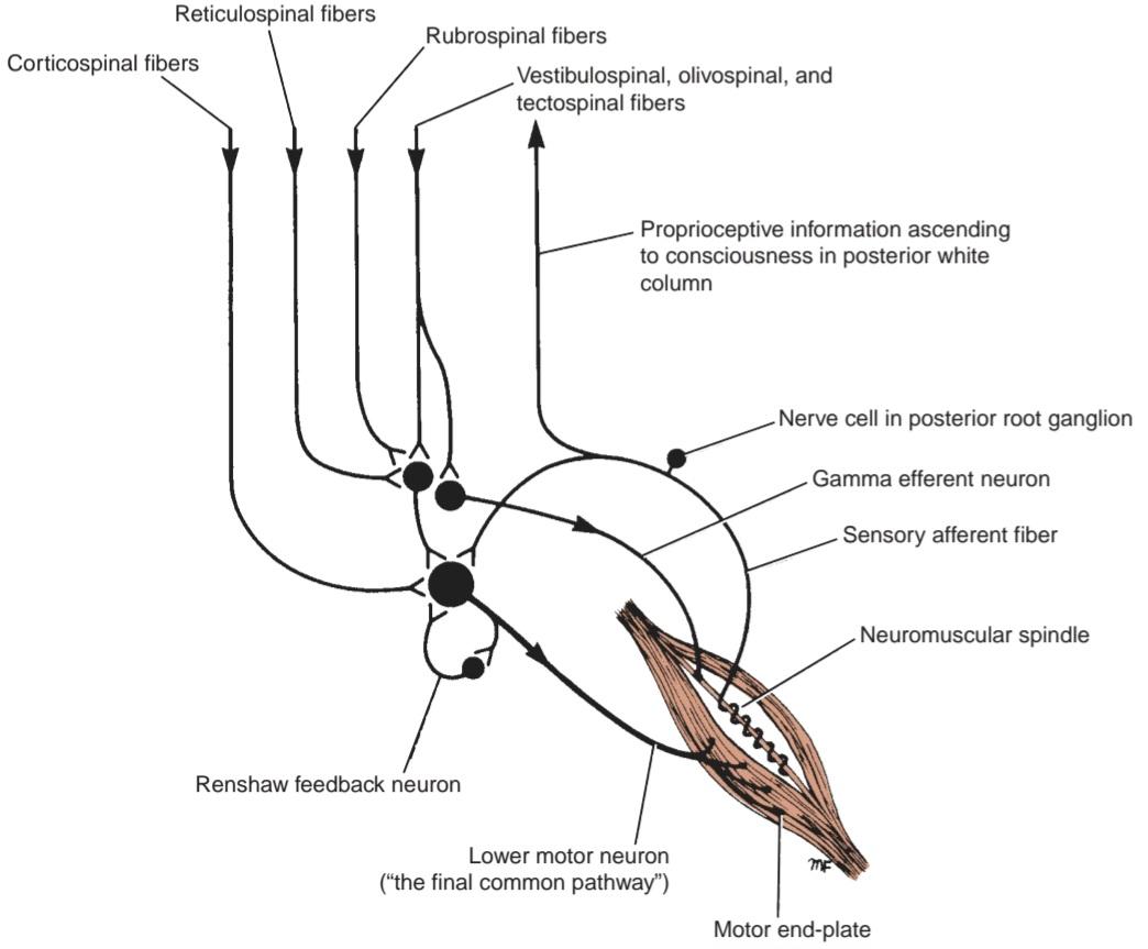
Figure 3-43 Normal postural tone of skeletal muscle is dependent not only on the integrity of the reflex arc but also on the summation of the nervous impulses received by the motor anterior gray column (horn) cells from other neurons of the nervous system.
شکل ۳-۴۳ تون وضعیتی طبیعی عضله اسکلتی نه تنها به یکپارچگی قوس رفلکسی، بلکه به مجموع تکانههای عصبی دریافتی توسط سلولهای ستون خاکستری قدامی حرکتی (شاخک) از سایر نورونهای سیستم عصبی نیز بستگی دارد.
«فصل سوم کتاب نورواناتومی بالینی اسنل»
🚀 با ما همراه شوید!
تازهترین مطالب و آموزشهای مغز و اعصاب را از دست ندهید. با فالو کردن کانال تلگرام آیندهنگاران مغز، از ما حمایت کنید!



