
نوروآناتومی بالینی اسنل | ساقه مغز؛ مغز میانی – مرور مبانی و اصول


دعای مطالعه [ نمایش ]
بِسْمِ الله الرَّحْمنِ الرَّحیمِ
اَللّهُمَّ اَخْرِجْنى مِنْ ظُلُماتِ الْوَهْمِ
خدايا مرا بيرون آور از تاريكىهاى وهم،
وَ اَكْرِمْنى بِنُورِ الْفَهْمِ
و به نور فهم گرامى ام بدار،
اَللّهُمَّ افْتَحْ عَلَيْنا اَبْوابَ رَحْمَتِكَ
خدايا درهاى رحمتت را به روى ما بگشا،
وَانْشُرْ عَلَيْنا خَزائِنَ عُلُومِكَ بِرَحْمَتِكَ يا اَرْحَمَ الرّاحِمينَ
و خزانههاى علومت را بر ما باز كن به امید رحمتت اى مهربانترين مهربانان.
آیندهنگاران مغز: «ما مغز را میشناسیم، تا آینده را بسازیم.»
کتاب «نوروآناتومی بالینی اسنل» اثر دکتر ریچارد اس. اسنل، با ویرایش جدید دکتر رایان اسپلیتگربر، همچنان بهعنوان مرجع طلایی این حوزه شناخته میشود. این اثر با تلفیق تازهترین پژوهشها و کاربردهای بالینی، پلی استوار میان دانش پایه و عمل پزشکی بنا میکند.
تلاشهای علمی و سالها تدریس ارزشمند استاد گرانقدر دکتر محمدتقی جغتایی الهامبخش ترجمه و گسترش این دانش در ایران بوده است.
ترجمه دقیق این کتاب توسط برند علمی آیندهنگاران مغز به مدیریت داریوش طاهری، امکان دسترسی فارسیزبانان به مرزهای نوین علوم اعصاب را فراهم کرده و رسالتی علمی در جهت شناخت عمیقتر مغز و ترسیم آیندهای روشنتر بر عهده گرفته است.
»» فصل ۵: ساقه مغز
»» Chapter 5: Brainstem
MIDBRAIN
The midbrain measures about 0.8 in (2 cm) in length and connects the pons and cerebellum with the forebrain (Fig. 5-23). Its long axis inclines anteriorly as it ascends through the opening in the tentorium cerebelli. The midbrain is traversed by a narrow channel, the cerebral aqueduct, which is filled with cerebrospinal fluid (Figs. 5-24 to 5-28).
مغز میانی
مغز میانی حدود 2 سانتیمتر (0.8 اینچ) طول دارد و پل مغزی و مخچه را به مغز پیشین متصل میکند (شکل 5-23). محور طولی آن هنگام بالا رفتن از طریق دهانه چادرینه مخچه به سمت جلو متمایل میشود. مغز میانی توسط یک کانال باریک، قنات مغزی، که پر از مایع مغزی نخاعی است، طی میشود (شکلهای 5-24 تا 5-28).

Figure 5-23 The midbrain. A: Anterior view. B: Lateral view.
شکل ۵-۲۳ مغز میانی. الف: نمای قدامی. ب: نمای جانبی.

Figure 5-24 Transverse section of the midbrain through the inferior colliculi shows the division of the midbrain into the tectum and the cerebral peduncles. Note that the cerebral peduncles are subdivided by the substantia nigra into the tegmentum and the crus cerebri.
شکل ۵-۲۴ برش عرضی مغز میانی از طریق کولیکولوس تحتانی، تقسیم مغز میانی به تکتوم و پدانکلهای مغزی را نشان میدهد. توجه داشته باشید که پدانکلهای مغزی توسط جسم سیاه به تگمنتوم و ساق مغزی تقسیم میشوند.
On the posterior surface are four colliculi (corpora quadrigemina). These are rounded eminences that are divided into superior and inferior pairs by a vertical and a transverse groove (see Fig. 5-26). The superior colliculi are centers for visual reflexes (see p. 213), and the inferior colliculi are lower auditory centers. In the midline below the inferior colliculi, the trochlear nerves emerge. These are small-diameter nerves that wind around the lateral aspect of the midbrain to enter the lateral wall of the cavernous sinus.
در سطح خلفی چهار کولیکولی وجود دارد. اینها برجستگیهای گردی هستند که توسط یک شیار عمودی و یک شیار عرضی به جفتهای بالایی و پایینی تقسیم میشوند (شکل 5-26 را ببینید). کولیکولیهای بالایی مراکز رفلکسهای بینایی هستند (به صفحه 213 مراجعه کنید) و کولیکولیهای پایینی مراکز شنوایی پایینی هستند. در خط میانی زیر کولیکولیهای پایینی، اعصاب تروکلئار خارج میشوند. اینها اعصابی با قطر کوچک هستند که در اطراف سطح جانبی مغز میانی میپیچند تا وارد دیواره جانبی سینوس غاری شوند.
On the lateral aspect of the midbrain, the superior and inferior brachia ascend in an anterolateral direction (see Fig. 5-23B). The superior brachium passes from the superior colliculus to the lateral geniculate body and the optic tract. The inferior brachium connects the inferior colliculus to the medial geniculate body.
در سطح جانبی مغز میانی، بازوییهای بالایی و پایینی در جهت قدامی-جانبی صعود میکنند (شکل 5-23B را ببینید). بازویی بالایی از کولیکولی بالایی به جسم زانویی جانبی و راه بینایی عبور میکند. بازویی پایینی، کولیکولی پایینی را به جسم زانویی داخلی متصل میکند.
On the anterior aspect of the midbrain, a deep depression in the midline, the interpeduncular fossa, is bounded on either side by the crus cerebri. Many small blood vessels perforate the floor of the interpeduncular fossa, and this region is termed the posterior perforated substance (see Fig. 5-23A). The oculomotor nerve emerges from a groove on the medial side of the crus cerebri and passes forward in the lateral wall of the cavernous sinus.
در قسمت قدامی مغز میانی، یک فرورفتگی عمیق در خط میانی، حفره بین پایهای، از دو طرف توسط ساق مغزی محدود شده است. رگهای خونی کوچک زیادی کف حفره بین پایهای را سوراخ میکنند و این ناحیه ماده سوراخدار خلفی نامیده میشود (شکل 5-23A را ببینید). عصب اوکولوموتور از شیاری در سمت داخلی ساق مغزی خارج میشود و در دیواره جانبی سینوس غاری به سمت جلو عبور میکند.
Internal Structure
The midbrain comprises two lateral halves, called the cerebral peduncles; each of these is divided into an anterior part, the crus cerebri, and a posterior part, the tegmentum, by a pigmented band of gray matter, the substantia nigra (see Figs. 5-24 and 5-25). The narrow cavity of the midbrain is the cerebral aqueduct, which connects the third and fourth ventricles. The tectum is the part of the midbrain posterior to the cerebral aqueduct; it has four small surface swellings referred to previously; these are the two superior and two inferior colliculi. The cerebral aqueduct is lined by ependyma and is surrounded by the central gray matter. On transverse sections of the midbrain, the interpeduncular fossa can be seen to separate the crura cerebri, whereas the tegmentum is continuous across the median plane (see Fig. 5-24).
ساختار داخلی
مغز میانی از دو نیمه جانبی به نام پایههای مغزی تشکیل شده است. هر یک از این پایهها توسط یک نوار رنگدانهدار از ماده خاکستری به نام جسم سیاه (به شکلهای 5-24 و 5-25 مراجعه کنید) به یک قسمت قدامی به نام ساق مغزی و یک قسمت خلفی به نام تگمنتوم تقسیم میشوند. حفره باریک مغز میانی، مجرای مغزی است که بطنهای سوم و چهارم را به هم متصل میکند. تکتوم بخشی از مغز میانی است که در خلف مجرای مغزی قرار دارد. این مجرا دارای چهار برآمدگی سطحی کوچک است که قبلاً به آنها اشاره شد. این برآمدگیها دو کولیکولی فوقانی و دو کولیکولی تحتانی هستند. مجرای مغزی توسط اپاندیم پوشیده شده و توسط ماده خاکستری مرکزی احاطه شده است. در مقاطع عرضی مغز میانی، میتوان حفره بین پایهای را مشاهده کرد که ساق مغزی را از هم جدا میکند، در حالی که تگمنتوم در سراسر صفحه میانی پیوسته است (به شکل 5-24 مراجعه کنید).

Figure 5-25 Transverse sections of the midbrain. A: At the level of the inferior colliculus. B: At the level of the superior colliculus. Note that trochlear nerves completely decussate within the superior medullary velum.
شکل ۵-۲۵ برشهای عرضی مغز میانی. الف: در سطح کولیکولوس تحتانی. ب: در سطح کولیکولوس فوقانی. توجه داشته باشید که اعصاب تروکلئار به طور کامل در داخل نرمکام مدولای فوقانی متقاطع میشوند.
Transverse Section of the Midbrain at the Level of the Inferior Colliculi
The inferior colliculus, consisting of a large nucleus of gray matter, lies beneath the corresponding surface elevation and forms part of the auditory pathway (see Figs. 5-25A and 5-27). It receives many of the terminal fibers of the lateral lemniscus. The pathway then continues through the infe- rior brachium to the medial geniculate body.
برش عرضی مغز میانی در سطح کالیکولوس تحتانی
کالیکولوس تحتانی، که از یک هسته بزرگ ماده خاکستری تشکیل شده است، در زیر برآمدگی سطح مربوطه قرار دارد و بخشی از مسیر شنوایی را تشکیل میدهد (شکلهای 5-25A و 5-27 را ببینید). این کالیکولوس بسیاری از فیبرهای انتهایی لمنیسکوس جانبی را دریافت میکند. سپس این مسیر از طریق بازوی تحتانی به جسم زانویی داخلی ادامه مییابد.
The trochlear nucleus is situated in the central gray matter close to the median plane just posterior to the medial longitudinal fasciculus. The emerging fibers of the trochlear nucleus pass laterally and posteriorly around the central gray matter and leave the midbrain just below the inferior colliculi. The fibers of the troch- lear nerve now decussate completely in the superior medullary velum. The mesencephalic nuclei of the trigeminal nerve are lateral to the cerebral aqueduct. The decussation of the superior cerebellar peduncles occupies the central part of the tegmentum anterior to the cerebral aqueduct. The reticular formation is smaller than that of the pons and is situated lateral to the decussation.
هسته تروکلئار در ماده خاکستری مرکزی نزدیک به صفحه میانی، درست در خلف دسته طولی داخلی قرار دارد. الیاف بیرون آمده از هسته تروکلئار به صورت جانبی و خلفی در اطراف ماده خاکستری مرکزی عبور میکنند و مغز میانی را درست زیر کولیکولهای تحتانی ترک میکنند. الیاف عصب تروکلئار اکنون به طور کامل در نرمکام فوقانی به صورت متقاطع قرار میگیرند. هستههای مزانسفال عصب سه قلو در سمت خارجی مجرای مغزی قرار دارند. محل تقاطع پایههای مخچه فوقانی، قسمت مرکزی تگمنتوم را در قدام مجرای مغزی اشغال میکند. تشکیلات مشبک کوچکتر از پل مغزی است و در سمت خارجی محل تقاطع قرار دارد.
The medial lemniscus ascends posterior to the substantia nigra; the spinal and trigeminal lemnisci are situated lateral to the medial lemniscus (see Figs. 5-25 and 5-27). The lateral lemniscus is located posterior to the trigeminal lemniscus.
لمنیسکوس داخلی در خلف جسم سیاه بالا میرود؛ لمنیسکوسهای نخاعی و سهقلو در سمت خارجی لمنیسکوس داخلی قرار دارند (شکلهای 5-25 و 5-27 را ببینید). لمنیسکوس جانبی در خلف لمنیسکوس سهقلو قرار دارد.
The substantia nigra is a large motor nucleus situated between the tegmentum, and the crus cerebri and is found throughout the midbrain. The nucleus is composed of medium-size multipolar neurons that possess inclusion granules of melanin pigment within their cytoplasm. The substantia nigra is concerned with muscle tone and is connected to the cerebral cortex, spinal cord, hypothalamus, and basal nuclei.
جسم سیاه یک هسته حرکتی بزرگ است که بین تگمنتوم و ساق مغزی قرار دارد و در سراسر مغز میانی یافت میشود. این هسته از نورونهای چندقطبی با اندازه متوسط تشکیل شده است که دارای گرانولهای انکلوزیونی رنگدانه ملانین در سیتوپلاسم خود هستند. جسم سیاه مربوط به تون عضلانی است و به قشر مغز، نخاع، هیپوتالاموس و هستههای پایه متصل است.
The crus cerebri contains important descending tracts and is separated from the tegmentum by the substantia nigra. The corticospinal and corticonuclear fibers occupy the middle two thirds of the crus. The frontopontine fibers occupy the medial part of the crus, and the temporopontine fibers occupy the lateral part of the crus. These descending tracts connect the cerebral cortex to the anterior gray column cells of the spinal cord, the CN nuclei, the pons, and the cerebellum (Table 5-4).
پایک مغزی حاوی مسیرهای نزولی مهمی است و توسط جسم سیاه از تگمنتوم جدا میشود. الیاف قشری-نخاعی و قشری-هستهای دو سوم میانی ساق را اشغال میکنند. فیبرهای فرونتوپونتین قسمت داخلی ساق و فیبرهای تمپوروپونتین قسمت جانبی ساق را اشغال میکنند. این مسیرهای نزولی، قشر مغز را به سلولهای ستون خاکستری قدامی نخاع، هستههای CN، پل مغزی و مخچه متصل میکنند (جدول 5-4).
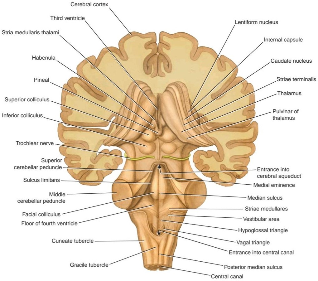
Figure 5-26 Posterior view of the brainstem showing the two superior and the two inferior colliculi of the tectum.
شکل ۵-۲۶ نمای خلفی ساقه مغز که دو کولیکولوس فوقانی و دو کولیکولوس تحتانی تکتوم را نشان میدهد.
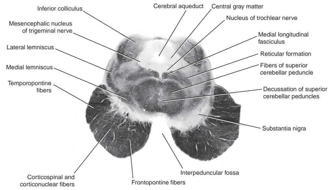
Figure 5-27 Photomicrograph of a transverse section of the midbrain at the level of the inferior colliculus. (Weigert stain.)
شکل ۵-۲۷ عکس میکروسکوپی از برش عرضی مغز میانی در سطح کولیکولوس تحتانی. (رنگآمیزی وایگرت.)
Table 5-4 Two Levels of the Midbrain and Their Major Structures a
جدول ۵-۴ دو سطح مغز میانی و ساختارهای اصلی آنهاa
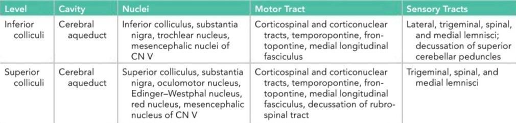
a Note that the reticular formation is present at all levels.
aتوجه داشته باشید که تشکیلات مشبک در تمام سطوح وجود دارد.
Transverse Section of the Midbrain at the Level of the Superior Colliculi
The superior colliculus (see Figs. 5-25B and 5-28), a large nucleus of gray matter that lies beneath the corresponding surface elevation, forms part of the visual reflexes. It is connected to the lateral geniculate body by the superior brachium. It receives afferent fibers from the optic nerve, the visual cortex, and the spinotectal tract. The efferent fibers form the tectospinal and tectobulbar tracts, which are probably responsible for the reflex movements of the eyes, head, and neck in response to visual stimuli. The afferent pathway for the light reflex ends in the pretectal nucleus. This is a small group of neurons situated close to the lateral part of the superior colliculus. After relaying in the pretectal nucleus, the fibers pass to the parasympathetic nucleus of the oculomotor nerve (Edinger-Westphal nucleus). The emerging fibers then pass to the oculomotor nerve. The oculomotor nucleus is situated in the central gray matter close to the median plane, just posterior to the medial longitudinal fasciculus. The fibers of the oculomotor nucleus pass anteriorly through the red nucleus to emerge on the medial side of the crus cerebri in the interpeduncular fossa. The nucleus of the oculomotor nerve is divisible into a number of cell groups.
برش عرضی مغز میانی در سطح کولیکولوس فوقانی
کولیکولوس فوقانی (به شکلهای 5-25B و 5-28 مراجعه کنید)، هسته بزرگی از ماده خاکستری که در زیر برآمدگی سطح مربوطه قرار دارد، بخشی از رفلکسهای بینایی را تشکیل میدهد. این هسته توسط بازوی فوقانی به جسم زانویی جانبی متصل است. این هسته فیبرهای آوران را از عصب بینایی، قشر بینایی و مسیر اسپینوتکتال دریافت میکند. فیبرهای وابران، مسیرهای تکتوسپینال و تکتوبولبار را تشکیل میدهند که احتمالاً مسئول حرکات رفلکسی چشمها، سر و گردن در پاسخ به محرکهای بینایی هستند. مسیر آوران برای رفلکس نور در هسته پره تکتال به پایان میرسد. این گروه کوچکی از نورونها است که در نزدیکی قسمت جانبی کولیکولوس فوقانی قرار دارند. پس از رله شدن در هسته پره تکتال، فیبرها به هسته پاراسمپاتیک عصب حرکتی چشم (هسته ادینگر-وستفال) منتقل میشوند. سپس فیبرهای بیرون آمده به عصب حرکتی چشم منتقل میشوند. هستهی عصب چشمی-حرکتی در مادهی خاکستری مرکزی نزدیک به صفحهی میانی، درست در خلف دستهی طولی داخلی قرار دارد. فیبرهای هستهی عصب چشمی-حرکتی به صورت قدامی از هستهی قرمز عبور میکنند تا در سمت داخلی ساقهی مغزی در حفرهی بین پایهای خارج شوند. هستهی عصب چشمی-حرکتی به تعدادی گروه سلولی قابل تقسیم است.
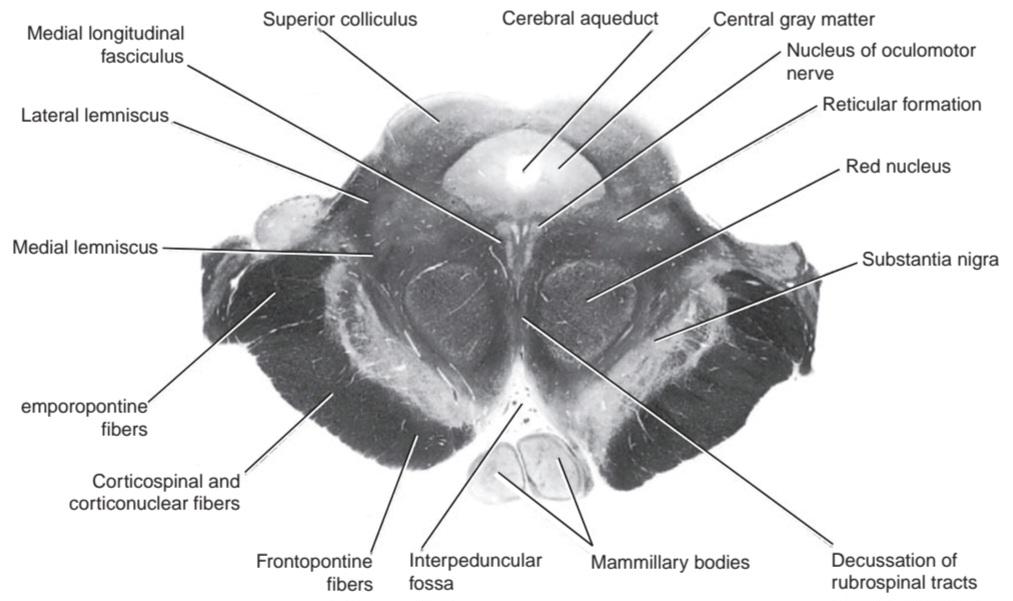
Figure 5-28 Photomicrograph of a transverse section of the midbrain at the level of the superior colliculus. (Weigert stain.)
شکل ۵-۲۸ عکس میکروسکوپی از برش عرضی مغز میانی در سطح کولیکولوس فوقانی. (رنگآمیزی وایگرت.)
The medial, spinal, and trigeminal lemnisci form a curved band posterior to the substantia nigra, but the lateral lemniscus does not extend superiorly to this level.
لمنیسهای میانی، نخاعی و سهقلو یک نوار خمیده در خلف جسم سیاه تشکیل میدهند، اما لمنیسهای جانبی به سمت بالا تا این سطح امتداد نمییابند.
The red nucleus is a rounded mass of gray matter situated between the cerebral aqueduct and the substantia nigra. Its reddish hue, seen in fresh specimens, is due to its vascularity and the presence of an iron-containing pigment in the cytoplasm of many of its neurons. Afferent fibers reach the red nucleus from (1) the cerebral cortex through the corticospinal fibers, (2) the cerebellum through the superior cerebellar peduncle, and (3) the lentiform nucleus, subthalamic and hypothalamic nuclei, substantia nigra, and spinal cord. Efferent fibers leave the red nucleus and pass to (1) the spinal cord through the rubrospinal tract (as this tract descends, it decussates), (2) the reticular formation through the rubroreticular tract, (3) the thalamus, and (4) the substantia nigra.
هسته قرمز، تودهای گرد از ماده خاکستری است که بین مجرای مغزی و جسم سیاه قرار دارد. رنگ مایل به قرمز آن، که در نمونههای تازه دیده میشود، به دلیل رگدار بودن و وجود رنگدانه حاوی آهن در سیتوپلاسم بسیاری از نورونهای آن است. الیاف آوران از (1) قشر مغز از طریق الیاف قشری-نخاعی، (2) مخچه از طریق پایه مخچهای فوقانی، و (3) هسته عدسی شکل، هستههای ساب تالامیک و هیپوتالامیک، جسم سیاه و نخاع به هسته قرمز میرسند. فیبرهای وابران هسته قرمز را ترک کرده و به (1) نخاع از طریق راه روبرواسپینال (با پایین آمدن این راه، به صورت متقاطع حرکت میکند)، (2) تشکیلات مشبک از طریق راه روبرورتیکولار، (3) تالاموس، و (4) جسم سیاه میروند.
The reticular formation is situated in the tegmentum lateral and posterior to the red nucleus.
تشکیلات مشبک در تگمنتوم جانبی و خلفی هسته قرمز قرار دارد.
The crus cerebri contains the identical important descending tracts-the corticospinal, corticonuclear, and corticopontine fibers that are present at the level of the inferior colliculus (see Table 5-4).
ستون فقرات شامل مسیرهای نزولی مهم یکسانی است – الیاف قشری-نخاعی، قشری-هستهای و قشری-پونتین که در سطح کولیکولوس تحتانی وجود دارند (به جدول 5-4 مراجعه کنید).
The continuity of the various CN nuclei through the different regions of the brainstem is shown diagrammatically in Figure 5-29.
پیوستگی هستههای مختلف CN در نواحی مختلف ساقه مغز به صورت نموداری در شکل 5-29 نشان داده شده است.

Figure 5-29 Position of some of the cranial nerve nuclei in the brainstem. A: Surface projection on the posterior aspect of the brainstem. B: Cross sections. The motor nuclei are in red and the sensory nuclei in blue.
شکل ۵-۲۹ موقعیت برخی از هستههای اعصاب جمجمهای در ساقه مغز. الف: تصویر سطحی در قسمت خلفی ساقه مغز. ب: مقاطع عرضی. هستههای حرکتی به رنگ قرمز و هستههای حسی به رنگ آبی هستند.
«فصل پنجم کتاب نورواناتومی بالینی اسنل»
🚀 با ما همراه شوید!
تازهترین مطالب و آموزشهای مغز و اعصاب را از دست ندهید. با فالو کردن کانال تلگرام آیندهنگاران مغز، از ما حمایت کنید!


