
نوروآناتومی بالینی اسنل | پرسشهای مروری و مسائل بالینی ساقه مغز


دعای مطالعه [ نمایش ]
بِسْمِ الله الرَّحْمنِ الرَّحیمِ
اَللّهُمَّ اَخْرِجْنى مِنْ ظُلُماتِ الْوَهْمِ
خدايا مرا بيرون آور از تاريكىهاى وهم،
وَ اَكْرِمْنى بِنُورِ الْفَهْمِ
و به نور فهم گرامى ام بدار،
اَللّهُمَّ افْتَحْ عَلَيْنا اَبْوابَ رَحْمَتِكَ
خدايا درهاى رحمتت را به روى ما بگشا،
وَانْشُرْ عَلَيْنا خَزائِنَ عُلُومِكَ بِرَحْمَتِكَ يا اَرْحَمَ الرّاحِمينَ
و خزانههاى علومت را بر ما باز كن به امید رحمتت اى مهربانترين مهربانان.
آیندهنگاران مغز: «ما مغز را میشناسیم، تا آینده را بسازیم.»
کتاب «نوروآناتومی بالینی اسنل» اثر دکتر ریچارد اس. اسنل، با ویرایش جدید دکتر رایان اسپلیتگربر، همچنان بهعنوان مرجع طلایی این حوزه شناخته میشود. این اثر با تلفیق تازهترین پژوهشها و کاربردهای بالینی، پلی استوار میان دانش پایه و عمل پزشکی بنا میکند.
تلاشهای علمی و سالها تدریس ارزشمند استاد گرانقدر دکتر محمدتقی جغتایی الهامبخش ترجمه و گسترش این دانش در ایران بوده است.
ترجمه دقیق این کتاب توسط برند علمی آیندهنگاران مغز به مدیریت داریوش طاهری، امکان دسترسی فارسیزبانان به مرزهای نوین علوم اعصاب را فراهم کرده و رسالتی علمی در جهت شناخت عمیقتر مغز و ترسیم آیندهای روشنتر بر عهده گرفته است.
»» فصل ۵: ساقه مغز
»» Chapter 5: Brainstem
? Clinical Problem Solving
1. While carrying out a physical examination of a patient with an intracranial tumor, the neurologist turns to a medical student and asks, “What signs or symptoms would you look for that would enable you to localize the tumor to the region of the medulla oblongata?” How would you have answered that question?
؟ حل مسئله بالینی
۱. متخصص مغز و اعصاب هنگام معاینه فیزیکی بیماری که تومور داخل جمجمهای دارد، رو به یک دانشجوی پزشکی میکند و میپرسد: «چه علائم یا نشانههایی را باید جستجو کنید تا بتوانید محل تومور را در ناحیه بصل النخاع پیدا کنید؟» شما چگونه به این سوال پاسخ میدادید؟
2. A 6-month-old boy dies with hydrocephalus and a myelocele in the lower thoracic region. At autopsy, the hindbrain is found to be deformed. The lower part of the medulla oblongata extends inferiorly through the foramen magnum into the vertebral canal as far as the third cervical vertebra. The lower four cranial nerves are longer than normal, and the upper cervical nerve roots ascend to reach their exit from the vertebral canal. The cerebellum on the left side extends inferiorly through the foramen magnum to the third cervical vertebra, where it adheres to the spinal cord. The roof of the fourth ventricle is abnormally low. (a) What is the name of this malformation? (b) Is hydrocephalus common in this condition? (c) Is an association possible between the thoracic myelocele and the presence of part of the hindbrain in the vertebral canal?
۲. یک پسر بچه ۶ ماهه به دلیل هیدروسفالی و میلوسل در ناحیه تحتانی قفسه سینه فوت میکند. در کالبدشکافی، مغز خلفی دچار تغییر شکل شده است. قسمت تحتانی بصل النخاع از طریق سوراخ بزرگ به سمت پایین و به داخل کانال مهرهای تا مهره سوم گردنی امتداد مییابد. چهار عصب جمجمهای پایینی بلندتر از حد معمول هستند و ریشههای عصبی گردنی بالایی برای رسیدن به محل خروج خود از کانال مهرهای به سمت بالا میروند. مخچه در سمت چپ از طریق سوراخ بزرگ به سمت پایین و به مهره سوم گردنی امتداد مییابد، جایی که به نخاع میچسبد. سقف بطن چهارم به طور غیرطبیعی پایین است. (الف) نام این ناهنجاری چیست؟ (ب) آیا هیدروسفالی در این بیماری شایع است؟ (ج) آیا ارتباطی بین میلوسل قفسه سینه و وجود بخشی از مغز خلفی در کانال مهرهای وجود دارد؟
3. A 68-year-old man is admitted to the hospital with the sudden onset of severe dizziness (vertigo), hiccups, and vomiting. He also complains of a hot, painful sensation in the skin of the right side of the face. On physical examination, the soft palate is drawn up to the left side when the patient was asked to say “ah,” and the right vocal cord lacked mobility as seen on laryngoscopic examination. The patient also shows drooping of the right upper eyelid (ptosis), sunken right eye (enophthalmos), and a constricted right pupil (myosis). When asked to protrude his tongue straight out of his mouth, the patient tries to do so, but the tip of the tongue pointed to the right side. Impaired pain and temperature sensation is evident in the trunk and extremities on the left side. Using your knowledge of anatomy, make the diagnosis.
۳. مردی ۶۸ ساله با شروع ناگهانی سرگیجه شدید (vertigo)، سکسکه و استفراغ در بیمارستان بستری میشود. او همچنین از احساس گرما و درد در پوست سمت راست صورت شکایت دارد. در معاینه فیزیکی، وقتی از بیمار خواسته میشود «آه» بگوید، کام نرم به سمت چپ کشیده میشود و همانطور که در معاینه لارنگوسکوپی مشاهده میشود، تار صوتی سمت راست فاقد حرکت است. بیمار همچنین افتادگی پلک فوقانی راست (پتوز)، فرورفتگی چشم راست (انوفتالموس) و انقباض مردمک راست (میوز) را نشان میدهد. وقتی از بیمار خواسته میشود زبان خود را مستقیماً از دهانش بیرون بیاورد، سعی میکند این کار را انجام دهد، اما نوک زبان به سمت راست اشاره میکند. اختلال در حس درد و دما در تنه و اندامهای سمت چپ مشهود است. با استفاده از دانش خود در مورد آناتومی، تشخیص را انجام دهید.
4. A pathologist, while exploring the posterior cranial fossa during an autopsy, is endeavoring to determine where the 9th, the 10th, and the cranial part of the 11th cranial nerves emerge from the hindbrain. Describe where these nerves emerge from the hindbrain.
۴. یک آسیبشناس، هنگام بررسی حفره جمجمهای خلفی در طول کالبدشکافی، تلاش میکند تا محل خروج اعصاب جمجمهای نهم، دهم و قسمت جمجمهای یازدهم از مغز خلفی را مشخص کند. محل خروج این اعصاب از مغز خلفی را شرح دهید.
5. A 10-year-old girl is taken to a physician because her mother has noticed that the right half of her face was weak and does not appear to react to emotional changes. Her mouth is pulled over slightly to the left, especially when she is tired. On questioning, the patient admits that food tends to stick inside her right cheek and that the right side of her face “felt funny.” The mother had first noticed the facial changes 3 months previously, and the condition has progressively worsened. On examination, definite weakness of the facial muscles on the right side is noted; the facial muscles on the left side are normal. Skin sensation on stimulation of the face is normal. On testing of the ocular movements, slight weakness of the lateral rectus muscle is evident on the right side. Examination of the movements of the arm and leg shows slight weakness on the left side. Using your knowledge of neuroanatomy, relate these symptoms and signs to a lesion in the pons.
۵. دختری ۱۰ ساله به پزشک مراجعه میکند زیرا مادرش متوجه شده است که نیمه راست صورتش ضعیف است و به نظر نمیرسد به تغییرات عاطفی واکنش نشان دهد. دهانش کمی به سمت چپ کشیده میشود، به خصوص وقتی خسته است. در پرسشها، بیمار اذعان میکند که غذا تمایل دارد داخل گونه راست او گیر کند و سمت راست صورتش «احساس عجیبی» دارد. مادر اولین بار ۳ ماه پیش متوجه تغییرات صورت شده بود و این وضعیت به تدریج بدتر شده است. در معاینه، ضعف قطعی عضلات صورت در سمت راست مشاهده میشود؛ عضلات صورت در سمت چپ طبیعی هستند. حس پوست در هنگام تحریک صورت طبیعی است. در آزمایش حرکات چشم، ضعف جزئی عضله رکتوس جانبی در سمت راست مشهود است. بررسی حرکات بازو و پا، ضعف جزئی در سمت چپ را نشان میدهد. با استفاده از دانش خود در مورد نوروآناتومی، این علائم و نشانهها را به ضایعهای در پل مغزی ربط دهید.
6. A 65-year-old man is admitted to the emergency department with a diagnosis of a severe pontine hemorrhage. On examination, he is found to have bilateral “pinpoint” pupils and quadriplegia. How can you explain the presence of the “pinpoint” pupils?
۶. مردی ۶۵ ساله با تشخیص خونریزی شدید پل مغزی در اورژانس بستری شده است. در معاینه، مشخص شد که او مردمکهای «نقطهای» دو طرفه و فلج چهار اندام دارد. چگونه میتوانید وجود مردمکهای «نقطهای» را توضیح دهید؟
7. A 46-year-old man with symptoms of deafness, vertigo, and double vision (diplopia) visits his physician. On questioning, he says that he also suffers from severe headaches, which are increasing in frequency and severity. The week before, he vomited several times during one of the headache attacks. On examination, he is found to have a slight right internal strabismus, a flattening of the skin furrows on the right side of his forehead, and a slight drooping of the right corner of his mouth. Hearing is impaired on the right side. On testing for sensory loss, sensory impairment is evident on the right side of the face in the areas supplied by the max- illary and mandibular divisions of the trigeminal nerve. Using your knowledge of anatomy, explain the symptoms and signs.
۷. مردی ۴۶ ساله با علائم ناشنوایی، سرگیجه و دوبینی (دیپلوپی) به پزشک مراجعه میکند. در پاسخ به سوالات، او میگوید که از سردردهای شدیدی نیز رنج میبرد که تعداد و شدت آنها رو به افزایش است. هفته قبل، او در یکی از حملات سردرد چندین بار استفراغ کرده است. در معاینه، مشخص شد که او دچار انحراف خفیف چشم داخلی سمت راست، صاف شدن چین و چروکهای پوستی در سمت راست پیشانی و افتادگی خفیف گوشه سمت راست دهان است. شنوایی در سمت راست مختل است. در آزمایش از دست دادن حس، اختلال حسی در سمت راست صورت در نواحی که توسط شاخههای فک بالا و پایین عصب سه قلو عصبدهی میشوند، مشهود است. با استفاده از دانش خود در مورد آناتومی، علائم و نشانهها را توضیح دهید.
8. After a severe automobile accident that results in the death of the driver of one of the vehicles, an autopsy is performed, and the skull is opened. A massive subdural hematoma is found in the middle cranial fossa. The rapid accumulation of blood within the skull had exerted pressure on the brain above the tentorium cerebelli. The uncus of the temporal lobe has been forced inferiorly through the hiatus in the tentorium cerebelli. What effect do you think these intracranial changes had on the midbrain of this patient?
۸. پس از یک تصادف شدید رانندگی که منجر به مرگ راننده یکی از وسایل نقلیه شد، کالبدشکافی انجام شد و جمجمه باز شد. یک هماتوم ساب دورال عظیم در حفره جمجمه میانی یافت شد. تجمع سریع خون در جمجمه، به مغز بالای چادرینه مخچه فشار وارد کرده بود. استخوان گیجگاهی لوب گیجگاهی از طریق شکاف چادرینه مخچه به سمت پایین رانده شده بود. به نظر شما این تغییرات داخل جمجمه چه تأثیری بر مغز میانی این بیمار داشته است؟
9. A 3-month-old girl is taken to a pediatrician because her mother is concerned about the large size of her head. The child is perfectly normal in every other respect. Examination of the child shows that the diameter of the head was larger than normal for the age; the fontanelles are larger than normal and are moderately tense. The scalp is shiny, and the scalp veins are dilated. The eyes are normal, and the mental and physical development of the child is within normal limits. Computed tomography and magnetic resonance imaging of the head reveal gross dilation of the third and lateral ventricles of the brain. What is your diagnosis? What possible treatment should be suggested to the mother of this child?
۹. یک دختر ۳ ماهه به دلیل نگرانی مادرش از اندازه بزرگ سر او، به متخصص اطفال برده میشود. کودک از هر نظر دیگر کاملاً طبیعی است. معاینه کودک نشان میدهد که قطر سر برای سن او بزرگتر از حد معمول بوده است؛ فونتانلها بزرگتر از حد معمول و نسبتاً سفت هستند. پوست سر براق است و رگهای پوست سر گشاد شدهاند. چشمها طبیعی هستند و رشد ذهنی و جسمی کودک در محدوده طبیعی است. توموگرافی کامپیوتری و تصویربرداری رزونانس مغناطیسی از سر، گشاد شدن آشکار بطن سوم و جانبی مغز را نشان میدهد. تشخیص شما چیست؟ چه درمان احتمالی باید به مادر این کودک پیشنهاد شود؟
10. A 20-year-old man is seen by a neurologist because he has a 3-month history of double vision. On examination of the patient, both eyes at rest are turned downward and laterally. The patient is unable to move the eyes upward or medially. Both upper lids are drooping (ptosis). Examination of both pupils shows them to be dilated, and they do not constrict when a light is shone into either eye. Facial movements and sensation are normal. Movements of the upper and lower limbs are normal. Loss of or altered skin sensations is not evident in the upper or the lower limbs. Using your knowledge of neuroanatomy, make a diagnosis and accurately locate the site of the lesion. Is the lesion unilateral or bilateral?
۱۰. مردی ۲۰ ساله به دلیل سابقه ۳ ماهه دوبینی توسط متخصص مغز و اعصاب ویزیت میشود. در معاینه بیمار، هر دو چشم در حالت استراحت به سمت پایین و طرفین چرخیدهاند. بیمار قادر به حرکت دادن چشمها به سمت بالا یا داخل نیست. هر دو پلک بالایی افتاده (پتوز) هستند. معاینه هر دو مردمک نشان میدهد که گشاد شدهاند و با تاباندن نور به هر یک از چشمها، منقبض نمیشوند. حرکات و حس صورت طبیعی است. حرکات اندامهای فوقانی و تحتانی طبیعی است. از دست دادن یا تغییر حس پوست در اندامهای فوقانی یا تحتانی مشهود نیست. با استفاده از دانش خود در مورد نوروآناتومی، تشخیص را انجام دهید و محل ضایعه را به طور دقیق مشخص کنید. آیا ضایعه یک طرفه است یا دو طرفه؟
11. A 57-year-old man with hypertension is admitted to the hospital with a diagnosis of hemorrhage into the midbrain, possibly from a branch of the posterior cerebral artery. He is found, on physical examination, to have paralysis on the right side of the levator palpebrae superioris, the superior rectus, medial rectus, inferior rectus, and inferior oblique muscles. Furthermore, his right pupil is dilated and fails to constrict on exposure to light or on accommodation. The left eye is normal in every respect. He displays hypersensitivity to touch on the skin of the left side of his face and has loss of skin sensation on the greater part of his left arm and left leg. The left leg also displays some spontaneous slow writhing movements (athetosis). Using your knowledge of neuroanatomy, explain the signs and symptoms exhibited by this patient.
۱۱. مردی ۵۷ ساله مبتلا به فشار خون بالا با تشخیص خونریزی در مغز میانی، احتمالاً از شاخهای از شریان مغزی خلفی، در بیمارستان بستری میشود. در معاینه فیزیکی مشخص میشود که او در سمت راست عضله بالابرنده پلک فوقانی، عضله راست فوقانی، عضله راست داخلی، عضله راست تحتانی و عضلات مایل تحتانی فلج دارد. علاوه بر این، مردمک چشم راست او گشاد شده و در معرض نور یا تطابق منقبض نمیشود. چشم چپ از هر نظر طبیعی است. او حساسیت بیش از حد به لمس پوست سمت چپ صورت خود نشان میدهد و در قسمت بیشتر بازوی چپ و پای چپ خود بیحس است. پای چپ نیز برخی حرکات پیچشی آهسته خود به خودی (آتتوز) را نشان میدهد. با استفاده از دانش خود در مورد نوروآناتومی، علائم و نشانههای بروز یافته توسط این بیمار را توضیح دهید.
12. A 41-year-old woman is diagnosed as having a lesion in the midbrain. Physical examination reveals an oculomotor nerve palsy on the left side (paralysis of the left extraocular muscles except the lateral rectus and the superior oblique muscles) and an absence of the light and accommodation reflexes on the left side. Some weakness is evident but no atrophy of the muscles of the lower part of the face and the tongue on the right side. The right arm and leg show spastic paralysis. Sensory loss on either side of the head, trunk, or limbs is not noted. Using your knowledge of neuroanatomy, precisely place the lesion in the midbrain of this patient.
۱۲. یک زن ۴۱ ساله مبتلا به ضایعهای در مغز میانی تشخیص داده میشود. معاینه فیزیکی، فلج عصب چشمی-حرکتی در سمت چپ (فلج عضلات خارج چشمی چپ به جز عضله رکتوس جانبی و عضله مایل فوقانی) و فقدان رفلکسهای نور و تطابق در سمت چپ را نشان میدهد. مقداری ضعف مشهود است اما آتروفی عضلات قسمت تحتانی صورت و زبان در سمت راست وجود ندارد. فلج اسپاستیک در دست و پای راست مشاهده میشود. از دست دادن حس در هر دو طرف سر، تنه یا اندامها مشاهده نمیشود. با استفاده از دانش خود در مورد نوروآناتومی، محل دقیق ضایعه را در مغز میانی این بیمار مشخص کنید.
√ Answers and Explanations to Clinical Problem Solving
1. Until involvement of one of the last four cranial nerves occurs, localization of a lesion to the medulla oblongata remains uncertain. For example, involve- ment of the main ascending sensory pathways or descending pathways may be caused by a lesion in the medulla, the pons, the midbrain, or the spinal cord. Involvement of the glossopharyngeal nerve can be detected by inadequacy of the gag reflex and loss of taste sensation on the posterior third of the tongue. Involvement of the vagus nerve can be assumed if the patient demonstrates some or all of the following symptoms: impairment of pharyngeal sensibility, difficulty in swallowing, nasal regurgitation of fluids with asymmetry of movement of the soft palate, and hoarseness of the voice with paralysis of the laryngeal muscles. The cranial part of the accessory nerve is distributed within the vagus nerve so that testing for this nerve alone is not possible. The spinal part of the accessory nerve, which supplies the sternocleidomastoid and trapezius muscles, arises from the spinal cord and is therefore unaffected by tumors of the medulla. The hypoglossal nerve involvement may be tested by looking for wasting, fasciculation, and paralysis of half of the tongue.
√ پاسخها و توضیحات برای حل مسئله بالینی
۱. تا زمانی که یکی از چهار عصب جمجمهای آخر درگیر نشود، محل ضایعه در بصل النخاع نامشخص است. به عنوان مثال، درگیری مسیرهای حسی صعودی اصلی یا مسیرهای نزولی ممکن است ناشی از ضایعه در بصل النخاع، پل مغزی، مغز میانی یا نخاع باشد. درگیری عصب زبانی-حلقی را میتوان با ناکافی بودن رفلکس تهوع و از دست دادن حس چشایی در یک سوم خلفی زبان تشخیص داد. در صورتی که بیمار برخی یا همه علائم زیر را نشان دهد، میتوان درگیری عصب واگ را فرض کرد: اختلال در حساسیت حلق، مشکل در بلع، برگشت مایعات از بینی با عدم تقارن حرکت کام نرم و گرفتگی صدا با فلج عضلات حنجره. قسمت جمجمهای عصب فرعی در داخل عصب واگ توزیع شده است، بنابراین آزمایش این عصب به تنهایی امکانپذیر نیست. بخش نخاعی عصب فرعی، که عضلات جناغی-چنبری-ماستوئید و ذوزنقهای را عصبدهی میکند، از نخاع منشأ میگیرد و بنابراین تحت تأثیر تومورهای بصل النخاع قرار نمیگیرد. درگیری عصب زیرزبانی را میتوان با بررسی تحلیل رفتن، فاسیکولاسیون و فلج نیمی از زبان بررسی کرد.
2. (a) The malformation in which the cerebellum and the medulla oblongata are found in the cervical part of the vertebral canal is known as the Arnold-Chiari malformation. (b) Yes. Hydrocephalus is common in this condition. The hydrocephalus may be due to distortion or malformation of the openings in the roof of the fourth ventricle, which normally allow the cerebrospinal fluid to escape into the subarachnoid space. (c) Yes. A myelocele is commonly associated with this malformation. The reason for this is not exactly known, although several investigators believe that the myelocele is the primary cause and that it tethers the lower part of the spinal cord to the surrounding tissues at the time when disproportionate growth of the spinal cord and the vertebral column occurs. This would serve to pull the medulla oblongata and the cerebellum inferiorly through the foramen magnum into the vertebral canal.
۲. (الف) ناهنجاری که در آن مخچه و بصل النخاع در قسمت گردنی کانال مهرهای یافت میشوند، به عنوان ناهنجاری آرنولد-کیاری شناخته میشود. (ب) بله. هیدروسفالی در این وضعیت شایع است. هیدروسفالی ممکن است به دلیل اعوجاج یا ناهنجاری دهانههای سقف بطن چهارم باشد که به طور معمول اجازه میدهند مایع مغزی نخاعی به فضای زیر عنکبوتیه فرار کند. (ج) بله. میلوسل معمولاً با این ناهنجاری مرتبط است. دلیل این امر دقیقاً مشخص نیست، اگرچه چندین محقق معتقدند که میلوسل علت اصلی است و در زمانی که رشد نامتناسب نخاع و ستون مهرهها رخ میدهد، قسمت پایینی نخاع را به بافتهای اطراف متصل میکند. این امر باعث میشود که بصل النخاع و مخچه از طریق سوراخ بزرگ به داخل کانال مهرهای کشیده شوند.
3. This patient is suffering from a thrombosis of the posterior inferior cerebellar artery or vertebral artery on the right side. The vertigo is caused by the involvement of the cerebellum or the vestibular nuclei or both. The hot, painful skin sensations are due to the involvement of the spinal tract and nucleus of the trigeminal nerve on the right side. The abnormal movement of the soft palate and the fixation of the right vocal cord are due to involvement of the nucleus of the vagus and accessory nerve on the right side. The ptosis, enophthalmos, and myosis (Horner syndrome) are due to involvement of the descending fibers of the sympathetic part of the autonomic nervous system. The tongue pointing to the right is caused by involvement of the right hypoglossal nucleus (the right genioglossus muscle is paralyzed). The loss of pain and temperature sensations on the opposite side of the body is due to involvement of the ascending lateral spinothalamic tracts. This characteristic clinical syndrome results from cutting off the arterial supply to a wedge-shaped area in the posterolateral part of the medulla oblongata and the inferior surface of the cerebellum.
۳. این بیمار از ترومبوز شریان مخچهای تحتانی خلفی یا شریان مهرهای در سمت راست رنج میبرد. سرگیجه ناشی از درگیری مخچه یا هستههای دهلیزی یا هر دو است. احساس گرما و درد پوست به دلیل درگیری دستگاه نخاعی و هسته عصب سه قلو در سمت راست است. حرکت غیرطبیعی کام نرم و ثابت شدن تارهای صوتی سمت راست به دلیل درگیری هسته عصب واگ و عصب فرعی در سمت راست است. پتوز، انوفتالموس و میوز (سندرم هورنر) به دلیل درگیری الیاف نزولی بخش سمپاتیک سیستم عصبی خودکار است. زبان که به سمت راست اشاره میکند به دلیل درگیری هسته هیپوگلوسال راست است (عضله جنیوگلوسوس راست فلج شده است). از دست دادن احساس درد و دما در سمت مقابل بدن به دلیل درگیری مسیرهای اسپینوتالامیک جانبی صعودی است. این سندرم بالینی مشخصه، ناشی از قطع خونرسانی شریانی به ناحیهای گوهای شکل در قسمت خلفی-جانبی بصل النخاع و سطح تحتانی مخچه است.
4. The 9th, the 10th, and the cranial part of the 11th cranial nerves emerge from the medulla oblongata in a groove between the olives and the inferior cerebellar peduncles.
۴. اعصاب نهم، دهم و بخش جمجمهای اعصاب یازدهم جمجمهای از بصل النخاع در شیاری بین زیتونها و پایههای مخچه تحتانی خارج میشوند.
5. This 10-year-old girl later was found to have an astrocytoma of the pons. The right unilateral facial weakness, together with weakness of the right lateral rectus muscle of the eye, was due to involvement of the right facial and abducens nuclei by the tumor. The absence of paresthesia of the face indicated that the principal sensory nucleus of the trigeminal nerve was intact on both sides. The weakness in the movements of the left arm and left leg was due to the involvement of the corticospinal fibers in the pons. (Remember that most of these fibers cross over to the opposite side at the decus- sation of the pyramids in the medulla.)
۵. بعداً مشخص شد که این دختر ۱۰ ساله مبتلا به آستروسیتومای پل مغزی است. ضعف یک طرفه صورت در سمت راست، همراه با ضعف عضله راست جانبی چشم، به دلیل درگیری هستههای صورت و ابدوسنس راست توسط تومور بود. عدم وجود پارستزی صورت نشان داد که هسته حسی اصلی عصب سه قلو در هر دو طرف سالم است. ضعف در حرکات دست چپ و پای چپ به دلیل درگیری فیبرهای قشری-نخاعی در پل مغزی بود. (به یاد داشته باشید که بیشتر این فیبرها در محل تقاطع هرمها در بصل النخاع به سمت مقابل عبور میکنند.)
6. “Pinpoint” pupils indicate that the constrictor pupillae muscles are strongly contracted and the dilator pupillae muscles are paralyzed. The dilator pupillae muscles are supplied by the sympathetic fibers, which descend through the pons (position not precisely known) to the lateral gray columns of the thoracic part of the spinal cord. Here, the fibers synapse, and the thoracolumbar sympathetic out- flow occurs.
۶. مردمکهای «سرسوزنی» نشان میدهند که عضلات تنگکننده مردمک به شدت منقبض و عضلات گشادکننده مردمک فلج شدهاند. عضلات گشادکننده مردمک توسط فیبرهای سمپاتیک تغذیه میشوند که از طریق پل مغزی (موقعیت دقیق آنها مشخص نیست) به ستونهای خاکستری جانبی قسمت سینهای نخاع میرسند. در اینجا، فیبرها سیناپس میکنند و خروج سمپاتیک سینهای-کمری رخ میدهد.
7. The deafness and vertigo were due to lesions in the cochlear and vestibular nuclei in the upper part of the pons. The double vision (diplopia) was produced by the involvement of the abducens nerve nucleus on the right side of the pons. The history of severe headaches and vomiting was due to a progressive rise in intracranial pressure caused by a tumor of the pons. The right unilateral facial palsy was due to the involvement of the right facial nerve nucleus. The sensory impairment of the skin of the middle and lower part of the right side of the face was due to the tumor involvement of the principal sensory nucleus of the right trigeminal nerve.
۷. ناشنوایی و سرگیجه به دلیل ضایعات هستههای حلزونی و دهلیزی در قسمت فوقانی پل مغزی بود. دوبینی (دیپلوپی) در اثر درگیری هسته عصب ابدوسنس در سمت راست پل مغزی ایجاد شده بود. سابقه سردردهای شدید و استفراغ به دلیل افزایش پیشرونده فشار داخل جمجمه ناشی از تومور پل مغزی بود. فلج یک طرفه صورت سمت راست به دلیل درگیری هسته عصب صورتی سمت راست بود. اختلال حسی پوست قسمت میانی و پایینی سمت راست صورت به دلیل درگیری هسته حسی اصلی عصب سه قلوی راست توسط تومور بود.
8. The herniated uncus and the subdural hemorrhage caused pressure of the opposite crus cerebri of the midbrain against the sharp edge of the tentorium. The distortion of the midbrain caused narrowing of the cerebral aqueduct, further raising the supratentorial pressure by blocking the passage of cerebrospinal fluid from the third to the fourth ventricle. Under these circumstances, severe hemorrhage may occur within the midbrain and affect the third and fourth cranial nerve nuclei and various important descending and ascending tracts.
۸. بیرونزدگی استخوان زیر سختشامه و خونریزی سابدورال باعث فشار ساق مغزی سمت مقابل مغز میانی به لبه تیز چادرینه شد. انحراف مغز میانی باعث تنگ شدن مجرای مغزی شده و با مسدود کردن مسیر مایع مغزی نخاعی از بطن سوم به بطن چهارم، فشار بالای چادرینه را بیشتر افزایش میدهد. در این شرایط، خونریزی شدید ممکن است در مغز میانی رخ دهد و هستههای اعصاب جمجمهای سوم و چهارم و مسیرهای مهم نزولی و صعودی مختلف را تحت تأثیر قرار دهد.
9. This child had hydrocephalus. The physical examination and the special tests showed that the third and lateral ventricles of the brain were grossly dilated owing to the accumulation of cerebrospinal fluid (CSF) in these cavities. Mechanical obstruction to CSF flow from the third into the fourth ventricle through the cerebral aqueduct was present. After the possibility of the presence of cysts or resectable tumors had been excluded, it was assumed that the cause of the obstruction was a congenital atresia or malformation of the cerebral aqueduct. If the condition were progressing-that is, the block in the aqueduct was complete and the head continued to increase in size at an abnormal rate-some form of neurosurgical procedure should have been performed to shunt CSF from the third or lateral ventricles into the subarachnoid space or into the venous system of the neck.
۹. این کودک هیدروسفالی داشت. معاینه فیزیکی و آزمایشهای ویژه نشان داد که بطنهای سوم و جانبی مغز به دلیل تجمع مایع مغزی نخاعی (CSF) در این حفرهها، به شدت متسع شدهاند. انسداد مکانیکی جریان CSF از بطن سوم به بطن چهارم از طریق مجرای مغزی وجود داشت. پس از رد احتمال وجود کیست یا تومورهای قابل برداشت، فرض بر این بود که علت انسداد، آترزی مادرزادی یا ناهنجاری مجرای مغزی است. اگر وضعیت در حال پیشرفت بود – یعنی انسداد مجرای مغزی کامل بود و اندازه سر با سرعت غیرطبیعی افزایش مییافت – باید نوعی عمل جراحی مغز و اعصاب برای شنت کردن CSF از بطن سوم یا جانبی به فضای زیر عنکبوتیه یا به سیستم وریدی گردن انجام میشد.
10. Two years later, the patient died. At autopsy, a large astrocytoma that involved the central part of the tegmentum at the level of the superior colliculi was found. The patient had exhibited all signs and symptoms associated with a raised intracranial pressure. The raised pressure was due in part to the expand- ing tumor, but the problem was compounded by the developing hydrocephalus resulting from blockage of the cerebral aqueduct.
۱۰. دو سال بعد، بیمار فوت کرد. در کالبدشکافی، یک آستروسیتومای بزرگ که قسمت مرکزی تگمنتوم را در سطح کولیکولوس فوقانی درگیر کرده بود، یافت شد. بیمار تمام علائم و نشانههای مرتبط با افزایش فشار داخل جمجمه را نشان داده بود. این افزایش فشار تا حدی به دلیل گسترش تومور بود، اما مشکل با هیدروسفالی در حال توسعه ناشی از انسداد مجرای مغزی تشدید شد.
The symptoms and signs exhibited by the patient when he was first seen by the neurologist could be explained by the presence of the tumor in the central gray matter at the level of the superior colliculi and involving the third cranial nerve nuclei on both sides. This resulted in bilateral ptosis; bilateral ophthalmoplegia; and bilateral fixed, dilated pupils. The resting position of the eyes in a downward and lat- eral position was due to the action of the superior oblique muscle (trochlear nerve) and lateral rectus muscle (abducens nerve).
علائم و نشانههایی که بیمار هنگام اولین ویزیت توسط متخصص مغز و اعصاب از خود نشان داد، میتوانست با وجود تومور در ماده خاکستری مرکزی در سطح کولیکولهای فوقانی و درگیری هستههای عصب جمجمهای سوم در هر دو طرف توضیح داده شود. این امر منجر به پتوز دو طرفه؛ فلج چشمی دو طرفه؛ و مردمکهای ثابت و متسع دو طرفه شد. وضعیت استراحت چشمها در حالت رو به پایین و جانبی به دلیل عملکرد عضله مایل فوقانی (عصب تروکلئار) و عضله رکتوس جانبی (عصب ابدوسنس) بود.
11. The patient had a hemorrhage in the right side of the tegmentum of the midbrain that involved the right third cranial nerve. The ascending tracts of the left trigeminal nerve also were involved. After emerging from the sensory nuclei of the left trigeminal nerve, they cross the midline and ascend through the trigeminal lemniscus on the right side. The loss of sensation seen in the left upper and lower limbs was due to involvement of the right medial and spinal lemnisci. The athetoid movements of the left leg could be explained on the basis of the involvement of the right red nucleus. The absence of spasticity of the left arm and leg would indicate that the lesion did not involve the right descending tracts. For further clarification, consult the descriptions of the various tracts (see Chapter 4).
۱۱. بیمار دچار خونریزی در سمت راست تگمنتوم مغز میانی بود که عصب سوم جمجمهای راست را درگیر میکرد. مسیرهای صعودی عصب سهقلو چپ نیز درگیر بودند. پس از خروج از هستههای حسی عصب سهقلو چپ، از خط وسط عبور کرده و از طریق لمنیسکوس سهقلو در سمت راست بالا میروند. از دست دادن حس مشاهده شده در اندام فوقانی و تحتانی چپ به دلیل درگیری لمنیسکوس میانی و نخاعی راست بود. حرکات آتتوئید پای چپ را میتوان بر اساس درگیری هسته قرمز راست توضیح داد. عدم وجود اسپاسم در بازو و پای چپ نشان میدهد که ضایعه مسیرهای نزولی راست را درگیر نکرده است. برای توضیح بیشتر، به توضیحات مسیرهای مختلف مراجعه کنید (به فصل ۴ مراجعه کنید).
12. Autopsy later revealed a vascular lesion involving a branch of the posterior cerebral artery. Considerable brain softening was found in the region of the substantia nigra and crus cerebri on the left side of the midbrain. The left oculomotor nerve was involved as it passed through the infarcted area. The corticonuclear fibers that pass to the facial nerve nucleus and the hypoglossal nucleus were involved as they descended through the left crus cerebri (they cross the midline at the level of the nuclei). The corticospinal fibers on the left side were also involved (they cross in the medulla oblongata), hence the spastic paralysis of the right arm and leg. The left trigeminal and left medial lemnisci were untouched, which explains the absence of sensory changes on the right side of the body. This is a good example of Weber syndrome.
۱۲. کالبدشکافی بعداً یک ضایعه عروقی را نشان داد که شاخهای از شریان مغزی خلفی را درگیر میکرد. نرم شدن قابل توجه مغز در ناحیه جسم سیاه و ساق مغزی در سمت چپ مغز میانی مشاهده شد. عصب چشمی-حرکتی چپ هنگام عبور از ناحیه انفارکتوس درگیر بود. الیاف قشری-هستهای که به هسته عصب صورت و هسته هیپوگلوسال میروند هنگام پایین آمدن از ساق مغزی چپ درگیر بودند (آنها از خط میانی در سطح هستهها عبور میکنند). الیاف قشری-نخاعی در سمت چپ نیز درگیر بودند (آنها در بصل النخاع عبور میکنند)، از این رو فلج اسپاستیک دست و پای راست ایجاد شد. عضله سه قلوی چپ و پرده میانی چپ دست نخورده بودند، که عدم وجود تغییرات حسی در سمت راست بدن را توضیح میدهد. این مثال خوبی از سندرم وبر است.
? Review Questions
Directions: Each of the numbered items in this section is followed by answers. Select the ONE lettered answer that is CORRECT.
؟ سوالات مروری
دستورالعمل: هر یک از موارد شمارهگذاری شده در این بخش با پاسخهایی دنبال میشوند. تنها پاسخ صحیح با یک حرف را انتخاب کنید.
1. The following statements concern the anterior surface of the medulla oblongata:
(a) The pyramids taper inferiorly and give rise to the decussation of the pyramids.
(b) On each side of the midline, an ovoid swelling called the olive contains the corticospinal fibers.
(c) The hypoglossal nerve emerges between the olive and the inferior cerebellar peduncle.
(d) The vagus nerve emerges between the pyramid and the olive.
(e) The abducens nerve emerges between the pons and the midbrain.
۱. عبارات زیر مربوط به سطح قدامی بصل النخاع است:
(الف) هرمها به سمت پایین باریک میشوند و باعث ایجاد تقاطع هرمها میشوند.
(ب) در هر طرف خط میانی، یک برآمدگی تخممرغی شکل به نام زیتون حاوی الیاف قشری-نخاعی است.
(ج) عصب زیرزبانی بین زیتون و پایه مخچه تحتانی خارج میشود.
(د) عصب واگ بین هرم و زیتون خارج میشود.
(ه) عصب ابدوسنس بین پل مغزی و مغز میانی خارج میشود.
2. The following general statements concern the medulla oblongata:
(a) The caudal half of the floor of the fourth ventricle is formed by the rostral half of the medulla.
(b) The central canal extends throughout the length of the medulla oblongata.
(c) The nucleus gracilis is situated beneath the gracile tubercle on the anterior surface of the medulla.
(d) The decussation of the medial lemnisci takes place in the rostral half of the medulla.
(e) The cerebellum lies anterior to the medulla.
(ب) کانال مرکزی در سراسر طول بصل النخاع امتداد دارد.
(ج) هسته گراسیلیس در زیر برجستگی گراسیل در سطح قدامی بصل النخاع قرار دارد.
(د) تقاطع لمنیسهای داخلی در نیمه جلویی بصل النخاع انجام میشود.
(ه) مخچه در جلوی بصل النخاع قرار دارد.
3. The following statements concern the interior of the lower part of the medulla:
(a) The decussation of the pyramids represents the crossing over from one side of the medulla to the other of a quarter of the corticospinal fibers.
(b) The central canal of the spinal cord is not continuous upward into the medulla.
(c) The substantia gelatinosa is not continuous with the nucleus of the spinal tract of the trigeminal nerve.
(d) The medial lemniscus is formed by the anterior spinothalamic tract and the spinotectal tract.
(e) The internal arcuate fibers emerge from the nucleus gracilis and nucleus cuneatus.
(ب) کانال مرکزی نخاع به سمت بالا و به سمت بصل النخاع ادامه ندارد.
(ج) جسم ژلاتینی با هسته راه نخاعی عصب سه قلو ادامه ندارد.
(د) نوار میانی توسط راه نخاعی-تالامیک قدامی و راه نخاعی-تکتال تشکیل شده است.
(ه) فیبرهای قوسی داخلی از هسته گراسیلیس و هسته کونئاتوس خارج میشوند.
4. The following statements concern the interior of the upper part of the medulla:
(a) The reticular formation consists of nerve fibers but no nerve cells.
(b) The nucleus ambiguus constitutes the motor nucleus of the vagus, cranial part of the accessory, and hypoglossal nerves.
(c) Beneath the floor of the fourth ventricle are located the dorsal nucleus of the vagus and the vestibular nuclei.
(d) The medial longitudinal fasciculus is a bundle of ascending fibers on each side of the midline.
(e) The inferior cerebellar peduncle connects the pons to the cerebellum.
(ب) هسته مبهم، هسته حرکتی عصب واگ، قسمت جمجمهای عصب فرعی و اعصاب زیرزبانی را تشکیل میدهد.
(ج) در زیر کف بطن چهارم، هسته پشتی عصب واگ و هستههای دهلیزی قرار دارند.
(د) دستهای از الیاف صعودی در هر طرف خط میانی است.
(ه) پایه مخچهای تحتانی، پل مغزی را به مخچه متصل میکند.
5. The following statements concern the Arnold-Chiari phenomenon:
(a) It is an acquired anomaly.
(b) The exits in the roof of the fourth ventricle may be blocked.
(c) The cerebellum never herniates through the foramen magnum.
(d) It is not associated with various forms of spina bifida.
(e) Performing a spinal tap in this condition is safe.
(ب) ممکن است خروجیهای سقف بطن چهارم مسدود شده باشند.
(ج) مخچه هرگز از طریق سوراخ بزرگ بیرون نمیزند.
(د) با اشکال مختلف اسپینا بیفیدا مرتبط نیست.
(ه) انجام پونکسیون نخاعی در این شرایط بیخطر است.
6. The following statements concern the medial medullary syndrome:
(a) The tongue is paralyzed on the contralateral side.
(b) Ipsilateral hemiplegia is evident.
(c) Ipsilateral sensations of position and movement are impaired.
(d) It is commonly caused by thrombosis of a branch of the vertebral artery to the medulla oblongata.
(e) Contralateral facial paralysis is evident.
۶. اظهارات زیر مربوط به سندرم مدولای داخلی است:
(الف) زبان در سمت مقابل فلج شده است.
(ب) فلج نیمه بدن در همان سمت مشهود است.
(ج) حس موقعیت و حرکت در همان سمت مختل شده است.
(د) معمولاً در اثر ترومبوز شاخهای از شریان مهرهای به بصل النخاع ایجاد میشود.
(ه) فلج صورت در سمت مقابل مشهود است.
7. The following statements concern the lateral medullary syndrome:
(a) It may be caused by a thrombosis of the anterior inferior cerebellar artery.
(b) The nucleus ambiguus of the same side may be damaged.
(c) Analgesia and thermoanesthesia may be evident on the contralateral side of the face.
(d) Contralateral trunk and extremity hypalgesia and thermoanesthesia may occur.
(e) Seizures may occur.
(ب) هسته آمبیگوس همان طرف ممکن است آسیب دیده باشد.
(ج) بیدردی و بیحسی حرارتی ممکن است در طرف مقابل صورت مشهود باشد.
(د) بیحسی و بیحسی حرارتی تنه و اندامهای طرف مقابل ممکن است رخ دهد.
(ه) ممکن است تشنج رخ دهد.
Directions: Matching Questions. The following questions apply to Figure 5-34. Match the numbers listed on the left with the appropriate lettered structure listed on the right. Each lettered option may be selected once, more than once, or not at all.
دستورالعمل: سوالات تطبیقی. سوالات زیر در مورد شکل ۵-۳۴ صدق میکند. اعداد ذکر شده در سمت چپ را با ساختار حروفی مناسب ذکر شده در سمت راست مطابقت دهید. هر گزینه حروفی را میتوان یک بار، بیش از یک بار یا اصلاً انتخاب نکرد.

9. Number 2 (b) Medial lemniscus
10. Number 3 (c) Hypoglossal nucleus
11. Number 4 (d) Reticular formation
12. Number 5 (e) None of the above
13. Number 6
۹. شماره ۲ (ب) نوار میانی
۱۰. شماره ۳ (ج) هسته زیرزبانی
۱۱. شماره ۴ (د) تشکیلات مشبک
۱۲. شماره ۵ (ه) هیچکدام
۱۳. شماره ۶
(b) The glossopharyngeal nerve emerges on the anterior aspect of the brainstem in the groove between the pons and the medulla oblongata.
(c) The basilar artery lies in a centrally placed groove on the anterior aspect of the pons.
(d) Many nerve fibers present on the posterior aspect of the pons converge laterally to form the middle cerebellar peduncle.
(e) The pons forms the lower half of the floor of the fourth ventricle.
۱۴. عبارات زیر مربوط به پل مغزی هستند:
(الف) عصب سه قلو از سطح جانبی پل مغزی خارج میشود.
(ب) عصب زبانی-حلقی از سطح قدامی ساقه مغز در شیار بین پل مغزی و بصل النخاع خارج میشود.
(ج) شریان بازیلار در یک شیار مرکزی در سطح قدامی پل مغزی قرار دارد.
(د) بسیاری از رشتههای عصبی موجود در سطح خلفی پل مغزی به صورت جانبی همگرا میشوند تا پایه مخچهای میانی را تشکیل دهند.
(ه) پل مغزی نیمه پایینی کف بطن چهارم را تشکیل میدهد.
15. The following important structures are located in the brainstem at the level stated:
(a) The red nucleus lies within the midbrain.
(b) The facial colliculus lies in the cranial part of the pons.
(c) The motor nucleus of the trigeminal nerve lies within the caudal part of the pons.
(d) The abducens nucleus lies within the cranial part of the pons.
(e) The trochlear nucleus lies within the midbrain at the level of the superior colliculus.
(ب) کولیکولوس فاسیال در قسمت جمجمهای پل مغزی قرار دارد.
(ج) هسته حرکتی عصب سه قلو در قسمت دمی پل مغزی قرار دارد.
(د) هسته ابدوسنس در قسمت جمجمهای پل مغزی قرار دارد.
(ه) هسته تروکلئار در مغز میانی در سطح کولیکولوس فوقانی قرار دارد.
16. The following statements concern the posterior surface of the pons:
(a) Lateral to the median sulcus is an elongated swelling called the lateral eminence.
(b) The facial colliculus is produced by the root of the facial nerve winding around the nucleus of the abducens nerve.
(c) The floor of the inferior part of the sulcus limitans is pigmented and is called the substantia ferruginea.
(d) The vestibular area lies medial to the sulcus limitans.
(e) The cerebellum lies anterior to the pons.
۱۶. عبارات زیر مربوط به سطح خلفی پل مغزی هستند:
(الف) در سمت خارجی شیار میانی، یک برآمدگی کشیده به نام برجستگی جانبی وجود دارد.
(ب) برآمدگی صورتی توسط ریشه عصب صورتی که به دور هسته عصب ابدوسنس میپیچد، ایجاد میشود.
(ج) کف قسمت تحتانی شیار محدودکننده رنگدانهدار است و جسم آهنین نامیده میشود.
(د) ناحیه دهلیزی در سمت داخلی شیار محدودکننده قرار دارد.
(ه) مخچه در قدام پل مغزی قرار دارد.
17. The following statements concern a transverse section through the caudal part of the pons:
(a) The pontine nuclei lie between the transverse pontine fibers.
(b) The vestibular nuclei lie medial to the abducens nucleus.
(c) The trapezoid body is made up of fibers derived from the facial nerve nuclei.
(d) The tegmentum is the part of the pons lying anterior to the trapezoid body.
(e) The medial longitudinal fasciculus lies above the floor of the fourth ventricle on either side of the midline.
(ب) هستههای دهلیزی در قسمت داخلی هسته ابدوسنس قرار دارند.
(ج) جسم ذوزنقهای از فیبرهایی تشکیل شده است که از هستههای عصب صورتی مشتق شدهاند.
(د) تگمنتوم بخشی از پل مغزی است که در جلوی جسم ذوزنقهای قرار دارد.
(ه) فاسیکولوس طولی داخلی در بالای کف بطن چهارم در دو طرف خط وسط قرار دارد.
18. The following statements concern a transverse section through the cranial part of the pons:
(a) The motor nucleus of the trigeminal nerve lies lateral to the main sensory nucleus in the tegmentum.
(b) The medial lemniscus has rotated so that its long axis lies vertically.
(c) Bundles of corticospinal fibers lie among the transverse pontine fibers.
(d) The medial longitudinal fasciculus joins the thalamus to the spinal nucleus of the trigeminal nerve.
(e) The motor root of the trigeminal nerve is much larger than the sensory root.
(ب) لمنیسکوس داخلی طوری چرخیده است که محور طولی آن به صورت عمودی قرار دارد.
(ج) دستههایی از الیاف قشری-نخاعی در میان الیاف پل مغزی عرضی قرار دارند.
(د) فاسیکول طولی داخلی، تالاموس را به هسته نخاعی عصب سهقلو متصل میکند.
(ه) ریشه حرکتی عصب سهقلو بسیار بزرگتر از ریشه حسی است.
19. The following statements concern the pons:
(a) It is related superiorly to the dorsum sellae of the sphenoid bone.
(b) It lies in the middle cranial fossa.
(c) Glial tumors of the pons are rare.
(d) The corticopontine fibers terminate in the pon- tine nuclei.
(e) The pons receives its blood supply from the internal carotid artery.
۱۹. عبارات زیر مربوط به پل مغزی است:
(الف) از بالا به پشت زین استخوان اسفنوئید متصل است.
(ب) در حفره جمجمه میانی قرار دارد.
(ج) تومورهای گلیال پل مغزی نادر هستند.
(د) الیاف کورتیکوپونتین به هستههای پل مغزی ختم میشوند.
(ه) پل مغزی خون خود را از شریان کاروتید داخلی دریافت میکند.
Directions: Matching Questions. The following questions apply to Figure 5-35. Match the numbers listed on the left with the appropriate lettered structure listed on the right. Each lettered option may be selected once, more than once, or not at all.
دستورالعمل: سوالات تطبیقی. سوالات زیر در مورد شکل ۵-۳۵ صدق میکند. اعداد ذکر شده در سمت چپ را با ساختار حروفی مناسب ذکر شده در سمت راست مطابقت دهید. هر گزینه حروفی میتواند یک بار، بیش از یک بار یا اصلاً انتخاب نشود.

Figure 5-35 Photomicrograph of transverse section of the pons. (Weigert stain.)
شکل ۵-۳۵ عکس میکروسکوپی از برش عرضی پل مغزی. (رنگآمیزی وایگرت.)
21. Number 2 (b) Medial longitudinal fasciculus
22. Number 3 (c) Superior cerebellar peduncle
23. Number 4 (d) Superior medullary velum
24. Number 5 (e) None of the above
25. Number 6
۲۱. شماره ۲ (ب) دسته طولی داخلی
۲۲. شماره ۳ (ج) پایه مخچهای فوقانی
۲۳. شماره ۴ (د) نرمکامهی فوقانی
۲۴. شماره ۵ (ه) هیچکدام
۲۵. شماره ۶
(a) It passes superiorly between the fixed and free borders of the tentorium cerebelli.
(b) The oculomotor nerve emerges from the posterior surface below the inferior colliculi.
(c) The superior brachium passes from the superior colliculus to the medial geniculate body.
(d) The cavity of the midbrain is called the cerebral aqueduct.
(e) The interpeduncular fossa is bounded laterally by the cerebellar peduncles.
۲۶. عبارات زیر مربوط به مغز میانی است:
(الف) از بین لبههای ثابت و آزاد چادرینه مخچهای در بالا عبور میکند.
(ب) عصب اوکولوموتور از سطح خلفی زیر کولیکولوس تحتانی خارج میشود.
(ج) بازوی فوقانی از کولیکولوس فوقانی به جسم زانویی داخلی عبور میکند.
(د) حفره مغز میانی، قنات مغزی نامیده میشود.
(ه) حفره بین پایهای به صورت جانبی توسط پایههای مخچهای محدود میشود.
27. The following statements concern the midbrain:
(a) The oculomotor nucleus is found within it at the level of the inferior colliculus.
(b) The trochlear nerve emerges on the anterior surface of the midbrain and decussates completely in the superior medullary velum.
(c) The trochlear nucleus is situated in the central gray matter at the level of the inferior colliculus.
(d) The lemnisci are situated medial to the central gray matter.
(e) The trigeminal lemniscus lies anterior to the medial lemniscus.
۲۷. عبارات زیر مربوط به مغز میانی است:
(الف) هسته حرکتی چشم در داخل آن و در سطح کولیکولوس تحتانی قرار دارد.
(ب) عصب تروکلئار از سطح قدامی مغز میانی خارج میشود و به طور کامل در نرمکام فوقانی به صورت متقاطع امتداد مییابد.
(ج) هسته تروکلئار در ماده خاکستری مرکزی و در سطح کولیکولوس تحتانی قرار دارد.
(د) لمنیسکوسها در قسمت داخلی ماده خاکستری مرکزی قرار دارند.
(ه) لمنیسکوس سه قلو در قسمت قدامی لمنیسکوس داخلی قرار دارد.
(a) The tectum is the part situated posterior to the cerebral aqueduct.
(b) The crus cerebri on each side lies posterior to the substantia nigra.
(c) The tegmentum lies anterior to the substantia nigra.
(d) The central gray matter encircles the red nuclei.
(e) The reticular formation is limited to the lower part of the midbrain.
۲۸. عبارات زیر مربوط به ساختارهای داخلی مغز میانی هستند:
(الف) تکتوم بخشی است که در خلف مجرای مغزی قرار دارد.
(ب) ساق مغزی در هر طرف در خلف جسم سیاه قرار دارد.
(ج) تگمنتوم در قدام جسم سیاه قرار دارد.
(د) ماده خاکستری مرکزی هستههای قرمز را احاطه کرده است.
(ه) تشکیلات مشبک به قسمت پایینی مغز میانی محدود میشود.
29. The following statements concern the colliculi of the midbrain:
(a) They are located in the tegmentum.
(b) The superior colliculi are concerned with sight reflexes.
(c) The inferior colliculi lie at the level of the oculomotor nerve nuclei.
(d) The inferior colliculi are concerned with reflexes of smell.
(e) The superior colliculi lie at the level of the trochlear nuclei.
۲۹. عبارات زیر مربوط به کولیکولهای مغز میانی است:
(الف) آنها در تگمنتوم قرار دارند.
(ب) کولیکولهای فوقانی مربوط به رفلکسهای بینایی هستند.
(ج) کولیکولهای تحتانی در سطح هستههای عصب حرکتی چشم قرار دارند.
(د) کولیکولهای تحتانی مربوط به رفلکسهای بویایی هستند.
(ه) کولیکولهای فوقانی در سطح هستههای قرقرهای قرار دارند.
30. The following statements concern the third cranial nerve nuclei:
(a) The oculomotor nucleus is situated lateral to the central gray matter.
(b) The sympathetic part of the oculomotor nucleus is called the Edinger-Westphal nucleus.
(c) The oculomotor nucleus lies posterior to the cerebral aqueduct.
(d) The nerve fibers from the oculomotor nucleus pass through the red nucleus.
۳۰. عبارات زیر مربوط به هستههای عصب جمجمهای سوم است:
(الف) هسته حرکتی چشم در سمت خارجی ماده خاکستری مرکزی قرار دارد.
(ب) بخش سمپاتیک هسته حرکتی چشم، هسته ادینگر-وستفال نامیده میشود.
(ج) هسته حرکتی چشم در خلف مجرای مغزی قرار دارد.
(د) رشتههای عصبی هسته حرکتی چشم از هسته قرمز عبور میکنند.
(ه) هسته حرکتی چشم نزدیک به دسته عصبی طولی جانبی قرار دارد.
Directions: Matching Questions. The following questions apply to Figure 5-36. Match the numbers listed on the left with the appropriate lettered structure listed on the right. Each lettered option may be selected once, more than once, or not at all.
دستورالعمل: سوالات تطبیقی. سوالات زیر در مورد شکل ۵-۳۶ صدق میکند. اعداد ذکر شده در سمت چپ را با ساختار حروفی مناسب ذکر شده در سمت راست مطابقت دهید. هر گزینه حروفی را میتوان یک بار، بیش از یک بار یا اصلاً انتخاب نکرد.
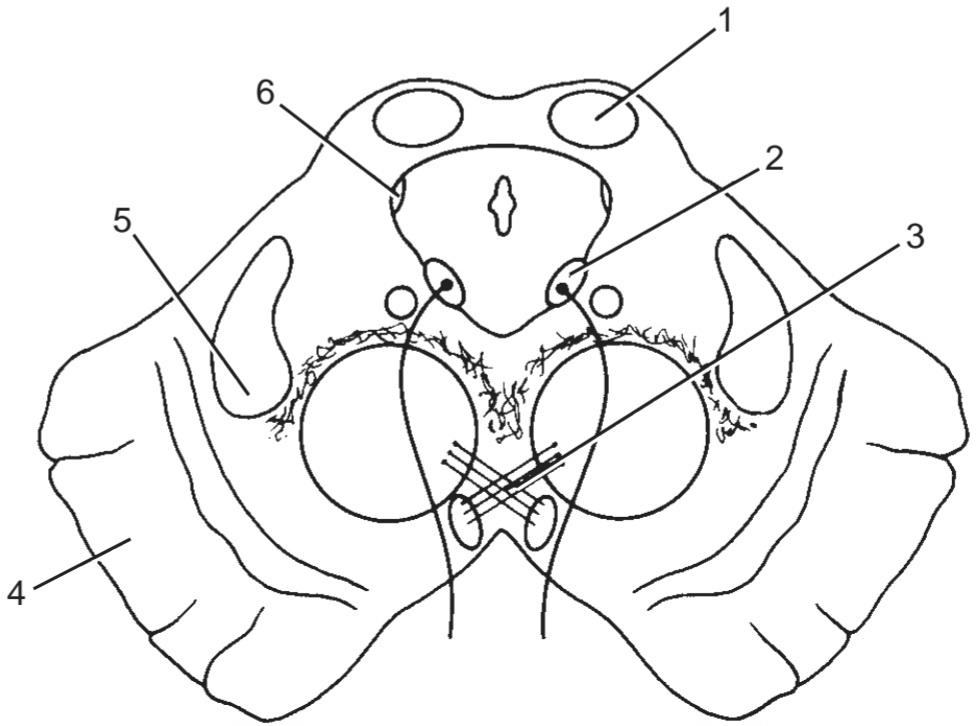
شکل ۵-۳۶ برش عرضی مغز میانی.
31. Number 1 (a) Medial longitudinal fasciculus
32. Number 2 (b) Inferior colliculus
33. Number 3 (c) Medial lemniscus
34. Number 4 (d) Trochlear nucleus
35. Number 5 (e) None of the above
36. Number 6
۳۱. شماره ۱ (الف) فاسیکولوس طولی داخلی
۳۲. شماره ۲ (ب) کولیکولوس تحتانی
۳۳. شماره ۳ (ج) نوار داخلی
۳۴. شماره ۴ (د) هسته تروکلئار
۳۵. شماره ۵ (ه) هیچکدام
۳۶. شماره ۶
Directions: Each case history is followed by questions. Read the case history, then select the ONE BEST lettered answer.
دستورالعمل: پس از هر شرح حال، سوالاتی مطرح شده است. شرح حال را بخوانید، سپس بهترین پاسخ با حروف مشخص شده را انتخاب کنید.
A 63-year-old man complaining of difficulty in swallowing, some hoarseness of his voice, and giddiness was seen by a neurologist. All these symptoms started suddenly 4 days previously. On physical examination, he was found to have a loss of the pharyngeal gagging reflex on the left side, left-sided facial analgesia, and left-sided paralysis of the vocal cord.
مردی ۶۳ ساله که از مشکل در بلع، گرفتگی صدا و سرگیجه شکایت داشت، توسط متخصص مغز و اعصاب ویزیت شد. همه این علائم به طور ناگهانی ۴ روز قبل شروع شده بود. در معاینه فیزیکی، مشخص شد که او دچار از دست دادن رفلکس عق زدن حلق در سمت چپ، بیحسی صورت در سمت چپ و فلج تارهای صوتی در سمت چپ است.
37. Based on the clinical history and the results of the physical examination, select the most likely diagnosis.
(a) Meningeal tumor in the posterior cranial fossa on the right side
(b) Lateral medullary syndrome on the left side
(c) Medial medullary syndrome on the left side
(d) Lateral medullary syndrome on the right side
(e) Medial medullary syndrome on the right side
۳۷. بر اساس شرح حال بالینی و نتایج معاینه فیزیکی، محتملترین تشخیص را انتخاب کنید.
(الف) تومور مننژ در حفره جمجمهای خلفی در سمت راست
(ب) سندرم مدولاری جانبی در سمت چپ
(ج) سندرم مدولاری داخلی در سمت چپ
(د) سندرم مدولاری جانبی در سمت راست
(ه) سندرم مدولاری داخلی در سمت راست
A 7-year-old girl was seen by a neurologist because she complained to her mother that she was seeing double. Careful physical examination revealed that the double vision became worse when she looked toward the left. The patient also had evidence of a mild motor paralysis of her right lower limb without spasticity. There was also a slight facial paralysis involving the whole left side of the face.
یک دختر ۷ ساله به دلیل شکایت از دوبینی به مادرش، توسط متخصص مغز و اعصاب ویزیت شد. معاینه فیزیکی دقیق نشان داد که وقتی به سمت چپ نگاه میکند، دوبینی او بدتر میشود. بیمار همچنین شواهدی از فلج حرکتی خفیف اندام تحتانی راست بدون اسپاسم داشت. همچنین فلج خفیف صورت که تمام سمت چپ صورت را درگیر میکرد، وجود داشت.
(a) The double vision caused by weakness of the left lateral rectus muscle.
(b) The complete left-sided facial paralysis caused by involvement of the left seventh cranial nerve nucleus or its nerve.
(c) The mild right hemiparesis produced by damage to the corticospinal tract on the right side.
(d) Magnetic resonance imaging revealed the presence of a tumor of the lower part of the pons on the left side.
(e) The left sixth cranial nerve nucleus was damaged.
۳۸. بر اساس شرح حال بالینی و معاینه بالینی، نقصهای عصبی زیر میتوانستند وجود داشته باشند، به جز:
(الف) دوبینی ناشی از ضعف عضله راست جانبی چپ.
(ب) فلج کامل صورت در سمت چپ ناشی از درگیری هسته عصب جمجمهای هفتم چپ یا عصب آن.
(ج) همیپارزی خفیف سمت راست ناشی از آسیب به دستگاه قشری نخاعی در سمت راست.
(د) تصویربرداری رزونانس مغناطیسی وجود توموری در قسمت پایینی پل مغزی در سمت چپ را نشان داد.
(ه) هسته عصب جمجمهای ششم چپ آسیب دیده بود.
A 42-year-old woman complaining of a severe, persistent headache visited her physician. At first, the headache was not continuous and tended to occur during the night. Now, the headache was present all the time and was felt over the whole head. Recently, she has begun to feel nauseous, and this has resulted in several episodes of vomiting. Last week, on looking in the mirror, she noted that her right pupil looked much larger than the left. Her right upper lid appeared to droop.
زنی ۴۲ ساله با شکایت از سردرد شدید و مداوم به پزشک مراجعه کرد. در ابتدا، سردرد مداوم نبود و بیشتر در طول شب رخ میداد. اکنون، سردرد همیشه وجود داشت و در تمام سر احساس میشد. اخیراً، او احساس تهوع کرده است و این منجر به چندین دوره استفراغ شده است. هفته گذشته، با نگاه کردن به آینه، متوجه شد که مردمک چشم راستش بسیار بزرگتر از چپ به نظر میرسد. پلک بالایی سمت راست او افتاده به نظر میرسید.
39. The physical examination revealed the following most likely findings except:
(a) Weakness in raising the right eyelid upward
(b) Severe ptosis of the right eye
(c) Obvious dilatation of the right pupil
(d) Ophthalmoscopic examination revealing bilateral papilledema
(e) Evidence of paralysis of either superior oblique muscle
(f) Examination of the lower limbs revealing mild spasticity of the left lower limb muscles
(g) Ataxia of the right upper limb
(h) Loss of taste sensation on the posterior third of the tongue on the left side
۳۹. معاینه فیزیکی یافتههای محتمل زیر را نشان داد، به جز:
(الف) ضعف در بالا بردن پلک راست
(ب) پتوز شدید چشم راست
(ج) اتساع واضح مردمک چشم راست
(د) معاینه افتالموسکوپی که ادم پاپی دو طرفه را نشان میدهد
(ه) شواهدی از فلج هر یک از عضلات مایل فوقانی
(و) معاینه اندام تحتانی که اسپاسم خفیف عضلات اندام تحتانی چپ را نشان میدهد
(ز) آتاکسی اندام فوقانی راست
(ح) از دست دادن حس چشایی در یک سوم خلفی زبان در سمت چپ
40. The combination of the clinical history and the findings in the physical examination enabled the physician to make the following most likely diagnosis.
(a) Tumor involving the left cerebral hemisphere
(b) Tumor involving the right side of the midbrain at the level of the superior colliculi
(c) Severe migraine
(d) Cerebral hemorrhage involving the left cerebral hemisphere
(e) Tumor of the left side of the midbrain
(ب) توموری که سمت راست مغز میانی را در سطح کولیکولهای فوقانی درگیر میکند
(ج) میگرن شدید
(د) خونریزی مغزی که نیمکره چپ مغز را درگیر میکند
(ه) تومور سمت چپ مغز میانی
√ پاسخها و توضیحات مربوط به سوالات مروری
۱. الف صحیح است. هرمهای بصل النخاع به سمت پایین باریک میشوند و باعث ایجاد تقاطع هرمها میشوند (شکل ۵-۹ را ببینید). ب. در هر طرف خط میانی روی سطح قدامی بصل النخاع در سمت جانبی هرمها، یک برآمدگی تخممرغی شکل به نام زیتون وجود دارد که حاوی هسته زیتونی است و حاوی الیاف قشری-نخاعی نیست. ج. عصب زیرزبانی بین هرم و زیتونی خارج میشود. د. عصب واگ بین زیتونی و پایه مخچهای تحتانی خارج میشود. ه. عصب ابدوسنس بین پل مغزی و بصل النخاع خارج میشود (شکل ۵-۹ را ببینید).
۷. ب صحیح است. در سندرم بصل النخاع جانبی، هسته آمبیگوس همان طرف ممکن است آسیب ببیند. الف) این وضعیت ممکن است در اثر ترومبوز شریان مخچهای خلفی تحتانی ایجاد شود. ج) بیدردی و بیحسی حرارتی در طرف همان طرف صورت ممکن است رخ دهد. د) بیحسی و بیحسی حرارتی تنه و اندام همان طرف ممکن است رخ دهد. ه) معمولاً تشنج رخ نمیدهد.
8. C is correct.
9. E is correct. The structure is the medial longitudinal fasciculus.
10. B is correct.
11. E is correct. The structure is the inferior olivary nucleus.
12. D is correct.
13. A is correct.
۸. ج صحیح است.
۹. ه صحیح است. ساختار، دسته عصبی طولی داخلی است.
۱۰. ب صحیح است.
۱۱. ه صحیح است. ساختار، هسته زیتونی تحتانی است.
۱۲. د صحیح است.
۱۳. الف صحیح است.
14. C is correct. The basilar artery lies in a centrally placed groove on the anterior aspect of the pons. The trigeminal nerve emerges on the anterior aspect of the pons. B. The glossopharyngeal nerve emerges on the anterior aspect of the medulla oblongata in the groove between the olive and the inferior cerebellar peduncle (see Fig. 5-9). D. The nerve fibers on the anterior aspect of the pons converge laterally to form the middle cerebellar peduncle. E. The pons forms the upper half of the floor of the fourth ventricle (see Fig. 5-18).
۱۴. ج صحیح است. شریان بازیلار در یک شیار مرکزی در قسمت قدامی پل مغزی قرار دارد. عصب سه قلو از قسمت قدامی پل مغزی خارج میشود. ب. عصب زبانی-حلقی از قسمت قدامی بصل النخاع در شیار بین زیتون و پایه مخچه تحتانی خارج میشود (شکل ۵-۹ را ببینید). د. رشتههای عصبی در قسمت قدامی پل مغزی به صورت جانبی همگرا میشوند تا پایه مخچه میانی را تشکیل دهند. ه. پل مغزی نیمه بالایی کف بطن چهارم را تشکیل میدهد (شکل ۵-۱۸ را ببینید).
15. A is correct. The red nucleus lies within the midbrain (see Fig. 5-25). B. The facial colliculus lies in the caudal part of the pons (see Fig. 5-18). C. The motor nucleus of the trigeminal nerve lies within the cranial part of the pons (see Fig. 5-20). D. The abducens nucleus lies within the caudal part of the pons (see Fig. 5-19). E. The trochlear nucleus lies within the midbrain at the level of the inferior colliculus (see Fig. 5-25).
۱۵. الف صحیح است. هسته قرمز در مغز میانی قرار دارد (شکل ۵-۲۵ را ببینید). ب. کولیکولوس فاسیال در قسمت دمی پل مغزی قرار دارد (شکل ۵-۱۸ را ببینید). ج. هسته حرکتی عصب سه قلو در قسمت جمجمهای پل مغزی قرار دارد (شکل ۵-۲۰ را ببینید). د. هسته ابدوسنس در قسمت دمی پل مغزی قرار دارد (شکل ۵-۱۹ را ببینید). ه. هسته تروکلئار در مغز میانی در سطح کولیکولوس تحتانی قرار دارد (شکل ۵-۲۵ را ببینید).
16. B is correct. On the posterior surface of the pons is the facial colliculus, which is produced by the root of the facial nerve winding around the nucleus of the abducens nerve (see Fig. 5-19). A. The medial emi- nence is an elongated swelling lateral to the median sulcus (see Fig. 5-26). C. The floor of the superior part of the sulcus limitans is pigmented and is called the substantia ferruginea (see Fig. 5-18). D. The vestibular area lies lateral to the sulcus limitans (see Fig. 5-18). E. The cerebellum lies posterior to the pons.
۱۶. ب صحیح است. در سطح خلفی پل مغزی، برآمدگی صورتی قرار دارد که توسط پیچ خوردن ریشه عصب صورتی به دور هسته عصب ابدوسنس ایجاد میشود (شکل ۵-۱۹ را ببینید). الف. برجستگی داخلی یک برآمدگی کشیده در سمت خارجی شیار میانی است (شکل ۵-۲۶ را ببینید). ج. کف قسمت فوقانی شیار محدودکننده رنگدانهدار است و جسم آهنی نامیده میشود (شکل ۵-۱۸ را ببینید). د. ناحیه دهلیزی در سمت خارجی شیار محدودکننده قرار دارد (شکل ۵-۱۸ را ببینید). ه. مخچه در خلف پل مغزی قرار دارد.
17. A is correct. The pontine nuclei lie between the transverse pontine fibers (see Fig. 5-12). B. The vestibular nuclei lie lateral to the abducens nucleus (see Fig. 5-19). C. The trapezoid body is made up of fibers derived from the cochlear nuclei and the nuclei of the trapezoid body. D. The tegmentum is the part of the pons lying posterior to the trapezoid body. E. The medial longitudinal fasciculus lies below the floor of the fourth ventricle on either side of the midline (see Fig. 5-19).
۱۷. الف صحیح است. هستههای پل مغزی بین فیبرهای پل مغزی عرضی قرار دارند (شکل ۵-۱۲ را ببینید). ب. هستههای دهلیزی در سمت خارجی هسته ابدوسنس قرار دارند (شکل ۵-۱۹ را ببینید). ج. جسم ذوزنقهای از فیبرهای مشتق شده از هستههای حلزونی و هستههای جسم ذوزنقهای تشکیل شده است. د. تگمنتوم بخشی از پل مغزی است که در خلف جسم ذوزنقهای قرار دارد. ه. فاسیکولوس طولی داخلی در زیر کف بطن چهارم در دو طرف خط وسط قرار دارد (شکل ۵-۱۹ را ببینید).
18. C is correct. In the pons, bundles of corticopontine fibers lie among the transverse pontine fibers (see Fig. 5-19). A. The motor nucleus of the trigeminal nerve lies medial to the main sensory nucleus in the tegmentum of the pons (see Fig. 5-20). B. In the cranial part of the pons, the medial lemniscus has rotated so that its long axis lies transversely (see Fig. 5-20). D. The medial longitudinal fasciculus is the main pathway that connects the vestibular and cochlear nuclei with the nuclei controlling the extraocular muscles (oculomotor, trochlear, and abducens nuclei). E. The motor root of the trigeminal nerve is much smaller than the sensory root.
۱۸. ج صحیح است. در پل مغزی، دستههایی از الیاف کورتیکوپونتین در میان الیاف پل مغزی عرضی قرار دارند (شکل ۵-۱۹ را ببینید). الف. هسته حرکتی عصب سهقلو در قسمت داخلی هسته حسی اصلی در تگمنتوم پل مغزی قرار دارد (شکل ۵-۲۰ را ببینید). ب. در قسمت جمجمهای پل مغزی، لمنیسکوس داخلی طوری چرخیده است که محور طولی آن به صورت عرضی قرار دارد (شکل ۵-۲۰ را ببینید). د. فاسیکول طولی داخلی مسیر اصلی است که هستههای دهلیزی و حلزونی را به هستههای کنترلکننده عضلات خارج چشمی (هستههای چشمی-حرکتی، قرقرهای و ابدوسنس) متصل میکند. ه. ریشه حرکتی عصب سهقلو بسیار کوچکتر از ریشه حسی است.
19. D is correct. In the pons, the corticopontine fibers terminate in the pontine nuclei. A. The pons is related anteriorly to the dorsum sellae of the sphenoid bone. B. The pons lies in the posterior cranial fossa. C. Astrocytoma of the pons is the most common tumor of the brainstem. E. The pons receives its blood supply from the basilar artery.
۱۹. د صحیح است. در پل مغزی، الیاف کورتیکوپونتین به هستههای پل مغزی ختم میشوند. الف) پل مغزی از جلو به زین پشتی استخوان اسفنوئید متصل است. ب) پل مغزی در حفره جمجمهای خلفی قرار دارد. ج) آستروسیتومای پل مغزی شایعترین تومور ساقه مغز است. ه) پل مغزی خون خود را از شریان بازیلار دریافت میکند.
20. D is correct.
21. C is correct.
22. E is correct. The structure is the transverse pontine fibers.
23. A is correct.
24. E is correct. The structure is the medial lemniscus.
25. B is correct.
۲۰. د صحیح است.
۲۱. ج صحیح است.
۲۲. ه صحیح است. ساختار، الیاف عرضی پل مغزی است.
۲۳. الف صحیح است.
۲۴. ه صحیح است. ساختار، لمنیسکوس داخلی است.
۲۵. ب صحیح است.
26. D is correct. The cavity of the midbrain is called the cerebral aqueduct (see Fig. 5-28). A. The midbrain passes superiorly through the opening in the tentorium cerebelli posterior to the dorsum sellae. B. The oculomotor nerve emerges from the anterior surface of the midbrain at the level of the superior colliculi (see Fig. 5-25). C. The superior brachium passes from the superior colliculus to the lateral geniculate body and the optic tract and is associated with visual functions (see Fig. 5-23). E. The inter- peduncular fossa is bounded laterally by the crus cerebri (see Fig. 5-25).
۲۶. د صحیح است. حفره مغز میانی، مجرای مغزی نامیده میشود (شکل ۵-۲۸ را ببینید). الف. مغز میانی از بالای سوراخ چادرینه مخچه در خلف عضله پشتی زین عبور میکند. ب. عصب اوکولوموتور از سطح قدامی مغز میانی در سطح کولیکولوس فوقانی خارج میشود (شکل ۵-۲۵ را ببینید). ج. بازوی فوقانی از کولیکولوس فوقانی به جسم زانویی جانبی و راه بینایی عبور میکند و با عملکردهای بینایی مرتبط است (شکل ۵-۲۳ را ببینید). ه. حفره بین پایهای به صورت جانبی توسط ساق مغزی محدود میشود (شکل ۵-۲۵ را ببینید).
27. C is correct. The trochlear nucleus is situated in the central gray matter of the midbrain at the level of the inferior colliculus (see Fig. 5-25). A. In the midbrain, the oculomotor nucleus is found at the level of the superior colliculus (see Fig. 5-25). B. The trochlear nerve emerges on the posterior surface of the midbrain and decussates completely in the superior medullary velum (see Fig. 5-25). D. The lemnisci are situated lateral to the central gray matter (see Fig. 5-25). E. The trigeminal lemniscus lies posterior to the medial lemniscus (see Fig. 5-25).
۲۷. ج صحیح است. هسته تروکلئار در ماده خاکستری مرکزی مغز میانی در سطح کولیکولوس تحتانی قرار دارد (شکل ۵-۲۵ را ببینید). الف. در مغز میانی، هسته اوکولوموتور در سطح کولیکولوس فوقانی قرار دارد (شکل ۵-۲۵ را ببینید). ب. عصب تروکلئار در سطح خلفی مغز میانی ظاهر میشود و به طور کامل در نرمکام فوقانی به صورت متقاطع قطع میشود (شکل ۵-۲۵ را ببینید). د. لمنیسکوسها در سمت خارجی ماده خاکستری مرکزی قرار دارند (شکل ۵-۲۵ را ببینید). ه. لمنیسکوس سهقلو در خلف لمنیسکوس داخلی قرار دارد (شکل ۵-۲۵ را ببینید).
28. A is correct. The tectum is the part of the midbrain situated posterior to the cerebral aqueduct (see Fig. 5-24). B. In the midbrain, the crus cerebri lies anterior to the substantia nigra (see Fig. 5-25). C. The tegmentum lies posterior to the substantia nigra (see Fig. 5-25). D. The central gray matter encircles the cerebral aqueduct (see Fig. 5-25). E. The reticular formation is present throughout the midbrain.
۲۸. الف صحیح است. تکتوم بخشی از مغز میانی است که در خلف مجرای مغزی قرار دارد (شکل ۵-۲۴ را ببینید). ب. در مغز میانی، ساقه مغزی در قدام جسم سیاه قرار دارد (شکل ۵-۲۵ را ببینید). ج. تگمنتوم در خلف جسم سیاه قرار دارد (شکل ۵-۲۵ را ببینید). د. ماده خاکستری مرکزی، مجرای مغزی را احاطه کرده است (شکل ۵-۲۵ را ببینید). ه. تشکیلات مشبک در سراسر مغز میانی وجود دارد.
29. B is correct. The superior colliculi of the midbrain are concerned with site reflexes. A. The colliculi are located in the tectum (see Fig. 5-25). C. The inferior colliculi lie at the level of the trochlear nerve nuclei (see Fig. 5-25). D. The inferior colliculi are concerned with auditory reflexes. E. The superior colliculi lie at the level of the red nuclei (see Fig. 5-25).
۲۹. ب صحیح است. کولیکولهای فوقانی مغز میانی مربوط به رفلکسهای موضعی هستند. الف. کولیکولها در تکتوم قرار دارند (شکل ۵-۲۵ را ببینید). ج. کولیکولهای تحتانی در سطح هستههای عصب قرقرهای قرار دارند (شکل ۵-۲۵ را ببینید). د. کولیکولهای تحتانی مربوط به رفلکسهای شنوایی هستند. ه. کولیکولهای فوقانی در سطح هستههای قرمز قرار دارند (شکل ۵-۲۵ را ببینید).
30. D is correct. The nerve fibers from the oculomotor nucleus pass through the red nucleus (see Fig. 5-25). A. The oculomotor nucleus is situated in the cen- tral gray matter (see Fig. 5-25). B. The parasympathetic part of the oculomotor nucleus is called the Edinger-Westphal nucleus. C. The oculomotor nucleus lies anterior to the cerebral aqueduct (see Fig. 5-25). E. The oculomotor nucleus lies close to the medial longitudinal fasciculus (see Fig. 5-25).
32. E is correct. The structure is the oculomotor nucleus.
33. E is correct. The structure is decussation of the rubrospinal tract.
34. E is correct. The structures are corticospinal and corticonuclear fibers.
35. C is correct.
36. E is correct. The structure is the mesencephalic nucleus of cranial nerve V.
37. B is correct.
۳۲. E صحیح است. ساختار، هسته اوکولوموتور است.
۳۳. E صحیح است. ساختار، تقاطع مسیر روبرواسپینال است.
۳۴. E صحیح است. ساختارها، فیبرهای قشری-نخاعی و قشری-هستهای هستند.
۳۵. C صحیح است.
۳۶. E صحیح است. ساختار، هسته مزانسفالی عصب جمجمهای V است.
۳۷. B صحیح است.
«فصل پنجم کتاب نورواناتومی بالینی اسنل»
🚀 با ما همراه شوید!
تازهترین مطالب و آموزشهای مغز و اعصاب را از دست ندهید. با فالو کردن کانال تلگرام آیندهنگاران مغز، از ما حمایت کنید!



