نوروآناتومی بالینی اسنل | ساقه مغز؛ آناتومی جمجمه – مرور مبانی و اصول


دعای مطالعه [ نمایش ]
بِسْمِ الله الرَّحْمنِ الرَّحیمِ
اَللّهُمَّ اَخْرِجْنى مِنْ ظُلُماتِ الْوَهْمِ
خدايا مرا بيرون آور از تاريكىهاى وهم،
وَ اَكْرِمْنى بِنُورِ الْفَهْمِ
و به نور فهم گرامى ام بدار،
اَللّهُمَّ افْتَحْ عَلَيْنا اَبْوابَ رَحْمَتِكَ
خدايا درهاى رحمتت را به روى ما بگشا،
وَانْشُرْ عَلَيْنا خَزائِنَ عُلُومِكَ بِرَحْمَتِكَ يا اَرْحَمَ الرّاحِمينَ
و خزانههاى علومت را بر ما باز كن به امید رحمتت اى مهربانترين مهربانان.
آیندهنگاران مغز: «ما مغز را میشناسیم، تا آینده را بسازیم.»
کتاب «نوروآناتومی بالینی اسنل» اثر دکتر ریچارد اس. اسنل، با ویرایش جدید دکتر رایان اسپلیتگربر، همچنان بهعنوان مرجع طلایی این حوزه شناخته میشود. این اثر با تلفیق تازهترین پژوهشها و کاربردهای بالینی، پلی استوار میان دانش پایه و عمل پزشکی بنا میکند.
تلاشهای علمی و سالها تدریس ارزشمند استاد گرانقدر دکتر محمدتقی جغتایی الهامبخش ترجمه و گسترش این دانش در ایران بوده است.
ترجمه دقیق این کتاب توسط برند علمی آیندهنگاران مغز به مدیریت داریوش طاهری، امکان دسترسی فارسیزبانان به مرزهای نوین علوم اعصاب را فراهم کرده و رسالتی علمی در جهت شناخت عمیقتر مغز و ترسیم آیندهای روشنتر بر عهده گرفته است.
»» فصل ۵: ساقه مغز
»» Chapter 5: Brainstem
CHAPTER OBJECTIVES
• To review the anatomy of the brainstem
• To develop a three-dimensional picture of the interior of the brainstem
• To know the positions of several of the cranial nerve nuclei, the olivary nuclear complex, and the paths taken by the various ascending and descending nerve tracts as they ascend to the higher brain centers or descend to the spinal cord
• To assess the signs and symptoms presented by a patient and identify the exact location of a structural lesion
اهداف فصل
• مرور آناتومی ساقه مغز
• ایجاد یک تصویر سه بعدی از داخل ساقه مغز
• شناخت موقعیت چندین هسته عصب جمجمهای، کمپلکس هستهای زیتونی و مسیرهایی که توسط مسیرهای عصبی صعودی و نزولی مختلف هنگام صعود به مراکز بالاتر مغز یا نزول به نخاع طی میشوند
• ارزیابی علائم و نشانههای ارائه شده توسط بیمار و شناسایی محل دقیق ضایعه ساختاری
A 58-year-old woman is referred to a neurologist because of recent onset of difficulty with walking. The neurologist notes that she stands and walks with her left arm flexed at the elbow and the left leg extended (left hemiparesis). While walking, she has difficulty flexing the left hip and knee and dorsiflexing the ankle; the forward motion is possible by swinging the left leg outward at the hip to avoid dragging the foot on the ground. Her left arm remains motionless.
زنی ۵۸ ساله به دلیل شروع اخیر مشکل در راه رفتن به متخصص مغز و اعصاب ارجاع داده میشود. متخصص مغز و اعصاب متوجه میشود که او با دست چپ خود که از آرنج خم شده و پای چپ که کشیده شده (همیپارزی چپ) میایستد و راه میرود. هنگام راه رفتن، او در خم کردن لگن و زانوی چپ و خم کردن مچ پا به سمت عقب مشکل دارد. حرکت رو به جلو با چرخاندن پای چپ به سمت بیرون از لگن برای جلوگیری از کشیده شدن پا روی زمین امکانپذیر است. دست چپ او بیحرکت میماند.
Neurologic examination shows no signs of facial paralysis, but tongue weakness is evident. On protrusion, the tongue deviates toward the right side (right hypoglossal nerve palsy). Cutaneous sensations are found to be normal, but muscle joint sense, tactile discrimination, and vibratory sense on the left side of the body are impaired.
معاینه عصبی هیچ نشانهای از فلج صورت نشان نمیدهد، اما ضعف زبان مشهود است. هنگام بیرون آوردن زبان، انحراف آن به سمت راست (فلج عصب زیرزبانی راست) مشاهده میشود. حسهای پوستی طبیعی هستند، اما حس مفاصل عضلانی، تمایز لمسی و حس ارتعاش در سمت چپ بدن مختل شدهاند.
Based on the neurologic findings, a diagnosis of rightsided medial medullary syndrome is made. The medial part of the right side of the medulla oblongata receives its arterial supply from the right vertebral artery. Occlusion of this artery or its branch to the medulla results in destruction of the right pyramid (left hemiparesis), destruction of the right hypoglossal nucleus and nerve (right hypoglossal palsy), and destruction of the medial lemniscus on the right side (left-sided loss of muscle joint sense, vibratory sense, and tactile discrimination). The absence of facial palsy showed that the facial nerve nuclei, the facial nerves, and the corticobulbar fibers to the facial nuclei were intact. The sparing of the sensations of touch, pain, and temperature showed that the spinal lemniscus was intact.
بر اساس یافتههای عصبی، تشخیص سندرم مدولای داخلی سمت راست داده میشود. بخش داخلی سمت راست بصل النخاع، خونرسانی شریانی خود را از شریان مهرهای راست دریافت میکند. انسداد این شریان یا شاخه آن به بصل النخاع منجر به تخریب هرم راست (همیپارزی چپ)، تخریب هسته و عصب هیپوگلوسال راست (فلج هیپوگلوسال راست) و تخریب لمنیسکوس داخلی در سمت راست (از دست دادن حس مفصل عضله، حس ارتعاش و تمایز لمسی در سمت چپ) میشود. عدم وجود فلج صورت نشان داد که هستههای عصب صورت، اعصاب صورت و فیبرهای قشری-بولبار به هستههای صورت سالم هستند. حفظ حس لمس، درد و دما نشان داد که لمنیسکوس نخاعی سالم است.
This diagnosis is possible as the result of carefully sorting out the neurologic findings. A clear knowledge of the position and function of the various nerve tracts and nuclei in the medulla oblongata is essential before a clinician can reach a diagnosis in this case.
این تشخیص در نتیجهی بررسی دقیق یافتههای عصبی امکانپذیر است. آگاهی دقیق از موقعیت و عملکرد مسیرهای عصبی و هستههای مختلف در بصل النخاع، قبل از اینکه پزشک بتواند در این مورد به تشخیص برسد، ضروری است.
SKULL ANATOMY
Head injuries from blunt trauma and penetrating missiles are associated with a high mortality and disabling morbidity. Because of the close relationship that exists between the skull and the underlying brain and cranial nerves (CNs), as well as their common involvement in many diseases, a brief review of the anatomy of the skull will first be considered.
آناتومی جمجمه
آسیبهای سر ناشی از ترومای غیرنافذ و موشکهای نفوذی با مرگ و میر بالا و عوارض ناتوانکننده همراه هستند. به دلیل ارتباط نزدیک بین جمجمه و مغز و اعصاب جمجمهای (CN) زیرین و همچنین درگیری مشترک آنها در بسیاری از بیماریها، ابتدا مروری مختصر بر آناتومی جمجمه خواهیم داشت.
Adult Skull
The skull is composed of several separate bones united at immobile joints called sutures. The connective tissue between the bones is called a sutural ligament. The mandible is an exception, united to the skull by the mobile temporomandibular joint.
جمجمه بزرگسالان
جمجمه از چندین استخوان جداگانه تشکیل شده است که در مفاصل بیحرکتی به نام درز به هم متصل شدهاند. بافت همبند بین استخوانها رباط درز نامیده میشود. فک پایین یک استثنا است که توسط مفصل گیجگاهی فکی متحرک به جمجمه متصل میشود.
The bones of the skull can be divided into those of the cranium and those of the face. The vault is the upper part of the cranium, and the base of the skull is the lowest part of the cranium (Fig. 5-1).
استخوانهای جمجمه را میتوان به استخوانهای جمجمه و استخوانهای صورت تقسیم کرد. طاق جمجمه قسمت بالایی جمجمه و قاعده جمجمه پایینترین قسمت جمجمه است (شکل 5-1).
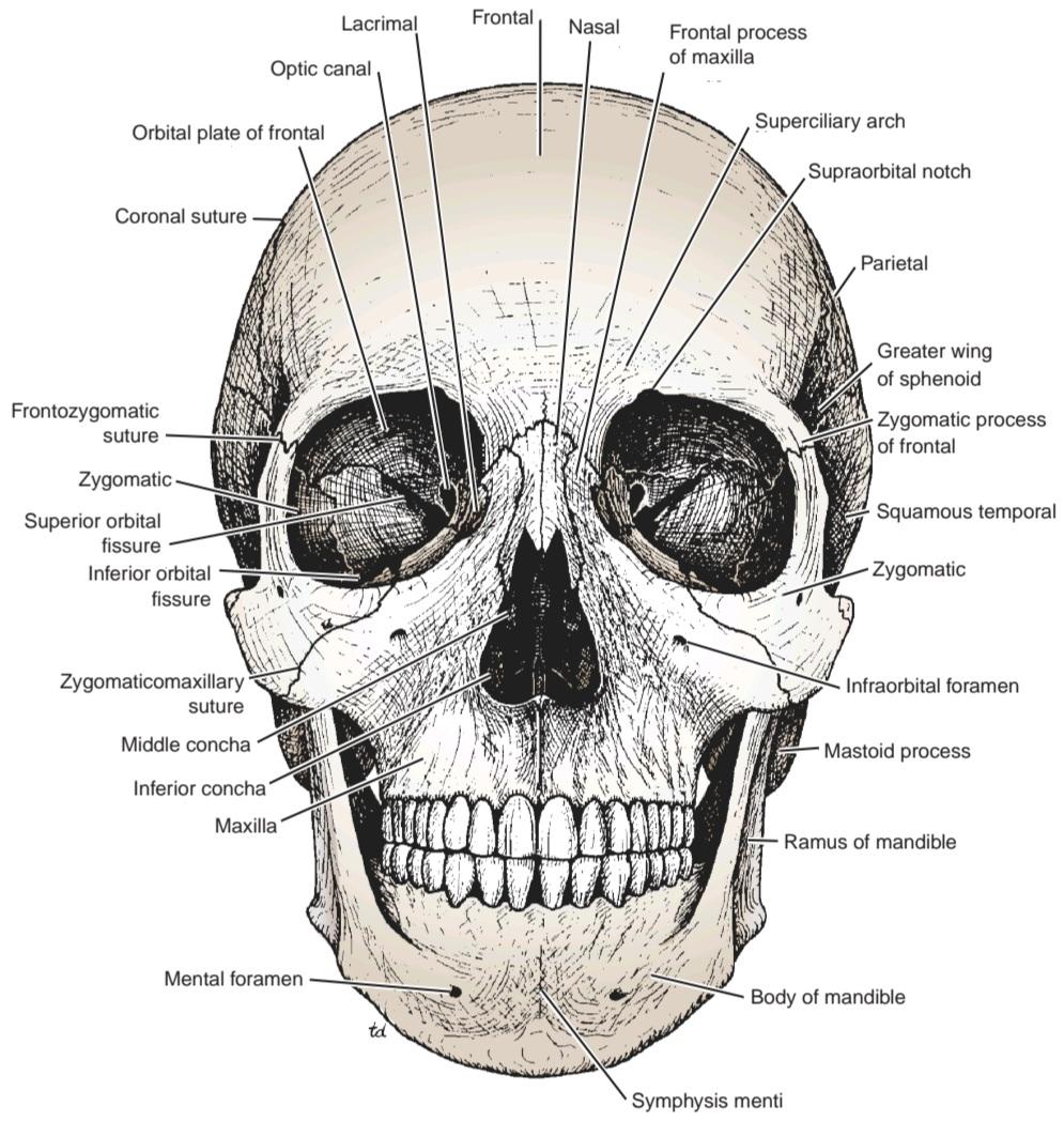
Figure 5-1 Bones of the anterior aspect of the skull.
شکل ۵-۱ استخوانهای قسمت قدامی جمجمه.
The skull bones are made up of external and internal tables of compact bone separated by a layer of spongy bone called the diploë (Fig. 5-2). The internal table is thinner and more brittle than the external table. The bones are covered on the outer and inner surfaces with periosteum.
استخوانهای جمجمه از صفحات خارجی و داخلی استخوان فشرده تشکیل شدهاند که توسط لایهای از استخوان اسفنجی به نام دیپلو از هم جدا شدهاند (شکل 5-2). صفحه داخلی نازکتر و شکنندهتر از صفحه خارجی است. استخوانها در سطوح خارجی و داخلی با پریوست پوشیده شدهاند.

Figure 5-2 Coronal section of the upper part of the head showing the layers of the scalp, the sagittal suture of the skull, the falx cerebri, the superior and inferior sagittal venous sinuses, the arachnoid granulations, the emissary veins, and the relation of cerebral blood vessels to the subarachnoid space.
شکل ۵-۲ برش کرونال از قسمت بالای سر که لایههای پوست سر، درز ساژیتال جمجمه، داس مغزی، سینوسهای وریدی ساژیتال فوقانی و تحتانی، گرانولهای عنکبوتیه، وریدهای فرستنده و ارتباط رگهای خونی مغز با فضای زیر عنکبوتیه را نشان میدهد.
The cranium consists of the following bones, two of which are paired (Fig. 5-3; also see Fig. 5-1):
جمجمه از استخوانهای زیر تشکیل شده است که دو تا از آنها جفت هستند (شکل 5-3؛ همچنین به شکل 5-1 مراجعه کنید):
• Frontal bone 1
• Parietal bones 2
• Occipital bone 1
• Temporal bones 2
• Sphenoid bone 1
• Ethmoid bone 1
• استخوان پیشانی ۱
• استخوانهای آهیانه ۲
• استخوان پسسری ۱
• استخوانهای گیجگاهی ۲
• استخوان اسفنوئید ۱
• استخوان اتموئید ۱
The facial bones consist of the following, two of which are single:
استخوانهای صورت از موارد زیر تشکیل شدهاند که دو مورد از آنها تکی هستند:
• Zygomatic bones 2
• Maxillae 2
• Nasal bones 2
• Lacrimal bones 2
• Vomer 1
• Palatine bones 2
• Inferior conchae 2
• Mandible 1
• استخوانهای زیگوماتیک ۲
• فک بالا ۲
• استخوان بینی ۲
• استخوان های اشکی 2
• Vomer 1
• استخوان های پالاتین ۲
• مخروط زیرین 2
• مندیبل 1
Although students do not need to know the detailed structure of each individual skull bone, they should be familiar with the skull as a whole. If possible, have a dried skull available for reference as you read the fol- lowing description.
اگرچه دانشآموزان نیازی به دانستن ساختار دقیق تک تک استخوانهای جمجمه ندارند، اما باید با جمجمه به طور کلی آشنا باشند. در صورت امکان، هنگام خواندن توضیحات زیر، یک جمجمه خشک شده برای مرجع در دسترس داشته باشید.
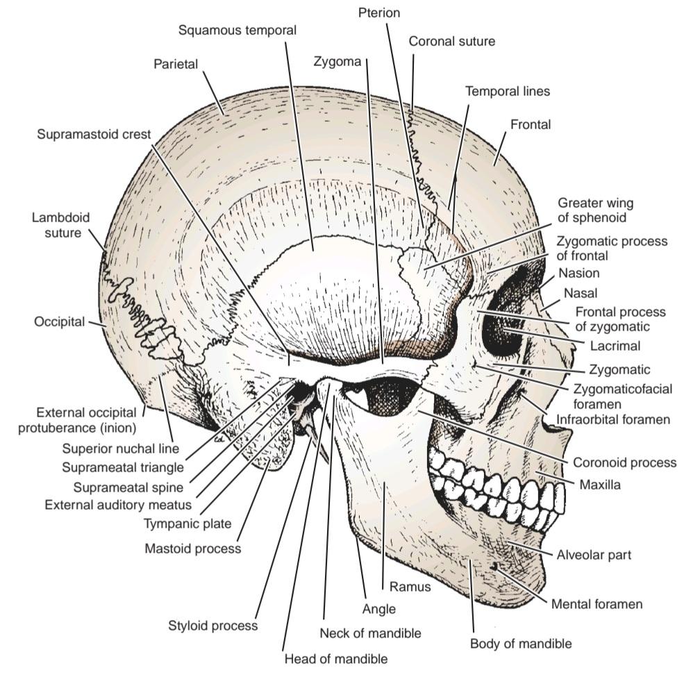
Figure 5-3 Bones of the lateral aspect of the skull.
شکل ۵-۳ استخوانهای سطح جانبی جمجمه.
Anterior View
The frontal bone, or forehead bone, curves downward to make the upper margins of the orbits (see Fig. 5-1). The superciliary arches can be seen on either side, and the supraorbital notch, or foramen, can be recognized. Medially, the frontal bone articulates with the frontal processes of the maxillae and with the nasal bones. Laterally, the frontal bone articulates with the zygomatic bone.
نمای قدامی
استخوان پیشانی یا استخوان پیشانی، به سمت پایین انحنا دارد تا حاشیههای بالایی حدقه چشم را تشکیل دهد (شکل 5-1 را ببینید). قوسهای فوقانی در هر دو طرف قابل مشاهده هستند و بریدگی فوق حدقهای یا سوراخ را میتوان تشخیص داد. در قسمت میانی، استخوان پیشانی با زوائد پیشانی فک بالا و استخوانهای بینی مفصل میشود. در قسمت جانبی، استخوان پیشانی با استخوان گونه مفصل میشود.
The orbital margins are bounded by the frontal bone superiorly, the zygomatic bone laterally, the maxilla inferiorly, and the processes of the maxilla and frontal bone medially.
حاشیههای کاسه چشم از بالا به استخوان پیشانی، از پهلو به استخوان گونه، از پایین به فک بالا و از داخل به زوائد فک بالا و استخوان پیشانی محدود میشوند.
Within the frontal bone, just above the orbital margins, are two hollow spaces lined with mucous membrane called the frontal air sinuses. These communicate with the nose and serve as voice resonators.
درون استخوان پیشانی، درست بالای حاشیههای کاسه چشم، دو فضای توخالی پوشیده شده با غشای مخاطی به نام سینوسهای هوایی پیشانی وجود دارد. این سینوسها با بینی ارتباط برقرار میکنند و به عنوان تشدیدکنندههای صدا عمل میکنند.
The two nasal bones form the bridge of the nose. Their lower borders, with the maxillae, make the anterior nasal aperture. The nasal cavity is divided into two by the bony nasal septum, which is largely formed by the vomer. The superior and middle conchae are shelves of bone that project into the nasal cavity from the ethmoid on each side; the inferior conchae are separate bones.
دو استخوان بینی، پل بینی را تشکیل میدهند. کنارههای پایینی آنها، به همراه فک بالا، دهانه قدامی بینی را میسازند. حفره بینی توسط تیغه استخوانی بینی که عمدتاً توسط استخوان استفراغ تشکیل شده است، به دو قسمت تقسیم میشود. شاخکهای فوقانی و میانی، قفسههایی از استخوان هستند که از اتموئید در هر طرف به داخل حفره بینی بیرون زدهاند. شاخکهای تحتانی استخوانهای جداگانهای هستند.
The two maxillae form the upper jaw, the anterior part of the hard palate, part of the lateral walls of the nasal cavities, and part of the floors of the orbital cavities. The two bones meet in the midline at the intermaxillary suture and form the lower margin of the nasal aperture. Below the orbit, the maxilla is perforated by the infraorbital foramen. The alveolar process projects downward and, together with the fellow of the opposite side, forms the alveolar arch, which carries the upper teeth. Within each maxilla is a large, pyramid-shaped cavity lined with mucous membrane called the maxillary sinus. This communicates with the nasal cavity and serves as a voice resonator.
دو فک بالا، فک بالا، قسمت قدامی کام سخت، بخشی از دیوارههای جانبی حفرههای بینی و بخشی از کف حفرههای کاسه چشم را تشکیل میدهند. این دو استخوان در خط وسط در محل درز بین فکی به هم میرسند و حاشیه پایینی دهانه بینی را تشکیل میدهند. در زیر کاسه چشم، فک بالا توسط سوراخ زیر کاسه چشم سوراخ میشود. زائده آلوئولی به سمت پایین بیرون زده و همراه با زائده طرف مقابل، قوس آلوئولی را تشکیل میدهد که دندانهای بالایی را در خود جای داده است. در داخل هر فک بالا، یک حفره بزرگ هرمی شکل وجود دارد که با غشای مخاطی پوشیده شده است و سینوس فک بالا نامیده میشود. این حفره با حفره بینی ارتباط برقرار میکند و به عنوان تشدیدکننده صدا عمل میکند.
The zygomatic bone forms the prominence of the cheek and part of the lateral wall and floor of the orbital cavity. It articulates with the maxilla medially and with the zygomatic process of the temporal bone laterally to form the zygomatic arch. The zygomatic bone is perforated by two foramina for the zygomaticofacial and zygomaticotemporal nerves.
استخوان گونه، برجستگی گونه و بخشی از دیواره جانبی و کف حفره کاسه چشم را تشکیل میدهد. این استخوان از سمت داخل با فک بالا و از سمت خارج با زائده گونه استخوان گیجگاهی مفصل میشود و قوس گونه را تشکیل میدهد. استخوان گونه توسط دو سوراخ برای اعصاب گونهصورتی و گونهگیجگاهی سوراخ میشود.
The mandible, or lower jaw, consists of a horizontal body and two vertical rami.
فک پایین یا مندیبل از یک بدنه افقی و دو شاخه عمودی تشکیل شده است.
Lateral View
The frontal bone forms the anterior part of the side of the skull and articulates with the parietal bone at the coronal suture (see Fig. 5-3).
نمای جانبی
استخوان پیشانی، قسمت قدامی کنار جمجمه را تشکیل میدهد و در محل درز تاجی با استخوان آهیانه مفصل میشود (شکل 5-3 را ببینید).
The parietal bones form the sides and roof of the cranium and articulate with each other in the midline at the sagittal suture. They articulate with the occipital bone behind, at the lambdoid suture.
استخوانهای جداری، کنارهها و سقف جمجمه را تشکیل میدهند و در خط میانی در محل درز ساژیتال با یکدیگر مفصل میشوند. آنها در پشت، در محل درز لامبوئید، با استخوان پسسری مفصل میشوند.
The skull is completed at the side by the squamous part of the occipital bone; parts of the temporal bone, namely, the squamous, tympanic, mastoid process, styloid process, and zygomatic process; and the greater wing of the sphenoid. Note the position of the external auditory meatus. The ramus and body of the mandible lie inferiorly.
جمجمه در کنار توسط قسمت سنگفرشی استخوان پسسری؛ بخشهایی از استخوان گیجگاهی، یعنی سنگفرشی، صماخ، زائده ماستوئید، زائده استایلوئید و زائده زایگوماتیک؛ و بال بزرگ استخوان اسفنوئید تکمیل میشود. به موقعیت مجرای شنوایی خارجی توجه کنید. شاخ و بدنه فک پایین در پایین قرار دارند.
Note that the thinnest part of the lateral wall of the skull is where the anteroinferior corner of the parietal bone articulates with the greater wing of the sphenoid; this point is referred to as the pterion.
توجه داشته باشید که نازکترین قسمت دیواره جانبی جمجمه جایی است که گوشه قدامی-تحتانی استخوان آهیانه با بال بزرگ استخوان اسفنوئید مفصل میشود؛ این نقطه به عنوان پتریون شناخته میشود.
Clinically, the pterion is an important area because it overlies the anterior division of the middle meningeal artery and vein.
از نظر بالینی، پتریون ناحیه مهمی است زیرا روی تقسیم قدامی شریان و ورید مننژیال میانی قرار دارد.
Identify the superior and inferior temporal lines, which begin as a single line from the posterior margin of the zygomatic process of the frontal bone and diverge as they arch backward. The temporal fossa lies below the inferior temporal line.
خطوط گیجگاهی فوقانی و تحتانی را که به صورت یک خط واحد از حاشیه خلفی زائده گونه استخوان پیشانی شروع میشوند و با قوس به سمت عقب از هم جدا میشوند، مشخص کنید. حفره گیجگاهی در زیر خط گیجگاهی تحتانی قرار دارد.
The infratemporal fossa lies below the infratemporal crest on the greater wing of the sphenoid. The pterygomaxillary fissure is a vertical fissure that lies within the fossa between the pterygoid process of the sphenoid bone and back of the maxilla. It leads medially into the pterygopalatine fossa.
حفره زیر گیجگاهی در زیر تاج زیر گیجگاهی روی بال بزرگ استخوان اسفنوئید قرار دارد. شکاف پتریگوماگزیلاری یک شکاف عمودی است که در داخل حفره بین زائده پتریگوئید استخوان اسفنوئید و پشت فک بالا قرار دارد. این شکاف از سمت داخل به حفره پتریگوپالاتین منتهی میشود.
The inferior orbital fissure is a horizontal fissure between the greater wing of the sphenoid bone and the maxilla. It leads forward into the orbit.
شکاف کاسه چشم تحتانی یک شکاف افقی بین بال بزرگ استخوان اسفنوئید و فک بالا است. این شکاف به سمت جلو به داخل کاسه چشم منتهی میشود.
The pterygopalatine fossa is a small space behind and below the orbital cavity. It communicates laterally with the infratemporal fossa through the pterygomaxillary fissure, medially with the nasal cavity through the sphenopalatine foramen, superiorly with the skull through the foramen rotundum, and anteriorly with the orbit through the inferior orbital fissure.
حفره پتریگوپالاتین فضای کوچکی در پشت و پایین حفره کاسه چشم است. این حفره از سمت خارج از طریق شکاف پتریگوماگزیلاری با حفره زیر گیجگاهی، از سمت داخل از طریق سوراخ اسفنوپالاتین با حفره بینی، از سمت بالا از طریق سوراخ روتندوم با جمجمه و از سمت جلو از طریق شکاف کاسه چشم تحتانی با کاسه چشم ارتباط دارد.
Posterior View
The posterior parts of the two parietal bones (Fig. 5-4A) with the intervening sagittal suture are seen above. Below, the parietal bones articulate with the squamous part of the occipital bone at the lambdoid suture. On each side, the occipital bone articulates with the temporal bone. In the midline of the occipital bone is a roughened elevation called the external occipital protuberance, which gives attachment to muscles and the ligamentum nuchae. On either side of the protuberance, the superior nuchal lines extend laterally toward the temporal bone.
نمای خلفی
قسمتهای خلفی دو استخوان جداری (شکل 5-4A) به همراه درز ساژیتال میانی در بالا دیده میشوند. در پایین، استخوانهای جداری با قسمت سنگفرشی استخوان پسسری در درز لامبوئید مفصل میشوند. در هر طرف، استخوان پسسری با استخوان گیجگاهی مفصل میشود. در خط میانی استخوان پسسری، یک برآمدگی زبر به نام برآمدگی پسسری خارجی وجود دارد که به عضلات و رباط پسسری متصل میشود. در هر دو طرف برآمدگی، خطوط پسسری فوقانی به صورت جانبی به سمت استخوان گیجگاهی امتداد مییابند.

Figure 5-4 Bones of the skull viewed from the posterior (A) and superior (B) aspects.
شکل ۵-۴ استخوانهای جمجمه از نمای خلفی (A) و فوقانی (B).
Superior View
Anteriorly, the frontal bone (see Fig. 5-4B) articulates with the two parietal bones at the coronal suture. Occasionally, the two halves of the frontal bone fail to fuse, leaving a midline metopic suture. Behind, the two parietal bones articulate in the midline at the sagittal suture.
نمای بالا
در جلو، استخوان پیشانی (شکل 5-4B را ببینید) با دو استخوان آهیانه در محل درز تاجی مفصل میشود. گاهی اوقات، دو نیمه استخوان پیشانی به هم جوش نمیخورند و یک درز میانی به نام درز متاپیک باقی میگذارند. در پشت، دو استخوان آهیانه در محل درز ساژیتال در خط وسط به هم متصل میشوند.
Inferior View
If the mandible is discarded, the anterior part of this aspect of the skull is seen to be formed by the hard palate (Fig. 5-5).
نمای تحتانی
اگر فک پایین کنار گذاشته شود، دیده میشود که قسمت قدامی این وجه جمجمه توسط کام سخت تشکیل شده است (شکل 5-5).

Figure 5-5 Inferior surface of the base of the skull.
شکل ۵-۵ سطح تحتانی قاعده جمجمه.
The palatal processes of the maxillae and the horizontal plates of the palatine bones can be identified. In the midline anteriorly is the incisive fossa and foramen. Posterolaterally are the greater and lesser palatine foramina.
زوائد کامی فک بالا و صفحات افقی استخوانهای کامی قابل تشخیص هستند. در خط میانی قدامی، حفره ثنایایی و سوراخ قرار دارد. در سمت خلفی-خارجی، سوراخهای کامی بزرگ و کوچک قرار دارند.
Above the posterior edge of the hard palate are the choanae (posterior nasal apertures). These are separated from each other by the posterior margin of the vomer and are bounded laterally by the medial pterygoid plates of the sphenoid bone. The inferior end of the medial pterygoid plate is prolonged as a curved spike of bone, the pterygoid hamulus.
در بالای لبه خلفی کام سخت، چوآناها (منافذ بینی خلفی) قرار دارند. این سوراخها توسط لبه خلفی وومر از یکدیگر جدا شده و از سمت جانبی توسط صفحات پتریگوئید داخلی استخوان اسفنوئید محدود میشوند. انتهای تحتانی صفحه پتریگوئید داخلی به صورت یک خار استخوانی خمیده، هامولوس پتریگوئید، امتداد یافته است.
Posterolateral to the lateral pterygoid plate, the greater wing of the sphenoid is pierced by the large foramen ovale and the small foramen spinosum. Posterolateral to the foramen spinosum is the spine of the sphenoid.
در سمت خلفی-خارجی صفحه پتریگوئید جانبی، بال بزرگ اسفنوئید توسط سوراخ بیضی بزرگ و سوراخ خاردار کوچک سوراخ میشود. در سمت خلفی-خارجی سوراخ خاردار، خار اسفنوئید قرار دارد.
Behind the spine of the sphenoid, in the interval between the greater wing of the sphenoid and the petrous part of the temporal bone, is a groove for the cartilaginous part of the auditory tube. The opening of the bony part of the tube can be identified.
پشت خار اسفنوئید، در فاصله بین بال بزرگ اسفنوئید و قسمت خاری استخوان گیجگاهی، شیاری برای قسمت غضروفی لوله شنوایی وجود دارد. دهانه قسمت استخوانی لوله قابل تشخیص است.
The mandibular fossa of the temporal bone and the articular tubercle form the upper articular surfaces for the temporomandibular joint. Separating the mandibular fossa from the tympanic plate posteriorly is the squamotympanic fissure, through the medial end of which the chorda tympani exits from the tympanic cavity.
حفره فک پایین استخوان گیجگاهی و برجستگی مفصلی، سطوح مفصلی بالایی مفصل گیجگاهی فکی را تشکیل میدهند. شکاف اسکواموتمپانیک، حفره فک پایین را از صفحه تمپان در قسمت خلفی جدا میکند که از طریق انتهای داخلی آن، طناب تمپانی از حفره تمپان خارج میشود.
The styloid process of the temporal bone projects downward and forward from its inferior aspect. The opening of the carotid canal can be seen on the inferior surface of the petrous part of the temporal bone.
زائده استیلوئید استخوان گیجگاهی از قسمت تحتانی آن به سمت پایین و جلو امتداد دارد. دهانه کانال کاروتید را میتوان در سطح تحتانی قسمت خاری استخوان گیجگاهی مشاهده کرد.
The medial end of the petrous part of the temporal bone is irregular and, together with the basilar part of the occipital bone and the greater wing of the sphenoid, forms the foramen lacerum. During life, the foramen lacerum is closed with fibrous tissue, and only a few small vessels pass through this foramen from the cavity of the skull to the exterior.
انتهای داخلی قسمت خارهای استخوان گیجگاهی نامنظم است و همراه با قسمت قاعدهای استخوان پسسری و بال بزرگ استخوان اسفنوئید، سوراخ لاسروما را تشکیل میدهد. در طول زندگی، سوراخ لاسروما با بافت فیبری بسته میشود و تنها چند رگ کوچک از طریق این سوراخ از حفره جمجمه به خارج عبور میکنند.
The tympanic plate, which forms part of the temporal bone, is C shaped on section and forms the bony part of the external auditory meatus. While examining this region, identify the suprameatal crest on the lateral surface of the squamous part of the temporal bone, the suprameatal triangle, and the suprameatal spine.
صفحه صماخ که بخشی از استخوان گیجگاهی را تشکیل میدهد، در برش C شکل است و قسمت استخوانی مجرای شنوایی خارجی را تشکیل میدهد. هنگام بررسی این ناحیه، تاج سوپرامئال را در سطح جانبی قسمت سنگفرشی استخوان گیجگاهی، مثلث سوپرامئال و خار سوپرامئال مشخص کنید.
In the interval between the styloid and mastoid processes, the stylomastoid foramen can be seen. Medial to the styloid process, the petrous part of the temporal bone has a deep notch, which, together with a shallower notch on the occipital bone, forms the jugular foramen.
در فاصله بین زوائد استیلوئید و ماستوئید، سوراخ استیلوماستوئید قابل مشاهده است. قسمت پتروس استخوان گیجگاهی در قسمت داخلی زائده استیلوئید دارای یک بریدگی عمیق است که همراه با یک بریدگی کمعمقتر روی استخوان پسسری، سوراخ ژوگولار را تشکیل میدهد.
Behind the posterior apertures of the nose and in front of the foramen magnum are the sphenoid bone and the basilar part of the occipital bone.
در پشت دهانههای خلفی بینی و در جلوی سوراخ مگنوم، استخوان اسفنوئید و قسمت پایه استخوان پسسری قرار دارند.
The occipital condyles should be identified; they articulate with the superior aspect of the lateral mass of the first cervical vertebra, the atlas. Superior to the occipital condyle is the hypoglossal canal for transmission of the hypoglossal nerve (Fig. 5-6).
کندیلهای پسسری باید شناسایی شوند؛ آنها با وجه فوقانی توده جانبی اولین مهره گردنی، یعنی مهره اطلس، مفصل میشوند. بالای کندیل پسسری، کانال زیرزبانی برای انتقال عصب زیرزبانی قرار دارد (شکل ۵-۶).
Posterior to the foramen magnum in the midline is the external occipital protuberance.
در خط وسط، در خلف سوراخ بزرگ، برآمدگی خارجی پسسری قرار دارد.

Figure 5-6 Internal surface of the base of the skull.
شکل ۵-۶ سطح داخلی قاعده جمجمه.
Neonatal Skull
The newborn skull (see Fig. 5-6), compared with the adult skull, has a disproportionately large cranium relative to the face. In childhood, the growth of the mandible, the maxillary sinuses, and the alveolar processes of the maxillae results in a great increase in length of the face.
جمجمه نوزاد
جمجمه نوزاد (شکل ۵-۶ را ببینید)، در مقایسه با جمجمه بزرگسال، جمجمهای به طور نامتناسبی بزرگ نسبت به صورت دارد. در دوران کودکی، رشد فک پایین، سینوسهای فک بالا و زوائد آلوئولی فک بالا منجر به افزایش زیاد طول صورت میشود.
The bones of the skull are smooth and unilaminar, there being no diploë present. Most of the skull bones are ossified at birth, but the process is incomplete, and the bones are mobile on each other, being connected by fibrous tissue or cartilage. The bones of the vault are not closely knit at sutures, as in the adult, but are separated by unossified membranous intervals called fontanelles. Clinically, the anterior and posterior fontanelles are most important and are easily examined in the midline of the vault.
استخوانهای جمجمه صاف و تک لایه هستند و هیچ دولایهای وجود ندارد. بیشتر استخوانهای جمجمه در بدو تولد استخوانی میشوند، اما این فرآیند ناقص است و استخوانها روی یکدیگر متحرک هستند و توسط بافت فیبری یا غضروف به هم متصل میشوند. استخوانهای طاق جمجمه مانند بزرگسالان در محل درزها به هم چسبیده نیستند، بلکه توسط فواصل غشایی غیر استخوانی به نام فونتانل از هم جدا میشوند. از نظر بالینی، فونتانلهای قدامی و خلفی مهمترین هستند و به راحتی در خط میانی طاق جمجمه معاینه میشوند.
The anterior fontanelle is diamond shaped and lies between the two halves of the frontal bone in front and the two parietal bones behind. The fibrous membrane forming the floor of the anterior fontanelle is replaced by bone and is closed by 18 months of age. The posterior fontanelle is triangular and lies between the two parietal bones in front and the occipital bone behind. By the end of the first year, the fontanelle is usually closed and can no longer be palpated.
فونتانل قدامی به شکل لوزی است و بین دو نیمه استخوان پیشانی در جلو و دو استخوان آهیانه در پشت قرار دارد. غشای فیبری که کف فونتانل قدامی را تشکیل میدهد با استخوان جایگزین میشود و تا ۱۸ ماهگی بسته میشود. فونتانل خلفی مثلثی است و بین دو استخوان آهیانه در جلو و استخوان پس سری در پشت قرار دارد. در پایان سال اول، فونتانل معمولاً بسته میشود و دیگر قابل لمس نیست.
The tympanic part of the temporal bone is merely a C-shaped ring at birth, compared with a C-shaped curved plate in the adult. The mastoid process is not present at birth (Fig. 5-7) and develops later in response to the pull of the sternocleidomastoid muscle when the child moves his or her head.
قسمت تمپانیک استخوان گیجگاهی در بدو تولد صرفاً یک حلقه C شکل است، در مقایسه با یک صفحه خمیده C شکل در بزرگسالان. زائده ماستوئید در بدو تولد وجود ندارد (شکل 5-7) و بعداً در پاسخ به کشش عضله استرنوکلیدوماستوئید هنگام حرکت سر کودک ایجاد میشود.
The mandible has right and left halves at birth, united in the midline with fibrous tissue. The two halves fuse at the symphysis menti by the end of the first year.
فک پایین در بدو تولد دارای نیمههای راست و چپ است که در خط وسط با بافت فیبروزی به هم متصل میشوند. این دو نیمه تا پایان سال اول در محل سمفیز منتی به هم جوش میخورند.

Figure 5-7 Neonatal skull as seen from the anterior (A) and lateral (B) aspects.
شکل ۵-۷ جمجمه نوزاد از نمای قدامی (A) و جانبی (B).
«فصل پنجم کتاب نورواناتومی بالینی اسنل»
🚀 با ما همراه شوید!
تازهترین مطالب و آموزشهای مغز و اعصاب را از دست ندهید. با فالو کردن کانال تلگرام آیندهنگاران مغز، از ما حمایت کنید!