مخچه و ارتباطات آن؛ فصل ششم نوروآناتومی بالینی اسنل


دعای مطالعه [ نمایش ]
بِسْمِ الله الرَّحْمنِ الرَّحیمِ
اَللّهُمَّ اَخْرِجْنى مِنْ ظُلُماتِ الْوَهْمِ
خدايا مرا بيرون آور از تاريكىهاى وهم،
وَ اَكْرِمْنى بِنُورِ الْفَهْمِ
و به نور فهم گرامى ام بدار،
اَللّهُمَّ افْتَحْ عَلَيْنا اَبْوابَ رَحْمَتِكَ
خدايا درهاى رحمتت را به روى ما بگشا،
وَانْشُرْ عَلَيْنا خَزائِنَ عُلُومِكَ بِرَحْمَتِكَ يا اَرْحَمَ الرّاحِمينَ
و خزانههاى علومت را بر ما باز كن به امید رحمتت اى مهربانترين مهربانان.
آیندهنگاران مغز: «ما مغز را میشناسیم، تا آینده را بسازیم.»
کتاب «نوروآناتومی بالینی اسنل» اثر دکتر ریچارد اس. اسنل، با ویرایش جدید دکتر رایان اسپلیتگربر، همچنان بهعنوان مرجع طلایی این حوزه شناخته میشود. این اثر با تلفیق تازهترین پژوهشها و کاربردهای بالینی، پلی استوار میان دانش پایه و عمل پزشکی بنا میکند.
تلاشهای علمی و سالها تدریس ارزشمند استاد گرانقدر آقای دکتر محمدتقی جغتایی الهامبخش ترجمه و گسترش این دانش در ایران بوده است.
ترجمه دقیق این کتاب توسط برند علمی آیندهنگاران مغز به مدیریت داریوش طاهری، امکان دسترسی فارسیزبانان به مرزهای نوین علوم اعصاب را فراهم کرده و رسالتی علمی در جهت شناخت عمیقتر مغز و ترسیم آیندهای روشنتر بر عهده گرفته است.
»» فصل ۶: مخچه و ارتباطات آن؛ بخش مرور مبانی و اصول
»» Chapter 6: Cerebellum and Its Connections
CHAPTER OBJECTIVES
• To review the structure and functions of the cerebellum
• To describe the afferent and efferent connections of the cerebellum within the central nervous system
اهداف فصل
• بررسی ساختار و عملکردهای مخچه
• توصیف ارتباطات آوران و وابران مخچه در سیستم عصبی مرکزی
A 56-year-old woman is examined by a neurologist for a variety of complaints, including an irregular swaying gait and a tendency to drift to the right when walking. Her family recently noticed that she has difficulty in keeping her balance when standing still, and she finds that standing with her feet apart helps her keep her balance.
یک زن ۵۶ ساله توسط یک متخصص مغز و اعصاب به دلیل شکایات مختلف، از جمله راه رفتن نامنظم و تمایل به انحراف به راست هنگام راه رفتن، معاینه میشود. خانواده او اخیراً متوجه شدهاند که او در حفظ تعادل خود هنگام ایستادن مشکل دارد و او متوجه میشود که ایستادن با پاهای باز به او کمک میکند تا تعادل خود را حفظ کند.
On examination, she has diminished tone of the muscles of her right upper limb, as seen when the elbow and wrist joints are passively flexed and extended. Similar evidence is found in the right lower limb. When asked to stretch out her arms in front of her and hold them in position, she demonstrates obvious signs of right-sided tremor. When asked to touch the tip of her nose with the left index finger, she performs the movement without any difficulty, but when she repeats the movement with her right index finger, she either misses her nose or hits it due to the irregularly contracting muscles. When she is asked to quickly pronate and supinate the forearms, the movements are normal on the left side but jerky and slow on the right side. A mild papilledema of both eyes is found. No other abnormal signs are seen.
در معاینه، او کاهش تون عضلات اندام فوقانی راست خود را نشان میدهد، همانطور که هنگام خم و راست کردن غیرفعال مفاصل آرنج و مچ دیده میشود. شواهد مشابهی در اندام تحتانی راست نیز یافت میشود. وقتی از او خواسته میشود دستانش را جلوی خود دراز کند و آنها را در حالت خود نگه دارد، علائم واضحی از لرزش سمت راست را نشان میدهد. وقتی از او خواسته میشود نوک بینی خود را با انگشت اشاره چپ لمس کند، این حرکت را بدون هیچ مشکلی انجام میدهد، اما وقتی این حرکت را با انگشت اشاره راست خود تکرار میکند، به دلیل انقباض نامنظم عضلات، یا بینی خود را گم میکند یا به آن ضربه میزند. وقتی از او خواسته میشود ساعدها را به سرعت به داخل و خارج بچرخاند، حرکات در سمت چپ طبیعی اما در سمت راست تند و کند است. ادم پاپی خفیف هر دو چشم مشاهده میشود. هیچ علامت غیرطبیعی دیگری مشاهده نمیشود.
The right-sided hypotonia, static tremor, and intention tremor associated with voluntary movements, right-sided dysdiadochokinesia, and the history are characteristic of right-sided cerebellar disease. A computed tomography scan reveals a tumor in the right cerebellar hemisphere.
هیپوتونی سمت راست، لرزش ایستا و لرزش عمدی مرتبط با حرکات ارادی، اختلال دیادوکوکینزی سمت راست و شرح حال، از ویژگیهای بیماری مخچه سمت راست هستند. اسکن توموگرافی کامپیوتری، توموری را در نیمکره راست مخچه نشان میدهد.
Understanding the structure and the nervous connections of the cerebellum and, in particular, knowing that the right cerebellar hemisphere influences voluntary muscle tone on the same side of the body enable the neurologist to make an accurate diagnosis and institute treatment.
درک ساختار و ارتباطات عصبی مخچه و به ویژه دانستن اینکه نیمکره راست مخچه بر تون عضلانی ارادی در همان سمت بدن تأثیر میگذارد، متخصص مغز و اعصاب را قادر میسازد تا تشخیص دقیقی انجام دهد و درمان را آغاز کند.
The cerebellum plays a very important role in the control of posture and voluntary movements. It unconsciously influences the smooth contraction of voluntary muscles and carefully coordinates their actions, together with the relaxation of their antagonists. Students should commit the functions of the connections of the cerebellum to the remainder of the central nervous system (CNS) to memory, as this will greatly assist in the retention of the material. In this chapter, great emphasis is placed on the fact that each cerebellar hemisphere controls muscular movements on the same side of the body and that the cerebellum has no direct pathway to the lower motor neurons but exerts its control via the cerebral cortex and the brainstem.
مخچه نقش بسیار مهمی در کنترل وضعیت بدن و حرکات ارادی ایفا میکند. این اندام به طور ناخودآگاه بر انقباض روان عضلات ارادی تأثیر میگذارد و اعمال آنها را به همراه شل شدن عضلات مخالفشان به دقت هماهنگ میکند. دانشآموزان باید عملکردهای اتصالات مخچه به بقیه سیستم عصبی مرکزی (CNS) را به حافظه بسپارند، زیرا این امر به حفظ مطالب کمک زیادی میکند. در این فصل، تأکید زیادی بر این واقعیت شده است که هر نیمکره مخچه حرکات عضلانی را در همان طرف بدن کنترل میکند و مخچه هیچ مسیر مستقیمی به نورونهای حرکتی تحتانی ندارد، بلکه کنترل خود را از طریق قشر مغز و ساقه مغز اعمال میکند.
GROSS APPEARANCE
The cerebellum is situated in the posterior cranial fossa and is covered superiorly by the tentorium cerebelli. It is the largest part of the hindbrain and lies posterior to the fourth ventricle, the pons, and the medulla oblongata (Fig. 6-1). The cerebellum is somewhat ovoid in shape and constricted in its median part. It consists of two cerebellar hemispheres joined by a narrow median vermis. The cerebellum is connected to the posterior aspect of the brainstem by three symmetrical bundles of nerve fibers called the superior, middle, and inferior cerebellar peduncles (see Figures 1-12 and 5-18).
ظاهر کلی
مخچه در حفره جمجمهای خلفی قرار دارد و در بالا توسط چادرینه مخچه پوشیده شده است. این بزرگترین قسمت مغز خلفی است و در خلف بطن چهارم، پل مغزی و بصل النخاع قرار دارد (شکل 6-1). مخچه تا حدودی تخم مرغی شکل است و در قسمت میانی خود تنگ است. از دو نیمکره مخچه تشکیل شده است که توسط یک ورمیس میانی باریک به هم متصل شدهاند. مخچه توسط سه دسته متقارن از الیاف عصبی به نام پایههای مخچهای فوقانی، میانی و تحتانی به قسمت خلفی ساقه مغز متصل میشود (به شکلهای 1-12 و 5-18 مراجعه کنید).
The cerebellum is divided into three main lobes: the anterior lobe, the middle lobe, and the flocculonodular lobe. The anterior lobe may be seen on the superior surface of the cerebellum and is separated from the middle lobe by a wide V-shaped fissure called the primary fissure (Figs. 6-2 and 6-3). The middle lobe (sometimes called the posterior lobe), which is the largest part of the cerebellum, is situated between the primary and uvulonodular fissures. The flocculonodular lobe is situated posterior to the uvulonodular fissure (Fig. 6-3). A deep horizontal fissure that is found along the margin of the cerebellum separates the superior from the inferior surfaces but has no morphologic or functional significance (Figs. 6-2 and 6-3).
مخچه به سه لوب اصلی تقسیم میشود: لوب قدامی، لوب میانی و لوب فلوکولوندولار. لوب قدامی را میتوان در سطح فوقانی مخچه مشاهده کرد و توسط یک شکاف V شکل پهن به نام شکاف اولیه از لوب میانی جدا میشود (شکلهای 6-2 و 6-3). لوب میانی (که گاهی اوقات لوب خلفی نامیده میشود) که بزرگترین قسمت مخچه است، بین شکاف اولیه و اوولونودولار قرار دارد. لوب فلوکولوندولار در خلف شکاف اوولونودولار قرار دارد (شکل 6-3). یک شکاف افقی عمیق که در امتداد حاشیه مخچه یافت میشود، سطوح فوقانی را از تحتانی جدا میکند اما هیچ اهمیت مورفولوژیکی یا عملکردی ندارد (شکلهای 6-2 و 6-3).
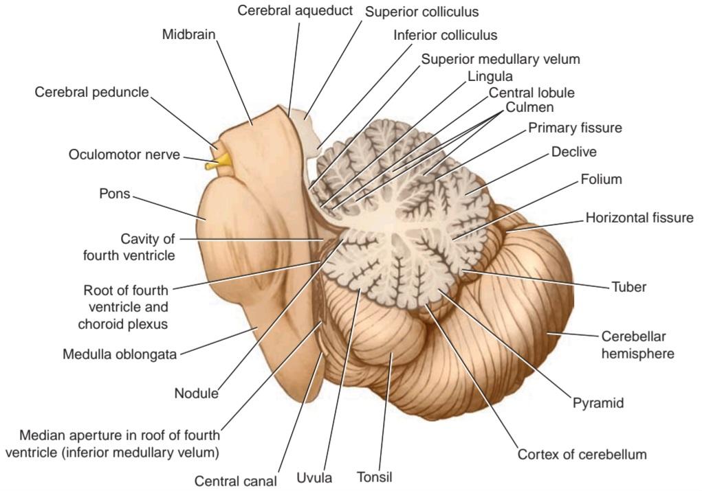
Figure 6-1 Sagittal section through the brainstem and the vermis of the cerebellum.
شکل ۶-۱ برش ساژیتال از ساقه مغز و ورمیس مخچه.
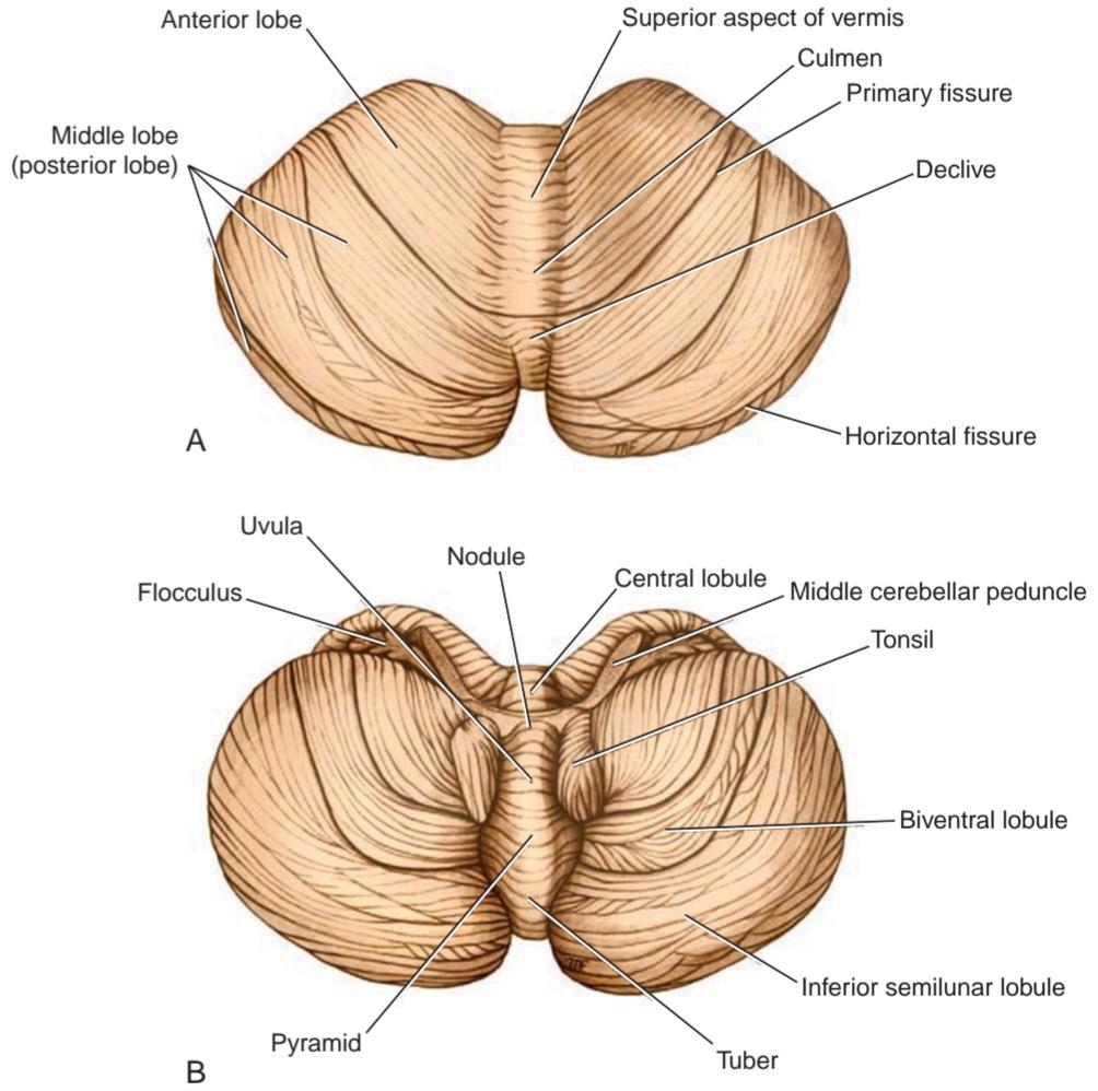
Figure 6-2 The cerebellum. A: Superior view. B: Inferior view.
شکل ۶-۲ مخچه. الف: نمای فوقانی. ب: نمای تحتانی.
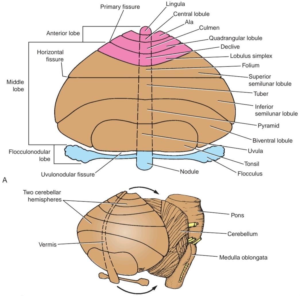
Figure 6-3 A: Flattened view of the cerebellar cortex showing the main cerebellar lobes, lobules, and fissures. B: Relationship between the diagram in (A) and the cerebellum.
شکل ۶-۳ الف: نمای مسطح از قشر مخچه که لوبها، لوبولها و شیارهای اصلی مخچه را نشان میدهد. ب: رابطه بین نمودار در (الف) و مخچه.
STRUCTURES
The cerebellum is composed of an outer covering of gray matter called the cortex and inner white matter. Embedded in the white matter of each hemisphere are three masses of gray matter forming the intracerebellar nuclei.
ساختارها
مخچه از یک پوشش بیرونی از ماده خاکستری به نام قشر مغز و ماده سفید داخلی تشکیل شده است. در ماده سفید هر نیمکره، سه توده ماده خاکستری وجود دارد که هستههای داخل مخچهای را تشکیل میدهند.
Cerebellar Cortex
The cerebellar cortex can be regarded as a large sheet with folds lying in the coronal or transverse plane. Each fold or folium contains a core of white matter covered superficially by gray matter (see Fig. 6-1).
قشر مخچه
قشر مخچه را میتوان به عنوان یک صفحه بزرگ با چینهایی در صفحه تاجی یا عرضی در نظر گرفت. هر چین یا فولیوم حاوی یک هسته از ماده سفید است که به صورت سطحی توسط ماده خاکستری پوشانده شده است (شکل 6-1 را ببینید).
A section made through the cerebellum parallel with the median plane divides the folia at right angles, and the cut surface has a branched appearance, called the arbor vitae.
برشی که از مخچه به موازات صفحه میانی ایجاد میشود، فولیا را با زاویه قائمه تقسیم میکند و سطح برش ظاهری شاخهدار دارد که به آن آربور ویتا میگویند.
The gray matter of the cortex throughout its extent has a uniform structure. It may be divided into three layers: (1) an external layer, the molecular layer; (2) a middle layer, the Purkinje cell layer; and (3) an internal layer, the granular layer (Figs. 6-4 and 6-5).
ماده خاکستری قشر مغز در سراسر وسعت خود دارای ساختار یکنواختی است. میتوان آن را به سه لایه تقسیم کرد: (1) یک لایه خارجی، لایه مولکولی؛ (2) یک لایه میانی، لایه سلولهای پورکنژ؛ و (3) یک لایه داخلی، لایه دانهای (شکلهای 6-4 و 6-5).
Molecular Layer
The molecular layer contains two types of neurons: the outer stellate cell and the inner basket cell (see Fig. 6-4). These neurons are scattered among dendritic arborizations and numerous thin axons that run parallel to the long axis of the folia. Neuroglial cells are found between these structures.
لایه مولکولی
لایه مولکولی شامل دو نوع نورون است: سلول ستارهای بیرونی و سلول سبدی داخلی (شکل 6-4 را ببینید). این نورونها در میان شاخههای دندریتی و آکسونهای نازک متعددی که به موازات محور طولی فولیا امتداد دارند، پراکنده شدهاند. سلولهای نوروگلیا بین این ساختارها یافت میشوند.
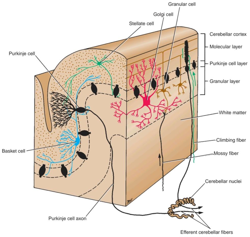
Figure 6-4 Cellular organization of the cerebellar cortex. Note the afferent and efferent fibers.
شکل ۶-۴ سازماندهی سلولی قشر مخچه. به فیبرهای آوران و وابران توجه کنید.
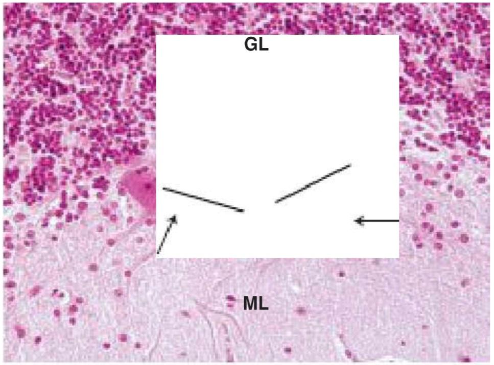
Figure 6-5 Light micrograph of the cerebellum (x132). Observe the Purkinje cells (P) with their dendritic trees (arrows) protruding into the molecular layer (ML). The heavily populated and deeply stained region is the granular layer (GL) of the cerebellum. (From Gartner, L. P. [2018]. Color atlas and text of histology [7th ed.]. Baltimore, MD: Wolters Kluwer.)
شکل ۶-۵ تصویر میکروسکوپی نوری از مخچه (132x). سلولهای پورکنژ (P) را با درختان دندریتی (فلشها) که به لایه مولکولی (ML) بیرون زدهاند، مشاهده کنید. ناحیه پر جمعیت و عمیقاً رنگآمیزی شده، لایه دانهای (GL) مخچه است. (از گارتنر، ال. پی. [2018]. اطلس رنگی و متن بافتشناسی [ویرایش هفتم]. بالتیمور، مریلند: ولترز کلوور.)
Purkinje Cell Layer
The Purkinje cells are large Golgi type I neurons. They are flask shaped and are arranged in a single layer (see Figs. 6-4 and 6-5). In a plane trans- verse to the folium, the dendrites of these cells are seen to pass into the molecular layer, where they undergo profuse branching. The primary and secondary branches are smooth, and subsequent branches are covered by short, thick dendritic spines. It has been shown that the spines form synaptic contacts with the parallel fibers derived from the granule cell axons.
لایه سلولی پورکنژ
سلولهای پورکنژ نورونهای بزرگ گلژی نوع I هستند. آنها به شکل فلاسک هستند و در یک لایه قرار گرفتهاند (شکلهای 6-4 و 6-5 را ببینید). در صفحهای که عمود بر چینه است، دندریتهای این سلولها به لایه مولکولی وارد میشوند، جایی که شاخههای فراوانی ایجاد میکنند. شاخههای اولیه و ثانویه صاف هستند و شاخههای بعدی توسط خارهای دندریتیک کوتاه و ضخیم پوشیده شدهاند. نشان داده شده است که خارها با فیبرهای موازی مشتق شده از آکسونهای سلولهای گرانول، تماسهای سیناپسی تشکیل میدهند.
At the base of the Purkinje cell, the axon arises and passes through the granular layer to enter the white matter. On entering the white matter, the axon acquires a myelin sheath, and it terminates by synapsing with cells of one of the intracerebellar nuclei. Collateral branches of the Purkinje axon make synaptic contacts with the dendrites of basket and stellate cells of the granular layer in the same area or in distant folia. A few of the Purkinje cell axons pass directly to end in the vestibular nuclei of the brainstem.
در پایه سلول پورکنژ، آکسون از لایه دانهدار خارج شده و از آن عبور میکند تا وارد ماده سفید شود. آکسون با ورود به ماده سفید، یک غلاف میلین به دست میآورد و با سیناپس زدن با سلولهای یکی از هستههای داخل مخچهای خاتمه مییابد. شاخههای جانبی آکسون پورکنژ با دندریتهای سلولهای سبدی و ستارهای لایه دانهدار در همان ناحیه یا در شاخکهای دور، تماس سیناپسی برقرار میکنند. تعداد کمی از آکسونهای سلول پورکنژ مستقیماً عبور میکنند تا در هستههای دهلیزی ساقه مغز خاتمه یابند.
Granular Layer
The granular layer is packed with small cells with densely staining nuclei and scanty cytoplasm. Each cell gives rise to four or five dendrites, which make claw-like endings and have synaptic contact with mossy fiber input (see Fig. 6-4). The axon of each granule cell passes into the molecular layer, where it bifurcates at a T junction, the branches running parallel to the long axis of the cerebellar folium (see Fig. 6-4). These fibers, known as parallel fibers, run at right angles to the dendritic processes of the Purkinje cells. Most of the parallel fibers make synaptic contacts with the spinous processes of the dendrites of the Purkinje cells. Neuroglial Fastigial nucleus Globose nucleus cells are found throughout this layer. Scattered throughout the granular layer are Golgi cells. Their dendrites ramify in the molecular layer, and their axons terminate by splitting up into branches that synapse with the dendrites of the granular cells (see Fig. 6-5).
لایه دانهای
لایه دانهای مملو از سلولهای کوچک با هستههای متراکم و سیتوپلاسم کم است. هر سلول چهار یا پنج دندریت ایجاد میکند که انتهای پنجه مانند ایجاد میکنند و با ورودی فیبر خزهای تماس سیناپسی دارند (شکل 6-4 را ببینید). آکسون هر سلول دانهای به لایه مولکولی منتقل میشود، جایی که در یک اتصال T دو شاخه میشود، شاخههایی که به موازات محور طولی چین مخچه امتداد دارند (شکل 6-4 را ببینید). این فیبرها که به عنوان فیبرهای موازی شناخته میشوند، با زاویه قائمه نسبت به زوائد دندریتیک سلولهای پورکنژ قرار دارند. بیشتر فیبرهای موازی با زوائد خاردار دندریتهای سلولهای پورکنژ تماس سیناپسی برقرار میکنند. هسته نوروگلیال فاستیژیال سلولهای هسته کروی در سراسر این لایه یافت میشوند. سلولهای گلژی در سراسر لایه دانهای پراکنده هستند. دندریتهای آنها در لایه مولکولی منشعب میشوند و آکسونهای آنها با تقسیم شدن به شاخههایی که با دندریتهای سلولهای دانهای سیناپس برقرار میکنند، خاتمه مییابند (شکل 6-5 را ببینید).
Functional Areas
Clinical observations by neurologists and neurosurgeons and the experimental use of positron emission tomography have shown that the cerebellar cortex can be divided into three functional areas.
حوزههای عملکردی
مشاهدات بالینی توسط متخصصان مغز و اعصاب و جراحان مغز و اعصاب و استفاده تجربی از توموگرافی گسیل پوزیترون نشان داده است که قشر مخچه را میتوان به سه ناحیه عملکردی تقسیم کرد.
The cortex of the vermis influences the movements of the long axis of the body, namely, the neck, the shoulders, the thorax, the abdomen, and the hips (Fig. 6-6). Immediately lateral to the vermis is a so-called intermediate zone of the cerebellar hemisphere. This area has been shown to control the muscles of the distal parts of the limbs, especially the hands and feet. The lateral zone of each cerebellar hemisphere appears to be concerned with the planning of sequential movements of the entire body and is involved with the conscious assessment of movement errors.
قشر کرمینه بر حرکات محور طولی بدن، یعنی گردن، شانهها، قفسه سینه، شکم و باسن تأثیر میگذارد (شکل 6-6). بلافاصله در کنار کرمینه، ناحیهای به نام ناحیه میانی نیمکره مخچه قرار دارد. نشان داده شده است که این ناحیه عضلات قسمتهای انتهایی اندامها، به ویژه دستها و پاها را کنترل میکند. به نظر میرسد ناحیه جانبی هر نیمکره مخچه مربوط به برنامهریزی حرکات متوالی کل بدن است و در ارزیابی آگاهانه خطاهای حرکتی نقش دارد.
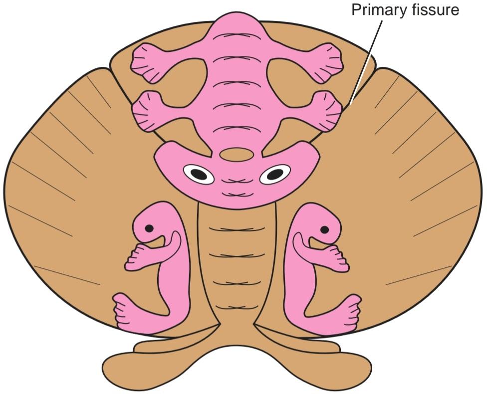
Figure 6-6 Somatosensory projection areas in the cerebellar cortex.
شکل ۶-۶ نواحی فرافکنی حس پیکری در قشر مخچه.
Intracerebellar Nuclei
Four masses of gray matter are embedded in the white matter of the cerebellum on each side of the midline (Fig. 6-7). From lateral to medial, these nuclei are the dentate, the emboliform, the globose, and the fastigial.
هستههای داخل مخچهای
چهار توده ماده خاکستری در ماده سفید مخچه در هر طرف خط میانی قرار دارند (شکل 6-7). این هستهها از جانبی به داخلی عبارتند از: دندانهای، آمبولیفرم، کروی و فاستیژیال.
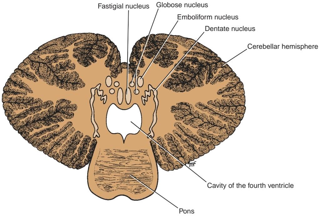
Figure 6-7 Position of the intracerebellar nuclei.
شکل ۶-۷ موقعیت هستههای داخل مخچهای.
The dentate nucleus is the largest of the cerebellar nuclei. It has the shape of a crumpled bag with the opening facing medially. The interior of the bag is filled with white matter made up of efferent fibers that leaves the nucleus through the opening to form a large part of the superior cerebellar peduncle.
هسته دندانهدار بزرگترین هسته مخچه است. این هسته به شکل یک کیسه مچاله شده است که دهانه آن به سمت داخل است. داخل کیسه پر از ماده سفیدی است که از الیاف وابران تشکیل شده و از طریق دهانه از هسته خارج میشود و بخش بزرگی از پایه مخچه فوقانی را تشکیل میدهد.
The emboliform nucleus is ovoid and is situated medial to the dentate nucleus, partially covering its hilus.
هسته آمبولیفرم بیضی شکل است و در قسمت داخلی هسته دندانهدار قرار دارد و تا حدی ناف آن را میپوشاند.
The globose nucleus consists of one or more rounded cell groups that lie medial to the emboliform nucleus.
هسته کروی از یک یا چند گروه سلولی گرد تشکیل شده است که در قسمت داخلی هسته آمبولیفرم قرار دارند.
The fastigial nucleus lies near the midline in the vermis and close to the roof of the fourth ventricle; it is larger than the globose nucleus.
هسته فاستیژیال در نزدیکی خط میانی ورمیس و نزدیک به سقف بطن چهارم قرار دارد. این هسته از هسته کروی بزرگتر است.
The intracerebellar nuclei are composed of large, multipolar neurons with simple branching dendrites. The axons form the cerebellar outflow in the superior and inferior cerebellar peduncles.
هستههای داخل مخچهای از نورونهای بزرگ و چندقطبی با دندریتهای منشعب ساده تشکیل شدهاند. آکسونها جریان خروجی مخچه را در پایههای مخچهای فوقانی و تحتانی تشکیل میدهند.
White Matter
There is a small amount of white matter in the vermis; it closely resembles the trunk and branches of a tree and thus is termed the arbor vitae (see Fig. 6-1). There is a large amount of white matter in each cerebellar hemisphere. The white matter is made up of three groups of fibers: (1) intrinsic, (2) afferent, and (3) efferent.
ماده سفید
مقدار کمی ماده سفید در ورمیس وجود دارد؛ این ماده شباهت زیادی به تنه و شاخههای درخت دارد و به همین دلیل به آن arbor vitae میگویند (شکل 6-1 را ببینید). مقدار زیادی ماده سفید در هر نیمکره مخچه وجود دارد. ماده سفید از سه گروه فیبر تشکیل شده است: (1) داخلی، (2) آوران و (3) وابران.
The intrinsic fibers do not leave the cerebellum but connect different regions of the organ. Some interconnect folia of the cerebellar cortex and vermis on the same side; others connect the two cerebellar hemispheres together.
الیاف داخلی از مخچه خارج نمیشوند، بلکه نواحی مختلف این اندام را به هم متصل میکنند. برخی از آنها، شاخکهای قشر مخچه و ورمیس را در یک طرف به هم متصل میکنند؛ برخی دیگر، دو نیمکره مخچه را به هم متصل میکنند.
The afferent fibers form the greater part of the white matter and proceed to the cerebellar cortex. They enter the cerebellum mainly through the inferior and middle cerebellar peduncles.
الیاف آوران بخش بزرگتری از ماده سفید را تشکیل میدهند و به قشر مخچه میروند. آنها عمدتاً از طریق پایههای مخچهای تحتانی و میانی وارد مخچه میشوند.
The efferent fibers constitute the output of the cerebellum and commence as the axons of the Purkinje cells of the cerebellar cortex. The great majority of the Purkinje cell axons pass to and synapse with the neurons of the cerebellar nuclei (fastigial, globose, emboliform, and dentate). The axons of the neurons then leave the cerebellum. A few Purkinje cell axons in the flocculonodular lobe and in parts of the vermis bypass the cerebellar nuclei and leave the cerebellum without synapsing.
الیاف وابران، خروجی مخچه را تشکیل میدهند و به عنوان آکسونهای سلولهای پورکنژ قشر مخچه شروع میشوند. اکثریت قریب به اتفاق آکسونهای سلولهای پورکنژ به نورونهای هستههای مخچه (فاستیژیال، کروی، آمبولیفرم و دندانهای) منتقل میشوند و با آنها سیناپس برقرار میکنند. سپس آکسونهای نورونها مخچه را ترک میکنند. تعداد کمی از آکسونهای سلولهای پورکنژ در لوب فلوکولوندولار و در بخشهایی از ورمیس، هستههای مخچه را دور میزنند و بدون سیناپس از مخچه خارج میشوند.
Fibers from the dentate, emboliform, and globose nuclei leave the cerebellum through the superior cerebellar peduncle. Fibers from the fastigial nucleus leave through the inferior cerebellar peduncle.
الیاف هستههای دندانهای، آمبولیفرم و کروی از طریق پایه مخچهای فوقانی از مخچه خارج میشوند. الیاف هسته فاستیژیال از طریق پایه مخچهای تحتانی خارج میشوند.
«فصل ششم کتاب نورواناتومی بالینی اسنل»
🚀 با ما همراه شوید!
تازهترین مطالب و آموزشهای مغز و اعصاب را از دست ندهید. با فالو کردن کانال تلگرام آیندهنگاران مغز، از ما حمایت کنید!