
مسیرهای آوران و مسیرهای وابران مخچه و نقش آنها در کنترل حرکات دقیق


دعای مطالعه [ نمایش ]
بِسْمِ الله الرَّحْمنِ الرَّحیمِ
اَللّهُمَّ اَخْرِجْنى مِنْ ظُلُماتِ الْوَهْمِ
خدايا مرا بيرون آور از تاريكىهاى وهم،
وَ اَكْرِمْنى بِنُورِ الْفَهْمِ
و به نور فهم گرامى ام بدار،
اَللّهُمَّ افْتَحْ عَلَيْنا اَبْوابَ رَحْمَتِكَ
خدايا درهاى رحمتت را به روى ما بگشا،
وَانْشُرْ عَلَيْنا خَزائِنَ عُلُومِكَ بِرَحْمَتِكَ يا اَرْحَمَ الرّاحِمينَ
و خزانههاى علومت را بر ما باز كن به امید رحمتت اى مهربانترين مهربانان.
آیندهنگاران مغز: «ما مغز را میشناسیم، تا آینده را بسازیم.»
کتاب «نوروآناتومی بالینی اسنل» اثر دکتر ریچارد اس. اسنل، با ویرایش جدید دکتر رایان اسپلیتگربر، همچنان بهعنوان مرجع طلایی این حوزه شناخته میشود. این اثر با تلفیق تازهترین پژوهشها و کاربردهای بالینی، پلی استوار میان دانش پایه و عمل پزشکی بنا میکند.
تلاشهای علمی و سالها تدریس ارزشمند استاد گرانقدر آقای دکتر محمدتقی جغتایی الهامبخش ترجمه و گسترش این دانش در ایران بوده است.
ترجمه دقیق این کتاب توسط برند علمی آیندهنگاران مغز به مدیریت داریوش طاهری، امکان دسترسی فارسیزبانان به مرزهای نوین علوم اعصاب را فراهم کرده و رسالتی علمی در جهت شناخت عمیقتر مغز و ترسیم آیندهای روشنتر بر عهده گرفته است.
»» فصل ۶: مخچه و ارتباطات آن؛ بخش مرور مبانی و اصول
»» Chapter 6: Cerebellum and Its Connections
CEREBELLAR AFFERENT FIBERS
The cerebellum receives major afferent tracts from the cerebral cortex, pons, medulla oblongata, and the spinal cord.
رشتههای آوران مخچه
مخچه رشتههای آوران اصلی را از قشر مغز، پل مغزی، بصل النخاع و نخاع دریافت میکند.
Cerebellar Afferent Fibers From the Cerebral Cortex
The cerebral cortex sends information to the cerebellum by three pathways: (1) the corticopontocerebellar pathway, (2) the cerebro-olivocerebellar pathway, and (3) the cerebroreticulocerebellar pathway.
فیبرهای آوران مخچه از قشر مغز
قشر مغز از طریق سه مسیر اطلاعات را به مخچه ارسال میکند: (1) مسیر کورتیکوپونتوسربلار، (2) مسیر مغزی-زیتونی-مخچهای، و (3) مسیر مغزی-مشبکی-مخچهای.
Corticopontocerebellar Pathway
The corticopontine fibers arise from nerve cells in the frontal, parietal, temporal, and occipital lobes of the cerebral cortex and descend through the corona radiata and internal capsule and terminate on the pontine nuclei (Fig. 6-10). The pontine nuclei give rise to the transverse fibers of the pons, which cross the midline and enter the opposite cerebellar hemisphere as the middle cerebellar peduncle (see Figs. 5-18, 5-19).
مسیر کورتیکوپونتوسربلار
الیاف کورتیکوپونتوسربلار از سلولهای عصبی در لوبهای پیشانی، آهیانه، گیجگاهی و پسسری قشر مغز منشأ میگیرند و از طریق تاج شعاعی و کپسول داخلی پایین میآیند و در هستههای پل مغزی خاتمه مییابند (شکل 6-10). هستههای پل مغزی باعث ایجاد الیاف عرضی پل مغزی میشوند که از خط میانی عبور کرده و به عنوان پایک میانی مخچه وارد نیمکره مخچه مقابل میشوند (شکلهای 5-18، 5-19 را ببینید).
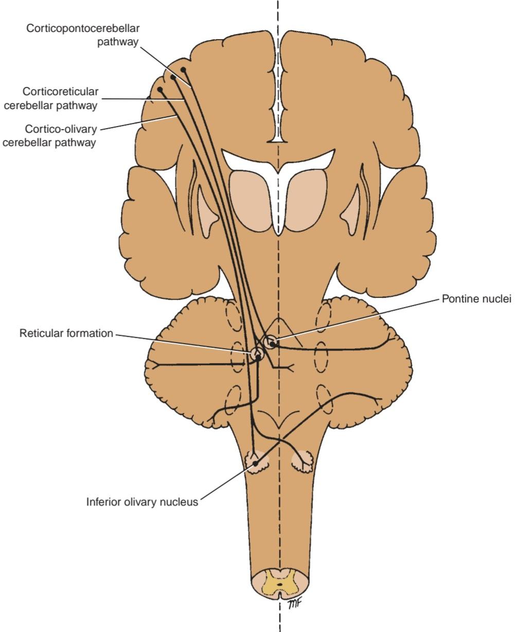
Figure 6-10 Cerebellar afferent fibers from the cerebral cortex. The cerebellar peduncles are shown as ovoid dotted lines.
شکل ۶-۱۰ فیبرهای آوران مخچه از قشر مغز. پایکهای مخچه به صورت خطوط نقطهچین بیضیشکل نشان داده شدهاند.
Cerebro-Olivocerebellar Pathway
The cortico-olivary fibers arise from nerve cells in the frontal, parietal, temporal, and occipital lobes of the cerebral cortex and descend through the corona radiata and internal capsule to terminate bilaterally on the inferior olivary nuclei. The inferior olivary nuclei give rise to fibers that cross the midline and enter the opposite cerebellar hemisphere through the inferior cerebellar peduncle. These fibers terminate as the climbing fibers in the cerebellar cortex.
مسیر مغزی-زیتونی مخچهای
فیبرهای مغزی-زیتونی مخچهای از سلولهای عصبی در لوبهای پیشانی، آهیانه، گیجگاهی و پسسری قشر مغز منشأ میگیرند و از طریق تاج شعاعی و کپسول داخلی پایین میآیند تا به صورت دو طرفه در هستههای زیتونی تحتانی خاتمه یابند. هستههای زیتونی تحتانی باعث ایجاد فیبرهایی میشوند که از خط میانی عبور میکنند و از طریق پایه مخچهای تحتانی وارد نیمکره مخچه مقابل میشوند. این فیبرها به عنوان فیبرهای بالارونده در قشر مخچه خاتمه مییابند.
Cerebroreticulocerebellar Pathway
The corticoreticular fibers arise from nerve cells from many areas of the cerebral cortex, particularly the sensorimotor areas. They descend to terminate in the reticular formation on the same side and on the opposite side in the pons and medulla. The cells in the reticular formation give rise to the reticulocerebellar fibers that enter the cerebellar hemisphere on the same side through the inferior and middle cerebellar peduncles.
مسیر مخچهای-مشبکی-مخچهای
الیاف قشری-مخچهای از سلولهای عصبی بسیاری از نواحی قشر مغز، به ویژه نواحی حسی-حرکتی، منشأ میگیرند. آنها به سمت پایین حرکت میکنند تا در تشکیلات مشبک در همان طرف و در طرف مقابل در پل مغزی و بصل النخاع خاتمه یابند. سلولهای موجود در تشکیلات مشبک، الیاف مشبک-مخچهای را ایجاد میکنند که از طریق پایکهای مخچهای تحتانی و میانی وارد نیمکره مخچه در همان طرف میشوند.
This connection between the cerebrum and the cerebellum is important in the control of voluntary movement. Information regarding the initiation of movement in the cerebral cortex is probably transmitted to the cerebellum so that the movement can be monitored and appropriate adjustments in the muscle activity can be made.
این ارتباط بین مخ و مخچه در کنترل حرکت ارادی مهم است. اطلاعات مربوط به شروع حرکت در قشر مغز احتمالاً به مخچه منتقل میشود تا بتوان حرکت را کنترل کرد و تنظیمات مناسب در فعالیت عضلات را انجام داد.
Cerebellar Afferent Fibers From the Spinal Cord
The spinal cord sends information to the cerebellum from somatosensory receptors by three pathways:
(1) the anterior spinocerebellar tract, (2) the posterior spinocerebellar tract, and (3) the cuneocerebellar tract.
فیبرهای آوران مخچه از نخاع
نخاع اطلاعات را از گیرندههای حسی-پیکری از طریق سه مسیر به مخچه ارسال میکند:
(1) راه نخاعی-مخچهای قدامی، (2) راه نخاعی-مخچهای خلفی، و (3) راه کونئوسربلار.
Anterior Spinocerebellar Tract
The axons entering the spinal cord from the posterior root ganglion terminate by synapsing with the neurons in the nucleus dorsalis (Clarke column) at the base of the posterior gray column. Most of the axons of these neurons cross to the opposite side and ascend as the anterior spinocerebellar tract in the contralateral white column; some of the axons ascend as the anterior spinocerebellar tract in the lateral white column on the same side (Fig. 6-11). The fibers enter the cerebellum through the superior cerebellar peduncle and terminate as mossy fibers in the cerebellar cortex. Collateral branches that end in the deep cerebellar nuclei are also given off. Those fibers that cross over to the opposite side in the spinal cord are thought to cross back within the cerebellum.
راه نخاعی-مخچهای قدامی
آکسونهایی که از گانگلیون ریشه خلفی وارد نخاع میشوند، با سیناپس با نورونهای موجود در هسته پشتی (ستون کلارک) در پایه ستون خاکستری خلفی خاتمه مییابند. بیشتر آکسونهای این نورونها به سمت مقابل عبور میکنند و به عنوان راه نخاعی-مخچهای قدامی در ستون سفید طرف مقابل بالا میروند؛ برخی از آکسونها به عنوان راه نخاعی-مخچهای قدامی در ستون سفید جانبی در همان طرف بالا میروند (شکل 6-11). فیبرها از طریق پایه مخچهای فوقانی وارد مخچه میشوند و به عنوان فیبرهای خزهای در قشر مخچه خاتمه مییابند. شاخههای جانبی که در هستههای عمقی مخچه ختم میشوند نیز جدا میشوند. تصور میشود آن دسته از فیبرهایی که از سمت مقابل در نخاع عبور میکنند، در داخل مخچه دوباره از هم عبور میکنند.
The anterior spinocerebellar tract is found at all segments of the spinal cord, and its fibers convey muscle joint information from the muscle spindles, tendon organs, and joint receptors of the upper and lower limbs. The cerebellum likely receives information from the skin and superficial fascia by this tract.
راه نخاعی-مخچهای قدامی در تمام بخشهای نخاع یافت میشود و فیبرهای آن اطلاعات مفصل عضلانی را از دوکهای عضلانی، اندامهای تاندونی و گیرندههای مفصلی اندامهای فوقانی و تحتانی منتقل میکنند. مخچه احتمالاً از طریق این راه اطلاعاتی را از پوست و فاسیای سطحی دریافت میکند.

Figure 6-11 Cerebellar afferent fibers from the spinal cord and internal ear. The cerebellar peduncles are shown as ovoid dotted lines.
شکل ۶-۱۱ فیبرهای آوران مخچه از نخاع و گوش داخلی. پایههای مخچه به صورت خطوط نقطهچین بیضیشکل نشان داده شدهاند.
Posterior Spinocerebellar Tract
The axons entering the spinal cord from the posterior root ganglion enter the posterior gray column and terminate by synapsing on the neurons at the base of the posterior gray column. These neurons are known collectively as the nucleus dorsalis (Clarke column). The axons of these neurons enter the posterolateral part of the lateral white column on the same side and ascend as the posterior spinocerebellar tract to the medulla oblongata. Here, the tract enters the cerebellum through the inferior cerebellar peduncle and terminates as mossy fibers in the cerebellar cortex. Collateral branches that end in the deep cerebellar nuclei are also given off. The posterior spinocerebellar tract receives muscle joint information from the muscle spindles, tendon organs, and joint receptors of the trunk and lower limbs.
راه نخاعی-مخچهای خلفی
آکسونهایی که از گانگلیون ریشه خلفی وارد نخاع میشوند، وارد ستون خاکستری خلفی میشوند و با سیناپس روی نورونهای پایه ستون خاکستری خلفی خاتمه مییابند. این نورونها در مجموع به عنوان هسته پشتی (ستون کلارک) شناخته میشوند. آکسونهای این نورونها از همان طرف وارد قسمت خلفی-جانبی ستون سفید جانبی میشوند و به عنوان راه نخاعی-مخچهای خلفی به بصل النخاع صعود میکنند. در اینجا، این راه از طریق پایه مخچهای تحتانی وارد مخچه میشود و به صورت الیاف خزهای در قشر مخچه خاتمه مییابد. شاخههای جانبی که به هستههای عمقی مخچه ختم میشوند نیز از آن جدا میشوند. راه نخاعی-مخچهای خلفی اطلاعات مفصل عضله را از دوکهای عضلانی، اندامهای تاندونی و گیرندههای مفصلی تنه و اندامهای تحتانی دریافت میکند.
Cuneocerebellar Tract
These fibers originate in the nucleus cuneatus of the medulla oblongata and enter the cerebellar hemisphere on the same side through the inferior cerebellar peduncle (see Fig. 6-10). The fibers terminate as mossy fibers in the cerebellar cortex. Collateral branches that end in the deep cerebellar nuclei are also given off. The cuneocerebellar tract receives muscle joint information from the muscle spindles, tendon organs, and joint receptors of the upper limb and upper part of the thorax.
راه کونئوسربلار
این فیبرها از هسته کونئاتوس بصل النخاع منشأ میگیرند و از طریق پایه مخچه تحتانی وارد نیمکره مخچه در همان طرف میشوند (شکل 6-10 را ببینید). این فیبرها به صورت فیبرهای خزهای در قشر مخچه خاتمه مییابند. شاخههای جانبی که به هستههای عمقی مخچه ختم میشوند نیز از آنها جدا میشوند. راه کونئوسربلار اطلاعات مفصل عضله را از دوکهای عضلانی، اندامهای تاندونی و گیرندههای مفصلی اندام فوقانی و قسمت فوقانی قفسه سینه دریافت میکند.
Cerebellar Afferent Fibers From the Vestibular Nerve
The vestibular nerve receives information from the inner ear concerning motion from the semicircular canals and position relative to gravity from the utricle and saccule. The vestibular nerve sends many afferent fibers directly to the cerebellum through the inferior cerebellar peduncle on the same side. Other vestibular afferent fibers pass first to the vestibular nuclei in the brainstem, where they synapse and are relayed to the cerebellum (see Fig. 6-11). They enter the cerebellum through the inferior cerebellar peduncle on the same side. All the afferent fibers from the inner ear terminate as mossy fibers in the flocculonodular lobe of the cerebellum.
الیاف آوران مخچه از عصب دهلیزی
عصب دهلیزی اطلاعات مربوط به حرکت را از مجاری نیمدایرهای و موقعیت نسبت به جاذبه را از اتریکول و ساکول از گوش داخلی دریافت میکند. عصب دهلیزی بسیاری از الیاف آوران را مستقیماً از طریق پایه مخچهای تحتانی در همان طرف به مخچه میفرستد. سایر الیاف آوران دهلیزی ابتدا به هستههای دهلیزی در ساقه مغز میروند، جایی که سیناپس تشکیل میدهند و به مخچه منتقل میشوند (شکل 6-11 را ببینید). آنها از طریق پایه مخچهای تحتانی در همان طرف وارد مخچه میشوند. تمام الیاف آوران از گوش داخلی به عنوان الیاف خزهای در لوب فلوکولوندولار مخچه خاتمه مییابند.
Other Afferent Fibers
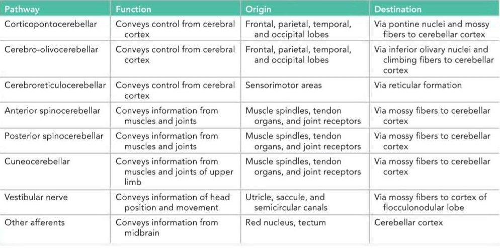
In addition, the cerebellum receives small bundles of afferent fibers from the red nucleus and the tectum. The afferent cerebellar pathways are summarized in Table 6-1.
سایر فیبرهای آوران
علاوه بر این، مخچه دستههای کوچکی از فیبرهای آوران را از هسته قرمز و تکتوم دریافت میکند. مسیرهای آوران مخچه در جدول 6-1 خلاصه شدهاند.
Table 6-1 Afferent Cerebellar Pathways
جدول ۶-۱ مسیرهای آوران مخچه
CEREBELLAR EFFERENT FIBERS
The entire output of the cerebellar cortex is through the axons of the Purkinje cells. Most of the axons of the Purkinje cells end by synapsing on the neurons of the deep cerebellar nuclei (see Fig. 6-4). The axons of the neurons that form the cerebellar nuclei constitute the efferent outflow from the cerebellum. A few Purkinje cell axons pass directly out of the cerebellum to the lateral vestibular nucleus. The efferent fibers from the cerebellum connect with the red nucleus, thalamus, vestibular complex, and reticular formation.
فیبرهای وابران مخچه
تمام خروجی قشر مخچه از طریق آکسونهای سلولهای پورکنژ است. بیشتر آکسونهای سلولهای پورکنژ با سیناپس روی نورونهای هستههای عمقی مخچه به پایان میرسند (شکل 6-4 را ببینید). آکسونهای نورونهایی که هستههای مخچه را تشکیل میدهند، جریان وابران از مخچه را تشکیل میدهند. چند آکسون سلول پورکنژ مستقیماً از مخچه به هسته دهلیزی جانبی منتقل میشوند. فیبرهای وابران از مخچه به هسته قرمز، تالاموس، کمپلکس دهلیزی و تشکیلات مشبک متصل میشوند.
Globose-Emboliform-Rubral Pathway
Axons of neurons in the globose and emboliform nuclei travel through the superior cerebellar peduncle and Cerebellar Efferent Fibers cross the midline to the opposite side in the decussation of the superior cerebellar peduncles (Fig. 6-12). The fibers end by synapsing with cells of the contralateral red nucleus, which give rise to axons of the rubrospinal tract. Thus, this pathway crosses twice, once in the decussation of the superior cerebellar peduncle and again in the rubrospinal tract close to its origin. By this means, the globose and emboliform nuclei influence motor activity on the same side of the body.
مسیر کروی-آمبولی-روبرال
آکسونهای نورونهای هستههای کروی و آمبولی شکل از طریق پایه مخچهای فوقانی و فیبرهای وابران مخچهای از خط میانی به سمت مخالف در تقاطع پایههای مخچهای فوقانی عبور میکنند (شکل 6-12). فیبرها با سیناپس با سلولهای هسته قرمز طرف مقابل که آکسونهای راه روبرواسپینال را ایجاد میکنند، به پایان میرسند. بنابراین، این مسیر دو بار، یک بار در تقاطع پایه مخچهای فوقانی و بار دیگر در راه روبرواسپینال نزدیک به مبدا خود، عبور میکند. به این ترتیب، هستههای کروی و آمبولی شکل بر فعالیت حرکتی در همان طرف بدن تأثیر میگذارند.
Dentatothalamic Pathway
Axons of neurons in the dentate nucleus travel through the superior cerebellar peduncle and cross the midline to the opposite side in the decussation of the superior cerebellar peduncle. The fibers end by synapsing with cells in the contralateral ventrolateral nucleus of the thalamus. The axons of the thalamic neurons ascend through the internal capsule and corona radiata and terminate in the primary motor area of the cerebral cortex. By this pathway, the dentate nucleus can influence motor activity by acting on the motor neurons of the opposite cerebral cortex; impulses from the motor cortex are transmitted to spinal segmental levels through the corticospinal tract. Remember that most of the fibers of the corticospinal tract cross to the opposite side in the decussation of the pyramids or later at the spinal segmental levels. Thus, the dentate nucleus is able to coordinate muscle activity on the same side of the body.
مسیر دندانی-تالاموسی
آکسونهای نورونهای هسته دندانهای از طریق ساقه مخچهای فوقانی عبور میکنند و از خط میانی به سمت مقابل در تقاطع ساقه مخچهای فوقانی عبور میکنند. فیبرها با سیناپس با سلولهای هسته شکمی-جانبی طرف مقابل تالاموس به پایان میرسند. آکسونهای نورونهای تالاموس از طریق کپسول داخلی و تاج شعاعی بالا میروند و در ناحیه حرکتی اولیه قشر مغز خاتمه مییابند. از طریق این مسیر، هسته دندانهای میتواند با تأثیر بر نورونهای حرکتی قشر مغز مقابل، بر فعالیت حرکتی تأثیر بگذارد. تکانههای قشر حرکتی از طریق راه قشری-نخاعی به سطوح سگمنتال نخاعی منتقل میشوند. به یاد داشته باشید که بیشتر فیبرهای راه قشری-نخاعی در تقاطع هرمها یا بعداً در سطوح سگمنتال نخاعی به سمت مقابل عبور میکنند. بنابراین، هسته دندانهای قادر به هماهنگی فعالیت عضلات در همان طرف بدن است.

Figure 6-12 Cerebellar efferent fibers. The cerebellar peduncles are shown as ovoid dotted lines.
شکل ۶-۱۲ فیبرهای وابران مخچه. پایههای مخچه به صورت خطوط نقطهچین بیضیشکل نشان داده شدهاند.
Fastigial Vestibular Pathway
The axons of neurons in the fastigial nucleus travel through the inferior cerebellar peduncle and end by projecting on the neurons of the lateral vestibular nucleus on both sides. Remember that some Purkinje cell axons project directly to the lateral vestibular nucleus. The neurons of the lateral vestibular nucleus form the vestibulospinal tract. The fastigial nucleus exerts a facilitatory influence mainly on the ipsilateral extensor muscle tone.
مسیر دهلیزی فاستیژیال
آکسونهای نورونهای هسته فاستیژیال از طریق پایه مخچهای تحتانی عبور میکنند و با بیرون زدن روی نورونهای هسته دهلیزی جانبی در دو طرف، پایان مییابند. به یاد داشته باشید که برخی از آکسونهای سلولهای پورکنژ مستقیماً به هسته دهلیزی جانبی بیرون زدهاند. نورونهای هسته دهلیزی جانبی، مسیر دهلیزی-نخاعی را تشکیل میدهند. هسته فاستیژیال تأثیر تسهیلکنندهای عمدتاً بر تون عضله بازکننده همان طرف اعمال میکند.
Fastigial Reticular Pathway
The axons of neurons in the fastigial nucleus travel through the inferior cerebellar peduncle and end by synapsing with neurons of the reticular formation. Axons of these neurons influence spinal segmental motor activity through the reticulospinal tract. The efferent cerebellar pathways are summarized in Table 6-2.
مسیر مشبک فاستیژیال
آکسونهای نورونهای هسته فاستیژیال از طریق پایه مخچهای تحتانی حرکت میکنند و با سیناپس با نورونهای تشکیلات مشبک به پایان میرسند. آکسونهای این نورونها از طریق مسیر مشبک-نخاعی بر فعالیت حرکتی قطعهای نخاعی تأثیر میگذارند. مسیرهای وابران مخچه در جدول 6-2 خلاصه شدهاند.
Table 6-2 Efferent Cerebellar Pathways a
جدول ۶-۲ مسیرهای وابران مخچه a
 a Note that each cerebellar hemisphere influences the voluntary muscle tone on the same side of the body.
aتوجه داشته باشید که هر نیمکره مخچه بر تون عضلانی ارادی همان طرف بدن تأثیر میگذارد.
FUNCTIONS OF THE CEREBELLUM
The cerebellum receives afferent information concerning voluntary movement from the cerebral cortex and from the muscles, tendons, and joints. It also receives information concerning balance from the vestibular nerve and possibly concerning sight through the tectocerebellar tract. All this information is fed into the cerebellar cortical circuitry by the mossy fibers and the climbing fibers and converges on the Purkinje cells (see Fig. 6-8). The axons of the Purkinje cells project with few exceptions on the deep cerebellar nuclei. The output of the vermis projects to the fastigial nucleus, the intermediate regions of the cortex project to the globose and emboliform nuclei, and the output of the lateral part of the cerebellar hemisphere projects to the dentate nucleus. A few Purkinje cell axons pass directly out of the cerebellum and end on the lateral vestibular nucleus in the brainstem. Purkinje axons are generally believed to exert an inhibitory influence on the neurons of the cerebellar nuclei and the lateral vestibular nuclei.
عملکردهای مخچه
مخچه اطلاعات آوران مربوط به حرکت ارادی را از قشر مغز و از عضلات، تاندونها و مفاصل دریافت میکند. همچنین اطلاعات مربوط به تعادل را از عصب دهلیزی و احتمالاً از طریق مسیر تکتوسربلار دریافت میکند. تمام این اطلاعات توسط فیبرهای خزهای و فیبرهای بالارونده به مدار قشر مخچه منتقل شده و روی سلولهای پورکنژ همگرا میشوند (شکل 6-8 را ببینید). آکسونهای سلولهای پورکنژ با چند استثنا روی هستههای عمقی مخچه امتداد دارند. خروجی ورمیس به هسته فاستیجیال، نواحی میانی قشر به هستههای کروی و آمبولیفرم و خروجی قسمت جانبی نیمکره مخچه به هسته دندانهدار امتداد دارند. چند آکسون سلول پورکنژ مستقیماً از مخچه خارج شده و به هسته دهلیزی جانبی در ساقه مغز ختم میشوند. عموماً اعتقاد بر این است که آکسونهای پورکنژ تأثیر مهاری بر نورونهای هستههای مخچه و هستههای دهلیزی جانبی دارند.
The cerebellar output is conducted to the sites of origin of the descending pathways that influence motor activity at the segmental spinal level. In this respect, the cerebellum has no direct neuronal connections with the lower motor neurons but exerts its influence indirectly through the cerebral cortex and brainstem.
خروجی مخچه به محلهای مبدا مسیرهای نزولی که بر فعالیت حرکتی در سطح قطعهای نخاع تأثیر میگذارند، هدایت میشود. از این نظر، مخچه هیچ ارتباط عصبی مستقیمی با نورونهای حرکتی تحتانی ندارد، اما تأثیر خود را به طور غیرمستقیم از طریق قشر مغز و ساقه مغز اعمال میکند.
Physiologists have postulated that the cerebellum functions as a coordinator of precise movements by continually comparing the output of the motor area of the cerebral cortex with the proprioceptive information received from the site of muscle action; it is then able to bring about the necessary adjustments by influencing the activity of the lower motor neurons (Fig. 6-13). This is accomplished by controlling the timing and sequence of firing of the a and y motor neurons. The cerebellum can possibly send back information to the motor cerebral cortex to inhibit the agonist muscles and stimulate the antagonist muscles, thus limiting the extent of voluntary movement.
فیزیولوژیستها فرض کردهاند که مخچه با مقایسه مداوم خروجی ناحیه حرکتی قشر مغز با اطلاعات حس عمقی دریافتی از محل عمل عضلات، به عنوان هماهنگکننده حرکات دقیق عمل میکند. سپس میتواند با تأثیرگذاری بر فعالیت نورونهای حرکتی تحتانی، تنظیمات لازم را انجام دهد (شکل 6-13). این کار با کنترل زمانبندی و توالی شلیک نورونهای حرکتی a و y انجام میشود. مخچه احتمالاً میتواند اطلاعات را به قشر مغزی حرکتی ارسال کند تا عضلات آگونیست را مهار و عضلات آنتاگونیست را تحریک کند و در نتیجه میزان حرکت ارادی را محدود کند.

Figure 6-13 Cerebellum serving as a comparator.
شکل ۶-۱۳ مخچه به عنوان مقایسه کننده عمل میکند.
«فصل ششم کتاب نورواناتومی بالینی اسنل»
🚀 با ما همراه شوید!
تازهترین مطالب و آموزشهای مغز و اعصاب را از دست ندهید. با فالو کردن کانال تلگرام آیندهنگاران مغز، از ما حمایت کنید!



