
نوروآناتومی بالینی اسنل | ساقه مغز – نکات و کاربردهای بالینی


دعای مطالعه [ نمایش ]
بِسْمِ الله الرَّحْمنِ الرَّحیمِ
اَللّهُمَّ اَخْرِجْنى مِنْ ظُلُماتِ الْوَهْمِ
خدايا مرا بيرون آور از تاريكىهاى وهم،
وَ اَكْرِمْنى بِنُورِ الْفَهْمِ
و به نور فهم گرامى ام بدار،
اَللّهُمَّ افْتَحْ عَلَيْنا اَبْوابَ رَحْمَتِكَ
خدايا درهاى رحمتت را به روى ما بگشا،
وَانْشُرْ عَلَيْنا خَزائِنَ عُلُومِكَ بِرَحْمَتِكَ يا اَرْحَمَ الرّاحِمينَ
و خزانههاى علومت را بر ما باز كن به امید رحمتت اى مهربانترين مهربانان.
آیندهنگاران مغز: «ما مغز را میشناسیم، تا آینده را بسازیم.»
کتاب «نوروآناتومی بالینی اسنل» اثر دکتر ریچارد اس. اسنل، با ویرایش جدید دکتر رایان اسپلیتگربر، همچنان بهعنوان مرجع طلایی این حوزه شناخته میشود. این اثر با تلفیق تازهترین پژوهشها و کاربردهای بالینی، پلی استوار میان دانش پایه و عمل پزشکی بنا میکند.
تلاشهای علمی و سالها تدریس ارزشمند استاد گرانقدر دکتر محمدتقی جغتایی الهامبخش ترجمه و گسترش این دانش در ایران بوده است.
ترجمه دقیق این کتاب توسط برند علمی آیندهنگاران مغز به مدیریت داریوش طاهری، امکان دسترسی فارسیزبانان به مرزهای نوین علوم اعصاب را فراهم کرده و رسالتی علمی در جهت شناخت عمیقتر مغز و ترسیم آیندهای روشنتر بر عهده گرفته است.
»» فصل ۵: ساقه مغز
»» Chapter 5: Brainstem
Clinical Notes
Clinical Significance of the Medulla Oblongata
The medulla oblongata not only contains many cranial nerve nuclei that are concerned with vital functions (e.g., regulation of heart rate and respiration), but it also serves as a conduit for the passage of ascending and descending tracts connecting the spinal cord to the higher centers of the nervous system. These tracts may become involved in demyelinating diseases, neoplasms, and vascular disorders.
یادداشتهای بالینی
اهمیت بالینی بصل النخاع
بصل النخاع نه تنها حاوی هستههای عصبی جمجمهای زیادی است که با عملکردهای حیاتی (مانند تنظیم ضربان قلب و تنفس) مرتبط هستند، بلکه به عنوان مجرایی برای عبور مسیرهای صعودی و نزولی که نخاع را به مراکز بالاتر سیستم عصبی متصل میکنند نیز عمل میکند. این مسیرها ممکن است در بیماریهای دمیلینه کننده، نئوپلاسمها و اختلالات عروقی دخیل باشند.
Raised Pressure in the Posterior Cranial Fossa and Its Effect on the Medulla Oblongata
The medulla oblongata is situated in the posterior cranial fossa, lying beneath the tentorium cerebelli and above the foramen magnum. It is related anteriorly to the basal portion of the occipital bone and the upper part of the odontoid process of the axis and posteriorly to the cerebellum.
افزایش فشار در حفره جمجمهای خلفی و تأثیر آن بر بصل النخاع
بصل النخاع در حفره جمجمهای خلفی، زیر چادرینه مخچه و بالای سوراخ بزرگ قرار دارد. از جلو با قسمت قاعدهای استخوان پسسری و قسمت بالایی زائده دندانی محور و از عقب با مخچه مرتبط است.
In patients with tumors of the posterior cranial fossa, the intracranial pressure is raised, and the brain––that is, the cerebellum and the medulla oblongata––tends to be pushed toward the area of least resistance; there is a downward herniation of the medulla and cerebellar tonsils through the foramen magnum. This will produce the symptoms of headache, neck stiffness,and paralysis of the glossopharyngeal,vagus,accessory, and hypoglossal nerves owing to traction. In these circumstances, it is extremely dangerous to perform a lumbar puncture because the sudden withdrawal of cerebrospinal fluid may precipitate further herniation of the brain through the foramen magnum and a sudden failure of vital functions,resulting from pressure and ischemia of the cranial nerve nuclei present in the medulla oblongata.
در بیماران مبتلا به تومورهای حفره جمجمهای خلفی، فشار داخل جمجمه افزایش مییابد و مغز – یعنی مخچه و بصل النخاع – تمایل به رانده شدن به سمت ناحیهای با کمترین مقاومت دارد. فتق رو به پایین لوزههای بصل النخاع و مخچه از طریق سوراخ بزرگ وجود دارد. این امر باعث ایجاد علائم سردرد، سفتی گردن و فلج اعصاب زبانی-حلقی، واگ، فرعی و زیرزبانی به دلیل کشش میشود. در این شرایط، انجام پونکسیون کمری بسیار خطرناک است زیرا خروج ناگهانی مایع مغزی نخاعی ممکن است باعث فتق بیشتر مغز از طریق سوراخ بزرگ و اختلال ناگهانی عملکردهای حیاتی شود که ناشی از فشار و ایسکمی هستههای اعصاب جمجمهای موجود در بصل النخاع است.
Arnold-Chiari Phenomenon
The Arnold-Chiari malformation is a congenital anomaly in which there is a herniation of the tonsils of the cerebellum and the medulla oblongata through the foramen magnum into the vertebral canal (Fig. 5-30). This results in the blockage of the exits in the roof of the fourth ventricle to the cerebrospinal fluid, causing internal hydrocephalus. It is commonly associated with craniovertebral anomalies or various forms of spina bifida. Signs and symptoms related to pressure on the cerebellum and medulla oblongata and involvement of the last four cranial nerves are associated with this condition.
پدیده آرنولد-کیاری
ناهنجاری آرنولد-کیاری یک ناهنجاری مادرزادی است که در آن لوزههای مخچه و بصل النخاع از طریق سوراخ بزرگ به داخل کانال مهرهای بیرون زده میشوند (شکل 5-30). این امر منجر به انسداد خروجیهای سقف بطن چهارم به مایع مغزی نخاعی میشود و باعث هیدروسفالی داخلی میشود. این بیماری معمولاً با ناهنجاریهای جمجمهای-ستون فقراتی یا اشکال مختلف اسپینا بیفیدا همراه است. علائم و نشانههای مربوط به فشار بر مخچه و بصل النخاع و درگیری چهار عصب جمجمهای آخر با این بیماری مرتبط هستند.

Figure 5-30 Arnold-Chiari phenomenon. This coronal section of the skull shows the herniation of the cerebellar tonsil and the medulla oblongata through the foramen magnum into the vertebral canal. (From Dudek, R.W., Louis, T. M. [2015]. High-yield gross anatomy (5th ed.). Baltimore, MD: Wolters Kluwer.)
شکل ۵-۳۰ پدیده آرنولد-کیاری. این برش کرونال از جمجمه، فتق لوزه مخچه و بصل النخاع را از طریق سوراخ بزرگ به داخل کانال مهرهای نشان میدهد. (از Dudek, R.W., Louis, T. M. [2015]. High-yield gross anatomy (5th ed.). Baltimore, MD: Wolters Kluwer.)
Vascular Disorders of the Medulla Oblongata
Lateral Medullary Syndrome of Wallenberg
The lateral part of the medulla oblongata is supplied by the posterior inferior cerebellar artery, which is usually a branch of the vertebral artery.Thrombosis of either of these arteries (Fig.5-31) produces the following signs and symptoms: dysphagia and dysarthria due to paralysis of the ipsilateral palatal and laryngeal muscles (innervated by the nucleus ambiguus); analgesia and thermoanesthesia on the ipsilateral side of the face (nucleus and spinal tract of the trigeminal nerve); vertigo, nausea, vomiting, and nystagmus (vestibular nuclei); ipsilateral Horner syndrome (descending sympathetic fibers); ipsilateral cerebellar signs—gait and limb ataxia (cerebellum or inferior cerebellar peduncle); and contralateral loss of sensations of pain and temperature (spinal lemniscus—spinothalamic tract).
اختلالات عروقی بصل النخاع
سندرم والنبرگ مدولای جانبی
بخش جانبی بصل النخاع توسط شریان مخچهای خلفی تحتانی، که معمولاً شاخهای از شریان مهرهای است، خونرسانی میشود. ترومبوز هر یک از این شریانها (شکل 5-31) علائم و نشانههای زیر را ایجاد میکند: دیسفاژی و دیسآرتری به دلیل فلج عضلات کامی و حنجرهای همان طرف (که توسط هسته آمبیگوس عصبدهی میشوند)؛ بیدردی و بیحسی حرارتی در سمت همان طرف صورت (هسته و مسیر نخاعی عصب سهقلو)؛ سرگیجه، تهوع، استفراغ و نیستاگموس (هستههای دهلیزی)؛ سندرم هورنر همان طرف (الیاف سمپاتیک نزولی)؛ علائم مخچهای همان طرف – آتاکسی راه رفتن و اندام (مخچه یا پایه مخچهای تحتانی)؛ و از دست دادن حس درد و دما در سمت مقابل (لنیسک نخاعی – راه اسپینوتالامیک).
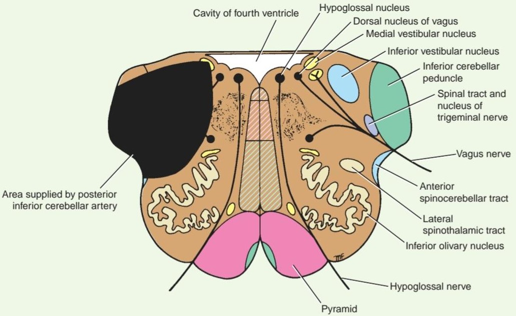
Figure 5-31 Transverse section of the medulla oblongata at the level of the inferior olivary nuclei showing the extent of the lesion producing the lateral medullary syndrome.
شکل ۵-۳۱ برش عرضی بصل النخاع در سطح هستههای زیتونی تحتانی که میزان ضایعه ایجاد کننده سندرم بصل النخاع جانبی را نشان میدهد.
MEDIAL MEDULLARY SYNDROME
The medial part of the medulla oblongata is supplied by the vertebral artery. Thrombosis of the medullary branch (Fig. 5-32) produces the following signs and symptoms: contralateral hemiparesis (pyramidal tract), contralateral impaired sensations of position and movement and tactile discrimination (medial lemniscus), and ipsilateral paralysis of tongue muscles with deviation to the paralyzed side when the tongue is protruded (hypoglossal nerve).
سندرم مدولای داخلی
بخش داخلی بصل النخاع توسط شریان مهرهای خونرسانی میشود. ترومبوز شاخه مدولا (شکل 5-32) علائم و نشانههای زیر را ایجاد میکند: همی پارزی طرف مقابل (راه هرمی)، اختلال در حس موقعیت و حرکت و تمایز لمسی طرف مقابل (لنیسک داخلی) و فلج عضلات زبان در همان طرف با انحراف به سمت فلج شده هنگام بیرون زدن زبان (عصب زیرزبانی).

Figure 5-32 Transverse section of the medulla oblongata at the level of the inferior olivary nuclei showing the extent of the lesion producing the medial medullary syndrome.
شکل ۵-۳۲ برش عرضی بصل النخاع در سطح هستههای زیتونی تحتانی که میزان ضایعه ایجاد کننده سندرم بصل النخاع داخلی را نشان میدهد.
Clinical Significance of the Pons
The pons, like the medulla oblongata and the cerebellum, is situated in the posterior cranial fossa lying beneath the tentorium cerebelli. It is related anteriorly to the basilar artery, the dorsum sellae of the sphenoid bone, and the basilar part of the occipital bone. In addition to forming the upper half of the floor of the fourth ventricle, it possesses several important CN nuclei (trigeminal, abducens, facial, and vestibulocochlear) and serves as a conduit for important ascending and descending tracts (corticonuclear, corticopontine, corticospinal, medial longitudinal fasciculus and medial, spinal, and lateral lemnisci). Not surprisingly, therefore, tumors, hemorrhage, or infarcts in this area of the brain produce a variety of symptoms and signs. For example, involvement of the corticopontocerebellar tracts will produce marked cerebellar ataxia, and voluntary movements are accompanied by a rhythmic tremor that develops and becomes further accentuated as the movements proceed (intention tumor).
اهمیت بالینی پل مغزی
پل مغزی، مانند بصل النخاع و مخچه، در حفره جمجمهای خلفی قرار دارد که در زیر چادرینه مخچه قرار دارد. این پل مغزی از جلو با شریان بازیلار، عضله پشتی استخوان اسفنوئید و قسمت بازیلار استخوان پسسری مرتبط است. علاوه بر تشکیل نیمه بالایی کف بطن چهارم، دارای چندین هسته مهم CN (هسته سه قلو، ابدوسنس، فاسیال و دهلیزی-حلزونی) است و به عنوان مجرایی برای مسیرهای صعودی و نزولی مهم (کورتیکونوکلئار، کورتیکوپونتین، کورتیکواسپاینال، فاسیکولوس طولی داخلی و لمنیسهای داخلی، نخاعی و جانبی) عمل میکند. بنابراین، جای تعجب نیست که تومورها، خونریزی یا انفارکتوس در این ناحیه از مغز علائم و نشانههای متنوعی ایجاد میکنند. برای مثال، درگیری راههای قشری-پونتوسربلار باعث آتاکسی مخچهای قابل توجه میشود و حرکات ارادی با لرزش ریتمیک همراه هستند که با ادامه حرکات ایجاد شده و تشدید میشود (تومور عمدی).
Tumors
Astrocytoma of the pons occurring in childhood is the most common tumor of the brainstem. The symptoms and signs are those of ipsilateral CN paralysis and contralateral hemiparesis: weakness of the facial muscles on the same side (facial nerve nucleus), weakness of the lateral rectus muscle on one or both sides (abducens nerve nucleus), nystagmus (vestibular nucleus), weakness of the jaw muscles (trigeminal nerve nucleus), impairment of hearing (cochlear nuclei), contralateral hemiparesis, quadriparesis (corticospinal fibers), anesthesia to light touch with the preservation of appreciation of pain over the skin of the face (principal sensory nucleus of trigeminal nerve involved, leaving spinal nucleus and tract of trigeminal intact), and contralateral sensory defects of the trunk and limbs (medial and spinal lemnisci). Involvement of the corticopontocerebellar tracts may cause ipsilateral cerebellar signs and symptoms. There may be impairment of conjugate deviation of the eyeballs due to involvement of the medial longitudinal fasciculus, which connects the oculomotor, trochlear, and abducens nerve nuclei.
تومورها
آستروسیتومای پل مغزی که در دوران کودکی رخ میدهد، شایعترین تومور ساقه مغز است. علائم و نشانههای آن شامل فلج CN همان طرف و همی پارزی طرف مقابل است: ضعف عضلات صورت در همان طرف (هسته عصب صورتی)، ضعف عضله رکتوس جانبی در یک یا هر دو طرف (هسته عصب ابدوسنس)، نیستاگموس (هسته دهلیزی)، ضعف عضلات فک (هسته عصب سه قلو)، اختلال شنوایی (هسته حلزونی)، همی پارزی طرف مقابل، کوادری پارزی (فیبرهای قشری-نخاعی)، بیحسی در برابر لمس سبک با حفظ درک درد روی پوست صورت (هسته حسی اصلی عصب سه قلو درگیر میشود، هسته نخاعی و مسیر عصب سه قلو دست نخورده باقی میماند) و نقصهای حسی طرف مقابل تنه و اندامها (لنیسکهای داخلی و نخاعی). درگیری مسیرهای قشری-نخاعی مخچهای ممکن است باعث علائم و نشانههای مخچهای طرف مقابل شود. ممکن است اختلال در انحراف مزدوج کره چشم به دلیل درگیری فاسیکول طولی داخلی، که هستههای عصب حرکتی چشم، قرقرهای و ابدوسنس را به هم متصل میکند، وجود داشته باشد.
Hemorrhage
The pons is supplied by the basilar artery and the anterior, inferior, and superior cerebellar arteries. If the hemorrhage occurs from one of those arteries and is unilateral, facial paralysis on the side of the lesion (involvement of the facial nerve nucleus and, therefore, a lower motor neuron palsy) and paralysis of the limbs on the opposite side (involvement of the corticospinal fibers as they pass through the pons) will result. Paralysis of conjugate ocular deviation (involvement of the abducens nerve nucleus and the medial longitudinal fasciculus) is common.
خونریزی
پل مغزی توسط شریان بازیلار و شریانهای مخچهای قدامی، تحتانی و فوقانی خونرسانی میشود. اگر خونریزی از یکی از این شریانها رخ دهد و یکطرفه باشد، فلج صورت در سمت ضایعه (درگیری هسته عصب صورتی و بنابراین فلج نورون حرکتی تحتانی) و فلج اندامها در سمت مقابل (درگیری فیبرهای قشری نخاعی هنگام عبور از پل مغزی) ایجاد میشود. فلج انحراف چشم (درگیری هسته عصب ابدوسنس و فاسیکول طولی داخلی) شایع است.
When the hemorrhage is extensive and bilateral, the pupils may be “pinpoint” (involvement of the ocular sympathetic fibers); bilateral paralysis of the face and the limbs is common. The patient may become poikilothermic because severe damage to the pons has cut off the body from the heat-regulating centers in the hypothalamus.
هنگامی که خونریزی گسترده و دو طرفه باشد، مردمکها ممکن است “نوک سوزنی” باشند (درگیری فیبرهای سمپاتیک چشمی)؛ فلج دو طرفه صورت و اندامها شایع است. بیمار ممکن است دچار پویکیلوترمی شود زیرا آسیب شدید به پل مغزی، ارتباط بدن را با مراکز تنظیم گرما در هیپوتالاموس قطع کرده است.
Infarctions
Usually, infarction of the pons is due to thrombosis or embolism of the basilar artery or its branches. If it involves the paramedian area of the pons, the corticospinal tracts, the pontine nuclei, and the fibers passing to the cerebellum through the middle cerebellar peduncle may be damaged. A laterally situated infarct will involve the trigeminal nerve, the medial lemniscus, and the middle cerebellar peduncle; the corticospinal fibers to the lower limbs also may be affected.
انفارکتوسها
معمولاً انفارکتوس پل مغزی به دلیل ترومبوز یا آمبولی شریان بازیلار یا شاخههای آن رخ میدهد. اگر ناحیه پارامدین پل مغزی را درگیر کند، ممکن است مسیرهای قشری-نخاعی، هستههای پل مغزی و فیبرهایی که از طریق پایه مخچه میانی به مخچه میروند، آسیب ببینند. انفارکتوس جانبی، عصب سه قلو، لمنیسکوس داخلی و پایه مخچه میانی را درگیر میکند؛ فیبرهای قشری-نخاعی به اندامهای تحتانی نیز ممکن است تحت تأثیر قرار گیرند.
The clinical conditions mentioned above will be understood more clearly if the ascending and descend- ing tracts of the brain and spinal cord are reviewed (see Chapter 4).
شرایط بالینی ذکر شده در بالا با بررسی مسیرهای صعودی و نزولی مغز و نخاع واضحتر درک خواهند شد (به فصل ۴ مراجعه کنید).
Clinical Significance of the Midbrain
The midbrain forms the upper end of the narrow stalk of the brain or brainstem. As it ascends out of the posterior cranial fossa through the relatively small rigid opening in the tentorium cerebelli, it is vulnerable to traumatic injury. It possesses two important CN nuclei (oculomotor and trochlear), reflex centers (the colliculi), and the red nucleus and substantia nigra, which greatly influence motor function, and the midbrain serves as a conduit for many important ascending and descending tracts. As in other parts of the brainstem, it is a site for tumors, hemorrhage, or infarcts that will produce a wide variety of symptoms and signs.
اهمیت بالینی مغز میانی
مغز میانی انتهای فوقانی ساقه باریک مغز یا ساقه مغز را تشکیل میدهد. از آنجا که از حفره جمجمهای خلفی از طریق دهانه نسبتاً کوچک و سفت در چادرینه مخچه بالا میرود، در برابر آسیبهای تروماتیک آسیبپذیر است. این بخش دارای دو هسته مهم CN (حرکتی چشمی و تروکلئار)، مراکز رفلکس (کولیکولها) و هسته قرمز و جسم سیاه است که تأثیر زیادی بر عملکرد حرکتی دارند و مغز میانی به عنوان مجرایی برای بسیاری از مسیرهای صعودی و نزولی مهم عمل میکند. مانند سایر قسمتهای ساقه مغز، این بخش نیز محلی برای تومورها، خونریزی یا انفارکتوسهایی است که علائم و نشانههای متنوعی را ایجاد میکنند.
Trauma
Among the mechanisms of injuries to the midbrain, a sudden lateral movement of the head could result in the cerebral peduncles impinging against the sharp rigid free edge of the tentorium cerebelli. Sudden movements of the head resulting from trauma cause different regions of the brain to move at different velocities relative to one another. For example, the large anatomical unit, the forebrain, may move at a different velocity from the remainder of the brain, such as the cerebellum. This will result in the midbrain being bent, stretched, twisted, or torn.
تروما
در میان مکانیسمهای آسیب به مغز میانی، حرکت ناگهانی جانبی سر میتواند منجر به برخورد ساقههای مغزی به لبه تیز و سفت و آزاد چادرینه مخچه شود. حرکات ناگهانی سر ناشی از تروما باعث میشود مناطق مختلف مغز با سرعتهای متفاوتی نسبت به یکدیگر حرکت کنند. به عنوان مثال، واحد آناتومیک بزرگ، مغز پیشین، ممکن است با سرعتی متفاوت از بقیه مغز، مانند مخچه، حرکت کند. این امر منجر به خم شدن، کشیده شدن، پیچ خوردن یا پارگی مغز میانی میشود.
Involvement of the oculomotor nucleus will produce ipsilateral paralysis of the levator palpebrae superioris; the superior, inferior, and medial rectus muscles; and the inferior oblique muscle. Malfunction of the parasympathetic nucleus of the oculomotor nerve produces a dilated pupil that is insensitive to light and does not constrict on accommodation.
درگیری هستهی چشمی-حرکتی باعث فلج همان طرف عضلهی بالابرندهی پلک فوقانی؛ عضلات راست فوقانی، تحتانی و داخلی؛ و عضلهی مایل تحتانی میشود. اختلال در عملکرد هستهی پاراسمپاتیک عصب چشمی-حرکتی باعث گشاد شدن مردمک چشم میشود که به نور حساس نیست و در هنگام تطابق منقبض نمیشود.
Involvement of the trochlear nucleus will produce contralateral paralysis of the superior oblique muscle of the eyeball. Thus, involvement of one or both of these nuclei, or the corticonuclear fibers that converge on them, will cause impairment of ocular movements.
درگیری هستهی تروکلئار باعث فلج طرف مقابل عضلهی مایل فوقانی کرهی چشم میشود. بنابراین، درگیری یک یا هر دوی این هستهها یا فیبرهای قشری-هستهای که روی آنها همگرا میشوند، باعث اختلال در حرکات چشمی خواهد شد.
Cerebral Aqueduct Blockage
The cavity of the midbrain, the cerebral aqueduct, is one of the narrower parts of the ventricular system. Normally, CSF that has been produced in the lateral and third ventricles passes through this channel to enter the fourth ventricle and so escapes through the foramina in its roof to enter the subarachnoid space. In congenital hydrocephalus, the cerebral aqueduct may be blocked or replaced by numerous small tubular passages that are insufficient for normal CSF flow. A tumor of the midbrain (Fig. 5-33A) or pressure on the midbrain from a tumor arising outside the midbrain may compress the aqueduct and produce hydrocephalus. When the cerebral aqueduct is blocked, the accumulating CSF within the third and lateral ventricles produces lesions in the midbrain. The presence of the oculomotor and trochlear nerve nuclei, together with the important descending corticospinal and corticonuclear tracts, will provide symptoms and signs that are helpful in accurately localizing a lesion in the brainstem.
انسداد مجرای مغزی
حفره مغز میانی، مجرای مغزی، یکی از باریکترین بخشهای سیستم بطنی است. به طور معمول، مایع مغزی نخاعی که در بطنهای جانبی و سوم تولید شده است، از این کانال عبور میکند تا وارد بطن چهارم شود و بنابراین از طریق سوراخهای سقف آن خارج شده و وارد فضای زیر عنکبوتیه میشود. در هیدروسفالی مادرزادی، مجرای مغزی ممکن است مسدود شود یا توسط مجاری لولهای کوچک متعددی جایگزین شود که برای جریان طبیعی CSF کافی نیستند. تومور مغز میانی (شکل 5-33A) یا فشار بر مغز میانی از توموری که از خارج از مغز میانی منشأ میگیرد، ممکن است مجرای مغزی را فشرده کرده و باعث هیدروسفالی شود. هنگامی که مجرای مغزی مسدود میشود، CSF انباشته شده در بطنهای سوم و جانبی باعث ایجاد ضایعاتی در مغز میانی میشود. وجود هستههای عصب اوکولوموتور و تروکلئار، همراه با مسیرهای مهم نزولی قشری-نخاعی و قشری-هستهای، علائم و نشانههایی را ایجاد میکند که در تعیین دقیق محل ضایعه در ساقه مغز مفید هستند.
Vascular Lesions
The midbrain houses CN III nuclei and serves as a conduit for all ascending and descending fibers between the cerebrum and brainstem; lesions of each will result in the following syndromes.
ضایعات عروقی
مغز میانی محل قرارگیری هستههای CN III است و به عنوان مجرایی برای تمام فیبرهای صعودی و نزولی بین مخ و ساقه مغز عمل میکند؛ ضایعات هر یک منجر به سندرمهای زیر میشود.
WEBER SYNDROME
Weber syndrome (see Fig. 5-33B), which is commonly produced by occlusion of a branch of the posterior cerebral artery that supplies the midbrain, results in the necrosis of brain tissue involving the oculomotor nerve and the crus cerebri. Ipsilateral ophthalmoplegia and contralateral paralysis of the lower part of the face, the tongue, and the arm and leg also result. The eyeball is deviated laterally because of the paralysis of the medial rectus muscle; the upper lid droops (ptosis), and the pupil is dilated and fixed to light and accommodation.
سندرم وبر
سندرم وبر (شکل 5-33B را ببینید)، که معمولاً در اثر انسداد شاخهای از شریان مغزی خلفی که مغز میانی را تغذیه میکند، ایجاد میشود، منجر به نکروز بافت مغز میشود که عصب اوکولوموتور و ساق پا را درگیر میکند. فلج چشمی همان طرف و فلج طرف مقابل قسمت تحتانی صورت، زبان و بازو و پا نیز ایجاد میشود. کره چشم به دلیل فلج عضله رکتوس داخلی به طرفین منحرف میشود؛ پلک فوقانی افتادگی (پتوز) پیدا میکند و مردمک گشاد شده و به نور و تطابق ثابت میشود.
BENEDIKT SYNDROME
Benedikt syndrome (see Fig. 5-33C) is similar to Weber syndrome, but the necrosis involves the medial lemniscus and red nucleus, producing contralateral hemianesthesia and involuntary movements of the limbs of the opposite side.
سندرم بندیکت
سندرم بندیکت (شکل 5-33C را ببینید) مشابه سندرم وبر است، اما نکروز، لمنیسکوس داخلی و هسته قرمز را درگیر میکند و باعث بیحسی موضعی در طرف مقابل و حرکات غیرارادی اندامهای طرف مقابل میشود.
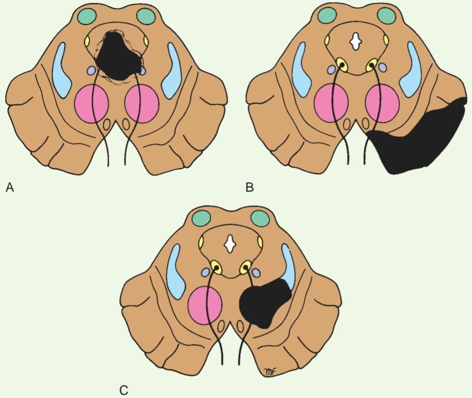
Figure 5-33 Pathology of the midbrain. A: Tumor of the midbrain blocking the cerebral aqueduct. B: Weber syndrome involving the oculomotor nerve and the crus cerebri following occlusion of the blood supply to the midbrain. C: Benedikt syndrome involving the red nucleus and the medial lemniscus following occlusion of the blood supply to the midbrain.
شکل ۵-۳۳ آسیب شناسی مغز میانی. الف: تومور مغز میانی که مجرای مغزی را مسدود میکند. ب: سندرم وبر که عصب اکولوموتور و ساق مغزی را پس از انسداد خونرسانی به مغز میانی درگیر میکند. ج: سندرم بندیکت که هسته قرمز و لمنیسکوس داخلی را پس از انسداد خونرسانی به مغز میانی درگیر میکند.
Key Concepts
Medulla Oblongata
• The medulla oblongata connects the pons superiorly and spinal cord inferiorly. On either side of the anterior median sulcus are the two pyramids that taper inferiorly, whereas most fibers cross at the decussation of the pyramids.
مفاهیم کلیدی
بصل النخاع
• بصل النخاع، پل مغزی را در بالا و نخاع را در پایین به هم متصل میکند. در دو طرف شیار میانی قدامی، دو هرم وجود دارد که به سمت پایین باریک میشوند، در حالی که بیشتر فیبرها در محل تقاطع هرمها از هم عبور میکنند.
• Posterolateral to the pyramids are the olives, which are elevations produced by the underlying inferior olivary nuclei.
• در قسمت خلفی-جانبی هرمها، زیتونها قرار دارند که برآمدگیهایی هستند که توسط هستههای زیتونی تحتانی زیرین ایجاد میشوند.
• The medulla oblongata contains multiple CN and cerebellar nuclei, the olivary nuclear complex, nucleus ambiguous, hypoglossal nucleus, vestibulocochlear nucleus, dorsal nucleus of the vagus, and nucleus of the tractus solitarius, and spinal nucleus of the trigeminal nerve.
• بصل النخاع شامل هستههای متعدد CN و مخچه، کمپلکس هستهای زیتونی، هسته مبهم، هسته زیرزبانی، هسته دهلیزی-حلزونی، هسته پشتی عصب واگ، و هسته مسیر منزوی، و هسته نخاعی عصب سه قلو است.
Pons
• The pons is anterior to the cerebellum and connects the medulla oblongata to the midbrain. The anterior surface is convex, and the trigeminal nerve emerges anterolaterally.
پل مغزی
• پل مغزی در جلوی مخچه قرار دارد و بصل النخاع را به مغز میانی متصل میکند. سطح قدامی آن محدب است و عصب سه قلو به صورت قدامی-جانبی از آن خارج میشود.
• The anterior or basal part of the pons consists of transversely running fibers, called the trapezoid body, and descending bundles of the corticospinal tract.
• بخش قدامی یا پایه پل مغزی شامل فیبرهایی است که به صورت عرضی امتداد دارند و جسم ذوزنقهای نامیده میشوند و دستههای نزولی راه قشری-نخاعی را تشکیل میدهند.
• The posterior part, or tegmentum, contains multiple nuclei, including facial, abducens, vestibular, pontine, trapezoid, and trigeminal (main sensory, spinal, and motor).
• قسمت خلفی یا تگمنتوم، شامل هسته های متعدد از جمله صورت، ابدوسنس، وستیبولار، پونتین، تراپزوئید و تری ژمینال (هسته های اصلی حسی، نخاعی و حرکتی) است.
Midbrain
• The midbrain connects the pons and the cerebellum with the forebrain.
مغز میانی
• مغز میانی، پل مغزی و مخچه را به مغز پیشین متصل میکند.
• The lateral halves, called the cerebral peduncles, are comprised of the crus cerebri, which contain corticospinal fibers, and the substantia nigra, a pigmented band of gray matter.
• نیمههای جانبی، که پایههای مغزی نامیده میشوند، از ساقه مغزی که حاوی الیاف قشری-نخاعی است و جسم سیاه، یک نوار رنگدانهدار از ماده خاکستری، تشکیل شدهاند.
• The cerebral aqueduct, which connects the third and fourth ventricles, passes through the midbrain and divides the anterior (tegmentum) from the posterior (tectum).
• مجرای مغزی که بطنهای سوم و چهارم را به هم متصل میکند، از مغز میانی عبور میکند و قسمت قدامی (تگمنتوم) را از قسمت خلفی (تکتوم) جدا میکند.
• The tectum consists of four swellings, two superior and two inferior colliculi, that are associated with vision and hearing, respectively.
• تکتوم از چهار برآمدگی، دو کولیکولی فوقانی و دو کولیکولی تحتانی، تشکیل شده است که به ترتیب با بینایی و شنوایی مرتبط هستند.
• The midbrain contains multiple nuclei including, inferior and superior colliculi, substantia nigra, trochlear, mesencephalic (trigeminal), oculomotor, Edinger-Westphal, and the red nucleus.
• مغز میانی شامل چندین هسته از جمله کولیکولی تحتانی و فوقانی، جسم سیاه، تروکلئار، مزانسفال (سه قلو)، اکولوموتور، ادینگر-وستفال و هسته قرمز است.
«فصل پنجم کتاب نورواناتومی بالینی اسنل»
🚀 با ما همراه شوید!
تازهترین مطالب و آموزشهای مغز و اعصاب را از دست ندهید. با فالو کردن کانال تلگرام آیندهنگاران مغز، از ما حمایت کنید!



