نوروآناتومی بالینی اسنل | ساقه مغز؛ پل مغزی – مرور مبانی و اصول


دعای مطالعه [ نمایش ]
بِسْمِ الله الرَّحْمنِ الرَّحیمِ
اَللّهُمَّ اَخْرِجْنى مِنْ ظُلُماتِ الْوَهْمِ
خدايا مرا بيرون آور از تاريكىهاى وهم،
وَ اَكْرِمْنى بِنُورِ الْفَهْمِ
و به نور فهم گرامى ام بدار،
اَللّهُمَّ افْتَحْ عَلَيْنا اَبْوابَ رَحْمَتِكَ
خدايا درهاى رحمتت را به روى ما بگشا،
وَانْشُرْ عَلَيْنا خَزائِنَ عُلُومِكَ بِرَحْمَتِكَ يا اَرْحَمَ الرّاحِمينَ
و خزانههاى علومت را بر ما باز كن به امید رحمتت اى مهربانترين مهربانان.
آیندهنگاران مغز: «ما مغز را میشناسیم، تا آینده را بسازیم.»
کتاب «نوروآناتومی بالینی اسنل» اثر دکتر ریچارد اس. اسنل، با ویرایش جدید دکتر رایان اسپلیتگربر، همچنان بهعنوان مرجع طلایی این حوزه شناخته میشود. این اثر با تلفیق تازهترین پژوهشها و کاربردهای بالینی، پلی استوار میان دانش پایه و عمل پزشکی بنا میکند.
تلاشهای علمی و سالها تدریس ارزشمند استاد گرانقدر دکتر محمدتقی جغتایی الهامبخش ترجمه و گسترش این دانش در ایران بوده است.
ترجمه دقیق این کتاب توسط برند علمی آیندهنگاران مغز به مدیریت داریوش طاهری، امکان دسترسی فارسیزبانان به مرزهای نوین علوم اعصاب را فراهم کرده و رسالتی علمی در جهت شناخت عمیقتر مغز و ترسیم آیندهای روشنتر بر عهده گرفته است.
»» فصل ۵: ساقه مغز
»» Chapter 5: Brainstem
PONS
The pons is anterior to the cerebellum (Fig. 5-17; see also Fig. 6-1) and connects the medulla oblongata to the midbrain. It is about 1 inch (2.5 cm) long and owes its name to the appearance presented on the anterior surface, which is that of a bridge connecting the right and left cerebellar hemispheres.
پل مغزی
پل مغزی در جلوی مخچه قرار دارد (شکل 5-17؛ همچنین به شکل 6-1 مراجعه کنید) و بصل النخاع را به مغز میانی متصل میکند. طول آن حدود 1 اینچ (2.5 سانتیمتر) است و نام خود را از ظاهری که در سطح قدامی آن وجود دارد، گرفته است که مانند پلی است که نیمکرههای مخچه راست و چپ را به هم متصل میکند.
The anterior surface is convex from side to side and shows many transverse fibers that converge on each side to form the middle cerebellar peduncle. A shallow groove in the midline, the basilar groove, lodges the basilar artery. On the anterolateral surface of the pons, the trigeminal nerve emerges on each side. Each nerve consists of a smaller, medial part, known as the motor root, and a larger, lateral part, known as the sensory root. In the groove between the pons and the medulla oblongata, from medial to lateral, the abducens, facial, and vestibulocochlear nerves emerge.
سطح قدامی از یک طرف به طرف دیگر محدب است و فیبرهای عرضی زیادی را نشان میدهد که در هر طرف به هم میرسند و پایه مخچه میانی را تشکیل میدهند. یک شیار کم عمق در خط وسط، شیار بازیلار، شریان بازیلار را در خود جای میدهد. در سطح قدامی-جانبی پل مغزی، عصب سه قلو در هر طرف خارج میشود. هر عصب از یک قسمت کوچکتر میانی، معروف به ریشه حرکتی، و یک قسمت بزرگتر جانبی، معروف به ریشه حسی، تشکیل شده است. در شیار بین پل مغزی و بصل النخاع، از داخلی به خارجی، اعصاب ابدوسنس، فاسیال و وستیبولوکوکلئار خارج میشوند.
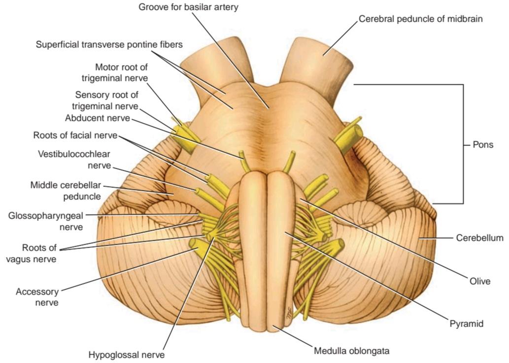
Figure 5-17 Anterior surface of the brainstem showing the pons.
شکل ۵-۱۷ سطح قدامی ساقه مغز که پل مغزی را نشان میدهد.

Figure 5-18 Posterior surface of the brainstem showing the pons. The cerebellum has been removed.
شکل ۵-۱۸ سطح خلفی ساقه مغز که پل مغزی را نشان میدهد. مخچه برداشته شده است.
The posterior surface of the pons is hidden from view by the cerebellum (Fig. 5-18). It forms the upper half of the floor of the fourth ventricle and is triangular in shape. The posterior surface is limited laterally by the superior cerebellar peduncles and is divided into symmetrical halves by a median sulcus. Lateral to this sulcus is an elongated elevation, the medial eminence, which is bounded laterally by a sulcus, the sulcus limitans. The inferior end of the medial eminence is slightly expanded to form the facial colliculus, which is produced by the root of the facial nerve winding around the nucleus of the abducens nerve (Fig. 5-19). The floor of the superior part of the sulcus limitans is bluish-gray in color and is called the substantia ferruginea; it owes its color to a group of deeply pigmented nerve cells. Lateral to the sulcus limitans is the area vestibuli produced by the underlying vestibular nuclei (see Fig. 5-18).
سطح خلفی پل مغزی توسط مخچه از دید پنهان است (شکل 5-18). این سطح، نیمه بالایی کف بطن چهارم را تشکیل میدهد و شکلی مثلثی دارد. سطح خلفی از طرفین توسط پایههای مخچهای فوقانی محدود شده و توسط یک شیار میانی به دو نیمه متقارن تقسیم میشود. در طرفین این شیار، یک برآمدگی کشیده به نام برجستگی داخلی وجود دارد که از طرفین توسط یک شیار به نام شیار محدودکننده محدود میشود. انتهای تحتانی برجستگی داخلی کمی گسترش یافته و برآمدگی صورت را تشکیل میدهد که توسط پیچ خوردن ریشه عصب صورت به دور هسته عصب ابدوسنس تولید میشود (شکل 5-19). کف قسمت فوقانی شیار محدودکننده به رنگ خاکستری مایل به آبی است و جسم آهنین نامیده میشود. رنگ آن به دلیل گروهی از سلولهای عصبی با رنگدانههای عمیق است. در سمت جانبی شیار محدودکننده، ناحیه دهلیزی قرار دارد که توسط هستههای دهلیزی زیرین تولید میشود (شکل 5-18 را ببینید).

Figure 5-19 Transverse section through the caudal part of the pons at the level of the facial colliculus.
شکل ۵-۱۹ برش عرضی از قسمت خلفی پل مغزی در سطح کولیکولوس فاسیال.
Internal Structure
For purposes of description, the pons is commonly divided into a posterior part, the tegmentum, and an anterior basal part by the transversely running fibers of the trapezoid body (see Fig. 5-19).
ساختار داخلی
برای توصیف، پل مغزی معمولاً توسط الیاف عرضی جسم ذوزنقهای به یک بخش خلفی، تگمنتوم، و یک بخش قدامی-قاعدهای تقسیم میشود (شکل 5-19 را ببینید).
The structure of the pons may be studied at two levels: (1) transverse section through the caudal part, passing through the facial colliculus, and (2) transverse section through the cranial part, passing through the trigeminal nuclei. Table 5-3 compares the two levels of the pons and the major structures present at each level.
ساختار پل مغزی را میتوان در دو سطح مطالعه کرد: (1) برش عرضی از طریق بخش دمی، که از کولیکولوس فاسیال عبور میکند، و (2) برش عرضی از طریق بخش جمجمهای، که از هستههای سه قلو عبور میکند. جدول 5-3 دو سطح پل مغزی و ساختارهای اصلی موجود در هر سطح را با هم مقایسه میکند.
Transverse Section Through the Caudal Part
The medial lemniscus rotates as it passes from the medulla into the pons. It is situated in the most anterior part of the tegmentum, with its long axis running transversely. The medial lemniscus is accompanied by the spinal and lateral lemnisci.
برش عرضی از طریق بخش خلفی
لنیسک داخلی هنگام عبور از بصل النخاع به پل مغزی میچرخد. این لمنیس در قدامیترین قسمت تگمنتوم قرار دارد و محور طولی آن به صورت عرضی امتداد یافته است. لمنیس داخلی با لمنیسهای نخاعی و جانبی همراه است.
The facial nucleus lies posterior to the lateral part of the medial lemniscus. The fibers of the facial nerve wind around the nucleus of the abducens nerve, producing the facial colliculus. The fibers of the facial nerve then pass anteriorly between the facial nucleus and the superior end of the nucleus of the spinal tract of the trigeminal nerve.
هسته فاسیال در خلف قسمت جانبی لمنیسکوس داخلی قرار دارد. فیبرهای عصب فاسیال به دور هسته عصب ابدوسنس میپیچند و کولیکولوس فاسیال را تشکیل میدهند. سپس فیبرهای عصب فاسیال به صورت قدامی بین هسته فاسیال و انتهای فوقانی هسته مسیر نخاعی عصب سه قلو عبور میکنند.
The medial longitudinal fasciculus is situated beneath the floor of the fourth ventricle on either side of the midline. The medial longitudinal fasciculus is the main pathway that connects the vestibular and cochlear nuclei with the nuclei controlling the extraocular muscles (oculomotor, trochlear, and abducens nuclei).
فاسیکولوس طولی داخلی در زیر کف بطن چهارم در دو طرف خط وسط قرار دارد. فاسیکولوس طولی داخلی مسیر اصلی است که هستههای دهلیزی و حلزونی را به هستههای کنترل کننده عضلات خارج چشمی (هستههای چشمی-حرکتی، قرقرهای و ابدوسنس) متصل میکند.
The medial vestibular nucleus is situated lateral to the abducens nucleus and is in close relationship to the inferior cerebellar peduncle. The superior part of the lateral and the inferior part of the superior vestibular nucleus are found at this level. The posterior and anterior cochlear nuclei are also found at this level.
هسته وستیبولار داخلی در سمت خارجی هسته ابدوسنس قرار دارد و در ارتباط نزدیک با پایه مخچه تحتانی است. قسمت فوقانی هسته جانبی و قسمت تحتانی هسته وستیبولار فوقانی در این سطح یافت میشوند. هستههای حلزونی خلفی و قدامی نیز در این سطح یافت میشوند.
The spinal nucleus of the trigeminal nerve and its tract lie on the anteromedial aspect of the inferior cer- ebellar peduncle.
هسته نخاعی عصب سه قلو و مسیر آن در قسمت قدامی-داخلی پایه مخچه تحتانی قرار دارد.
The trapezoid body is made up of fibers derived from the cochlear nuclei and the nuclei of the trapezoid body. They run transversely in the anterior part of the tegmentum (see p. 210).
جسم ذوزنقهای از الیاف مشتق شده از هستههای حلزونی و هستههای جسم ذوزنقهای تشکیل شده است. آنها به صورت عرضی در قسمت قدامی تگمنتوم امتداد دارند (به صفحه 210 مراجعه کنید).
The basilar part of the pons, at this level, contains small masses of nerve cells called pontine nuclei. The corticopontine fibers of the crus cerebri of the midbrain terminate in the pontine nuclei. The axons of these cells give origin to the transverse fibers of the pons, which cross the midline and intersect the corticospinal and corticonuclear tracts, breaking them up into small bundles. The transverse fibers of the pons enter the middle cerebellar peduncle and are distributed to the cerebellar hemisphere. This connection forms the main pathway linking the cerebral cortex to the cerebellum.
بخش قاعدهای پل مغزی، در این سطح، حاوی تودههای کوچکی از سلولهای عصبی به نام هستههای پل مغزی است. فیبرهای قشری پل مغزی ساق مغز میانی در هستههای پل مغزی خاتمه مییابند. آکسونهای این سلولها منشأ فیبرهای عرضی پل مغزی هستند که از خط میانی عبور کرده و مسیرهای قشری-نخاعی و قشری-هستهای را قطع میکنند و آنها را به دستههای کوچک تقسیم میکنند. فیبرهای عرضی پل مغزی وارد پایه مخچهای میانی شده و در نیمکره مخچه توزیع میشوند. این اتصال، مسیر اصلی اتصال قشر مغز به مخچه را تشکیل میدهد.
Table 5-3 Levels of the Pons and Their Major Structures a
جدول ۵-۳ سطوح پلها و ساختارهای اصلی آنهاa
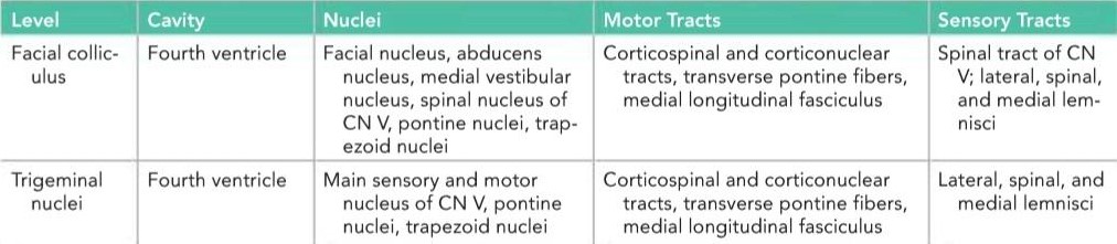
a Note that the reticular formation is present at all levels. CN, cranial nerve.
aتوجه داشته باشید که تشکیلات مشبک در تمام سطوح وجود دارد. CN، عصب جمجمهای.
Transverse Section Through the Cranial Part
The internal structure of the cranial part of the pons is similar to that seen at the caudal level (Figs. 5-20 to 5-22), but it now contains the motor and principal sensory nuclei of the trigeminal nerve.
برش عرضی از طریق بخش جمجمهای
ساختار داخلی بخش جمجمهای پل مغزی مشابه ساختار مشاهده شده در سطح خلفی است (شکلهای 5-20 تا 5-22)، اما اکنون حاوی هستههای حرکتی و حسی اصلی عصب سه قلو است.

Figure 5-20 Transverse section through the pons at the level of the trigeminal nuclei.
شکل ۵-۲۰ برش عرضی از طریق پل مغزی در سطح هستههای سه قلو.

Figure 5-21 Photomicrograph of a transverse section of the pons at the level of the trigeminal nuclei.
شکل ۵-۲۱ عکس میکروسکوپی از برش عرضی پل مغزی در سطح هستههای سه قلو.
The motor nucleus of the trigeminal nerve is situated beneath the lateral part of the fourth ventricle within the reticular formation (see Figs. 5-20 and 5-21). The emerging motor fibers travel anteriorly through the substance of the pons and exit on its anterior surface.
هسته حرکتی عصب سهقلو در زیر قسمت جانبی بطن چهارم در داخل تشکیلات مشبک قرار دارد (به شکلهای 5-20 و 5-21 مراجعه کنید). فیبرهای حرکتی بیرون آمده از طریق ماده پل مغزی به سمت جلو حرکت کرده و از سطح قدامی آن خارج میشوند.
The principal sensory nucleus of the trigeminal nerve is situated on the lateral side of the motor nucleus; it is continuous inferiorly with the nucleus of the spinal tract. The entering sensory fibers travel through the substance of the pons and lie lateral to the motor fibers (see Fig. 5-20).
هسته حسی اصلی عصب سهقلو در سمت جانبی هسته حرکتی قرار دارد؛ این هسته در امتداد تحتانی هسته دستگاه نخاعی قرار دارد. فیبرهای حسی ورودی از طریق ماده پل مغزی عبور کرده و در سمت خارجی فیبرهای حرکتی قرار دارند (به شکل 5-20 مراجعه کنید).
The superior cerebellar peduncle is situated posterolateral to the motor nucleus of the trigeminal nerve (see Figs. 5-20 and 5-21). It is joined by the anterior spinocerebellar tract.
پایه مخچهای فوقانی در سمت خلفی-خارجی هسته حرکتی عصب سهقلو قرار دارد (به شکلهای 5-20 و 5-21 مراجعه کنید). این پایه توسط دستگاه نخاعی-مخچهای قدامی به هم متصل میشود.
The trapezoid body and the medial lemniscus are situated in the same position as they were in the previous section (see Fig. 5-20). The lateral and spinal lemnisci lie at the lateral extremity of the medial lemniscus (see Figs. 5-20 and 5-22).
جسم ذوزنقهای و لمنیسکوس داخلی در همان موقعیتی قرار دارند که در بخش قبلی بودند (به شکل 5-20 مراجعه کنید). لمنیسکوس جانبی و نخاعی در انتهای جانبی لمنیسکوس داخلی قرار دارند (شکلهای 5-20 و 5-22 را ببینید).
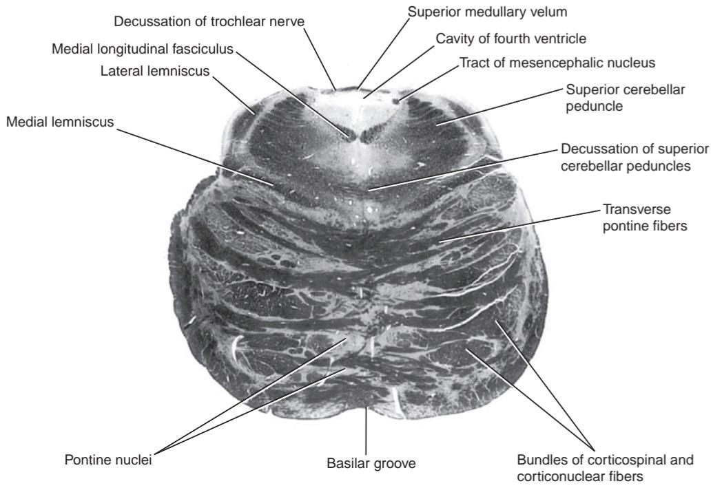
Figure 5-22 Photomicrograph of a transverse section of the most rostral part of the pons.
شکل ۵-۲۲ عکس میکروسکوپی از برش عرضی از قسمت قدامیترین قسمت پل مغزی.
«فصل پنجم کتاب نورواناتومی بالینی اسنل»
🚀 با ما همراه شوید!
تازهترین مطالب و آموزشهای مغز و اعصاب را از دست ندهید. با فالو کردن کانال تلگرام آیندهنگاران مغز، از ما حمایت کنید!