
نورونها و نوروگلیا؛ فصل دوم نوروآناتومی بالینی اسنل


دعای مطالعه [ نمایش ]
بِسْمِ الله الرَّحْمنِ الرَّحیمِ
اَللّهُمَّ اَخْرِجْنى مِنْ ظُلُماتِ الْوَهْمِ
خدايا مرا بيرون آور از تاريكىهاى وهم،
وَ اَكْرِمْنى بِنُورِ الْفَهْمِ
و به نور فهم گرامى ام بدار،
اَللّهُمَّ افْتَحْ عَلَيْنا اَبْوابَ رَحْمَتِكَ
خدايا درهاى رحمتت را به روى ما بگشا،
وَانْشُرْ عَلَيْنا خَزائِنَ عُلُومِكَ بِرَحْمَتِكَ يا اَرْحَمَ الرّاحِمينَ
و خزانههاى علومت را بر ما باز كن به امید رحمتت اى مهربانترين مهربانان.
آیندهنگاران مغز: «ما مغز را میشناسیم، تا آینده را بسازیم.»
کتاب «نوروآناتومی بالینی اسنل» اثر دکتر ریچارد اس. اسنل، با ویرایش جدید دکتر رایان اسپلیتگربر، همچنان بهعنوان مرجع طلایی این حوزه شناخته میشود. این اثر با تلفیق تازهترین پژوهشها و کاربردهای بالینی، پلی استوار میان دانش پایه و عمل پزشکی بنا میکند.
تلاشهای علمی و سالها تدریس ارزشمند استاد گرانقدر آقای دکتر محمدتقی جغتایی الهامبخش ترجمه و گسترش این دانش در ایران بوده است.
ترجمه دقیق این کتاب توسط برند علمی آیندهنگاران مغز به مدیریت داریوش طاهری، امکان دسترسی فارسیزبانان به مرزهای نوین علوم اعصاب را فراهم کرده و رسالتی علمی در جهت شناخت عمیقتر مغز و ترسیم آیندهای روشنتر بر عهده گرفته است.
»» فصل ۲: نورونها و نوروگلیا؛ مبانی و مرور
»» Chapter 2: Neurons and Neuroglia
CHAPTER OBJECTIVES
• To define the neuron and name its processes To learn the varieties of neurons and identify them in different parts of the nervous system
• To review the cell biology of a neuron and understand the function of a nerve cell and its processes
• To review the structure of the plasma membrane as it is related to its physiology
• To learn the transport of materials from the cell body to the axon terminals
• To understand the structure and function of synapses and neurotransmitters
• To review the supporting function of the neuroglial cells for nerve cells and the possible role that they play in neuronal metabolism, function, and neuronal death
اهداف فصل
• تعریف نورون و نامگذاری فرآیندهای آن
• یادگیری انواع نورونها و شناسایی آنها در بخشهای مختلف سیستم عصبی
• بررسی زیستشناسی سلولی یک نورون و درک عملکرد یک سلول عصبی و فرآیندهای آن
• بررسی ساختار غشای پلاسمایی و ارتباط آن با فیزیولوژی آن
• یادگیری انتقال مواد از جسم سلولی به پایانههای آکسون
• درک ساختار و عملکرد سیناپسها و انتقالدهندههای عصبی
• بررسی عملکرد پشتیبانی سلولهای نوروگلیا برای سلولهای عصبی و نقش احتمالی آنها در متابولیسم، عملکرد و مرگ نورونی
A 38-year-old man with a history of involuntary movements, personality changes, and mental deterioration is referred to a neurologist. The symptoms started insidiously 8 years ago and are getting progressively worse. The first symptoms are involuntary, abrupt, and purposeless movements of the upper limbs associated with clumsiness and dropping things. At presentation, the patient has difficulty walking, speaking, and swallowing. Associated with these movement defects are memory impairment and loss of intellectual capacity. Impulsive behavior and bouts of depression also occur. Close questioning of the patient and his wife reveals that the patient’s father and his older brother had similar symptoms before they died. A diagnosis of Huntington disease is made.
مردی ۳۸ ساله با سابقه حرکات غیرارادی، تغییرات شخصیتی و زوال عقل به متخصص مغز و اعصاب ارجاع داده میشود. علائم از ۸ سال پیش به طور نامحسوس شروع شده و به تدریج بدتر میشوند. اولین علائم، حرکات غیرارادی، ناگهانی و بیهدف اندام فوقانی است که با دست و پا چلفتی بودن و انداختن اشیا همراه است. در بدو مراجعه، بیمار در راه رفتن، صحبت کردن و بلعیدن مشکل دارد. اختلال حافظه و از دست دادن ظرفیت فکری نیز با این نقایص حرکتی همراه است. رفتار تکانشی و دورههای افسردگی نیز رخ میدهد. پرسش دقیق از بیمار و همسرش نشان میدهد که پدر و برادر بزرگتر بیمار قبل از مرگ علائم مشابهی داشتهاند. تشخیص بیماری هانتینگتون داده میشود.
Huntington disease is an autosomal-dominant disorder with the defect localized to the short arm of chromosome 4. Histologically, the caudate nucleus and the putamen show extensive degeneration, mainly involving the acetyl- choline and y-aminobutyric acid (GABA)-producing neurons; the dopamine neurons are unaffected. Secondary degeneration of the cerebral cortex also occurs. This case is an example of a hereditary disorder that mainly involves a particular group of neurons.
بیماری هانتینگتون یک اختلال اتوزومال غالب است که نقص آن در بازوی کوتاه کروموزوم ۴ قرار دارد. از نظر بافتشناسی، هسته دمدار و پوتامن دژنراسیون گستردهای را نشان میدهند که عمدتاً نورونهای تولیدکننده استیل کولین و گاما آمینوبوتیریک اسید (GABA) را درگیر میکند؛ نورونهای دوپامین تحت تأثیر قرار نمیگیرند. دژنراسیون ثانویه قشر مغز نیز رخ میدهد. این مورد نمونهای از یک اختلال ارثی است که عمدتاً گروه خاصی از نورونها را درگیر میکند.
The purpose of this chapter is to help students understand how the basic excitable cell-the neuron-communicates with other neurons. It also considers certain injuries to the neuron and the effects of drugs on the mechanism by which neurons communicate with one another.
هدف این فصل کمک به دانشآموزان در درک چگونگی ارتباط سلول تحریکپذیر پایه – نورون – با سایر نورونها است. همچنین آسیبهای خاص به نورون و اثرات داروها بر مکانیسم ارتباط نورونها با یکدیگر را در نظر میگیرد.

NEURONS
Neuron is the name given to the nerve cell and all its processes (Fig. 2-1). Neurons are excitable cells that are specialized for the reception of stimuli and the conduction of the nerve impulse. They vary considerably in size and shape, but each possesses a cell body from which one or more processes called neurites project (Fig. 2-2). Neurites responsible for receiving information and conducting it toward the cell body are dendrites. The single long tubular neurite that conducts impulses away from the cell body is the axon. Dendrites and axons are commonly referred to as nerve fibers.
نورونها
نورون نامی است که به سلول عصبی و تمام زوائد آن داده شده است (شکل 2-1). نورونها سلولهای تحریکپذیری هستند که برای دریافت محرکها و هدایت تکانه عصبی تخصص یافتهاند. آنها از نظر اندازه و شکل به طور قابل توجهی متفاوت هستند، اما هر کدام دارای یک جسم سلولی هستند که از آن یک یا چند زائده به نام نوریتها بیرون زده میشوند (شکل 2-2). نوریتهایی که مسئول دریافت اطلاعات و هدایت آن به سمت جسم سلولی هستند، دندریتها هستند. نوریت لولهای بلند و منفردی که تکانهها را از جسم سلولی دور میکند، آکسون نام دارد. دندریتها و آکسونها معمولاً به عنوان فیبرهای عصبی شناخته میشوند.
Neurons are found in the brain and spinal cord and in ganglia. Unlike most other cells in the body, normal neurons in the mature individual do not undergo division and replication.
نورونها در مغز و نخاع و در گانگلیونها یافت میشوند. برخلاف اکثر سلولهای دیگر بدن، نورونهای طبیعی در فرد بالغ تقسیم و تکثیر نمیشوند.

Figure 2-1 A neuron.
شکل ۲-۱ یک نورون.

Figure 2-2 Photomicrograph of a smear preparation of the spinal cord showing a neuron with its cell body and its processes or neurites.
شکل ۲-۲ عکس میکروسکوپی از نمونه تهیه شده از نخاع که یک نورون را به همراه جسم سلولی و زوائد یا نوریتهای آن نشان میدهد.
Neuron Types
Although the cell body of a neuron may be as small as 5 μm or as large as 135 μm in diameter, the processes or neurites may extend over a distance of more than 1 m. Neurons can be classified morphologically based on the number, length, and mode of branching of their neurites (Fig. 2-3).
انواع نورون
اگرچه قطر جسم سلولی یک نورون ممکن است به کوچکی ۵ میکرومتر یا به بزرگی ۱۳۵ میکرومتر باشد، زوائد یا نوریتها ممکن است تا مسافتی بیش از ۱ متر امتداد داشته باشند. نورونها را میتوان از نظر مورفولوژیکی بر اساس تعداد، طول و نحوه انشعاب نوریتهایشان طبقهبندی کرد (شکل ۲-۳).
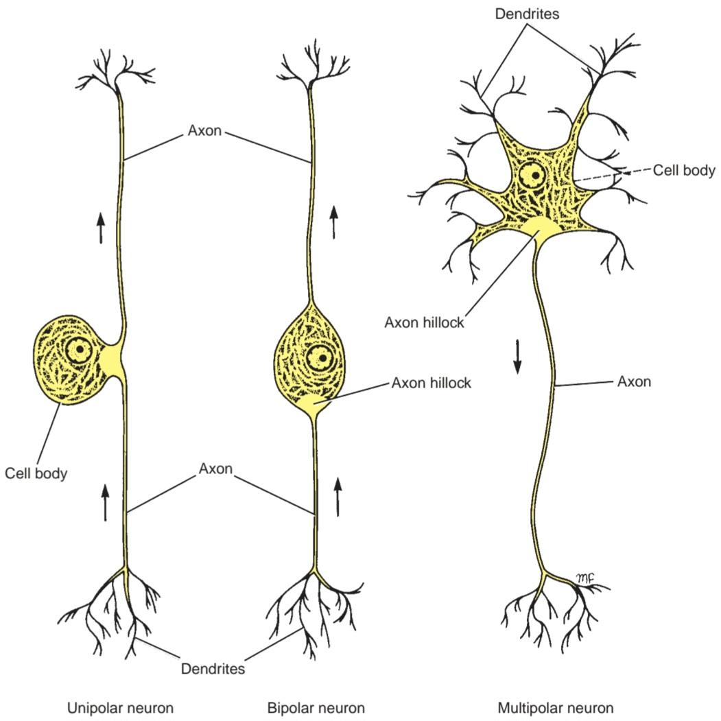
Figure 2-3 The classification of neurons according to the number, length, and mode of branching of the neurites.
شکل ۲-۳ طبقهبندی نورونها بر اساس تعداد، طول و نحوهی انشعاب نوریتها.
Unipolar neurons have a single neurite that divides a short distance from the cell body into two branches, one proceeding to some peripheral structure and the other entering the central nervous system (CNS). The branches of this single neurite have the structural and functional characteristics of an axon. In this type of neuron, the fine terminal branches found at the peripheral end of the axon at the receptor site are often referred to as the dendrites. Examples of this form of neuron are found in the posterior root ganglion.
نورونهای تکقطبی دارای یک نوریت واحد هستند که در فاصله کوتاهی از جسم سلولی به دو شاخه تقسیم میشود، یکی به سمت ساختار محیطی میرود و دیگری وارد سیستم عصبی مرکزی (CNS) میشود. شاخههای این نوریت واحد، ویژگیهای ساختاری و عملکردی یک آکسون را دارند. در این نوع نورون، شاخههای انتهایی ظریف که در انتهای محیطی آکسون در محل گیرنده یافت میشوند، اغلب به عنوان دندریت شناخته میشوند. نمونههایی از این شکل نورون در گانگلیون ریشه خلفی یافت میشوند.
Bipolar neurons have an elongated cell body, with a single neurite emerging from each end. Examples of this type of neuron are found in the retinal bipolar cells and the cells of the sensory cochlear and vestibular ganglia.
نورونهای دوقطبی دارای جسم سلولی کشیدهای هستند که از هر انتهای آن یک نوریت خارج میشود. نمونههایی از این نوع نورون در سلولهای دوقطبی شبکیه و سلولهای گانگلیونهای حسی حلزونی و دهلیزی یافت میشوند.
Multipolar neurons have a number of neurites arising from the cell body. With the exception of the long process, the axon, the remainder of the neurites are dendrites. Most neurons of the brain and spinal cord are of this type.
نورونهای چندقطبی تعدادی نوریت دارند که از جسم سلولی منشأ میگیرند. به استثنای زائده بلند، آکسون، بقیه نوریتها دندریت هستند. اکثر نورونهای مغز و نخاع از این نوع هستند.
Neurons may also be classified according to size. Golgi type I neurons have a long axon that can stretch 1 m or more in length in extreme cases (Figs. 2-4 to 2-6). The axons of these neurons form the long fiber tracts of the brain and spinal cord and the nerve fibers of peripheral nerves. The pyramidal cells of the cerebral cortex, the Purkinje cells of the cerebellar cortex, and the motor cells of the spinal cord are good examples.
نورونها را میتوان بر اساس اندازه نیز طبقهبندی کرد. نورونهای گلژی نوع I دارای یک آکسون بلند هستند که در موارد شدید میتواند تا ۱ متر یا بیشتر امتداد یابد (شکلهای ۲-۴ تا ۲-۶). آکسونهای این نورونها، رشتههای بلند مغز و نخاع و رشتههای عصبی اعصاب محیطی را تشکیل میدهند. سلولهای هرمی قشر مغز، سلولهای پورکینژ قشر مخچه و سلولهای حرکتی نخاع نمونههای خوبی هستند.
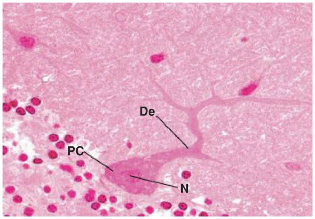
Figure 2-4 Photomicrograph of a silver-stained section of the cerebellar cortex showing two Purkinje cells. These are examples of Golgi type I neurons. De, dendrite; PC, Purkinje cell; N, Nucleus. (From Gartner, L. P.
(2018). Color atlas and text of histology (7th ed). Baltimore, MD: Wolters Kluwer.)
شکل ۲-۴ عکس میکروسکوپی از بخشی از قشر مخچه که با رنگ نقره رنگآمیزی شده و دو سلول پورکنژ را نشان میدهد. اینها نمونههایی از نورونهای گلژی نوع I هستند. De، دندریت؛ PC، سلول پورکنژ؛ N، هسته. (از گارتنر، ال. پی. (۲۰۱۸). اطلس رنگی و متن بافتشناسی (ویرایش هفتم). بالتیمور، مریلند: ولترز کلوور.)
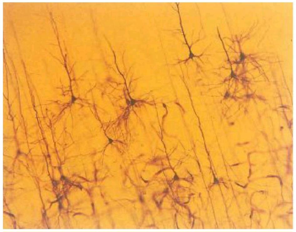
Figure 2-5 Photomicrograph of a silver-stained section of the cerebral cortex. Note the presence of large pyramidal cells, which are examples of Golgi type I neurons, and numerous Golgi type Il neurons. (From Bear, M. F., Connors, B. W., & Paradiso, M. A. (2016). Neuroscience: Exploring the brain (4th ed). Baltimore, MD: Wolters Kluwer.)
شکل ۲-۵ فتومیکروگراف از بخشی از قشر مغز که با رنگ نقره رنگآمیزی شده است. به وجود سلولهای هرمی بزرگ، که نمونههایی از نورونهای گلژی نوع I هستند، و نورونهای متعدد گلژی نوع II توجه کنید. (از Bear, M. F., Connors, B. W., & Paradiso, M. A. (2016). Neuroscience: Exploring the brain (4th ed. Baltimore, MD: Wolters Kluwer.)

Figure 2-6 Different types of neurons.
شکل ۲-۶ انواع مختلف نورونها.
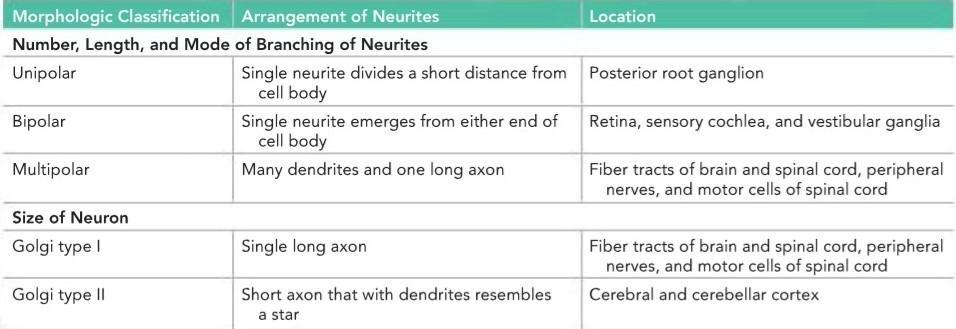
Golgi type II neurons have a short axon that terminates in the neighborhood of the cell body or is entirely absent (Figs. 2-5 and 2-6). They greatly outnumber the Golgi type I neurons. The short dendrites that arise from these neurons give them a star-shaped appearance. These neurons are numerous in the cerebral and cerebellar cortex and are often inhibitory in function. Table 2-1 summarizes neuronal classification.
نورونهای گلژی نوع II دارای یک آکسون کوتاه هستند که در همسایگی جسم سلولی خاتمه مییابد یا کاملاً وجود ندارد (شکلهای 2-5 و 2-6). تعداد آنها بسیار بیشتر از نورونهای گلژی نوع I است. دندریتهای کوتاهی که از این نورونها منشأ میگیرند، ظاهری ستارهای شکل به آنها میدهند. این نورونها در قشر مغز و مخچه به تعداد زیاد وجود دارند و اغلب عملکرد مهاری دارند. جدول 2-1 خلاصهای از طبقهبندی نورونها را ارائه میدهد.
Table 2-1 Neuron Classification
جدول ۲-۱ طبقهبندی نورون
Neuronal Structure
A neuron’s cell body, like that of other cells, consists essentially of a mass of cytoplasm in which a nucleus is embedded (Figs. 2-7 and 2-8), bounded externally by a plasma membrane.
ساختار نورونی
جسم سلولی یک نورون، مانند سایر سلولها، اساساً از تودهای از سیتوپلاسم تشکیل شده است که در آن یک هسته قرار دارد (شکلهای ۲-۷ و ۲-۸) که از خارج توسط غشای پلاسمایی محدود شده است.

Figure 2-7 Photomicrograph of a section of the anterior gray column of the spinal cord showing two large motor nerve cells with nuclei. Note the prominent nucleolus in one of the nuclei. GL, granular layer; ML, molecular layer.
شکل ۲-۷ عکس میکروسکوپی از بخشی از ستون خاکستری قدامی نخاع که دو سلول عصبی حرکتی بزرگ دارای هسته را نشان میدهد. به هستک برجسته در یکی از هستهها توجه کنید. GL، لایه دانهای؛ ML، لایه مولکولی.

Figure 2-8 Diagrammatic representation of the fine structure of a neuron.
شکل ۲-۸ نمایش نموداری ساختار ظریف یک نورون.
Nerve Cell Body
Interestingly, the volume of cytoplasm within the nerve cell body is often far less than the total volume of cytoplasm in the neurites. The cell bodies of the small granular cells of the cerebellar cortex measure about 5 μm in diameter, whereas those of the large anterior horn cells may measure as much as 135 um in diameter. The main structures of a nerve cell body are summarized in Table 2-2.
جسم سلولی عصبی
جالب توجه است که حجم سیتوپلاسم درون جسم سلولی عصبی اغلب بسیار کمتر از حجم کل سیتوپلاسم در نوریتها است. جسم سلولی سلولهای دانهای کوچک قشر مخچه حدود 5 میکرومتر قطر دارد، در حالی که قطر سلولهای شاخ قدامی بزرگ ممکن است به 135 میکرومتر برسد. ساختارهای اصلی جسم سلولی عصبی در جدول 2-2 خلاصه شده است.
Table 2-2 Main Structures in a Nerve Cell Body
جدول ۲-۲ ساختارهای اصلی در جسم سلولی عصبی

Nucleus
The nucleus, which stores the genes, is commonly centrally located within the cell body and is typically large and rounded. Do not confuse the term nucleus in cytology with the term nucleus in neuroanatomy, which refers to a discrete group of nerve cell bodies in the CNS. In mature neurons, the chromosomes no longer duplicate themselves and function only in gene expression. Therefore, the chromosomes are not arranged as compact structures but exist in an uncoiled state. Thus, the nucleus is pale, and the fine chromatin granules are widely dispersed (Figs. 2-6 and 2-7).
هسته
هسته، که ژنها را ذخیره میکند، معمولاً در مرکز جسم سلولی قرار دارد و معمولاً بزرگ و گرد است. اصطلاح هسته در سیتولوژی را با اصطلاح هسته در نوروآناتومی اشتباه نگیرید، که به گروهی مجزا از اجسام سلولی عصبی در سیستم عصبی مرکزی اشاره دارد. در نورونهای بالغ، کروموزومها دیگر خود را تکثیر نمیکنند و فقط در بیان ژن عمل میکنند. بنابراین، کروموزومها به صورت ساختارهای فشرده مرتب نشدهاند، بلکه در حالت بدون پیچ خوردگی وجود دارند. بنابراین، هسته رنگپریده است و گرانولهای ریز کروماتین به طور گسترده پراکنده شدهاند (شکلهای 2-6 و 2-7).
A single prominent nucleolus synthesizes ribosomal ribonucleic acid (rRNA) and assists with ribosome subunit assembly. The large size of the nucleolus probably is due to the high rate of protein synthesis, which is necessary to maintain the protein level in the large cytoplasmic volume that is present in the long neurites as well as in the cell body.
یک هستک برجسته، اسید ریبونوکلئیک ریبوزومی (rRNA) را سنتز میکند و به مونتاژ زیرواحدهای ریبوزوم کمک میکند. اندازه بزرگ هستک احتمالاً به دلیل سرعت بالای سنتز پروتئین است که برای حفظ سطح پروتئین در حجم بزرگ سیتوپلاسمی که در نوریتهای بلند و همچنین در جسم سلولی وجود دارد، ضروری است.
In the female, one of the two X chromosomes is compact and is known as the Barr body. It is composed of sex chromatin and sits at the inner surface of the nuclear envelope.
در ماده، یکی از دو کروموزوم X فشرده است و به عنوان جسم بار شناخته میشود. این جسم از کروماتین جنسی تشکیل شده و در سطح داخلی پوشش هستهای قرار دارد.
The nuclear envelope (Fig. 2-9; also see Fig. 2-8) is continuous with the cytoplasmic rough, or granular, endoplasmic reticulum (RER). The envelope is double layered and possesses fine nuclear pores, through which materials can diffuse into and out of the nucleus. Therefore, the substance in the nucleus and the cytoplasm can be considered as functionally continuous. Newly formed ribosomal subunits can be passed into the cytoplasm through the nuclear pores.
پوشش هستهای (شکل 2-9؛ همچنین به شکل 2-8 مراجعه کنید) در امتداد شبکه آندوپلاسمی خشن یا دانهای سیتوپلاسمی (RER) قرار دارد. این پوشش دو لایه است و دارای منافذ هستهای ظریفی است که از طریق آنها مواد میتوانند به داخل و خارج هسته منتشر شوند. بنابراین، ماده موجود در هسته و سیتوپلاسم را میتوان از نظر عملکردی پیوسته در نظر گرفت. زیر واحدهای ریبوزومی تازه تشکیل شده میتوانند از طریق منافذ هستهای به سیتوپلاسم منتقل شوند.
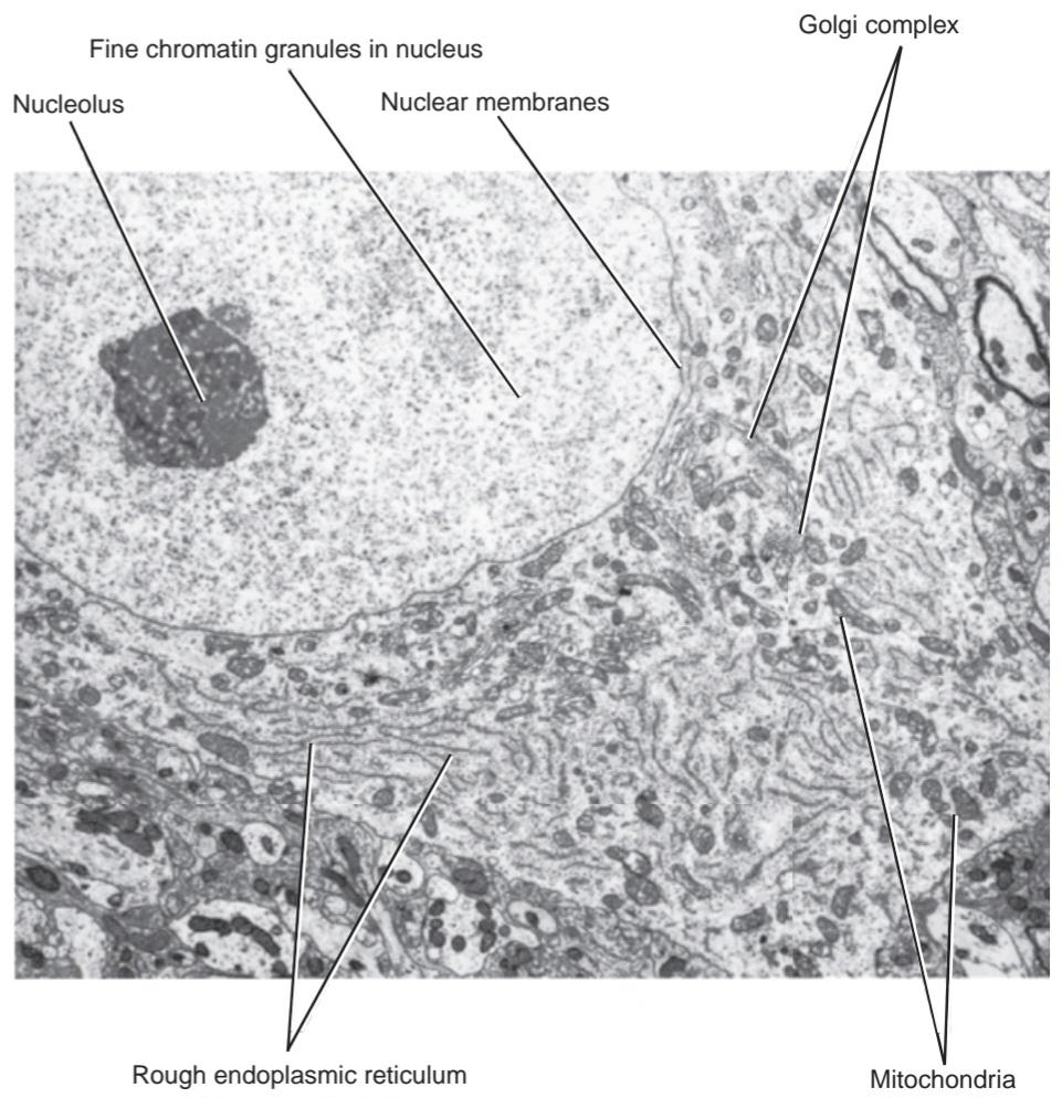
Figure 2-9 Electron micrograph of a neuron showing the structure of the nucleus and a number of cytoplasmic organelles. (Courtesy Dr. J. M. Kerns.)
شکل ۲-۹ تصویر میکروسکوپ الکترونی از یک نورون که ساختار هسته و تعدادی از اندامکهای سیتوپلاسمی را نشان میدهد. (با تشکر از دکتر جی. ام. کرنز.)
Cytoplasm
The cytoplasm is rich in rough (granular) and smooth (agranular) endoplasmic reticulum (Fig. 2-10; also see Fig. 2-9) and contains the following organelles and inclusions: (a) Nissl substance; (b) the Golgi complex; (c) mitochondria; (d) microfilaments; (e) microtubules; (f) lysosomes; (g) centrioles; and (h) lipofuscin, melanin, glycogen, and lipid.
سیتوپلاسم
سیتوپلاسم غنی از شبکه آندوپلاسمی خشن (دانهدار) و صاف (بدون دانه) است (شکل 2-10؛ همچنین به شکل 2-9 مراجعه کنید) و شامل اندامکها و اجزای زیر است: (الف) ماده نیسل؛ (ب) کمپلکس گلژی؛ (ج) میتوکندری؛ (د) ریزرشتهها؛ (ه) میکروتوبولها؛ (و) لیزوزومها؛ (ز) سانتریولها؛ و (ح) لیپوفوشین، ملانین، گلیکوژن و لیپید.

Figure 2-10 Electron micrograph of a neuron showing nuclear and plasma membranes and cytoplasmic organelles. (Courtesy Dr. J. M. Kerns.)
شکل ۲-۱۰ تصویر میکروسکوپ الکترونی از یک نورون که غشاهای هسته و پلاسما و اندامکهای سیتوپلاسمی را نشان میدهد. (با تشکر از دکتر جی. ام. کرنز.)
Nissl substance consists of granules that are distributed throughout the cytoplasm of the cell body, except for the region close to the axon, called the axon hillock (Fig. 2-11). The granular material also extends into the proximal parts of the dendrites but is not present in the axon.
ماده نیسل از گرانولهایی تشکیل شده است که در سراسر سیتوپلاسم جسم سلولی، به جز ناحیه نزدیک به آکسون، که تپه آکسون نامیده میشود، توزیع شدهاند (شکل 2-11). ماده گرانولی همچنین به قسمتهای پروگزیمال دندریتها گسترش مییابد اما در آکسون وجود ندارد.
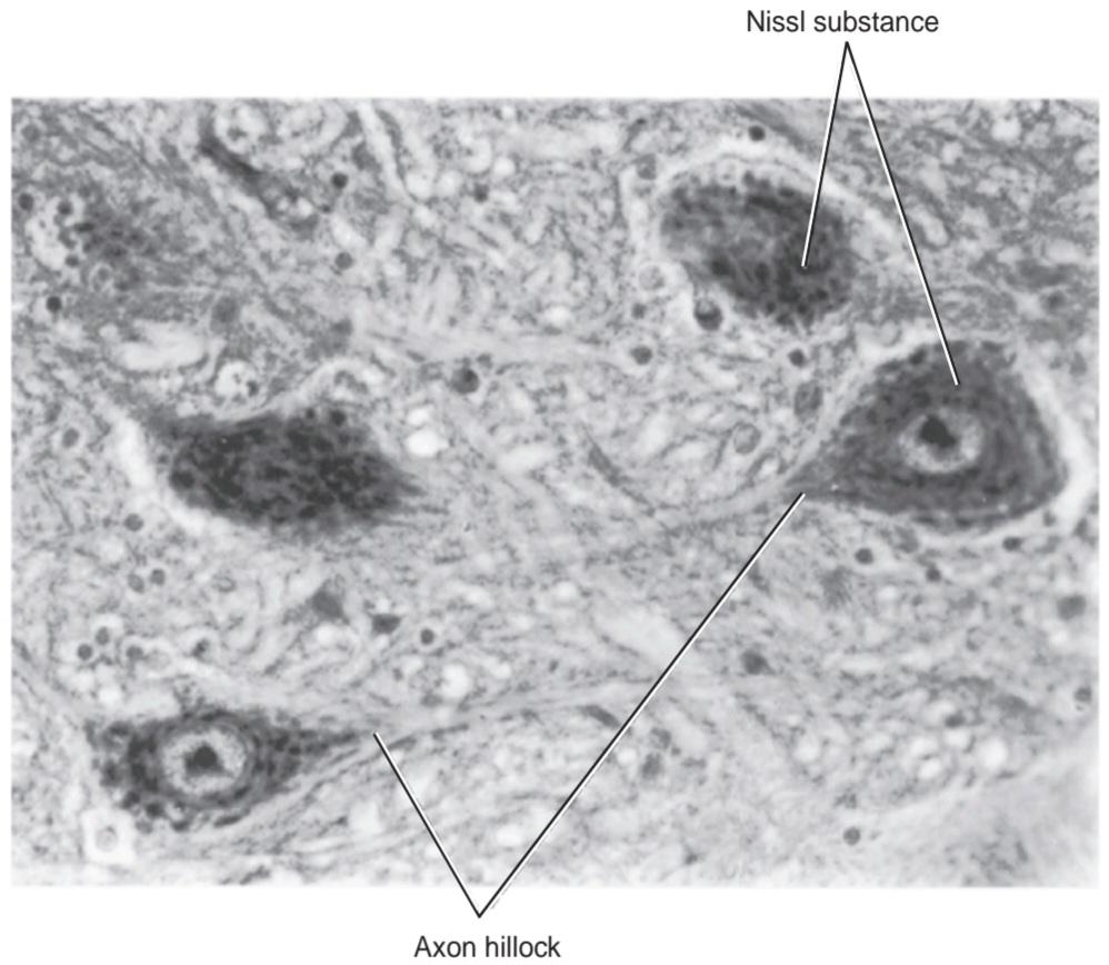
Figure 2-11 Photomicrograph of a section of the anterior gray column of the spinal cord stained with toluidine blue. Note the presence of dark-staining Nissl substance in the cytoplasm of four neurons.
شکل ۲-۱۱ عکس میکروسکوپی از بخشی از ستون خاکستری قدامی نخاع که با آبی تولوئیدین رنگآمیزی شده است. به وجود ماده نیسل با رنگ تیره در سیتوپلاسم چهار نورون توجه کنید.
Electron micrographs show that the Nissl substance is composed of RER (Fig. 2-12) arranged in the form of broad cisternae stacked one on top of the other. Although many of the ribosomes are attached to the surface of the endoplasmic reticulum, many more lie free in the intervals between the cisternae. Because the ribosomes contain RNA, the Nissl substance is basophilic and can be well demonstrated by staining with toluidine blue or other basic aniline dyes (Fig. 2-11) and using the light microscope.
تصاویر میکروسکوپ الکترونی نشان میدهند که ماده نیسل از RER (شکل 2-12) تشکیل شده است که به شکل سیسترنهای پهن روی هم چیده شدهاند. اگرچه بسیاری از ریبوزومها به سطح شبکه آندوپلاسمی متصل هستند، اما تعداد بیشتری از آنها به صورت آزاد در فواصل بین سیسترنها قرار دارند. از آنجا که ریبوزومها حاوی RNA هستند، ماده نیسل بازوفیل است و میتوان آن را با رنگآمیزی با آبی تولوئیدین یا سایر رنگهای آنیلین بازی (شکل 2-11) و با استفاده از میکروسکوپ نوری به خوبی نشان داد.

Figure 2-12 Electron micrograph of the cytoplasm of two neurons showing the structure of Nissl bodies (substance). (Courtesy Dr. J. M. Kerns.)
شکل ۲-۱۲ تصویر میکروسکوپ الکترونی از سیتوپلاسم دو نورون که ساختار اجسام نیسل (ماده) را نشان میدهد. (با احترام از دکتر جی. ام. کرنز.)
The Nissl substance is responsible for synthesizing protein, which flows along the dendrites and the axon and replaces the proteins that are broken down during cellular activity. Fatigue or neuronal damage causes the Nissl substance to move and become concentrated at the periphery of the cytoplasm. This phenomenon, which gives the impression that the Nissl substance has disappeared, is known as chromatolysis.
ماده نیسل مسئول سنتز پروتئین است که در امتداد دندریتها و آکسون جریان مییابد و جایگزین پروتئینهایی میشود که در طول فعالیت سلولی تجزیه میشوند. خستگی یا آسیب عصبی باعث میشود ماده نیسل حرکت کند و در حاشیه سیتوپلاسم متمرکز شود. این پدیده که این تصور را ایجاد میکند که ماده نیسل ناپدید شده است، به عنوان کروماتولیز شناخته میشود.
The Golgi complex, when seen with the light microscope after staining with a silver-osmium method, appears as a network of irregular wavy threads around the nucleus. In electron micrographs, it appears as clusters of flattened cisternae and small vesicles made up of smooth endoplasmic reticulum (SER) (Figs. 2-8 and 2-9).
کمپلکس گلژی، وقتی با میکروسکوپ نوری پس از رنگآمیزی با روش نقره-اسمیوم دیده میشود، به صورت شبکهای از رشتههای موجدار نامنظم در اطراف هسته ظاهر میشود. در میکروگرافهای الکترونی، به صورت خوشههایی از سیسترنهای مسطح و وزیکولهای کوچک ساخته شده از شبکه آندوپلاسمی صاف (SER) دیده میشود (شکلهای 2-8 و 2-9).
The protein produced by the Nissl substance is transferred to the inside of the Golgi complex in transport vesicles, where it is temporarily stored and where carbohydrate may be added to the protein to form glycoproteins. The proteins are believed to travel from one cisterna to another via transport vesicles. Each cisterna of the Golgi complex is specialized for different types of enzymatic reaction. At the trans side of the complex, the macromolecules are packaged in vesicles for transport to the nerve terminals. The Golgi complex is also thought to be active in lysosome production and in the synthesis of cell membranes. The latter function is particularly important in the formation of synaptic vesicles at the axon terminals.
پروتئین تولید شده توسط ماده نیسل در وزیکولهای انتقالی به داخل کمپلکس گلژی منتقل میشود، جایی که به طور موقت ذخیره میشود و کربوهیدرات ممکن است برای تشکیل گلیکوپروتئینها به پروتئین اضافه شود. اعتقاد بر این است که پروتئینها از طریق وزیکولهای انتقالی از یک سیسترن به سیسترن دیگر منتقل میشوند. هر سیسترن از کمپلکس گلژی برای انواع مختلف واکنشهای آنزیمی تخصص یافته است. در سمت ترانس کمپلکس، ماکرومولکولها در وزیکولهایی برای انتقال به پایانههای عصبی بستهبندی میشوند. همچنین تصور میشود که کمپلکس گلژی در تولید لیزوزوم و سنتز غشاهای سلولی فعال است. عملکرد اخیر به ویژه در تشکیل وزیکولهای سیناپسی در پایانههای آکسون اهمیت دارد.
Mitochondria are found scattered throughout the cell body, dendrites, and axons (Figs. 2-8 and 2-9). They are spherical or rod shaped. In electron micrographs, the walls show a characteristic double membrane. The inner membrane is thrown into folds or cristae that project into the center of the mitochondrion. Mitochondria possess many enzymes, which are localized chiefly on the inner mitochondrial membrane. These enzymes take part in the tricarboxylic acid cycle and the cytochrome chains of respiration. Therefore, mitochondria are important in nerve cells, as in other cells, in the production of energy.
میتوکندریها در سراسر جسم سلولی، دندریتها و آکسونها پراکنده هستند (شکلهای ۲-۸ و ۲-۹). آنها کروی یا میلهای شکل هستند. در تصاویر میکروسکوپ الکترونی، دیوارهها یک غشای دوگانه مشخص را نشان میدهند. غشای داخلی به صورت چینخوردگیها یا کریستاهایی که به مرکز میتوکندری بیرون زدهاند، در آمده است. میتوکندریها دارای آنزیمهای زیادی هستند که عمدتاً در غشای داخلی میتوکندری قرار دارند. این آنزیمها در چرخه اسید تری کربوکسیلیک و زنجیرههای سیتوکروم تنفس شرکت میکنند. بنابراین، میتوکندریها در سلولهای عصبی، مانند سایر سلولها، در تولید انرژی اهمیت دارند.
Neurofibrils, as seen with the light microscope after staining with silver, are numerous and run parallel to each other through the cell body into the neurites (Fig. 2-13). With the electron microscope, the neurofibrils resolve into bundles of neurofilaments-each filament measuring about 10 nm in diameter (Fig. 2-14). The neurofilaments form the main component of the cytoskeleton. Chemically, neurofilaments are very stable and belong to the cytokeratin family.
نوروفیبریلها، همانطور که با میکروسکوپ نوری پس از رنگآمیزی با نقره دیده میشوند، متعدد هستند و به موازات یکدیگر از جسم سلولی به داخل نوریتها امتداد مییابند (شکل ۲-۱۳). با میکروسکوپ الکترونی، نوروفیبریلها به دستههایی از نوروفیلامنتها تجزیه میشوند – هر فیلامنت قطری حدود ۱۰ نانومتر دارد (شکل ۲-۱۴). نوروفیلامنتها جزء اصلی اسکلت سلولی را تشکیل میدهند. از نظر شیمیایی، نوروفیلامنتها بسیار پایدار هستند و به خانواده سیتوکراتین تعلق دارند.
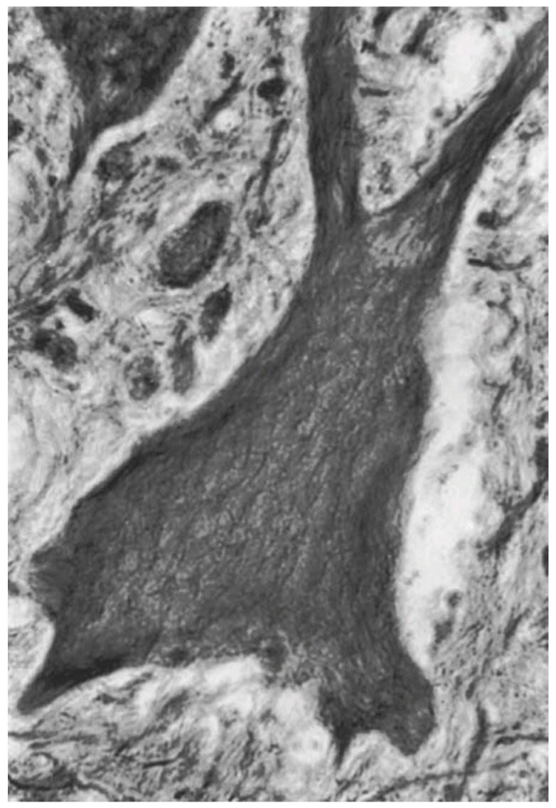
Figure 2-13 Photomicrograph of a silver-stained section of a neuron showing the presence of large numbers of neurofibrils in the cytoplasm of the cell body and the neurites.
شکل ۲-۱۳ عکس میکروسکوپی از بخشی از یک نورون که با رنگ نقره رنگآمیزی شده است، وجود تعداد زیادی نوروفیبریل را در سیتوپلاسم جسم سلولی و نوریتها نشان میدهد.
Microfilaments measure about 3 to 5 nm in diameter and are formed of actin. Microfilaments are concentrated at the periphery of the cytoplasm just beneath the plasma membrane where they form a dense network. Together with microtubules, microfilaments play a key role in the formation of new cell processes and the retraction of old ones. They also assist the microtubules in axon transport.
ریزرشتهها قطری حدود ۳ تا ۵ نانومتر دارند و از اکتین تشکیل شدهاند. ریزرشتهها در حاشیه سیتوپلاسم، درست زیر غشای پلاسمایی، متمرکز شدهاند و در آنجا یک شبکه متراکم تشکیل میدهند. ریزرشتهها به همراه میکروتوبولها نقش کلیدی در تشکیل زوائد سلولی جدید و جمع شدن زوائد قدیمی دارند. آنها همچنین به میکروتوبولها در انتقال آکسون کمک میکنند.
Microtubules are similar to those seen in other types of cells. They measure about 25 nm in diameter and are found interspersed among the neurofilaments (Fig. 2-14). They extend throughout the cell body and its processes. In the axon, all the microtubules are arranged in parallel, with one end pointing to the cell body and the other end pointing distally away from the cell body.
میکروتوبولها مشابه میکروتوبولهایی هستند که در انواع دیگر سلولها دیده میشوند. قطر آنها حدود 25 نانومتر است و در میان نوروفیلامنتها پراکنده شدهاند (شکل 2-14). آنها در سراسر جسم سلولی و زوائد آن امتداد دارند. در آکسون، تمام میکروتوبولها به صورت موازی چیده شدهاند، به طوری که یک سر آنها به سمت جسم سلولی و سر دیگر آنها به سمت دور از جسم سلولی قرار دارد.

Figure 2-14 Electron micrograph of dendrites showing the presence of neurofilaments and microtubules within their cytoplasm. A: Longitudinal section of two adjacent dendrites. B: Transverse section of a dendrite. (Courtesy Dr. J. M. Kerns.)
شکل ۲-۱۴ تصویر میکروسکوپ الکترونی از دندریتها که وجود نوروفیلامنتها و میکروتوبولها را در سیتوپلاسم آنها نشان میدهد. الف: برش طولی از دو دندریت مجاور. ب: برش عرضی از یک دندریت. (با احترام از دکتر جی. ام. کرنز.)
Microtubules and microfilaments provide a stationary track that permits specific organelles to move by molecular motors. The stop-and-start movement is caused by the periodic dissociation of the organelles from the track or the collision with other structures.
میکروتوبولها و ریزرشتهها یک مسیر ثابت فراهم میکنند که به اندامکهای خاص اجازه میدهد توسط موتورهای مولکولی حرکت کنند. حرکت توقف و شروع به دلیل جدا شدن دورهای اندامکها از مسیر یا برخورد با سایر ساختارها ایجاد میشود.
Cell transport involves the movement of membrane organelles, secretory material, synaptic precursor mem- branes, large dense core vesicles, mitochondria, and SER.
انتقال سلولی شامل حرکت اندامکهای غشایی، مواد ترشحی، غشاهای پیشساز سیناپسی، وزیکولهای هسته متراکم بزرگ، میتوکندریها و SER است.
Cell transport can take place in both directions in the cell body and its processes. Rapid transport (100 to 400 mm/day) is mediated by two motor proteins associated with the microtubule adenosine triphosphate (ATP)-ase sites. In anterograde (away from the cell) movement, kinesin-coated organelles are thought to move toward one end of the tubule, and, in retrograde (toward the cell) movement, dynein-coated organelles are thought to move toward the other end of the tubule. The direction and speed of the movement of an organelle can be brought about by the activation of one of the motor proteins or of both simultaneously.
انتقال سلولی میتواند در هر دو جهت در جسم سلولی و فرآیندهای آن انجام شود. انتقال سریع (100 تا 400 میلیمتر در روز) توسط دو پروتئین حرکتی مرتبط با جایگاههای آنزیم آدنوزین تری فسفات (ATP)-آز میکروتوبول انجام میشود. در حرکت رو به جلو (به دور از سلول)، تصور میشود اندامکهای پوشیده از کینزین به سمت یک انتهای توبول حرکت میکنند و در حرکت رو به عقب (به سمت سلول)، تصور میشود اندامکهای پوشیده از دینئین به سمت انتهای دیگر توبول حرکت میکنند. جهت و سرعت حرکت یک اندامک میتواند با فعال شدن یکی از پروتئینهای حرکتی یا هر دو به طور همزمان ایجاد شود.
Slow transport (0.1 to 3.0 mm/day) involves the bulk movement of the cytoplasm and includes the move- ment of mitochondria and other organelles. Slow axonal transport occurs only in the anterograde direction. The molecular motor has not been identified but is probably one of the kinesin families.
انتقال آهسته (0.1 تا 3.0 میلیمتر در روز) شامل حرکت حجمی سیتوپلاسم و شامل حرکت میتوکندریها و سایر اندامکها میشود. انتقال آهسته آکسونی فقط در جهت قدامی-جهشی رخ میدهد. موتور مولکولی شناسایی نشده است، اما احتمالاً یکی از خانوادههای کینزین است.
Lysosomes are membrane-bound vesicles measuring about 8 nm in diameter. They serve the cell by acting as intracellular scavengers and contain hydrolytic enzymes. They are formed by the budding off of the Golgi apparatus. Lysosomes exist in three forms: (1) primary lysosomes, which have just been formed; (2) secondary lysosomes, which contain partially digested material (myelin figures); and (3) residual bodies, in which the enzymes are inactive and the bodies have evolved from digestible materials such as pigment and lipid.
لیزوزومها وزیکولهای متصل به غشاء با قطر حدود 8 نانومتر هستند. آنها با عمل به عنوان رفتگرهای درون سلولی به سلول خدمت میکنند و حاوی آنزیمهای هیدرولیتیک هستند. آنها با جوانه زدن دستگاه گلژی تشکیل میشوند. لیزوزومها به سه شکل وجود دارند: (1) لیزوزومهای اولیه که تازه تشکیل شدهاند؛ (2) لیزوزومهای ثانویه که حاوی مواد نیمه هضم شده (شکلهای میلین) هستند؛ و (3) اجسام باقیمانده که در آنها آنزیمها غیرفعال هستند و اجسام از مواد قابل هضم مانند رنگدانه و لیپید تکامل یافتهاند.
Centrioles are small, paired structures found in immature dividing nerve cells. Each centriole is a hollow cylinder whose wall is made up of microtubule bundles. They are associated with the formation of the spindle during cell division and in the formation of microtubules. Centrioles are also found in mature nerve cells, where they are likely involved in the maintenance of microtubules.
سانتریولها ساختارهای کوچک و جفتی هستند که در سلولهای عصبی در حال تقسیم نابالغ یافت میشوند. هر سانتریول یک استوانه توخالی است که دیواره آن از دستههای میکروتوبول تشکیل شده است. آنها با تشکیل دوک تقسیم در طول تقسیم سلولی و در تشکیل میکروتوبولها مرتبط هستند. سانتریولها همچنین در سلولهای عصبی بالغ یافت میشوند، جایی که احتمالاً در نگهداری میکروتوبولها نقش دارند.
Lipofuscin (pigment material) occurs as yellowishbrown granules within the cytoplasm (Fig. 2-15). It probably forms as the result of lysosomal activity, and it represents a harmless metabolic byproduct. Lipofuscin accumulates with age.
لیپوفوسین (ماده رنگدانهای) به صورت گرانولهای قهوهای مایل به زرد در سیتوپلاسم وجود دارد (شکل 2-15). احتمالاً در نتیجه فعالیت لیزوزومی تشکیل میشود و یک محصول جانبی متابولیک بیضرر است. لیپوفووسین با افزایش سن تجمع مییابد.
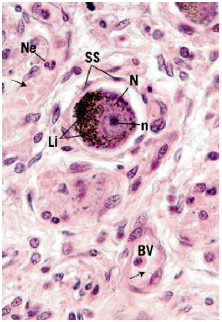
Figure 2-15 Photomicrograph of a longitudinal section of a posterior root ganglion showing the presence of lipofuscin granules within the cytoplasm of sensory neurons. Ne, neutrophil; arrow, red blood cell; SS, satellite cells; N, nucleus; n, nucleolus; Li, lipofuscin; BV, blood vessel.(From Gartner, L. P. (2018). Color atlas and text of histology (7th ed). Baltimore, MD: Wolters Kluwer.)
شکل ۲-۱۵ فتومیکروگراف از برش طولی گانگلیون ریشه خلفی که وجود گرانولهای لیپوفوشین را در سیتوپلاسم نورونهای حسی نشان میدهد. Ne، نوتروفیل؛ پیکان، گلبول قرمز؛ SS، سلولهای ماهوارهای؛ N، هسته؛ n، هستک؛ Li، لیپوفوشین؛ BV، رگ خونی. (از گارتنر، ال. پی. (2018). اطلس رنگی و متن بافتشناسی (ویرایش هفتم). بالتیمور، مریلند: ولترز کلوور.)
Melanin granules are found in the cytoplasm of cells in certain parts of the brain (e.g., the substantia nigra of the midbrain). Their presence may be related to the catecholamine-synthesizing ability of these neurons, whose neurotransmitter is dopamine.
گرانولهای ملانین در سیتوپلاسم سلولهای بخشهای خاصی از مغز (مثلاً جسم سیاه مغز میانی) یافت میشوند. وجود آنها ممکن است با توانایی سنتز کاتکولآمینها در این نورونها که انتقالدهنده عصبی آنها دوپامین است، مرتبط باشد.
Plasma Membrane
The plasma membrane forms the continuous external boundary of the cell body and its processes, and, in the neuron, it is the site for the initiation and conduction of the nerve impulse (Figs. 2-10 and 2-14). The membrane is about 8 nm thick, which is too thin to be seen with the light microscope. When viewed under the electron microscope, the plasma membrane appears as two dark lines with a light line between them.
غشای پلاسمایی
غشای پلاسمایی مرز خارجی پیوسته جسم سلولی و زوائد آن را تشکیل میدهد و در نورون، محل شروع و هدایت تکانه عصبی است (شکلهای 2-10 و 2-14). ضخامت غشا حدود 8 نانومتر است که برای دیدن با میکروسکوپ نوری بسیار نازک است. وقتی زیر میکروسکوپ الکترونی مشاهده میشود، غشای پلاسمایی به صورت دو خط تیره با یک خط روشن بین آنها ظاهر میشود.
The plasma membrane is composed of an inner and an outer layer of very loosely arranged protein molecules, each layer being about 2.5 nm thick, separated by a middle layer of lipid about 3 nm thick. The lipid layer is made up of two rows of phospholipid molecules arranged so that their hydrophobic ends are in contact with each other and their polar ends are in contact with the protein layers. Certain protein molecules lie within the phospholipid layer and span the entire width of the lipid layer. These molecules provide the membrane with hydrophilic channels through which inorganic ions may enter and leave the cell. Carbohydrate molecules are attached to the outside of the plasma membrane and are linked to the proteins or the lipids, forming what is known as the cell coat or glycocalyx.
غشای پلاسمایی از یک لایه داخلی و یک لایه خارجی از مولکولهای پروتئینی با آرایش بسیار سست تشکیل شده است که هر لایه حدود ۲.۵ نانومتر ضخامت دارد و توسط یک لایه میانی لیپیدی حدود ۳ نانومتر از هم جدا میشوند. لایه لیپیدی از دو ردیف مولکول فسفولیپیدی تشکیل شده است که به گونهای آرایش یافتهاند که انتهای آبگریز آنها با یکدیگر و انتهای قطبی آنها با لایههای پروتئینی در تماس باشد. مولکولهای پروتئینی خاصی در داخل لایه فسفولیپیدی قرار دارند و در کل عرض لایه لیپیدی امتداد دارند. این مولکولها کانالهای آبدوستی را برای غشا فراهم میکنند که از طریق آنها یونهای معدنی میتوانند وارد سلول شده و از آن خارج شوند. مولکولهای کربوهیدرات به قسمت بیرونی غشای پلاسمایی متصل شده و به پروتئینها یا لیپیدها متصل میشوند و چیزی را تشکیل میدهند که به عنوان پوشش سلولی یا گلیکوکالیکس شناخته میشود.
The plasma membrane and the cell coat together form a semipermeable membrane that allows diffusion of certain ions through it but restricts others. In the resting state (unstimulated state), K+ ions diffuse through the plasma membrane from the cell cytoplasm to the tissue fluid (Fig. 2-16). The permeability of the membrane to K+ ions is much greater than that to Na+ ions; thus, the passive efflux of K+ is much greater than the influx of Na+. This results in a steady potential difference of about 280 mV, which can be measured across the plasma membrane because the inside of the membrane is negative with respect to the outside. This potential is known as the resting potential.
غشای پلاسما و پوشش سلولی با هم یک غشای نیمهتراوا تشکیل میدهند که امکان انتشار یونهای خاصی را از طریق آن فراهم میکند اما سایر یونها را محدود میکند. در حالت استراحت (حالت تحریک نشده)، یونهای +K از طریق غشای پلاسما از سیتوپلاسم سلول به مایع بافتی انتشار مییابند (شکل 2-16). نفوذپذیری غشا به یونهای +K بسیار بیشتر از نفوذ به یونهای +Na است؛ بنابراین، خروج غیرفعال +K بسیار بیشتر از ورود +Na است. این امر منجر به اختلاف پتانسیل ثابتی در حدود 280 میلیولت میشود که میتوان آن را در سراسر غشای پلاسما اندازهگیری کرد زیرا داخل غشا نسبت به خارج منفی است. این پتانسیل به عنوان پتانسیل استراحت شناخته میشود.
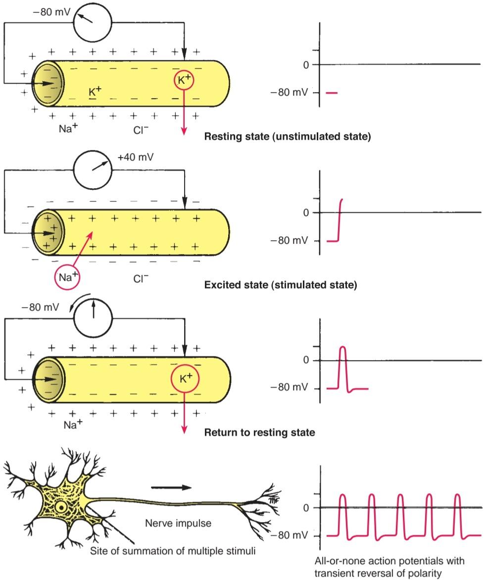
Figure 2-16 lonic and electrical changes that occur in a neuron when it is stimulated.
شکل ۲-۱۶ تغییرات حرکتی و الکتریکی که هنگام تحریک در یک نورون رخ میدهد.
Membrane Excitation and Conduction
In the resting unstimulated state, a nerve fiber is polarized so that the interior is negative to the exterior; the potential difference across the plasma membrane (axolemma) is about -80 mV and is called the resting membrane potential (Fig. 2-17).
تحریک و هدایت غشاء
در حالت استراحت و بدون تحریک، یک فیبر عصبی قطبی میشود به طوری که قسمت داخلی آن نسبت به قسمت خارجی منفی است؛ اختلاف پتانسیل در غشای پلاسما (آکسولما) حدود -80 میلی ولت است و پتانسیل غشاء در حال استراحت نامیده میشود (شکل 2-17).

Figure 2-17 lonic and electrical changes that occur in a nerve fiber when it is conducting an impulse.
شکل ۲-۱۷ تغییرات حرکتی و الکتریکی که در یک فیبر عصبی هنگام هدایت یک تکانه رخ میدهد.
When the nerve cell is excited (stimulated) by electrical, mechanical, or chemical means, a rapid change in membrane permeability to Na+ ions takes place, and Na ions diffuse through the plasma membrane into the cell cytoplasm from the tissue fluid. This results in the membrane becoming progressively depolarized. The sudden influx of Na+ ions followed by the altered polarity produces an action potential (AP), which is approximately 140 mV. This potential is very brief, lasting about 5 msec. The increased membrane permeability for Na+ ions quickly ceases, and membrane permeability for K+ ions increases. Therefore, K+ ions start to flow from the cell cytoplasm and return the localized area of the cell to the resting state.
وقتی سلول عصبی به وسیلهی وسایل الکتریکی، مکانیکی یا شیمیایی تحریک میشود، تغییر سریعی در نفوذپذیری غشاء به یونهای +Na رخ میدهد و یونهای +Na از طریق غشای پلاسما به داخل سیتوپلاسم سلول از مایع بافتی منتشر میشوند. این امر منجر به دپلاریزه شدن تدریجی غشاء میشود. هجوم ناگهانی یونهای +Na و به دنبال آن تغییر قطبیت، پتانسیل عمل (AP) ایجاد میکند که تقریباً 140 میلیولت است. این پتانسیل بسیار کوتاه است و حدود 5 میلیثانیه طول میکشد. افزایش نفوذپذیری غشاء برای یونهای +Na به سرعت متوقف میشود و نفوذپذیری غشاء برای یونهای +K افزایش مییابد. بنابراین، یونهای +K شروع به جریان یافتن از سیتوپلاسم سلول میکنند و ناحیهی موضعی سلول را به حالت استراحت بازمیگردانند.
A nerve impulse (AP) starts at the initial segment of the axon and is a self-propagating wave of electrical negativity that passes rapidly along the surface of the axolemma. Once generated, the AP spreads away from the site of initiation and is conducted along neurites as the nerve impulse. The wave of electrical negativity is initiated by an adequate stimulus being applied to the surface of the neuron (Fig. 2-18). Under normal circumstances, this occurs at the initial segment of the axon, which is the most sensitive part of the neuron. The stimulus alters the permeability of the membrane to Na+ ions at the point of stimulation. Now, Na+ ions rapidly enter the axon (Fig. 2-17). The positive ions outside the axolemma quickly decrease to zero. Therefore, the membrane potential is reduced to zero and is said to be depolarized. A typical resting potential is -80 mV, with the outside of the membrane positive to the inside; the AP is about 140 mV, with the outside of the membrane negative to the inside. In small-diameter axons, the AP may not rise to as much as 40 mV.
یک تکانه عصبی (AP) از بخش اولیه آکسون شروع میشود و یک موج خودانتشار از منفی الکتریکی است که به سرعت در امتداد سطح آکسولما عبور میکند. پس از تولید، AP از محل شروع پخش میشود و در امتداد نوریتها به عنوان تکانه عصبی هدایت میشود. موج منفی الکتریکی با اعمال یک محرک کافی به سطح نورون آغاز میشود (شکل 2-18). در شرایط عادی، این اتفاق در بخش اولیه آکسون، که حساسترین قسمت نورون است، رخ میدهد. محرک، نفوذپذیری غشا را نسبت به یونهای +Na در نقطه تحریک تغییر میدهد. اکنون، یونهای +Na به سرعت وارد آکسون میشوند (شکل 2-17). یونهای مثبت خارج از آکسولما به سرعت به صفر کاهش مییابند. بنابراین، پتانسیل غشا به صفر کاهش مییابد و گفته میشود که دپلاریزه شده است. یک پتانسیل استراحت معمول 80- میلیولت است، که در آن قسمت بیرونی غشا مثبت به سمت داخل است. پتانسیل عمل (AP) حدود ۱۴۰ میلیولت است و قسمت بیرونی غشا نسبت به داخل آن منفی است. در آکسونهای با قطر کوچک، پتانسیل عمل ممکن است تا ۴۰ میلیولت افزایش نیابد.
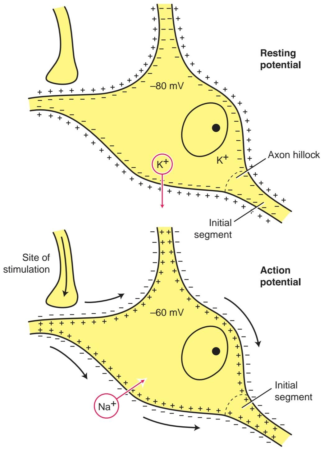
Figure 2-18 Creation of the action potential by the arrival of a stimulus from a single presynaptic terminal. Note that the action potential generated at the initial segment will only occur if the threshold for excitation is reached at the initial segment. (From Snell, R. S. Clinical neuroanatomy: A review with questions and explanations (3rd ed., p. 7). Baltimore, MD: Lippincott Williams & Wilkins.)
شکل ۲-۱۸ ایجاد پتانسیل عمل با ورود یک محرک از یک پایانه پیشسیناپسی. توجه داشته باشید که پتانسیل عمل تولید شده در قطعه اولیه تنها در صورتی رخ میدهد که آستانه تحریک در قطعه اولیه حاصل شود. (از اسنل، آر. اس. نوروآناتومی بالینی: مروری با سوالات و توضیحات (ویرایش سوم، صفحه 7). بالتیمور، مریلند: لیپینکات ویلیامز و ویلکینز.)
The negatively charged point on the outside of the axolemma now acts as a stimulus to the adjacent positively charged axolemma, and, in less than 1 msec, the polarity of the adjacent resting potential is reversed. The AP now has moved along the axolemma from the point originally stimulated to the adjacent point on the membrane. In this manner, the AP travels along the full length of a nerve fiber to its end.
نقطه با بار منفی در قسمت بیرونی آکسولما اکنون به عنوان محرکی برای آکسولمای با بار مثبت مجاور عمل میکند و در کمتر از ۱ میلیثانیه، قطبیت پتانسیل استراحت مجاور معکوس میشود. نقطه فعال (AP) اکنون در امتداد آکسولما از نقطهای که در ابتدا تحریک شده بود تا نقطه مجاور روی غشاء حرکت کرده است. به این ترتیب، نقطه فعال در امتداد کل طول یک فیبر عصبی تا انتهای آن حرکت میکند.
As the AP moves along the nerve fiber, entry of the Na+ ions into the axon ceases, and the permeability of the axolemma to K+ ions increases. Now, K+ ions rapidly diffuse outside the axon because the concentration is much higher within the axon than outside so that the original resting membrane potential is restored. The permeability of the axolemma now decreases, and the status quo is restored by the active transport of the Na+ ions out of the axon and the K+ ions into the axon. The outer surface of the axolemma is again electrically positive compared with that of the inner surface. This is a simplistic description of the movements of the Na+ and K+ ions. (For further details on the voltage-gated Na+ and K+ channels, the Na+ and K+ pumps, and the Na+ and K+ leak channels, refer to a textbook of physiology.)
با حرکت AP در امتداد فیبر عصبی، ورود یونهای +Na به آکسون متوقف میشود و نفوذپذیری آکسولما به یونهای +K افزایش مییابد. اکنون، یونهای +K به سرعت در خارج از آکسون منتشر میشوند زیرا غلظت آنها در داخل آکسون بسیار بیشتر از خارج است، به طوری که پتانسیل استراحت اولیه غشاء بازیابی میشود. نفوذپذیری آکسولما اکنون کاهش مییابد و با انتقال فعال یونهای +Na به خارج از آکسون و یونهای +K به داخل آکسون، وضعیت به حالت قبل برمیگردد. سطح بیرونی آکسولما در مقایسه با سطح داخلی، دوباره از نظر الکتریکی مثبت است. این یک توصیف ساده از حرکات یونهای +Na و +K است. (برای جزئیات بیشتر در مورد کانالهای +Na و +K وابسته به ولتاژ، پمپهای +Na و +K و کانالهای نشت +Na و +K، به یک کتاب درسی فیزیولوژی مراجعه کنید.)
For a short time after the passage of a nerve impulse along a nerve fiber, while the axolemma is still depolarized, a second stimulus, however strong, is unable to excite the nerve. This period of time is called the absolute refractory period. The underlying reason for the refractory period is that the Na channels become inactivated, and no stimulation, however strong, will open the Na+ gates. This period is followed by a further short interval during which the excitability of the nerve gradually returns to normal. This latter period is called the relative refractory period. Therefore, the refractory period makes a continuous excitatory state of the nerve impossible and limits the frequency of the impulses.
برای مدت کوتاهی پس از عبور یک تکانه عصبی در امتداد یک فیبر عصبی، در حالی که آکسولما هنوز دپلاریزه است، محرک دوم، هر چقدر هم قوی باشد، قادر به تحریک عصب نیست. این دوره زمانی، دوره تحریکناپذیری مطلق نامیده میشود. دلیل اصلی دوره تحریکناپذیری این است که کانالهای سدیم غیرفعال میشوند و هیچ تحریکی، هر چقدر هم قوی، دروازههای +Na را باز نمیکند. پس از این دوره، یک فاصله کوتاه دیگر وجود دارد که در طی آن تحریکپذیری عصب به تدریج به حالت عادی برمیگردد. این دوره اخیر، دوره تحریکناپذیری نسبی نامیده میشود. بنابراین، دوره تحریکناپذیری، حالت تحریکی مداوم عصب را غیرممکن میکند و فرکانس تکانهها را محدود میکند.
The greater the strength of the initial stimulus, the larger the initial depolarization and the greater will be the spread into the surrounding areas of the plasma membrane. Multiple excitatory stimuli applied to a neuron’s surface result in summation. For example, subthreshold stimuli may pass over the surface of the cell body and be summated at the origin of the axon and so initiate an AP. Inhibitory stimuli are believed to produce their effect by causing an influx of CI ions through the plasma membrane into the neuron, thus producing hyperpolarization and reducing the excitatory state of the cell (Fig. 2-19).
هرچه قدرت محرک اولیه بیشتر باشد، دپلاریزاسیون اولیه بیشتر و گسترش آن به نواحی اطراف غشای پلاسما بیشتر خواهد بود. محرکهای تحریکی متعدد اعمال شده به سطح نورون منجر به جمع شدن میشوند. به عنوان مثال، محرکهای زیرآستانهای ممکن است از سطح جسم سلولی عبور کرده و در مبدا آکسون جمع شوند و بنابراین یک AP را آغاز کنند. اعتقاد بر این است که محرکهای مهاری با ایجاد هجوم یونهای CI از طریق غشای پلاسما به داخل نورون، اثر خود را ایجاد میکنند و در نتیجه باعث ایجاد هایپرپلاریزاسیون و کاهش حالت تحریکی سلول میشوند (شکل 2-19).
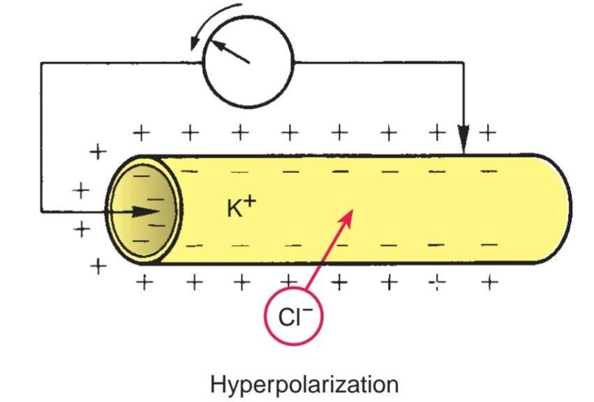
Figure 2-19 lonic and electrical changes that occur in a neuron during hyperpolarization.
شکل ۲-۱۹ تغییرات الکتریکی و حرکتی که در یک نورون در طول هایپرپلاریزاسیون رخ میدهد.
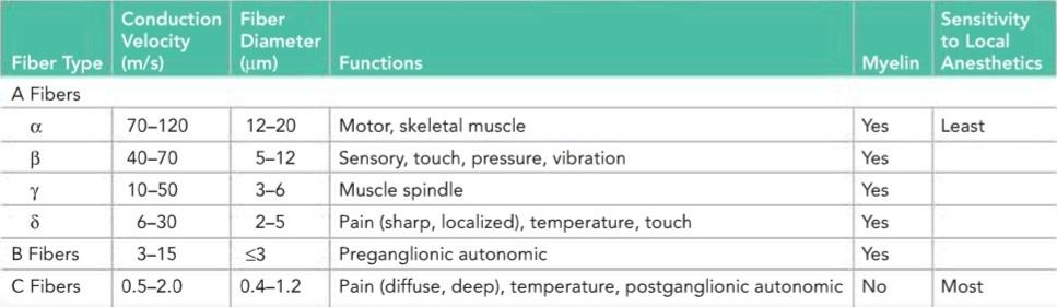
The conduction velocity of a nerve fiber is proportional to the cross-sectional area of the axon, with the thicker fibers conducting more rapidly than those of smaller diameter. In the large motor fibers (a fibers), the rate may be as high as 70 to 120 m/s; the smaller sensory fibers have slower conduction rates (Table 2-3).
سرعت هدایت یک فیبر عصبی متناسب با سطح مقطع آکسون است، به طوری که فیبرهای ضخیمتر سریعتر از فیبرهای با قطر کوچکتر هدایت میشوند. در فیبرهای حرکتی بزرگ (فیبرهای a)، این سرعت ممکن است به 70 تا 120 متر بر ثانیه برسد؛ فیبرهای حسی کوچکتر سرعت هدایت کمتری دارند (جدول 2-3).
Table 2-3 Classification of Nerve Fibers by Speed of Conduction and Size
جدول ۲-۳ طبقهبندی فیبرهای عصبی بر اساس سرعت هدایت و اندازه
In nonmyelinated fibers, the AP passes continuously along the axolemma, progressively exciting neighboring areas of membrane (Fig. 2-20). In myelinated fibers, the presence of a myelin sheath serves as an insulator, and few ions can flow through the sheath. Consequently, a myelinated nerve fiber can be stimulated only at the nodes of Ranvier, where the axon is naked and the ions can pass freely through the plasma membrane between the extracellular fluid and the axoplasm. In these fibers, the AP jumps from one node to the next. The AP at one node sets up a current in the surrounding tissue fluid, which quickly produces depolarization at the next node. This leaping of the AP from one node to the next is referred to as saltatory conduction. This is a more rapid mechanism than is found in nonmyelinated fibers (120.0 m/s in a large myelinated fiber compared with 0.5 m/s in a very small unmyelinated fiber).
در فیبرهای بدون میلین، AP به طور مداوم در امتداد آکسولما عبور میکند و به تدریج نواحی مجاور غشاء را تحریک میکند (شکل 2-20). در فیبرهای میلیندار، وجود غلاف میلین به عنوان یک عایق عمل میکند و یونهای کمی میتوانند از طریق غلاف جریان یابند. در نتیجه، یک فیبر عصبی میلیندار فقط در گرههای رانویه قابل تحریک است، جایی که آکسون برهنه است و یونها میتوانند آزادانه از طریق غشای پلاسما بین مایع خارج سلولی و آکوپلاسم عبور کنند. در این فیبرها، AP از یک گره به گره بعدی میپرد. AP در یک گره جریانی را در مایع بافت اطراف ایجاد میکند که به سرعت باعث دپلاریزاسیون در گره بعدی میشود. این جهش AP از یک گره به گره بعدی به عنوان هدایت جهشی شناخته میشود. این مکانیسم سریعتر از آن چیزی است که در فیبرهای بدون میلین یافت میشود (120.0 متر بر ثانیه در یک فیبر میلیندار بزرگ در مقایسه با 0.5 متر بر ثانیه در یک فیبر بدون میلین بسیار کوچک).

Figure 2-20 Electrical changes that occur in stimulated myelinated axon (saltatory conduction) (A) and stimulated nonmyelinated axon (B).
شکل 2-20 تغییرات الکتریکی که در آکسون میلیندار تحریکشده (هدایت جهشی) (A) و آکسون بدون میلین تحریکشده (B) رخ میدهد.
Sodium and Potassium Channels
Sodium and potassium channels, through which the sodium and potassium ions diffuse through the plasma membrane, are formed of the protein molecules that extend through the full thickness of the plasma membrane (Fig. 2-21). Why a particular channel permits the passage of K+ ions while excluding Na+ ions is difficult to explain. The selectivity cannot be due to the diameter of the ions, insofar as the K+ ion is larger than the Na+ ion. However, the movement of ions in solution depends not only on the size of the ion but also on the size of the shell of water surrounding it. K+ ions have weaker electric fields than Na+ ions; thus, K+ ions attract less water than Na+ ions. Therefore, K+ ions behave as if they are smaller than Na+ ions. However, this physicochemical explanation does not entirely account for why a channel is selective. The channels may have narrow regions along their length that act as sieves or molecular filters. The ions may also participate in electrostatic interactions with the amino acid residues lining the walls of the channel.
کانالهای سدیم و پتاسیم
کانالهای سدیم و پتاسیم، که یونهای سدیم و پتاسیم از طریق آنها از غشای پلاسما عبور میکنند، از مولکولهای پروتئینی تشکیل شدهاند که در تمام ضخامت غشای پلاسما امتداد دارند (شکل 2-21). توضیح اینکه چرا یک کانال خاص اجازه عبور یونهای +K را میدهد در حالی که یونهای +Na را حذف میکند، دشوار است. گزینشپذیری نمیتواند به دلیل قطر یونها باشد، تا جایی که یون +K بزرگتر از یون +Na است. با این حال، حرکت یونها در محلول نه تنها به اندازه یون، بلکه به اندازه پوسته آب اطراف آن نیز بستگی دارد. یونهای +K میدانهای الکتریکی ضعیفتری نسبت به یونهای +Na دارند. بنابراین، یونهای +K آب کمتری نسبت به یونهای +Na جذب میکنند. بنابراین، یونهای +K طوری رفتار میکنند که گویی از یونهای +Na کوچکتر هستند. با این حال، این توضیح فیزیکوشیمیایی به طور کامل دلیل گزینشی بودن یک کانال را توضیح نمیدهد. کانالها ممکن است در طول خود دارای نواحی باریکی باشند که به عنوان غربال یا فیلترهای مولکولی عمل میکنند. یونها همچنین ممکن است در برهمکنشهای الکترواستاتیکی با باقیماندههای اسید آمینه که دیوارههای کانال را پوشاندهاند، شرکت کنند.
Ion channel proteins are relatively stable, but they exist in at least two conformational states, open and closed functional states. The mechanism responsible for the opening and closing of a channel is not understood but can be thought of as a gate. Gating may involve the twisting and distortion of the channel, thus creating a wider or narrower lumen. Gating appears to occur in response to such stimuli as voltage change, the presence of a ligand, or stretch or pressure.
پروتئینهای کانال یونی نسبتاً پایدار هستند، اما حداقل در دو حالت ساختاری، حالتهای عملکردی باز و بسته، وجود دارند. مکانیسم مسئول باز و بسته شدن یک کانال مشخص نیست، اما میتوان آن را به عنوان یک دروازه در نظر گرفت. دروازهبندی ممکن است شامل پیچاندن و اعوجاج کانال باشد، در نتیجه یک لومن پهنتر یا باریکتر ایجاد میکند. به نظر میرسد که دروازهبندی در پاسخ به محرکهایی مانند تغییر ولتاژ، وجود لیگاند یا کشش یا فشار رخ میدهد.
In the nonstimulated state, potassium channel gates are open wider than sodium channel gates, which are nearly closed. This permits the K+ ions to diffuse out of the cell cytoplasm more readily than the Na+ ions can diffuse in. In the stimulated state, the sodium channel gates are at first wide open; then, the potassium channel gates are opened, and the sodium channel gates are nearly closed again. The opening and closing of these channels is thought to produce the depolarization and repolarization of the plasma membrane.
در حالت غیرتحریکی، دریچههای کانال پتاسیم نسبت به دریچههای کانال سدیم که تقریباً بسته هستند، بازتر هستند. این امر به یونهای +K اجازه میدهد تا راحتتر از یونهای +Na به داخل سیتوپلاسم سلول نفوذ کنند. در حالت تحریکشده، دریچههای کانال سدیم ابتدا کاملاً باز هستند؛ سپس دریچههای کانال پتاسیم باز میشوند و دریچههای کانال سدیم دوباره تقریباً بسته میشوند. تصور میشود که باز و بسته شدن این کانالها باعث دپلاریزاسیون و رپلاریزاسیون غشای پلاسما میشود.
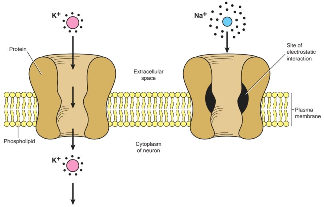
Figure 2-21 lonic permeability of the plasma membrane. Diagram shows the interactions of the ions with water, the membrane lipid bilayer, and the ion channels.
شکل ۲-۲۱ نفوذپذیری یونی غشای پلاسما. نمودار، برهمکنش یونها با آب، دولایه لیپیدی غشا و کانالهای یونی را نشان میدهد.
Nerve Cell Processes
Neurites the processes of a nerve cell-may be divided into dendrites and an axon.
زوائد سلول عصبی
نوریتها زوائد یک سلول عصبی هستند که میتوانند به دندریتها و آکسونها تقسیم شوند.
Dendrites are the short processes of the cell body (Fig. 2-22). Their diameter tapers as they extend from the cell body, and they often branch profusely. In many neurons, the finer branches bear large numbers of small projections called dendritic spines. The cytoplasm of the dendrites closely resembles that of the cell body and contains Nissl granules, mitochondria, microtubules, microfilaments, ribosomes, and SER. Dendrites are extensions of the cell body to increase the surface area for the reception of axons from other neurons. Essentially, they conduct the nerve impulse toward the cell body.
دندریتها زوائد کوتاه جسم سلولی هستند (شکل 2-22). قطر آنها با امتداد یافتن از جسم سلولی باریک میشود و اغلب به طور فراوان شاخه شاخه میشوند. در بسیاری از نورونها، شاخههای ظریفتر تعداد زیادی برآمدگی کوچک به نام خارهای دندریتی دارند. سیتوپلاسم دندریتها شباهت زیادی به جسم سلولی دارد و حاوی گرانولهای نیسل، میتوکندری، میکروتوبولها، میکروفیلامنتها، ریبوزومها و گیرندههای SER است. دندریتها امتداد جسم سلولی هستند تا سطح دریافت آکسونها از سایر نورونها را افزایش دهند. اساساً، آنها تکانه عصبی را به سمت جسم سلولی هدایت میکنند.
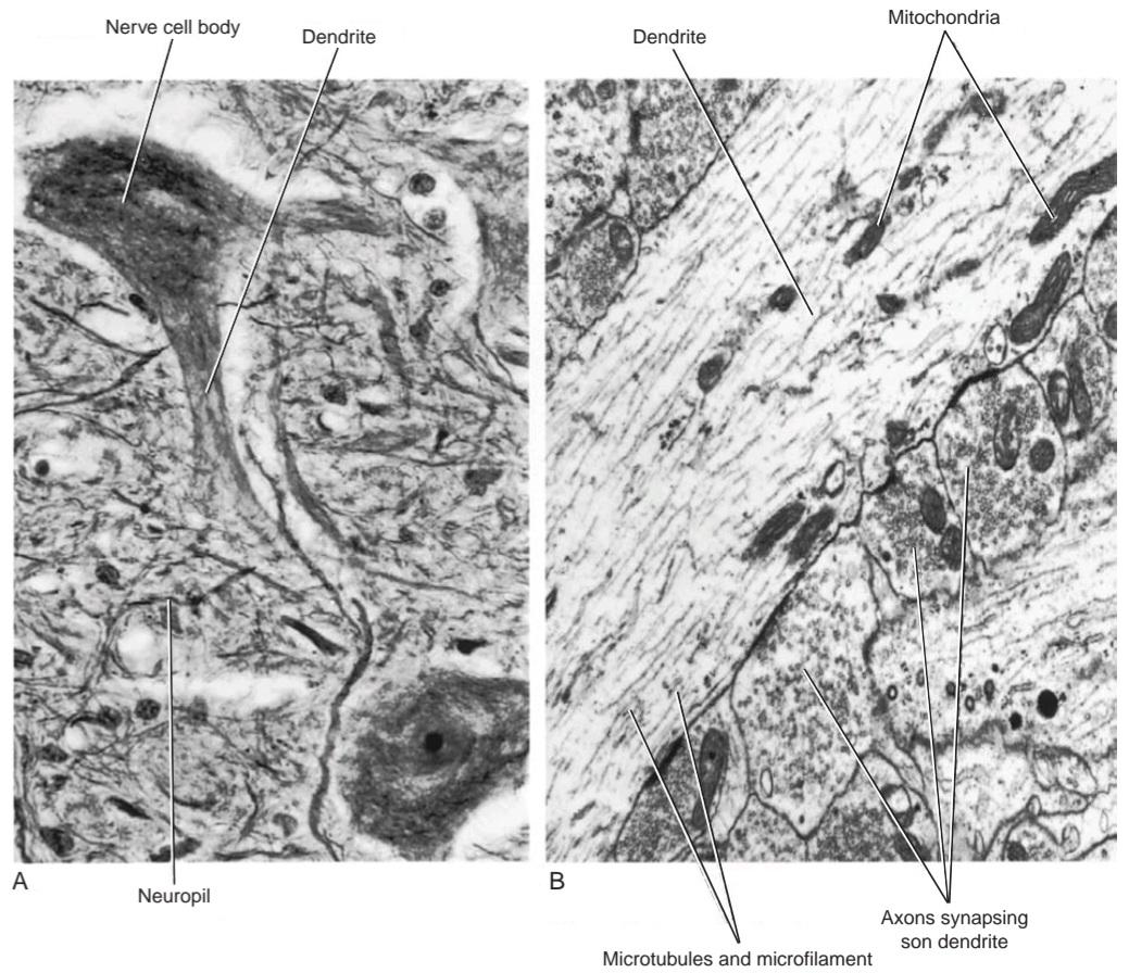
Figure 2-22 A: Light photomicrograph of a motor neuron in the anterior gray column of the spinal cord showing the nerve cell body, two dendrites, and the surrounding neuropil. B: Electron micrograph of a dendrite showing axodendritic synapses. (Courtesy Dr. J. M. Kerns.)
شکل ۲-۲۲ الف: عکس میکروسکوپی نوری از یک نورون حرکتی در ستون خاکستری قدامی نخاع که جسم سلولی عصبی، دو دندریت و نوروپیل اطراف آن را نشان میدهد. ب: عکس میکروسکوپی الکترونی از یک دندریت که سیناپسهای آکسودندریتی را نشان میدهد. (با احترام از دکتر جی. ام. کرنز.)
During early embryonic development, dendrites are overproduced. Later, they are reduced in number and size in response to altered functional demand from afferent axons. Evidence shows that dendrites remain plastic throughout life and elongate and branch or contract in response to afferent activity.
در طول رشد اولیه جنینی، دندریتها بیش از حد تولید میشوند. بعداً، در پاسخ به تغییر تقاضای عملکردی آکسونهای آوران، تعداد و اندازه آنها کاهش مییابد. شواهد نشان میدهد که دندریتها در طول زندگی انعطافپذیر باقی میمانند و در پاسخ به فعالیت آوران، کشیده و منشعب میشوند یا منقبض میشوند.
The axon is the longest process of the cell body. It arises from a small conical elevation on the cell body, devoid of Nissl granules, called the axon hillock (Fig. 2-23; also see Fig. 2-8). Occasionally, an axon arises from the proximal part of a dendrite. An axon is tubular and is uniform in diameter; it tends to have a smooth surface.
آکسون طولانیترین زائده جسم سلولی است. از یک برآمدگی مخروطی کوچک روی جسم سلولی، عاری از گرانولهای نیسل، به نام تپه آکسون، ناشی میشود (شکل 2-23؛ همچنین به شکل 2-8 مراجعه کنید). گاهی اوقات، یک آکسون از قسمت پروگزیمال یک دندریت ناشی میشود. آکسون لولهای شکل است و قطر یکنواختی دارد؛ تمایل دارد سطح صافی داشته باشد.
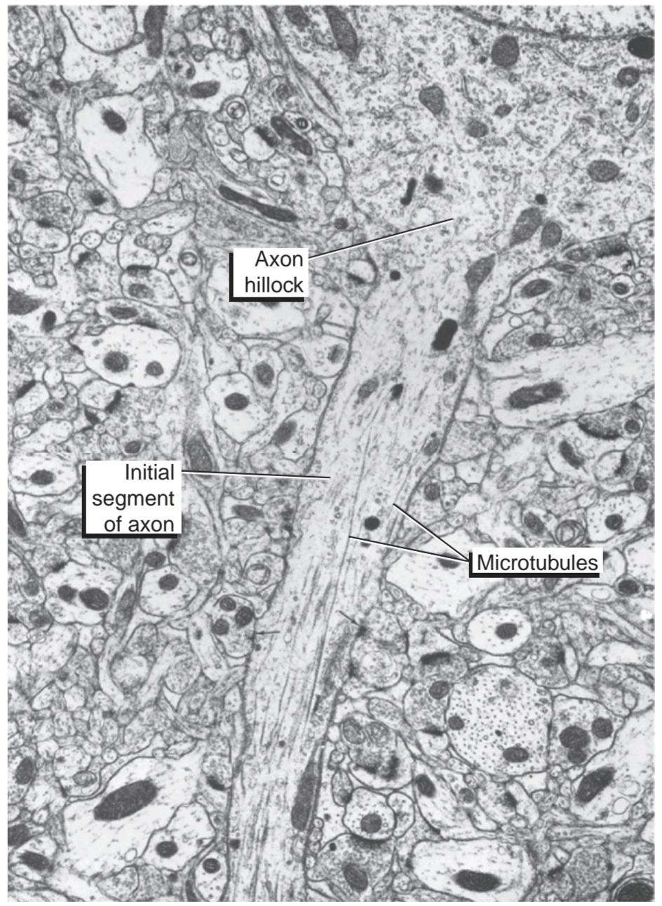
Figure 2-23 Electron micrograph of a longitudinal section of a neuron from the cerebral cortex showing the detailed structure of the region of the axon hillock and the initial segment of the axon. Note the absence of Nissl substance (rough endoplasmic reticulum) in the axon hillock and the presence of numerous microtubules in the axoplasm. Note also the axon terminals (arrows) forming axoaxonal synapses with the initial segment of the axon. (Courtesy Dr. A. Peters.)
شکل ۲-۲۳ تصویر میکروسکوپ الکترونی از برش طولی یک نورون از قشر مغز که ساختار دقیق ناحیه تپه آکسون و بخش اولیه آکسون را نشان میدهد. به عدم وجود ماده نیسل (شبکه آندوپلاسمی خشن) در تپه آکسون و وجود میکروتوبولهای متعدد در آکسون توجه کنید. همچنین به پایانههای آکسون (فلشها) که سیناپسهای آکسونی را با بخش اولیه آکسون تشکیل میدهند، توجه کنید. (با احترام از دکتر A. Peters.)
Axons usually do not branch close to the cell body; collateral branches may occur along their length. Shortly before their termination, axons commonly branch profusely. The distal ends of the terminal branches of the axons are often enlarged; they are called terminals (Fig. 2-24). Some axons (especially those of autonomic nerves) show a series of swellings resembling a string of beads near their termination; these swellings are called varicosities.
آکسونها معمولاً نزدیک به جسم سلولی شاخه نمیشوند؛ شاخههای جانبی ممکن است در امتداد طول آنها ایجاد شوند. کمی قبل از خاتمه، آکسونها معمولاً به طور گسترده شاخه میشوند. انتهای دیستال شاخههای انتهایی آکسونها اغلب بزرگ میشوند؛ به آنها ترمینال میگویند (شکل 2-24). برخی از آکسونها (به ویژه آکسونهای اعصاب خودکار) یک سری تورم شبیه به یک رشته مهره در نزدیکی انتهای خود نشان میدهند؛ این تورمها واریس نامیده میشوند.

Figure 2-24 Electron micrograph showing multiple axodendritic synapses. Note the presence of large numbers of presynaptic vesicles within the axons. The definition has come to include the site at which a neuron comes into close proximity with a skeletal muscle cell and functional communication occurs. (Courtesy Dr. J. M. Kerns.)
شکل ۲-۲۴ تصویر میکروسکوپ الکترونی که سیناپسهای آکسودندریتیک متعددی را نشان میدهد. به وجود تعداد زیادی وزیکول پیشسیناپسی در آکسونها توجه کنید. این تعریف شامل محلی میشود که در آن یک نورون به نزدیکی سلول عضله اسکلتی میرسد و ارتباط عملکردی رخ میدهد. (با احترام از دکتر جی. ام. کرنز.)
Axons may be very short (0.1 mm), as seen in many neurons of the CNS, or extremely long (3.0 m), as seen when they extend from a peripheral receptor in the skin of the toe to the spinal cord and thence to the brain.
آکسونها ممکن است بسیار کوتاه (0.1 میلیمتر) باشند، همانطور که در بسیاری از نورونهای سیستم عصبی مرکزی دیده میشود، یا بسیار بلند (3.0 متر) باشند، همانطور که وقتی از یک گیرنده محیطی در پوست انگشت پا تا نخاع و از آنجا به مغز امتداد مییابند، دیده میشوند.
Axon diameter varies considerably with different neurons. Those of larger diameter conduct impulses rapidly, and those of smaller diameter conduct impulses very slowly.
قطر آکسون در نورونهای مختلف به طور قابل توجهی متفاوت است. آنهایی که قطر بزرگتری دارند، تکانهها را به سرعت و آنهایی که قطر کمتری دارند، تکانهها را بسیار آهسته هدایت میکنند.
The plasma membrane bounding the axon is called the axolemma; the cytoplasm of the axon is the axoplasm. Unlike the cytoplasm of the cell body, axoplasm lacks Nissl granules and a Golgi complex. The sites for the production of protein, namely RNA and ribosomes, are absent. Thus, axonal survival depends on the trans- port of substances from the cell bodies.
غشای پلاسمایی که آکسون را احاطه کرده است، آکسولما نامیده میشود؛ سیتوپلاسم آکسون، آکسوپلاسم است. برخلاف سیتوپلاسم جسم سلولی، آکسوپلاسم فاقد گرانولهای نیسل و کمپلکس گلژی است. جایگاههای تولید پروتئین، یعنی RNA و ریبوزومها، وجود ندارند. بنابراین، بقای آکسون به انتقال مواد از جسم سلولی بستگی دارد.
The initial segment of the axon is the first 50 to 100 um after it leaves the axon hillock of the nerve cell body (Fig. 2-23). This is the most excitable part of the axon and is the site at which an AP originates.
بخش اولیه آکسون، ۵۰ تا ۱۰۰ میکرومتر اول پس از خروج از برآمدگی آکسون جسم سلولی عصبی است (شکل ۲-۲۳). این بخش، تحریکپذیرترین بخش آکسون است و محلی است که AP از آن سرچشمه میگیرد.
Remember that, under normal conditions, an AP does not originate on the plasma membrane of the cell body but, instead, always at the initial segment.
به یاد داشته باشید که در شرایط عادی، AP از غشای پلاسمایی جسم سلولی سرچشمه نمیگیرد، بلکه همیشه از بخش اولیه است.
An axon always conducts impulses away from the cell body. The axons of sensory posterior root ganglion cells are an exception; here, the long neurite, which is indistinguishable from an axon, carries the impulse toward the cell body. (See unipolar neurons, p. 33.)
آکسون همیشه تکانهها را از جسم سلولی دور میکند. آکسونهای سلولهای گانگلیون ریشه خلفی حسی یک استثنا هستند؛ در اینجا، نوریت بلند، که از آکسون قابل تشخیص نیست، تکانه را به سمت جسم سلولی حمل میکند. (به نورونهای تکقطبی، صفحه ۳۳ مراجعه کنید.)
Axon Transport
Materials are transported from the cell body to the axon terminals (anterograde transport) and to a lesser extent in the opposite direction (retrograde transport).
انتقال آکسون
مواد از جسم سلولی به پایانههای آکسون (انتقال آنتروگراد) و به میزان کمتر در جهت مخالف (انتقال رتروگراد) منتقل میشوند.
Fast anterograde transport of 100 to 400 mm/day refers to the transport of proteins and transmitter substances or their precursors. Slow anterograde transport of 0.1 to 3.0 mm/day refers to the transport of axoplasm and includes the microfilaments and microtubules.
انتقال سریع رو به جلو با سرعت ۱۰۰ تا ۴۰۰ میلیمتر در روز به انتقال پروتئینها و مواد ناقل یا پیشسازهای آنها اشاره دارد. انتقال آهسته رو به جلو با سرعت ۰.۱ تا ۳ میلیمتر در روز به انتقال آکسوپلاسم اشاره دارد و شامل ریزرشتهها و میکروتوبولها میشود.
Retrograde transport explains how the cell bodies of nerve cells respond to changes in the distal end of the axons. For example, activated growth factor receptors can be carried along the axon to their site of action in the nucleus. Pinocytotic vesicles arising at the axon terminals can be quickly returned to the cell body. Worn-out organelles can be returned to the cell body for breakdown by the lysosomes.
انتقال رتروگراد توضیح میدهد که چگونه جسم سلولی سلولهای عصبی به تغییرات در انتهای دیستال آکسونها پاسخ میدهد. به عنوان مثال، گیرندههای فاکتور رشد فعال شده میتوانند در امتداد آکسون به محل عمل خود در هسته منتقل شوند. وزیکولهای پینوسیتوزی که در پایانههای آکسون ایجاد میشوند، میتوانند به سرعت به جسم سلولی بازگردانده شوند. اندامکهای فرسوده میتوانند برای تجزیه توسط لیزوزومها به جسم سلولی بازگردانده شوند.
Axon transport is brought about by microtubules assisted by the microfilaments.
انتقال آکسون توسط میکروتوبولها و با کمک ریزرشتهها انجام میشود.
Synapses
The nervous system consists of a large number of neurons that are linked together to form functional conducting pathways. The site where two neurons (or a neuron and a skeletal muscle or gland cell) come into close proximity and functional interneuronal communication occurs is referred to as a synapse (Fig. 2-25). Most neurons may make synapse with 1,000 or more other neurons and may receive up to 10,000 connections from other neurons. Communication at a synapse, under physiologic conditions, takes place in one direction only. Synapses occur in a number of forms. The most common type is that which occurs between an axon of one neuron and the dendrite or cell body of the second neuron. As the axon approaches the synapse, it may have a terminal expansion (bouton terminal), or it may have a series of expansions (bouton de passage), each of which makes synaptic contact. In other types of synapses, the axon synapses on the initial segment of another axon-that is, proximal to where the myelin sheath begins or synapses may exist between terminal expansions from different neurons. Depending on the site of the synapse, they are referred to as axodendritic, axosomatic, or axoaxonic.
سیناپسها
سیستم عصبی از تعداد زیادی نورون تشکیل شده است که برای تشکیل مسیرهای هدایت عملکردی به یکدیگر متصل شدهاند. محلی که دو نورون (یا یک نورون و یک سلول عضله اسکلتی یا غده) در مجاورت هم قرار میگیرند و ارتباط بین نورونی عملکردی رخ میدهد، سیناپس نامیده میشود (شکل 2-25). اکثر نورونها ممکن است با 1000 یا بیشتر نورون دیگر سیناپس برقرار کنند و ممکن است تا 10000 اتصال از نورونهای دیگر دریافت کنند. ارتباط در یک سیناپس، تحت شرایط فیزیولوژیکی، فقط در یک جهت انجام میشود. سیناپسها به اشکال مختلفی رخ میدهند. رایجترین نوع، سیناپسی است که بین آکسون یک نورون و دندریت یا جسم سلولی نورون دوم رخ میدهد. با نزدیک شدن آکسون به سیناپس، ممکن است یک انبساط انتهایی (bouton terminal) داشته باشد، یا ممکن است یک سری انبساط (bouton de passage) داشته باشد که هر کدام تماس سیناپسی برقرار میکنند. در انواع دیگر سیناپسها، سیناپسهای آکسون در قطعه اولیه آکسون دیگر – یعنی نزدیک به جایی که غلاف میلین شروع میشود – سیناپس ایجاد میکنند یا سیناپسها ممکن است بین گسترشهای انتهایی از نورونهای مختلف وجود داشته باشند. بسته به محل سیناپس، به آنها آکسودندریتیک، آکسوسوماتیک یا آکسوآکسونیک گفته میشود.
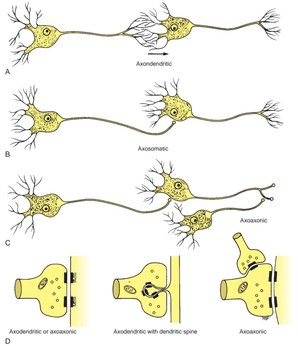
Figure 2-25 A-D: Different types of chemical synapses.
شکل ۲-۲۵ A-D: انواع مختلف سیناپسهای شیمیایی.
How an axon terminates varies considerably in different parts of the nervous system. For example, a single axon may terminate on a single neuron, or a single axon may synapse with multiple neurons, as in the case of the parallel fibers of the cerebellar cortex synapsing with multiple Purkinje cells. In the same way, a single neuron may have synaptic junctions with axons of many different neurons. The arrangement of these synapses will determine the means by which a neuron can be stimulated or inhibited. Synaptic spines, extensions of the surface of a neuron, form receptive sites for synaptic contact with afferent boutons.
نحوهی پایان یافتن یک آکسون در بخشهای مختلف سیستم عصبی به طور قابل توجهی متفاوت است. به عنوان مثال، یک آکسون ممکن است به یک نورون ختم شود، یا یک آکسون ممکن است با چندین نورون سیناپس برقرار کند، همانطور که در مورد فیبرهای موازی قشر مخچه که با چندین سلول پورکنژ سیناپس برقرار میکنند، صادق است. به همین ترتیب، یک نورون ممکن است با آکسونهای بسیاری از نورونهای مختلف، اتصالات سیناپسی داشته باشد. ترتیب این سیناپسها، روشی را که یک نورون میتواند تحریک یا مهار شود، تعیین میکند. خارهای سیناپسی، امتدادهای سطح یک نورون، مکانهای گیرندهای را برای تماس سیناپسی با دستههای آوران تشکیل میدهند.
Synapses are of two types: chemical and electrical. Most synapses are chemical, in which a chemical substance, the neurotransmitter, passes across the narrow space between the cells and becomes attached to a protein molecule in the postsynaptic membrane called the receptor.
سیناپسها دو نوع هستند: شیمیایی و الکتریکی. اکثر سیناپسها شیمیایی هستند که در آنها یک ماده شیمیایی، انتقالدهنده عصبی، از فضای باریک بین سلولها عبور میکند و به یک مولکول پروتئینی در غشای پسسیناپسی به نام گیرنده متصل میشود.
In most chemical synapses, several neurotransmitters may be present. One neurotransmitter is usually the principal activator and acts directly on the postsynaptic membrane, while the other transmitters function as modulators and modify the activity of the principal transmitter.
در بیشتر سیناپسهای شیمیایی، ممکن است چندین انتقالدهنده عصبی وجود داشته باشد. یک انتقالدهنده عصبی معمولاً فعالکننده اصلی است و مستقیماً روی غشای پسسیناپسی عمل میکند، در حالی که انتقالدهندههای دیگر به عنوان تعدیلکننده عمل میکنند و فعالیت انتقالدهنده اصلی را تغییر میدهند.
Chemical Synapses
On examination with an electron microscope, synapses are seen to be areas of structural specialization (Fig. 2-26; also see Fig. 2-24). The apposed surfaces of the terminal axonal expansion and the neuron are called the presynaptic and postsynaptic membranes, respectively, and they are separated by a synaptic cleft measuring about 20 to 30 nm wide. The presynaptic and postsynaptic membranes are thickened, and the adjacent underlying cytoplasm shows increased density. On the presynaptic side, the dense cytoplasm is broken up into groups; on the postsynaptic side, the density often extends into a subsynaptic web. Presynaptic ves icles, mitochondria, and occasional lysosomes are present in the cytoplasm close to the presynaptic membrane (see Fig. 2-26). On the postsynaptic side, the cytoplasm often contains parallel cisternae. The synaptic cleft contains polysaccharides.
سیناپسهای شیمیایی
در بررسی با میکروسکوپ الکترونی، سیناپسها به عنوان نواحی تخصص ساختاری دیده میشوند (شکل 2-26؛ همچنین به شکل 2-24 مراجعه کنید). سطوح روبروی گسترش آکسون انتهایی و نورون به ترتیب غشاهای پیشسیناپسی و پسسیناپسی نامیده میشوند و توسط یک شکاف سیناپسی به عرض حدود 20 تا 30 نانومتر از هم جدا میشوند. غشاهای پیشسیناپسی و پسسیناپسی ضخیم شدهاند و سیتوپلاسم زیرین مجاور، تراکم بیشتری را نشان میدهد. در سمت پیشسیناپسی، سیتوپلاسم متراکم به گروههایی تقسیم میشود؛ در سمت پسسیناپسی، تراکم اغلب به یک شبکه زیرسیناپسی گسترش مییابد. وزیکولهای پیشسیناپسی، میتوکندریها و گاهی اوقات لیزوزومها در سیتوپلاسم نزدیک به غشای پیشسیناپسی وجود دارند (شکل 2-26 را ببینید). در سمت پسسیناپسی، سیتوپلاسم اغلب حاوی سیسترنهای موازی است. شکاف سیناپسی حاوی پلیساکاریدها است.
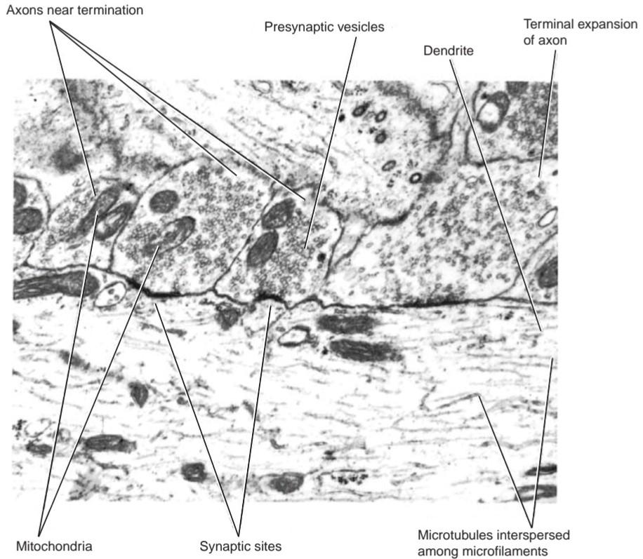
Figure 2-26 High-power electron micrograph of axodendritic synapses showing the thickening of the cell membranes at the synaptic sites, presynaptic vesicles, and the presence of mitochondria within the axons near their termination. (Courtesy Dr. J. M. Kerns.)
شکل ۲-۲۶ تصویر میکروسکوپ الکترونی با قدرت بالا از سیناپسهای آکسودندریتی که ضخیم شدن غشاهای سلولی در مکانهای سیناپسی، وزیکولهای پیشسیناپسی و وجود میتوکندری در آکسونهای نزدیک به انتهای آنها را نشان میدهد. (با احترام از دکتر جی. ام. کرنز.)
The presynaptic terminal contains many small presynaptic vesicles that contain the molecules of the neurotransmitter(s). The vesicles fuse with the presynaptic membrane and discharge the neurotransmitter(s) into the synaptic cleft by a process of exocytosis (Fig. 2-27).
پایانه پیشسیناپسی حاوی تعداد زیادی وزیکول کوچک پیشسیناپسی است که حاوی مولکولهای انتقالدهنده عصبی (یا انتقالدهندههای عصبی) هستند. این وزیکولها با غشای پیشسیناپسی ادغام میشوند و انتقالدهنده عصبی (یا انتقالدهندههای عصبی) را از طریق فرآیند اگزوسیتوز به شکاف سیناپسی تخلیه میکنند (شکل 2-27).
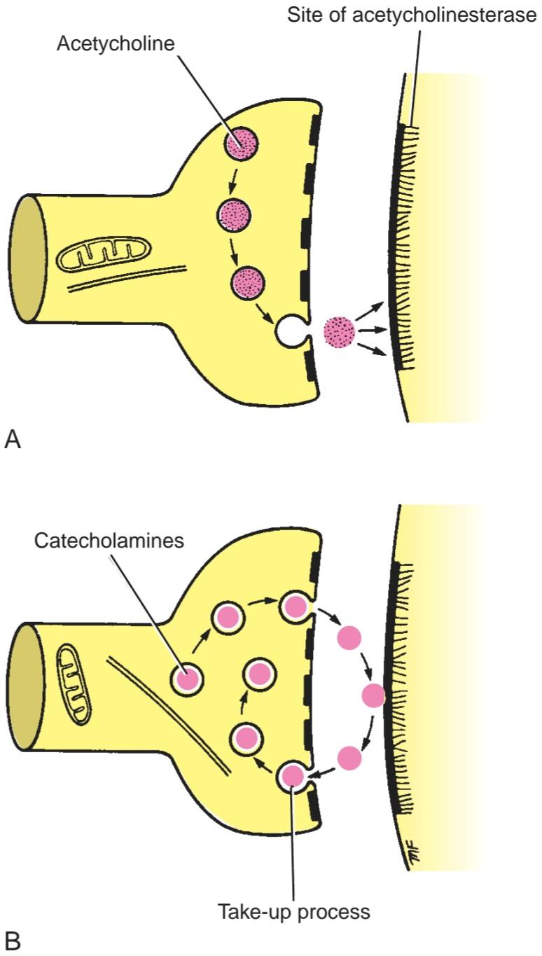
Figure 2-27 Release of neurotransmitters. A: Acetylcholine. B: Catecholamines.
شکل ۲-۲۷ آزادسازی انتقالدهندههای عصبی. الف: استیل کولین. ب: کاتکولآمینها.
When synapses are first formed in the embryo, they are recognized as small zones of density separated by a synaptic cleft. Later, they mature into well-differentiated structures. The presence of simple, undifferentiated synapses in the postnatal nervous system has led to the suggestion that synapses can be developed as required and possibly undergo atrophy when redundant. This plasticity of synapses may be of great importance in the process of learning and in the development and maintenance of memory.
وقتی سیناپسها برای اولین بار در جنین تشکیل میشوند، به صورت مناطق کوچکی از تراکم که توسط یک شکاف سیناپسی از هم جدا شدهاند، تشخیص داده میشوند. بعدها، آنها به ساختارهای کاملاً تمایز یافته بالغ میشوند. وجود سیناپسهای ساده و تمایز نیافته در سیستم عصبی پس از تولد منجر به این پیشنهاد شده است که سیناپسها میتوانند در صورت نیاز توسعه یابند و احتمالاً در صورت اضافی بودن، دچار آتروفی شوند. این انعطافپذیری سیناپسها ممکن است در فرآیند یادگیری و در توسعه و نگهداری حافظه از اهمیت زیادی برخوردار باشد.
Neurotransmitters
Presynaptic vesicles and mitochondria play a key role in the release of neurotransmitter substances at synapses. The vesicles contain the neurotransmitter substance that is released into the synaptic cleft; the mitochondria provide ATP for the synthesis of new transmitter substance.
انتقالدهندههای عصبی
وزیکولهای پیشسیناپسی و میتوکندریها نقش کلیدی در آزادسازی مواد انتقالدهنده عصبی در سیناپسها دارند. این وزیکولها حاوی ماده انتقالدهنده عصبی هستند که به شکاف سیناپسی آزاد میشود؛ میتوکندریها ATP را برای سنتز ماده انتقالدهنده جدید فراهم میکنند.
Most neurons produce and release only one principal transmitter at all their nerve endings. For example, acetylcholine (ACH) is widely used as a transmitter by different neurons in the central and peripheral parts of the nervous system, whereas dopamine is released by neurons in the substantia nigra. Glycine, another transmitter, is found principally in synapses in the spinal cord.
بیشتر نورونها فقط یک فرستنده اصلی را در تمام انتهای عصبی خود تولید و آزاد میکنند. به عنوان مثال، استیل کولین (ACH) به طور گسترده به عنوان یک فرستنده توسط نورونهای مختلف در بخشهای مرکزی و محیطی سیستم عصبی استفاده میشود، در حالی که دوپامین توسط نورونهای جسم سیاه آزاد میشود. گلیسین، یک فرستنده دیگر، عمدتاً در سیناپسهای نخاع یافت میشود.
The following chemical substances act as neurotransmitters (many more exist): ACh, norepinephrine, epinephrine, dopamine, glycine, serotonin, GABA, enkephalins, substance P, and glutamic acid.
مواد شیمیایی زیر به عنوان انتقالدهندههای عصبی عمل میکنند (تعداد بسیار بیشتری وجود دارد): استیلکولین، نوراپی نفرین، اپی نفرین، دوپامین، گلیسین، سروتونین، گابا، انکفالینها، ماده P و اسید گلوتامیک.
Note that all skeletal neuromuscular junctions use only ACh as the transmitter, whereas synapses between neurons use a large number of different transmitters.
توجه داشته باشید که تمام اتصالات عصبی-عضلانی اسکلتی فقط از استیلکولین به عنوان فرستنده استفاده میکنند، در حالی که سیناپسهای بین نورونها از تعداد زیادی فرستنده مختلف استفاده میکنند.
NEUROTRANSMITTER ACTION
All neurotransmitters are released from their nerve endings by the arrival of the nerve impulse (AP). This results in an influx of Ca2+ ions, which causes the synaptic vesicles to fuse with the presynaptic membrane. The neurotransmitters are then ejected into the extracellular fluid in the synaptic cleft. Once in the cleft, they diffuse across the gap to the postsynaptic membrane. There they achieve their objective by raising or lowering the resting potential of the postsynaptic membrane for a brief period of time.
عملکرد انتقالدهنده عصبی
تمام انتقالدهندههای عصبی با رسیدن تکانه عصبی (AP) از انتهای عصبی خود آزاد میشوند. این امر منجر به هجوم یونهای +Ca2 میشود که باعث میشود وزیکولهای سیناپسی با غشای پیشسیناپسی ادغام شوند. سپس انتقالدهندههای عصبی به مایع خارج سلولی در شکاف سیناپسی منتقل میشوند. پس از ورود به شکاف، از طریق شکاف به غشای پسسیناپسی پخش میشوند. در آنجا با افزایش یا کاهش پتانسیل استراحت غشای پسسیناپسی برای مدت کوتاهی به هدف خود میرسند.
The receptor proteins on the postsynaptic membrane bind the transmitter substance and undergo an immediate conformational change that opens the ion channel, generating an immediate but brief excitatory postsynaptic potential (EPSP) or an inhibitory postsynaptic potential (IPSP). The rapid excitation is seen with ACh (nicotinic) and L-glutamate, or the inhibition is seen with GABA (Table 2-4). Other receptor proteins bind the transmitter substance and activate a second-messenger system, usually through a molecular transducer, a G protein. These receptors have a longer latent period, and the duration of the response may last several minutes or longer. ACh (muscarinic), serotonin, histamine, neuropeptides, and adenosine are good examples of this type of transmitter, which is often referred to as a neuromodulator (see next section, Neuromodulators).
پروتئینهای گیرنده روی غشای پسسیناپسی به مادهی انتقالدهنده متصل میشوند و دچار یک تغییر ساختاری فوری میشوند که کانال یونی را باز میکند و یک پتانسیل پسسیناپسی تحریکی فوری اما کوتاه (EPSP) یا یک پتانسیل پسسیناپسی مهاری (IPSP) ایجاد میکند. تحریک سریع با ACh (نیکوتین) و L-گلوتامات یا مهار با GABA مشاهده میشود (جدول 2-4). سایر پروتئینهای گیرنده به مادهی انتقالدهنده متصل میشوند و یک سیستم پیامرسان ثانویه را فعال میکنند، معمولاً از طریق یک مبدل مولکولی، یک پروتئین G. این گیرندهها دورهی نهفتهی طولانیتری دارند و مدت زمان پاسخ ممکن است چند دقیقه یا بیشتر طول بکشد. ACh (موسکارینی)، سروتونین، هیستامین، نوروپپتیدها و آدنوزین نمونههای خوبی از این نوع انتقالدهنده هستند که اغلب به عنوان یک تعدیلکنندهی عصبی شناخته میشوند (به بخش بعدی، تعدیلکنندههای عصبی مراجعه کنید).
The excitatory and the inhibitory effects on the postsynaptic membrane of the neuron will depend on the summation of the postsynaptic responses at the different synapses. If the overall effect is one of depolarization, the neuron will be excited, an AP will be initiated at the initial segment of the axon, and a nerve impulse will travel along the axon. If, on the other hand, the overall effect is one of hyperpolarization, the neuron will be inhibited and no nerve impulse will arise.
اثرات تحریکی و مهاری بر غشای پس سیناپسی نورون به مجموع پاسخهای پس سیناپسی در سیناپسهای مختلف بستگی دارد. اگر اثر کلی از نوع دپلاریزاسیون باشد، نورون تحریک میشود، یک AP در قطعه اولیه آکسون آغاز میشود و یک تکانه عصبی در امتداد آکسون حرکت میکند. از سوی دیگر، اگر اثر کلی از نوع هایپرپلاریزاسیون باشد، نورون مهار میشود و هیچ تکانه عصبی ایجاد نمیشود.
Table 2-4 Examples of Principal (Classic) Neurotransmitters and Neuromodulators at Synapses
جدول ۲-۴ نمونههایی از انتقالدهندههای عصبی اصلی (کلاسیک) و تعدیلکنندههای عصبی در سیناپسها

a Note that these are only a few examples of an ever-increasing number of known neuromediators. ACh, acetylcholine; GABA, y-aminobutyric acid; EPSP, excitatory postsynaptic potential; IPSP, inhibitory postsynaptic potential.
a توجه داشته باشید که اینها تنها چند نمونه از تعداد فزایندهای از واسطههای عصبی شناخته شده هستند. ACh، استیل کولین؛ GABA، γ-آمینوبوتیریک اسید؛ EPSP، پتانسیل پس سیناپسی تحریکی؛ IPSP، پتانسیل پس سیناپسی مهاری.
NEUROTRANSMITTER DISTRIBUTION AND FATE
The distribution of the neurotransmitters varies in different parts of the nervous system. ACh, for example, is found at the neuromuscular junction, in autonomic ganglia, and at parasympathetic nerve endings. In the CNS, the motor neuron collaterals to the Renshaw cells are cholinergic. In the hippocampus, the ascending reticular pathways, and the afferent fibers for the visual and auditory systems, the neurotransmitters are also cholinergic.
توزیع و سرنوشت انتقالدهندههای عصبی
توزیع انتقالدهندههای عصبی در بخشهای مختلف سیستم عصبی متفاوت است. به عنوان مثال، استیلکولین در محل اتصال عصبی-عضلانی، در گانگلیونهای اتونوم و در انتهای اعصاب پاراسمپاتیک یافت میشود. در سیستم عصبی مرکزی، نورونهای حرکتی جانبی سلولهای رنشاو کولینرژیک هستند. در هیپوکامپ، مسیرهای مشبک صعودی و فیبرهای آوران برای سیستمهای بینایی و شنوایی، انتقالدهندههای عصبی نیز کولینرژیک هستند.
Norepinephrine is found at sympathetic nerve endings. In the CNS, it is found in high concentration in the hypothalamus. Dopamine is found in high concentration in different parts of the CNS, such as in the basal nuclei (ganglia).
نوراپی نفرین در انتهای اعصاب سمپاتیک یافت میشود. در سیستم عصبی مرکزی (CNS)، با غلظت بالا در هیپوتالاموس یافت میشود. دوپامین با غلظت بالا در قسمتهای مختلف سیستم عصبی مرکزی، مانند هستههای قاعدهای (گانگلیونها)، یافت میشود.
The effect produced by a neurotransmitter is limited by its destruction or reabsorption. For example, in the case of ACh, the effect is limited by the destruction of the transmitter in the synaptic cleft by the enzyme acetylcholinesterase (AChE) (Fig. 2-27). However, with the catecholamines, the effect is limited by the return of the transmitter to the presynaptic nerve ending.
اثر ایجاد شده توسط یک انتقالدهنده عصبی توسط تخریب یا بازجذب آن محدود میشود. به عنوان مثال، در مورد ACh، اثر توسط تخریب انتقالدهنده در شکاف سیناپسی توسط آنزیم استیل کولین استراز (AChE) محدود میشود (شکل 2-27). با این حال، در مورد کاتکولآمینها، اثر توسط بازگشت انتقالدهنده به انتهای عصب پیشسیناپسی محدود میشود.
Neuromodulators
Interestingly, in many synapses, certain substances other than the principal neurotransmitters are ejected from the presynaptic membrane into the synaptic cleft. These substances are capable of modulating and mod- ifying the activity of the postsynaptic neuron and are called neuromodulators.
تعدیلکنندههای عصبی
جالب توجه است که در بسیاری از سیناپسها، مواد خاصی غیر از انتقالدهندههای عصبی اصلی از غشای پیشسیناپسی به شکاف سیناپسی خارج میشوند. این مواد قادر به تعدیل و اصلاح فعالیت نورون پسسیناپسی هستند و تعدیلکنندههای عصبی نامیده میشوند.
NEUROMODULATOR ACTION
Neuromodulators can coexist with the principal neurotransmitter at a single synapse. Usually, but not always, the neuromodulators are in separate presynaptic vesicles. Although the principal neurotransmitters have a rapid, brief effect on the postsynaptic membrane on release into the synaptic cleft, neuromodulators do not have a direct effect on the postsynaptic membrane. Rather, they enhance, prolong, inhibit, or limit the principal neurotransmitter’s effect on the postsynaptic membrane. Neuromodulators act through a second-messenger system, usually through a molecular transducer, such as a G protein, and alter the response of the receptor to the neurotransmitter. In a given area of the nervous system, many different afferent neurons can release several different neuromodulators that affect the postsynaptic neuron. Such an arrangement can lead to a wide variety of responses, depending on input from the afferent neurons.
عملکرد تعدیلکننده عصبی
تعدیلکنندههای عصبی میتوانند با انتقالدهنده عصبی اصلی در یک سیناپس واحد همزیستی داشته باشند. معمولاً، اما نه همیشه، تعدیلکنندههای عصبی در وزیکولهای پیشسیناپسی جداگانه قرار دارند. اگرچه انتقالدهندههای عصبی اصلی هنگام آزاد شدن به شکاف سیناپسی، تأثیر سریع و کوتاهی بر غشای پسسیناپسی دارند، تعدیلکنندههای عصبی تأثیر مستقیمی بر غشای پسسیناپسی ندارند. در عوض، آنها اثر انتقالدهنده عصبی اصلی را بر غشای پسسیناپسی افزایش، طولانی، مهار یا محدود میکنند. تعدیلکنندههای عصبی از طریق یک سیستم پیامرسان ثانویه، معمولاً از طریق یک مبدل مولکولی، مانند پروتئین G، عمل میکنند و پاسخ گیرنده به انتقالدهنده عصبی را تغییر میدهند. در یک ناحیه مشخص از سیستم عصبی، بسیاری از نورونهای آوران مختلف میتوانند چندین تعدیلکننده عصبی مختلف را آزاد کنند که بر نورون پسسیناپسی تأثیر میگذارند. چنین ترتیبی میتواند بسته به ورودی نورونهای آوران، منجر به طیف گستردهای از پاسخها شود.
Electrical Synapses
Electrical synapses are gap junctions containing channels that extend from the cytoplasm of the presynaptic neuron to that of the postsynaptic neuron: They are rare in the human CNS. The neurons communicate electrically; a chemical transmitter is not present. The bridging channels permit ionic current flow to take place from one cell to the other with a minimum of delay. In electrical synapses, the rapid spread of activity from one neuron to another ensures that a group of neurons performing an identical function act together. Electrical synapses also have the advantage that they are bidirectional; chemical synapses are not.
سیناپسهای الکتریکی
سیناپسهای الکتریکی، اتصالات شکافداری هستند که حاوی کانالهایی هستند که از سیتوپلاسم نورون پیشسیناپسی تا سیتوپلاسم نورون پسسیناپسی امتداد دارند: آنها در سیستم عصبی مرکزی انسان نادر هستند. نورونها به صورت الکتریکی ارتباط برقرار میکنند؛ فرستنده شیمیایی وجود ندارد. کانالهای پلزننده اجازه میدهند جریان یونی با حداقل تأخیر از یک سلول به سلول دیگر منتقل شود. در سیناپسهای الکتریکی، گسترش سریع فعالیت از یک نورون به نورون دیگر تضمین میکند که گروهی از نورونها که عملکرد یکسانی دارند، با هم عمل میکنند. سیناپسهای الکتریکی همچنین این مزیت را دارند که دو طرفه هستند؛ سیناپسهای شیمیایی اینطور نیستند.
NEUROGLIA
The neurons of the CNS are supported by several varieties of nonexcitable cells, which together are called neuroglia (Fig. 2-28). Neuroglial cells are generally smaller than neurons and outnumber them by 5 to 10 times; they comprise about half the total volume of the brain and spinal cord.
نوروگلیا
نورونهای سیستم عصبی مرکزی توسط چندین نوع سلول غیرقابل تحریک پشتیبانی میشوند که در مجموع نوروگلیا نامیده میشوند (شکل 2-28). سلولهای نوروگلیا عموماً از نورونها کوچکتر هستند و تعداد آنها 5 تا 10 برابر بیشتر است. آنها حدود نیمی از کل حجم مغز و نخاع را تشکیل میدهند.
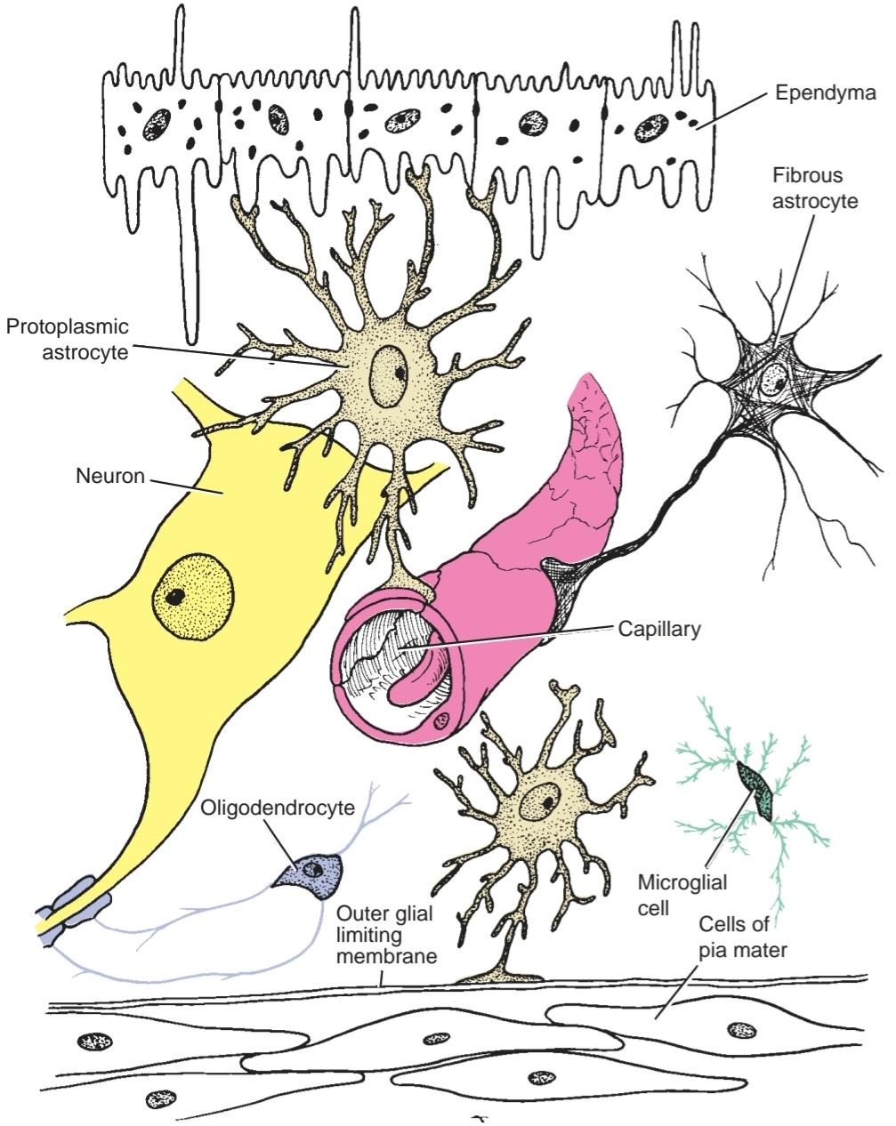
Figure 2-28 Diagrammatic representation of the arrangement of different types of neuroglial cells.
شکل ۲-۲۸ نمایش نموداری از چیدمان انواع مختلف سلولهای نوروگلیا.
The four types of neuroglial cells are (1) astrocytes, (2) oligodendrocytes, (3) microglia, and (4) ependyma. A summary of the structural features, location, and functions of the different neuroglial cells is provided in Table 2-5.
چهار نوع سلول نوروگلیا عبارتند از (1) آستروسیتها، (2) الیگودندروسیتها، (3) میکروگلیا و (4) اپاندیم. خلاصهای از ویژگیهای ساختاری، محل و عملکرد سلولهای نوروگلیای مختلف در جدول 2-5 ارائه شده است.
Table 2-5 Structural Features, Location, and Functions of Neuroglial Cells
جدول ۲-۵ ویژگیهای ساختاری، موقعیت و وظایف سلولهای نوروگلیا

CNS, central nervous system; CSF, cerebrospinal fluid.
سیستم عصبی مرکزی (CNS)؛ مایع مغزی نخاعی (CSF)؛ مایع مغزی نخاعی
Astrocytes
Astrocytes have small cell bodies with branching processes that extend in all directions. The two types of astrocytes include fibrous and protoplasmic.
آستروسیتها
آستروسیتها دارای اجسام سلولی کوچکی با زوائد شاخهدار هستند که در همه جهات امتداد دارند. دو نوع آستروسیت شامل فیبری و پروتوپلاسمی هستند.
Fibrous astrocytes are found mainly in the white matter, where their processes pass between the nerve fibers (Fig. 2-29). Each process is long, slender, smooth, and not much branched. The cell bodies and processes contain many filaments in their cytoplasm.
آستروسیتهای فیبری عمدتاً در ماده سفید یافت میشوند، جایی که زوائد آنها از بین فیبرهای عصبی عبور میکنند (شکل 2-29). هر زائده بلند، باریک، صاف و خیلی شاخهدار نیست. اجسام سلولی و زوائد حاوی رشتههای زیادی در سیتوپلاسم خود هستند.
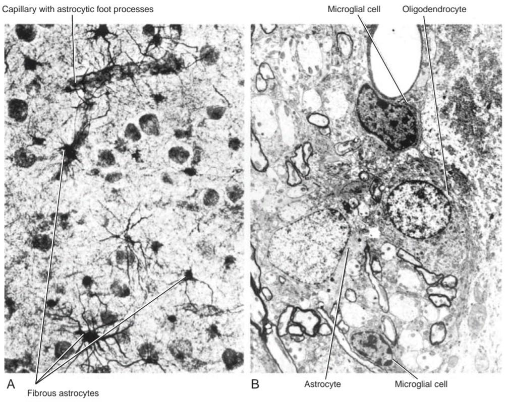
Figure 2-29 A: Photomicrograph of a section of the gray matter of the spinal cord showing fibrous astrocytes. B: Electron micrograph showing an astrocyte. (Courtesy Dr. J. M. Kerns.)
شکل ۲-۲۹ الف: عکس میکروسکوپی از بخشی از ماده خاکستری نخاع که آستروسیتهای فیبری را نشان میدهد. ب: عکس میکروسکوپی الکترونی که یک آستروسیت را نشان میدهد. (با تشکر از دکتر جی. ام. کرنز.)
Protoplasmic astrocytes are found mainly in the gray matter, where their processes pass between the nerve cell bodies (Figs. 2-30 and 2-31). The processes are shorter, thicker, and more branched than those of the fibrous astrocyte, and their cytoplasm contains fewer filaments.
آستروسیتهای پروتوپلاسمی عمدتاً در ماده خاکستری یافت میشوند، جایی که زوائد آنها از بین اجسام سلولهای عصبی عبور میکند (شکلهای 2-30 و 2-31). این زوائد کوتاهتر، ضخیمتر و شاخهدارتر از زوائد آستروسیتهای فیبری هستند و سیتوپلاسم آنها حاوی رشتههای کمتری است.

Figure 2-30 Photomicrograph of a protoplasmic astrocyte in the cerebral cortex.
شکل ۲-۳۰ عکس میکروسکوپی از یک آستروسیت پروتوپلاسمی در قشر مغز.
Many of the processes of astrocytes end in expansions on blood vessels (perivascular feet), where they form an almost complete covering on the external surface of capillaries. Large numbers of astrocytic processes are interwoven at the outer and inner surfaces of the CNS, where they form the outer and inner glial limiting membranes. Thus, the outer glial limiting membrane is found beneath the pia mater, and the inner glial limiting membrane lies beneath the ependyma lining the ventricles of the brain and the central canal of the spinal cord.
بسیاری از زوائد آستروسیتها به گسترشهایی روی رگهای خونی (پاهای اطراف عروقی) ختم میشوند، جایی که تقریباً پوشش کاملی روی سطح خارجی مویرگها تشکیل میدهند. تعداد زیادی از زوائد آستروسیتی در سطوح خارجی و داخلی سیستم عصبی مرکزی (CNS) به هم بافته شدهاند، جایی که غشاهای محدودکننده گلیال خارجی و داخلی را تشکیل میدهند. بنابراین، غشای محدودکننده گلیال خارجی در زیر نرمشامه یافت میشود و غشای محدودکننده گلیال داخلی در زیر اپاندیم قرار دارد که بطنهای مغز و کانال مرکزی نخاع را میپوشاند.
Astrocytic processes are also found in large numbers around the initial segment of most axons and in the bare segments of axons at the nodes of Ranvier. Axon terminals at many sites are separated from other nerve cells and their processes by an envelope of astrocytic processes.
زوائد آستروسیتی همچنین به تعداد زیاد در اطراف قطعه اولیه اکثر آکسونها و در بخشهای لخت آکسونها در گرههای رانویه یافت میشوند. پایانههای آکسون در بسیاری از نقاط توسط پوششی از زوائد آستروسیتی از سایر سلولهای عصبی و زوائد آنها جدا میشوند.
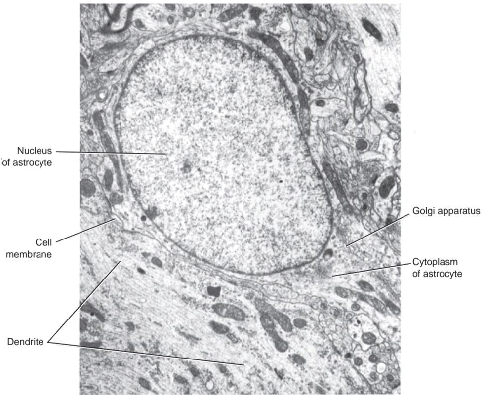
Figure 2-31 Electron micrograph of a protoplasmic astrocyte in the cerebral cortex. (Courtesy Dr. A. Peters.)
شکل ۲-۳۱ تصویر میکروسکوپ الکترونی از یک آستروسیت پروتوپلاسمی در قشر مغز. (با تشکر از دکتر A. Peters.)
Astrocyte Functions
Astrocytes, with their branching processes, form a supporting framework for the nerve cells and nerve fibers. Their processes are functionally coupled at gap junctions. In the embryo, they serve as a scaffolding for the migration of immature neurons. By covering the synaptic contacts between neurons, they may serve as electrical insulators preventing axon terminals from influencing neighboring and unrelated neurons. They may even form barriers for the spread of neurotransmitter substances released at synapses. Astrocytes have been shown to be affected by GABA and glutamic acid secreted by the nerve terminals, thereby limiting the influence of these neurotransmitters. Astrocytes appear to be able to take up excess K+ ions from the extracellular space so that they may have an important function during repetitive firing of a neuron. They store glycogen within their cytoplasm. The glycogen can be broken down into glucose and even further into lactate, both of which are released to surrounding neurons in response to norepinephrine.
عملکردهای آستروسیت
آستروسیتها، با زوائد شاخهدار خود، چارچوبی پشتیبان برای سلولهای عصبی و فیبرهای عصبی تشکیل میدهند. زوائد آنها از نظر عملکردی در اتصالات شکافدار به هم متصل هستند. در جنین، آنها به عنوان داربستی برای مهاجرت نورونهای نابالغ عمل میکنند. با پوشاندن تماسهای سیناپسی بین نورونها، آنها ممکن است به عنوان عایقهای الکتریکی عمل کنند و از تأثیر پایانههای آکسون بر نورونهای همسایه و غیرمرتبط جلوگیری کنند. آنها حتی ممکن است موانعی برای گسترش مواد انتقالدهنده عصبی آزاد شده در سیناپسها ایجاد کنند. نشان داده شده است که آستروسیتها تحت تأثیر گابا و اسید گلوتامیک ترشح شده توسط پایانههای عصبی قرار میگیرند و در نتیجه تأثیر این انتقالدهندههای عصبی را محدود میکنند. به نظر میرسد آستروسیتها میتوانند یونهای +K اضافی را از فضای خارج سلولی جذب کنند تا بتوانند در طول شلیک مکرر یک نورون عملکرد مهمی داشته باشند. آنها گلیکوژن را در سیتوپلاسم خود ذخیره میکنند. گلیکوژن میتواند به گلوکز و حتی بیشتر به لاکتات تجزیه شود که هر دو در پاسخ به نوراپی نفرین به نورونهای اطراف آزاد میشوند.
Astrocytes may serve as phagocytes by taking up degenerating synaptic axon terminals. Following the death of neurons due to disease, astrocytes proliferate and fill in the spaces previously occupied by the neurons, a process called replacement gliosis. Astrocytes possibly serve as a conduit for the passage of metabolites or raw materials from blood capillaries to the neurons through their perivascular feet. Because astrocytes are linked together by gap junctions, they enable ions to pass from one cell to another without entering the extracellular space. Astrocytes may produce substances that have a trophic influence on neighboring neurons. Recent research has suggested that astrocytes secrete cytokines that regulate the activity of immune cells entering the nervous system in disease. Finally, astrocytes play an important role in the structure of the blood-brain barrier. Here, the astrocyte processes terminate as expanded feet at the basement membrane of blood vessels.
آستروسیتها ممکن است با جذب پایانههای آکسون سیناپسی در حال تخریب، به عنوان فاگوسیت عمل کنند. پس از مرگ نورونها به دلیل بیماری، آستروسیتها تکثیر میشوند و فضاهایی را که قبلاً توسط نورونها اشغال شده بود، پر میکنند، فرآیندی که گلیوز جایگزینی نامیده میشود. آستروسیتها احتمالاً به عنوان مجرایی برای عبور متابولیتها یا مواد خام از مویرگهای خونی به نورونها از طریق پاهای اطراف عروقی خود عمل میکنند. از آنجا که آستروسیتها توسط اتصالات شکافدار به هم متصل هستند، یونها را قادر میسازند تا بدون ورود به فضای خارج سلولی از یک سلول به سلول دیگر منتقل شوند. آستروسیتها ممکن است موادی تولید کنند که تأثیر تغذیهای بر نورونهای همسایه دارند. تحقیقات اخیر نشان داده است که آستروسیتها سیتوکینهایی ترشح میکنند که فعالیت سلولهای ایمنی وارد شده به سیستم عصبی را در بیماری تنظیم میکنند. در نهایت، آستروسیتها نقش مهمی در ساختار سد خونی-مغزی دارند. در اینجا، فرآیندهای آستروسیت به عنوان پاهای منبسط شده در غشای پایه رگهای خونی خاتمه مییابند.
Oligodendrocytes
Oligodendrocytes have small cell bodies and a few delicate processes; their cytoplasm does not contain filaments. Oligodendrocytes are found in rows along myelinated nerve fibers and surround nerve cell bodies (Fig. 2-32). Electron micrographs show the processes of a single oligodendrocyte joining the myelin sheaths of several nerve fibers (Fig. 2-33). However, only one process joins the myelin between two adjacent nodes of Ranvier.
الیگودندروسیتها
الیگودندروسیتها دارای جسم سلولی کوچک و چند زائده ظریف هستند؛ سیتوپلاسم آنها حاوی رشته نیست. الیگودندروسیتها در ردیفهایی در امتداد فیبرهای عصبی میلیندار یافت میشوند و اجسام سلولهای عصبی را احاطه میکنند (شکل 2-32). تصاویر میکروسکوپ الکترونی، زائدههای یک الیگودندروسیت واحد را نشان میدهند که به غلافهای میلین چندین فیبر عصبی متصل میشود (شکل 2-33). با این حال، تنها یک زائده، میلین را بین دو گره مجاور رانویه متصل میکند.

Figure 2-32 A: Photomicrograph of a group of oligodendrocytes. B: Electron micrograph of two oligodendrocytes. (Courtesy Dr. J. M. Kerns.)
شکل ۲-۳۲ الف: تصویر میکروسکوپی از گروهی از الیگودندروسیتها. ب: تصویر میکروسکوپی الکترونی از دو الیگودندروسیت. (با تشکر از دکتر جی. ام. کرنز.)
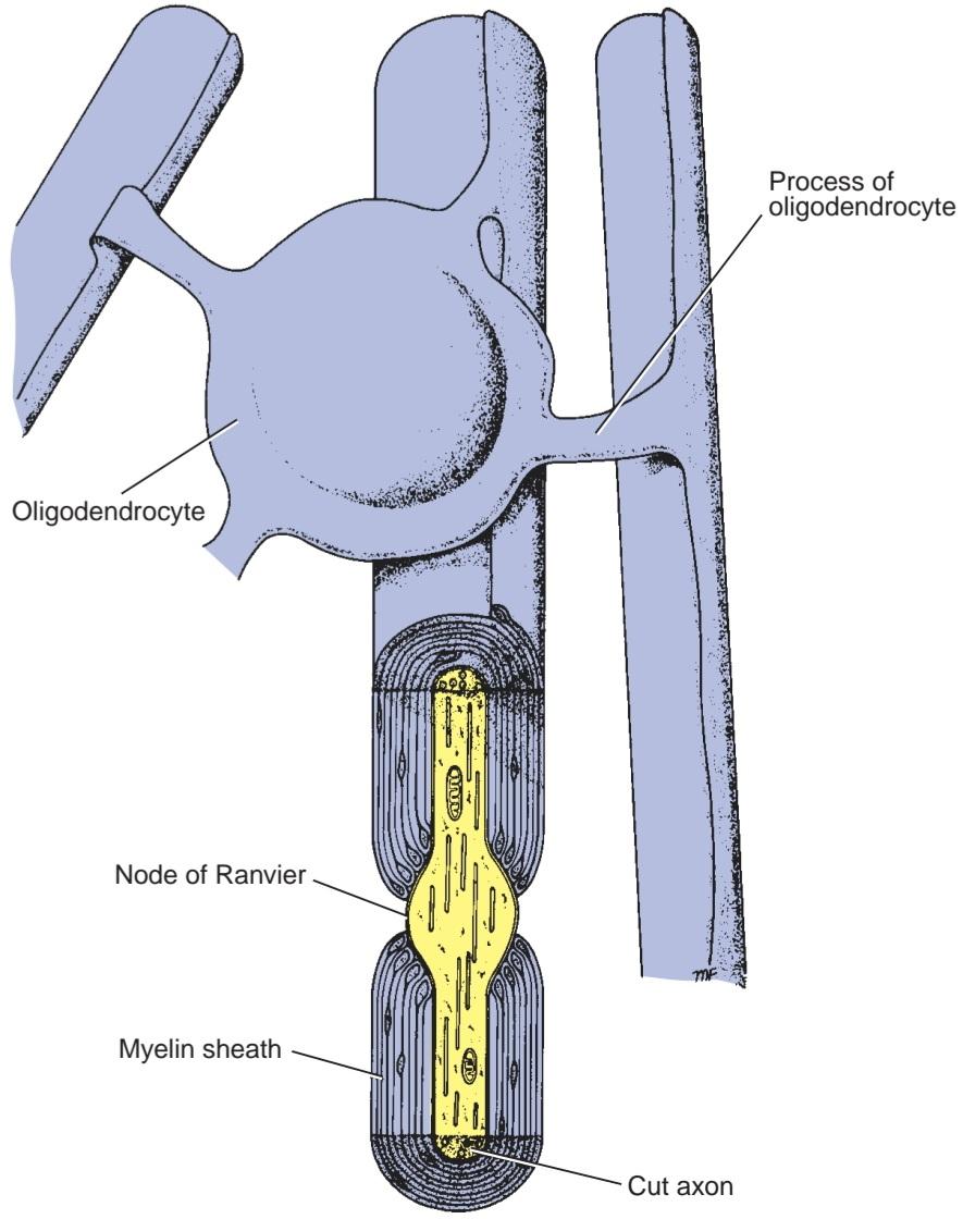
Figure 2-33 A single oligodendrocyte whose processes are continuous with the myelin sheaths of four nerve fibers within the central nervous system.
شکل ۲-۳۳ یک الیگودندروسیت واحد که زوائد آن در امتداد غلافهای میلین چهار فیبر عصبی در سیستم عصبی مرکزی قرار دارند.
Oligodendrocyte Functions
Oligodendrocytes are responsible for the formation of the myelin sheath of nerve fibers in the CNS, much as the myelin of peripheral nerves is formed from Schwann cells. This formation and maintenance of myelin around many CNS axons provides them with an insulating coat and greatly increases the speed of nerve conduction along them (see p. 47). Because oligodendrocytes have several processes, unlike Schwann cells, they can each form several internodal segments of myelin on the same or different axons. A single oligodendrocyte can form as many as 60 internodal segments. Note that, unlike Schwann cells in the peripheral nervous system (PNS), oligodendrocytes and their associated axons are not surrounded by a basement membrane. Myelination begins at about the 16th week of intrauterine life and continues postnatally until practically all the major nerve fibers are myelinated by the time the child is walking.
وظایف الیگودندروسیتها
الیگودندروسیتها مسئول تشکیل غلاف میلین فیبرهای عصبی در سیستم عصبی مرکزی هستند، همانطور که میلین اعصاب محیطی از سلولهای شوان تشکیل میشود. این تشکیل و نگهداری میلین در اطراف بسیاری از آکسونهای سیستم عصبی مرکزی، یک پوشش عایق برای آنها فراهم میکند و سرعت هدایت عصبی را در امتداد آنها به میزان زیادی افزایش میدهد (به صفحه ۴۷ مراجعه کنید). از آنجایی که الیگودندروسیتها برخلاف سلولهای شوان دارای چندین زائده هستند، هر کدام میتوانند چندین قطعه بین گرهای میلین را روی آکسونهای یکسان یا متفاوت تشکیل دهند. یک الیگودندروسیت واحد میتواند تا ۶۰ قطعه بین گرهای تشکیل دهد. توجه داشته باشید که برخلاف سلولهای شوان در سیستم عصبی محیطی (PNS)، الیگودندروسیتها و آکسونهای مرتبط با آنها توسط غشای پایه احاطه نشدهاند. میلینسازی از حدود هفته شانزدهم زندگی داخل رحمی شروع میشود و پس از تولد ادامه مییابد تا زمانی که عملاً تمام فیبرهای عصبی اصلی تا زمانی که کودک راه میرود، میلیندار شوند.
Oligodendrocytes also surround nerve cell bodies (satellite oligodendrocytes) and probably have a similar function to the satellite or capsular cells of peripheral sensory ganglia. They are thought to influence the biochemical environment of neurons.
الیگودندروسیتها همچنین اجسام سلولهای عصبی (الیگودندروسیتهای ماهوارهای) را احاطه کردهاند و احتمالاً عملکردی مشابه سلولهای ماهوارهای یا کپسولی گانگلیونهای حسی محیطی دارند. تصور میشود که آنها بر محیط بیوشیمیایی نورونها تأثیر میگذارند.
Microglia
The microglial cells are embryologically unrelated to the other neuroglial cells and are derived from mac- rophages outside the nervous system. They are the smallest of the neuroglial cells and are found scattered throughout the CNS (Fig. 2-34). Wavy branching processes arise from their small cell bodies that give off numerous spinelike projections. They closely resemble connective tissue macrophages. They migrate into the nervous system during fetal life. Microglial cells increase in number in the presence of damaged nervous tissue resulting from trauma and ischemic injury and in the presence of diseases including Alzheimer disease, Parkinson disease, multiple sclerosis, and AIDS. Many of these new cells are monocytes that have migrated from the blood.
میکروگلیا
سلولهای میکروگلیا از نظر جنینی هیچ ارتباطی با سایر سلولهای نوروگلیا ندارند و از ماکروفاژهای خارج از سیستم عصبی مشتق شدهاند. آنها کوچکترین سلولهای نوروگلیا هستند و در سراسر سیستم عصبی مرکزی پراکنده شدهاند (شکل 2-34). از اجسام سلولی کوچک آنها، زوائد شاخهای موجی شکل ناشی میشود که برآمدگیهای خارمانند متعددی ایجاد میکنند. آنها شباهت زیادی به ماکروفاژهای بافت همبند دارند. آنها در طول زندگی جنینی به سیستم عصبی مهاجرت میکنند. تعداد سلولهای میکروگلیا در حضور بافت عصبی آسیبدیده ناشی از تروما و آسیب ایسکمیک و در حضور بیماریهایی از جمله بیماری آلزایمر، بیماری پارکینسون، مولتیپل اسکلروزیس و ایدز افزایش مییابد. بسیاری از این سلولهای جدید، مونوسیتهایی هستند که از خون مهاجرت کردهاند.
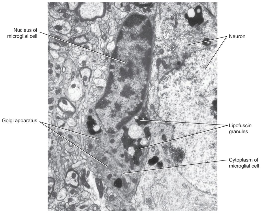
Figure 2-34 Electron micrograph of a microglial cell in the cerebral cortex. (Courtesy Dr. A. Peters.)
شکل ۲-۳۴ تصویر میکروسکوپ الکترونی از یک سلول میکروگلیا در قشر مغز. (با تشکر از دکتر ای. پیترز.)
Microglia Function
Microglial cells in the normal brain and spinal cord appear to be inactive and are sometimes called resting microglial cells. In inflammatory disease of the CNS, they become the immune effector cells. They retract their processes and migrate to the site of the lesion. Here, they proliferate and become antigen-presenting cells, which, together with the invading T lymphocytes, confront invading organisms. They are also actively phagocytic; their cytoplasm becomes filled with lipids and cell remnants. The microglial cells are joined by monocytes from neighboring blood vessels.
عملکرد میکروگلیا
سلولهای میکروگلیا در مغز و نخاع طبیعی غیرفعال به نظر میرسند و گاهی اوقات سلولهای میکروگلیا در حال استراحت نامیده میشوند. در بیماری التهابی سیستم عصبی مرکزی، آنها به سلولهای مؤثر ایمنی تبدیل میشوند. آنها زوائد خود را جمع میکنند و به محل ضایعه مهاجرت میکنند. در اینجا، آنها تکثیر میشوند و به سلولهای ارائه دهنده آنتیژن تبدیل میشوند که همراه با لنفوسیتهای T مهاجم، با ارگانیسمهای مهاجم مقابله میکنند. آنها همچنین به طور فعال فاگوسیتوز میکنند. سیتوپلاسم آنها پر از لیپیدها و بقایای سلولی میشود. سلولهای میکروگلیا توسط مونوسیتهای رگهای خونی مجاور به هم متصل میشوند.
Ependyma
Ependymal cells line the cavities of the brain and the central canal of the spinal cord. They form a single layer of cells that are cuboidal or columnar in shape and possess microvilli and cilia (Fig. 2-35). The cilia are often motile, and their movements contribute to the flow of the cerebrospinal fluid (CSF). The bases of the ependymal cells lie on the internal glial limiting membrane.
اپاندیما
سلولهای اپاندیمال حفرههای مغز و کانال مرکزی نخاع را میپوشانند. آنها یک لایه واحد از سلولها را تشکیل میدهند که به شکل مکعبی یا ستونی هستند و دارای میکروویلی و مژک میباشند (شکل 2-35). مژکها اغلب متحرک هستند و حرکات آنها به جریان مایع مغزی نخاعی (CSF) کمک میکند. پایههای سلولهای اپاندیمال روی غشای محدودکننده گلیال داخلی قرار دارند.
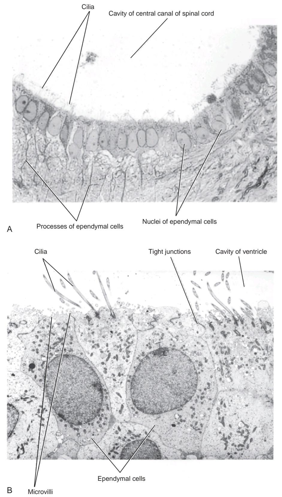
Figure 2-35 A: Photomicrograph of ependymal cells lining the central canal of the spinal cord. B: Electron micrograph of ependymal cells lining the cavity of the third ventricle. (Courtesy Dr. J. M. Kerns.)
شکل ۲-۳۵ الف: عکس میکروسکوپی از سلولهای اپاندیمی که مجرای مرکزی نخاع را میپوشانند. ب: عکس میکروسکوپی الکترونی از سلولهای اپاندیمی که حفره بطن سوم را میپوشانند. (با تشکر از دکتر جی. ام. کرنز.)
Ependymal cells can be divided into three groups:
سلولهای اپندیمی را میتوان به سه گروه تقسیم کرد:
1. Ependymocytes line the ventricles of the brain and the central canal of the spinal cord and are in contact with the CSF. Their adjacent surfaces have gap junctions, but the CSF is in free communication with CNS intercellular spaces.
۱. اپندیموسیتها بطنهای مغز و کانال مرکزی نخاع را میپوشانند و با مایع مغزی نخاعی (CSF) در تماس هستند. سطوح مجاور آنها دارای اتصالات شکافدار هستند، اما مایع مغزی نخاعی با فضاهای بین سلولی سیستم عصبی مرکزی (CNS) ارتباط آزاد دارد.
2. Tanycytes line the floor of the third ventricle overlying the median eminence of the hypothalamus. These cells have long basal processes that pass between the cells of the median eminence and place endfeet on blood capillaries.
۲. تانیسیتها کف بطن سوم را که روی برجستگی میانی هیپوتالاموس قرار دارد، میپوشانند. این سلولها دارای زوائد پایهای بلندی هستند که از بین سلولهای برجستگی میانی عبور میکنند و پاهای انتهایی را روی مویرگهای خونی قرار میدهند.
3. Choroidal epithelial cells cover the surfaces of the choroid plexuses. The sides and bases of these cells are thrown into folds; near their luminal surfaces, the cells are held together by tight junctions that encircle the cells. The presence of tight junctions prevents the leakage of CSF into the underlying tissues.
۳. سلولهای اپیتلیال مشیمیهای سطوح شبکههای مشیمیهای را میپوشانند. کنارهها و پایههای این سلولها به صورت چینخوردههایی درآمده است. در نزدیکی سطوح لومینال آنها، سلولها توسط اتصالات محکمی که سلولها را احاطه کردهاند، در کنار هم نگه داشته میشوند. وجود اتصالات محکم از نشت مایع مغزی نخاعی به بافتهای زیرین جلوگیری میکند.
Ependymal Cell Functions
Ependymocytes assist in CSF circulation within the cavities of the brain and the central canal of the spinal cord by the movements of the cilia. The microvilli on the free surfaces of the ependymocytes indicate that they also have an absorptive function. Tanycytes are thought to transport chemical substances from the CSF to the hypophyseal portal system. In this manner, they may play a part in the control of the hormone production by the anterior lobe of the pituitary. Choroidal epithelial cells are involved in the production and secretion of CSF from the choroid plexuses.
عملکردهای سلولهای اپندیمی
اپندیموسیتها با حرکات مژکها به گردش مایع مغزی نخاعی در حفرههای مغز و کانال مرکزی نخاع کمک میکنند. میکروویلیهای روی سطوح آزاد اپندیموسیتها نشان میدهد که آنها عملکرد جذبی نیز دارند. تصور میشود که تانیسیتها مواد شیمیایی را از مایع مغزی نخاعی به سیستم پورتال هیپوفیز منتقل میکنند. به این ترتیب، آنها ممکن است در کنترل تولید هورمون توسط لوب قدامی هیپوفیز نقش داشته باشند. سلولهای اپیتلیال مشیمیه در تولید و ترشح مایع مغزی نخاعی از شبکههای مشیمیه نقش دارند.
EXTRACELLULAR SPACE
When nervous tissue is examined under an electron microscope, a very narrow gap separates the neurons and the neuroglial cells. These gaps are linked together and filled with tissue fluid; they are called the extracellular space. The extracellular space is in almost direct continuity with the CSF in the subarachnoid space externally and with the CSF in the ventricles of the brain and the central canal of the spinal cord internally. The extracellular space also surrounds the blood capillaries in the brain and spinal cord. (The CNS does not have lymphatic capillaries.)
فضای خارج سلولی
هنگامی که بافت عصبی زیر میکروسکوپ الکترونی بررسی میشود، یک شکاف بسیار باریک، نورونها و سلولهای نوروگلیا را از هم جدا میکند. این شکافها به هم متصل شده و با مایع بافتی پر شدهاند؛ به آنها فضای خارج سلولی گفته میشود. فضای خارج سلولی تقریباً در امتداد مستقیم با مایع مغزی نخاعی (CSF) در فضای زیر عنکبوتیه از خارج و با مایع مغزی نخاعی در بطنهای مغز و کانال مرکزی نخاع از داخل است. فضای خارج سلولی همچنین مویرگهای خونی را در مغز و نخاع احاطه کرده است. (سیستم عصبی مرکزی مویرگهای لنفاوی ندارد.)
The extracellular space thus provides a pathway for the exchange of ions and molecules between the blood and the neurons and glial cells. The plasma membrane of the endothelial cells of most capillaries is impermeable to many chemicals, and this forms the blood-brain barrier.
بنابراین فضای خارج سلولی مسیری را برای تبادل یونها و مولکولها بین خون و نورونها و سلولهای گلیال فراهم میکند. غشای پلاسمایی سلولهای اندوتلیال اکثر مویرگها نسبت به بسیاری از مواد شیمیایی نفوذناپذیر است و این سد خونی-مغزی را تشکیل میدهد.
«فصل دوم کتاب نورواناتومی بالینی اسنل»
🚀 با ما همراه شوید!
تازهترین مطالب و آموزشهای مغز و اعصاب را از دست ندهید. با فالو کردن کانال تلگرام آیندهنگاران مغز، از ما حمایت کنید!



