نوروآناتومی بالینی اسنل | رشتههای عصبی و عصبدهی محیطی – نکات و کاربردهای بالینی


دعای مطالعه [ نمایش ]
بِسْمِ الله الرَّحْمنِ الرَّحیمِ
اَللّهُمَّ اَخْرِجْنى مِنْ ظُلُماتِ الْوَهْمِ
خدايا مرا بيرون آور از تاريكىهاى وهم،
وَ اَكْرِمْنى بِنُورِ الْفَهْمِ
و به نور فهم گرامى ام بدار،
اَللّهُمَّ افْتَحْ عَلَيْنا اَبْوابَ رَحْمَتِكَ
خدايا درهاى رحمتت را به روى ما بگشا،
وَانْشُرْ عَلَيْنا خَزائِنَ عُلُومِكَ بِرَحْمَتِكَ يا اَرْحَمَ الرّاحِمينَ
و خزانههاى علومت را بر ما باز كن به امید رحمتت اى مهربانترين مهربانان.
کتاب «نوروآناتومی بالینی اسنل» اثر دکتر ریچارد اس. اسنل، با ویرایش جدید دکتر رایان اسپلیتگربر، همچنان بهعنوان مرجع طلایی این حوزه شناخته میشود. این اثر با تلفیق تازهترین پژوهشها و کاربردهای بالینی، پلی استوار میان دانش پایه و عمل پزشکی بنا میکند.
تلاشهای علمی و سالها تدریس ارزشمند استاد گرانقدر دکتر محمدتقی جغتایی الهامبخش ترجمه و گسترش این دانش در ایران بوده است.
ترجمه دقیق این کتاب توسط برند علمی آیندهنگاران مغز به مدیریت داریوش طاهری، امکان دسترسی فارسیزبانان به مرزهای نوین علوم اعصاب را فراهم کرده و رسالتی علمی در جهت شناخت عمیقتر مغز و ترسیم آیندهای روشنتر بر عهده گرفته است.
»» فصل ۳: رشتههای عصبی و عصبدهی محیطی
»» Chapter 3: Nerve Fibers and Peripheral Innervation
Clinical Notes
Neuronal Response to Injury
The survival of a neuron’s cytoplasm depends on its being connected, however indirectly, with the nucleus. The nucleus plays a key role in the synthesis of proteins, which pass into the cell processes and replace proteins that have been metabolized by cell activity. Thus, the cytoplasm of axons and dendrites will undergo degeneration quickly if these processes are separated from the nerve cell body.
یادداشتهای بالینی
پاسخ عصبی به آسیب
بقای سیتوپلاسم یک نورون به اتصال آن، هرچند غیرمستقیم، به هسته بستگی دارد. هسته نقش کلیدی در سنتز پروتئینها دارد که به فرآیندهای سلولی منتقل میشوند و جایگزین پروتئینهایی میشوند که توسط فعالیت سلولی متابولیزه شدهاند. بنابراین، اگر این فرآیندها از جسم سلولی عصبی جدا شوند، سیتوپلاسم آکسونها و دندریتها به سرعت دچار انحطاط میشوند.
Nerve Cell Body Injury
Severe damage of the nerve cell body due to trauma, interference with the blood supply, or disease may result in degeneration of the entire neuron, including its dendrites and synaptic endings. In the brain and spinal cord, the neuronal debris and the fragments of myelin (if the processes are myelinated) are engulfed and phagocytosed by the microglial cells. Later, the neighboring astrocytes proliferate and replace the neuron with scar tissue.
آسیب جسم سلولی عصبی
آسیب شدید جسم سلولی عصبی به دلیل ضربه، اختلال در خونرسانی یا بیماری ممکن است منجر به تخریب کل نورون، از جمله دندریتها و پایانههای سیناپسی آن شود. در مغز و نخاع، بقایای نورونی و قطعات میلین (اگر زوائد میلیندار باشند) توسط سلولهای میکروگلیا بلعیده و فاگوسیتوز میشوند. بعداً، آستروسیتهای همسایه تکثیر میشوند و نورون را با بافت اسکار جایگزین میکنند.
In the peripheral nervous system (PNS), the tissue macrophages remove the debris, and the local fibroblasts replace the neuron with scar tissue.
در سیستم عصبی محیطی (PNS)، ماکروفاژهای بافتی، بقایای عصبی را حذف میکنند و فیبروبلاستهای موضعی، نورون را با بافت اسکار جایگزین میکنند.
Nerve Cell Process Injury
If the axon of the nerve cell is divided, degenerative changes will take place in (1) the distal segment that is separated from the cell body, (2) a portion of the axon proximal to the injury, and (3) possibly the cell body from which the axon arises.
آسیب به فرآیند سلول عصبی
اگر آکسون سلول عصبی تقسیم شود، تغییرات دژنراتیو در (1) بخش انتهایی که از جسم سلولی جدا شده است، (2) بخشی از آکسون که نزدیک به محل آسیب قرار دارد، و (3) احتمالاً جسم سلولی که آکسون از آن منشأ میگیرد، رخ خواهد داد.
CHANGES IN THE DISTAL SEGMENT OF THE AXON
The changes spread distally from the site of the lesion (Fig. 3-44) and include its terminations; the process is referred to as wallerian degeneration. In the PNS, on the first day, the axon becomes swollen and irregular; by the third or fourth day, the axon is broken into fragments, and the debris is digested by the surrounding Schwann cells and tissue macrophages. The entire axon is destroyed within a week.
تغییرات در بخش انتهایی آکسون
این تغییرات از محل ضایعه به سمت انتهایی گسترش مییابند (شکل 3-44) و شامل انتهای آن میشوند؛ این فرآیند دژنراسیون والرین نامیده میشود. در سیستم عصبی محیطی (PNS)، در روز اول، آکسون متورم و نامنظم میشود؛ تا روز سوم یا چهارم، آکسون به قطعاتی شکسته میشود و بقایای آن توسط سلولهای شوان اطراف و ماکروفاژهای بافتی هضم میشود. کل آکسون ظرف یک هفته از بین میرود.
Figure 3-44 A-D: Degeneration and regeneration in a divided nerve.
شکل ۳-۴۴ A-D: تخریب و بازسازی در یک عصب تقسیم شده.
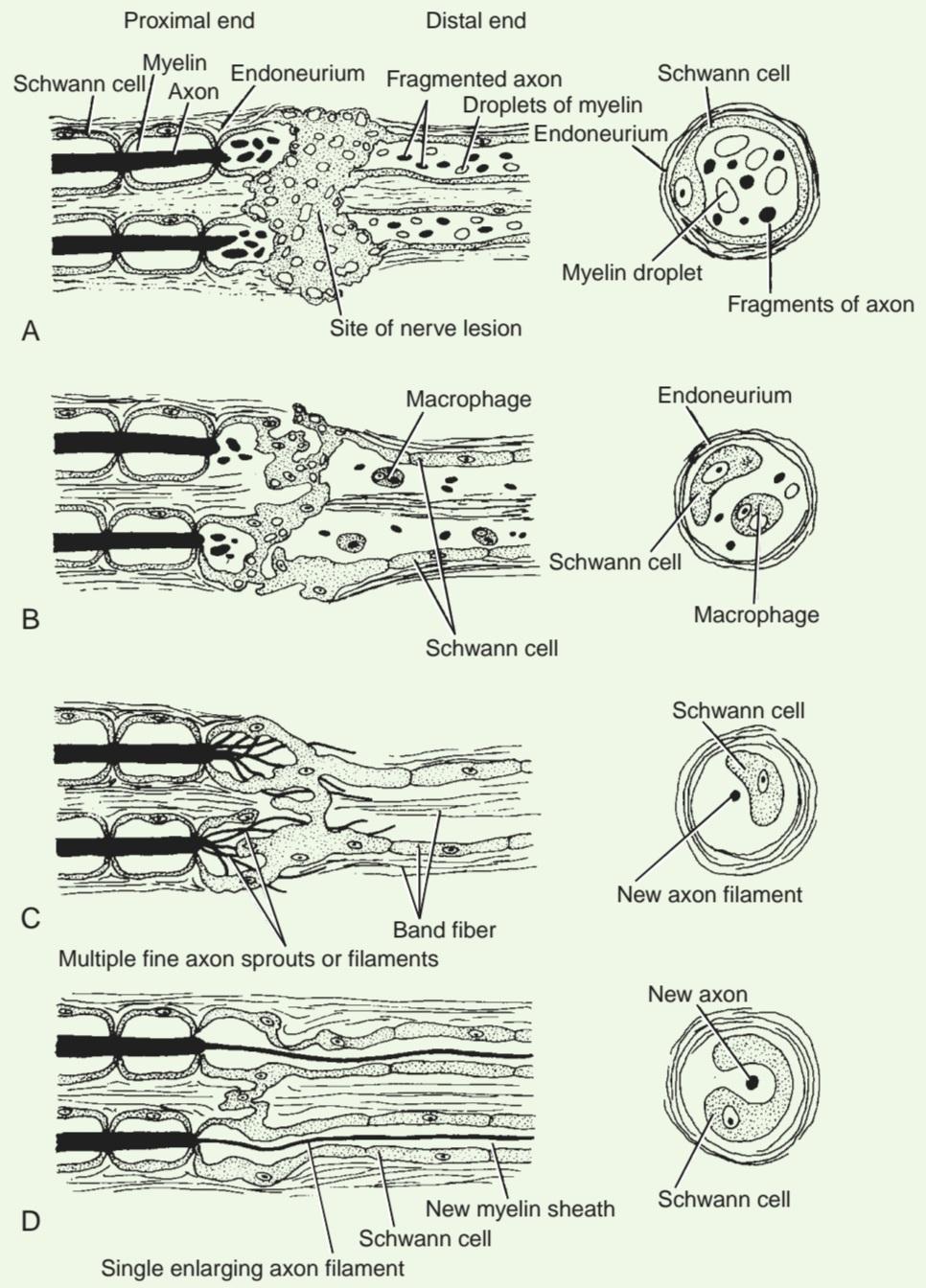
Meanwhile, the myelin sheath slowly breaks down, and lipid droplets appear within the Schwann cell cytoplasm. Later, the droplets are extruded from the Schwann cell and subsequently phagocytosed by tissue macrophages. The Schwann cells now begin to proliferate rapidly and become arranged in parallel cords within the persistent basement membrane. The endoneurial sheath and the contained cords of Schwann cells are referred to as a band fiber. If regeneration does not occur, the axon and the Schwann cells are replaced by fibrous tissue produced by local fibroblasts.
در همین حال، غلاف میلین به آرامی تجزیه میشود و قطرات چربی در سیتوپلاسم سلول شوان ظاهر میشوند. بعداً، قطرات از سلول شوان خارج شده و متعاقباً توسط ماکروفاژهای بافتی فاگوسیتوز میشوند. سلولهای شوان اکنون به سرعت شروع به تکثیر میکنند و به صورت طنابهای موازی در غشای پایه پایدار قرار میگیرند. غلاف اندونوریال و طنابهای حاوی سلولهای شوان به عنوان فیبر نواری شناخته میشوند. اگر بازسازی رخ ندهد، آکسون و سلولهای شوان با بافت فیبری تولید شده توسط فیبروبلاستهای موضعی جایگزین میشوند.
In the central nervous system (CNS), degeneration of the axons and the myelin sheaths follows a similar course, and the debris is removed by the phagocytic activity of the microglial cells. Little is known about the role of oligodendrocytes in this process. The astrocytes now proliferate and replace the axons.
در سیستم عصبی مرکزی (CNS)، تخریب آکسونها و غلافهای میلین روند مشابهی را دنبال میکند و بقایای آن توسط فعالیت فاگوسیتیک سلولهای میکروگلیال حذف میشوند. اطلاعات کمی در مورد نقش الیگودندروسیتها در این فرآیند وجود دارد. اکنون آستروسیتها تکثیر شده و جایگزین آکسونها میشوند.
CHANGES IN THE PROXIMAL SEGMENT OF THE AXON
The changes in the proximal segment of the axon are similar to those that take place in the distal segment (see Fig. 3-44) but extend only proximally above the lesion as far as the first node of Ranvier. The proliferating cords of Schwann cells in the peripheral nerves bulge from the cut surfaces of the endoneurial tubes.
تغییرات در بخش پروگزیمال آکسون
تغییرات در بخش پروگزیمال آکسون مشابه تغییراتی است که در بخش دیستال رخ میدهد (شکل 3-44 را ببینید) اما فقط به صورت پروگزیمال بالای ضایعه تا اولین گره رانویر امتداد مییابد. طنابهای تکثیر شونده سلولهای شوان در اعصاب محیطی از سطوح بریده شده لولههای اندونوریال بیرون میزنند.
CHANGES IN THE NERVE CELL BODY
The changes that occur in the cell body following injury to its axon are referred to as retrograde degeneration; the changes that take place in the proximal segment of the axon commonly are included under this heading. The possible reason for these changes is that section of the axon cuts off the cell body from its supply of trophic factors derived from the target organs at the distal end of the axon.
تغییرات در جسم سلولی عصبی
تغییراتی که پس از آسیب به آکسون در جسم سلولی رخ میدهد، دژنراسیون رتروگراد نامیده میشود؛ تغییراتی که در بخش پروگزیمال آکسون رخ میدهد، معمولاً تحت این عنوان قرار میگیرند. دلیل احتمالی این تغییرات این است که آن بخش از آکسون، جسم سلولی را از منبع فاکتورهای تغذیهای مشتق شده از اندامهای هدف در انتهای دیستال آکسون، محروم میکند.
The most characteristic change occurs in the cell body within the first 2 days following injury and reaches its maximum within 2 weeks. The Nissl material becomes fine, granular (Figs. 3-45 and 3-46), and dispersed throughout the cytoplasm, a process known as chromatolysis. Chromatolysis begins near the axon hillock and spreads to all parts of the cell body. In addition, the nucleus moves from its central location toward the periphery of the cell, and the cell body swells and becomes rounded. The degree of chromatolysis and the degree of swelling of the cell are greatest when the injury to the axon is close to the cell body. In some neurons, very severe damage to the axon close to the cell body may lead to death of the neuron. On the other hand, damage to the most distal process may lead to little or no detectable change in the cell body. The dispersal of the Nissl material-that is, the cytoplasmic RNA and the swelling of the cell are caused by cellular edema. The apparent loss of staining affinity of the Nissl material is due to the wide dispersal of the cytoplasmic RNA. The movement of the nucleus away from the center of the cell may be due to the cellular edema.
مشخصترین تغییر در جسم سلولی در عرض ۲ روز اول پس از آسیب رخ میدهد و ظرف ۲ هفته به حداکثر خود میرسد. ماده نیسل ریز و دانهدار میشود (شکلهای ۳-۴۵ و ۳-۴۶) و در سراسر سیتوپلاسم پراکنده میشود، فرآیندی که به عنوان کروماتولیز شناخته میشود. کروماتولیز در نزدیکی تپه آکسون شروع میشود و به تمام قسمتهای جسم سلولی گسترش مییابد. علاوه بر این، هسته از محل مرکزی خود به سمت حاشیه سلول حرکت میکند و جسم سلولی متورم و گرد میشود. درجه کروماتولیز و درجه تورم سلول زمانی که آسیب به آکسون نزدیک جسم سلولی باشد، بیشترین مقدار را دارد. در برخی از نورونها، آسیب بسیار شدید به آکسون نزدیک جسم سلولی ممکن است منجر به مرگ نورون شود. از سوی دیگر، آسیب به انتهاییترین زائده ممکن است منجر به تغییر کم یا بدون تغییر قابل تشخیص در جسم سلولی شود. پراکندگی ماده نیسل – یعنی RNA سیتوپلاسمی و تورم سلول ناشی از ادم سلولی است. از دست رفتن ظاهری میل ترکیبی رنگآمیزی ماده نیسل به دلیل پراکندگی وسیع RNA سیتوپلاسمی است. حرکت هسته از مرکز سلول ممکن است به دلیل ادم سلولی باشد.

Figure 3-45 Photomicrographs of motor neurons of the anterior gray column of the spinal cord. A: Nissl substance in normal neurons. B: Following section of anterior roots of spinal nerve, showing chromatolysis.
شکل ۳-۴۵ فتومیکروگرافهای نورونهای حرکتی ستون خاکستری قدامی نخاع. الف: ماده نیسل در نورونهای طبیعی. ب: برش زیر از ریشههای قدامی عصب نخاعی، کروماتولیز را نشان میدهد.

Figure 3-46 The changes that may take place in a nerve cell body following division of one of its processes.
شکل ۳-۴۶ تغییراتی که ممکن است پس از تقسیم یکی از زوائد جسم سلولی عصبی در آن رخ دهد.
Synaptic terminals are seen to withdraw from the surface of the injured nerve cell body and its dendrites and are replaced by Schwann cells in the PNS and microglial cells or astrocytes in the CNS, a process called synaptic stripping. Its possible causes are (1) loss of plasma membrane adhesiveness following injury and (2) stimulation of the supporting cells by chemicals released from the injured neuron. If the injury is sufficiently great, the cells of the immune system—namely, monocytes and macrophagesmay migrate into the area.
دیده میشود که پایانههای سیناپسی از سطح جسم سلولی عصبی آسیبدیده و دندریتهای آن جدا میشوند و توسط سلولهای شوان در سیستم عصبی محیطی (PNS) و سلولهای میکروگلیال یا آستروسیتها در سیستم عصبی مرکزی (CNS) جایگزین میشوند، فرآیندی که به آن سلب سیناپسی میگویند. علل احتمالی آن عبارتند از (1) از دست دادن چسبندگی غشای پلاسمایی پس از آسیب و (2) تحریک سلولهای پشتیبان توسط مواد شیمیایی آزاد شده از نورون آسیبدیده. اگر آسیب به اندازه کافی شدید باشد، سلولهای سیستم ایمنی – یعنی مونوسیتها و ماکروفاژها – ممکن است به این ناحیه مهاجرت کنند.
Neuronal Recovery
In contrast to the rapid onset of retrograde degeneration, the recovery of the nerve cell body and regeneration of its processes may take several months.
بازیابی نورونی
برخلاف شروع سریع دژنراسیون رتروگراد، بازیابی جسم سلول عصبی و بازسازی فرآیندهای آن ممکن است چندین ماه طول بکشد.
Nerve Cell Body Recovery
The nucleolus moves to the periphery of the nucleus, and polysome clusters reappear in the cytoplasm. This indicates that RNA and protein synthesis is being accelerated in preparation for the reformation of the axon. Thus, the original Nissl structure reconstitutes, cell body swelling decreases, and the nucleus returns to its characteristic central position (Fig. 3-46).
بازیابی جسم سلولی عصبی
هستک به حاشیه هسته منتقل میشود و خوشههای پلیزوم دوباره در سیتوپلاسم ظاهر میشوند. این نشان میدهد که سنتز RNA و پروتئین در آمادهسازی برای بازسازی آکسون تسریع میشود. بنابراین، ساختار نیسل اصلی بازسازی میشود، تورم جسم سلولی کاهش مییابد و هسته به موقعیت مرکزی مشخصه خود باز میگردد (شکل 3-46).
Regeneration of Peripheral Nerve Axons
Regrowth of the axons (motor, sensory, and autonomic) is possible in peripheral nerves and appears to depend on the presence of endoneurial tubes and the special qualities possessed by Schwann cells. Sprouts from the axons grow from the proximal stump and into the distal stump toward the nerve’s end organs. The following mechanisms are thought to be involved: (1) the axons are attracted by chemotropic factors secreted by the Schwann cells in the distal stump, (2) growth-stimulating factors exist within the distal stump, and (3) inhibitory factors are present in the perineurium to inhibit the axons from leaving the nerve.
بازسازی آکسونهای اعصاب محیطی
رشد مجدد آکسونها (حرکتی، حسی و اتونومیک) در اعصاب محیطی امکانپذیر است و به نظر میرسد به وجود لولههای اندونوریال و ویژگیهای خاص سلولهای شوان بستگی دارد. جوانههای آکسونها از کنده پروگزیمال و به داخل کنده دیستال به سمت اندامهای انتهایی عصب رشد میکنند. تصور میشود مکانیسمهای زیر در این امر دخیل باشند: (1) آکسونها توسط عوامل شیمیدرمانی ترشح شده توسط سلولهای شوان در کنده دیستال جذب میشوند، (2) عوامل محرک رشد در کنده دیستال وجود دارند، و (3) عوامل مهارکننده در پرینوریوم وجود دارند تا مانع خروج آکسونها از عصب شوند.
The satisfactory regeneration of axons and the return of normal function depend on the following factors:
بازسازی رضایتبخش آکسونها و بازگشت عملکرد طبیعی به عوامل زیر بستگی دارد:
1. In crush nerve injuries, in which the axon is divided or its blood supply has been interfered with but the endoneurial sheaths remain intact, regeneration is likely.
۱. در آسیبهای عصبی ناشی از له شدن عصب، که در آن آکسون تقسیم شده یا خونرسانی آن مختل شده است اما غلافهای اندونوریال دستنخورده باقی ماندهاند، احتمال بازسازی وجود دارد.
2. In nerves that have been completely severed, the chance of recovery is much less because the regenerating fibers from the proximal stump may be directed to an incorrect destination in the distal stump-that is, cutaneous fibers entering incorrect nerve endings or motor nerves supplying incorrect muscles.
۲. در اعصابی که کاملاً قطع شدهاند، احتمال بهبودی بسیار کمتر است زیرا الیاف در حال بازسازی از ریشه پروگزیمال ممکن است به مقصد نادرستی در ریشه دیستال هدایت شوند – یعنی الیاف پوستی وارد انتهای عصبی نادرست یا اعصاب حرکتی که عضلات نادرست را تغذیه میکنند، شوند.
3. If the distance between the proximal and distal stumps of the completely severed nerve is greater than a few millimeters or the gap becomes filled with proliferating fibrous tissue or is simply filled by adjacent muscles that bulge into the gap, then the chances of recovery are very poor. The outgrowing axonal sprouts escape into the surrounding connective tissue and form a tangled mass or neuroma. In these cases, early close surgical approximation of the severed ends, if possible, greatly facilitates the chances of recovery.
۳. اگر فاصله بین انتهای پروگزیمال و دیستال عصب کاملاً قطع شده بیش از چند میلیمتر باشد یا شکاف با بافت فیبری در حال تکثیر پر شود یا به سادگی توسط عضلات مجاور که به داخل شکاف برآمده میشوند، پر شود، احتمال بهبودی بسیار کم است. جوانههای آکسونی که از محل خود خارج میشوند، به بافت همبند اطراف فرار میکنند و یک توده درهمتنیده یا نوروم تشکیل میدهند. در این موارد، در صورت امکان، نزدیک کردن زودهنگام انتهای قطع شده با جراحی، احتمال بهبودی را تا حد زیادی افزایش میدهد.
4. When mixed nerves (those containing sensory, motor, and autonomic fibers) are completely severed, the chances of a good recovery are very much less than when the nerve is purely sensory or purely motor. The reason for this is that the regenerating fibers from the proximal stump may be guided to an incorrect destination in the distal stump; for example, cutaneous fibers may enter motor endoneurial tubes and vice versa.
۴. وقتی اعصاب مختلط (اعصابی که حاوی فیبرهای حسی، حرکتی و اتونوم هستند) کاملاً قطع میشوند، احتمال بهبودی خوب بسیار کمتر از زمانی است که عصب کاملاً حسی یا کاملاً حرکتی باشد. دلیل این امر این است که فیبرهای در حال بازسازی از استامپ پروگزیمال ممکن است به مقصد نادرستی در استامپ دیستال هدایت شوند. به عنوان مثال، فیبرهای پوستی ممکن است وارد لولههای اندونوریال حرکتی شوند و برعکس.
5. Inadequate physiotherapy to the paralyzed muscles will result in their degeneration before the regenerating motor axons have reached them.
۵. فیزیوتراپی ناکافی برای عضلات فلج شده منجر به تخریب آنها قبل از رسیدن آکسونهای حرکتی در حال بازسازی به آنها میشود.
6. The presence of infection at the site of the wound will seriously interfere with the process of regeneration.
۶. وجود عفونت در محل زخم به طور جدی در روند بازسازی اختلال ایجاد میکند.
If the proximal and distal stumps of the severed nerve are in close apposition, the following regenerative processes take place (see Fig. 3-44). The Schwann cells, having undergone mitotic division, now fill the space within the basal lamina of the endoneurial tubes of the proximal stump as far proximally as the next node of Ranvier and in the distal stump as far distally as the end organs. Where a small gap exists between the proximal and distal stumps, the multiplying Schwann cells form a number of cords to bridge the gap.
اگر استامپهای پروگزیمال و دیستال عصب قطع شده در مجاورت نزدیک باشند، فرآیندهای بازسازی زیر رخ میدهد (شکل ۳-۴۴ را ببینید). سلولهای شوان، که تقسیم میتوزی را پشت سر گذاشتهاند، اکنون فضای داخل لایه پایه لولههای اندونوریال استامپ پروگزیمال را تا نزدیکی گره بعدی رانویه و در استامپ دیستال تا نزدیکی اندامهای انتهایی پر میکنند. در جایی که شکاف کوچکی بین استامپهای پروگزیمال و دیستال وجود دارد، سلولهای شوان در حال تکثیر، تعدادی طناب تشکیل میدهند تا این شکاف را پر کنند.
Each proximal axon end now gives rise to multiple fine sprouts or filaments with bulbous tips. These filaments, as they grow, advance along the clefts between the Schwann cells and thus cross the interval between the proximal and distal nerve stumps. Many such filaments now enter the proximal end of each endoneurial tube and grow distally in contact with the Schwann cells (Fig. 3-47). Although the filaments from many different axons may enter a single endoneurial tube, only one filament persists, the remainder degenerate, and that one filament grows distally to reinnervate a motor or sensory end organ. While crossing the gap between the severed nerve ends, many filaments fail to enter an endoneurial tube and grow out into the surrounding connective tissue. Note that the formation of multiple sprouts or filaments from a single proximal axon greatly increases the chances that a neuron will become connected to a sensory or motor ending. Why one filament within a single endoneurial tube should be selected to persist while the remainder degenerate remains unknown.
هر انتهای آکسون پروگزیمال اکنون جوانهها یا رشتههای ظریف متعددی با نوکهای پیازی ایجاد میکند. این رشتهها، همزمان با رشد، در امتداد شکافهای بین سلولهای شوان پیشروی میکنند و بنابراین از فاصله بین کندههای عصبی پروگزیمال و دیستال عبور میکنند. بسیاری از این رشتهها اکنون وارد انتهای پروگزیمال هر لوله اندونوریال میشوند و در تماس با سلولهای شوان به سمت دیستال رشد میکنند (شکل 3-47). اگرچه رشتههای آکسونهای مختلف ممکن است وارد یک لوله اندونوریال واحد شوند، اما تنها یک رشته باقی میماند، بقیه تخریب میشوند و آن یک رشته به سمت دیستال رشد میکند تا یک اندام انتهایی حرکتی یا حسی را دوباره عصبدهی کند. هنگام عبور از شکاف بین انتهای عصبی قطع شده، بسیاری از رشتهها نمیتوانند وارد یک لوله اندونوریال شوند و به بافت همبند اطراف رشد کنند. توجه داشته باشید که تشکیل جوانهها یا رشتههای متعدد از یک آکسون پروگزیمال واحد، احتمال اتصال یک نورون به یک انتهای حسی یا حرکتی را تا حد زیادی افزایش میدهد. اینکه چرا باید یک رشته در یک لوله اندونوریال واحد برای باقی ماندن انتخاب شود در حالی که بقیه رشتهها تخریب میشوند، هنوز ناشناخته است.
Once the axon has reached the end organ, the adjacent Schwann cells start to lay down a myelin sheath. This process begins at the site of the original lesion and extends in a distal direction. By this means, the nodes of Ranvier and the Schmidt-Lanterman incisures are formed.
پس از رسیدن آکسون به اندام انتهایی، سلولهای شوان مجاور شروع به ایجاد غلاف میلین میکنند. این فرآیند از محل ضایعه اصلی شروع میشود و در جهت دیستال امتداد مییابد. به این ترتیب، گرههای رانویه و شیارهای اشمیت-لانترمن تشکیل میشوند.
Many months may elapse before the axon reaches its appropriate end organ, depending on the site of the nerve injury. The rate of growth has been estimated to be approximately 2 to 4 mm per day. However, in light of the almost certain delay incurred by the axons as they cross the site of the injury, an overall regeneration rate of 1.5 mm per day is a useful figure to remember clinically. Even if all the difficulties outlined above are overcome and a given neuron reaches the original end organ, the enlarging axonal filament within the endoneurial tube reaches only about 80% of its original diameter. For this reason, the conduction velocity will not be as great as that of the original axon. Moreover, a given motor axon tends to innervate more muscle fibers than formerly; thus, the control of muscle is less precise.
بسته به محل آسیب عصبی، ممکن است ماهها طول بکشد تا آکسون به اندام انتهایی مناسب خود برسد. سرعت رشد تقریباً 2 تا 4 میلیمتر در روز تخمین زده شده است. با این حال، با توجه به تأخیر تقریباً قطعی آکسونها هنگام عبور از محل آسیب، سرعت کلی بازسازی 1.5 میلیمتر در روز، رقمی مفید برای به خاطر سپردن بالینی است. حتی اگر تمام مشکلات ذکر شده در بالا برطرف شود و یک نورون مشخص به اندام انتهایی اصلی برسد، رشته آکسونی در حال بزرگ شدن در لوله اندونوریال تنها به حدود 80٪ از قطر اصلی خود میرسد. به همین دلیل، سرعت هدایت به اندازه آکسون اصلی نخواهد بود. علاوه بر این، یک آکسون حرکتی معین تمایل دارد که فیبرهای عضلانی بیشتری را نسبت به قبل عصبدهی کند؛ بنابراین، کنترل عضله دقت کمتری دارد.

Figure 3-47 Photomicrograph of a longitudinal section of the distal stump of the sciatic nerve showing evidence of degeneration and axon regeneration following injury. (Courtesy Dr. M. J. T. Fitzgerald.)
شکل ۳-۴۷ عکس میکروسکوپی از برش طولی انتهای انتهایی عصب سیاتیک که شواهدی از تخریب و بازسازی آکسون پس از آسیب را نشان میدهد. (با تشکر از دکتر ام. جی. تی. فیتزجرالد.)
Regeneration of Central Nervous System Axons
In the CNS, axons attempt to regenerate, as evidenced by sprouting of the axons, but the process ceases after about 2 weeks. Long-distance regeneration is rare, and the injured axons make few new synapses. Evidence that restoration of function takes place does not exist. The regeneration process is aborted by the absence of endoneurial tubes (which are necessary to guide the regenerating axons), the failure of oligodendrocytes to serve in the same manner as Schwann cells, and the laying down of scar tissue by the active astrocytes. Absence of nerve growth factors in the CNS or that the neuroglial cells may produce nerve growth-inhibiting factors is also possible.
بازسازی آکسونهای سیستم عصبی مرکزی
در سیستم عصبی مرکزی، آکسونها تلاش میکنند تا بازسازی شوند، همانطور که جوانه زدن آکسونها نشان میدهد، اما این فرآیند پس از حدود ۲ هفته متوقف میشود. بازسازی در فواصل دور نادر است و آکسونهای آسیبدیده سیناپسهای جدید کمی ایجاد میکنند. شواهدی مبنی بر وقوع بازیابی عملکرد وجود ندارد. فرآیند بازسازی به دلیل عدم وجود لولههای اندونوریال (که برای هدایت آکسونهای در حال بازسازی ضروری هستند)، عدم عملکرد الیگودندروسیتها به همان شیوه سلولهای شوان و ایجاد بافت اسکار توسط آستروسیتهای فعال، متوقف میشود. عدم وجود فاکتورهای رشد عصبی در سیستم عصبی مرکزی یا اینکه سلولهای نوروگلیال ممکن است فاکتورهای مهار کننده رشد عصبی تولید کنند نیز محتمل است.
Research has shown that the Schwann cell basal laminae contain laminin and cell-adhesion molecules of the immunoglobulin family, both of which stimulate axon growth. The CNS contains only low concentrations of these molecules. In the embryo, when axon growth actively takes place in both the CNS and PNS, growth-promoting factors are present in both systems. Later in development, these factors disappear in the CNS. Myelin in the CNS inhibits axonal growth, and myelination in the CNS occurs late in the development pro- cess when growth of the main nervous pathways is complete.
تحقیقات نشان داده است که لایههای پایه سلولهای شوان حاوی لامینین و مولکولهای چسبندگی سلولی از خانواده ایمونوگلوبولین هستند که هر دو رشد آکسون را تحریک میکنند. سیستم عصبی مرکزی (CNS) فقط غلظت کمی از این مولکولها را دارد. در جنین، هنگامی که رشد آکسون به طور فعال در هر دو سیستم CNS و PNS رخ میدهد، عوامل محرک رشد در هر دو سیستم وجود دارند. بعداً در طول رشد، این عوامل در سیستم عصبی مرکزی ناپدید میشوند. میلین در سیستم عصبی مرکزی رشد آکسون را مهار میکند و میلینسازی در سیستم عصبی مرکزی در اواخر فرآیند رشد، زمانی که رشد مسیرهای عصبی اصلی کامل میشود، رخ میدهد.
Central axons may not be as good at regeneration as peripheral axons. In tissue culture, peripheral axons are more successful at growth than central axons. Moreover, the ability of central axons to grow decreases with age.
آکسونهای مرکزی ممکن است به خوبی آکسونهای محیطی در بازسازی نباشند. در کشت بافت، آکسونهای محیطی در رشد موفقتر از آکسونهای مرکزی هستند. علاوه بر این، توانایی آکسونهای مرکزی برای رشد با افزایش سن کاهش مییابد.
NEUROBIOLOGIC RESEARCH INTO CENTRAL NERVOUS SYSTEM REGENERATION
Because traumatic injury to the CNS produces such devastating disabilities that are largely irreversible, neurobiologists are now enthusiastically pressing forward with research in this area. Differences are known to exist between the environment in the CNS and PNS. Moreover, the ability of central axons in lower vertebrates, such as frogs, to regenerate provides an enormous stimulus for future work. Research has taken the following directions:
تحقیقات نوروبیولوژی در مورد بازسازی سیستم عصبی مرکزی
از آنجا که آسیبهای تروماتیک به سیستم عصبی مرکزی (CNS) منجر به ناتوانیهای ویرانگری میشود که تا حد زیادی غیرقابل برگشت هستند، متخصصان نوروبیولوژی اکنون با اشتیاق به تحقیقات در این زمینه ادامه میدهند. تفاوتهایی بین محیط CNS و PNS شناخته شده است. علاوه بر این، توانایی آکسونهای مرکزی در مهرهداران پستتر، مانند قورباغهها، برای بازسازی، انگیزه زیادی برای کارهای آینده فراهم میکند. تحقیقات در جهتهای زیر انجام شده است:
1. Molecules present in the PNS, such as laminins and neurotrophins, have been introduced into the CNS at the site of injury to promote axon growth.
۱. مولکولهای موجود در سیستم عصبی محیطی، مانند لامینینها و نوروتروفینها، در محل آسیب به سیستم عصبی مرکزی (CNS) وارد شدهاند تا رشد آکسون را افزایش دهند.
2. Schwann cells have been grafted into the CNS, and central axons will grow into the graft.
۲. سلولهای شوان به سیستم عصبی مرکزی پیوند زده شدهاند و آکسونهای مرکزی به داخل پیوند رشد خواهند کرد.
3. Attempts have been made to reduce the inhibitory factors present in the CNS. Infusion of antibodies at the site of injury has been carried out with some success.
۳. تلاشهایی برای کاهش عوامل مهارکننده موجود در سیستم عصبی مرکزی انجام شده است. تزریق آنتیبادیها در محل آسیب با موفقیت نسبی انجام شده است.
4. The introduction of anti-inflammatory agents to suppress the neuroglial and monocyte response has been used with success. Methylprednisolone is now commonly used as soon as possible after the incident in all patients with spinal cord injuries.
۴. معرفی عوامل ضد التهابی برای سرکوب پاسخ نوروگلیا و مونوسیت با موفقیت انجام شده است. متیل پردنیزولون اکنون معمولاً در اسرع وقت پس از حادثه در تمام بیماران مبتلا به آسیبهای نخاعی استفاده میشود.
Although a vast amount of research still needs to be done, a combination of treatments may provide the return of some function to these patients with CNS injuries.
اگرچه هنوز تحقیقات زیادی باید انجام شود، اما ترکیبی از درمانها میتواند بازگشت برخی از عملکردها را به این بیماران مبتلا به آسیبهای سیستم عصبی مرکزی (CNS) فراهم کند.
Transneuronal Degeneration
The responses of a single neuron to injury were considered in the previous section. In the CNS, if one group of neurons is injured, then a second group farther along the pathway, serving the same function, may also show degenerative changes. This phenomenon is referred to as anterograde transneuronal degeneration. For example, if the axons of the ganglion cells of the retina are severed, not only do the distal ends of the axons that go to the lateral geniculate bodies undergo degeneration but also the neurons in the lateral geniculate bodies with which these axons form synapses undergo degeneration. In fact, a further set of neurons may be involved in the degenerative process in the visual cortex.
تخریب ترانس نورونی
پاسخهای یک نورون به آسیب در بخش قبلی مورد بررسی قرار گرفت. در سیستم عصبی مرکزی، اگر یک گروه از نورونها آسیب ببینند، گروه دوم که در امتداد مسیر قرار دارند و همان عملکرد را انجام میدهند، ممکن است تغییرات تخریبی نیز نشان دهند. این پدیده به عنوان تخریب ترانس نورونی قدامی شناخته میشود. به عنوان مثال، اگر آکسونهای سلولهای گانگلیونی شبکیه قطع شوند، نه تنها انتهای دیستال آکسونهایی که به اجسام زانویی جانبی میروند، دچار تخریب میشوند، بلکه نورونهای اجسام زانویی جانبی که این آکسونها با آنها سیناپس تشکیل میدهند نیز دچار تخریب میشوند. در واقع، ممکن است مجموعه دیگری از نورونها در فرآیند تخریب در قشر بینایی دخیل باشند.
In situations in the CNS in which multiple neurons synapse with a single distal neuron, injury to one of the proximal neurons is not followed by degeneration of the distal neuron.
در موقعیتهایی در سیستم عصبی مرکزی که چندین نورون با یک نورون انتهایی سیناپس میکنند، آسیب به یکی از نورونهای پروگزیمال منجر به تخریب نورون انتهایی نمیشود.
Experimentation on animals with artificial lesions of the CNS has shown that retrograde transneuronal degeneration may occur in certain situations.
آزمایش روی حیوانات با ضایعات مصنوعی سیستم عصبی مرکزی نشان داده است که تخریب ترانس نورونی رتروگراد ممکن است در موقعیتهای خاصی رخ دهد.
Neuronal Degeneration Associated with Senescence
Many neurons degenerate and disappear during fetal development. This process is believed to be due to their failure to establish adequate functional connections. During postnatal life, further gradual neuronal degeneration continues to occur. In old age, an individual may have lost up to 20% of the original number of neurons. This may account to some extent for the loss of efficiency of the nervous system that is associated with senescence.
انحطاط عصبی مرتبط با پیری
بسیاری از نورونها در طول رشد جنینی تخریب و ناپدید میشوند. اعتقاد بر این است که این فرآیند به دلیل عدم ایجاد ارتباطات عملکردی کافی توسط آنها است. در طول زندگی پس از تولد، انحطاط تدریجی نورونها همچنان ادامه دارد. در دوران پیری، یک فرد ممکن است تا 20٪ از تعداد اولیه نورونهای خود را از دست داده باشد. این امر میتواند تا حدودی دلیل از دست دادن کارایی سیستم عصبی مرتبط با پیری باشد.
Atrophy of Voluntary Muscle and Other End Organs Following Peripheral Nerve Degeneration
Voluntary muscle undergoes degenerative changes following motor nerve section. First, the response to acetylcholine (ACH) is altered, followed by gradual wasting of the sarcoplasm, and finally loss of the fibrils and striations. Eventually, the muscle completely atrophies and is replaced by fibrous tissue. Reinnervation of the muscle halts its degeneration, and, if the muscle atrophy is not too advanced, normal structure and function return.
آتروفی عضله ارادی و سایر اندامهای انتهایی پس از تخریب عصب محیطی
عضله ارادی پس از قطع عصب حرکتی، دچار تغییرات دژنراتیو میشود. ابتدا، پاسخ به استیل کولین (ACH) تغییر میکند، و به دنبال آن تحلیل تدریجی سارکوپلاسم و در نهایت از بین رفتن فیبریلها و مخططها رخ میدهد. در نهایت، عضله به طور کامل آتروفی میشود و با بافت فیبری جایگزین میشود. عصبدهی مجدد عضله، تخریب آن را متوقف میکند و اگر آتروفی عضله خیلی پیشرفته نباشد، ساختار و عملکرد طبیعی آن بازمیگردد.
Furthermore, if the motor nerve that supplies fast white voluntary muscle fibers is exchanged for a motor nerve that supplies slow red voluntary muscle fibers, the muscle fibers change their structural and functional properties to comply with the new type of innervation. This experimental result strongly suggests not only that voluntary muscle cells are dependent on the presence of intact motor nerves but also that the nerve has some trophic influence on the muscle and even determines the type of muscle that it innervates.
علاوه بر این، اگر عصب حرکتی که فیبرهای عضلانی ارادی سفید تند را تغذیه میکند با یک عصب حرکتی که فیبرهای عضلانی ارادی قرمز کند را تغذیه میکند، تعویض شود، فیبرهای عضلانی خواص ساختاری و عملکردی خود را تغییر میدهند تا با نوع جدید عصبدهی مطابقت داشته باشند. این نتیجه تجربی قویاً نشان میدهد که نه تنها سلولهای عضلانی ارادی به وجود اعصاب حرکتی سالم وابسته هستند، بلکه عصب تأثیر تغذیهای بر عضله دارد و حتی نوع عضلهای را که عصبدهی میکند تعیین میکند.
Another end organ, the taste bud, also depends on the integrity of the sensory nerve. If the nerve is sectioned, the taste bud quickly atrophies. Once the sensory nerve regenerates into the mucous membrane, new taste buds develop.
عضو انتهایی دیگر، جوانه چشایی، نیز به یکپارچگی عصب حسی بستگی دارد. اگر عصب بریده شود، جوانه چشایی به سرعت تحلیل میرود. هنگامی که عصب حسی در غشای مخاطی بازسازی میشود، جوانههای چشایی جدید ایجاد میشوند.
Traumatic Lesions of Peripheral Nerves
Seddon (1944) described three clinical types of nerve injury:
ضایعات تروماتیک اعصاب محیطی
سدون (۱۹۴۴) سه نوع بالینی آسیب عصبی را شرح داد:
• Neuropraxia is a transient block. The paralysis is incomplete, recovery is rapid and complete, and microscopic evidence of nerve degeneration does not exist. Pressure is the most common cause. It is essentially a temporary interference in function.
• نوروپراکسی یک انسداد گذرا است. فلج ناقص است، بهبودی سریع و کامل است و شواهد میکروسکوپی از تخریب عصب وجود ندارد. فشار شایعترین علت است. اساساً یک تداخل موقت در عملکرد است.
• Axonotmesis is a nerve lesion in which the axons are damaged but the surrounding connective tissue sheaths remain more or less intact. Wallerian degeneration occurs peripherally. Functional recovery is more rapid and more complete than after complete section of the nerve trunk because the nerve fibers, although severely damaged, for the most part retain their normal anatomical relationships to one another, owing to the preservation of the connective tissue sheaths. Crush injuries, traction, and compression are the most common causes.
• آکسونوتمزیس یک ضایعه عصبی است که در آن آکسونها آسیب میبینند اما غلافهای بافت همبند اطراف کم و بیش دست نخورده باقی میمانند. دژنراسیون والرین به صورت محیطی رخ میدهد. بهبودی عملکردی سریعتر و کاملتر از زمانی است که تنه عصبی به طور کامل قطع میشود، زیرا فیبرهای عصبی، اگرچه به شدت آسیب دیدهاند، اما به دلیل حفظ غلافهای بافت همبند، تا حد زیادی روابط آناتومیکی طبیعی خود را با یکدیگر حفظ میکنند. آسیبهای ناشی از له شدن، کشش و فشردگی شایعترین علل هستند.
• Neurotmesis is complete section of the nerve trunk.
• نوروتمزیس، برش کامل تنه عصبی است.
Symptoms and Signs of Neurotmesis
Completely dividing a peripheral nerve can be seen in the clinic as paralysis or anesthesia, or both. The magnitude of injury is assessed by reflexive response, muscle strength, and distribution of cutaneous sensation loss.
علائم و نشانههای نوروتمزیس
قطع کامل عصب محیطی میتواند در کلینیک به صورت فلج یا بیحسی یا هر دو مشاهده شود. میزان آسیب با پاسخ رفلکسی، قدرت عضلات و توزیع از دست دادن حس پوست ارزیابی میشود.
MOTOR CHANGES
The muscles that are innervated show flaccid paralysis and rapidly waste. The reflexes in which the muscles participate are lost. The paralyzed muscle ceases to respond to faradic stimulation after 4 to 7 days. After 10 days, the muscle responds only sluggishly to galvanic stimulation, and the strength of the current must be greater than that required for a normal innervated muscle. This altered response of muscle to electrical stimulation is known as the reaction of degeneration.
تغییرات حرکتی
عضلاتی که عصبدهی شدهاند، فلج شل نشان میدهند و به سرعت تحلیل میروند. رفلکسهایی که عضلات در آنها شرکت میکنند از بین میروند. عضله فلج شده پس از ۴ تا ۷ روز به تحریک فارادیک پاسخ نمیدهد. پس از ۱۰ روز، عضله فقط به کندی به تحریک گالوانیک پاسخ میدهد و قدرت جریان باید بیشتر از مقدار مورد نیاز برای یک عضله عصبدهی شده طبیعی باشد. این پاسخ تغییر یافته عضله به تحریک الکتریکی به عنوان واکنش دژنراسیون شناخته میشود.
SENSORY CHANGES
Cutaneous sensibility is completely lost over the area exclusively supplied by the nerve. This area is surrounded by a zone of partial sensory loss where adjacent sensory nerves overlap. The skin area in which the sensation of light touch is lost is much greater than the area lost to pinprick.
تغییرات حسی
حساسیت پوست در ناحیهای که منحصراً توسط عصب تغذیه میشود، کاملاً از بین میرود. این ناحیه توسط ناحیهای با فقدان نسبی حس احاطه شده است که در آن اعصاب حسی مجاور روی هم قرار میگیرند. ناحیهای از پوست که در آن حس لامسه سبک از بین میرود، بسیار بزرگتر از ناحیهای است که در اثر سوزن زدن از بین میرود.
VASOMOTOR, SUDOMOTOR, AND TROPHIC CHANGES
Section of a peripheral nerve results in the interruption of postganglionic sympathetic fibers traveling in the nerve. As a result of the loss of vascular control, the skin area at first becomes red and hot. Later, the affected area becomes blue and colder than normal, especially in cold weather. Because of the loss of sudomotor control, the sweat glands cease to produce sweat, and the skin becomes dry and scaly. Nail growth becomes retarded as the direct result of poor peripheral circulation. If a large area of the body is dener- vated, as in cases in which the sciatic nerve is sectioned, the bones undergo decalcification as a result of disuse and loss of circulatory control.
تغییرات وازوموتور، سودوموتور و تروفیک
قطع عصب محیطی منجر به قطع فیبرهای سمپاتیک پسگانگلیونی که در عصب حرکت میکنند، میشود. در نتیجه از دست دادن کنترل عروقی، ابتدا پوست ناحیه قرمز و داغ میشود. بعداً، ناحیه آسیبدیده، بهویژه در هوای سرد، آبی و سردتر از حالت عادی میشود. به دلیل از دست دادن کنترل سودوموتور، غدد عرق تولید عرق را متوقف میکنند و پوست خشک و پوسته پوسته میشود. رشد ناخن به دلیل گردش خون محیطی ضعیف، به تأخیر میافتد. اگر ناحیه بزرگی از بدن عصبکشی شود، مانند مواردی که عصب سیاتیک قطع میشود، استخوانها در نتیجه عدم استفاده و از دست دادن کنترل گردش خون، دچار کلسیمزدایی میشوند.
RECOVERY FROM NEUROTMESIS
Assuming that the divided peripheral nerve has been carefully sutured together, a clinician must be aware of the symptoms and signs of recovery and their sequence.
بهبودی از نوروتمزیس
با فرض اینکه عصب محیطی جدا شده با دقت بخیه زده شده است، پزشک باید از علائم و نشانههای بهبودی و توالی آنها آگاه باشد.
MOTOR RECOVERY
Regenerating motor axons grow at an average rate of about 1.5 mm per day. The proximal muscles will recover first, and the distal muscles will recover later. The muscles may respond to faradic stimulation before voluntary control returns.
بازیابی حرکتی
آکسونهای حرکتی در حال بازسازی با سرعت متوسط حدود ۱.۵ میلیمتر در روز رشد میکنند. عضلات پروگزیمال ابتدا و عضلات دیستال بعداً بهبود مییابند. عضلات ممکن است قبل از بازگشت کنترل ارادی به تحریک فارادیک پاسخ دهند.
SENSORY RECOVERY
Sensory recovery occurs before voluntary movement returns. The part of the nerve distal to the section becomes very sensitive to mechanical stimulation once the regenerating sensory axons have entered the distal segment. Simple tapping of the distal nerve trunk gives rise to a tingling sensation in the area of cutaneous distribution of the nerve. This sign is referred to as the Tinel sign. Recovery of deep cutaneous sensibility-that is, pain caused by deep pressure is the first sign of recovery. This is followed by the return of poorly localized, superficial cutaneous pain. Vasomotor control also returns at about this time. Later, the sensations of heat and cold are recovered. Light touch and tactile discrimination are the last sensations to return; these sensations return many months later and are often incomplete.
بازیابی حسی
بهبود حسی قبل از بازگشت حرکت ارادی رخ میدهد. بخشی از عصب که در انتهای این بخش قرار دارد، پس از ورود آکسونهای حسی در حال بازسازی به بخش انتهایی، به تحریک مکانیکی بسیار حساس میشود. ضربه زدن ساده به تنه عصب انتهایی باعث ایجاد احساس سوزن سوزن شدن در ناحیه توزیع پوستی عصب میشود. این علامت به عنوان علامت تینل شناخته میشود. بازیابی حساسیت عمیق پوستی – یعنی درد ناشی از فشار عمیق – اولین علامت بهبودی است. به دنبال آن، درد سطحی و موضعی ضعیف پوست بازمیگردد. کنترل وازوموتور نیز تقریباً در همین زمان بازمیگردد. بعداً، حس گرما و سرما بازیابی میشوند. لمس سبک و تمایز لمسی آخرین حسهایی هستند که برمیگردند. این حسها ماهها بعد برمیگردند و اغلب ناقص هستند.
Specific Spinal Nerve Injuries
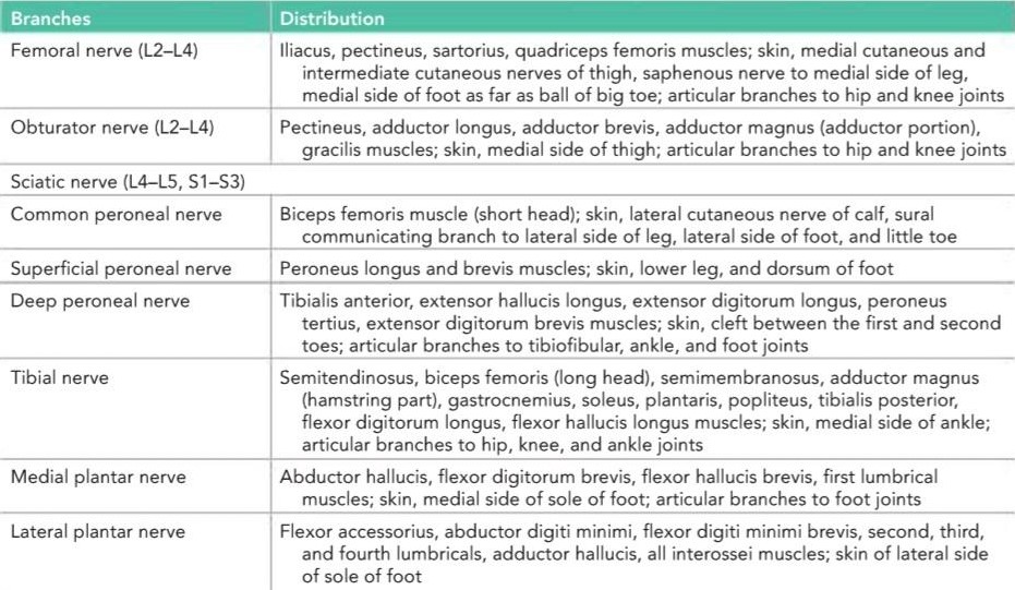
Table 3-3 summarizes the important features found in cervical and lumbosacral root syndromes (a detailed description of the neurologic deficits following the many spinal nerve injuries seen in clinical practice is beyond the scope of this book). Tables that summarize the branches of the brachial (Table 3-4) and lumbar and sacral (Table 3-5) plexuses and their distribution are also included. These tables can assist the reader in determining the specific nerve lesion associated with a particular motor or sensory deficit in the upper or lower limbs.
آسیبهای خاص اعصاب نخاعی
جدول 3-3 ویژگیهای مهم موجود در سندرمهای ریشه گردنی و کمری-خاجی را خلاصه میکند (شرح مفصلی از نقصهای عصبی پس از آسیبهای متعدد اعصاب نخاعی که در عمل بالینی مشاهده میشوند، فراتر از محدوده این کتاب است). جداولی که شاخههای شبکههای عصبی بازویی (جدول 3-4) و کمری و خاجی (جدول 3-5) و توزیع آنها را خلاصه میکنند نیز گنجانده شده است. این جداول میتوانند به خواننده در تعیین ضایعه عصبی خاص مرتبط با نقص حرکتی یا حسی خاص در اندام فوقانی یا تحتانی کمک کنند.
Cranial nerve injuries are considered in Chapter 11.
آسیبهای اعصاب جمجمهای در فصل ۱۱ بررسی شدهاند.
Table 3-3 Important Features Found in Cervical and Lumbosacral Root Syndromes
جدول ۳-۳ ویژگیهای مهم موجود در سندرمهای ریشه گردنی و کمری-خاجی

Table 3-4 Branches of the Brachial Plexus and Their Distribution
جدول ۳-۴ شاخههای شبکه بازویی و توزیع آنها

Table 3-5 Branches of the Lumbar and Sacral Plexuses and Their Distribution
جدول ۳-۵ شاخههای شبکههای کمری و خاجی و توزیع آنها
Basic Clinical Principles Underlying Peripheral Nerve Injuries
• In open, dirty wounds, which carry a high risk of infection, the sectioned nerve should be ignored, and the wound infection should be treated. Later, when the wound has healed satisfactorily, the nerve should be explored, and the cut ends of the nerve should be sutured together.
اصول بالینی پایه آسیبهای اعصاب محیطی
• در زخمهای باز و کثیف که خطر عفونت بالایی دارند، باید عصب بریده شده نادیده گرفته شود و عفونت زخم درمان شود. بعداً، هنگامی که زخم به طور رضایتبخشی بهبود یافت، باید عصب بررسی شود و انتهای بریده شده عصب به هم بخیه زده شود.
• For a patient with a healed wound and no evidence of nerve recovery, the treatment should be conservative. Sufficient time should be allowed to elapse to enable the regenerating nerve fibers to reach the proximal muscles. If recovery fails to occur, the nerve should be explored surgically.
• برای بیماری که زخمش التیام یافته و هیچ نشانهای از بهبودی عصب ندارد، درمان باید محافظهکارانه باشد. باید زمان کافی برای رسیدن فیبرهای عصبی در حال بازسازی به عضلات پروگزیمال در نظر گرفته شود. در صورت عدم بهبودی، عصب باید با جراحی بررسی شود.
• In cases in which connective tissue, bone fragments, or muscles come to lie between the cut ends of a severed nerve, the nerve should be explored; if possible, the cut ends of the nerve should be brought together and sutured.
• در مواردی که بافت همبند، قطعات استخوان یا عضلات بین انتهای بریده شده عصب قطع شده قرار میگیرند، عصب باید بررسی شود؛ در صورت امکان، انتهای بریده شده عصب باید به هم نزدیک شده و بخیه زده شود.
• The nutrition of the paralyzed muscles must be maintained with adequate physiotherapy. Warm baths, massage, and warm clothing help to maintain adequate circulation.
• تغذیه عضلات فلج شده باید با فیزیوتراپی کافی حفظ شود. حمام گرم، ماساژ و لباس گرم به حفظ گردش خون کافی کمک میکند.
• The paralyzed muscles must not be allowed to be stretched by antagonist muscles or by gravity. Moreover, excessive shortening of the paralyzed muscles leads to contracture of these muscles.
• نباید اجازه داد که عضلات فلج شده توسط عضلات آنتاگونیست یا نیروی جاذبه کشیده شوند. علاوه بر این، کوتاه شدن بیش از حد عضلات فلج شده منجر به انقباض این عضلات میشود.
• Mobility must be preserved by daily passive movements of all joints in the affected area. Failure to do this results in the formation of adhesions and consequent limitation of movement.
• تحرک باید با حرکات غیرفعال روزانه تمام مفاصل در ناحیه آسیب دیده حفظ شود. عدم انجام این کار منجر به ایجاد چسبندگی و در نتیجه محدودیت حرکت میشود.
Once voluntary movement returns in the most proximal muscles, the physiotherapist can assist the patient in performing active exercises. This not only aids in the return of a normal circulation to the affected part but also helps the patient to learn once again the complicated muscular performance of skilled movements.
پس از بازگشت حرکت ارادی در نزدیکترین عضلات، فیزیوتراپیست میتواند به بیمار در انجام تمرینات فعال کمک کند. این کار نه تنها به بازگشت گردش خون طبیعی به قسمت آسیب دیده کمک میکند، بلکه به بیمار کمک میکند تا بار دیگر عملکرد پیچیده عضلانی حرکات ماهرانه را بیاموزد.
Nerve Transplantation
Nerve grafts have been used with some success to restore muscle tone in facial nerve palsy. In mixed nerve injuries, nerve grafts have succeeded only in restoring some sensory function and slight muscle activity. The presence of two suture lines and the increased possibility of mixing the nerve fibers is probably the reason for the lack of success with nerve grafts. In most nerve injuries, even when the gap between the proximal and distal ends is as great as 10 cm, mobilizing the nerve or altering its position in relation to joints so that the proximal and distal ends may be brought together without undue tension is usually possible; the ends are then sutured together.
پیوند عصب
پیوندهای عصبی با موفقیت نسبی برای بازیابی تون عضلانی در فلج عصب صورت استفاده شدهاند. در آسیبهای عصبی مختلط، پیوند عصبی تنها در بازیابی برخی از عملکردهای حسی و فعالیت جزئی عضلات موفق بوده است. وجود دو خط بخیه و افزایش احتمال اختلاط رشتههای عصبی احتمالاً دلیل عدم موفقیت پیوند عصبی است. در بیشتر آسیبهای عصبی، حتی زمانی که فاصله بین انتهای پروگزیمال و دیستال به بزرگی 10 سانتیمتر باشد، معمولاً میتوان عصب را تحریک کرد یا موقعیت آن را نسبت به مفاصل تغییر داد تا انتهای پروگزیمال و دیستال بدون کشش بیش از حد به هم نزدیک شوند. سپس انتهای عصب به هم بخیه زده میشود.
Peripheral Nerve Tumors
A peripheral nerve consists essentially of nerve fibers (axons), each of which is associated with Schwann cells; the fibers are either myelinated or nonmyelinated. The nerve fibers are arranged in parallel bundles and are surrounded by connective tissue sheaths.
تومورهای عصبی محیطی
یک عصب محیطی اساساً از فیبرهای عصبی (آکسونها) تشکیل شده است که هر کدام با سلولهای شوان مرتبط هستند؛ این فیبرها یا میلیندار هستند یا بدون میلین. فیبرهای عصبی به صورت دستههای موازی چیده شدهاند و توسط غلافهای بافت همبند احاطه شدهاند.
A benign fibroma or a malignant sarcoma may arise in the connective tissue of the nerve and does not differ from similar tumors elsewhere. Neurilemmomas are believed to arise from Schwann cells. They arise from any nerve trunk, cranial or spinal, and in any part of its course. Primary tumors of the axons are very rare.
یک فیبروم خوشخیم یا یک سارکوم بدخیم ممکن است در بافت همبند عصب ایجاد شود و با تومورهای مشابه در جاهای دیگر تفاوتی ندارد. اعتقاد بر این است که نوریلموماها از سلولهای شوان ناشی میشوند. آنها از هر تنه عصبی، جمجمهای یا نخاعی و در هر بخشی از مسیر آن ایجاد میشوند. تومورهای اولیه آکسونها بسیار نادر هستند.
Blood Vessels, Lymphatics, and Endoneurial Spaces within Peripheral Nerves
Peripheral nerves receive branches from arteries in the regions they pass through. The anastomotic network that exists within a nerve is considerable, and local ischemia does not occur if a single artery is obstructed.
رگهای خونی، لنفاوی و فضاهای اندونوریال در اعصاب محیطی
اعصاب محیطی شاخههایی از شریانها را در مناطقی که از آنها عبور میکنند، دریافت میکنند. شبکه آناستوموزی که در یک عصب وجود دارد قابل توجه است و اگر یک شریان مسدود شود، ایسکمی موضعی رخ نمیدهد.
A plexus of lymph vessels lies within the epineurial connective tissues, and this drains to regional lymph nodes.
شبکهای از رگهای لنفاوی در بافتهای همبند اپینوریال قرار دارد و این رگها به غدد لنفاوی ناحیهای تخلیه میشوند.
As demonstrated by the results of experiments in which dyes have been injected into peripheral nerves, spaces exist between individual nerve fibers. These endoneurial spaces evidently provide a potential route for the ascent of tetanus toxin to the spinal cord.
همانطور که نتایج آزمایشهایی که در آنها رنگها به اعصاب محیطی تزریق شدهاند نشان میدهد، بین فیبرهای عصبی منفرد فضاهایی وجود دارد. این فضاهای اندونوریال آشکارا مسیر بالقوهای برای صعود سم کزاز به نخاع فراهم میکنند.
Local Anesthetic Action on Nerve Conduction
Local anesthetics are drugs that block nerve conduction when applied locally to a nerve fiber in suitable concentrations. Their site of action is the axolemma (plasma membrane), and they interfere with the transient increase in permeability of the axolemma to Na, K, and other ions. The sensitivity of nerve fibers to local anesthetics is related to the size of the nerve fibers (see Table 2-3). Small nerve fibers are more susceptible than large fibers; small fibers are also slower to recover.
اثر بیحسکنندههای موضعی بر هدایت عصبی
بیحسکنندههای موضعی داروهایی هستند که وقتی به صورت موضعی در غلظتهای مناسب روی یک فیبر عصبی اعمال میشوند، هدایت عصبی را مسدود میکنند. محل اثر آنها آکسولما (غشای پلاسما) است و با افزایش گذرای نفوذپذیری آکسولما به سدیم، پتاسیم و سایر یونها تداخل میکنند. حساسیت فیبرهای عصبی به بیحسکنندههای موضعی به اندازه فیبرهای عصبی مربوط میشود (به جدول 2-3 مراجعه کنید). فیبرهای عصبی کوچک نسبت به فیبرهای بزرگ حساستر هستند؛ فیبرهای کوچک نیز دیرتر بهبود مییابند.
Cocaine has been used clinically to block nerve conduction. Unfortunately, it is a strong stimulant of the cerebral cortex and readily causes addiction. Procaine is a synthetic compound that is widely used as a local anesthetic agent.
کوکائین به صورت بالینی برای مسدود کردن هدایت عصبی مورد استفاده قرار گرفته است. متأسفانه، این ماده یک محرک قوی برای قشر مغز است و به راحتی باعث اعتیاد میشود. پروکائین یک ترکیب مصنوعی است که به طور گسترده به عنوان یک عامل بیحس کننده موضعی استفاده میشود.
Apparent Recovery of Central Nervous System Function
Axon regeneration in the brain and spinal cord is minimal following a lesion, yet considerable functional recovery often occurs. Several explanations exist, and more than one mechanism may be involved.
بهبودی ظاهری عملکرد سیستم عصبی مرکزی
بازسازی آکسون در مغز و نخاع پس از ضایعه حداقل است، با این حال اغلب بهبودی عملکردی قابل توجهی رخ میدهد. توضیحات متعددی وجود دارد و ممکن است بیش از یک مکانیسم در این امر دخیل باشد.
1. Nerve fiber function may be impaired from compression by edema fluid. Once the edema subsides, a substantial recovery may take place.
۱. عملکرد فیبر عصبی ممکن است در اثر فشرده شدن توسط مایع ادم مختل شود. پس از فروکش کردن ادم، بهبودی قابل توجهی ممکن است رخ دهد.
2. The damaged nerve fiber proximal to the lesion may form new synapses with neighboring normal neurons.
۲. فیبر عصبی آسیب دیده نزدیک به ضایعه ممکن است سیناپسهای جدیدی با نورونهای طبیعی مجاور تشکیل دهد.
3. Following a lesion to branches of a nerve, all the neurotransmitters may pass down the remaining branches, producing a greater effect.
۳. پس از ضایعه شاخههای یک عصب، تمام انتقالدهندههای عصبی ممکن است از شاخههای باقی مانده عبور کنند و اثر بیشتری ایجاد کنند.
4. Following a lesion of an afferent neuron, an increased number of receptor sites may develop on a postsynaptic membrane. This may result in the second neuron responding to neurotransmitter substances from neighboring neurons.
۴. پس از ضایعه یک نورون آوران، ممکن است تعداد بیشتری از جایگاههای گیرنده روی غشای پس سیناپسی ایجاد شود. این ممکن است منجر به پاسخ نورون دوم به مواد انتقالدهنده عصبی از نورونهای همسایه شود.
5. Nonfunctioning neurons may take over the function of damaged neurons.
۵. نورونهای غیرفعال ممکن است عملکرد نورونهای آسیبدیده را بر عهده بگیرند.
6. The damaged nerve fiber proximal to the lesion may form new synapses with neighboring normal neurons.
۶. فیبر عصبی آسیبدیده نزدیک به ضایعه ممکن است سیناپسهای جدیدی با نورونهای طبیعی مجاور تشکیل دهد.
7. The normal neighboring nerve fibers may give off branches distal to the lesion, which then follow the path- way previously occupied by the damaged fibers.
۷. فیبرهای عصبی طبیعی مجاور ممکن است شاخههایی را در دیستال ضایعه ایجاد کنند که سپس مسیری را که قبلاً توسط فیبرهای آسیبدیده اشغال شده بود، دنبال میکنند.
8. If a particular function, such as the contraction of voluntary muscle, is served by two neural pathways in the CNS and one pathway is damaged, the remaining undamaged pathway may take over the entire function. Thus, conceivably, if the corticospinal tract is injured, the corticoreticulospinal tract may take over the major role of controlling the muscle movement.
۸. اگر یک عملکرد خاص، مانند انقباض عضله ارادی، توسط دو مسیر عصبی در سیستم عصبی مرکزی انجام شود و یک مسیر آسیب ببیند، مسیر آسیبندیده باقیمانده ممکن است کل عملکرد را بر عهده بگیرد. بنابراین، احتمالاً، اگر مسیر قشری-نخاعی آسیب ببیند، مسیر قشری-نخاعی ممکن است نقش اصلی کنترل حرکت عضلات را بر عهده بگیرد.
9. With intensive physiotherapy, patients may be trained to use other muscles to compensate for the loss of paralyzed muscles.
۹. با فیزیوتراپی فشرده، میتوان به بیماران آموزش داد تا از عضلات دیگر برای جبران از دست دادن عضلات فلج استفاده کنند.
Herpes Zoster
Herpes zoster, or shingles, is a relatively common condi- tion caused by the reactivation of the latent varicella zoster virus in a patient who has previously had chickenpox. The infection is found in the first sensory neuron in a cranial or spinal nerve. The lesion is seen as an inflammation and degeneration of the sensory neuron with the formation of vesicles with inflammation of the skin. The first symptom is pain in the distribution of the sensory neuron, followed in a few days by a skin eruption. The condition occurs most frequently in patients over age 50 years.
هرپس زوستر
هرپس زوستر یا زونا، یک بیماری نسبتاً شایع است که در اثر فعال شدن مجدد ویروس نهفته واریسلا زوستر در بیماری که قبلاً آبله مرغان داشته است، ایجاد میشود. عفونت در اولین نورون حسی در عصب جمجمهای یا نخاعی یافت میشود. ضایعه به صورت التهاب و تخریب نورون حسی با تشکیل وزیکولهایی با التهاب پوست دیده میشود. اولین علامت، درد در محل توزیع نورون حسی است که پس از چند روز با بثورات پوستی دنبال میشود. این بیماری بیشتر در بیماران بالای ۵۰ سال رخ میدهد.
Polyneuropathy
Polyneuropathy is an impairment of function of many peripheral nerves simultaneously. The many causes include infection (endotoxin of diphtheria, Guillain-Barré syndrome [see Clinical Example at beginning of chapter]), metabolic disorders (vitamins B, and B12 deficiency, poisoning by heavy metals, drugs), and endocrine disorders (diabetes). Axon degeneration and/or segmental demyelination may take place, and the neuron cell body may be involved. In mild cases, the condition is reversible, but in severe cases, it may be permanent. Both sensory and motor symptoms and signs may be evident.
پلینوروپاتی
پلینوروپاتی اختلال عملکرد همزمان بسیاری از اعصاب محیطی است. علل متعدد آن شامل عفونت (اندوتوکسین دیفتری، سندرم گیلن باره [به مثال بالینی در ابتدای فصل مراجعه کنید])، اختلالات متابولیک (کمبود ویتامینهای B و B12، مسمومیت با فلزات سنگین، داروها) و اختلالات غدد درونریز (دیابت) است. دژنراسیون آکسون و/یا دمیلیناسیون قطعهای ممکن است رخ دهد و جسم سلولی نورون ممکن است درگیر شود. در موارد خفیف، این بیماری برگشتپذیر است، اما در موارد شدید، ممکن است دائمی باشد. علائم و نشانههای حسی و حرکتی ممکن است مشهود باشند.
Receptors
Sensory endings are found throughout the body in both somatic and visceral areas. Their wide distribution is fortunate, because they enable us to react to changes in the external and internal environment.
گیرندهها
پایانههای حسی در سراسر بدن، هم در نواحی سوماتیک و هم در نواحی احشایی یافت میشوند. توزیع گسترده آنها خوشبختانه است، زیرا ما را قادر میسازند تا به تغییرات در محیط خارجی و داخلی واکنش نشان دهیم.
To make a diagnosis or study the effect of treatment on a disease process, a clinician relies almost entirely on the patient’s ability to describe changes in subjective sensations or to respond to specific stimuli during a physical examination. Such descriptions, such as “knifelike pain,” “dull and aching pain,” “colicky pain,” “pins and needles,” and “cannot feel anything” are very familiar to the practicing clinician. Each main type of sensation that can be experienced, such as pain, temperature, and touch and pressure, is called a modality of sensation. The type of modality felt by a patient from a particular part of the body is determined by the specific area of the CNS to which the afferent nerve fiber passes. However, remembering that axons carrying specific modalities are associated with one or more anatomically distinct receptors is clinically useful (Table 3-6).
برای تشخیص یا مطالعه تأثیر درمان بر روند بیماری، یک پزشک تقریباً به طور کامل به توانایی بیمار در توصیف تغییرات در احساسات ذهنی یا پاسخ به محرکهای خاص در طول معاینه فیزیکی متکی است. چنین توصیفاتی، مانند “درد چاقو مانند”، “درد مبهم و دردناک”، “درد قولنجی”، “سوزن سوزن شدن” و “احساس نکردن چیزی” برای پزشک متخصص بسیار آشنا هستند. هر نوع حس اصلی که میتواند تجربه شود، مانند درد، دما و لمس و فشار، یک روش حسی نامیده میشود. نوع روشی که بیمار از یک قسمت خاص بدن احساس میکند، توسط ناحیه خاصی از سیستم عصبی مرکزی که فیبر عصبی آوران به آن منتقل میشود، تعیین میشود. با این حال، به خاطر داشتن این نکته که آکسونهای حامل مدالیتههای خاص با یک یا چند گیرندهی آناتومیک متمایز مرتبط هستند، از نظر بالینی مفید است (جدول 3-6).
Table 3-6 Receptors and Associated Funcitons
جدول ۳-۶ گیرندهها و عملکردهای مرتبط

Sensory Receptors and Age
With life expectancy increasing, many patients now reach the age when sensory receptor degeneration can cause disequilibrium. This critical age varies in different individuals, but once it starts, the sensory systems progressively deteriorate, involving not only visual and auditory systems but also proprioception and the ability to integrate the afferent information entering the CNS.
گیرندههای حسی و سن
با افزایش امید به زندگی، بسیاری از بیماران اکنون به سنی میرسند که تخریب گیرندههای حسی میتواند باعث عدم تعادل شود. این سن بحرانی در افراد مختلف متفاوت است، اما به محض شروع، سیستمهای حسی به تدریج رو به زوال میروند و نه تنها سیستمهای بینایی و شنوایی، بلکه حس عمقی و توانایی ادغام اطلاعات آوران ورودی به سیستم عصبی مرکزی را نیز درگیر میکنند.
Sensory Modality Examination
An accurate physical examination may enable the neurologist to make a precise diagnosis. He or she may be able to determine whether a particular sensation can or cannot be appreciated or whether it is less than normal. The clinician will be able to determine the precise area over the surface of the body where impairment of sensation is found. The following sensations are usually tested:
معاینه روش حسی
یک معاینه فیزیکی دقیق میتواند متخصص مغز و اعصاب را قادر به تشخیص دقیق کند. او ممکن است بتواند تشخیص دهد که آیا یک حس خاص قابل درک است یا خیر، یا اینکه آیا کمتر از حد طبیعی است یا خیر. پزشک قادر خواهد بود ناحیه دقیق روی سطح بدن را که در آن اختلال حس وجود دارد، تعیین کند. معمولاً حسهای زیر آزمایش میشوند:
1. Light touch. This is tested by gently touching the skin with a wisp of cotton; the patient has the eyes closed and responds “yes” whenever the stimulus is felt. Recognize that different areas of the skin normally exhibit different thresholds for touch. The back and buttocks are less sensitive than the face or fingertips. On hairy surfaces, the slightest movement of a hair usually can be felt.
۱. لمس سبک. این آزمایش با لمس آرام پوست با یک تکه پنبه انجام میشود؛ بیمار چشمانش را بسته نگه میدارد و هر زمان که محرک احساس شد، «بله» میگوید. توجه داشته باشید که نواحی مختلف پوست معمولاً آستانههای لمسی متفاوتی دارند. پشت و باسن نسبت به صورت یا نوک انگشتان حساسیت کمتری دارند. روی سطوح مودار، معمولاً کوچکترین حرکت مو قابل احساس است.
2. Localization of touch. After detecting the light touch with the eyes closed, the patient is asked to place a finger on the exact site touched. Failure to accomplish this may be due to damage to the cerebral cortex.
۲. تعیین محل لمس. پس از تشخیص لمس سبک با چشمان بسته، از بیمار خواسته میشود که انگشت خود را دقیقاً روی محل لمس شده قرار دهد. عدم انجام این کار ممکن است به دلیل آسیب به قشر مغز باشد.
3. Two-point tactile discrimination. Two blunt points are applied to the skin surface while the patient’s eyes are closed. Gradually, the points are brought closer together until the patient is unable to distinguish two definite points. A normal person is able to distinguish two separate points on the tip of the index finger when they are separated by a distance greater than about 3 mm. On the back, however, they have to be separated by as much as 3 to 4 cm.
۳. تشخیص لمسی دو نقطهای. دو نقطه صاف در حالی که چشمان بیمار بسته است روی سطح پوست اعمال میشود. به تدریج، نقاط به هم نزدیکتر میشوند تا زمانی که بیمار قادر به تشخیص دو نقطه مشخص نباشد. یک فرد عادی قادر است دو نقطه جداگانه را روی نوک انگشت اشاره تشخیص دهد، زمانی که فاصله آنها بیش از حدود ۳ میلیمتر باشد. با این حال، در پشت، آنها باید تا ۳ تا ۴ سانتیمتر از هم فاصله داشته باشند.
4. Pain. The skin may be touched with the sharp end of a pin. First, the pain threshold is established and then the areas of diminished or lost pain sensation are mapped out. Apply the stimulus in an irregular manner, first using the sharp end of the pin and then the dull head, with the patient responding “sharp” or “dull.” Certain diseases, such as tabes dorsalis or polyneuropathy (polyneuritis), may produce a delay of up to 3 seconds before the patient recognizes the sharp pain.
۴. درد. پوست را میتوان با انتهای تیز یک سنجاق لمس کرد. ابتدا آستانه درد تعیین میشود و سپس نواحی کاهش یا از دست دادن حس درد مشخص میشوند. محرک را به صورت نامنظم اعمال کنید، ابتدا از انتهای تیز سنجاق و سپس از سر کند آن استفاده کنید، در حالی که بیمار به صورت «تیز» یا «کند» پاسخ میدهد. برخی بیماریها، مانند تابس دورسالیس یا پلینوروپاتی (پلینوریت)، ممکن است تا ۳ ثانیه قبل از اینکه بیمار درد تیز را تشخیص دهد، تأخیر ایجاد کنند.
5. Pressure pain. This poorly localized pain is perceived by deep pressure on a muscle or by squeezing a tendon.
۵. درد فشاری. این درد که به خوبی موضعی نیست، با فشار عمیق روی عضله یا فشردن تاندون احساس میشود.
6. Temperature testing. Test tubes filled with hot or cold water may be used. When the test tubes are applied to the skin, the patient responds with either “warm” or “cold.” First, the temperature threshold is established and then the areas of diminished or lost temperature sensation are mapped out.
۶. آزمایش دما. میتوان از لولههای آزمایش پر از آب گرم یا سرد استفاده کرد. وقتی لولههای آزمایش روی پوست قرار میگیرند، بیمار با «گرم» یا «سرد» پاسخ میدهد. ابتدا آستانه دما تعیین میشود و سپس نواحی که حس دما در آنها کاهش یافته یا از بین رفته است، مشخص میشوند.
7. Vibration. When the handle of a vibrating tuning fork is applied to the skin over bone (e.g., the medial malleolus of the tibia or the olecranon process of the ulna), a tingling sensation is felt. This is due to the stimulation of superficial and deep pressure receptors. The patient is asked to respond when the first vibration is felt as well as when the vibration can no longer be detected. The perception of vibration in the legs is usually diminished after age 60 years.
۷. ارتعاش. وقتی دسته یک دیاپازون مرتعش روی پوست روی استخوان (مثلاً قوزک داخلی تیبیا یا زائده اولکرانون اولنا) قرار میگیرد، احساس سوزن سوزن شدن احساس میشود. این به دلیل تحریک گیرندههای فشار سطحی و عمقی است. از بیمار خواسته میشود که چه زمانی که اولین ارتعاش احساس میشود و چه زمانی که دیگر نمیتوان ارتعاش را تشخیص داد، پاسخ دهد. درک ارتعاش در پاها معمولاً پس از ۶۰ سالگی کاهش مییابد.
8. Appreciation of form (stereognosis). With the patient’s eyes closed, the examiner places common objects, such as coins or keys, in the patient’s hands. The patient normally should be able to identify objects by moving them around in the hand and feeling them with the fingers.
۸. درک فرم (استریگنوزیس). در حالی که چشمان بیمار بسته است، معاینهکننده اشیاء رایج مانند سکه یا کلید را در دستان بیمار قرار میدهد. بیمار معمولاً باید بتواند اشیاء را با حرکت دادن آنها در دست و لمس کردن آنها با انگشتان شناسایی کند.
9. Passive movements of joints. This test may be carried out on the fingers or toes. With the patient completely relaxed and in the supine position with eyes closed, the digit is flexed or extended irregularly. After each movement, the patient is asked, “Is the digit ‘up’ or ‘down”?” A normal individual not only can determine that passive movement is taking place but also is aware of the direction of the movement.
۹. حرکات غیرفعال مفاصل. این آزمایش ممکن است روی انگشتان دست یا پا انجام شود. در حالی که بیمار کاملاً آرام و در حالت خوابیده به پشت با چشمان بسته است، انگشت به طور نامنظم خم یا باز میشود. پس از هر حرکت، از بیمار پرسیده میشود: «آیا انگشت «بالا» است یا «پایین»؟» یک فرد عادی نه تنها میتواند تشخیص دهد که حرکت غیرفعال در حال انجام است، بلکه از جهت حرکت نیز آگاه است.
10. Postural sensibility. This is the ability to describe the position of a limb when it is placed in that position while the patient’s eyes are closed. Another way to perform the test is to ask the patient, with eyes closed, to place the limb on the opposite side in the same position as the other limb. The application and interpretation of the results of these tests will be understood more fully when the afferent or sensory pathways have been discussed (see Chapter 4).
۱۰. حساسیت وضعیتی. این توانایی توصیف موقعیت یک اندام است وقتی که در آن موقعیت قرار میگیرد در حالی که چشمان بیمار بسته است. روش دیگر برای انجام آزمایش این است که از بیمار خواسته شود با چشمان بسته، اندام را در طرف مقابل در همان موقعیت اندام دیگر قرار دهد. کاربرد و تفسیر نتایج این آزمایشها زمانی که مسیرهای آوران یا حسی مورد بحث قرار گرفته باشند، به طور کاملتری درک خواهد شد (به فصل ۴ مراجعه کنید).
Phantom Limb
Wherever a particular sensory pathway is stimulated along its course from the receptor to the sensory cortex of the brain, the sensation experienced by the individual is referred to the site of the receptor. For example, if the pain fibers from the receptors in the little finger are stimulated in the ulnar nerve at the elbow, the individual will experience pain in the little finger and not at the elbow.
اندام خیالی
هر جا که یک مسیر حسی خاص در طول مسیر خود از گیرنده تا قشر حسی مغز تحریک شود، حسی که فرد تجربه میکند به محل گیرنده ارجاع داده میشود. به عنوان مثال، اگر فیبرهای درد از گیرندههای انگشت کوچک در عصب اولنار در آرنج تحریک شوند، فرد درد را در انگشت کوچک و نه در آرنج تجربه خواهد کرد.
Following the amputation of a limb, the patient may experience severe pain in the absent limb due to pressure on the nerve fibers at the end of the stump. This phenomenon is referred to clinically as phantom limb.
پس از قطع عضو، بیمار ممکن است به دلیل فشار بر روی فیبرهای عصبی در انتهای استامپ، درد شدیدی را در اندام غایب تجربه کند. این پدیده از نظر بالینی به عنوان اندام خیالی شناخته میشود.
Action of Drugs and Other Agents on Skeletal Neuromuscular Junctions
Table 3-7 gives some examples of drugs and diseases affecting the motor endplates in skeletal muscle.
تأثیر داروها و سایر عوامل بر اتصالات عصبی-عضلانی اسکلتی
جدول 3-7 نمونههایی از داروها و بیماریهایی را که بر صفحات انتهایی حرکتی در عضله اسکلتی تأثیر میگذارند، نشان میدهد.
Table 3-7 Drugs and Diseases Affecting the Motor Endplates in Skeletal Muscle
جدول ۳-۷ داروها و بیماریهای مؤثر بر صفحات انتهایی حرکتی در عضله اسکلتی

ACh, acetylcholine; AChE, acetylcholinesterase.
ACh، استیل کولین؛ AChE، استیل کولین استراز.
Neuromuscular Blocking Agents
d-Tubocurarine produces flaccid paralysis of skeletal muscle, first affecting the extrinsic muscles of the eyes and then those of the face, the extremities, and finally the diaphragm.
عوامل مسدودکننده عصبی-عضلانی
d-توبوکورارین باعث فلج شل عضلات اسکلتی میشود که ابتدا عضلات خارجی چشمها و سپس عضلات صورت، اندامها و در نهایت دیافراگم را تحت تأثیر قرار میدهد.
Dimethyltubocurarine, gallamine, and benzoquinonium have similar effects.
دی متیل توبوکورارین، گالامین و بنزوکینونیوم اثرات مشابهی دارند.
These drugs combine with the receptor sites at the postsynaptic membrane normally used by ACh and thus block the neurotransmitter action of ACh. Therefore, they are referred to as competitive blocking agents, as they are competing for the same receptor site as ACh. As these drugs are slowly metabolized, the paralysis passes off.
این داروها با محلهای گیرنده در غشای پسسیناپسی که معمولاً توسط ACh استفاده میشوند، ترکیب میشوند و بنابراین عملکرد انتقالدهنده عصبی ACh را مسدود میکنند. بنابراین، به آنها عوامل مسدودکننده رقابتی گفته میشود، زیرا برای همان محل گیرنده ACh رقابت میکنند. از آنجایی که این داروها به آرامی متابولیزه میشوند، فلج از بین میرود.
Decamethonium and succinylcholine also paralyze skeletal muscle, but their action differs from that of competitive blocking agents because they produce their effect by causing depolarization of the motor endplate. Acting like ACh, they depolarize the postsynaptic membrane, and the muscle contracts once. This is followed by flaccid paralysis and blockage of neuromuscular activity. Although the blocking action endures for some time, the drugs are metabolized, and the paralysis passes off. The actual paralysis is produced by the continued depolarization of the postsynaptic membrane. Remember that continuous depolarization does not produce continuous skeletal muscle contraction. Repolarization has to take place before further depolarization can occur.
دکامتونیوم و سوکسینیل کولین نیز عضله اسکلتی را فلج میکنند، اما عملکرد آنها با عوامل مسدودکننده رقابتی متفاوت است زیرا آنها با ایجاد دپلاریزاسیون صفحه انتهایی حرکتی، اثر خود را ایجاد میکنند. آنها مانند استیل کولین عمل میکنند، غشای پس سیناپسی را دپلاریزه میکنند و عضله یک بار منقبض میشود. به دنبال آن فلج شل و انسداد فعالیت عصبی-عضلانی رخ میدهد. اگرچه اثر مسدودکننده برای مدتی ادامه دارد، اما داروها متابولیزه میشوند و فلج از بین میرود. فلج واقعی با دپلاریزاسیون مداوم غشای پس سیناپسی ایجاد میشود. به یاد داشته باشید که دپلاریزاسیون مداوم باعث انقباض مداوم عضله اسکلتی نمیشود. قبل از وقوع دپلاریزاسیون بیشتر، رپلاریزاسیون باید انجام شود.
Neuromuscular blocking agents are commonly used with general anesthetics to produce the desired degree of muscle relaxation without using larger doses of general anesthetics. Because the respiratory muscles are paralyzed, facilities for artificial respiration are essential.
عوامل مسدودکننده عصبی-عضلانی معمولاً با داروهای بیهوشی عمومی برای ایجاد درجه مطلوب شل شدن عضلات بدون استفاده از دوزهای بالاتر داروهای بیهوشی عمومی استفاده میشوند. از آنجا که عضلات تنفسی فلج میشوند، امکانات تنفس مصنوعی ضروری است.
Anticholinesterases
Physostigmine and neostigmine have the capacity to combine with acetylcholinesterase (AChE) and prevent the esterase from inactivating ACh. In addition, neostigmine has a direct stimulating action on skeletal muscle. The actions of both drugs are reversible, and they have been used with success in the treatment of myasthenia gravis.
آنتی کولین استرازها
فیزوستیگمین و نئوستیگمین توانایی ترکیب با استیل کولین استراز (AChE) و جلوگیری از غیرفعال شدن استیل کولین توسط استراز را دارند. علاوه بر این، نئوستیگمین اثر تحریکی مستقیمی بر عضله اسکلتی دارد. عملکرد هر دو دارو برگشتپذیر است و با موفقیت در درمان میاستنی گراویس مورد استفاده قرار گرفتهاند.
Bacterial Toxins
Clostridium botulinum, the causative organism in certain cases of food poisoning, produces a toxin that inhibits the release of ACh at the neuromuscular junction. Death results from paralysis of the respiratory muscles. The course of the disease can be improved by the administration of calcium gluconate or guanidine, which promotes the release of ACh from the nerve terminals.
سموم باکتریایی
کلستریدیوم بوتولینوم، ارگانیسم عامل در موارد خاصی از مسمومیت غذایی، سمی تولید میکند که مانع از آزاد شدن استیلکولین (ACh) در محل اتصال عصبی-عضلانی میشود. مرگ ناشی از فلج عضلات تنفسی است. روند بیماری را میتوان با تجویز گلوکونات کلسیم یا گوانیدین که باعث آزاد شدن استیلکولین از پایانههای عصبی میشود، بهبود بخشید.
Motor Nerve and Skeletal Muscle
Not only does the motor nerve control the activity of the muscle it supplies but also its integrity is essential for the muscle’s normal maintenance. Following section of a motor nerve, the muscle fibers rapidly atrophy and are replaced by connective tissue. The total bulk of the muscle may be 75% reduced in as little as 3 months. This degree of atrophy does not occur if the muscle simply is immobilized; that is, it is not just disuse atrophy. Apparently, the maintenance of normal muscle is dependent on the continued reception of ACh at the postsynaptic membrane at the neuromuscular junction.
عصب حرکتی و عضله اسکلتی
عصب حرکتی نه تنها فعالیت عضلهای را که به آن عصب میدهد کنترل میکند، بلکه یکپارچگی آن نیز برای حفظ طبیعی عضله ضروری است. پس از قطع عصب حرکتی، فیبرهای عضلانی به سرعت آتروفی میشوند و با بافت همبند جایگزین میشوند. حجم کل عضله ممکن است در کمتر از ۳ ماه ۷۵٪ کاهش یابد. اگر عضله به سادگی بیحرکت شود، این درجه از آتروفی رخ نمیدهد؛ یعنی فقط آتروفی ناشی از عدم استفاده نیست. ظاهراً حفظ عضله طبیعی به دریافت مداوم استیلکولین در غشای پسسیناپسی در محل اتصال عصبی-عضلانی بستگی دارد.
Denervation Supersensitivity of Skeletal Muscle
After approximately 2 weeks of denervation, skeletal muscle fibers respond to externally applied ACh at sites other than the neuromuscular junctions. This supersensitivity could be explained on the basis that many new ACh receptors have developed along the length of the muscle fibers following denervation.
حساسیت بیش از حد عضله اسکلتی ناشی از قطع عصب
پس از تقریباً ۲ هفته از قطع عصب، فیبرهای عضله اسکلتی به استیل کولین اعمال شده خارجی در مکانهایی غیر از اتصالات عصبی-عضلانی پاسخ میدهند. این حساسیت بیش از حد را میتوان بر این اساس توضیح داد که بسیاری از گیرندههای جدید استیل کولین پس از قطع عصب در امتداد طول فیبرهای عضلانی ایجاد شدهاند.
Myasthenia Gravis
Myasthenia gravis is a common disease characterized by drooping of the upper eyelids (ptosis), double vision (diplopia), difficulty in swallowing (dysphagia), difficulty in talking (dysarthria), and general muscle weakness and fatigue. Initially, the disease most often involves the muscles of the eye and the pharynx, and the symptoms can be relieved with rest. In the progressive form of the disease, the weakness becomes steadily worse, and ultimately death occurs.
میاستنی گراویس
میاستنی گراویس یک بیماری شایع است که با افتادگی پلکهای فوقانی (پتوز)، دوبینی (دیپلوپی)، مشکل در بلع (دیسفاژی)، مشکل در صحبت کردن (دیسآرتریا) و ضعف و خستگی عمومی عضلات مشخص میشود. در ابتدا، این بیماری اغلب عضلات چشم و حلق را درگیر میکند و علائم با استراحت تسکین مییابند. در شکل پیشرونده بیماری، ضعف به طور پیوسته بدتر میشود و در نهایت مرگ رخ میدهد.
The condition is an autoimmune disorder in which anti- bodies are produced against the nicotinic ACh receptors on the postsynaptic membrane. The cause of the autoimmune disorder is unknown. The antibodies interfere with the synaptic transmission by reducing the number of receptors or by blocking the interaction of ACh with its receptors. The size of the junctional folds is also reduced, and the width of the synaptic cleft is increased. Together, these changes result in reduced amplitude in endplate potentials. The condition can be temporarily relieved by AChE drugs such as neostigmine, which potentiates ACh’s action.
این بیماری یک اختلال خودایمنی است که در آن آنتیبادیهایی علیه گیرندههای نیکوتینی استیلکولین (ACh) روی غشای پسسیناپسی تولید میشوند. علت این اختلال خودایمنی ناشناخته است. آنتیبادیها با کاهش تعداد گیرندهها یا با مسدود کردن تعامل استیلکولین با گیرندههایش، در انتقال سیناپسی اختلال ایجاد میکنند. اندازه چینهای اتصالی نیز کاهش مییابد و عرض شکاف سیناپسی افزایش مییابد. در مجموع، این تغییرات منجر به کاهش دامنه پتانسیلهای صفحه انتهایی میشوند. این بیماری میتواند به طور موقت توسط داروهای استیلکولین استئارات (AChE) مانند نئوستیگمین که عملکرد استیلکولین استئارات را تقویت میکند، تسکین یابد.
In adults with myasthenia gravis, about 70% show evidence of hyperplasia of their thymus glands. In the thymus, T cells, which mediate immune protection, undergo maturation. Excessive synthesis of thymic hormones that stimulate the development of T cells may contribute to the autoimmune response.
در بزرگسالان مبتلا به میاستنی گراویس، حدود ۷۰٪ شواهدی از هیپرپلازی غدد تیموس خود را نشان میدهند. در تیموس، سلولهای T که واسطه محافظت ایمنی هستند، بالغ میشوند. سنتز بیش از حد هورمونهای تیموسی که باعث تحریک رشد سلولهای T میشوند، ممکن است در پاسخ خودایمنی نقش داشته باشد.
A rare congenital form of myasthenia gravis may exist from birth, with no abnormal antibody present. The causes of the congenital disease include AChE deficiency at the motor endplates, impaired release of ACh, impaired capac- ity of the receptors to interact with ACh, and a reduced number of ACh receptors.
یک نوع نادر مادرزادی میاستنی گراویس ممکن است از بدو تولد وجود داشته باشد، بدون اینکه آنتیبادی غیرطبیعی وجود داشته باشد. علل بیماری مادرزادی شامل کمبود AChE در صفحات انتهایی حرکتی، اختلال در آزادسازی ACh، اختلال در ظرفیت گیرندهها برای تعامل با ACh و کاهش تعداد گیرندههای ACh است.
Hypokalemic Periodic Paralysis and Hyperkalemic Paralysis
Hypokalemic periodic paralysis and hyperkalemic paralysis are diseases due to decreased or increased blood potassium levels. The ability of ACh to initiate electrical changes in the postsynaptic membrane of the neuromuscular junction can be greatly influenced by the level of blood potassium, and this blood change is responsible for the paralysis in these patients.
فلج دورهای هیپوکالمیک و فلج هایپرکالمیک
فلج دورهای هیپوکالمیک و فلج هایپرکالمیک بیماریهایی هستند که به دلیل کاهش یا افزایش سطح پتاسیم خون ایجاد میشوند. توانایی استیلکولین در شروع تغییرات الکتریکی در غشای پسسیناپسی محل اتصال عصبی-عضلانی میتواند تا حد زیادی تحت تأثیر سطح پتاسیم خون قرار گیرد و این تغییر خون مسئول فلج در این بیماران است.
Action of Drugs on Neuromuscular Junctions in Smooth and Cardiac Muscle and Nerve Endings on Secretory Cells
In normal body physiology, ACh released from postganglionic parasympathetic fibers can bring about depolarization and resulting contraction of smooth muscle fibers. ACh, however, is a useless drug to be administered by the clinician, because it is rapidly destroyed by the cholinesterases. Also, its actions are so widespread that it cannot be used selectively. By slightly changing the structure, as in the case of methacholine chloride or carbachol, the drugs are less susceptible to destruction by the cholinesterases but still possess the ability to react with the receptors.
عملکرد داروها بر اتصالات عصبی-عضلانی در عضلات صاف و قلبی و انتهای عصب بر سلولهای ترشحی
در فیزیولوژی طبیعی بدن، استیلکولین آزاد شده از فیبرهای پاراسمپاتیک پسگانگلیونی میتواند باعث دپلاریزاسیون و در نتیجه انقباض فیبرهای عضله صاف شود. با این حال، استیلکولین دارویی بیفایده برای تجویز توسط پزشک است، زیرا به سرعت توسط کولیناسترازها تخریب میشود. همچنین، عملکرد آن چنان گسترده است که نمیتوان آن را به صورت انتخابی استفاده کرد. با تغییر جزئی ساختار، مانند مورد متاکولین کلرید یا کارباکول، داروها کمتر در معرض تخریب توسط کولیناسترازها قرار میگیرند، اما همچنان توانایی واکنش با گیرندهها را دارند.
Atropine and scopolamine are drugs that compete with ACh for the same receptors. These drugs are competitive antagonists of ACh at receptor sites of smooth muscle, cardiac muscle, and various secretory cells.
آتروپین و اسکوپولامین داروهایی هستند که با ACh برای گیرندههای مشابه رقابت میکنند. این داروها آنتاگونیستهای رقابتی ACh در محلهای گیرنده عضله صاف، عضله قلب و سلولهای ترشحی مختلف هستند.
Norepinephrine is released from postganglionic sympathetic fibers and can bring about depolarization of smooth muscle in the walls of arteries, for example, resulting in their contraction. At other sites, such as the bronchi, it causes smooth muscle relaxation. Sympathetic receptors have been classified as a and B. The functions associated with α- receptors are vasoconstriction, mydriasis (dilatation of the pupil), and relaxation of the smooth muscle of the intestine. B-Receptors are associated with vasodilatation, cardioacceleration, bronchial relaxation, and intestinal relaxation.
نوراپی نفرین از فیبرهای سمپاتیک پسگانگلیونی آزاد میشود و میتواند باعث دپلاریزاسیون عضله صاف در دیواره شریانها شود، به عنوان مثال، در نتیجه انقباض آنها. در سایر نقاط، مانند برونشها، باعث شل شدن عضلات صاف میشود. گیرندههای سمپاتیک به عنوان a و B طبقهبندی شدهاند. عملکردهای مرتبط با گیرندههای α- عبارتند از انقباض عروق، میدریاز (گشاد شدن مردمک چشم) و شل شدن عضله صاف روده. گیرندههای B- با گشاد شدن عروق، تسریع قلبی، شل شدن برونشها و شل شدن روده مرتبط هستند.
Phenoxybenzamine has been found to block a-receptors, whereas propranolol blocks ẞ-receptors. The structure of these receptors is not known.
فنوکسی بنزامین گیرندههای α- را مسدود میکند، در حالی که پروپرانولول گیرندههای ẞ را مسدود میکند. ساختار این گیرندهها مشخص نیست.
Sensory Perception Abnormalities
Abnormalities in sensory perception should be looked for on the face, trunk, and limbs. Areas of diminished pain sensation (hypalgesia) or touch sensation (hypesthesia) or heightened sensation (hyperesthesia) should be identified. Abnormal sensations (paresthesia), such as pins and needles, may be experienced by a patient who has a lesion located anywhere along the sensory pathway from the peripheral nerve to the cerebral cortex. The areas of sensory abnormality should be precisely defined and recorded, with each modality being recorded separately.
ناهنجاریهای ادراک حسی
ناهنجاریهای ادراک حسی باید در صورت، تنه و اندامها بررسی شوند. نواحی با کاهش حس درد (هیپالژزی) یا حس لامسه (هیپستزی) یا افزایش حس (هیپراستزی) باید شناسایی شوند. حسهای غیرطبیعی (پارستزی)، مانند سوزن سوزن شدن، ممکن است توسط بیماری که ضایعهای در هر کجای مسیر حسی از عصب محیطی تا قشر مغز دارد، تجربه شود. نواحی ناهنجاری حسی باید به طور دقیق تعریف و ثبت شوند و هر روش به طور جداگانه ثبت شود.
Testing sensory function requires practice and experience. Many patients have difficulty in responding to a clinician’s examination of the sensory system. Some individuals try to assist the examiner by wrongfully anticipating the correct response. This problem can largely be overcome by testing for cutaneous sensibility with the patient’s eyes closed. In this way, the patient cannot see which areas of skin are being tested. Other patients find it difficult to understand exactly what information is required of them. Some intelligent patients respond more to differences in intensity of stimulation rather than giving a simple “yes” or “no” answer to the question “Can you feel anything?” The clinician must always be aware of the possibility of hysteria, which occurs when a patient complains of sensory loss that has no neuroanatomical explanation. For example, a total loss of skin sensation on one side of the face, including the angle of the jaw, would infer that the patient has a lesion involving the fifth cranial nerve in the pons and the greater auricular nerve (C2-C3), which is anatomically very unlikely. Patience and objectivity are required, and if doubt exists as to the accuracy of the assessment, the patient should be reexamined on another occasion.
آزمایش عملکرد حسی نیاز به تمرین و تجربه دارد. بسیاری از بیماران در پاسخ به معاینه سیستم حسی توسط پزشک مشکل دارند. برخی افراد سعی میکنند با پیشبینی نادرست پاسخ صحیح، به معاینهکننده کمک کنند. این مشکل را میتوان تا حد زیادی با آزمایش حساسیت پوستی با چشمان بسته بیمار برطرف کرد. به این ترتیب، بیمار نمیتواند ببیند کدام نواحی پوست مورد آزمایش قرار میگیرند. برخی دیگر از بیماران در درک دقیق اطلاعات مورد نیاز از خود مشکل دارند. برخی از بیماران باهوش بیشتر به تفاوت در شدت تحریک پاسخ میدهند تا اینکه به سوال “آیا میتوانید چیزی حس کنید؟” پاسخ ساده “بله” یا “خیر” بدهند. پزشک باید همیشه از احتمال هیستری آگاه باشد، که زمانی رخ میدهد که بیمار از از دست دادن حسی شکایت میکند که هیچ توضیح نوروآناتومیکی ندارد. به عنوان مثال، از دست دادن کامل حس پوست در یک طرف صورت، از جمله زاویه فک، نشان میدهد که بیمار ضایعهای دارد که عصب پنجم جمجمهای در پل مغزی و عصب گوش بزرگ (C2-C3) را درگیر میکند، که از نظر آناتومیکی بسیار بعید است. صبر و بیطرفی لازم است و اگر در مورد دقت ارزیابی شک وجود دارد، بیمار باید در فرصت دیگری مجدداً معاینه شود.
Segmental Innervation of the Skin
Because large nerve plexuses are present at the roots of the upper and lower limbs, a single spinal nerve may send both motor and sensory fibers to several peripheral nerves, and conversely, a single peripheral nerve may receive nerve fibers from many spinal nerves. Moreover, a lesion of a segment of the spinal cord, or posterior root, or spinal nerve will result in a sensory loss that is different from that occurring after a lesion of a peripheral nerve.
عصبدهی قطعهای پوست
از آنجا که شبکههای عصبی بزرگی در ریشههای اندامهای فوقانی و تحتانی وجود دارند، یک عصب نخاعی واحد ممکن است هم فیبرهای حرکتی و هم فیبرهای حسی را به چندین عصب محیطی ارسال کند و برعکس، یک عصب محیطی واحد ممکن است فیبرهای عصبی را از بسیاری از اعصاب نخاعی دریافت کند. علاوه بر این، ضایعه یک قطعه از نخاع، یا ریشه خلفی، یا عصب نخاعی منجر به از دست دادن حسی میشود که با آنچه پس از ضایعه یک عصب محیطی رخ میدهد، متفاوت است.
The area of skin supplied by a single spinal nerve and, therefore, a single segment of the spinal cord is called a dermatome. A clinician should remember that dermatomes overlap and that, in the trunk, at least three contiguous spinal nerves have to be sectioned to produce a region of complete anesthesia. Remember also that the degree of overlap for painful and thermal sensations is much greater than that for tactile sensation. A clinician should have a working knowledge of the segmental (dermatomal) innervation of skin, because with a pin or a piece of cotton, he or she can determine whether the sensory function of a particular spinal nerve or segment of the spinal cord is normal. When examining the dermatomal charts, note that because of the development of the upper limbs, the anterior rami of the lower cervical and first thoracic spinal nerves have lost their cutaneous innervation of the trunk anteriorly, and at the level of the second costal cartilage, the fourth cervical dermatome is contiguous with the second thoracic dermatome. In the sensory innervation of the head, the trigeminal (fifth cranial) nerve supplies a large area of the face and scalp, and its cutaneous area is contiguous with that of the second cervical segment.
ناحیهای از پوست که توسط یک عصب نخاعی واحد و بنابراین، یک بخش از نخاع عصبدهی میشود، درماتوم نامیده میشود. یک پزشک باید به خاطر داشته باشد که درماتومها همپوشانی دارند و در تنه، حداقل سه عصب نخاعی مجاور باید بریده شوند تا ناحیهای با بیحسی کامل ایجاد شود. همچنین به یاد داشته باشید که میزان همپوشانی برای حسهای دردناک و حرارتی بسیار بیشتر از حس لامسه است. یک پزشک باید دانش عملی در مورد عصبدهی سگمنتال (پوستی) پوست داشته باشد، زیرا با یک سنجاق یا یک تکه پنبه میتواند تشخیص دهد که آیا عملکرد حسی یک عصب نخاعی یا بخش خاصی از نخاع طبیعی است یا خیر. هنگام بررسی نمودارهای پوستی، توجه داشته باشید که به دلیل رشد اندامهای فوقانی، شاخههای قدامی اعصاب نخاعی گردنی تحتانی و اولین اعصاب نخاعی سینهای، عصبدهی پوستی تنه خود را به سمت قدام از دست دادهاند و در سطح غضروف دندهای دوم، درماتوم چهارم گردنی با درماتوم دوم سینهای مجاور است. در عصبدهی حسی سر، عصب سهقلو (پنجمین عصب جمجمهای) ناحیه وسیعی از صورت و پوست سر را عصبدهی میکند و ناحیه پوستی آن با ناحیه پوستی قطعه دوم گردنی مجاور است.
Because the dermatomes run longitudinally along the long axis of the upper limbs, sensation should be tested by dragging a wisp of cotton or a pin along the long axis of the medial and lateral borders of the limbs. On the trunk, the dermatomes run almost horizontally, so the stimulus should be applied by moving in a vertical direction.
از آنجا که درماتومها به صورت طولی در امتداد محور طولی اندامهای فوقانی قرار دارند، حس باید با کشیدن یک تکه پنبه یا سنجاق در امتداد محور طولی مرزهای داخلی و خارجی اندامها آزمایش شود. در تنه، درماتومها تقریباً به صورت افقی قرار دارند، بنابراین محرک باید با حرکت در جهت عمودی اعمال شود.
Segmental Innervation of the Muscles
Remember that most skeletal muscles are innervated by more than one spinal nerve and, therefore, by the same number of segments of the spinal cord. Complete destruction of one segment of the spinal cord as the result of trauma or pressure from a tumor will cause weakness of all the muscles that are innervated from that segment. To paralyze a muscle completely, several adjacent segments of the spinal cord have to be destroyed.
عصبدهی قطعهای عضلات
به یاد داشته باشید که بیشتر عضلات اسکلتی توسط بیش از یک عصب نخاعی عصبدهی میشوند و بنابراین، به همان تعداد از بخشهای نخاع. تخریب کامل یک بخش از نخاع در نتیجه ضربه یا فشار ناشی از تومور باعث ضعف تمام عضلاتی میشود که از آن بخش عصبدهی میشوند. برای فلج کامل یک عضله، باید چندین بخش مجاور نخاع تخریب شوند.
Because of the presence of the cervical, brachial, and lumbosacral plexuses, the axons of motor anterior gray column cells are redistributed into a number of peripheral nerves. Knowing this, the clinician, is able to distinguish between a lesion of a segment of the spinal cord, an anterior root, or a spinal nerve on the one hand and a lesion of a peripheral nerve on the other hand. For example, the musculocutaneous nerve of the arm, which receives nerve fibers from the fifth, sixth, and seventh cervical segments of the spinal cord, supplies a finite number of musclesnamely, the biceps brachii, the brachialis, and the coracobrachialis muscles-and section of that nerve would result in total paralysis of these muscles; a lesion of the fifth, sixth, and seventh cervical spinal segments, or their anterior roots or their spinal nerves, would result in paralysis of these muscles and also partial paralysis of many other muscles, including the deltoid, supraspinatus, teres minor, and infraspinatus.
به دلیل وجود شبکههای عصبی گردنی، بازویی و کمری-خاجی، آکسونهای سلولهای ستون خاکستری حرکتی قدامی در تعدادی از اعصاب محیطی توزیع میشوند. با دانستن این موضوع، پزشک قادر است بین ضایعه یک بخش از نخاع، یک ریشه قدامی یا یک عصب نخاعی از یک طرف و ضایعه یک عصب محیطی از طرف دیگر تمایز قائل شود. به عنوان مثال، عصب عضلانی-پوستی بازو که فیبرهای عصبی را از بخشهای پنجم، ششم و هفتم گردنی نخاع دریافت میکند، به تعداد محدودی از عضلات، یعنی عضلات دو سر بازویی، براکیالیس و کوراکوبراکیالیس، عصبرسانی میکند و قطع آن عصب منجر به فلج کامل این عضلات میشود. ضایعه بخشهای پنجم، ششم و هفتم ستون فقرات گردنی یا ریشههای قدامی یا اعصاب نخاعی آنها منجر به فلج این عضلات و همچنین فلج نسبی بسیاری از عضلات دیگر، از جمله دلتوئید، سوپرااسپیناتوس، ترس مینور و اینفرااسپیناتوس میشود.
The segmental innervation of the biceps brachii, triceps, brachioradialis, muscles of the anterior abdominal wall, quadriceps femoris, gastrocnemius, and soleus should be memorized, as testing them is possible by eliciting their reflex contraction (see p. 110).
عصبدهی قطعهای عضلات دو سر بازویی، سه سر بازویی، بازویی-عضلانی راست، عضلات دیواره قدامی شکم، چهار سر ران، دوقلو و نعلی باید به خاطر سپرده شود، زیرا آزمایش آنها با ایجاد انقباض رفلکسی آنها امکانپذیر است (به صفحه 110 مراجعه کنید).
Muscle Tone
Skeletal muscle tone is due to the presence of a few muscle fibers within a muscle being in a state of full contraction all the time. Muscle tone is controlled reflexly from afferent nerve endings situated in the muscle itself. Therefore, any disease process that interferes with any part of the reflex arc will abolish the muscle tone. Some examples are syphilitic infection of the posterior root (tabes dorsalis); destruction of the motor anterior gray column cells, as in poliomyelitis or syringomyelia; destruction of a segment of the spinal cord by trauma or pressure from a tumor; section of an anterior root; pressure on a spinal nerve by a prolapsed intervertebral disc; and section of a peripheral nerve, as in a stab wound. All these clinical conditions will result in loss of muscle tone.
تون عضلانی
تون عضلانی اسکلتی به دلیل وجود چند فیبر عضلانی در یک عضله است که همیشه در حالت انقباض کامل قرار دارند. تون عضلانی به صورت رفلکسی توسط انتهای اعصاب آوران واقع در خود عضله کنترل میشود. بنابراین، هر فرآیند بیماری که با هر بخشی از قوس رفلکسی تداخل داشته باشد، تون عضلانی را از بین میبرد. برخی از نمونهها عبارتند از عفونت سیفلیسی ریشه خلفی (tabes dorsalis)؛ تخریب سلولهای ستون خاکستری قدامی حرکتی، مانند فلج اطفال یا سیرنگومیلی؛ تخریب بخشی از نخاع در اثر ضربه یا فشار ناشی از تومور؛ برش ریشه قدامی؛ فشار بر عصب نخاعی توسط دیسک بین مهرهای بیرون زده؛ و برش عصب محیطی، مانند زخم چاقو. همه این شرایط بالینی منجر به از دست دادن تون عضلانی میشوند.
Although the basic mechanism underlying muscle tone is the integrity of the spinal segmental reflex, this reflex activity is influenced by nervous impulses received by the anterior horn cells from all levels of the brain and spinal cord. Spinal shock, which follows injury to the spinal cord and is caused by loss of functional activity of neurons, will result in diminished muscle tone. Cerebellar disease also results in diminished muscle tone because the cerebellum facilitates the stretch reflex. The reticular formation normally tends to increase muscle tone, but its activity is inhibited by higher cerebral centers. Therefore, if the higher cerebral control is interfered with by trauma or disease, the inhibition is lost and the muscle tone is exaggerated (decerebrate rigidity). Primary degeneration of the muscles themselves (myopathies) can also cause loss of muscle tone.
اگرچه مکانیسم اساسی زیربنایی تون عضلانی، یکپارچگی رفلکس سگمنتال نخاعی است، اما این فعالیت رفلکسی تحت تأثیر تکانههای عصبی دریافت شده توسط سلولهای شاخ قدامی از تمام سطوح مغز و نخاع قرار میگیرد. شوک نخاعی که پس از آسیب به نخاع ایجاد میشود و در اثر از دست دادن فعالیت عملکردی نورونها ایجاد میشود، منجر به کاهش تون عضلانی خواهد شد. بیماری مخچه نیز منجر به کاهش تون عضلانی میشود زیرا مخچه رفلکس کششی را تسهیل میکند. تشکیلات مشبک معمولاً تمایل به افزایش تون عضلانی دارد، اما فعالیت آن توسط مراکز مغزی بالاتر مهار میشود. بنابراین، اگر کنترل مغزی بالاتر توسط تروما یا بیماری مختل شود، مهار از بین میرود و تون عضلانی تشدید میشود (سفتی دِسِربرِیت). دژنراسیون اولیه خود عضلات (میوپاتیها) نیز میتواند باعث از دست رفتن تون عضلانی شود.
Posture
The posture of an individual depends on the degree and distribution of muscle tone and, therefore, on the activity of the motor neurons that supply the muscles. The motor neurons in the anterior gray columns of the spinal cord are the points where the nervous impulses converge from many posterior nerve roots and the descending fibers from many different levels of the brain and spinal cord. The successful coordination of all these nervous influences results in a normal posture.
وضعیت بدن
وضعیت بدن یک فرد به میزان و توزیع تون عضلانی و بنابراین به فعالیت نورونهای حرکتی که عضلات را تغذیه میکنند بستگی دارد. نورونهای حرکتی در ستونهای خاکستری قدامی نخاع، نقاطی هستند که تکانههای عصبی از بسیاری از ریشههای عصبی خلفی و الیاف نزولی از سطوح مختلف مغز و نخاع به هم میرسند. هماهنگی موفقیتآمیز همه این تأثیرات عصبی منجر به وضعیت طبیعی بدن میشود.
When someone is in the standing posture, remarkably little muscular activity is taking place in the muscles of the limbs and trunk. This is because the center of gravity of any part of the body is mainly above the joints on which its weight is directed. Moreover, in many joints, such as the hip and the knee, the ligaments are very strong and support the body in the erect posture. However, a person cannot remain standing if all muscles are paralyzed. Once a person starts to fall, either forward, backward, or laterally, the muscle spindles and other stretch receptors immediately increase their activity, and the reflex arcs come into play; thus, reflex compensatory muscle contractions take place to restore the state of balance. The eyes and the receptors in the membranous labyrinth also play a vital part in the maintenance of balance. The importance of the eyes in maintaining the erect position can easily be tested in a normal person. Once the eyes are closed, the person shows a tendency to sway slightly because he or she now must rely exclusively on muscle and labyrinthine receptors to preserve his or her balance.
وقتی کسی در حالت ایستاده قرار دارد، فعالیت عضلانی بسیار کمی در عضلات اندامها و تنه رخ میدهد. دلیل این امر این است که مرکز ثقل هر قسمت از بدن عمدتاً بالای مفاصلی است که وزن بدن روی آنها قرار دارد. علاوه بر این، در بسیاری از مفاصل، مانند لگن و زانو، رباطها بسیار قوی هستند و بدن را در حالت ایستاده نگه میدارند. با این حال، اگر تمام عضلات فلج شوند، فرد نمیتواند بایستد. به محض اینکه فرد شروع به افتادن میکند، چه به جلو، چه به عقب یا به طرفین، دوکهای عضلانی و سایر گیرندههای کششی بلافاصله فعالیت خود را افزایش میدهند و قوسهای رفلکسی وارد عمل میشوند. بنابراین، انقباضات جبرانی رفلکسی عضلات برای بازیابی حالت تعادل انجام میشود. چشمها و گیرندههای موجود در لابیرنت غشایی نیز نقش حیاتی در حفظ تعادل دارند. اهمیت چشمها در حفظ حالت ایستاده را میتوان به راحتی در یک فرد عادی آزمایش کرد. هنگامی که چشمها بسته میشوند، فرد تمایل به کمی تاب خوردن نشان میدهد زیرا اکنون باید منحصراً به گیرندههای عضلانی و لابیرنت برای حفظ تعادل خود تکیه کند.
Pathologic alteration in muscle tone will therefore affect posture. For example, in hemiplegia or in Parkinson disease, in which hypertonicity exists, posture will be changed. As with cerebellar disease, hypotonicity will cause drooping of the shoulder on the affected side. Lesions involving peripheral nerves that innervate antigravity muscles will produce wristdrop (radial nerve) and footdrop (common peroneal nerve).
بنابراین، تغییرات پاتولوژیک در تون عضلانی بر وضعیت بدن تأثیر میگذارد. به عنوان مثال، در همی پلژی یا بیماری پارکینسون، که در آنها هیپرتونی وجود دارد، وضعیت بدن تغییر خواهد کرد. همانند بیماری مخچه، هیپوتونی باعث افتادگی شانه در سمت آسیب دیده میشود. ضایعاتی که اعصاب محیطی که عضلات ضد جاذبه را عصبدهی میکنند را درگیر میکنند، باعث افتادگی مچ دست (عصب رادیال) و افتادگی پا (عصب پرونئال مشترک) میشوند.
Clinical Observation of Muscular Activity
Magnitude of muscle injury is evaluated through observation of size, tone, and the presence of involuntary movements.
مشاهده بالینی فعالیت عضلانی
میزان آسیب عضلانی از طریق مشاهده اندازه، تون و وجود حرکات غیرارادی ارزیابی میشود.
Muscular Power
Ask the patient to perform movements for which the muscle under examination is primarily responsible. Next, ask the patient to perform each movement against resistance and compare the strengths of the muscles on the two sides of the body. Section of the peripheral nerve that supplies the muscle or disease affecting the anterior gray column cells (e.g., poliomyelitis) will clearly reduce the power of or paralyze the muscles involved.
قدرت عضلانی
از بیمار بخواهید حرکاتی را انجام دهد که عضله مورد بررسی در درجه اول مسئول آنهاست. سپس، از بیمار بخواهید هر حرکت را در برابر مقاومت انجام دهد و قدرت عضلات دو طرف بدن را مقایسه کند. بخشی از عصب محیطی که عضله را تغذیه میکند یا بیماری که سلولهای ستون خاکستری قدامی را تحت تأثیر قرار میدهد (مثلاً فلج اطفال) به وضوح قدرت عضلات مربوطه را کاهش میدهد یا آنها را فلج میکند.
Muscle Wasting
Muscle wasting occurs within 2 to 3 weeks after section of the motor nerve. In the limbs, it is easily tested by measuring the diameter of the limbs at a given point over the involved muscle and comparing the measurement obtained with that at the same site on the opposite limb.
تحلیل رفتن عضلات
تحلیل رفتن عضلات ظرف ۲ تا ۳ هفته پس از قطع عصب حرکتی رخ میدهد. در اندامها، این موضوع به راحتی با اندازهگیری قطر اندامها در یک نقطه معین روی عضله درگیر و مقایسه اندازهگیری به دست آمده با اندازهگیری در همان محل در اندام مقابل قابل آزمایش است.
Muscular Fasciculation
Twitching of groups of muscle fibers is seen most often in patients with chronic disease that affects the anterior horn cells (e.g., progressive muscular atrophy).
فاسیکولاسیون عضلانی
پرش ناگهانی گروههایی از فیبرهای عضلانی اغلب در بیماران مبتلا به بیماریهای مزمن که سلولهای شاخ قدامی را تحت تأثیر قرار میدهند (مثلاً آتروفی عضلانی پیشرونده) مشاهده میشود.
Muscular Contracture
Muscular contracture occurs most commonly in the muscles that normally oppose paralyzed muscles. The muscles contract and undergo permanent shortening.
انقباض عضلانی
انقباض عضلانی بیشتر در عضلاتی رخ میدهد که معمولاً در مقابل عضلات فلج قرار دارند. عضلات منقبض شده و دچار کوتاه شدن دائمی میشوند.
Muscle Tone
A muscle without tone-that is, one in which the simple spinal reflex arcs are not functioning-is noncontractile and doughlike on palpation. Degrees of loss of tone may be tested by passively moving the joints and comparing the resistance to the movements by the muscles on the two sides of the body. Increase in muscle tone can occur following the removal of the cerebral inhibition on the reticular formation.
تون عضلانی
عضلهای که تون ندارد – یعنی عضلهای که در آن قوسهای رفلکسی ساده نخاعی کار نمیکنند – در لمس غیرانقباضی و خمیری شکل است. درجات از دست دادن تون را میتوان با حرکت دادن غیرفعال مفاصل و مقایسه مقاومت عضلات دو طرف بدن در برابر حرکات آزمایش کرد. افزایش تون عضلانی میتواند پس از حذف مهار مغزی روی تشکیل شبکهای رخ دهد.
Muscular Coordination
To determine muscular coordination, ask the patient to touch, with the eyes open, the tip of the nose with the tip of the forefinger and then ask to repeat the process with the eyes closed. A similar test of the lower limbs may be carried out with the patient lying down. Ask the patient to place one heel on the opposite knee, with the eyes open and then ask to repeat the process with the eyes closed.
هماهنگی عضلانی
برای تعیین هماهنگی عضلانی، از بیمار بخواهید که با چشمان باز، نوک بینی را با نوک انگشت اشاره لمس کند و سپس از او بخواهید که این کار را با چشمان بسته تکرار کند. میتوان آزمایش مشابهی را روی اندامهای تحتانی بیمار در حالت درازکش انجام داد. از بیمار بخواهید که یک پاشنه پا را روی زانوی مقابل قرار دهد و چشمان خود را باز نگه دارد و سپس از او بخواهید که این کار را با چشمان بسته تکرار کند.
Another test is to ask the patient to quickly supinate and pronate both forearms simultaneously. Disease of the cerebellum, for example, which coordinates muscular activity, would result in impaired ability to perform these rapid repetitive movements.
آزمایش دیگر این است که از بیمار بخواهید که هر دو ساعد خود را به سرعت و همزمان به حالت سوپینیشن و پرونیشن درآورد. به عنوان مثال، بیماری مخچه که فعالیت عضلانی را هماهنگ میکند، منجر به اختلال در توانایی انجام این حرکات تکراری سریع میشود.
Involuntary Movement of Muscles
Tic is a coordinated, repetitive movement involving one or more muscles.
حرکات غیرارادی عضلات
تیک یک حرکت هماهنگ و تکراری است که یک یا چند عضله را درگیر میکند.
Choreiform movements are quick, jerky, irregular movements that are nonrepetitive. Swift grimaces and sudden movements of the head or limbs are examples of this condition.
حرکات کرهشکل، حرکات سریع، تند و نامنظمی هستند که تکرار نمیشوند. اخمهای سریع و حرکات ناگهانی سر یا اندامها نمونههایی از این وضعیت هستند.
Athetosis consists of slow, sinuous, writhing movements that most commonly involve the distal segments of the limbs.
آتتوز شامل حرکات آهسته، سینوسی و پیچشی است که معمولاً بخشهای انتهایی اندامها را درگیر میکند.
Tremor is the alternating contraction of the agonists and antagonists of a joint.
لرزش انقباض متناوب آگونیستها و آنتاگونیستهای یک مفصل است.
Myoclonus consists of shocklike muscular contractions of a portion of a muscle, an entire muscle, or a group of muscles.
میوکلونوس شامل انقباضات عضلانی شوک مانند بخشی از یک عضله، کل یک عضله یا گروهی از عضلات است.
Tonic spasm refers to a sustained contraction of a muscle or group of muscles, as in the tonic phase of an epileptic seizure.
اسپاسم تونیک به انقباض پایدار یک عضله یا گروهی از عضلات اشاره دارد، مانند مرحله تونیک تشنج صرعی.
Neurologic Sensory and Motor Symptoms-Are They Always of Primary Neurologic Origin?
A neurologic diagnosis depends on determining the site of the lesion and the nature of the pathology causing the disease. The clinician cannot consider the nervous system in isolation, because the neurologic symptoms and signs may depend on disorders mainly involving another system. For example, a cerebral embolism may follow the formation of a blood clot on the ventricular wall of a patient with coronary thrombosis. A cerebral abscess may follow the formation of a lung abscess. Therefore, neurologic examination in many patients should be accompanied by a more general physical examination involving other systems.
علائم حسی و حرکتی عصبی – آیا آنها همیشه منشأ عصبی اولیه دارند؟
تشخیص عصبی به تعیین محل ضایعه و ماهیت آسیب شناسی ایجاد کننده بیماری بستگی دارد. پزشک نمیتواند سیستم عصبی را به طور جداگانه در نظر بگیرد، زیرا علائم و نشانههای عصبی ممکن است به اختلالاتی بستگی داشته باشند که عمدتاً سیستم دیگری را درگیر میکنند. به عنوان مثال، آمبولی مغزی ممکن است پس از تشکیل لخته خون روی دیواره بطن بیمار مبتلا به ترومبوز کرونری ایجاد شود. آبسه مغزی ممکن است پس از تشکیل آبسه ریه ایجاد شود. بنابراین، معاینه عصبی در بسیاری از بیماران باید با معاینه فیزیکی کلیتری که شامل سایر سیستمها میشود، همراه باشد.
Key Concepts
Nerve Fibers
• Myelinated nerve fibers are surrounded by segmented, discontinuous myelin sheaths from supporting neuroglia cells.
مفاهیم کلیدی
فیبرهای عصبی
• فیبرهای عصبی میلیندار توسط غلافهای میلین قطعه قطعه و ناپیوسته از سلولهای نوروگلیای پشتیبان احاطه شدهاند.
• In the PNS, multiple Schwann cells wrap around a single axon in a tight spiral.
• در سیستم عصبی محیطی (PNS)، چندین سلول شوان به صورت مارپیچی محکم به دور یک آکسون واحد میپیچند.
• In the CNS, a single oligodendrocyte will extend multiple myelin processes that cover portions of multiple nerve fibers.
• در سیستم عصبی مرکزی (CNS)، یک الیگودندروسیت واحد، چندین زائده میلین را امتداد میدهد که بخشهایی از چندین رشته عصبی را میپوشانند.
• Some smaller axons do not require myelination, such as fine sensory or postganglionic autonomic nerves.
• برخی از آکسونهای کوچکتر نیازی به میلینسازی ندارند، مانند اعصاب حسی ظریف یا اعصاب خودکار پسگانگلیونی.
Peripheral Nerves
• The 31 pairs of spinal nerves and 12 pairs of cranial nerves each carry sensory and/or motor nerve fibers.
اعصاب محیطی
• ۳۱ جفت عصب نخاعی و ۱۲ جفت عصب جمجمهای هر کدام حامل رشتههای عصبی حسی و/یا حرکتی هستند.
• Spinal nerves carrying sensory information have a dorsal root ganglion, formed by the collection of unipolar cell bodies. Cranial nerves carrying sensory fibers also have sensory ganglia.
• اعصاب نخاعی که اطلاعات حسی را حمل میکنند، دارای یک گانگلیون ریشه پشتی هستند که از تجمع اجسام سلولی تک قطبی تشکیل شده است. اعصاب جمجمهای که فیبرهای حسی را حمل میکنند نیز دارای گانگلیونهای حسی هستند.
• Sympathetic and parasympathetic nerves have autonomic ganglia, formed by the collection of postsynaptic autonomic cell bodies, located away from the brain and spinal cord.
• اعصاب سمپاتیک و پاراسمپاتیک دارای گانگلیونهای اتونوم هستند که از تجمع اجسام سلولی اتونوم پس سیناپسی تشکیل شدهاند و دور از مغز و نخاع قرار دارند.
• Peripheral nerve bundles may divide into branches that join neighboring peripheral nerves, forming a nerve plexus. This allows nerve fibers from different spinal cord segments to be distributed efficiently in different nerve trunks to various parts of the body.
• دستههای عصبی محیطی ممکن است به شاخههایی تقسیم شوند که به اعصاب محیطی مجاور متصل میشوند و یک شبکه عصبی را تشکیل میدهند. این امر به فیبرهای عصبی از بخشهای مختلف نخاع اجازه میدهد تا به طور موثر در تنههای عصبی مختلف به قسمتهای مختلف بدن توزیع شوند.
Conduction in Peripheral Nerves
• Reversing the negative resting potential by diffusion of sodium (Na) and potassium (K1) across the plasma membrane, resulting in propagation of electrical depolarization down the fiber, is called an action potential.
هدایت در اعصاب محیطی
• معکوس کردن پتانسیل استراحت منفی با انتشار سدیم (Na) و پتاسیم (K1) از طریق غشای پلاسما، که منجر به انتشار دپلاریزاسیون الکتریکی در امتداد فیبر میشود، پتانسیل عمل نامیده میشود.
• For a short time after the action potential, the nerve is unable to be excited because the sodium channels are inactivated. This is called the absolute refractory period.
• برای مدت کوتاهی پس از پتانسیل عمل، عصب قادر به تحریک نیست زیرا کانالهای سدیم غیرفعال هستند. این دوره، دوره تحریکناپذیری مطلق نامیده میشود.
• Conduction velocity is positively affected by factors such as fiber diameter and myelination.
• سرعت هدایت به طور مثبت تحت تأثیر عواملی مانند قطر فیبر و میلینسازی قرار میگیرد.
Receptor Endings
• Sensory receptors are classified by functional type (mechanoreceptors, thermoreceptors, nociceptors, electromagnetic receptors, and chemoreceptors) and anatomical type (encapsulated or nonencapsulated).
انتهای گیرنده
• گیرندههای حسی بر اساس نوع عملکردی (گیرندههای مکانیکی، گیرندههای حرارتی، گیرندههای درد، گیرندههای الکترومغناطیسی و گیرندههای شیمیایی) و نوع آناتومیکی (کپسولدار یا بدون کپسول) طبقهبندی میشوند.
• Free nerve endings are nonencapsulated and typically detect pain, crude touch, pressure, and tickle sensations.
• پایانههای عصبی آزاد بدون کپسول هستند و معمولاً درد، لمس خام، فشار و حس قلقلک را تشخیص میدهند.
• Merkel discs are nonencapsulated endings found in hairless skin and are considered to be slowly adapting touch receptors.
• دیسکهای مرکل پایانههای بدون کپسولی هستند که در پوست بدون مو یافت میشوند و به عنوان گیرندههای لمسی با سازگاری آهسته در نظر گرفته میشوند.
• Hair follicle receptors are nonencapsulated endings that surround the hair follicle and respond to bending the hair.
• گیرندههای فولیکول مو پایانههای بدون کپسولی هستند که فولیکول مو را احاطه کرده و به خم شدن مو پاسخ میدهند.
• Meissner corpuscles are encapsulated ending found in the dermal papillae of the skin and function as rapidly adapting fine touch detectors.
• اجسام مایسنر، انتهای کپسولداری هستند که در پاپیلای پوستی پوست یافت میشوند و به عنوان حسگرهای لمسی ظریف با قابلیت تطبیق سریع عمل میکنند.
• Pacinian corpuscles are encapsulated endings in the skin that respond to vibration sensations.
• اجسام پاچینی، انتهای کپسولداری در پوست هستند که به حس ارتعاش پاسخ میدهند.
• Ruffini corpuscles are encapsulated stretch receptors in the skin.
• اجسام رافینی، گیرندههای کششی کپسولدار در پوست هستند.
• Neuromuscular and neurotendinous spindles are found in muscles and tendons to detect muscle stretch and muscle tension, respectively.
• دوکهای عصبی-عضلانی و عصبی-تاندونی در عضلات و تاندونها یافت میشوند تا به ترتیب کشش و تنش عضلانی را تشخیص دهند.
Effector Endings
• Skeletal muscle is innervated by large myelinated nerve fibers derived from motor neurons in the anterior gray horns of the spinal cord.
پایانههای مؤثر
• عضله اسکلتی توسط فیبرهای عصبی میلیندار بزرگ مشتق شده از نورونهای حرکتی در شاخهای خاکستری قدامی نخاع عصبدهی میشود.
• A single nerve fiber terminates on multiple muscle fibers at a site referred to as a neuromuscular junction or motor endplate.
• یک فیبر عصبی واحد در محلی که به عنوان اتصال عصبی-عضلانی یا صفحه انتهایی حرکتی شناخته میشود، به چندین فیبر عضلانی ختم میشود.
• Smooth muscle and cardiac muscle fibers are innervated by sympathetic and parasympathetic parts of the autonomic system.
• فیبرهای عضله صاف و عضله قلبی توسط بخشهای سمپاتیک و پاراسمپاتیک سیستم اتونوم عصبدهی میشوند.
🚀 با ما همراه شوید!
تازهترین مطالب و آموزشهای مغز و اعصاب را از دست ندهید. با فالو کردن کانال تلگرام آیندهنگاران مغز، از ما حمایت کنید!