نوروآناتومی بالینی اسنل | پرسشهای مروری و مسائل بالینی نخاع و مسیرهای صعودی، نزولی و بین قطعهای


دعای مطالعه [ نمایش ]
بِسْمِ الله الرَّحْمنِ الرَّحیمِ
اَللّهُمَّ اَخْرِجْنى مِنْ ظُلُماتِ الْوَهْمِ
خدايا مرا بيرون آور از تاريكىهاى وهم،
وَ اَكْرِمْنى بِنُورِ الْفَهْمِ
و به نور فهم گرامى ام بدار،
اَللّهُمَّ افْتَحْ عَلَيْنا اَبْوابَ رَحْمَتِكَ
خدايا درهاى رحمتت را به روى ما بگشا،
وَانْشُرْ عَلَيْنا خَزائِنَ عُلُومِكَ بِرَحْمَتِكَ يا اَرْحَمَ الرّاحِمينَ
و خزانههاى علومت را بر ما باز كن به امید رحمتت اى مهربانترين مهربانان.
کتاب «نوروآناتومی بالینی اسنل» اثر دکتر ریچارد اس. اسنل، با ویرایش جدید دکتر رایان اسپلیتگربر، همچنان بهعنوان مرجع طلایی این حوزه شناخته میشود. این اثر با تلفیق تازهترین پژوهشها و کاربردهای بالینی، پلی استوار میان دانش پایه و عمل پزشکی بنا میکند.
تلاشهای علمی و سالها تدریس ارزشمند استاد گرانقدر دکتر محمدتقی جغتایی الهامبخش ترجمه و گسترش این دانش در ایران بوده است.
ترجمه دقیق این کتاب توسط برند علمی آیندهنگاران مغز به مدیریت داریوش طاهری، امکان دسترسی فارسیزبانان به مرزهای نوین علوم اعصاب را فراهم کرده و رسالتی علمی در جهت شناخت عمیقتر مغز و ترسیم آیندهای روشنتر بر عهده گرفته است.
»» فصل ۴: نخاع و مسیرهای صعودی، نزولی و بین قطعهای
»» Chapter 4: Spinal Cord and Ascending, Descending, and Intersegmental Tracts
? Clinical Problem Solving
1. A 53-year-old widower is admitted to the hospital complaining of a burning pain over his right shoulder region and the upper part of his right arm. The pain started 3 weeks previously and, since that time, has progressively worsened. The pain is accentuated when the patient moves his neck or coughs. Two years previously, he had been treated for osteoarthritis of his vertebral column. The patient states that he had been a football player at college and continued to take an active part in the game until he was 42 years old. Physical examination reveals weakness, wasting, and fasciculation of the right deltoid and biceps brachii muscles. The right biceps tendon reflex is absent. Radiologic examination reveals extensive spur formation on the bodies of the fourth, fifth, and sixth cervical vertebrae. The patient demonstrates hyperesthesia and partial analgesia in the skin over the lower part of the right deltoid and down the lateral side of the arm. Using your knowledge of neuroanatomy, make the diagnosis. How is the pain produced? Why is the pain made worse by coughing?
؟ حل مسئله بالینی
۱. یک مرد 53 ساله بیوه با شکایت از درد سوزشی در ناحیه شانه راست و قسمت بالای بازوی راست خود در بیمارستان بستری میشود. درد از 3 هفته پیش شروع شده و از آن زمان به تدریج بدتر شده است. درد با حرکت گردن یا سرفه بیمار تشدید میشود. دو سال پیش، او به دلیل آرتروز ستون فقرات تحت درمان قرار گرفته بود. بیمار اظهار میکند که در دانشگاه بازیکن فوتبال بوده و تا 42 سالگی به طور فعال در این بازی شرکت داشته است. معاینه فیزیکی ضعف، تحلیل رفتن و فاسیکولاسیون عضلات دلتوئید راست و دو سر بازویی را نشان میدهد. رفلکس تاندون دو سر بازویی راست وجود ندارد. معاینه رادیولوژیک تشکیل خارهای گسترده روی بدنه مهرههای چهارم، پنجم و ششم گردن را نشان میدهد. بیمار هیپراستزی و بیحسی نسبی در پوست قسمت پایینی دلتوئید راست و پایین سمت جانبی بازو را نشان میدهد. با استفاده از دانش خود در مورد نوروآناتومی، تشخیص را انجام دهید. درد چگونه ایجاد میشود؟ چرا درد با سرفه بدتر میشود؟
2. A 66-year-old woman is admitted to the hospital because of her increasing difficulty with walking. Two weeks before admission, she had been able to walk with the help of a stick. Since that time, walking has become increasingly difficult, and for the past 2 days, she cannot walk at all. She has complete control of micturition and defecation. On examination, the handgrip is weak on both sides, but power is normal in the proximal segments of the upper extremities. The tendon reflexes of the upper limbs and the sensory functions are normal. Both lower limbs show muscular weakness with increased muscle tone, especially on the left side. The knee jerks and ankle jerks (tendon reflexes) in both lower limbs are grossly exaggerated, and extensor plantar responses are seen bilaterally. The patient has a loss of sensation of pain below the fifth thoracic dermatome on both sides of the body. Postural sense is impaired in both great toes, and vibration sense is absent below the fifth thoracic segmental level. Radiologic examination, including an MRI, of the vertebral column shows nothing abnormal. A myelogram in the lumbar region reveals a complete block at the lower border of the fourth thoracic vertebra. Using your knowledge of neuroanatomy, suggest a possible diagnosis. How would you treat this patient? Name the tracts in the spinal cord that are responsible for pain sensation conduction. What is the position of these tracts in the spinal cord? Name the tracts responsible for the conduction of postural sense and vibration sense from the spinal cord to the brain. Why did the patient have increas- ing difficulty in walking? Why were the tendon reflexes in the lower limbs exaggerated, and why did the patient exhibit bilateral extensor plantar responses?
۲. زنی ۶۶ ساله به دلیل افزایش مشکل در راه رفتن در بیمارستان بستری شده است. دو هفته قبل از بستری، او میتوانست با کمک عصا راه برود. از آن زمان، راه رفتن به طور فزایندهای دشوار شده است و در ۲ روز گذشته، او اصلاً نمیتواند راه برود. او کنترل کامل ادرار و مدفوع را دارد. در معاینه، قدرت گرفتن دست در هر دو طرف ضعیف است، اما قدرت در بخشهای پروگزیمال اندام فوقانی طبیعی است. رفلکسهای تاندونی اندام فوقانی و عملکردهای حسی طبیعی هستند. هر دو اندام تحتانی ضعف عضلانی با افزایش تون عضلانی، به ویژه در سمت چپ، نشان میدهند. پرشهای زانو و مچ پا (رفلکسهای تاندونی) در هر دو اندام تحتانی به شدت تشدید شده است و پاسخهای کف پایی اکستانسور به صورت دو طرفه مشاهده میشود. بیمار در هر دو طرف بدن، احساس درد در زیر درماتوم پنجم سینهای را از دست داده است. حس وضعیتی در هر دو انگشت شست پا مختل شده است و حس ارتعاش در زیر سطح سگمنتال پنجم سینهای وجود ندارد. معاینه رادیولوژیک، شامل MRI، از ستون فقرات هیچ چیز غیرطبیعی نشان نمیدهد. میلوگرام در ناحیه کمری، انسداد کامل در مرز پایینی مهره چهارم سینهای را نشان میدهد. با استفاده از دانش خود در مورد نوروآناتومی، تشخیص احتمالی را پیشنهاد دهید. چگونه این بیمار را درمان میکنید؟ مسیرهایی را در نخاع که مسئول هدایت حس درد هستند، نام ببرید. موقعیت این مسیرها در نخاع چگونه است؟ مسیرهایی را که مسئول هدایت حس وضعیتی و حس ارتعاش از نخاع به مغز هستند، نام ببرید. چرا بیمار در راه رفتن دچار مشکل فزایندهای بود؟ چرا رفلکسهای تاندونی در اندامهای تحتانی تشدید شده بود و چرا بیمار پاسخهای کف پایی اکستانسور دو طرفه را نشان میداد؟
3. A 20-year-old male student celebrates the passing of an examination by drinking several beers at a party. On the way home, he drives his car head-on into a bridge abutment. On examination in the emergency department, he is found to have a fracture dislocation of the ninth thoracic vertebra with signs and symptoms of severe damage to the spinal cord. On physical examination, he has an upper motor neuron paralysis of the left leg. He also has loss of muscle joint sense of the left leg. On testing of cutaneous sensibility, he has a band of cutaneous hyperesthesia extending around the abdominal wall on the left side at the level of the umbilicus. Just below this, he has a narrow band of anesthesia and analgesia. On the right side, total analgesia, thermoanesthesia, and partial loss of tactile sense of the skin of the abdominal wall below the level of the umbilicus and involving the whole of the right leg are present. Using your knowledge of neuroanatomy, state the level at which the spinal cord was damaged. Was the spinal cord completely sectioned? If not, on which side did the hemisection occur? Explain the sensory losses found on examination in this patient.
۳. یک دانشجوی پسر ۲۰ ساله با نوشیدن چندین آبجو در یک مهمانی، قبولی در امتحان را جشن میگیرد. در راه خانه، او با ماشین خود به تکیهگاه یک پل برخورد میکند. در معاینه در بخش اورژانس، مشخص میشود که او دچار شکستگی و دررفتگی مهره نهم سینهای با علائم و نشانههای آسیب شدید نخاع است. در معاینه فیزیکی، او فلج نورون حرکتی فوقانی پای چپ دارد. او همچنین دچار از دست دادن حس مفصل عضلانی پای چپ است. در آزمایش حساسیت پوستی، او یک نوار هیپراستزی پوستی دارد که در اطراف دیواره شکم در سمت چپ در سطح ناف امتداد یافته است. درست زیر این، او یک نوار باریک بیحسی و بیدردی دارد. در سمت راست، بیدردی کامل، بیحسی حرارتی و از دست دادن نسبی حس لامسه پوست دیواره شکم در زیر سطح ناف و درگیر کردن کل پای راست وجود دارد. با استفاده از دانش خود در مورد نوروآناتومی، سطح آسیب نخاعی را بیان کنید. آیا نخاع به طور کامل بریده شده است؟ اگر نه، در کدام طرف، نیمهقطعه شدن نخاع رخ داده است؟ از دست دادن حس در معاینه این بیمار را توضیح دهید.
4. A 35-year-old woman is admitted to the hospital for investigation. She has symptoms of analgesia and thermoanesthesia on the medial side of the left hand that persist for 6 months. Three weeks prior to her admittance, she had severely burned the little finger of her left hand on a hot stove and was unaware that the burn had occurred until she smelled the burning skin. On physical examination, she is found to have considerably reduced pain and temperature sense involving the eighth cervical and first thoracic dermatomes of the left hand. However, her sense of tactile discrimination is perfectly normal in these areas. Examination of the right arm shows a similar but much less severe dissociated sensory loss involving the same areas. No further abnormal signs are discovered. Using your knowledge of neuroanatomy, state which tract or tracts were involved in this pathologic process. Name this disease.
۴. زنی ۳۵ ساله برای بررسی در بیمارستان بستری میشود. او علائم بیدردی و بیحسی حرارتی در سمت داخلی دست چپ دارد که به مدت ۶ ماه ادامه داشته است. سه هفته قبل از بستری شدن، انگشت کوچک دست چپ خود را به شدت روی اجاق گاز داغ سوزانده بود و تا زمانی که بوی سوختگی پوست را حس نکرده بود، از وقوع سوختگی بیخبر بود. در معاینه فیزیکی، مشخص شد که حس درد و دما در هشتمین درماتوم گردنی و اول قفسه سینه دست چپ او به طور قابل توجهی کاهش یافته است. با این حال، حس تشخیص لامسه او در این نواحی کاملاً طبیعی است. معاینه بازوی راست، از دست دادن حس تجزیهای مشابه اما با شدت بسیار کمتر را در همان نواحی نشان میدهد. هیچ علامت غیرطبیعی دیگری کشف نشده است. با استفاده از دانش خود در مورد نوروآناتومی، بیان کنید که کدام مسیر یا مسیرها در این فرآیند پاتولوژیک دخیل بودهاند. نام این بیماری را بنویسید.
5. A 60-year-old man walks into the neurology clinic, and the clinician pays particular attention to his gait. The patient raises his feet unnecessarily high and brings them to the ground in a stamping manner. While he is waiting for the clinician, he is seen to stand with his feet wide apart. On questioning, the patient says that he is finding it increasingly difficult to walk and is starting to use a stick, especially when he goes out for walks in the dark. The clinician asks the patient to stand with his toes and heels together and to close his eyes. The patient immediately starts to sway, and the nurse has to steady him to prevent him from falling. On further examination, the patient is found to have loss of muscle joint sense of both legs and is unable to detect any feeling of vibration when a vibrating tuning fork was placed on the medial malleolus of either leg. No other sensory losses are noted. Using your knowledge of neuroanatomy, name the ascending pathways that are involved, by disease, in this patient. Name a disease that could be responsible for these findings.
۵. مردی ۶۰ ساله وارد کلینیک مغز و اعصاب میشود و پزشک توجه ویژهای به نحوه راه رفتن او میکند. بیمار پاهایش را بیجهت بالا میآورد و آنها را به صورت کوبیدن روی زمین به زمین میکوبد. در حالی که منتظر پزشک است، مشاهده میشود که پاهایش را کاملاً از هم باز کرده است. در پرسش و پاسخ، بیمار میگوید که راه رفتن برایش به طور فزایندهای دشوار شده و شروع به استفاده از عصا کرده است، به خصوص وقتی که برای پیادهروی در تاریکی بیرون میرود. پزشک از بیمار میخواهد که با انگشتان پا و پاشنههایش کنار هم بایستد و چشمانش را ببندد. بیمار بلافاصله شروع به تلو تلو خوردن میکند و پرستار مجبور میشود او را ثابت نگه دارد تا از افتادنش جلوگیری کند. در معاینه بیشتر، مشخص میشود که بیمار حس عضلات و مفاصل هر دو پا را از دست داده است و هنگام قرار دادن یک دیاپازون مرتعش روی قوزک داخلی هر دو پا، قادر به تشخیص هیچ گونه احساس ارتعاشی نیست. هیچ گونه اختلال حسی دیگری مشاهده نمیشود. با استفاده از دانش خود در مورد نوروآناتومی، مسیرهای صعودی که در این بیمار، به دلیل بیماری، درگیر هستند را نام ببرید. یک بیماری را نام ببرید که میتواند مسئول این یافتهها باشد.
6. A 68-year-old man has an advanced inoperable carcinoma of the prostate with multiple metastases in the lumbar vertebrae and hip bones. Apart from the severe intractable pain, the patient is still able to enjoy life among his family. After a full discussion of the prognosis with the patient and his wife, the wife turned to the clinician and says, “Can’t you do something to stop this terrible pain so that my husband can die happy?” What can a clinician do to help a patient under these circumstances?
۶. مردی ۶۸ ساله مبتلا به سرطان پیشرفته و غیرقابل جراحی پروستات با متاستازهای متعدد در مهرههای کمری و استخوانهای لگن است. بیمار جدا از درد شدید و غیرقابل تحمل، هنوز هم میتواند از زندگی در کنار خانوادهاش لذت ببرد. پس از بحث کامل در مورد پیشآگهی با بیمار و همسرش، همسر به پزشک مراجعه کرد و گفت: «آیا نمیتوانید کاری برای متوقف کردن این درد وحشتناک انجام دهید تا شوهرم بتواند با خوشحالی بمیرد؟» پزشک در این شرایط چه کاری میتواند برای کمک به بیمار انجام دهد؟
7. A third-year medical student attends a lecture on the effects of vertebral column trauma. The orthopedic surgeon describes very superficially the different neurologic deficits that may follow injury to the spinal cord. At the end of the lecture, the student says he does not understand what was meant by the term spinal shock. He cannot understand what the underlying mechanism for this condition is. He also asks the surgeon to explain what was meant by paraplegia in extension and paraplegia in flexion. Can the surgeon explain why one condition sometimes passes into the other? These are good questions. Can you answer them?
۷. یک دانشجوی سال سوم پزشکی در یک سخنرانی در مورد اثرات ترومای ستون فقرات شرکت میکند. جراح ارتوپد نقصهای عصبی مختلفی را که ممکن است پس از آسیب به نخاع ایجاد شوند، بسیار سطحی توصیف میکند. در پایان سخنرانی، دانشجو میگوید که منظور از اصطلاح شوک نخاعی را نمیفهمد. او نمیتواند مکانیسم اساسی این بیماری را درک کند. او همچنین از جراح میخواهد که منظور از پاراپلژی در حالت اکستانسیون و پاراپلژی در حالت فلکسیون را توضیح دهد. آیا جراح میتواند توضیح دهد که چرا گاهی اوقات یک بیماری به بیماری دیگر منتقل میشود؟ اینها سوالات خوبی هستند. آیا میتوانید به آنها پاسخ دهید؟
8. While examining a patient with right-sided hemiplegia caused by a cerebrovascular accident, the neurologist asks the student which clinical signs can be attributed to an interruption of the corticospinal tracts and which signs can be attributed to damage to other descending tracts. Using your knowledge of neuroanatomy, answer this question.
۸. متخصص مغز و اعصاب هنگام معاینه بیماری که به دلیل سکته مغزی دچار فلج نیمه راست بدن شده است، از دانشجو میپرسد که کدام علائم بالینی را میتوان به قطع شدن راههای قشری-نخاعی و کدام علائم را میتوان به آسیب به سایر راههای نزولی نسبت داد. با استفاده از دانش خود در مورد نوروآناتومی، به این سوال پاسخ دهید.
9. A large civilian aircraft is forced to abort its take- off because three tires have burst as the plane speeds along the runway. The pilot miraculously manages to halt the plane as it veers off the runway and comes to an abrupt halt in a ditch. All the passengers escape injury, but one of the flight attendants is admitted to the emergency department with suspected spinal cord injury. On questioning, the 25-year-old patient says that although she had her seatbelt fastened, she was thrown violently forward on impact. She says she cannot feel anything in either leg and cannot move her legs. On examination, complete motor and sensory loss is noted in both legs below the inguinal ligament as well as absence of all deep tendon reflexes of both legs. Twelve hours later, she is able to move the toes and ankle of her left lower limb, and she has a return of sensations to her right leg except for loss of tactile discrimination, vibratory sense, and proprioceptive sense. She has a band of complete anesthesia over her right inguinal ligament. Her left leg shows total analgesia, thermoanesthesia, and partial loss of tactile sense. Her right leg is totally paralyzed, and the muscles are spastic. A right-sided Babinski response is seen as well as right-sided ankle clonus. The right knee jerk is exaggerated.
۹. یک هواپیمای بزرگ غیرنظامی به دلیل ترکیدن سه لاستیک در حین حرکت سریع هواپیما در امتداد باند فرودگاه، مجبور به لغو پرواز میشود. خلبان به طرز معجزهآسایی موفق میشود هواپیما را در حالی که از باند خارج شده و در یک گودال به طور ناگهانی متوقف میشود، متوقف کند. همه مسافران از آسیب دیدگی جان سالم به در میبرند، اما یکی از مهمانداران پرواز به دلیل مشکوک بودن به آسیب نخاعی در بخش اورژانس بستری میشود. در پاسخ به سوالات، بیمار ۲۵ ساله میگوید که اگرچه کمربند ایمنی خود را بسته بود، اما در اثر برخورد به شدت به جلو پرتاب شد. او میگوید که در هیچ یک از پاهایش چیزی حس نمیکند و نمیتواند پاهایش را حرکت دهد. در معاینه، از دست دادن کامل حس حرکتی و حسی در هر دو پا از زیر رباط کشاله ران و همچنین عدم وجود تمام رفلکسهای تاندون عمقی هر دو پا مشاهده میشود. دوازده ساعت بعد، او قادر به حرکت انگشتان پا و مچ پای چپ خود است و به جز از دست دادن تمایز لمسی، حس ارتعاش و حس عمقی، حس پای راستش به او بازگشته است. او یک نوار بیحسی کامل روی رباط کشاله ران راست خود دارد. پای چپ او بیحسی کامل، بیحسی حرارتی و از دست دادن نسبی حس لامسه را نشان میدهد. پای راست او کاملاً فلج است و عضلات آن منقبض شدهاند. واکنش بابینسکی در سمت راست و همچنین کلونوس مچ پای راست دیده میشود. پرش زانوی راست تشدید شده است.
Using your knowledge of neuroanatomy, explain the symptoms and signs found in this patient. Which vertebra was damaged?
با استفاده از دانش خود در زمینه نوروآناتومی، علائم و نشانههای موجود در این بیمار را توضیح دهید. کدام مهره آسیب دیده است؟
10. Why is moving a patient with suspected vertebral column fracture or dislocation so dangerous?
۱۰. چرا حرکت دادن بیماری که مشکوک به شکستگی یا دررفتگی ستون فقرات است، بسیار خطرناک است؟
11. An 18-year-old man is admitted to the hospital following a severe automobile accident. After a complete neurologic investigation, his family is told that he will be paralyzed from the waist down for the rest of his life. The neurologist outlines to the medical personnel the importance of preventing complications in these cases. The common complications are the following: (a) urinary infection, (b) bedsores, (c) nutritional deficiency, (d) muscular spasms, and (e) pain. Using your knowledge of neuroanatomy, explain the underlying reasons for these complications. How long after the accident would giving an accurate prognosis in this patient be possible?
۱۱. مردی ۱۸ ساله پس از یک تصادف شدید رانندگی در بیمارستان بستری میشود. پس از بررسی کامل نورولوژیک، به خانوادهاش گفته میشود که او تا آخر عمر از کمر به پایین فلج خواهد بود. متخصص مغز و اعصاب اهمیت پیشگیری از عوارض در این موارد را برای پرسنل پزشکی شرح میدهد. عوارض شایع عبارتند از: (الف) عفونت ادراری، (ب) زخم بستر، (ج) کمبود تغذیه، (د) اسپاسم عضلانی و (ه) درد. با استفاده از دانش خود در مورد نوروآناتومی، دلایل اصلی این عوارض را توضیح دهید. چه مدت پس از تصادف، پیشبینی دقیق در مورد این بیمار امکانپذیر است؟
12. A 67-year-old man is brought to the neurology clinic by his daughter because she notices that his right arm has a tremor. Apparently, this had started about 6 months previously and is becoming steadily worse. When questioned, the patient says he notices that the muscles of his limbs sometimes felt stiff, but he has attributed this to old age. While talking, the patient rarely smiles and then only with difficulty. He infrequently blinks his eyes. The patient tends to speak in a low, faint voice. When asked to walk, the patient has normal posture and gait, although he tends to hold his right arm flexed at the elbow joint. When he is sitting, the fingers of the right hand are alternately contracting and relax- ing, and a fine tremor involving the wrist and elbow on the right side is seen. The tremor is worse when the arm is at rest. When he is asked to hold a book in his right hand, the tremor stops momentarily, but it starts again immediately after the book is placed on the table. The daughter says that when her father falls asleep, the tremor stops immediately. On examination, passive movements of the right elbow and wrist show increased tone, and cogwheel rigidity is noted. The patient has neither cutaneous or deep sensibility sensory loss, and the reflexes are normal. Using your knowledge of neuroanat- omy, make a diagnosis. Which regions of the brain are diseased?
۱۲. مردی ۶۷ ساله توسط دخترش به کلینیک مغز و اعصاب آورده میشود زیرا دخترش متوجه لرزش دست راستش شده است. ظاهراً این لرزش حدود ۶ ماه پیش شروع شده و به طور پیوسته بدتر میشود. وقتی از بیمار سوال میشود، میگوید که متوجه میشود گاهی اوقات عضلات اندامهایش سفت میشوند، اما او این را به پیری نسبت میدهد. بیمار هنگام صحبت کردن به ندرت لبخند میزند و آن هم به سختی. او به ندرت پلک میزند. بیمار تمایل دارد با صدای آهسته و ضعیف صحبت کند. وقتی از او خواسته میشود راه برود، وضعیت و طرز راه رفتن طبیعی دارد، اگرچه تمایل دارد دست راستش را از مفصل آرنج خم نگه دارد. وقتی نشسته است، انگشتان دست راست به طور متناوب منقبض و شل میشوند و لرزش ظریفی در مچ دست و آرنج در سمت راست دیده میشود. لرزش هنگام استراحت دست بدتر است. وقتی از او خواسته میشود کتابی را در دست راست خود نگه دارد، لرزش لحظهای متوقف میشود، اما بلافاصله پس از قرار دادن کتاب روی میز دوباره شروع میشود. دختر میگوید وقتی پدرش به خواب میرود، لرزش فوراً متوقف میشود. در معاینه، حرکات غیرفعال آرنج و مچ دست راست افزایش تون عضلانی و سفتی چرخدندهها مشاهده میشود. بیمار نه از نظر پوستی و نه از نظر حسی عمقی دچار اختلال نیست و رفلکسها طبیعی هستند. با استفاده از دانش خود در مورد نوروآناتومی، تشخیص را مطرح کنید. کدام نواحی مغز بیمار هستند؟
13. Name a center in the central nervous system that may be responsible for the following clinical signs: (a) intention tremor, (b) athetosis, (c) chorea, (d) dystonia, and (e) hemiballismus.
۱۳. مرکزی را در سیستم عصبی مرکزی نام ببرید که ممکن است مسئول علائم بالینی زیر باشد: (الف) لرزش عمدی، (ب) آتتوز، (ج) کره، (د) دیستونی، و (ه) همی بالیسم.
√ Answers and Explanations to Clinical Problem Solving
1. This patient was suffering from spondylosis, which is a general term used for degenerative changes in the vertebral column caused by osteoarthritis. In the cervical region, the growth of osteophytes was exerting pressure on the anterior and posterior roots of the fifth and sixth spinal nerves. As the result of repeated trauma and aging, degenerative changes occurred at the articulating surfaces of the fourth, fifth, and sixth cervical vertebrae. Extensive spur formation resulted in narrowing of the intervertebral foramina with pressure on the nerve roots. The burning pain, hyperesthesia, and partial analgesia were due to pressure on the posterior roots, and weakness, wasting, and fasciculation of the deltoid and biceps brachii muscles were due to pressure on the anterior roots. Movements of the neck presumably intensified the symptoms by exerting further traction or pressure on the nerve roots. Coughing or sneezing raised the pressure within the vertebral canal and resulted in further pressure on the nerve roots.
√ پاسخها و توضیحات برای حل مسئله بالینی
۱. این بیمار از اسپوندیلوز رنج میبرد، که یک اصطلاح عمومی برای تغییرات دژنراتیو در ستون فقرات ناشی از آرتروز است. در ناحیه گردنی، رشد استئوفیتها به ریشههای قدامی و خلفی اعصاب نخاعی پنجم و ششم فشار وارد میکرد. در نتیجه ترومای مکرر و پیری، تغییرات دژنراتیو در سطوح مفصلی مهرههای چهارم، پنجم و ششم گردنی رخ داد. تشکیل گسترده خار منجر به تنگ شدن سوراخهای بین مهرهای با فشار بر ریشههای عصبی شد. درد سوزشی، هیپراستزی و بیحسی نسبی به دلیل فشار بر ریشههای خلفی و ضعف، تحلیل رفتن و فاسیکولاسیون عضلات دلتوئید و دوسر بازویی به دلیل فشار بر ریشههای قدامی بود. حرکات گردن احتمالاً با اعمال کشش یا فشار بیشتر بر ریشههای عصبی، علائم را تشدید میکرد. سرفه یا عطسه فشار داخل کانال مهرهای را افزایش میداد و منجر به فشار بیشتر بر ریشههای عصبی میشد.
2. The patient was operated on and laminectomy of the third, fourth, and fifth thoracic vertebrae was carried out. At the level of the fourth thoracic vertebra, a small swelling was seen on the posterior surface of the spinal cord; it was attached to the dura mater. Histologic examination showed that it was a meningioma. The tumor was easily removed, and the patient successfully recovered from the operation. Power of the lower limbs progressively returned, with the patient walking without a stick. This patient emphasizes the importance of making an early, accurate diagnosis because benign extramedullary spinal tumors are readily treatable. The lateral spinal thalamic tracts are responsible for the conduction of pain impulses up the spinal cord. These tracts are sit- uated in the lateral white columns of the spinal cord (see p. 141). Postural sense and vibration sense are conducted up the spinal cord in the posterior white column through the fasciculus cuneatus from the upper limbs and the upper part of the thorax and in the fasciculus gracilis from the lower part of the trunk and the leg. The difficulty in walking was due to pressure on the corticospinal tracts in the lateral white column. The exaggeration in the tendon reflexes of the lower limbs and the bilateral extensor plan- tar responses were due to the pressure on the descending tracts in the spinal cord at the level of the tumor. This also resulted in spastic paralysis of the muscles of the lower limbs.
۲. بیمار تحت عمل جراحی قرار گرفت و لامینکتومی مهرههای سوم، چهارم و پنجم قفسه سینه انجام شد. در سطح مهره چهارم قفسه سینه، تورم کوچکی در سطح خلفی نخاع مشاهده شد؛ این تورم به سختشامه متصل بود. بررسی بافتشناسی نشان داد که مننژیوم است. تومور به راحتی برداشته شد و بیمار با موفقیت از عمل بهبود یافت. قدرت اندامهای تحتانی به تدریج بازگشت و بیمار بدون عصا راه میرفت. این بیمار بر اهمیت تشخیص زودهنگام و دقیق تأکید میکند زیرا تومورهای خوشخیم خارج از مغز استخوان ستون فقرات به راحتی قابل درمان هستند. مسیرهای تالاموس جانبی نخاع مسئول هدایت تکانههای درد به سمت بالای نخاع هستند. این مسیرها در ستونهای سفید جانبی نخاع قرار دارند (به صفحه ۱۴۱ مراجعه کنید). حس وضعیت بدن و حس ارتعاش از طریق فاسیکولوس کونئاتوس از اندامهای فوقانی و قسمت فوقانی قفسه سینه و از طریق فاسیکولوس گراسیلیس از قسمت تحتانی تنه و پا به نخاع در ستون سفید خلفی منتقل میشوند. مشکل در راه رفتن به دلیل فشار بر روی مسیرهای قشری-نخاعی در ستون سفید جانبی بود. تشدید رفلکسهای تاندونی اندامهای تحتانی و پاسخهای پلانتار اکستانسور دو طرفه به دلیل فشار بر مسیرهای نزولی در نخاع در سطح تومور بود. این امر همچنین منجر به فلج اسپاستیک عضلات اندام تحتانی شد.
3. A fracture dislocation of the 9th thoracic vertebra would result in severe damage to the 10th thoracic segment of the spinal cord. The unequal sensory and motor losses on the two sides indicate a left hemisection of the cord. The narrow band of hyperesthesia on the left side was due to the irritation of the cord immediately above the site of the lesion. The band of anesthesia and analgesia was due to the destruction of the cord on the left side at the level of the 10th thoracic segment; that is, all afferent fibers entering the cord at that point were interrupted. The loss of pain and thermal sensibilities and the loss of light touch below the level of the umbilicus on the right side were caused by the interruption of the lateral and anterior spinothalamic tracts on the left side of the cord.
۳. شکستگی و دررفتگی مهره نهم سینهای منجر به آسیب شدید به قطعه دهم سینهای نخاع میشود. از دست دادن حس و حرکت نابرابر در دو طرف، نشاندهندهی پارگی نیمه چپ نخاع است. نوار باریک هیپراستزی در سمت چپ به دلیل تحریک نخاع بلافاصله بالای محل ضایعه بود. نوار بیحسی و بیدردی به دلیل تخریب نخاع در سمت چپ در سطح قطعه دهم سینهای بود؛ یعنی تمام فیبرهای آورانی که در آن نقطه وارد نخاع میشدند، قطع شده بودند. از دست دادن درد و حساسیت حرارتی و از دست دادن لمس سبک در زیر سطح ناف در سمت راست، ناشی از قطع شدن مسیرهای نخاعی-تالاموسی جانبی و قدامی در سمت چپ نخاع بود.
4. This patient has the early signs and symptoms of syringomyelia. The gliosis and cavitation had resulted in interruption of the lateral and anterior spinothalamic tracts as they decussated in the anterior gray and white commissures of the spinal cord at the level of the eighth cervical and first thoracic segments of the spinal cord. Because of uneven growth of the cavitation, the condition was worse on the left side than the right. Because tactile discrimination was normal in both upper limbs, the fasciculus cuneatus in both posterior white columns was unaffected. This dissociated sensory loss is characteristic of this disease.
۴. این بیمار علائم و نشانههای اولیه سیرنگومیلی را دارد. گلیوز و کاویتاسیون منجر به قطع شدن راههای اسپینوتالامیک جانبی و قدامی شده بود، زیرا آنها در رابطهای خاکستری و سفید قدامی نخاع در سطح هشتمین قطعه گردنی و اولین قطعه سینهای نخاع به صورت متقاطع قرار داشتند. به دلیل رشد ناهموار کاویتاسیون، وضعیت در سمت چپ بدتر از سمت راست بود. از آنجا که تشخیص لمسی در هر دو اندام فوقانی طبیعی بود، فاسیکولوس کونئاتوس در هر دو ستون سفید خلفی تحت تأثیر قرار نگرفته بود. این از دست دادن حسی تفکیک شده از ویژگیهای این بیماری است.
5. The peculiar stamping gait and the swaying posture on closing the eyes are the characteristic signs of loss of appreciation of proprioceptive sensation from the lower limbs. These, together with the inability to detect the vibrations of a tuning fork placed on the medial malleoli of both legs, indicated that the patient had a lesion involving the fasciculus gracilis in both posterior white columns. Further questioning of this patient indicated that he had been treated for syphilis. The diagnosis was tabes dorsalis.
۵. راه رفتن عجیب و غریب با ضربات محکم و وضعیت نوسانی هنگام بستن چشمها، نشانههای مشخص از دست دادن حس عمقی از اندامهای تحتانی هستند. این علائم، همراه با عدم توانایی در تشخیص ارتعاشات دیاپازون قرار داده شده روی قوزکهای داخلی هر دو پا، نشان میداد که بیمار ضایعهای در فاسیکولوس گراسیلیس در هر دو ستون سفید خلفی دارد. سوالات بیشتر از این بیمار نشان داد که او برای سیفلیس تحت درمان قرار گرفته است. تشخیص، تابس دورسالیس بود.
6. The treatment of intractable pain in terminal cancer is a difficult problem. Narcotic drugs with their strong analgesic action are generally used. The likelihood that these drugs will be habit-forming is accepted in a dying patient. Alternative treatments include the continuous infusion of morphine directly into the spinal cord (see p. 164) or the surgical section of the nerve fibers carrying the sensations of pain into the nervous system. The techniques of posterior rhizotomy and cordotomy are described on page 164.
۶. درمان درد مقاوم به درمان در سرطان لاعلاج یک مشکل دشوار است. داروهای مخدر با اثر ضد درد قوی معمولاً استفاده میشوند. احتمال اینکه این داروها در بیمار در حال مرگ اعتیادآور باشند، پذیرفته شده است. درمانهای جایگزین شامل تزریق مداوم مورفین مستقیماً به نخاع (به صفحه ۱۶۴ مراجعه کنید) یا برش جراحی رشتههای عصبی حامل حس درد به سیستم عصبی است. تکنیکهای ریزوتومی خلفی و کوردوتومی در صفحه ۱۶۴ شرح داده شده است.
7. Spinal shock is a temporary interruption of physiologic function of the spinal cord following injury. It may in part be a vascular phenomenon involving the gray matter of the spinal cord; on the other hand, some authorities believe it is due to the sudden interruption of the influence of the higher centers on the local segmental reflexes. Whatever the cause, it usually disappears after 1 to 4 weeks. The condition is characterized by flaccid paralysis and loss of sensation and reflex activity below the level of the lesion; this includes paralysis of the bladder and rectum.
۷. شوک نخاعی، وقفه موقت عملکرد فیزیولوژیکی نخاع پس از آسیب است. این حالت ممکن است تا حدی یک پدیده عروقی باشد که ماده خاکستری نخاع را درگیر میکند؛ از سوی دیگر، برخی از متخصصان معتقدند که این حالت به دلیل وقفه ناگهانی تأثیر مراکز بالاتر بر رفلکسهای سگمنتال موضعی است. علت هرچه باشد، معمولاً پس از ۱ تا ۴ هفته از بین میرود. این وضعیت با فلج شل و از دست دادن حس و فعالیت رفلکسی در زیر سطح ضایعه مشخص میشود؛ این شامل فلج مثانه و رکتوم نیز میشود.
Paraplegia in extension and paraplegia in flexion follow severe injury to the spinal cord. Para- plegia in extension indicates an increase in the extensor muscle tone owing to the overactivity of the y efferent nerve fibers to the muscle spindles as the result of the release of these neurons from the higher centers. However, some neurologists believe that the vestibulospinal tracts are intact in these cases. Should all the descending tracts be severed, the condition of paraplegia in flexion occurs where the reflex responses are flexor in nature when a noxious stimulus is applied. Recall that paraplegia in extension and paraplegia in flexion occur only after spinal shock has ceased. Paraplegia in extension may change to paraplegia in flexion if the damage to the spinal cord becomes more extensive and the vestibulospinal tracts are destroyed.
پاراپلژی در اکستانسیون و پاراپلژی در فلکسیون به دنبال آسیب شدید نخاع ایجاد میشوند. پاراپلژی در اکستانسیون نشان دهنده افزایش تون عضله اکستانسور به دلیل فعالیت بیش از حد فیبرهای عصبی وابران Y به دوکهای عضلانی در نتیجه آزاد شدن این نورونها از مراکز بالاتر است. با این حال، برخی از متخصصان مغز و اعصاب معتقدند که در این موارد، راههای دهلیزی-نخاعی سالم هستند. در صورت قطع شدن تمام راههای نزولی، وضعیت پاراپلژی در فلکسیون رخ میدهد که در آن پاسخهای رفلکسی هنگام اعمال یک محرک مضر، ماهیت فلکسوری دارند. به یاد داشته باشید که پاراپلژی در اکستانسیون و پاراپلژی در فلکسیون تنها پس از قطع شوک نخاعی رخ میدهند. اگر آسیب به نخاع گستردهتر شود و راههای دهلیزی-نخاعی از بین بروند، پاراپلژی در اکستانسیون ممکن است به پاراپلژی در فلکسیون تبدیل شود.
8. If this patient is assumed to have a lesion in the left internal capsule following a cerebral hem- orrhage, the corticospinal fibers would have been interrupted as they descended through the posterior limb of the internal capsule. Because most of these fibers crossed to the right side at the decussation of the pyramids or lower down at the segmental level of the spinal cord, the muscles of the opposite side would have been affected. Interruption of these corticospinal fibers would have produced the following clinical signs: (a) a positive Babinski sign; (b) loss of superficial abdominal and cremasteric reflexes; and (c) loss of performance of fine, skilled voluntary movements, especially at the distal ends of the limbs.
۸. اگر فرض شود که این بیمار پس از خونریزی مغزی دچار ضایعهای در کپسول داخلی چپ شده است، فیبرهای قشری-نخاعی هنگام پایین آمدن از اندام خلفی کپسول داخلی قطع شدهاند. از آنجا که بیشتر این فیبرها در محل تقاطع هرمها یا پایینتر در سطح سگمنتال نخاع به سمت راست عبور میکردند، عضلات طرف مقابل تحت تأثیر قرار میگرفتند. قطع این فیبرهای قشری-نخاعی علائم بالینی زیر را ایجاد میکرد: (الف) علامت بابینسکی مثبت؛ (ب) از بین رفتن رفلکسهای سطحی شکمی و کرماستر؛ و (ج) از بین رفتن حرکات ارادی ظریف و ماهرانه، به ویژه در انتهای دیستال اندامها.
In patients with severe hemorrhage into the internal capsule, subcortical connections between the cerebral cortex and the caudate nucleus and the globus pallidus and other subcortical nuclei may be damaged. Moreover, some of the nuclei themselves may be destroyed. The interruption of other descending tracts from these subcor- tical centers would produce the following clinical signs: (a) severe paralysis on the opposite side of the body; (b) spasticity of the paralyzed muscles; (c) exaggerated deep muscle reflexes on the opposite side of the body to the lesion (clonus may be demonstrated); and (d) clasp- knife reaction, which may be felt in the paralyzed muscles.
در بیمارانی که خونریزی شدید در کپسول داخلی دارند، اتصالات زیرقشری بین قشر مغز و هسته دمدار و گلوبوس پالیدوس و سایر هستههای زیرقشری ممکن است آسیب ببینند. علاوه بر این، برخی از خود هستهها نیز ممکن است از بین بروند. قطع سایر مسیرهای نزولی از این مراکز زیرقشری علائم بالینی زیر را ایجاد میکند: (الف) فلج شدید در طرف مقابل بدن؛ (ب) اسپاسم عضلات فلج شده؛ (ج) رفلکسهای عضلانی عمقی تشدید شده در طرف مقابل ضایعه بدن (ممکن است کلونوس نشان داده شود)؛ و (د) واکنش چاقوی ضامندار، که ممکن است در عضلات فلج شده احساس شود.
9. A lateral radiograph of the thoracic part of the vertebral column showed a fracture dislocation involving the 10th vertebra. The first lumbar segment of the spinal cord is related to this vertebra. The first lumbar dermatome overlies the inguinal ligament, and total anesthesia over the right ligament would suggest a partial lesion of the spinal cord involving the total sensory input at that level. Loss of tactile discrimination and vibratory and proprioceptive senses in the right leg was caused by interruption of the ascending tracts in the posterior white column on the right side of the spinal cord. Loss of pain and temperature senses in the skin of the left leg was due to destruction of the crossed lateral spinothalamic tracts on the right side at the level of the lesion. Loss of tactile sense in the skin of the left leg was caused by the destruction of the crossed anterior spinothalamic tracts on the right side. Spastic paralysis of the right leg and the right-sided ankle clonus were due to the interruption of the right-sided descending tracts other than the corticospinal tracts. The right-sided Babinski response was brought about by the interruption of the corticospinal fibers on the right side.
۹. رادیوگرافی جانبی از قسمت قفسه سینه ستون فقرات، شکستگی و دررفتگی مهره دهم را نشان داد. اولین قطعه کمری نخاع به این مهره مربوط میشود. اولین درماتوم کمری روی رباط کشاله ران قرار دارد و بیحسی کامل روی رباط راست، نشاندهنده ضایعه جزئی نخاع است که کل ورودی حسی را در آن سطح درگیر میکند. از دست دادن تشخیص لامسه و حسهای ارتعاشی و عمقی در پای راست ناشی از قطع مسیرهای صعودی در ستون سفید خلفی در سمت راست نخاع بود. از دست دادن حس درد و دما در پوست پای چپ به دلیل تخریب مسیرهای نخاعی-تالامیک جانبی متقاطع در سمت راست در سطح ضایعه بود. از دست دادن حس لامسه در پوست پای چپ ناشی از تخریب مسیرهای نخاعی-تالامیک قدامی متقاطع در سمت راست بود. فلج اسپاستیک پای راست و کلونوس مچ پای راست به دلیل قطع شدن مسیرهای نزولی سمت راست به غیر از مسیرهای قشری-نخاعی بود. پاسخ بابینسکی سمت راست با قطع شدن فیبرهای قشری-نخاعی در سمت راست ایجاد شد.
The complete motor and sensory loss of both legs and the absence of all deep tendon reflexes of both legs during the first 12 hours were due to spinal shock.
از دست دادن کامل حس و حرکت هر دو پا و فقدان تمام رفلکسهای تاندونی عمقی هر دو پا در طول ۱۲ ساعت اول به دلیل شوک نخاعی بود.
10. The spinal cord occupies the vertebral canal of the vertebral column, and therefore, under normal circumstances, it is well protected. Unfortunately, once the integrity of the bony protection is destroyed by a fracture dislocation, especially in the thoracic region, where the canal is of small diameter, the bone can damage the cord and sever it just as a knife cuts through butter. All patients suspected of having an injury to the spine must be handled with great care to prevent the bones undergoing further dislocation and causing further injury to the cord. The patient should be carefully lifted by multiple supports under the feet, knees, pelvis, back, shoulders, and head and placed on a rigid stretcher or board for transportation to the nearest medical center.
۱۰. نخاع کانال مهرهای ستون فقرات را اشغال میکند و بنابراین، در شرایط عادی، به خوبی محافظت میشود. متأسفانه، هنگامی که یکپارچگی محافظ استخوانی در اثر شکستگی و دررفتگی از بین میرود، به خصوص در ناحیه قفسه سینه، جایی که کانال قطر کمی دارد، استخوان میتواند به نخاع آسیب برساند و آن را درست مانند چاقویی که کره را میبرد، جدا کند. تمام بیمارانی که مشکوک به آسیب ستون فقرات هستند باید با احتیاط زیاد درمان شوند تا از دررفتگی بیشتر استخوانها و آسیب بیشتر به نخاع جلوگیری شود. بیمار باید با احتیاط توسط چندین تکیهگاه زیر پاها، زانوها، لگن، پشت، شانهها و سر بلند شود و برای انتقال به نزدیکترین مرکز پزشکی روی یک برانکارد یا تخته سفت قرار گیرد.
11. Urinary infection secondary to bladder dysfunction is extremely common in paraplegic patients. The patient has not only lost control of the bladder but also does not know when it is full. An indwelling Foley catheter is placed in the bladder immediately for continuous drainage, and antibiotic therapy is instituted.
۱۱. عفونت ادراری ثانویه به اختلال عملکرد مثانه در بیماران پاراپلژی بسیار شایع است. بیمار نه تنها کنترل مثانه خود را از دست داده است، بلکه نمیداند چه زمانی پر شده است. بلافاصله یک سوند فولی در مثانه قرار داده میشود تا تخلیه مداوم انجام شود و درمان آنتیبیوتیکی آغاز میگردد.
Bedsores are very common in patients who have lost all sensory perception over their bony points, such as the ischial tuberosities and the sacrum. Bedsores are best prevented by (a) keeping the skin scrupulously clean, (b) frequently changing the position of the patient, and (c) keeping soft padding beneath the bony points.
زخم بستر در بیمارانی که تمام ادراک حسی خود را در نقاط استخوانی، مانند توبروزیتههای ایسکیال و ساکروم، از دست دادهاند، بسیار رایج است. بهترین راه پیشگیری از زخم بستر (الف) تمیز نگه داشتن دقیق پوست، (ب) تغییر مکرر وضعیت بیمار و (ج) قرار دادن پد نرم در زیر نقاط استخوانی است.
Nutritional deficiency is common in active individuals who are suddenly confined to their beds and who are paralyzed. Loss of appetite must be combated by giving the patients a high-calorie diet that has all the required ingredients, especially vitamins.
کمبود تغذیهای در افراد فعالی که ناگهان در رختخواب خود محبوس میشوند و فلج میشوند، رایج است. با دادن یک رژیم غذایی پرکالری به بیماران که تمام مواد مورد نیاز، به ویژه ویتامینها را دارد، باید با از دست دادن اشتها مقابله کرد.
Muscle spasms occur in paraplegia in extension or paraplegia in flexion and may follow only minor stimuli. The cause is unknown, but neuronal irritation at the site of injury may be responsible. Warm baths are helpful, but, occasionally, in extreme cases, nerve section may be necessary.
اسپاسمهای عضلانی در پاراپلژی در حالت اکستانسیون یا پاراپلژی در حالت فلکسیون رخ میدهند و ممکن است فقط پس از محرکهای جزئی ایجاد شوند. علت آن ناشناخته است، اما تحریک عصبی در محل آسیب ممکن است مسئول باشد. حمام آب گرم مفید است، اما گاهی اوقات، در موارد شدید، ممکن است قطع عصب لازم باشد.
Pain occurs in the anesthetic areas in about 25% of patients who have a complete section of the spinal cord. The pain may be burning or shooting and superficial, or deep visceral. Here again, neuronal irritation at the site of injury may be responsible. Analgesics should be tried, but in some indi- viduals, rhizotomy or even chordotomy may be necessary.
درد در نواحی بیحسی در حدود ۲۵٪ از بیمارانی که دچار قطع کامل نخاع شدهاند، رخ میدهد. درد ممکن است سوزشی یا تیرکشنده باشد و سطحی یا احشایی عمیق باشد. در اینجا نیز، تحریک عصبی در محل آسیب ممکن است مسئول باشد. باید مسکنها امتحان شوند، اما در برخی افراد، ریزوتومی یا حتی کوردوتومی ممکن است ضروری باشد.
An accurate prognosis is not possible until the stage of spinal shock has ended, and this may last as long as 4 weeks.
پیشآگهی دقیق تا زمانی که مرحله شوک نخاعی به پایان نرسیده باشد، امکانپذیر نیست و این ممکن است تا ۴ هفته طول بکشد.
12. The characteristic coarse tremor of the right hand (pill rolling) and right arm, the unsmiling masklike face with unblinking eyes, and the cogwheel rigidity of the involved muscles make the diagnosis of early Parkinson disease (paralysis agitans) certain. Degenerative lesions occur in the substantia nigra and other subcortical nuclei, including the lentiform nucleus. The loss of normal function of these subcortical areas and the absence of their influence on the lower motor neurons are responsible for the increased tone and tremor.
۱۲. لرزش خشن مشخصه دست راست (غلتاندن قرص) و بازوی راست، صورت بیلبخند و ماسکمانند با چشمانی که پلک نمیزنند، و سفتی چرخدنده عضلات درگیر، تشخیص مراحل اولیه بیماری پارکینسون (فلج آژیتانس) را قطعی میکند. ضایعات دژنراتیو در جسم سیاه و سایر هستههای زیرقشری، از جمله هسته عدسی شکل، رخ میدهد. از دست دادن عملکرد طبیعی این نواحی زیرقشری و عدم تأثیر آنها بر نورونهای حرکتی تحتانی، مسئول افزایش تون عضلانی و لرزش هستند.
13. (a) Intention tremor occurs in cerebellar disease. (b) Athetosis occurs in lesions of the corpus striatum. (c) Chorea occurs in lesions of the corpus striatum. (d) Dystonia occurs in disease of the lentiform nucleus. (e) Hemiballismus occurs in disease of the opposite subthalamic nucleus.
۱۳. (الف) لرزش عمدی در بیماری مخچه رخ میدهد. (ب) آتتوز در ضایعات جسم مخطط رخ میدهد. (ج) کره در ضایعات جسم مخطط رخ میدهد. (د) دیستونی در بیماری هسته عدسی شکل رخ میدهد. (ه) همیبالیسموس در بیماری هسته زیرتالاموسی مقابل رخ میدهد.
? Review Questions
Directions: Each of the numbered items in this section is followed by answers. Select the ONE lettered answer that is CORRECT.
؟ سوالات مروری
دستورالعمل: هر یک از موارد شمارهگذاری شده در این بخش با پاسخهایی دنبال میشوند. تنها پاسخ صحیح با یک حرف را انتخاب کنید.
1. The following statements concern the spinal cord:
(a) The anterior and posterior gray columns on the two sides are united by a white commissure.
(b) The terminal ventricle is the expanded lower end of the fourth ventricle.
(c) The larger nerve cell bodies in the anterior gray horns give rise to a efferent nerve fibers in the anterior roots.
(d) The substantia gelatinosa groups of cells are located at the base of each posterior gray column.
(e) The nucleus dorsalis (Clarke column) is a group of nerve cells found in the posterior gray column and restricted to the lumbar segments of the cord.
۱. عبارات زیر مربوط به نخاع هستند:
(الف) ستونهای خاکستری قدامی و خلفی در دو طرف توسط یک رابط سفید به هم متصل میشوند.
(ب) بطن انتهایی، انتهای پایینی و منبسط شده بطن چهارم است.
(ج) اجسام سلولی عصبی بزرگتر در شاخهای خاکستری قدامی، رشتههای عصبی وابران را در ریشههای قدامی ایجاد میکنند.
(د) گروههای سلولی ماده ژلاتینی در پایه هر ستون خاکستری خلفی قرار دارند.
(ه) هسته پشتی (ستون کلارک) گروهی از سلولهای عصبی است که در ستون خاکستری خلفی یافت میشود و به بخشهای کمری نخاع محدود میشود.
2. The following statements concern the white columns of the spinal cord:
(a) The posterior spinocerebellar tract is situated in the posterior white column.
(b) The anterior spinothalamic tract is found in the anterior white column.
(c) The lateral spinothalamic tract is found in the anterior white column.
(d) The fasciculus gracilis is found in the lateral white column.
(e) The rubrospinal tract is found in the anterior white column.
۲. عبارات زیر مربوط به ستونهای سفید نخاع هستند:
(الف) راه نخاعی-مخچهای خلفی در ستون سفید خلفی قرار دارد.
(ب) راه نخاعی-تالاموسی قدامی در ستون سفید قدامی یافت میشود.
(ج) راه نخاعی-تالاموسی جانبی در ستون سفید قدامی یافت میشود.
(د) فاسیکولوس گراسیلیس در ستون سفید جانبی یافت میشود.
(ه) راه روبرواسپینال در ستون سفید قدامی یافت میشود.
3. The following statements concern the spinal cord:
(a) The spinal cord has a cervical enlargement for the brachial plexus.
(b) The spinal cord possesses spinal nerves that are attached to the cord by anterior and posterior rami.
(c) In the adult, the spinal cord usually ends inferiorly at the lower border of the fourth lumbar vertebra.
(d) The ligamentum denticulatum anchors the spinal cord to the pedicles of the vertebra along each side.
(e) The central canal does not communicate with the fourth ventricle of the brain.
۳. عبارات زیر مربوط به نخاع است:
(الف) نخاع دارای یک برآمدگی گردنی برای شبکه بازویی است.
(ب) نخاع دارای اعصاب نخاعی است که توسط شاخههای قدامی و خلفی به نخاع متصل میشوند.
(ج) در بزرگسالان، نخاع معمولاً در مرز پایینی مهره چهارم کمری به سمت پایین ختم میشود.
(د) رباط دندانهای، نخاع را به پایههای مهره در هر طرف متصل میکند.
(ه) کانال مرکزی با بطن چهارم مغز ارتباط برقرار نمیکند.
Directions: Matching Questions. Questions 4 through 9 apply to the following figure. Match the numbers listed on the left with the appropriate lettered structure listed on the right. Each lettered option may be selected once, more than once, or not at all.
دستورالعمل: سوالات تطبیقی. سوالات ۴ تا ۹ در مورد شکل زیر صدق میکند. اعداد ذکر شده در سمت چپ را با ساختار حروفی مناسب ذکر شده در سمت راست مطابقت دهید. هر گزینه حروفی را میتوان یک بار، بیش از یک بار یا اصلاً انتخاب نکرد.
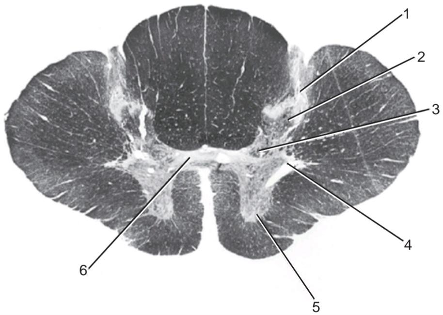
4. Number 1 (a) Nucleus proprius
5. Number 2 (b) Preganglionic sympathetic outflow
6. Number 3 (c) Nucleus dorsalis
7. Number 4 (d) Substantia gelatinosa
8. Number 5 (e) None of the above
9. Number 6
۴. شماره ۱ (الف) هسته پروپیوس
۵. شماره ۲ (ب) جریان سمپاتیک پیش عقدهای
۶. شماره ۳ (ج) هسته پشتی
۷. شماره ۴ (د) جسم ژلاتینی
۸. شماره ۵ (ه) هیچ یک از موارد فوق
۹. شماره ۶
Directions: Each of the numbered items in this section is followed by answers. Select the ONE lettered answer that is CORRECT.
دستورالعمل: هر یک از موارد شمارهگذاری شده در این بخش با پاسخهایی دنبال میشوند. پاسخی را که یک حرف دارد و صحیح است، انتخاب کنید.
10. The following statements concern the cell of origin of the tracts listed below:
(a) The fasciculus cuneatus arises from cells in the substantia gelatinosa.
(b) The anterior spinothalamic arises from cells in the posterior root ganglion.
(c) The fasciculus gracilis arises from cells in the nucleus dorsalis (Clarke column).
(d) The anterior spinocerebellar arises from cells in the posterior root ganglion.
(e) The lateral spinothalamic arises from cells in the substantia gelatinosa.
۱۰. عبارات زیر مربوط به سلول منشأ مسیرهای ذکر شده در زیر است:
(الف) فاسیکولوس کونئاتوس از سلولهای جسم ژلاتینی منشأ میگیرد.
(ب) اسپینوتالامیک قدامی از سلولهای گانگلیون ریشه خلفی منشأ میگیرد.
(ج) فاسیکولوس گراسیلیس از سلولهای هسته پشتی (ستون کلارک) منشأ میگیرد.
(د) اسپینوسربلار قدامی از سلولهای گانگلیون ریشه خلفی منشأ میگیرد.
(ه) اسپینوتالامیک جانبی از سلولهای جسم ژلاتینی منشأ میگیرد.
11. The following statements concern the courses taken by the tracts listed below:
(a) The fasciculus gracilis does not cross to the opposite side of the neural axis.
(b) The spinotectal tract does not cross to the opposite side of the spinal cord.
(c) The lateral spinothalamic tract does not cross to the opposite side of the spinal cord.
(d) The posterior spinocerebellar tract crosses to the opposite side of the neural axis.
(e) The anterior spinothalamic tract immediately crosses to the opposite side of the spinal cord.
۱۱. عبارات زیر مربوط به مسیرهایی است که توسط مسیرهای ذکر شده در زیر طی میشوند:
(الف) فاسیکولوس گراسیلیس از سمت مقابل محور عصبی عبور نمیکند.
(ب) مسیر اسپینوتکتال از سمت مقابل نخاع عبور نمیکند.
(ج) مسیر اسپینوتالامیک جانبی از سمت مقابل نخاع عبور نمیکند.
(د) مسیر اسپینوسربلار خلفی از سمت مقابل محور عصبی عبور میکند.
(ه) مسیر اسپینوتالامیک قدامی بلافاصله از سمت مقابل نخاع عبور میکند.
12. The following statements concern the nucleus of termination of the tracts listed below:
(a) The posterior white column tracts terminate in the inferior colliculus.
(b) The spinoreticular tract terminates on the neurons of the hippocampus.
(c) The spinotectal tract terminates in the inferior colliculus.
(d) The anterior spinothalamic tract terminates in the ventral posterolateral nucleus of the thalamus.
(e) The anterior spinocerebellar tract terminates in the dentate nucleus of the cerebellum.
۱۲. عبارات زیر مربوط به هسته انتهایی راههای عصبی ذکر شده در زیر است:
(الف) راههای عصبی ستون سفید خلفی در کولیکولوس تحتانی ختم میشوند.
(ب) راه عصبی نخاعی-شبکهای به نورونهای هیپوکامپ ختم میشود.
(ج) راه عصبی نخاعی-تکتال در کولیکولوس تحتانی ختم میشود.
(د) راه عصبی نخاعی-تالاموسی قدامی در هسته خلفی-جانبی شکمی تالاموس ختم میشود.
(ه) راه عصبی نخاعی-مخچهای قدامی در هسته دندانهای مخچه ختم میشود.
13. The following statements relate sensations with the appropriate nervous pathways:
(a) Two-point tactile discrimination travels in the lateral spinothalamic tract.
(b) Pain travels in the anterior spinothalamic tract.
(c) Unconscious muscle joint sense travels in the anterior spinocerebellar tract.
(d) Pressure travels in the posterior spinothalamic tract.
(e) Vibration travels in the posterior spinocerebellar tract.
۱۳. جملات زیر حسها را به مسیرهای عصبی مناسب مرتبط میکنند:
(الف) حس لامسه دو نقطهای در مسیر نخاعی-تالامیک جانبی حرکت میکند.
(ب) درد در مسیر نخاعی-تالامیک قدامی حرکت میکند.
(ج) حس ناخودآگاه مفصل عضلانی در مسیر نخاعی-مخچهای قدامی حرکت میکند.
(د) فشار در مسیر نخاعی-تالامیک خلفی حرکت میکند.
(ه) ارتعاش در مسیر نخاعی-مخچهای خلفی حرکت میکند.
14. The following statements concern the gating theory of pain:
(a) Stimulation of small non-pain-conducting fibers in a peripheral nerve may reduce pain sensitivity.
(b) Massage applied to the skin over a painful joint may reduce pain sensitivity.
(c) Stimulation of 8 A- and C-type fibers in a spinal nerve posterior root may decrease pain sensitivity.
(d) Degeneration of large non-pain-conducting fibers in a peripheral nerve decreases pain sensitivity.
(e) Inhibition of pain conduction in the spinal cord does not involve connector neurons.
۱۴. گزارههای زیر مربوط به نظریه دروازهای درد هستند:
(الف) تحریک فیبرهای کوچک غیر رسانای درد در یک عصب محیطی ممکن است حساسیت به درد را کاهش دهد.
(ب) ماساژ اعمال شده روی پوست روی مفصل دردناک ممکن است حساسیت به درد را کاهش دهد.
(ج) تحریک ۸ فیبر نوع A و C در ریشه خلفی عصب نخاعی ممکن است حساسیت به درد را کاهش دهد.
(د) تخریب فیبرهای بزرگ غیر رسانای درد در یک عصب محیطی حساسیت به درد را کاهش میدهد.
(ه) مهار هدایت درد در نخاع شامل نورونهای رابط نمیشود.
15. The following statements concern the reception of pain:
(a) Serotonin is not a transmitter substance in the analgesic system.
(b) Substance P is thought to be the neurotransmitter at the synapses where the first-order neuron terminates on the cells in the posterior gray column of the spinal cord.
(c) Enkephalins and endorphins may stimulate substance P release in the posterior gray column of the spinal cord.
(d) Many of the tracts conducting the initial, sharp, pricking pain terminate in the dorsal anterolateral nucleus of the thalamus.
(e) The slow-conducting C-type fibers are responsible for prolonged, burning pain.
۱۵. جملات زیر مربوط به دریافت درد هستند:
(الف) سروتونین یک ماده فرستنده در سیستم ضد درد نیست.
(ب) تصور میشود ماده P انتقالدهنده عصبی در سیناپسهایی باشد که نورون درجه یک به سلولهای ستون خاکستری خلفی نخاع ختم میشود.
(ج) انکفالینها و اندورفینها ممکن است آزادسازی ماده P را در ستون خاکستری خلفی نخاع تحریک کنند.
(د) بسیاری از مسیرهایی که درد اولیه، تیز و سوزناک را هدایت میکنند، در هسته قدامی-جانبی پشتی تالاموس خاتمه مییابند.
(ه) فیبرهای نوع C با هدایت کند، مسئول درد طولانی و سوزشی هستند.
16. The following statements concern the corticospinal tracts:
(a) They occupy the posterior limb of the internal capsule.
(b) They are mainly responsible for controlling the voluntary movements in proximal limb muscles.
(c) They arise as axons of the pyramidal cells in the fourth layer of the cerebral cortex.
(d) Those that control the movements of the upper limb originate in the precentral gyrus on the medial side of the cerebral hemisphere.
(e) Those that are concerned with the movements of the lower limb are located in the medial area of the middle three fifths of the basis pedunculi.
۱۶. عبارات زیر مربوط به راههای قشری-نخاعی هستند:
(الف) آنها اندام خلفی کپسول داخلی را اشغال میکنند.
(ب) آنها عمدتاً مسئول کنترل حرکات ارادی در عضلات اندام پروگزیمال هستند.
(ج) آنها به عنوان آکسونهای سلولهای هرمی در لایه چهارم قشر مغز ایجاد میشوند.
(د) آنهایی که حرکات اندام فوقانی را کنترل میکنند از شکنج پیشمرکزی در سمت داخلی نیمکره مغزی منشأ میگیرند.
(ه) آنهایی که مربوط به حرکات اندام تحتانی هستند در ناحیه داخلی سه پنجم میانی پایه قرار دارند.
17. The following statements concern the course taken by the tracts listed below:
(a) The rubrospinal tract crosses the midline of the neuroaxis in the medulla oblongata.
(b) The tectospinal tract (most of the nerve fibers) crosses the midline in the posterior commis- sure.
(c) The vestibulospinal tract crosses the midline in the midbrain.
(d) The lateral corticospinal tract crosses the midline in the medulla oblongata.
(e) The anterior corticospinal tract crosses the midline in the midbrain.
۱۷. عبارات زیر مربوط به مسیر طی شده توسط مسیرهای ذکر شده در زیر است:
(الف) مسیر روبرو-نخاعی از خط میانی نورواکسیژن در بصل النخاع عبور میکند.
(ب) مسیر تکتو-نخاعی (بیشتر رشتههای عصبی) از خط میانی در رابط خلفی عبور میکند.
(ج) مسیر دهلیزی-نخاعی از خط میانی در مغز میانی عبور میکند.
(د) مسیر قشری-نخاعی جانبی از خط میانی در بصل النخاع عبور میکند.
(ه) مسیر قشری-نخاعی قدامی از خط میانی در مغز میانی عبور میکند.
18. The following statements concern the nerve cells of origin for the tracts listed below:
(a) The vestibulospinal tract originates from cells of the medial vestibular nucleus situated in the pons.
(b) The tectospinal tract originates from cells in the inferior colliculus.
(c) The lateral corticospinal tract originates from cells in area 4 of the cerebral cortex.
(d) The rubrospinal tract originates from cells in the reticular nucleus.
(e) The reticulospinal tract originates from cells in the reticular formation that is confined to the midbrain.
۱۸. عبارات زیر مربوط به سلولهای عصبی منشأ مسیرهای ذکر شده در زیر است:
(الف) مسیر دهلیزی-نخاعی از سلولهای هسته دهلیزی داخلی واقع در پل مغزی منشأ میگیرد.
(ب) مسیر تکتو-نخاعی از سلولهای موجود در کولیکولوس تحتانی منشأ میگیرد.
(ج) مسیر قشری-نخاعی جانبی از سلولهای ناحیه ۴ قشر مغز منشأ میگیرد.
(د) مسیر روبرو-نخاعی از سلولهای موجود در هسته مشبک منشأ میگیرد.
(ه) مسیر مشبک-نخاعی از سلولهای موجود در تشکیلات مشبک که به مغز میانی محدود میشود، منشأ میگیرد.
19. The following statements concern muscle movement:
(a) Muscular fasciculation is seen only with rapid destruction of lower motor neurons.
(b) Muscle spindle afferent nerve fibers send information only to the spinal cord.
(c) In Parkinson disease, dopamine-secreting neurons that originate in the vestibular nucleus degenerate.
(d) Brain neuronal activity preceding a voluntary movement is limited to the precentral gyrus (area 4).
(e) Hyperactive ankle-jerk reflexes and ankle clonus indicate release of lower motor neuron supraspinal inhibition.
۱۹. عبارات زیر مربوط به حرکت عضلات هستند:
(الف) فاسیکولاسیون عضلانی فقط با تخریب سریع نورونهای حرکتی تحتانی دیده میشود.
(ب) فیبرهای عصبی آوران دوک عضلانی فقط اطلاعات را به نخاع ارسال میکنند.
(ج) در بیماری پارکینسون، نورونهای ترشحکننده دوپامین که از هسته دهلیزی منشأ میگیرند، تخریب میشوند.
(د) فعالیت نورونی مغز قبل از حرکت ارادی به شکنج پیشمرکزی (ناحیه ۴) محدود میشود.
(ه) رفلکسهای پرش مچ پا و کلونوس مچ پا نشاندهنده آزادسازی مهار فوق نخاعی نورون حرکتی تحتانی است.
20. After a hemorrhage into the left internal capsule in a right-handed person, the following sign or symptom might be present:
(a) Left homonymous hemianopia
(b) Right astereognosis
(c) Left hemiplegia
(d) Normal speech
(e) Left-sided positive Babinski response
۲۰. پس از خونریزی در کپسول داخلی چپ در یک فرد راست دست، ممکن است علائم یا نشانههای زیر وجود داشته باشد:
(الف) همیآنوپی هومونیموس چپ
(ب) آسترئوگنوزیس راست
(ج) همیپلژی چپ
(د) گفتار طبیعی
(ه) پاسخ بابنسکی مثبت سمت چپ
21. A patient with a traumatic lesion of the left half of the spinal cord at the level of the eighth cervical segment might present the following sign(s) and symptom(s):
(a) Loss of pain and temperature sensations on the left side below the level of the lesion
(b) Loss of position sense of the right leg
(c) Right hemiplegia
(d) Left positive Babinski sign
(e) Right-sided lower motor paralysis in the segment of the lesion and muscular atrophy
۲۱. بیماری که دچار ضایعه تروماتیک نیمه چپ نخاع در سطح هشتمین قطعه گردنی شده است، ممکن است علائم و نشانههای زیر را نشان دهد:
(الف) از دست دادن حس درد و دما در سمت چپ زیر سطح ضایعه
(ب) از دست دادن حس وضعیت پای راست
(ج) همی پلژی سمت راست
(د) علامت بابینسکی مثبت در سمت چپ
(ه) فلج حرکتی تحتانی سمت راست در قطعه ضایعه و آتروفی عضلانی
Directions: Each of the numbered items in this section is followed by answers. Select the ONE lettered answer that is BEST in each case.
دستورالعمل: هر یک از موارد شمارهگذاری شده در این بخش با پاسخهایی دنبال میشوند. در هر مورد، بهترین پاسخ با یک حرف را انتخاب کنید.
22. Which of the signs and symptoms listed below indicate a cerebellar lesion?
(a) Cogwheel rigidity
(b) Hemiballismus
(c) Chorea
(d) Intention tremor
(e) Athetosis
۲۲. کدام یک از علائم و نشانههای ذکر شده در زیر نشان دهنده ضایعه مخچه است؟
(الف) سفتی چرخ دنده
(ب) همی بالیسم
(ج) کره
(د) لرزش عمدی
(ه) آتتوز
23. Which of the following regions of white matter would not contain corticospinal fibers?
(a) Pyramid of medulla oblongata
(b) Lateral white column of the spinal cord
(c) Cerebral peduncle of the midbrain
(d) Anterior limb of the internal capsule
(e) Corona radiata
۲۳. کدام یک از نواحی زیر از ماده سفید حاوی الیاف قشری-نخاعی نیست؟
(الف) هرم بصل النخاع
(ب) ستون سفید جانبی نخاع
(ج) ساقه مغزی مغز میانی
(د) اندام قدامی کپسول داخلی
(ه) تاج شعاعی
Directions: Each case history is followed by questions. Read the case history, then select the ONE BEST let- tered answer.
دستورالعمل: پس از هر شرح حال، سوالاتی مطرح شده است. شرح حال را بخوانید، سپس بهترین پاسخ با حروف الفبا را انتخاب کنید.
A 59-year-old woman was experiencing pain in the back and showed evidence of loss of pain and temperature sensations down the back of her left leg. Three years previously, she underwent a radical mastectomy followed by radiation and chemotherapy for advanced carcinoma of her right breast.
زنی ۵۹ ساله از درد کمر رنج میبرد و شواهدی از از دست دادن حس درد و دما در پشت پای چپ خود نشان میداد. سه سال قبل، او تحت عمل جراحی ماستکتومی رادیکال و به دنبال آن پرتودرمانی و شیمیدرمانی به دلیل سرطان پیشرفته سینه راست خود قرار گرفته بود.
Examination showed that she was experiencing pain over the lower part of the back, with loss of the skin sensations of pain and temperature down the back of her left leg in the area of the S1-S3 dermatomes. No other neurologic deficits were identified. Radiographic examination of the vertebral column showed evidence of metastases in the bodies of the 9th and 10th thoracic vertebrae. An MRI revealed an extension of one of the metastases into the vertebral canal, with slight indentation of the spinal cord on the right side.
معاینه نشان داد که او در قسمت تحتانی کمر درد دارد و حس درد و حرارت پوست در پشت پای چپش در ناحیه درماتومهای S1-S3 از بین رفته است. هیچ نقص عصبی دیگری شناسایی نشد. بررسی رادیوگرافی ستون فقرات، شواهدی از متاستاز در بدنه مهرههای نهم و دهم سینهای را نشان داد. MRI گسترش یکی از متاستازها به داخل کانال مهرهای را با فرورفتگی جزئی نخاع در سمت راست نشان داد.
24. The pain in the back could be explained in this patient by the following facts except:
(a) Osteoarthritis of the joints of the vertebral column
(b) Metastases in the bodies of the 9th and 10th thoracic vertebrae
(c) Tumor pressure on the spinal nerve posterior roots
(d) A prolapsed intervertebral disc pressing on the spinal nerves
(e) Spasm of the postvertebral muscles following tumor pressure on the posterior white columns of the spinal cord
۲۴. درد کمر در این بیمار را میتوان با حقایق زیر توضیح داد، به جز:
(الف) آرتروز مفاصل ستون فقرات
(ب) متاستاز در بدنه مهرههای ۹ و ۱۰ سینهای
(ج) فشار تومور بر ریشههای خلفی عصب نخاعی
(د) فشار دیسک بین مهرهای بیرون زده بر اعصاب نخاعی
(ه) اسپاسم عضلات پشت مهرهای پس از فشار تومور بر ستونهای سفید خلفی نخاع
25. Loss of pain and temperature sensations down the back of the patient’s left leg in the area of the $1-$3 dermatomes could be explained by the following factual statements except:
(a) Lateral spinothalamic tracts in the spinal cord conduct the sensations of pain.
(b) Lateral spinothalamic tracts are laminated, with the sacral segments of the body located most laterally.
(c) The sacral segments of the tracts are the most exposed to external cord pressure from a metastasizing tumor.
(d) Loss of temperature sensations in the leg could be explained by tumor pressure on the anterior spinothalamic tract.
۲۵. از دست دادن حس درد و دما در پشت پای چپ بیمار در ناحیه درماتومهای ۱ تا ۳ اینچی را میتوان با گزارههای واقعی زیر توضیح داد، به جز:
(الف) راههای اسپینوتالامیک جانبی در نخاع، حس درد را منتقل میکنند.
(ب) راههای اسپینوتالامیک جانبی لایه لایه هستند و بخشهای خاجی بدن در سمت خارجیترین قسمت قرار دارند.
(ج) بخشهای خاجی این راهها بیشترین آسیب را در معرض فشار طناب نخاعی خارجی ناشی از تومور متاستاز دهنده دارند.
(د) از دست دادن حس دما در پا را میتوان با فشار تومور بر راه اسپینوتالامیک قدامی توضیح داد.
26. The severe intractable pain in the back in this patient could be treated by the following methods except:
(a) Prescription of salicylates in large doses
(b) Intramuscular injection of morphine or even the direct injection of the opiate into the spinal cord
(c) Posterior rhizotomy
(d) Cordotomy
(e) Injection of opiates into the subarachnoid space
۲۶. درد شدید و غیرقابل تحمل کمر در این بیمار را میتوان با روشهای زیر درمان کرد، به جز:
(الف) تجویز سالیسیلاتها در دوزهای بالا
(ب) تزریق عضلانی مورفین یا حتی تزریق مستقیم مواد افیونی به نخاع
(ج) ریزوتومی خلفی
(د) کوردوتومی
(ه) تزریق مواد افیونی به فضای زیر عنکبوتیه
√ Answers and Explanations to Review Questions
1. C is correct. The larger nerve cell bodies in the anterior gray horns give rise to a efferent nerve fibers in the anterior roots. A. The anterior and posterior gray columns on the two sides of the spinal cord are united by a gray commissure formed of gray matter. B. The terminal ventricle is the expanded lower end of the central canal. D. The substantia gelatinosa group of cells is located at the apex of each posterior gray column throughout the length of the spinal cord. E. The nucleus dorsalis (Clarke column) is a group of nerve cells found in the posterior gray column and extend- ing from the eighth cervical segment of the cord to the third or fourth lumbar segment.
√ پاسخها و توضیحات برای مرور سوالات
۱. ج صحیح است. اجسام سلولی عصبی بزرگتر در شاخهای خاکستری قدامی، رشتههای عصبی وابران را در ریشههای قدامی ایجاد میکنند. الف. ستونهای خاکستری قدامی و خلفی در دو طرف نخاع توسط یک رابط خاکستری تشکیل شده از ماده خاکستری به هم متصل میشوند. ب. بطن انتهایی، انتهای پایینی گسترش یافته کانال مرکزی است. د. گروه سلولهای ماده ژلاتینی در راس هر ستون خاکستری خلفی در سراسر طول نخاع قرار دارد. ه. هسته پشتی (ستون کلارک) گروهی از سلولهای عصبی است که در ستون خاکستری خلفی یافت میشود و از هشتمین قطعه گردنی نخاع تا سومین یا چهارمین قطعه کمری امتداد دارد.
2. B is correct. In the spinal cord, the anterior spinothalamic tract is found in the anterior white column. A. The posterior spinocerebellar tract is situated in the lateral white column. C. The lateral spinothalamic tract is found in the lateral white column. D. The fasciculus gracilis is found in the posterior white column. E. The rubrospinal tract is found in the lateral white column.
۲. ب صحیح است. در نخاع، راه نخاعی-تالاموسی قدامی در ستون سفید قدامی یافت میشود. الف) راه نخاعی-مخچهای خلفی در ستون سفید جانبی قرار دارد. ج) راه نخاعی-تالاموسی جانبی در ستون سفید جانبی یافت میشود. د) فاسیکولوس گراسیلیس در ستون سفید خلفی یافت میشود. ه) راه روبرواسپینال در ستون سفید جانبی یافت میشود.
3. A is correct. The spinal cord has a cervical enlarge- ment for the brachial plexus (Fig. 4-5). B. The spinal cord possesses spinal nerves that are attached to the cord by anterior and posterior nerve roots. C. In the adult, the spinal cord usually ends inferiorly at the lower border of the first lumbar vertebra. D. The ligamentum denticulatum anchors the spinal cord to the dura mater along each side. E. The central canal, which contains cerebrospinal fluid, communicates with the fourth ventricle of the brain.
۳. الف صحیح است. نخاع دارای یک برآمدگی گردنی برای شبکه بازویی است (شکل ۴-۵). ب. نخاع دارای اعصاب نخاعی است که توسط ریشههای عصبی قدامی و خلفی به نخاع متصل میشوند. ج. در بزرگسالان، نخاع معمولاً در لبه پایینی اولین مهره کمری به سمت پایین ختم میشود. د. رباط دندانهای، نخاع را در امتداد هر طرف به سختشامه متصل میکند. ه. کانال مرکزی که حاوی مایع مغزی نخاعی است، با بطن چهارم مغز ارتباط برقرار میکند.
4. D is correct.
۴. د صحیح است.
5. A is correct.
۵. الف صحیح است.
6. C is correct.
۶. ج صحیح است.
7. B is correct.
۷. ب صحیح است.
8. E is correct. The structure is the anterior gray horn.
۸. ه صحیح است. ساختار آن شاخ خاکستری قدامی است.
9. E is correct. The structure is the gray commissure.
۹. ه صحیح است. ساختار آن رابط خاکستری است.
10. E is correct. In the spinal cord, the lateral spinothalamic tract arises from cells in the substantia gelatinosa. A. The fasciculus cuneatus arises from cells in the posterior root ganglion. B. The anterior spinal thalamic arises from cells in the substantia gelatinosa. C. The fasciculus gracilis arises from cells in the posterior root ganglion. D. The anterior spinocerebellar arises from cells in Clarke column.
۱۰. ه صحیح است. در نخاع، راه نخاعی-تالاموسی جانبی از سلولهای جسم ژلاتینی منشأ میگیرد. الف. فاسیکولوس کونئاتوس از سلولهای گانگلیون ریشه خلفی منشأ میگیرد. ب. تالاموس نخاعی قدامی از سلولهای جسم ژلاتینی منشأ میگیرد. ج. فاسیکولوس گراسیلیس از سلولهای گانگلیون ریشه خلفی منشأ میگیرد. د. مخچه نخاعی قدامی از سلولهای ستون کلارک منشأ میگیرد.
11. A is correct. The fasciculus gracilis does not cross to the opposite side of the neural axis. B. The spinotectal tract crosses to the opposite side of the spinal cord. C. The lateral spinothalamic tract crosses to the opposite side of the spinal cord. D. The posterior spinocerebellar tract does not cross to the opposite side of the spinal cord. E. The anterior spinothalamic tract crosses very obliquely to the opposite side of the spinal cord.
۱۱. الف صحیح است. فاسیکولوس گراسیلیس از سمت مخالف محور عصبی عبور نمیکند. ب. راه اسپینوتکتال از سمت مخالف نخاع عبور میکند. ج. راه اسپینوتالامیک جانبی از سمت مخالف نخاع عبور میکند. د. راه اسپینوسربلار خلفی از سمت مخالف نخاع عبور نمیکند. ه. راه اسپینوتالامیک قدامی به صورت بسیار مایل از سمت مخالف نخاع عبور میکند.
12. D is correct. The anterior spinothalamic tract terminates in the ventral posterolateral nucleus of the thalamus. A. The posterior white column tracts terminate in the nucleus gracilis and cuneatus. B. The spinoreticular tract terminates on the neu- rons of the reticular formation in the medulla, pons, and midbrain. C. The spinotectal tract terminates in the superior colliculus. E. The anterior spinocerebellar tract terminates in the cerebellar cortex.
۱۲. گزینه د صحیح است. راه نخاعی-تالاموسی قدامی در هسته خلفی-جانبی تالاموس خاتمه مییابد. الف) راههای ستون سفید خلفی در هستههای گراسیلیس و کونئاتوس خاتمه مییابند. ب) راه نخاعی-شبکهای به نورونهای تشکیلات مشبک در بصل النخاع، پل مغزی و مغز میانی ختم میشود. ج) راه نخاعی-تکتال در کولیکولوس فوقانی خاتمه مییابد. ه) راه نخاعی-مخچهای قدامی در قشر مخچه خاتمه مییابد.
13. C is correct. Unconscious muscle joint sense travels in the anterior spinocerebellar tract. A. Two- point tactile discrimination travels in the fasciculus cuneatus. B. Pain travels in the lateral spinothalamic tract. D. Pressure travels in the anterior spinothalamic tract. E. Vibration travels in the fas- ciculus gracilis.
۱۳. ج صحیح است. حس ناخودآگاه مفصل عضله در مسیر نخاعی-مخچهای قدامی حرکت میکند. الف) تشخیص لمسی دو نقطهای در فاسیکولوس کونئاتوس حرکت میکند. ب) درد در مسیر نخاعی-تالامیک جانبی حرکت میکند. د) فشار در مسیر نخاعی-تالامیک قدامی حرکت میکند. ه) ارتعاش در فاسیکولوس گراسیلیس حرکت میکند.
14. B is correct concerning the gating theory of pain. Massage applied to the skin over a painful joint may reduce pain sensitivity. A. Stimulation of large nonpain-conducting fibers in a peripheral nerve may reduce pain sensitivity. C. Stimulation of 8 A- and C-type fibers in a spinal nerve posterior root may increase pain sensitivity. D. Degeneration of large non-pain-conducting fibers in a peripheral nerve increases pain sensitivity. E. Inhibition of pain con- duction in the spinal cord could be brought about by means of connector neurons.
۱۴. مورد ب در مورد نظریه دروازهای درد صحیح است. ماساژ اعمال شده بر روی پوست روی مفصل دردناک ممکن است حساسیت به درد را کاهش دهد. الف. تحریک فیبرهای بزرگ غیر رسانای درد در یک عصب محیطی ممکن است حساسیت به درد را کاهش دهد. ج. تحریک ۸ فیبر نوع A و C در ریشه خلفی عصب نخاعی ممکن است حساسیت به درد را افزایش دهد. د. تخریب فیبرهای بزرگ غیر رسانای درد در یک عصب محیطی حساسیت به درد را افزایش میدهد. ه. مهار هدایت درد در نخاع میتواند به وسیله نورونهای رابط انجام شود.
15. E is correct. The slow-conducting C-type fibers are responsible for prolonged, burning pain. A. Serotonin is a transmitter substance in the analgesic system. B. Substance P is a peptide and is thought to be the neurotransmitter at the synapses where the first-order neuron terminates on the cells in the posterior gray column of the spinal cord. C. Enkephalins and endorphins may inhibit the release of substance P in the posterior gray column of the spinal cord. D. Many of the tracts conducting the initial, sharp, pricking pain terminate in the ventral posterolateral nucleus of the thalamus.
۱۵. ه صحیح است. فیبرهای نوع C با هدایت آهسته مسئول درد طولانی و سوزشی هستند. الف) سروتونین یک ماده فرستنده در سیستم ضد درد است. ب) ماده P یک پپتید است و تصور میشود که انتقال دهنده عصبی در سیناپسهایی است که نورون درجه یک به سلولهای ستون خاکستری خلفی نخاع ختم میشود. ج) انکفالینها و اندورفینها ممکن است آزادسازی ماده P را در ستون خاکستری خلفی نخاع مهار کنند. د) بسیاری از مسیرهایی که درد اولیه، تیز و سوزنزننده را هدایت میکنند، در هسته خلفی-جانبی شکمی تالاموس ختم میشوند.
16. A is correct. The corticospinal tracts occupy the posterior limb of the internal capsule (Fig. 4-11). B. The corticospinal tracts are mainly responsible for controlling the voluntary movements in the distal muscles of the limbs. C. They arise as axons of the pyramidal cells in the fifth layer of the cerebral cortex. D. Those that control the movements of the upper limb originate in the precentral gyrus on the lateral side of the cerebral hemisphere. E. Those that are concerned with the movements of the lower limb are located in the lateral area of the middle three fifths of the basis pedunculi.
۱۶. الف صحیح است. راههای قشری-نخاعی، اندام خلفی کپسول داخلی را اشغال میکنند (شکل ۴-۱۱). ب. راههای قشری-نخاعی عمدتاً مسئول کنترل حرکات ارادی در عضلات انتهایی اندامها هستند. ج. آنها به عنوان آکسونهای سلولهای هرمی در لایه پنجم قشر مغز ایجاد میشوند. د. آنهایی که حرکات اندام فوقانی را کنترل میکنند، از شکنج پیشمرکزی در سمت جانبی نیمکره مغزی منشأ میگیرند. ه. آنهایی که مربوط به حرکات اندام تحتانی هستند، در ناحیه جانبی سه پنجم میانی پایههای ساقه قرار دارند.
17. D is correct. The lateral corticospinal tract crosses the midline in the medulla oblongata (Fig. 4-21). A. The rubrospinal tract crosses the midline of the neuroaxis in the midbrain. B. The tectospinal tract (most of the nerve fibers) crosses the midline in the midbrain. C. The vestibulospinal tract does not cross the midline and descends through the medulla oblongata and spinal cord in the anterior white column (Figs. 4-20 and 4-25). E. The anterior corticospinal tract crosses the midline in the spinal cord.
۱۷. د صحیح است. راه قشری-نخاعی جانبی از خط وسط در بصل النخاع عبور میکند (شکل ۴-۲۱). الف. راه روبرواسپینال از خط وسط نورواکسیژن در مغز میانی عبور میکند. ب. راه تکتواسپینال (بیشتر رشتههای عصبی) از خط وسط در مغز میانی عبور میکند. ج. راه دهلیزی-نخاعی از خط وسط عبور نمیکند و از طریق بصل النخاع و نخاع در ستون سفید قدامی پایین میآید (شکلهای ۴-۲۰ و ۴-۲۵). ه. راه قشری-نخاعی قدامی از خط وسط در نخاع عبور میکند.
18. C is correct. The lateral corticospinal tract originates from cells in area 4 of the cerebral cortex. A. The vestibulospinal tract originates from cells of the lateral vestibular nucleus situated in the pons. B. The tectospinal tract originates from cells in the superior colliculus. D. The rubrospinal tract originates from cells in the red nucleus. E. The reticulospinal tract originates from cells in the retic- ular formation in the midbrain, pons, and medulla oblongata.
۱۸. ج صحیح است. راه قشری-نخاعی جانبی از سلولهای ناحیه ۴ قشر مغز منشأ میگیرد. الف) راه دهلیزی-نخاعی از سلولهای هسته دهلیزی جانبی واقع در پل مغزی منشأ میگیرد. ب) راه تکتو-نخاعی از سلولهای کولیکولوس فوقانی منشأ میگیرد. د) راه روبرو-نخاعی از سلولهای هسته قرمز منشأ میگیرد. ه) راه مشبک-نخاعی از سلولهای تشکیلات مشبک در مغز میانی، پل مغزی و بصل النخاع منشأ میگیرد.
19. E is correct. Hyperactive ankle-jerk reflexes and ankle clonus indicate lower motor neuron release from supraspinal inhibition. A. Muscular fasciculation is seen only with slow destruction of the lower motor neurons. B. Muscle spindle afferent nerve fibers send information to the brain as well as to the spinal cord. C. In Parkinson disease, dopamine-secreting neurons decrease in the substantia nigra. D. Brain neuronal activity preceding a voluntary movement is not lim- ited to the precentral gyrus (area 4).
۱۹. ه صحیح است. رفلکسهای پرش مچ پا و کلونوس مچ پا نشاندهندهی آزادسازی نورون حرکتی تحتانی از مهار فوق نخاعی هستند. الف. فاسیکولاسیون عضلانی فقط با تخریب آهسته نورونهای حرکتی تحتانی مشاهده میشود. ب. فیبرهای عصبی آوران دوک عضلانی اطلاعات را به مغز و همچنین به نخاع ارسال میکنند. ج. در بیماری پارکینسون، نورونهای ترشحکننده دوپامین در جسم سیاه کاهش مییابند. د. فعالیت نورونی مغز قبل از حرکت ارادی محدود به شکنج پیشمرکزی (ناحیه ۴) نیست.
20. B is correct. Right astereognosis. A. Right homonymous hemianopia is seen. C. Right hemiplegia is present. D. Aphasia is present. E. A right-sided positive Babinski response is seen.
۲۰. ب صحیح است. استریوگنوزیس راست. الف. همیآنوپی هومونیموس راست دیده میشود. ج. همیپلژی راست وجود دارد. د. آفازی وجود دارد. ه. پاسخ بابنسکی مثبت در سمت راست دیده میشود.
21. D is correct. Left positive Babinski sign is present. A. Pain and temperature sensations are lost on the right side below the level of the lesion. B. Position sense of the left leg is lost. C. Left hemiplegia is present. E. Left-sided lower motor paralysis is seen in the segment of the lesion and muscular atrophy.
۲۱. د صحیح است. علامت بابینسکی مثبت در سمت چپ وجود دارد. الف. حس درد و دما در سمت راست زیر سطح ضایعه از بین رفته است. ب. حس وضعیت پای چپ از بین رفته است. ج. همی پلژی سمت چپ وجود دارد. ه. فلج حرکتی تحتانی سمت چپ در قطعه ضایعه و آتروفی عضلانی مشاهده میشود.
22. D is correct. Intention tremor is present. A. Cog- wheel rigidity occurs in Parkinson disease when the muscle resistance is overcome as a series of jerks. B. Hemiballismus is a rare form of involuntary movement confined to one side of the body; it occurs in disease of the subthalamic nuclei. C. Chorea consists of a series of continuous, rapid, involuntary, jerky, coarse, purposeless movements, which may take place during sleep; it occurs with lesions of the corpus striatum. E. Athetosis consists of continuous, slow, involuntary, dysrhythmic movements that are always the same in the same individual, and they disappear during sleep; it occurs with lesions of the corpus striatum.
۲۲. د صحیح است. لرزش عمدی وجود دارد. الف) سفتی چرخ دنده در بیماری پارکینسون زمانی رخ میدهد که مقاومت عضله به صورت مجموعهای از حرکات ناگهانی برطرف میشود. ب) همیبالیسموس نوعی نادر از حرکت غیرارادی محدود به یک طرف بدن است؛ این حرکت در بیماری هستههای سابتالاموس رخ میدهد. ج) کره شامل مجموعهای از حرکات مداوم، سریع، غیرارادی، تند و تیز، خشن و بیهدف است که ممکن است در طول خواب رخ دهد؛ این حرکت با ضایعات جسم مخطط رخ میدهد. ه) آتتوز شامل حرکات مداوم، آهسته، غیرارادی و نامنظم است که همیشه در یک فرد یکسان هستند و در طول خواب از بین میروند؛ این حرکت با ضایعات جسم مخطط رخ میدهد.
23. D is correct. The anterior limb of the internal capsule does not contain corticospinal fibers.
۲۳. د صحیح است. شاخه قدامی کپسول داخلی حاوی الیاف قشری-نخاعی نیست.
24. E is correct. Spasm of the postvertebral muscles would not be produced by pressure on the posterior white columns of the spinal cord.
۲۴. ه صحیح است. اسپاسم عضلات پس مهرهای با فشار بر ستونهای سفید خلفی نخاع ایجاد نمیشود.
25. D is correct. The sensation of temperature travels in the lateral spinothalamic tract along with the pain impulses.
۲۵. د صحیح است. حس دما در مسیر اسپینوتالامیک جانبی همراه با تکانههای درد حرکت میکند.
26. A is correct. Salicylates, such as aspirin, sodium salicylate, and diflunisal, are used clinically only for the relief of mild to moderate pain, as found in patients suffering from headache and dysmenorrhea.
۲۶. الف صحیح است. سالیسیلاتها، مانند آسپرین، سدیم سالیسیلات و دیفلونیزال، از نظر بالینی فقط برای تسکین درد خفیف تا متوسط استفاده میشوند، همانطور که در بیمارانی که از سردرد و دیسمنوره رنج میبرند، یافت میشود.
🚀 با ما همراه شوید!
تازهترین مطالب و آموزشهای مغز و اعصاب را از دست ندهید. با فالو کردن کانال تلگرام آیندهنگاران مغز، از ما حمایت کنید!