مبانی علوم اعصاب اریک کندل؛ مولکولهای رنگدانهای شبکیه و سلولهای گانگلیونی شبکیه


دعای مطالعه [ نمایش ]
بِسْمِ الله الرَّحْمنِ الرَّحیمِ
اَللّهُمَّ اَخْرِجْنى مِنْ ظُلُماتِ الْوَهْمِ
خدايا مرا بيرون آور از تاريكىهاى وهم،
وَ اَكْرِمْنى بِنُورِ الْفَهْمِ
و به نور فهم گرامى ام بدار،
اَللّهُمَّ افْتَحْ عَلَيْنا اَبْوابَ رَحْمَتِكَ
خدايا درهاى رحمتت را به روى ما بگشا،
وَانْشُرْ عَلَيْنا خَزائِنَ عُلُومِكَ بِرَحْمَتِكَ يا اَرْحَمَ الرّاحِمينَ
و خزانههاى علومت را بر ما باز كن به امید رحمتت اى مهربانترين مهربانان.
«مبانی علوم اعصاب» اثر دکتر اریک کندل و همکاران، یکی از جامعترین و معتبرترین منابع در حوزه علوم اعصاب محسوب میشود.
ترجمه دقیق این کتاب، با مدیریت داریوش طاهری و تحت نظارت برند علمی آیندهنگاران مغز، با الهام از تلاشهای علمی و تدریس اساتیدی همچون دکتر محمدتقی جغتایی و دکتر ثریا مهرابی، به فارسی برگردانده شده است.
این ترجمه، دسترسی به دانش عمیق علوم اعصاب را برای دانشجویان و پژوهشگران رشتههای پزشکی، علوم اعصاب، روانشناسی، علوم شناختی، هوش مصنوعی، گفتاردرمانی، مهندسی پزشکی و تمام علاقهمندان به درک عملکرد ذهن و مغز فراهم کرده و گامی در راستای ارتقای دانش و بهبود کیفیت زندگی است.
» کتاب مبانی علوم اعصاب اریک کندل
» » فصل ۲۲: فصل پردازش بصری سطح پایین: شبکیه؛ قسمت دوم
در حال ویرایش
Principles of Neural Science; Eric R. Kandel
»» Chapter 22: Low-Level Visual Processing: The Retina
The Photoreceptor Layer Samples the Visual Image
لایه گیرنده نور از تصویر بصری نمونهبرداری میکند
Ocular Optics Limit the Quality of the Retinal Image
اپتیک چشم، کیفیت تصویر شبکیه را محدود میکند
There Are Two Types of Photoreceptors: Rods and Cones
دو نوع گیرنده نوری وجود دارد: میلهایها و مخروطیها
Phototransduction Links the Absorption of a Photon to a Change in Membrane Conductance
انتقال نوری، جذب فوتون را به تغییر در رسانایی غشاء مرتبط میکند
Light Activates Pigment Molecules in the Photoreceptors
نور مولکولهای رنگدانه را در گیرندههای نوری فعال میکند
Excited Rhodopsin Activates a Phosphodiesterase Through the G Protein Transducin
رودوپسین برانگیخته شده از طریق پروتئین G ترانسدوسین، یک فسفودی استراز را فعال میکند.
Multiple Mechanisms Shut Off the Cascade
چندین مکانیسم، آبشار را خاموش میکنند
Defects in Phototransduction Cause Disease
نقص در انتقال نوری باعث بیماری میشود
Ganglion Cells Transmit Neural Images to the Brain
سلولهای گانگلیون تصاویر عصبی را به مغز منتقل میکنند
The Two Major Types of Ganglion Cells Are ON Cells and OFF Cells
دو نوع اصلی سلولهای گانگلیون، سلولهای روشن (ON) و سلولهای خاموش (OFF) هستند.
Many Ganglion Cells Respond Strongly to Edges in the Image
بسیاری از سلولهای گانگلیون به شدت به لبههای تصویر پاسخ میدهند
The Output of Ganglion Cells Emphasizes Temporal Changes in Stimuli
خروجی سلولهای گانگلیون بر تغییرات زمانی در محرکها تأکید دارد
Retinal Output Emphasizes Moving Objects
خروجی شبکیه، اشیاء متحرک را برجسته میکند
Several Ganglion Cell Types Project to the Brain Through Parallel Pathways
چندین نوع سلول گانگلیون از طریق مسیرهای موازی به مغز میرسند
A Network of Interneurons Shapes the Retinal Output
شبکهای از نورونهای رابط، خروجی شبکیه را شکل میدهد
Parallel Pathways Originate in Bipolar Cells
مسیرهای موازی از سلولهای دوقطبی سرچشمه میگیرند
Spatial Filtering Is Accomplished by Lateral Inhibition
فیلترینگ مکانی با مهار جانبی انجام میشود
Temporal Filtering Occurs in Synapses and Feedback Circuits
فیلترینگ زمانی در سیناپسها و مدارهای بازخورد رخ میدهد
Color Vision Begins in Cone-Selective Circuits
بینایی رنگی از مدارهای انتخابی مخروطی آغاز میشود
Congenital Color Blindness Takes Several Forms
کوررنگی مادرزادی اشکال مختلفی دارد
Rod and Cone Circuits Merge in the Inner Retina
مدارهای میلهای و مخروطی در شبکیه داخلی ادغام میشوند
The Retina’s Sensitivity Adapts to Changes in Illumination
حساسیت شبکیه با تغییرات روشنایی سازگار میشود
Light Adaptation Is Apparent in Retinal Processing and Visual Perception
سازگاری با نور در پردازش شبکیه و ادراک بصری آشکار است
Multiple Gain Controls Occur within the Retina
کنترلهای چندگانهی بهره در شبکیه رخ میدهند
Light Adaptation Alters Spatial Processing
سازگاری با نور، پردازش فضایی را تغییر میدهد
Highlights
نکات برجسته
Phototransduction Links the Absorption of a Photon to a Change in Membrane Conductance
انتقال نوری، جذب فوتون را به تغییر در رسانایی غشاء مرتبط میکند
As in many other neurons, the membrane potential of a photoreceptor is regulated by the balance of membrane conductances to Nat and K+ ions, whose transmembrane gradients are maintained by metabolically active pumps (Chapter 9). In the dark, Na+ ions flow into the photoreceptor through nonselective cation channels that are activated by the second messenger cyclic guanosine 3-5′ monophosphate (cGMP).
همانند بسیاری از نورونهای دیگر، پتانسیل غشای یک گیرنده نوری توسط تعادل رسانایی غشایی به یونهای +Na و +K تنظیم میشود که شیبهای غشایی آنها توسط پمپهای فعال متابولیکی حفظ میشود (فصل 9). در تاریکی، یونهای +Na از طریق کانالهای کاتیونی غیرانتخابی که توسط پیامرسان ثانویه گوانوزین حلقوی 3-5′ مونوفسفات (cGMP) فعال میشوند، به گیرنده نوری جریان مییابند.
Absorption of a photon by the pigment protein sets in motion a biochemical cascade that ultimately lowers the concentration of cGMP, thus closing the cGMP-gated channels and moving the cell closer to the K+ equilibrium potential. In this way, light hyperpolarizes the photoreceptor (Figure 22-7). Here, we describe this sequence of events in detail. Most of this knowledge derives from studies of rods, but the mechanism in cones is very similar.
جذب یک فوتون توسط پروتئین رنگدانه، آبشاری بیوشیمیایی را به حرکت در میآورد که در نهایت غلظت cGMP را کاهش میدهد، بنابراین کانالهای دریچهدار cGMP را میبندد و سلول را به پتانسیل تعادل +K نزدیکتر میکند. به این ترتیب، نور گیرنده نوری را بیشقطبی میکند (شکل 22-7). در اینجا، این توالی رویدادها را با جزئیات شرح میدهیم. بیشتر این دانش از مطالعات روی میلهها حاصل شده است، اما مکانیسم در مخروطها بسیار مشابه است.

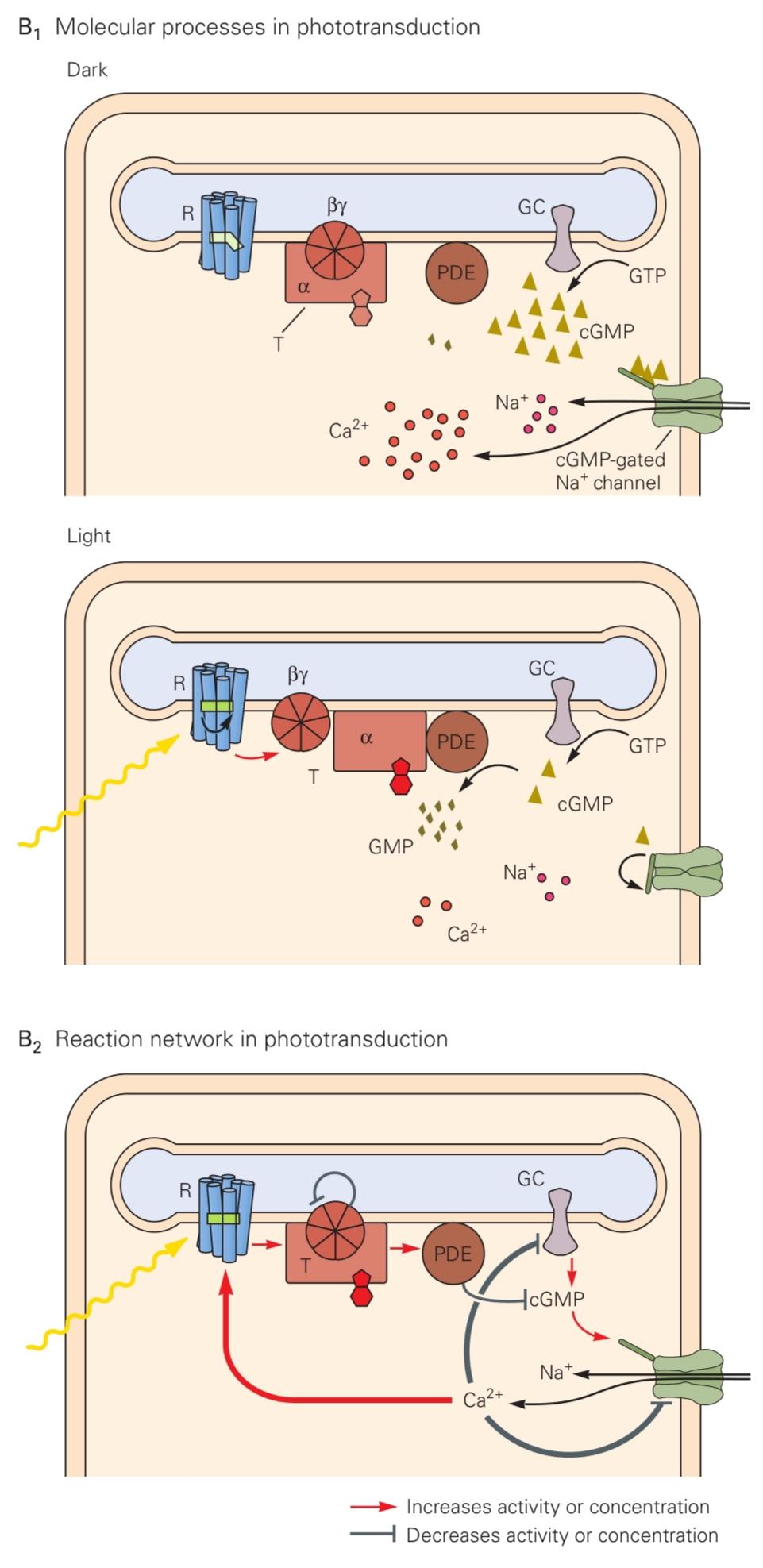

Figure 22-7 (Opposite) Phototransduction.
شکل 22-7 (روبرو) انتقال نوری.
A. The rod cell responds to light. Rhodopsin molecules in the outer-segment discs absorb photons, which leads to the closure of cyclic guanosine 3′-5′ monophosphate (cGMP)-gated channels in the plasma membrane. This channel closure hyperpolarizes the membrane and reduces the rate of release of the neurotransmitter glutamate. (Adapted from Alberts 2008.)
الف) سلول استوانهای به نور پاسخ میدهد. مولکولهای رودوپسین در دیسکهای بخش بیرونی، فوتونها را جذب میکنند که منجر به بسته شدن کانالهای دریچهدار گوانوزین حلقوی ۳’-۵’ مونوفسفات (cGMP) در غشای پلاسما میشود. این بسته شدن کانال، غشا را بیشقطبی میکند و سرعت آزادسازی گلوتامات، انتقالدهنده عصبی، را کاهش میدهد. (اقتباس از آلبرتز ۲۰۰۸.)
B. 1. Molecular processes in phototransduction. cGMP is produced by a guanylate cyclase (GC) from guanosine triphosphate (GTP) and hydrolyzed by a phosphodiesterase (PDE). In the dark, the phosphodiesterase activity is low, the cGMP concentration is high, and the cGMP-gated channels are open, allowing the influx of Na+ and Ca2+. In the light, rhodopsin (R) is excited by absorption of a photon, then activates transducin (T), which in turn activates the PDE; the cGMP level drops, the membrane channels close, and less Na+ and Ca2+ enter the cell. The transduction enzymes are all located in the internal membrane discs, and the soluble ligand cGMP serves as a messenger to the plasma membrane.
ب. ۱. فرآیندهای مولکولی در فوتوترانسداکشن. cGMP توسط یک گوانیلات سیکلاز (GC) از گوانوزین تری فسفات (GTP) تولید و توسط یک فسفودیاستراز (PDE) هیدرولیز میشود. در تاریکی، فعالیت فسفودیاستراز کم، غلظت cGMP بالا و کانالهای دریچهدار cGMP باز هستند و اجازه ورود +Na و +Ca2 را میدهند. در روشنایی، رودوپسین (R) با جذب یک فوتون تحریک میشود، سپس ترانسدوسین (T) را فعال میکند که به نوبه خود PDE را فعال میکند. سطح cGMP کاهش مییابد، کانالهای غشایی بسته میشوند و +Na و +Ca2 کمتری وارد سلول میشوند. آنزیمهای ترانسداکشن همگی در دیسکهای غشای داخلی قرار دارند و لیگاند محلول cGMP به عنوان پیامرسان به غشای پلاسما عمل میکند.
2. Calcium ions have a negative feedback role in the reaction cascade in phototransduction. Stimulation of the network by light leads to the closure of the cGMP-gated channels. This causes a drop in the intracellular concentration of Ca2+. Because Ca2+ modulates the function of at least three components of the cascade-rhodopsin, GC, and the cGMP-gated channel the drop in Ca2+ counteracts the excitation caused by light.
۲. یونهای کلسیم نقش بازخورد منفی در آبشار واکنش در انتقال نوری دارند. تحریک شبکه توسط نور منجر به بسته شدن کانالهای وابسته به cGMP میشود. این امر باعث کاهش غلظت درون سلولی +Ca2 میشود. از آنجا که +Ca2 عملکرد حداقل سه جزء آبشار – رودوپسین، GC و کانال وابسته به cGMP – را تعدیل میکند، کاهش +Ca2 تحریک ناشی از نور را خنثی میکند.
C. Voltage response of a primate rod and cone to brief flashes of light of increasing intensity. Higher numbers on the traces indicate greater intensities of illumination (not all traces are labeled). For dim flashes, the response amplitude increases linearly with intensity. At high intensities, the receptor saturates and remains hyperpolarized steadily for some time after the flash; this leads to the afterimages that we perceive after a bright flash. Note that the response peaks earlier for brighter flashes and that cones respond faster than rods. (Reproduced, with permission, from Schneeweis and Schnapf 1995. Copy- right © 1995 AAAS.)
ج. پاسخ ولتاژ یک سلول میلهای و مخروطی نخستی به جرقههای کوتاه نور با شدت فزاینده. اعداد بالاتر روی ردپاها نشان دهنده شدت بیشتر روشنایی هستند (همه ردپاها برچسب گذاری نشدهاند). برای جرقههای کم نور، دامنه پاسخ به صورت خطی با شدت افزایش مییابد. در شدتهای بالا، گیرنده اشباع میشود و برای مدتی پس از جرقه به طور پیوسته هایپرپلاریزه باقی میماند. این منجر به پستصویرهایی میشود که ما پس از یک جرقه روشن درک میکنیم. توجه داشته باشید که پاسخ برای جرقههای روشنتر زودتر به اوج میرسد و مخروطها سریعتر از میلهها پاسخ میدهند. (با اجازه، از Schneeweis and Schnapf 1995 بازتولید شده است. حق چاپ © 1995 AAAS.)
Light Activates Pigment Molecules in the Photoreceptors
نور مولکولهای رنگدانه را در گیرندههای نوری فعال میکند
Rhodopsin, the visual pigment in rod cells, has two components. The protein portion, opsin, is embedded in the disc membrane and does not by itself absorb visible light. The light-absorbing moiety, retinal, is a small molecule whose 11-cis isomer is covalently linked to a lysine residue of opsin (Figure 22–8A). Absorption of a photon by retinal causes it to flip from the 11-cis to the all-trans configuration. This reaction is the only light- dependent step in vision.
رودوپسین، رنگدانه بینایی در سلولهای میلهای، دو جزء دارد. بخش پروتئینی، اوپسین، در غشای دیسک جاسازی شده است و به خودی خود نور مرئی را جذب نمیکند. بخش جاذب نور، رتینال، مولکول کوچکی است که ایزومر 11-سیس آن به صورت کووالانسی به یک باقیمانده لیزین از اوپسین متصل شده است (شکل 22-8A). جذب یک فوتون توسط رتینال باعث میشود که از پیکربندی 11-سیس به پیکربندی تمام ترانس تغییر کند. این واکنش تنها مرحله وابسته به نور در بینایی است.
The change in shape of the retinal molecule causes a conformational change in the opsin to an activated state called metarhodopsin II, thus triggering the second step of phototransduction. Metarhodopsin II is unstable and splits within minutes, yielding opsin and free all-trans retinal. The all-trans retinal is then trans- ported from rods to pigment epithelial cells, where it is reduced to all-trans retinol (vitamin A), the precursor of 11-cis retinal, which is subsequently transported back to rods.
تغییر شکل مولکول رتینال باعث تغییر ساختاری در اوپسین به حالت فعال شده به نام متارودوپسین II میشود و بدین ترتیب مرحله دوم انتقال نوری را آغاز میکند. متارودوپسین II ناپایدار است و در عرض چند دقیقه تجزیه میشود و اوپسین و رتینال تمام ترانس آزاد را به وجود میآورد. سپس رتینال تمام ترانس از سلولهای استوانهای به سلولهای اپیتلیال رنگدانهای منتقل میشود، جایی که به رتینول تمام ترانس (ویتامین A)، پیشساز رتینال ۱۱-سیس، احیا میشود که متعاقباً دوباره به سلولهای استوانهای منتقل میشود.
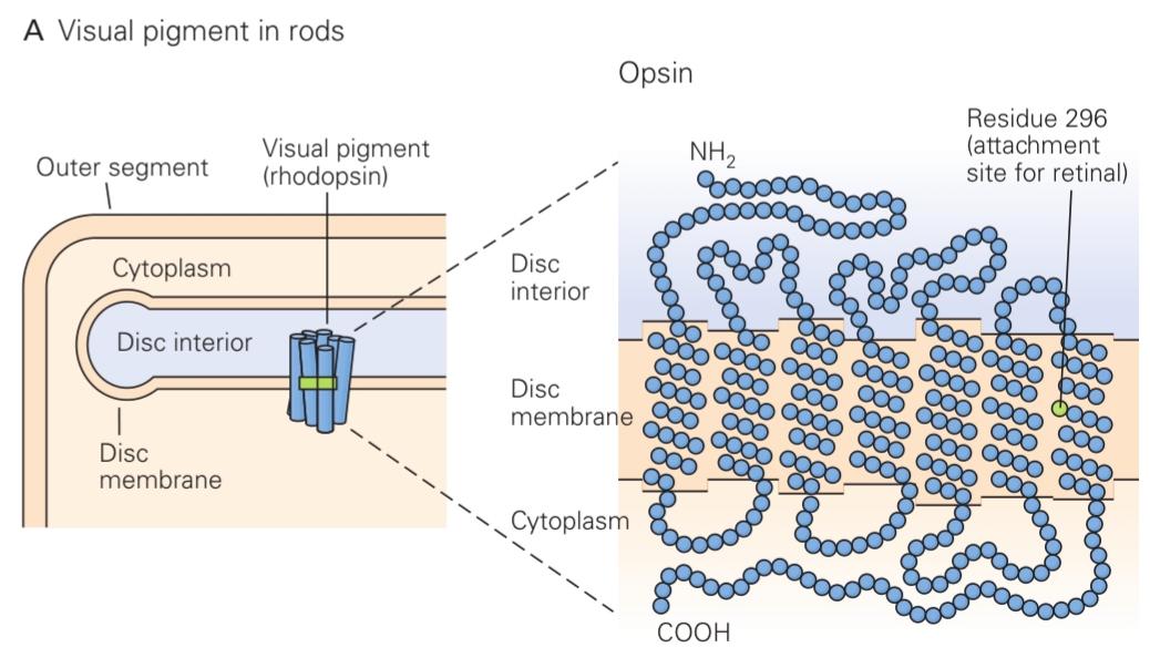


Figure 22-8 Structure of the visual pigments.
شکل 22-8 ساختار رنگدانههای بینایی.
A. The visual pigment in rod cells, rhodopsin, is the covalent complex of two components. Opsin is a large protein with 348 amino acids and a molecular mass of approximately 40,000 daltons. It loops back and forth seven times across the membrane of the rod disc. Retinal is a small light- absorbing compound covalently attached to a side chain of lysine 296 in opsin’s seventh membrane-spanning region. Absorption of light by 11-cis retinal causes a rotation around the double bond. As retinal adopts the more stable all-trans configuration, it causes a conformational change in opsin that triggers the subsequent events of visual transduction. (Adapted, with permission, from Nathans and Hogness 1984.)
الف) رنگدانه بینایی در سلولهای میلهای، رودوپسین، کمپلکس کووالانسی از دو جزء است. اوپسین پروتئین بزرگی با ۳۴۸ اسید آمینه و جرم مولکولی تقریباً ۴۰۰۰۰ دالتون است. این پروتئین هفت بار در سراسر غشای دیسک میلهای به جلو و عقب حلقه میزند. رتینال یک ترکیب کوچک جاذب نور است که به صورت کووالانسی به یک زنجیره جانبی لیزین ۲۹۶ در ناحیه هفتم غشایی اوپسین متصل شده است. جذب نور توسط رتینال ۱۱-سیس باعث چرخش حول پیوند دوگانه میشود. همانطور که رتینال پیکربندی تمام ترانس پایدارتر را اتخاذ میکند، باعث تغییر ساختاری در اوپسین میشود که وقایع بعدی انتقال بینایی را آغاز میکند. (اقتباس شده، با اجازه، از ناتانز و هاگنس ۱۹۸۴.)
B. The blue circles denote identical amino acids; black circles denote differences. The forms of opsin in the three types of cone cells (L, M, and S) resemble each other as well as the rhodopsin in rod cells, suggesting that all four evolved from a common precursor by duplication and divergence. The L and M opsins are most closely related, with 96% identity in their amino acid sequences. They are thought to have evolved from a gene- duplication event approximately 30 million years ago, after Old World monkeys, which have three visual pigments, separated from New World monkeys, which generally have only two.
ب. دایرههای آبی نشاندهنده اسیدهای آمینه یکسان هستند؛ دایرههای سیاه نشاندهنده تفاوتها هستند. اشکال اوپسین در سه نوع سلول مخروطی (L، M و S) و همچنین رودوپسین در سلولهای میلهای شبیه به یکدیگر هستند، که نشان میدهد هر چهار نوع از یک پیشساز مشترک از طریق تکثیر و واگرایی تکامل یافتهاند. L و موپسینها با ۹۶٪ شباهت در توالی اسیدهای آمینه خود، بیشترین ارتباط را با هم دارند. تصور میشود که آنها تقریباً ۳۰ میلیون سال پیش، پس از جدا شدن میمونهای دنیای قدیم که سه رنگدانه بینایی دارند از میمونهای دنیای جدید که عموماً فقط دو رنگدانه دارند، از یک رویداد تکثیر ژن تکامل یافتهاند.
All-trans retinal is thus a crucial compound in the visual system. Its precursors, such as vitamin A, can not be synthesized by humans and so must be a regular part of the diet. Deficiencies of vitamin A can lead to night blindness and, if untreated, to deterioration of receptor outer segments and eventually to blindness.
بنابراین، رتینال تمام ترانس یک ترکیب حیاتی در سیستم بینایی است. پیشسازهای آن، مانند ویتامین A، نمیتوانند توسط انسان سنتز شوند و بنابراین باید بخشی منظم از رژیم غذایی باشند. کمبود ویتامین A میتواند منجر به شبکوری و در صورت عدم درمان، به زوال بخشهای بیرونی گیرنده و در نهایت به نابینایی منجر شود.
Each type of cone in the human retina produces a variant of the opsin protein. These three cone pigments are distinguished by their absorption spectrum, the dependence on wavelength of the efficiency of light absorption (see Figure 22-6). The spectrum is determined by the protein sequence through the interaction between retinal and certain amino acid side chains near the binding pocket. Red light excites L cones more than the M cones, whereas green light excites the M cones more. Therefore, the relative degree of excitation in these cone types contains information about the spectrum of the light, independent of its intensity. The brain’s comparison of signals from different cone types is the basis for color vision.
هر نوع مخروط در شبکیه انسان نوعی از پروتئین اوپسین را تولید میکند. این سه رنگدانه مخروطی با طیف جذبی خود، وابستگی به طول موج و راندمان جذب نور، متمایز میشوند (شکل 22-6 را ببینید). این طیف توسط توالی پروتئین از طریق تعامل بین زنجیرههای جانبی شبکیه و برخی اسیدهای آمینه در نزدیکی محفظه اتصال تعیین میشود. نور قرمز مخروطهای L را بیشتر از مخروطهای M تحریک میکند، در حالی که نور سبز مخروطهای M را بیشتر تحریک میکند. بنابراین، درجه نسبی تحریک در این انواع مخروطها حاوی اطلاعاتی در مورد طیف نور، مستقل از شدت آن است. مقایسه سیگنالهای انواع مختلف مخروطها توسط مغز، اساس بینایی رنگی است.
In night vision, only the rods are active, so all functional photoreceptors have the same absorption spectrum. A green light consequently has exactly the same effect on the visual system as a red light of a greater intensity. Because a single-photoreceptor system cannot distinguish the spectrum of a light from its intensity, “at night all cats are gray.” By comparing the sensitivity of a rod to different wavelengths of light, one obtains the absorption spectrum of rhodopsin. It is a remarkable fact that one can measure this molecular property accurately just by asking human subjects about the appearance of various colored lights (Figure 22-9). The quantitative study of perception, or psychophysics, provides similar insights into other mechanisms of brain processing (Chapter 17).
در دید در شب، فقط سلولهای استوانهای فعال هستند، بنابراین همه گیرندههای نوری عملکردی طیف جذب یکسانی دارند. در نتیجه، یک نور سبز دقیقاً همان تأثیری را بر سیستم بینایی دارد که یک نور قرمز با شدت بیشتر دارد. از آنجا که یک سیستم تک گیرنده نوری نمیتواند طیف یک نور را از شدت آن تشخیص دهد، “در شب همه گربهها خاکستری هستند.” با مقایسه حساسیت یک سلول استوانهای به طول موجهای مختلف نور، میتوان طیف جذبی رودوپسین را به دست آورد. این یک واقعیت قابل توجه است که میتوان این ویژگی مولکولی را تنها با پرسیدن از افراد در مورد ظاهر نورهای رنگی مختلف، به طور دقیق اندازهگیری کرد (شکل 22-9). مطالعه کمی ادراک یا روانفیزیک، بینشهای مشابهی را در مورد سایر مکانیسمهای پردازش مغز ارائه میدهد (فصل 17).
Excited Rhodopsin Activates a Phosphodiesterase Through the G Protein Transducin
رودوپسین برانگیخته شده از طریق پروتئین G ترانسدوسین، یک فسفودی استراز را فعال میکند
Activated rhodopsin in the form of metarhodopsin II diffuses within the disc membrane where it encounters transducin, a member of the G protein family (Chapter 14). As is the case for other G proteins, the inactive form of transducin binds a molecule of guanosine diphosphate (GDP). Interaction with metarhodopsin II promotes the exchange of GDP for guanosine triphosphate (GTP). This leads to dissociation of transducin’s subunits into an active α-subunit carrying the GTP (Tα-GTP) and the β- and γ-subunits (Tẞγ). Metarhodopsin II can activate hundreds of additional transducin molecules, thus significantly amplifying the cell’s response.
رودوپسین فعالشده به شکل متارودوپسین II در غشای دیسک منتشر میشود و در آنجا با ترانسدوسین، عضوی از خانواده پروتئین G، مواجه میشود (فصل 14). همانند سایر پروتئینهای G، شکل غیرفعال ترانسدوسین به یک مولکول گوانوزین دیفسفات (GDP) متصل میشود. برهمکنش با متارودوپسین II، تبادل GDP با گوانوزین تریفسفات (GTP) را افزایش میدهد. این امر منجر به تفکیک زیرواحدهای ترانسدوسین به یک زیرواحد فعال α حامل GTP (Tα-GTP) و زیرواحدهای β و γ (Tẞγ) میشود. متارودوپسین II میتواند صدها مولکول ترانسدوسین اضافی را فعال کند و در نتیجه پاسخ سلول را به طور قابل توجهی تقویت کند.
The active transducin subunit Tα-GTP forms a complex with a cyclic nucleotide phosphodiesterase, another protein associated with the disc membrane. This interaction greatly increases the rate at which the enzyme hydrolyzes cGMP to 5′-GMP. Each phosphodiesterase molecule can hydrolyze more than 1,000 molecules of cGMP per second, thus increasing the degree of amplification.
زیر واحد فعال ترانسدوسین Tα-GTP با یک نوکلئوتید حلقوی فسفودیاستراز، پروتئین دیگری که با غشای دیسک مرتبط است، کمپلکس تشکیل میدهد. این برهمکنش سرعت هیدرولیز cGMP به 5′-GMP توسط آنزیم را به میزان زیادی افزایش میدهد. هر مولکول فسفودیاستراز میتواند بیش از ۱۰۰۰ مولکول cGMP را در ثانیه هیدرولیز کند، بنابراین درجه تکثیر را افزایش میدهد.
The concentration of cGMP controls the activity of the cGMP-gated channels in the plasma membrane of the outer segment. In darkness, when the cGMP concentration is high, a sizeable Na+ influx through the open channels maintains the cell at a depolarized level of approximately 40 mV. As a consequence, the cell’s synaptic terminal continuously releases the transmitter glutamate. The light-evoked decrease in cGMP results in the closure of the cGMP-gated channels, thus reducing the inward flux of Na+ ions and hyperpolarizing the cell (Figure 22-7B1). Hyperpolarization slows the release of neurotransmitter from the photoreceptor terminal, thereby initiating a neural signal.
غلظت cGMP فعالیت کانالهای دریچهدار cGMP را در غشای پلاسمایی بخش بیرونی کنترل میکند. در تاریکی، هنگامی که غلظت cGMP بالا است، هجوم قابل توجه Na+ از طریق کانالهای باز، سلول را در سطح دپلاریزه تقریباً 40 میلیولت نگه میدارد. در نتیجه، پایانه سیناپسی سلول به طور مداوم گلوتامات فرستنده را آزاد میکند. کاهش ناشی از نور در cGMP منجر به بسته شدن کانالهای دریچهدار cGMP میشود، در نتیجه جریان رو به داخل یونهای Na+ کاهش مییابد و سلول بیشقطبی میشود (شکل 22-7B1). بیشقطبی شدن، آزادسازی انتقالدهنده عصبی از پایانه گیرنده نوری را کند میکند و در نتیجه یک سیگنال عصبی را آغاز میکند.
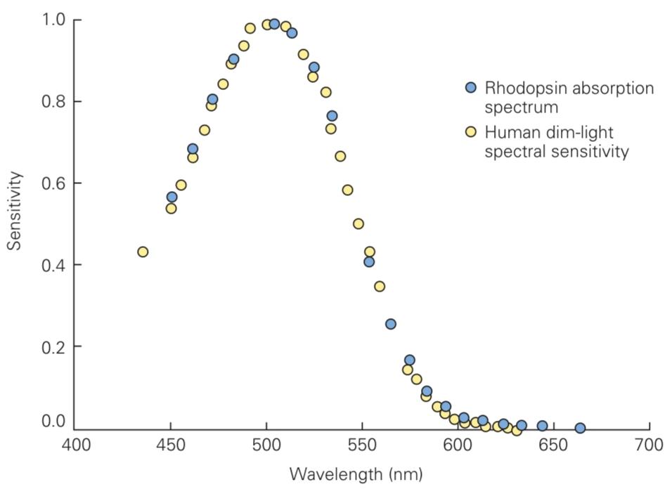
Figure 22-9 Absorption spectrum of rhodopsin. The absorption spectrum of human rhodopsin measured in a cuvette is compared with the spectral sensitivity of human observers to very dim light flashes. The psychophysical data have been corrected for absorption by the ocular media. (Reproduced, with permission, from Wald and Brown 1956. Copyright © 1956 Springer Nature.)
شکل ۲۲-۹ طیف جذبی رودوپسین. طیف جذبی رودوپسین انسانی که در یک کووت اندازهگیری شده است، با حساسیت طیفی ناظران انسانی به نورهای بسیار ضعیف مقایسه میشود. دادههای روانفیزیکی برای جذب توسط محیط چشمی اصلاح شدهاند. (با اجازه، از Wald and Brown 1956 بازتولید شده است. حق چاپ © ۱۹۵۶ Springer Nature.)
Multiple Mechanisms Shut Off the Cascade
چندین مکانیسم، آبشار را خاموش میکنند
The photoreceptor’s response to a single photon must be terminated so that the cell can respond to another photon. Metarhodopsin II is inactivated through phosphorylation by a specific rhodopsin kinase followed by binding of the soluble protein arrestin, which blocks the interaction with transducin.
پاسخ گیرنده نوری به یک فوتون واحد باید خاتمه یابد تا سلول بتواند به فوتون دیگری پاسخ دهد. متارودوپسین II از طریق فسفوریلاسیون توسط یک رودوپسین کیناز خاص غیرفعال میشود و به دنبال آن پروتئین محلول ارستین متصل میشود که تعامل با ترانسدوسین را مسدود میکند.
Active transducin (Tα-GTP) has an intrinsic GTPase activity, which eventually converts bound GTP to GDP. Tα-GDP then releases phosphodiesterase and recombines with Tẞγ, ready again for excitation by rhodopsin. Once the phosphodiesterase has been inactivated, the CGMP concentration is restored by a guanylate cyclase that produces cGMP from GTP. At this point, the membrane channels open, the Na+ current resumes, and the photoreceptor depolarizes back to its dark potential.
ترانسدوسین فعال (Tα-GTP) دارای فعالیت ذاتی GTPase است که در نهایت GTP متصل را به GDP تبدیل میکند. سپس Tα-GDP فسفودیاستراز را آزاد میکند و با Tẞγ نوترکیب میشود و دوباره برای تحریک توسط رودوپسین آماده میشود. پس از غیرفعال شدن فسفودیاستراز، غلظت CGMP توسط یک گوانیلات سیکلاز که cGMP را از GTP تولید میکند، بازیابی میشود. در این مرحله، کانالهای غشایی باز میشوند، جریان Na+ از سر گرفته میشود و گیرنده نوری به پتانسیل تاریک خود دپلاریزه میشود.
In addition to these independent mechanisms that shut off individual elements of the cascade, an important feedback mechanism ensures that large responses are terminated more quickly. This is mediated by a change in the Ca2+ concentration in the cell. Calcium ions enter the cell through the cGMP-gated channels and are extruded by rapid cation exchangers. In the dark, the intracellular Ca2+ concentration is high, but during the cell’s light response, when the cGMP-gated channels close, the Ca2+ level drops quickly to a few percent of the dark level.
علاوه بر این مکانیسمهای مستقل که عناصر منفرد آبشار را خاموش میکنند، یک مکانیسم بازخورد مهم تضمین میکند که پاسخهای بزرگ سریعتر خاتمه مییابند. این امر با تغییر در غلظت Ca2+ در سلول انجام میشود. یونهای کلسیم از طریق کانالهای وابسته به cGMP وارد سلول میشوند و توسط تبادلکنندههای کاتیونی سریع دفع میشوند. در تاریکی، غلظت Ca2+ درون سلولی بالا است، اما در طول پاسخ نوری سلول، هنگامی که کانالهای وابسته به cGMP بسته میشوند، سطح Ca2+ به سرعت به چند درصد از سطح تاریکی کاهش مییابد.
This reduction in Ca2+ concentration modulates the biochemical reactions in three ways (Figure 22-7B2). Rhodopsin phosphorylation is accelerated through the action of the calcium-binding protein recoverin on rhodopsin kinase, thus reducing activation of transducin. The activity of guanylyl cyclase is accelerated by calcium-dependent guanylyl cyclase-activating proteins. Finally, the affinity of the cGMP-gated channel for cGMP is increased through the action of Ca2+– calmodulin. All these effects promote the return of the photoreceptor to the dark state.
این کاهش غلظت Ca2+ واکنشهای بیوشیمیایی را از سه طریق تعدیل میکند (شکل 22-7B2). فسفوریلاسیون رودوپسین از طریق عملکرد پروتئین متصل شونده به کلسیم، ریکاورین، بر روی رودوپسین کیناز تسریع میشود و در نتیجه فعالسازی ترانسدوسین کاهش مییابد. فعالیت گوانیلیل سیکلاز توسط پروتئینهای فعالکننده گوانیلیل سیکلاز وابسته به کلسیم تسریع میشود. در نهایت، میل ترکیبی کانال وابسته به cGMP برای cGMP از طریق عملکرد Ca2+- کالمودولین افزایش مییابد. همه این اثرات، بازگشت گیرنده نوری به حالت تاریکی را تقویت میکنند.
Defects in Phototransduction Cause Disease
نقص در انتقال نوری باعث بیماری میشود
Not surprisingly, defects in the phototransduction machinery have serious consequences. One prominent defect is color blindness, which results from loss or abnormality in the genes for cone pigments, as discussed later.
جای تعجب نیست که نقص در دستگاه انتقال نور عواقب جدی دارد. یکی از نقصهای برجسته، کوررنگی است که همانطور که بعداً مورد بحث قرار میگیرد، ناشی از از دست دادن یا ناهنجاری در ژنهای مربوط به رنگدانههای مخروطی است.
Stationary night blindness results when rod function has been lost but cone function remains intact. This disease is heritable, and mutations have been identified in many components of the phototransduction cascade: rhodopsin, rod transducin, rod phosphodiesterase, rhodopsin kinase, and arrestin. In some cases, it appears that the rods are permanently activated, as if exposed to a constant blinding light.
شب کوری ثابت زمانی ایجاد میشود که عملکرد سلولهای استوانهای از بین رفته باشد اما عملکرد سلولهای مخروطی دست نخورده باقی مانده باشد. این بیماری ارثی است و جهشهایی در بسیاری از اجزای آبشار انتقال نور شناسایی شدهاند: رودوپسین، ترانسدوسین سلولهای استوانهای، فسفودیاستراز سلولهای استوانهای، رودوپسین کیناز و ارستین. در برخی موارد، به نظر میرسد که سلولهای استوانهای به طور دائم فعال هستند، گویی در معرض نور خیرهکنندهی مداوم قرار دارند.
Unfortunately, many defects in phototransduction lead to retinitis pigmentosa, a progressive degeneration of the retina that ultimately results in blindness. The disease has multiple forms, many of which have been associated with mutations that affect signal transduction in rods. Why these changes in function lead to death of the rods and subsequent degeneration of the cones is not understood.
متأسفانه، بسیاری از نقصها در انتقال نور منجر به رتینیت پیگمنتوزا، یک تخریب پیشرونده شبکیه که در نهایت منجر به نابینایی میشود، میشود. این بیماری اشکال مختلفی دارد که بسیاری از آنها با جهشهایی مرتبط هستند که بر انتقال سیگنال در سلولهای استوانهای تأثیر میگذارند. اینکه چرا این تغییرات در عملکرد منجر به مرگ سلولهای استوانهای و تخریب بعدی سلولهای مخروطی میشود، مشخص نیست.
Ganglion Cells Transmit Neural Images to the Brain
سلولهای گانگلیون تصاویر عصبی را به مغز منتقل میکنند
The photoreceptor layer produces a relatively simple neural representation of the visual scene: Neurons in bright regions are hyperpolarized, whereas those in dark regions are depolarized. Because the optic nerve has only about 1% as many axons as there are receptor cells, the retinal circuit must edit the information in the photoreceptors before it is conveyed to the brain.
لایه گیرنده نوری، نمایش عصبی نسبتاً سادهای از صحنه بصری تولید میکند: نورونهای موجود در مناطق روشن، بیشقطبی هستند، در حالی که نورونهای موجود در مناطق تاریک، دپلاریزه هستند. از آنجا که عصب بینایی تنها حدود ۱٪ از تعداد سلولهای گیرنده، آکسون دارد، مدار شبکیه باید اطلاعات موجود در گیرندههای نوری را قبل از انتقال به مغز ویرایش کند.
This step constitutes low-level visual processing, the first stage in deriving visual percepts from the pattern of light falling on the retina. To understand this process, we must first understand the organization of the retina’s output and how retinal ganglion cells respond to various patterns of light.
این مرحله، پردازش بصری سطح پایین را تشکیل میدهد، اولین مرحله در استخراج ادراکات بصری از الگوی نوری که بر شبکیه میتابد. برای درک این فرآیند، ابتدا باید سازماندهی خروجی شبکیه و نحوه پاسخ سلولهای گانگلیونی شبکیه به الگوهای مختلف نور را درک کنیم.
The Two Major Types of Ganglion Cells Are ON Cells and OFF Cells
دو نوع اصلی سلولهای گانگلیون، سلولهای روشن (ON) و سلولهای خاموش (OFF) هستند
Many retinal ganglion cells fire action potentials spontaneously even in darkness or constant illumination. If the light intensity is suddenly increased, so-called ON cells fire more rapidly. Other ganglion cells, the OFF cells, fire more slowly or cease firing altogether. When the intensity diminishes again, the ON cells fire less and OFF cells fire more. The retinal output thus includes two complementary representations that differ in the polarity of their response to light.
بسیاری از سلولهای گانگلیونی شبکیه حتی در تاریکی یا روشنایی مداوم، پتانسیل عمل خودبهخودی تولید میکنند. اگر شدت نور بهطور ناگهانی افزایش یابد، سلولهای به اصطلاح ON سریعتر تولید پتانسیل عمل میکنند. سایر سلولهای گانگلیونی، سلولهای OFF، کندتر تولید پتانسیل عمل میکنند یا بهطورکلی تولید پتانسیل عمل را متوقف میکنند. وقتی شدت نور دوباره کاهش مییابد، سلولهای ON کمتر و سلولهای OFF بیشتر تولید پتانسیل عمل میکنند. بنابراین، خروجی شبکیه شامل دو نمایش مکمل است که در قطبیت پاسخ آنها به نور متفاوت است.
This arrangement serves to communicate rapidly both brightening and dimming in the visual scene. If the retina had only ON cells, a dark object would be encoded by a decrease in firing rate. If the ganglion cell fired at a maintained rate of 10 spikes per second and then decreased its rate, it would take about 100 ms for the postsynaptic neuron to notice the change in frequency of action potentials. In contrast, an increase in firing rate to 200 spikes per second is noticeable within only 5 ms.
این چیدمان برای برقراری ارتباط سریع بین روشن شدن و کم شدن نور در صحنه بصری عمل میکند. اگر شبکیه فقط سلولهای ON داشته باشد، یک شیء تاریک با کاهش سرعت شلیک سیگنال رمزگذاری میشود. اگر سلول گانگلیونی با سرعت ثابت 10 اسپایک در ثانیه شلیک کند و سپس سرعت آن را کاهش دهد، حدود 100 میلیثانیه طول میکشد تا نورون پسسیناپسی متوجه تغییر در فرکانس پتانسیلهای عمل شود. در مقابل، افزایش سرعت شلیک به 200 اسپایک در ثانیه تنها در عرض 5 میلیثانیه قابل توجه است.
Many Ganglion Cells Respond Strongly to Edges in the Image
بسیاری از سلولهای گانگلیون به شدت به لبههای تصویر پاسخ میدهند
To probe the responses of a ganglion cell in more detail, one can test how the cell’s firing varies with the location and time course of a small spot of light focused on different portions of the retina.
برای بررسی دقیقتر پاسخهای یک سلول گانگلیون، میتوان آزمایش کرد که چگونه شلیک سلول با مکان و مسیر زمانی یک نقطه کوچک نور که بر بخشهای مختلف شبکیه متمرکز شده است، تغییر میکند.
A typical ganglion cell is sensitive to light in a compact region of the retina near the cell body called the cell’s receptive field. Within that area, one can often distinguish a center region and surround region where light produces opposite responses in the cell. An ON cell, for example, fires faster when a bright spot is focused in the cell’s receptive field center but decreases its firing when the spot is focused on the surround. If light covers both the center and the surround, the response is much weaker than for center-only illumination. A bright spot on the center combined with a dark annulus covering the surround elicits very strong firing. For an OFF cell, these relationships are reversed; the cell is strongly excited by a dark spot and a bright annulus (Figure 22-10).
یک سلول گانگلیونی معمولی در ناحیهای فشرده از شبکیه نزدیک جسم سلولی به نام میدان گیرنده سلول، به نور حساس است. در آن ناحیه، اغلب میتوان یک ناحیه مرکزی و ناحیه پیرامونی را تشخیص داد که در آن نور پاسخهای متضادی را در سلول ایجاد میکند. به عنوان مثال، یک سلول روشن، هنگامی که یک نقطه روشن در مرکز میدان گیرنده سلول متمرکز میشود، سریعتر شلیک میکند، اما هنگامی که آن نقطه روی محیط اطراف متمرکز میشود، شلیک آن کاهش مییابد. اگر نور هم مرکز و هم محیط اطراف را بپوشاند، پاسخ بسیار ضعیفتر از زمانی است که فقط مرکز روشن میشود. یک نقطه روشن در مرکز همراه با یک حلقه تاریک که محیط اطراف را پوشانده است، شلیک بسیار قوی ایجاد میکند. برای یک سلول خاموش، این روابط برعکس میشوند؛ سلول به شدت توسط یک نقطه تاریک و یک حلقه روشن تحریک میشود (شکل 22-10).
The output produced by a population of retinal ganglion cells thus enhances regions of spatial contrast in the input, such as an edge between two areas of different intensity, and gives less emphasis to regions of homogeneous illumination.
بنابراین، خروجی تولید شده توسط جمعیتی از سلولهای گانگلیونی شبکیه، نواحی با کنتراست مکانی در ورودی، مانند لبه بین دو ناحیه با شدت متفاوت، را افزایش میدهد و تأکید کمتری بر نواحی با روشنایی همگن دارد.
The Output of Ganglion Cells Emphasizes Temporal Changes in Stimuli
خروجی سلولهای گانگلیون بر تغییرات زمانی در محرکها تأکید دارد
When an effective light stimulus appears, a ganglion cell’s firing typically increases sharply from the resting level to a peak and then relaxes to an intermediate rate. When the stimulus turns off, the firing rate drops sharply then gradually recovers to the resting level.
وقتی یک محرک نوری مؤثر ظاهر میشود، شلیک یک سلول گانگلیونی معمولاً از سطح استراحت به شدت افزایش مییابد و به اوج خود میرسد و سپس تا حد متوسطی آرام میشود. وقتی محرک خاموش میشود، سرعت شلیک به شدت کاهش مییابد و سپس به تدریج به سطح استراحت برمیگردد.
The rapidity of decline from the peak to the resting level varies among ganglion cell types. Transient neurons produce a burst of spikes only at the onset of the stimulus, whereas sustained neurons maintain an almost steady firing rate for several seconds during stimulation (Figure 22–10).
سرعت کاهش از اوج تا سطح استراحت در بین انواع سلولهای گانگلیونی متفاوت است. نورونهای گذرا فقط در شروع محرک، انفجاری از اسپایکها تولید میکنند، در حالی که نورونهای پایدار در طول تحریک، سرعت شلیک تقریباً ثابتی را برای چند ثانیه حفظ میکنند (شکل ۲۲-۱۰).
In general, however, the output of ganglion cells favors temporal changes in visual input over periods of constant light intensity. In fact, when an image is stabilized on the retina with an eye-tracking device, it fades from view within seconds. Fortunately, this never happens in normal vision; even when we attempt to fix our gaze, small automatic eye movements (saccades) continually scan the image across the retina and prevent the world from disappearing (Chapter 25).
با این حال، به طور کلی، خروجی سلولهای گانگلیونی در طول دورههای شدت نور ثابت، تغییرات زمانی در ورودی بصری را ترجیح میدهد. در واقع، هنگامی که یک تصویر با استفاده از دستگاه ردیابی چشم روی شبکیه تثبیت میشود، در عرض چند ثانیه از دید محو میشود. خوشبختانه، این اتفاق هرگز در بینایی طبیعی رخ نمیدهد؛ حتی وقتی سعی میکنیم نگاه خود را ثابت نگه داریم، حرکات کوچک خودکار چشم (ساکادها) به طور مداوم تصویر را در سراسر شبکیه اسکن میکنند و از ناپدید شدن جهان جلوگیری میکنند (فصل ۲۵).
Retinal Output Emphasizes Moving Objects
خروجی شبکیه، اشیاء متحرک را برجسته میکند
Based on these observations, we can understand more generally the response of ganglion cells to visual inputs. For example, the edges of a moving object elicit strong firing in the ganglion cell population because these are the only regions of spatial contrast and the only regions where the light intensity changes over time (Figure 22-11).
بر اساس این مشاهدات، میتوانیم به طور کلیتر پاسخ سلولهای گانگلیونی به ورودیهای بصری را درک کنیم. به عنوان مثال، لبههای یک جسم متحرک باعث شلیک قوی در جمعیت سلولهای گانگلیونی میشوند زیرا اینها تنها مناطقی هستند که کنتراست مکانی دارند و تنها مناطقی هستند که شدت نور در طول زمان تغییر میکند (شکل 22-11).
We can easily appreciate why the retina selectively responds to these features. The outline of an object is particularly useful for inferring its shape and identity. Similarly, objects that move or change suddenly are more worthy of immediate attention than those that do not. Retinal processing thus extracts low-level features of the scene that are useful for guiding behavior and selectively transmits those to the brain. In fact, the rejection of features that are constant either in space or in time accounts for the spatiotemporal sensitivity of human perception (Box 22-1).
ما به راحتی میتوانیم درک کنیم که چرا شبکیه به طور انتخابی به این ویژگیها پاسخ میدهد. طرح کلی یک شیء به ویژه برای استنباط شکل و هویت آن مفید است. به طور مشابه، اشیایی که به طور ناگهانی حرکت میکنند یا تغییر میکنند، نسبت به اشیایی که حرکت نمیکنند، ارزش توجه فوری بیشتری دارند. بنابراین، پردازش شبکیه، ویژگیهای سطح پایین صحنه را که برای هدایت رفتار مفید هستند، استخراج میکند و آنها را به طور انتخابی به مغز منتقل میکند. در واقع، رد ویژگیهایی که چه در فضا و چه در زمان ثابت هستند، حساسیت مکانی-زمانی ادراک انسان را توضیح میدهد (کادر 22-1).
Several Ganglion Cell Types Project to the Brain Through Parallel Pathways
چندین نوع سلول گانگلیون از طریق مسیرهای موازی به مغز میرسند
Several different types of ganglion cells have been identified on the basis of their morphology and responses to light. The ON and OFF cells occur in every vertebrate retina, and in the primate retina, two major classes of cells, the P-cells and M-cells, each include ON and OFF types (see Figure 22-2B). At any given distance from the fovea, the receptive fields of M-cells (Latin magno, large) are much larger than those of P-cells (Latin parvo, small). The M-cells also have faster and more transient responses than P-cells. Some ganglion cells are intrinsically light-sensitive owing to expression of the visual pigment melanopsin.
چندین نوع مختلف از سلولهای گانگلیونی بر اساس مورفولوژی و پاسخهایشان به نور شناسایی شدهاند. سلولهای روشن و خاموش در شبکیه هر مهرهداری وجود دارند و در شبکیه نخستیسانان، دو دسته اصلی از سلولها، سلولهای P و سلولهای M، هر کدام شامل انواع روشن و خاموش هستند (شکل 22-2B را ببینید). در هر فاصله مشخصی از گودی مرکزی، میدانهای گیرنده سلولهای M (لاتین magno، بزرگ) بسیار بزرگتر از سلولهای P (لاتین parvo، کوچک) هستند. سلولهای M همچنین پاسخهای سریعتر و گذراتری نسبت به سلولهای P دارند. برخی از سلولهای گانگلیونی به دلیل بیان رنگدانه بینایی ملانوپسین، ذاتاً به نور حساس هستند.
In total, more than 20 types of ganglion cells have been described. The population of each type covers the retina in a tiled fashion, such that any point on the retina lies within the receptive field center of at least one ganglion cell. One can envision that the signals from each population together send a distinct neural representation of the visual field to the brain. In this view, the optic nerve conveys 20 or more neural representations that differ in polarity (ON or OFF), spatial resolution (fine or coarse), temporal responsiveness (sustained or transient), spectral filtering (broadband or dominated by red, green, or blue), and selectivity for other image features such as motion.
در مجموع، بیش از 20 نوع سلول گانگلیونی توصیف شده است. جمعیت هر نوع، شبکیه را به صورت کاشیکاری شده میپوشاند، به طوری که هر نقطه روی شبکیه در مرکز میدان دریافتی حداقل یک سلول گانگلیونی قرار دارد. میتوان تصور کرد که سیگنالهای هر جمعیت با هم، یک نمایش عصبی متمایز از میدان بینایی را به مغز ارسال میکنند. در این دیدگاه، عصب بینایی 20 یا بیشتر نمایش عصبی را منتقل میکند که از نظر قطبیت (روشن یا خاموش)، وضوح مکانی (ریز یا درشت)، پاسخگویی زمانی (پایدار یا گذرا)، فیلتر طیفی (پهن باند یا تحت سلطه قرمز، سبز یا آبی) و گزینشپذیری برای سایر ویژگیهای تصویر مانند حرکت متفاوت هستند.

Figure 22-10 Responses of retinal ganglion cells with center-surround receptive fields. In these idealized experiments, the stimulus changes from a uniform gray field to the pattern of bright (yellow) and dark (black) regions indicated on the left. This leads to the firing rate responses shown on the right. 1. ON cells are excited by a bright spot in the receptive field center, OFF cells by a dark spot. In sustained cells, the excitation persists throughout stimulation, whereas in transient cells, a brief burst of spikes occurs just after the onset of stimulation. 2. If the same stimulus that excites the center is applied to the surround, firing is suppressed. 3. Uniform stimulation of both center and surround elicits a response like that of the center, but much smaller in amplitude. 4. Stimulation of the center combined with the opposite stimulus in the surround produces the strongest response.
شکل 22-10 پاسخ سلولهای گانگلیونی شبکیه با میدانهای گیرنده مرکز-محیط. در این آزمایشهای ایدهآل، محرک از یک میدان خاکستری یکنواخت به الگویی از نواحی روشن (زرد) و تاریک (سیاه) که در سمت چپ نشان داده شده است، تغییر میکند. این منجر به پاسخهای سرعت شلیک نشان داده شده در سمت راست میشود. 1. سلولهای روشن توسط یک نقطه روشن در مرکز میدان گیرنده و سلولهای خاموش توسط یک نقطه تاریک تحریک میشوند. در سلولهای پایدار، تحریک در طول تحریک ادامه مییابد، در حالی که در سلولهای گذرا، انفجار کوتاهی از اسپایکها درست پس از شروع تحریک رخ میدهد. 2. اگر همان محرکی که مرکز را تحریک میکند به محیط اطراف اعمال شود، شلیک سرکوب میشود. 3. تحریک یکنواخت هر دو مرکز و محیط اطراف، پاسخی مانند مرکز ایجاد میکند، اما دامنه آن بسیار کوچکتر است. 4. تحریک مرکز همراه با محرک مخالف در محیط اطراف، قویترین پاسخ را ایجاد میکند.
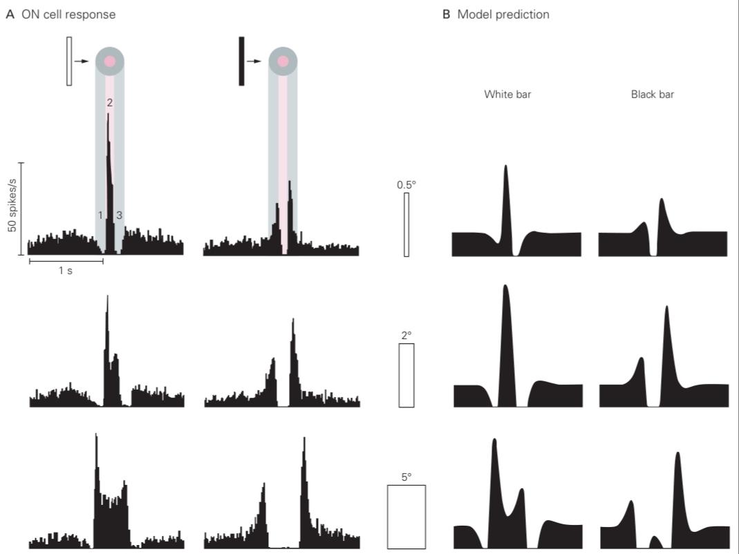
Figure 22-11 Responses of ganglion cells in the cat retina to moving objects.
شکل 22-11 پاسخ سلولهای گانگلیونی در شبکیه گربه به اشیاء متحرک.
A. The firing rate of an ON ganglion cell in response to a variety of bars (white or black, various widths) moving across the retina. Each bar moves at 10° per second; 1° corresponds to 180 μm on the retina. In response to the white bar, the firing rate first decreases as the bar passes over the receptive-field surround (1), increases as the bar enters the center (2), and decreases again as the bar passes through the surround on the opposite side (3). The dark bar elicits responses of the opposite sign. Because ganglion cells similar to this one are distributed throughout the retina, one can also interpret this curve as an instantaneous snapshot of activity in a population of ganglion cells, where the horizontal axis represents location on the retina. In effect, this activity profile is the neural representation of the moving bar transmitted to the brain. A complementary population of OFF ganglion cells (not shown) here) conveys another neural activity profile in parallel. In this way, both bright and dark edges can be signaled by a sharp increase in firing.
الف. نرخ شلیک یک سلول گانگلیون ON در پاسخ به میلههای مختلف (سفید یا سیاه، با عرضهای مختلف) که در سراسر شبکیه حرکت میکنند. هر میله با سرعت 10 درجه در ثانیه حرکت میکند؛ 1 درجه مربوط به 180 میکرومتر روی شبکیه است. در پاسخ به میله سفید، نرخ شلیک ابتدا با عبور میله از روی میدان گیرنده (1) کاهش مییابد، با ورود میله به مرکز (2) افزایش مییابد و با عبور میله از محیط در طرف مقابل (3) دوباره کاهش مییابد. میله تیره پاسخهایی با علامت مخالف را ایجاد میکند. از آنجا که سلولهای گانگلیون مشابه این در سراسر شبکیه توزیع شدهاند، میتوان این منحنی را به عنوان یک تصویر لحظهای از فعالیت در جمعیتی از سلولهای گانگلیون نیز تفسیر کرد، که در آن محور افقی نشان دهنده مکان روی شبکیه است. در واقع، این نمایه فعالیت، نمایش عصبی میله متحرک منتقل شده به مغز است. یک جمعیت مکمل از سلولهای گانگلیون OFF (که در اینجا نشان داده نشده است) نمایه فعالیت عصبی دیگری را به طور موازی منتقل میکند. به این ترتیب، هر دو لبه روشن و تاریک میتوانند با افزایش شدید شلیک سیگنال شوند.
B. A simple model of retinal processing that incorporates center-surround antagonism and a transient temporal filter is used to predict ganglion-cell firing rates. The predictions match the essential features of the responses in part A. (Reproduced, with permission, from Rodieck 1965. Copyright © 1965 Elsevier Ltd.)
ب. یک مدل ساده از پردازش شبکیه که شامل تضاد مرکز-اطراف و یک فیلتر زمانی گذرا است، برای پیشبینی نرخ شلیک سلولهای گانگلیونی استفاده میشود. پیشبینیها با ویژگیهای اساسی پاسخها در بخش الف مطابقت دارند. (با اجازه، از Rodieck 1965 بازتولید شده است. حق چاپ © 1965 Elsevier Ltd.)
»» فصل بعد: فصل پردازش بینایی سطح متوسط و اولیههای بینایی
»» تمامی کتاب