نوروآناتومی بالینی؛ سیستم عصبی خودمختار


ترجمه و بازنویسی علمی فصول کتاب «نوروآناتومی بالینی» | کتاب درسی جامع ساختارهای عصبی (ویرایش دوم)
کتاب درسی نوروآناتومی بالینی، ویرایش دوم. Textbook of Clinical Neuroanatomy (2nd ed.) یکی از منابع معتبر در حوزه نوروآناتومی بالینی (Clinical Neuroanatomy) است که با رویکردی کاربردی، ساختارها و مسیرهای سیستم عصبی (Nervous System) را بهصورت نظاممند و بالینی شرح میدهد.
این ترجمه آموزشی، حاصل تلاش تیم علمی آیندهنگاران مغز به سرپرستی داریوش طاهری است و با هدف ارتقای آموزش علوم اعصاب (Neuroscience) در زبان فارسی، با حفظ دقت علمی، به زبانی روان تدوین شده است.
در ۲۰ فصل این مجموعه، ساختارهایی چون مغز (Brain)، نخاع (Spinal Cord)، اعصاب مغزی (Cranial Nerves)، سیستم لیمبیک (Limbic System)، مسیرهای حسی و حرکتی (Sensory and Motor Pathways)، مخچه (Cerebellum)، دیانسفالون (Diencephalon) و سیستم عصبی خودمختار (Autonomic Nervous System) بررسی میشوند.
این مجموعه برای دانشجویان پزشکی، رزیدنتها و متخصصان علاقهمند به نوروساینس بالینی طراحی شده و تلفیقی از مفاهیم بنیادی، نکات بالینی و نمودارهای عملکردی را ارائه میدهد.
با سپاس از همراهی شما
تیم آیندهنگاران مغز | سرپرستی: داریوش طاهری
20. Autonomic Nervous System
20. سیستم عصبی خودکار
The autonomic (self-controlling) nervous system is that part of the nervous system which regulates most of the involuntary activities of the body, such as the activities of smooth muscles of bronchial tree, gut, genitourinary system, pupil, arrector pili muscles of the hair, cardiac muscle and secretion of the glands. Thus it represents the visceral component of the nervous system, hence sometimes also called ‘visceral nervous system’.
سیستم عصبی خودکار (خودکنترل) بخشی از سیستم عصبی است که بیشتر فعالیتهای غیرارادی بدن، مانند فعالیتهای عضلات صاف برونش، روده، دستگاه تناسلی-ادراری، مردمک چشم، عضلات راست کننده مو، عضله قلب و ترشح غدد را تنظیم میکند. بنابراین، این سیستم نمایانگر جزء احشایی سیستم عصبی است، از این رو گاهی اوقات “سیستم عصبی احشایی” نیز نامیده میشود.
The autonomic nervous system, like the somatic nervous system is made up of afferent and efferent pathways.
سیستم عصبی خودکار، مانند سیستم عصبی سوماتیک، از مسیرهای آوران و وابران تشکیل شده است.
The visceral afferent pathways resemble somatic afferent pathways. The cell bodies of first order sensory neurons (pseudo-unipolar) are located in the cranial and dorsal root ganglia. There peripheral processes are distributed through autonomic ganglia or plexuses or through somatic nerves. The central processes accompany somatic afferent fibres through cranial nerves or dorsal spinal roots into the central nervous system when they establish connections to mediate autonomic reflexes and visceral sensation.
مسیرهای آوران احشایی شبیه مسیرهای آوران سوماتیک هستند. اجسام سلولی نورونهای حسی درجه یک (شبه تک قطبی) در گانگلیونهای ریشه جمجمهای و پشتی قرار دارند. فرآیندهای محیطی آنها از طریق گانگلیونها یا شبکههای عصبی خودکار یا از طریق اعصاب سوماتیک توزیع میشوند. فرآیندهای مرکزی، فیبرهای آوران سوماتیک را از طریق اعصاب جمجمهای یا ریشههای نخاعی پشتی به سیستم عصبی مرکزی همراهی میکنند، زمانی که آنها ارتباطاتی را برای واسطهگری رفلکسهای خودکار و حس احشایی برقرار میکنند.
Thus the afferent fibres of the autonomic nervous system are identical to the afferent fibres of the somatic nervous system and form part of general afferent component of the entire nervous system. Visceral efferent pathways, however, differ from those of somatic efferent pathways (i.e. the efferent fibres, differ in the somatic and autonomic nervous systems).
بنابراین، فیبرهای آوران سیستم عصبی خودکار با فیبرهای آوران سیستم عصبی سوماتیک یکسان هستند و بخشی از جزء آوران عمومی کل سیستم عصبی را تشکیل میدهند. با این حال، مسیرهای وابران احشایی، با مسیرهای آوران سوماتیک متفاوت هستند (یعنی فیبرهای وابران در سیستمهای عصبی سوماتیک و خودکار متفاوت هستند).
Somatic efferent fibres pass directly from the CNS to the skeletal muscle, whereas autonomic (visceral) efferent fibres do not pass directly to the visceral effector organs from the CNS, instead they first relay in the autonomic ganglia outside the CNS, and then the postganglionic fibres supply the effector organs. The only exception is the innervation of the adrenal medulla.
فیبرهای وابران سوماتیک مستقیماً از سیستم عصبی مرکزی (CNS) به عضله اسکلتی منتقل میشوند، در حالی که فیبرهای وابران اتونوم (احشایی) مستقیماً از سیستم عصبی مرکزی (CNS) به اندامهای مؤثر احشایی منتقل نمیشوند، در عوض ابتدا در گانگلیونهای اتونوم خارج از سیستم عصبی مرکزی رله میشوند و سپس فیبرهای پسگانگلیونی اندامهای مؤثر را تغذیه میکنند. تنها استثنا عصبدهی مدولای آدرنال است.
Thus the efferent pathway of autonomic nervous system is made up of two neurons, preganglionic and postganglionic.
بنابراین مسیر وابران سیستم عصبی خودکار از دو نورون، پیشگانگلیونی و پسگانگلیونی، تشکیل شده است.
For all practical purposes, the autonomic nervous system (ANS) is a general visceral efferent motor system which controls and regulates smooth muscle, cardiac muscle and glands. These three divisions of autonomic nervous system differ in their organization and structure but they are closely related functionally.
برای تمام اهداف عملی، سیستم عصبی خودکار (ANS) یک سیستم حرکتی وابران احشایی عمومی است که عضلات صاف، عضله قلبی و غدد را کنترل و تنظیم میکند. این سه بخش سیستم عصبی خودکار از نظر سازماندهی و ساختار متفاوت هستند اما از نظر عملکردی ارتباط نزدیکی با هم دارند.
N.B. All the efferent peripheral nerve fibres belong to the autonomic nervous system except those to the skeletal muscles.
توجه: تمام فیبرهای عصبی محیطی وابران به جز آنهایی که به عضلات اسکلتی تعلق دارند، متعلق به سیستم عصبی خودکار هستند.
Divisions of the Autonomic Nervous System
The autonomic nervous system is divided into three divisions, the sympathetic nervous system and parasympathetic nervous system, and the enteric nervous system.
بخشهای سیستم عصبی خودکار
سیستم عصبی خودکار به سه بخش تقسیم میشود: سیستم عصبی سمپاتیک و سیستم عصبی پاراسمپاتیک و سیستم عصبی رودهای.
1. Sympathetic nervous system: The preganglionic sympathetic fibres arise from lateral horn cells of the thoracic and upper two lumbar segments (T1 to L2) of the spinal cord, hence sympathetic nervous system constitutes the thoracolumbar outflow (Fig. 20.1). The axons of preganglionic neurons project to the autonomic ganglia (the sympathetic ganglia). The ganglia are connected to each other and form a beaded chain.
۱. سیستم عصبی سمپاتیک: فیبرهای سمپاتیک پیش عقدهای از سلولهای شاخ جانبی دو بخش سینهای و کمری فوقانی (T1 تا L2) نخاع منشأ میگیرند، از این رو سیستم عصبی سمپاتیک جریان خروجی سینهای-کمری را تشکیل میدهد (شکل ۲۰.۱). آکسونهای نورونهای پیش عقدهای به گانگلیونهای اتونوم (گانگلیونهای سمپاتیک) منتهی میشوند. گانگلیونها به یکدیگر متصل شده و یک زنجیره مهرهای تشکیل میدهند.
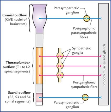
FIG. 20.1 Schematic diagram to show the craniosacral outflow (parasympathetic) and thoracolumbar outflow (sympathetic). Note that postganglionic parasympathetic fibres are shorter in length as compared to postganglionic sympathetic fibres.
شکل 20.1 نمودار شماتیک برای نشان دادن جریان خروجی جمجمهای-خاجی (پاراسمپاتیک) و سینهای-کمری (سمپاتیک). توجه داشته باشید که فیبرهای پاراسمپاتیک پسگانگلیونی در مقایسه با فیبرهای سمپاتیک پسگانگلیونی طول کوتاهتری دارند.
2. Parasympathetic nervous system: The preganglionic parasympathetic fibres arise from general visceral efferent (GVE) nuclei of brainstem and lateral horn cells of the second, third and fourth sacral segments of the spinal cord, hence parasympathetic nervous system constitutes the craniosacral outflow (Fig. 20.1). The axons of preganglionic fibres project to the autonomic ganglia (the parasympathetic ganglia) which are located near or embedded in the wall of viscera/glands.
2. سیستم عصبی پاراسمپاتیک: فیبرهای پاراسمپاتیک پیشگانگلیونی از هستههای وابران احشایی عمومی (GVE) ساقه مغز و سلولهای شاخ جانبی بخشهای دوم، سوم و چهارم خاجی نخاع منشأ میگیرند، از این رو سیستم عصبی پاراسمپاتیک جریان خروجی جمجمهای-خاجی را تشکیل میدهد (شکل 20.1). آکسونهای فیبرهای پیشگانگلیونی به گانگلیونهای اتونوم (گانگلیونهای پاراسمپاتیک) که در نزدیکی یا در دیواره احشاء/غدد قرار دارند، امتداد مییابند.
The sympathetic ganglia are located near the CNS along the sympathetic chain in the paravertebral region or in front of vertebral column, hence the postganglionic sympathetic fibres are longer in length, on the other hand the parasympathetic ganglia are located near effector organ, hence the postganglionic fibres are shorter in length (Fig. 20.1).
گانگلیونهای سمپاتیک در نزدیکی سیستم عصبی مرکزی (CNS) در امتداد زنجیره سمپاتیک در ناحیه پاراورتبرال یا در جلوی ستون فقرات قرار دارند، از این رو فیبرهای سمپاتیک پسگانگلیونی طول بلندتری دارند، از سوی دیگر گانگلیونهای پاراسمپاتیک در نزدیکی اندام مؤثر قرار دارند، از این رو فیبرهای پسگانگلیونی طول کوتاهتری دارند (شکل 20.1).
The effects of sympathetic and parasympathetic control are mainly stimulation or inhibition of glandular secretion and contraction or relaxation of smooth/cardiac muscle.
اثرات کنترل سمپاتیک و پاراسمپاتیک عمدتاً تحریک یا مهار ترشح غدد و انقباض یا شل شدن عضله صاف/قلبی است.
3. Enteric nervous system: It is a network of neurons in the wall of gastrointestinal tract.
۳. سیستم عصبی رودهای: شبکهای از نورونها در دیواره دستگاه گوارش است.
N.B. In general the sympathetic and parasympathetic nervous systems produce opposite effects, viz. sympathetic stimulation causes dilation of pupil, whereas parasympathetic stimulation causes constriction of the pupil. In tissues or organs innervated by both parasympathetic and sympathetic systems, the two systems function in an integrated reciprocal manner to produce a balanced action.
توجه: به طور کلی، سیستمهای عصبی سمپاتیک و پاراسمپاتیک اثرات متضادی ایجاد میکنند، یعنی تحریک سمپاتیک باعث گشاد شدن مردمک چشم میشود، در حالی که تحریک پاراسمپاتیک باعث تنگ شدن مردمک چشم میشود. در بافتها یا اندامهایی که توسط هر دو سیستم پاراسمپاتیک و سمپاتیک عصبدهی میشوند، این دو سیستم به صورت متقابل و یکپارچه عمل میکنند تا یک عمل متعادل ایجاد کنند.
Sympathetic Nervous System
The activities (functions) of the sympathetic nervous system are such that as if it prepares the body to deal with the emergency (exciting and stressful) situations. The heart rate is increased, arterioles of the skin and intestine are constricted, those of skeletal muscle are dilated, and the blood pressure is raised. There is redistribution of the blood so that it leaves the skin and GIT and pass to the brain, heart and skeletal muscles. In addition there is dilatation of pupils, inhibition of the smooth muscle of the bronchi, intestine, and urinary bladder, and closure of sphincters.
سیستم عصبی سمپاتیک
فعالیتها (عملکردهای) سیستم عصبی سمپاتیک به گونهای است که گویی بدن را برای مقابله با موقعیتهای اضطراری (هیجانانگیز و استرسزا) آماده میکند. ضربان قلب افزایش مییابد، شریانهای پوست و روده منقبض میشوند، شریانهای عضلات اسکلتی گشاد میشوند و فشار خون بالا میرود. توزیع مجدد خون به گونهای انجام میشود که از پوست و دستگاه گوارش خارج شده و به مغز، قلب و عضلات اسکلتی منتقل میشود. علاوه بر این، گشاد شدن مردمک چشم، مهار عضلات صاف برونشها، روده و مثانه و بسته شدن اسفنکترها نیز وجود دارد.
It is generally said that the sympathetic stimulation mobilizes the body energy for flight or fight.
به طور کلی گفته میشود که تحریک سمپاتیک، انرژی بدن را برای فرار یا مبارزه بسیج میکند.
Functions of the sympathetic nervous system
عملکردهای سیستم عصبی سمپاتیک
• Dilatation of pupil
• گشاد شدن مردمک چشم
• Widening of the palpebral fissure
• گشاد شدن شکاف پلکی
• Secretion of sweat glands
• ترشح غدد عرق
• Motor for arrector pili muscles
• محرک عضلات راست کننده پلک
• Vasoconstriction of blood vessels all over the body, except those in skeletal muscles
• انقباض عروق خونی در سراسر بدن، به جز عروق عضلات اسکلتی
• Stimulation of heart
• تحریک قلب
• Bronchodilatation and inhibition of secretion from bronchial glands
• گشاد شدن برونشها و مهار ترشح غدد برونشی
• Inhibition of gastrointestinal motility and secretions
• مهار حرکات و ترشحات دستگاه گوارش
• Sensory for pain from most of the viscera
• حسی برای درد ناشی از اکثر احشاء
• Contraction of sphincters of the bladder and bowel
• انقباض اسفنکترهای مثانه و روده
• Motor for ejaculation.
• محرک انزال.
Efferent nerve fibres (thoracolumbar outflow)
The preganglionic sympathetic fibres arise in the lateral grey column of the spinal cord from segments T1 to L2. From each of these segments they emerge as small myelinated axons into the corresponding anterior primary ramus and pass via white ramus communicans into the ganglion of a sympathetic trunk.
فیبرهای عصبی وابران (خروجی توراکولومبار)
فیبرهای سمپاتیک پیش عقدهای در ستون خاکستری جانبی نخاع از قطعات T1 تا L2 منشأ میگیرند. از هر یک از این قطعات، آنها به صورت آکسونهای کوچک میلیندار به شاخه اولیه قدامی مربوطه خارج میشوند و از طریق شاخههای سفید ارتباطی به گانگلیون تنه سمپاتیک میروند.
Once preganglionic fibres reach the paravertebral ganglia (ganglia in the sympathetic chain) they are distributed as follows (Figs 20.2, 20.3):
هنگامی که فیبرهای پیش عقدهای به گانگلیونهای پاراورتبرال (گانگلیونهای زنجیره سمپاتیک) میرسند، به صورت زیر توزیع میشوند (شکلهای 20.2، 20.3):
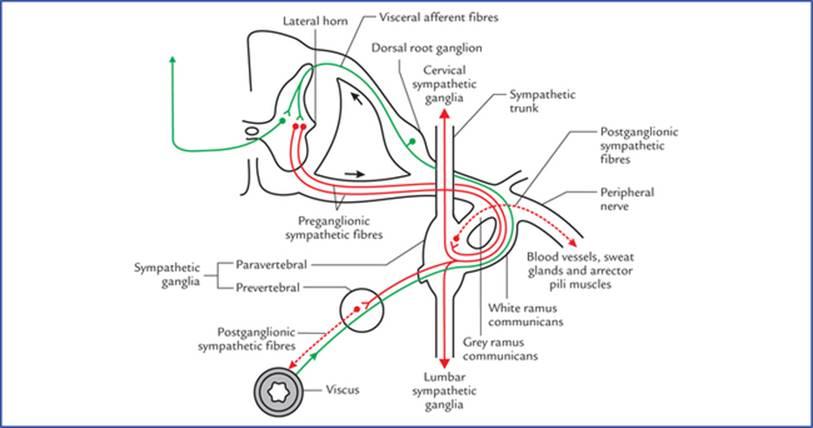
FIG. 20.2 Sympathetic efferent and afferent fibres. The preganglionic sympathetic fibres are shown by solid red lines and postganglionic sympathetic fibres by interrupted red lines. The sympathetic afferent fibres are shown by green lines. Note the fate of preganglionic sympathetic fibres. These may (a) relay in their corresponding ganglion and pass to their corresponding spinal nerve for distribution, (b) ascend or descend in the sympathetic chain and relay in higher or lower ganglia, or (c) pass without synapse to a peripheral (prevertebral) ganglion for relay.
شکل 20.2 فیبرهای آوران و وابران سمپاتیک. فیبرهای سمپاتیک پیش عقدهای با خطوط قرمز پررنگ و فیبرهای سمپاتیک پس عقدهای با خطوط قرمز منقطع نشان داده شدهاند. فیبرهای آوران سمپاتیک با خطوط سبز نشان داده شدهاند. به سرنوشت فیبرهای سمپاتیک پیش عقدهای توجه کنید. این فیبرها ممکن است (الف) در گانگلیون مربوطه خود رله کرده و برای توزیع به عصب نخاعی مربوطه خود منتقل شوند، (ب) در زنجیره سمپاتیک بالا یا پایین رفته و در گانگلیونهای بالاتر یا پایینتر رله کنند، یا (ج) بدون سیناپس برای رله به یک گانگلیون محیطی (پیش مهرهای) منتقل شوند.
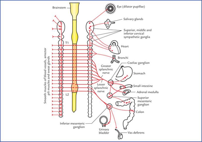
FIG. 20.3 Sympathetic nervous system (thoracolumbar outflow). The postganglionic sympathetic fibres are shown by interrupted lines.
شکل 20.3 سیستم عصبی سمپاتیک (خروجی قفسه سینه و کمر). فیبرهای سمپاتیک پسگانگلیونی با خطوط منقطع نشان داده شدهاند.
• Synapse with the cells in the ganglion. The postganglionic fibres (non-myelinated axons) arising from ganglion cells rejoin the spinal nerves (grey rami communicantes) and distributed through its branches to the blood vessels, sweat glands and arrector pili muscles.
• سیناپس با سلولهای گانگلیون. فیبرهای پسگانگلیونی (آکسونهای بدون میلین) که از سلولهای گانگلیون منشأ میگیرند، به اعصاب نخاعی (شاخههای خاکستری ارتباطی) متصل میشوند و از طریق شاخههای آن به رگهای خونی، غدد عرق و عضلات راستکننده مو توزیع میشوند.
• Ascend in the sympathetic trunk to synapse in the cervical sympathetic ganglia.
• در تنه سمپاتیک بالا میروند تا در گانگلیونهای سمپاتیک گردنی سیناپس کنند.
• Descend in the sympathetic trunk to synapse in lumbar sympathetic ganglia.
• در تنه سمپاتیک پایین میآیند تا در گانگلیونهای سمپاتیک کمری سیناپس کنند.
• Leave the ganglia without synapsing as splanchnic nerves and relay in the prevertebral ganglia. Some of these fibres pass to the suprarenal gland where they synapse with the cells within the medulla. These medullary cells which may be regarded as modified postganglionic sympathetic neurons, secrete epinephrine and norepinephrine.
• گانگلیونها را بدون سیناپس به عنوان اعصاب احشایی ترک میکنند و در گانگلیونهای پیشمهرهای رله میکنند. برخی از این فیبرها به غده فوق کلیوی میروند و در آنجا با سلولهای داخل بصلالنخاع سیناپس میکنند. این سلولهای مدولاری که میتوان آنها را به عنوان نورونهای سمپاتیک پسگانگلیونی اصلاحشده در نظر گرفت، اپینفرین و نوراپی نفرین ترشح میکنند.
Sympathetic trunks
The sympathetic trunks are two ganglionated chains of nerve fibres lying one on either side of the vertebral column, extending along its whole length, i.e. from atlas vertebra above to the coccyx below. Each chain (trunk) bears a number of knot-like enlargements, the sympathetic ganglia. The number of ganglia is variable, but generally there are three in the cervical region, 11 in the thoracic region, four in the lumbar region, and four in the sacral region. Thus each sympathetic chain possesses 22 sympathetic ganglia. The ganglia associated with sympathetic chain are termed paravertebral ganglia.
تنههای سمپاتیک
تنههای سمپاتیک دو زنجیره گانگلیونی از الیاف عصبی هستند که در دو طرف ستون مهرهها قرار دارند و در تمام طول آن، یعنی از مهره اطلس در بالا تا دنبالچه در پایین، امتداد دارند. هر زنجیره (تنه) دارای تعدادی زائده گره مانند به نام گانگلیونهای سمپاتیک است. تعداد گانگلیونها متغیر است، اما به طور کلی سه عدد در ناحیه گردنی، ۱۱ عدد در ناحیه سینهای، چهار عدد در ناحیه کمری و چهار عدد در ناحیه خاجی وجود دارد. بنابراین هر زنجیره سمپاتیک دارای ۲۲ گانگلیون سمپاتیک است. گانگلیونهای مرتبط با زنجیره سمپاتیک، گانگلیونهای پاراورتبرال نامیده میشوند.
N.B. The terminal ganglion, the ganglion impar is formed by fusion of two sympathetic trunks as they converge in front of the coccyx.
توجه: گانگلیون انتهایی، گانگلیون ایمپار، از ادغام دو تنه سمپاتیک هنگام همگرایی در جلوی دنبالچه تشکیل میشود.
Cervical sympathetic ganglia
The cervical part of sympathetic trunk extends from base of skull to the neck of first rib. It consists of three large ganglia, superior, middle and inferior. They are believed to be formed during development by the fusion of smaller segmental ganglia.
گانگلیونهای سمپاتیک گردنی
بخش گردنی تنه سمپاتیک از قاعده جمجمه تا گردن دنده اول امتداد دارد. این بخش از سه گانگلیون بزرگ، فوقانی، میانی و تحتانی تشکیل شده است. اعتقاد بر این است که آنها در طول تکامل از ادغام گانگلیونهای قطعهای کوچکتر تشکیل میشوند.
• The superior cervical ganglion is largest and formed by the fusion of upper 4 cervical ganglia. It is spindle-shaped and lies in front of transverse processes of C2 and C3 vertebra.
• گانگلیون فوقانی گردنی بزرگترین است و از ادغام ۴ گانگلیون فوقانی گردنی تشکیل میشود. این گانگلیون دوکی شکل است و در جلوی زوائد عرضی مهرههای C2 و C3 قرار دارد.
Branches
– Grey rami communicantes to ventral rami of upper four cervical spinal nerves.
شاخهها
– شاخههای خاکستری به شاخههای شکمی چهار عصب نخاعی گردنی فوقانی متصل میشوند.
– Arterial branches to, both external and internal carotid arteries. These branches form plexuses around these arteries and are distributed along their branches. The fibres carried by external carotid artery are vasomotor, sudomotor, and pilomotor and supply areas of skin mainly supplied by trigeminal nerve.
– شاخههای شریانی به شریانهای کاروتید خارجی و داخلی. این شاخهها در اطراف این شریانها شبکهای تشکیل میدهند و در امتداد شاخههای خود توزیع میشوند. فیبرهایی که توسط شریان کاروتید خارجی حمل میشوند، وازوموتور، سودوموتور و پیلوموتور هستند و نواحی تغذیهکننده پوست عمدتاً توسط عصب سهقلو تغذیه میشوند.
The fibres carried by internal carotid artery (internal carotid nerve) are (motor) to the dilator pupillae and smooth muscle of levator palpebrae superioris (Muller’s muscle).
فیبرهایی که توسط شریان کاروتید داخلی (عصب کاروتید داخلی) حمل میشوند، (حرکتی) به گشادکننده مردمک و عضله صاف عضله بالابرنده پلک فوقانی (عضله مولر) هستند.
– Cardiac branch (superior cervical cardiac branch) to the heart. It is motor (visceral) to cardiac muscle.
– شاخه قلبی (شاخه قلبی گردنی فوقانی) به قلب. این شاخه حرکتی (احشایی) به عضله قلب است.
• The middle cervical ganglion is small and formed by the fusion of 5th and 6th cervical ganglia. It lies in close relationship to inferior thyroid artery at the level of cricoid cartilage (C6).
• گانگلیون گردنی میانی کوچک است و از به هم پیوستن گانگلیونهای گردنی پنجم و ششم تشکیل شده است. این گانگلیون در سطح غضروف کریکوئید (C6) در ارتباط نزدیک با شریان تیروئید تحتانی قرار دارد.
Branches
– Grey rami communicantes to 5th and 6th cervical spinal nerves.
شاخهها
– شاخههای خاکستری به اعصاب نخاعی گردنی پنجم و ششم متصل میشوند.
– Arterial branch to inferior thyroid artery for distribution to pharynx, larynx, and upper parts of the trachea and oesophagus, thyroid and parathyroid.
– شاخه شریانی به شریان تیروئید تحتانی برای توزیع به حلق، حنجره و قسمتهای فوقانی نای و مری، تیروئید و پاراتیروئید.
– Cardiac branch (middle cervical cardiac branch) to the heart.
– شاخه قلبی (شاخه قلبی گردنی میانی) به قلب.
• The inferior cervical ganglion is formed by the fusion of 7th and 8th cervical ganglia. It fuses with the first thoracic ganglion to form the cervicothoracic ganglion (also called stellate ganglion because its numerous branches give it a star-shaped appearance).
• گانگلیون گردنی تحتانی از ادغام گانگلیونهای هفتم و هشتم گردنی تشکیل میشود. این گانگلیون با اولین گانگلیون سینهای ادغام شده و گانگلیون گردنی-سینهای را تشکیل میدهد (که به دلیل شاخههای متعددش، گانگلیون ستارهای نیز نامیده میشود).
Branches
– Grey rami communicantes to C7 and C8 (in most cases T1 also) spinal nerves.
شاخهها
– شاخههای خاکستری طناب ارتباطی به اعصاب نخاعی C7 و C8 (در بیشتر موارد همچنین T1) متصل میشوند.
– Arterial branches to both subclavian and vertebral arteries.
– شاخههای شریانی به هر دو شریان ساب کلاوین و مهرهای.
– Cardiac branch (inferior cardiac branch) to the heart.
– شاخه قلبی (شاخه قلبی تحتانی) به قلب.
N.B. The cervical ganglia receive their preganglionic fibres from first and second thoracic (T1 and T2) spinal segments by the white rami communicantes of first and second thoracic spinal nerves.
توجه: گانگلیونهای گردنی فیبرهای پیش عقدهای خود را از قطعات نخاعی اول و دوم سینهای (T1 و T2) توسط شاخههای سفید طناب ارتباطی اعصاب نخاعی اول و دوم سینهای دریافت میکنند.
Thoracic ganglia
The thoracic part of sympathetic trunk usually consists of 11 ganglia (because first thoracic ganglion gets fused with the inferior cervical ganglion).
گانگلیونهای سینهای
بخش سینهای تنه سمپاتیک معمولاً از ۱۱ گانگلیون تشکیل شده است (زیرا اولین گانگلیون سینهای با گانگلیون گردنی تحتانی ادغام میشود).
• They are connected by grey and white rami communicantes to the thoracic spinal nerves.
• The first five thoracic ganglia (T1-T5) give postganglionic fibres to the heart, lung and oesophagus (through cardiac, pulmonary and oesophageal plexuses).
• آنها توسط شاخههای خاکستری و سفید به اعصاب نخاعی سینهای متصل میشوند.
• پنج گانگلیون اول سینهای (T1-T5) فیبرهای پسگانگلیونی به قلب، ریه و مری میدهند (از طریق شبکههای قلبی، ریوی و مری).
• The lower eight ganglia give preganglionic fibres, which are grouped to form the three splanchnic nerves:
• Greater splanchnic nerve arises from 5th to 9th thoracic ganglia.
• Lesser splanchnic nerve arises from 10th and 11th thoracic ganglia.
• Least splanchnic nerve arises from 12th (or last thoracic) ganglion.
• هشت گانگلیون پایینی فیبرهای پیشگانگلیونی میدهند که برای تشکیل سه عصب احشایی گروهبندی میشوند:
• عصب احشایی بزرگ از گانگلیونهای پنجم تا نهم سینهای منشأ میگیرد.
• عصب احشایی کوچک از گانگلیونهای دهم و یازدهم سینهای منشأ میگیرد.
• کوچکترین عصب احشایی از گانگلیون دوازدهم (یا آخرین گانگلیون سینهای) منشأ میگیرد.
The three splanchnic nerves pierce the crus of diaphragm to end in coeliac and superior mesenteric plexuses and ganglia on abdominal aorta. From there postganglionic fibres arise and supply the abdominal viscera.
سه عصب احشایی از ساقه دیافراگم عبور میکنند و به شبکههای سلیاک و مزانتریک فوقانی و گانگلیونهای روی آئورت شکمی ختم میشوند. از آنجا فیبرهای پسگانگلیونی خارج شده و احشاء شکمی را تغذیه میکنند.
Lumbar ganglia
The abdominal part of sympathetic trunk consists of four ganglia:
گانگلیونهای کمری
بخش شکمی تنه سمپاتیک از چهار گانگلیون تشکیل شده است:
• Only upper two ganglia receive white rami communicantes from first and second lumbar spinal nerves.
• فقط دو گانگلیون بالایی، شاخههای ارتباطی سفید را از اعصاب نخاعی کمری اول و دوم دریافت میکنند.
• All ganglia give grey rami communicantes to corresponding lumbar spinal nerves.
• همه گانگلیونها، شاخههای ارتباطی خاکستری را به اعصاب نخاعی کمری مربوطه میدهند.
• Visceral branches and lumbar splanchnic nerves from ganglia pass downwards, to form a plexus in front of the 5th lumbar and upper sacral vertebrae, called superior hypogastric plexus. It lies between the common iliac vessels. It divides into two inferior hypogastric plexuses, one on each side of the rectum.
• شاخههای احشایی و اعصاب اسپلانکنیک کمری از گانگلیونها به سمت پایین عبور میکنند و یک شبکه در جلوی مهره پنجم کمری و مهره بالایی خاجی تشکیل میدهند که شبکه هیپوگاستریک فوقانی نامیده میشود. این شبکه بین عروق ایلیاک مشترک قرار دارد. به دو شبکه هیپوگاستریک تحتانی تقسیم میشود که هر کدام در یک طرف راست روده قرار دارند.
Sacral ganglia
The pelvic part of sympathetic trunk consists of four ganglia. They give grey rami communicantes to sacral and coccy-geal nerves to supply lower limbs, buttocks and perineum; and visceral branches to the pelvic plexuses.
گانگلیونهای خاجی
بخش لگنی تنه سمپاتیک از چهار گانگلیون تشکیل شده است. این گانگلیونها به اعصاب خاجی و دنبالچهای، شاخههای خاکستری ارتباطی میدهند تا اندامهای تحتانی، باسن و پرینه را عصبدهی کنند؛ و شاخههای احشایی به شبکههای لگنی میدهند.
Prevertebral ganglia
There are three prevertebral ganglia, which are situated in front of vertebral column in the abdominal cavity. They are located close to the origins of superior, middle and inferior mesenteric arteries and are named after them, viz.
گانگلیونهای پیشمهرهای
سه گانگلیون پیشمهرهای وجود دارد که در جلوی ستون فقرات در حفره شکم قرار دارند. آنها نزدیک به منشأ شریانهای مزانتریک فوقانی، میانی و تحتانی قرار دارند و به نام آنها نامگذاری شدهاند، یعنی.
• Coeliac ganglion, at the origin of coeliac artery
• Superior mesenteric ganglion, at the origin of superior mesenteric artery
• Inferior mesenteric ganglion, at the origin of inferior mesenteric artery.
• گانگلیون سلیاک، در مبدا شریان سلیاک
• گانگلیون مزانتریک فوقانی، در مبدا شریان مزانتریک فوقانی
• گانگلیون مزانتریک تحتانی، در مبدا شریان مزانتریک تحتانی.
Parasympathetic Nervous System
The activities of parasympathetic nervous system are directed towards conserving and restoring energy. The heart rate is slowed, the pupils are constricted, the peristalsis and glandular activity is increased, the sphincters are relaxed (opened), and the bladder wall is contracted.
سیستم عصبی پاراسمپاتیک
فعالیتهای سیستم عصبی پاراسمپاتیک معطوف به حفظ و بازیابی انرژی است. ضربان قلب کاهش مییابد، مردمک چشم منقبض میشود، حرکات دودی و فعالیت غدد افزایش مییابد، اسفنکترها شل (باز) میشوند و دیواره مثانه منقبض میشود.
In general the parasympathetic stimulation slows down the body processes except that of GIT and genitourinary system, and allows restoration processes to occur quietly and peacefully.
به طور کلی، تحریک پاراسمپاتیک فرآیندهای بدن به جز دستگاه گوارش و دستگاه ادراری تناسلی را کند میکند و اجازه میدهد فرآیندهای ترمیمی به آرامی و با آرامش انجام شوند.
Functions of the parasympathetic nervous system
عملکردهای سیستم عصبی پاراسمپاتیک
• Constriction of pupil.
• انقباض مردمک چشم.
• Increase in lens curvature (antero-posterior) for accommodation.
• افزایش انحنای عدسی (قدامی-خلفی) برای تطابق.
• Secretion of respiratory and digestive glands.
• ترشح غدد تنفسی و گوارشی.
• Inhibition of cardiac excitation, conduction and contraction.
• مهار تحریک، هدایت و انقباض قلب.
• Bronchoconstriction and increased secretion from bronchial glands.
• انقباض برونشها و افزایش ترشح غدد برونشی.
• Increased gastrointestinal motility and inhibition of pyloric and anal sphincters.
• افزایش حرکات دستگاه گوارش و مهار اسفنکترهای پیلور و مقعد.
• Sensory differentiation of faeces and flatus.
• تمایز حسی مدفوع و گاز.
• Contraction of muscular wall of urinary bladder (detrusor muscle) and inhibition of the internal urethral sphincter (sphincter vesicae).
• انقباض دیواره عضلانی مثانه (عضله دترسور) و مهار اسفنکتر داخلی مجرای ادرار (اسفنکتر وزیکا).
• Motor for initiation and maintenance of erection of penis.
• محرک شروع و حفظ نعوظ آلت تناسلی.
The functions of sympathetic and parasympathetic nervous system are compared in Table 20.1.
عملکردهای سیستم عصبی سمپاتیک و پاراسمپاتیک در جدول 20.1 با هم مقایسه شدهاند.
Table 20.1
Comparison of sympathetic and parasympathetic functions
جدول ۲۰.۱
مقایسه عملکردهای سمپاتیک و پاراسمپاتیک
Structure | Sympathetic function | Parasympathetic function |
Eye | Dilatation of pupil | Constriction of pupil |
Lacrimal gland and Salivary glands | Viscous secretion | Watery secretion |
Bronchial smooth muscle | Relaxation | Contraction |
Heart | Increases heart rate | Decreases heart rate |
GIT | Decreases peristalsis and constricts the sphincters | Increases peristalsis and relaxes sphincters |
Genitourinary tract | ||
• Bladder wall and sphincter | Relaxes bladder wall and constricts the sphincter | Contracts the bladder wall and relaxes the sphincter |
• Penis | Ejaculation | Erection |
Skin | ||
• Sweat glands | Produce sweating | No effect |
• Arrector pili | Contraction of arrector pili leading erection of hair | No effect |
Blood vessels | ||
• Skin, splanchnic vessels | Contraction | No effect |
• Skeletal muscles | Relaxation | No effect |
Efferent nerve fibres (Fig. 20.4)
The preganglionic parasympathetic fibres arise from brain-stem and spinal cord.
فیبرهای عصبی وابران (شکل 20.4)
فیبرهای پاراسمپاتیک پیش عقدهای از ساقه مغز و نخاع منشأ میگیرند.
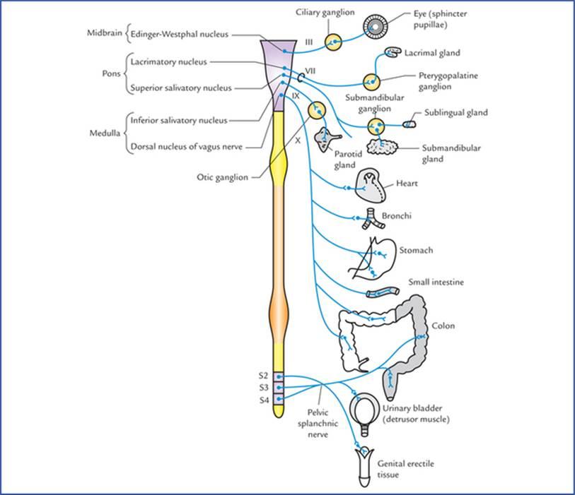
FIG. 20.4 Parasympathetic nervous system (craniosacral outflow).
شکل 20.4 سیستم عصبی پاراسمپاتیک (خروجی جمجمهای-خاجی).
From the brainstem, they arise from general visceral efferent nuclei (viz. Edinger-Westphal nucleus, lacrimatory nucleus and superior salivatory nucleus, inferior salivatory nucleus, and dorsal nucleus of vagus) and run through IIIrd (oculomotor), Vllth (facial), IXth (glossopharyngeal) and Xth (vagus) cranial nerves, respectively to terminate in the parasympathetic ganglia in the region of head and neck (viz. ciliary, pterygopalatine, submandibular, and otic) and ganglion cells in the autonomic nerve plexuses in the thorax and abdomen. These fibres form what is termed cranial outflow. Preganglionic fibres from Edinger-Westphal nucleus relay in the ciliary ganglion, from lacrimatory nucleus in the pterygopalatine ganglion, from superior salivatory nucleus in the submandibular ganglion, from inferior salivatory nucleus in the otic ganglion, and from dorsal nucleus of vagus in the ganglion cells of autonomic nerve plexuses in the thorax and abdomen.
از ساقه مغز، آنها از هستههای وابران احشایی عمومی (یعنی هسته ادینگر-وستفال، هسته اشکی و هسته بزاقی فوقانی، هسته بزاقی تحتانی و هسته پشتی واگ) منشأ میگیرند و به ترتیب از اعصاب جمجمهای IIIrd (حرکتی چشمی)، Vllth (صورتی)، IXth (زبانی-حلقی) و Xth (واگوس) عبور میکنند تا در گانگلیونهای پاراسمپاتیک در ناحیه سر و گردن (یعنی مژگانی، پتریگوپالاتین، تحت فکی و شنوایی) و سلولهای گانگلیون در شبکههای عصبی خودکار در قفسه سینه و شکم خاتمه یابند. این فیبرها چیزی را تشکیل میدهند که به عنوان خروج جمجمهای نامیده میشود. فیبرهای پیش عقدهای از هسته ادینگر-وستفال در گانگلیون مژگانی، از هسته اشکی در گانگلیون پتریگوپالاتین، از هسته بزاقی فوقانی در گانگلیون تحت فکی، از هسته بزاقی تحتانی در گانگلیون گوش و از هسته پشتی واگ در سلولهای گانگلیونی شبکههای عصبی خودکار در قفسه سینه و شکم رله میشوند.
From the spinal cord, the preganglionic parasympathetic fibres arise from the cells in the grey matter of the 2nd, 3rd, and 4th sacral spinal segments, pass through anterior nerve roots of the corresponding sacral spinal nerves (sacral outflow), leave the sacral nerves and form the pelvic splanchnic nerves, through which they terminate in parasympathetic ganglia located near or within the walls of pelvic organs.
فیبرهای پاراسمپاتیک پیش عقدهای از نخاع از سلولهای ماده خاکستری بخشهای نخاعی دوم، سوم و چهارم خاجی منشأ میگیرند، از ریشههای عصبی قدامی اعصاب نخاعی خاجی مربوطه عبور میکنند (خروجی خاجی)، اعصاب خاجی را ترک کرده و اعصاب اسپلانکنیک لگنی را تشکیل میدهند که از طریق آنها به گانگلیونهای پاراسمپاتیک واقع در نزدیکی یا درون دیوارههای اندامهای لگنی ختم میشوند.
The postganglionic parasympathetic fibres arise from parasympathetic ganglia or autonomic plexuses and supply the viscera.
فیبرهای پاراسمپاتیک پس عقدهای از گانگلیونهای پاراسمپاتیک یا شبکههای خودکار منشأ میگیرند و احشاء را تغذیه میکنند.
Autonomic plexuses
The large collections of afferent and efferent autonomic fibres and their associated ganglia form autonomic plexuses in the thorax, abdomen, and pelvis. The branches from these plexuses innervate the viscera.
شبکههای عصبی خودکار
مجموعههای بزرگ فیبرهای عصبی خودکار آوران و وابران و گانگلیونهای مرتبط با آنها، شبکههای عصبی خودکار را در قفسه سینه، شکم و لگن تشکیل میدهند. شاخههای این شبکهها، احشاء را عصبدهی میکنند.
The autonomic plexuses in different regions of body are listed in Table 20.2.
شبکههای عصبی خودکار در نواحی مختلف بدن در جدول 20.2 فهرست شدهاند.
Table 20.2
Autonomic plexuses and their locations
جدول 20.2
شبکههای عصبی خودکار و محل قرارگیری آنها
Region | Autonomic plexuses |
Thorax | Cardiac, pulmonary, and oesophageal |
Abdomen* | Coeliac, superior mesenteric, inferior mesenteric and aortic |
Pelvis | Superior and inferior hypogastric |
* In the abdomen the plexuses are associated with aorta and its branches, and named after them.
* در شکم، شبکهها با آئورت و شاخههای آن مرتبط هستند و به نام آنها نامگذاری شدهاند.
Autonomic ganglia
Autonomic ganglia are the sites where preganglionic fibres synapse on postganglionic neurons and thus serve as a relay station. The autonomic ganglia are situated along the course of peripheral efferent autonomic pathways.
گانگلیونهای اتونوم
گانگلیونهای اتونوم مکانهایی هستند که فیبرهای پیش عقدهای روی نورونهای پس عقدهای سیناپس میدهند و بنابراین به عنوان یک ایستگاه رله عمل میکنند. گانگلیونهای اتونوم در امتداد مسیرهای اتونوم وابران محیطی قرار دارند.
The sympathetic ganglia form part of sympathetic chain/trunk and are also located in front of vertebral column, away from the viscera.
گانگلیونهای سمپاتیک بخشی از زنجیره/تنه سمپاتیک را تشکیل میدهند و همچنین در جلوی ستون فقرات، دور از احشاء قرار دارند.
The parasympathetic ganglia, on the other hand, are situated close to or within the walls of the viscera.
از سوی دیگر، گانگلیونهای پاراسمپاتیک نزدیک یا درون دیوارههای احشاء قرار دارند.
Neurotransmitters involved in autonomic nerve impulse transmission
The autonomic nerve fibres release one of the two transmitter substances, acetylcholine (cholinergic endings) or noradrenaline (adrenergic endings).
انتقالدهندههای عصبی دخیل در انتقال تکانه عصبی خودکار
فیبرهای عصبی خودکار یکی از دو ماده انتقالدهنده، استیل کولین (انتهای کولینرژیک) یا نورآدرنالین (انتهای آدرنرژیک) را آزاد میکنند.
All the preganglionic autonomic fibres (i.e. both sympathetic as well as parasympathetic) and postganglionic parasympathetic fibres are cholinergic, i.e. their terminals liberate acetylcholine as their transmitter substance. All the postganglionic sympathetic fibres are adrenergic, i.e. their terminals liberate noradrenaline (norepinephrine) except for those supplying the sweat glands (i.e. sudomotor fibres), hair muscles (arrector pili) which are cholinergic (Fig. 20.5).
تمام فیبرهای خودکار پیشگانگلیونی (یعنی هم سمپاتیک و هم پاراسمپاتیک) و پسگانگلیونی پاراسمپاتیک کولینرژیک هستند، یعنی پایانههای آنها استیل کولین را به عنوان ماده انتقالدهنده آزاد میکنند. تمام فیبرهای سمپاتیک پسگانگلیونی آدرنرژیک هستند، یعنی پایانههای آنها نورآدرنالین (نوراپی نفرین) را آزاد میکنند، به جز آنهایی که غدد عرق را تغذیه میکنند (یعنی فیبرهای سودوموتور) و عضلات مو (پیلیهای راستکننده) که کولینرژیک هستند (شکل 20.5).
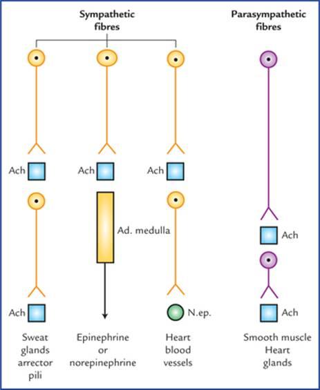
FIG. 20.5 The autonomic efferent fibres and chemical transmitter substances released by their terminals.
شکل 20.5 فیبرهای وابران اتونوم و مواد انتقالدهنده شیمیایی آزاد شده توسط پایانههای آنها.
Points to Note
نکات قابل توجه
• The postganglionic sympathetic transmitter substance, norepinephrine produces both excitatory and inhibitory actions at the postsynaptic sites. This is, due to presence of two types of adrenergic receptors, α and β in the target organs. The response depends upon the type of receptors being activated. In general the activation of α receptors causes excitation and activation of (β receptors causes inhibition.
• ماده انتقالدهنده سمپاتیک پسگانگلیونی، نوراپی نفرین، هم اثرات تحریکی و هم اثرات مهاری را در مکانهای پس سیناپسی ایجاد میکند. این به دلیل وجود دو نوع گیرنده آدرنرژیک، α و β در اندامهای هدف است. پاسخ به نوع گیرندههای فعال شده بستگی دارد. به طور کلی، فعال شدن گیرندههای α باعث تحریک و فعال شدن گیرندههای β باعث مهار میشود.
• The cholinergic receptors are also of two types, muscarinic and nicotinic. The muscarinic receptors are activated by muscarine which elicits the same effects as acetylcholine in the target organs. The nicotinic receptors are excited by low doses, while blocked by high doses of nicotine.
• گیرندههای کولینرژیک نیز دو نوع هستند، موسکارینی و نیکوتینی. گیرندههای موسکارینی توسط موسکارین فعال میشوند که همان اثرات استیل کولین را در اندامهای هدف ایجاد میکند. گیرندههای نیکوتینی با دوزهای پایین تحریک میشوند، در حالی که توسط دوزهای بالای نیکوتین مسدود میشوند.
Clinical Correlation
The classification of autonomic receptors has led to the development of many drugs which can modify autonomic activities. These drugs are commonly used to treat shock, hypertension and cardiac problems.
همبستگی بالینی
طبقهبندی گیرندههای خودکار منجر به توسعه داروهای زیادی شده است که میتوانند فعالیتهای خودکار را تغییر دهند. این داروها معمولاً برای درمان شوک، فشار خون بالا و مشکلات قلبی استفاده میشوند.
The anatomical, physiological and pharmacological differences between sympathetic and parasympathetic parts of autonomic nervous system are provided in Table 20.3.
تفاوتهای آناتومیکی، فیزیولوژیکی و دارویی بین بخشهای سمپاتیک و پاراسمپاتیک سیستم عصبی خودکار در جدول 20.3 ارائه شده است.
Table 20.3
Anatomical, physiological, and pharmacological differences between sympathetic and parasympathetic parts of the autonomic nervous system
جدول 20.3
تفاوتهای آناتومیکی، فیزیولوژیکی و دارویی بین بخشهای سمپاتیک و پاراسمپاتیک سیستم عصبی خودکار
Sympathetic system | Parasympathetic system | |
Anatomical differences | ||
• Cells of origin of preganglionic fibres | Are located in the thoracic and upper 2 or 3 lumbar segments (T1-L2/3) of the spinal cord (thoracolumbar outflow) | Are located in the brainstem and second, third, and fourth sacral segments (S2, S3 & S4) of the spinal cord (craniosacral outflow) |
• Ganglia | Are located along the sympathetic chain and in front of prevertebral region, hence preganglionic fibres are shorter in length and postganglionic sympathetic fibres are longer in length | Are located near or in the effector organs, hence preganglionic fibres are longer in length and postganglionic are shorter in length |
• Outflow from CNS occurs through: | Spinal nerves | Cranial nerves |
• Distribution of postganglionic fibres | Wide distribution | Limited distribution |
Physiological differences | Prepares the body for emergency | Conserves and restores the energy |
Pharmacological differences | (Also see page 239) | (Also see page 239) |
Neurotransmitter liberated by the postganglionic fibres | All postganglionic sympathetic fibres liberate norepinephrine except for those supplying the sweat glands or blood vessels in the skeletal muscles which liberate acetylcholine | All postganglionic parasympathetic fibres liberate acetylcholine |
Afferent nerve fibres of autonomic nervous system (visceral afferents)
The cell bodies of the afferent nerve fibres (first order sensory neurons) of the autonomic nervous system are located either in the dorsal root ganglia of spinal nerves or in the sensory ganglia of the cranial nerves. These neurons are general visceral afferent neurons as they carry general sensations from the viscera. The central processes of these neurons enter the central nervous system and either take part in the formation of local reflex arcs, or pass to higher centres of the autonomic nervous system, such as the hypothalamus.
فیبرهای عصبی آوران سیستم عصبی خودکار (آورانهای احشایی)
جسم سلولی فیبرهای عصبی آوران (نورونهای حسی درجه یک) سیستم عصبی خودکار یا در گانگلیونهای ریشه پشتی اعصاب نخاعی یا در گانگلیونهای حسی اعصاب جمجمهای قرار دارند. این نورونها، نورونهای آوران احشایی عمومی هستند زیرا حسهای عمومی را از احشاء حمل میکنند. فرآیندهای مرکزی این نورونها وارد سیستم عصبی مرکزی میشوند و یا در تشکیل قوسهای رفلکس موضعی شرکت میکنند یا به مراکز بالاتر سیستم عصبی خودکار مانند هیپوتالاموس میروند.
The impulses carried by these fibres are associated with:
• Visceral reflexes, usually at the unconscious level.
• Sensations of hunger, thirst, nausea, sex desire and bladder and bowel distension.
• Visceral pain.
تکانههایی که توسط این فیبرها حمل میشوند با موارد زیر مرتبط هستند:
• رفلکسهای احشایی، معمولاً در سطح ناخودآگاه.
• احساس گرسنگی، تشنگی، حالت تهوع، میل جنسی و اتساع مثانه و روده.
• درد احشایی.
Once the autonomic afferents enter the spinal cord or brain, they travel alongside, or mixed with, the somatic afferent fibres.
وقتی آورانهای خودکار وارد نخاع یا مغز میشوند، در کنار یا با الیاف آوران سوماتیک حرکت میکنند.
N.B. The nerve endings of autonomic afferents (visceral afferents) are not activated by heat, cut or touch but rather by stretch or lack of oxygen.
توجه: انتهای عصبی آورانهای خودکار (آورانهای احشایی) توسط گرما، بریدگی یا لمس فعال نمیشوند، بلکه توسط کشش یا کمبود اکسیژن فعال میشوند.
Visceral Pain
The visceral pain is of great clinical significance, hence needs an elaborate description.
درد احشایی
درد احشایی از اهمیت بالینی بالایی برخوردار است، بنابراین نیاز به توصیف مفصلی دارد.
Usually the viscera are insensitive to cutting and such sensations as touch and heat. However, visceral pain is experienced when it is stretched or suffers from ischaemia (lack of O2) or if there is accumulation of metabolites.
معمولاً احشای بدن نسبت به بریدگی و احساساتی مانند لمس و گرما حساس نیستند. با این حال، درد احشایی زمانی تجربه میشود که کشیده شود یا دچار ایسکمی (کمبود اکسیژن) شود یا اگر متابولیتها تجمع داشته باشند.
The visceral pain is dull, boring and poorly localized due to stimulation of large number of fibres, on the other hand the somatic pain is sharp, precise and well localized. The pain of appendicitis is a classical example to put forward these facts. Initially when there is an inflammation of appendix and its covering layer of visceral peritoneum (both innervated by autonomic nerve fibres), the pain is dull and vaguely located in the midline in the periumbilical region. The midgut from which the appendix is derived is a median structure during the development. As the inflammation progresses and involves parietal peritoneum (innervated by somatic nerve fibres), the pain is sharp and clearly located in the right iliac fossa at McBurney’s point.
درد احشایی به دلیل تحریک تعداد زیادی از فیبرها، کسلکننده، خستهکننده و با موقعیت مکانی ضعیف است، از سوی دیگر، درد جسمی تیز، دقیق و با موقعیت مکانی خوب است. درد آپاندیسیت یک مثال کلاسیک برای بیان این واقعیتها است. در ابتدا، هنگامی که التهاب آپاندیس و لایه پوشاننده آن یعنی صفاق احشایی (هر دو توسط فیبرهای عصبی خودکار عصبدهی میشوند) وجود دارد، درد کسلکننده و مبهم در خط وسط در ناحیه اطراف ناف است. روده میانی که آپاندیس از آن مشتق میشود، در طول رشد، یک ساختار میانی است. با پیشرفت التهاب و درگیر شدن صفاق جداری (که توسط فیبرهای عصبی سوماتیک عصبدهی میشود)، درد تیز است و به وضوح در حفره ایلیاک راست در نقطه مکبرنی قرار دارد.
Clinical Correlation
Referred pain
The visceral pain is frequently referred to the skin areas (dermatomes) that are innervated by the same segments of the spinal cord as the diseased viscus. Hence referred pain may be defined as pain felt at a site remote from its source of origin.
همبستگی بالینی
درد ارجاعی
درد احشایی اغلب به نواحی پوستی (درماتومها) ارجاع داده میشود که توسط همان بخشهای نخاع که احشای بیمار عصبدهی میشوند، عصبدهی میشوند. از این رو، درد ارجاعی را میتوان به عنوان دردی تعریف کرد که در محلی دور از منبع اصلی خود احساس میشود.
Anatomical basis of referred pain
The exact basis is not known. However, there are two theories to explain it:
اساس آناتومیکی درد ارجاعی
اساس دقیق آن مشخص نیست. با این حال، دو نظریه برای توضیح آن وجود دارد:
• One theory is that when nerve fibres from the viscus and the skin (dermatome) enter the same segment of spinal cord, they synapse with and stimulate the same (common) second order neurons in the sensory pathway to the cerebral cortex (Fig. 20.6). As a result stimuli from viscus and skin ascend to the central nervous system along a common pathway, consequently the cerebral cortex fails to distinguish between the sites of origin of stimuli.
• یک نظریه این است که وقتی فیبرهای عصبی از احشاء و پوست (درماتوم) وارد یک قطعه از نخاع میشوند، با نورونهای درجه دوم (مشترک) یکسانی در مسیر حسی به قشر مغز سیناپس برقرار کرده و آنها را تحریک میکنند (شکل 20.6). در نتیجه، محرکهای احشاء و پوست از طریق یک مسیر مشترک به سیستم عصبی مرکزی صعود میکنند، در نتیجه قشر مغز نمیتواند بین محلهای مبدا محرکها تمایز قائل شود.
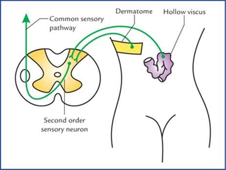
FIG. 20.6 The first order sensory neurons carrying sensations from viscera and dermatome are entering in the same segment of the spinal cord and synapsing with the common second order sensory neuron.
شکل ۲۰.۶ نورونهای حسی درجه یک که حس را از احشاء و پوست منتقل میکنند، در همان بخش از نخاع وارد میشوند و با نورون حسی درجه دو مشترک سیناپس برقرار میکنند.
• Another theory is that under normal conditions the viscera do not send painful stimuli to the brain, whereas the skin repeatedly sends noxious stimuli to the brain. Since afferent fibres from both enter the same segment of the spinal cord, the brain interprets that the information is coming from the skin rather than from the viscus.
• نظریه دیگر این است که در شرایط عادی، احشاء محرکهای دردناک را به مغز نمیفرستند، در حالی که پوست بارها محرکهای مضر را به مغز میفرستد. از آنجایی که فیبرهای آوران از هر دو وارد همان بخش از نخاع میشوند، مغز تفسیر میکند که اطلاعات از پوست میآید نه از احشاء.
Some important examples of referred pain (Fig. 20.7) are listed in Table 20.4.
برخی از نمونههای مهم درد ارجاعی (شکل ۲۰.۷) در جدول ۲۰.۴ فهرست شدهاند.
Table 20.4
The organs and sites of referred pain
جدول ۲۰.۴
اندامها و محلهای درد ارجاعی
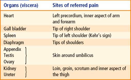
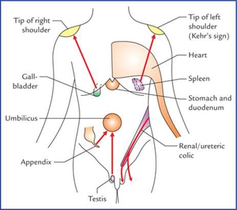
FIG. 20.7 Sites of referred pain from some viscera.
شکل ۲۰.۷ محلهای درد ارجاعی از برخی احشاء.
Higher control of the autonomic nervous system
The normal activities of autonomic nervous system are governed by regulatory centres in the brainstem, hypothalamus and cerebral cortex.
کنترل بالاتر سیستم عصبی خودکار
فعالیتهای طبیعی سیستم عصبی خودکار توسط مراکز تنظیمی در ساقه مغز، هیپوتالاموس و قشر مغز اداره میشوند.
The hypothalamus is regarded as the most important higher centre for the control of autonomic nervous system. Its anterior part controls the parasympathetic responses, whereas its posterior part controls the sympathetic responses.
هیپوتالاموس به عنوان مهمترین مرکز بالاتر برای کنترل سیستم عصبی خودکار در نظر گرفته میشود. قسمت قدامی آن پاسخهای پاراسمپاتیک را کنترل میکند، در حالی که قسمت خلفی آن پاسخهای سمپاتیک را کنترل میکند.
Almost all the autonomic responses can be modified by the activity of same area of the cerebral cortex, presumably through hypothalamus.
تقریباً همه پاسخهای خودکار میتوانند توسط فعالیت همان ناحیه از قشر مغز، احتمالاً از طریق هیپوتالاموس، اصلاح شوند.
Clinical Correlation
Miller et al. (1970) suggested that the autonomic nervous system can be brought under voluntary control to some extent and that the patients with hypertension, for example, possibly can be trained to reduce their blood pressure.
همبستگی بالینی
میلر و همکاران (1970) اظهار داشتند که سیستم عصبی خودکار را میتوان تا حدودی تحت کنترل ارادی درآورد و به عنوان مثال، بیماران مبتلا به فشار خون بالا را میتوان برای کاهش فشار خون آموزش داد.
The higher centres of the brain can abnormally influence the functions of autonomic nervous system and induce clinical conditions like cardiac palpitations and myocardial infarction.
مراکز بالاتر مغز میتوانند به طور غیرطبیعی بر عملکرد سیستم عصبی خودکار تأثیر بگذارند و باعث ایجاد شرایط بالینی مانند تپش قلب و انفارکتوس میوکارد شوند.
Enteric nervous system
The enteric nervous system is a network of intrinsic neurons and ganglia located in the wall of the gastrointestinal tract. These intrinsic neurons are derived from neural crest cells and are independent of sympathetic and parasympathetic nerves. There exists reflex pathway (local reflex mechanisms) in the enteric nervous system through which it controls contraction of muscle coats of the alimentary tract, the secretion of gastric acid, intestinal transport of water and electrolytes, mucosal blood flow, etc.
سیستم عصبی رودهای
سیستم عصبی رودهای شبکهای از نورونها و گانگلیونهای ذاتی است که در دیواره دستگاه گوارش قرار دارند. این نورونهای ذاتی از سلولهای تاج عصبی مشتق شدهاند و مستقل از اعصاب سمپاتیک و پاراسمپاتیک هستند. در سیستم عصبی رودهای، مسیر رفلکس (مکانیسمهای رفلکس موضعی) وجود دارد که از طریق آن انقباض پوششهای عضلانی دستگاه گوارش، ترشح اسید معده، انتقال آب و الکترولیتها از طریق روده، جریان خون مخاطی و غیره را کنترل میکند.
N.B. Though complex interactions occur between enteric and parasympathetic and sympathetic nervous systems, the enteric nervous system is capable of performing its reflex activities independent of CNS.
توجه: اگرچه تعاملات پیچیدهای بین سیستمهای عصبی رودهای و پاراسمپاتیک و سمپاتیک وجود دارد، سیستم عصبی رودهای قادر به انجام فعالیتهای رفلکسی خود مستقل از سیستم عصبی مرکزی است.
Clinical Correlation
Since intrinsic neurons survive following section of extrinsic sympathetic and parasympathetic nerves, the organs/parts of GIT that are transplanted are not truly denervated. Consequently there is no cessation of their activity, i.e. contraction remains unaffected and no structural changes occur.
همبستگی بالینی
از آنجایی که نورونهای داخلی پس از قطع اعصاب سمپاتیک و پاراسمپاتیک خارجی زنده میمانند، اندامها/بخشهایی از دستگاه گوارش که پیوند زده میشوند، واقعاً عصبگیری نمیشوند. در نتیجه، هیچ توقفی در فعالیت آنها وجود ندارد، یعنی انقباض بدون تغییر باقی میماند و هیچ تغییر ساختاری رخ نمیدهد.
Important autonomic innervations
The Eyeball
The main structures supplied by autonomic motor fibres in the eyeball are: sphincter pupillae, ciliary muscle and dilator pupillae.
عصبدهیهای مهم اتونومیک
کره چشم
ساختارهای اصلی که توسط فیبرهای حرکتی اتونومیک در کره چشم تغذیه میشوند عبارتند از: مردمک اسفنکتر، عضله مژگانی و مردمک گشادکننده.
• Sphincter pupillae and ciliary muscle, are innervated by parasympathetic fibres (seeChapter 9).
• مردمکهای اسفنکتر و عضله مژگانی، توسط فیبرهای پاراسمپاتیک عصبدهی میشوند (به فصل 9 مراجعه کنید).
• Dilator pupillae, it is innervated by sympathetic fibres. The preganglionic sympathetic fibres arise from lateral horn cells of first thoracic spinal segment (T1), pass through anterior root of the first thoracic nerve, to enter the inferior cervical sympathetic ganglion and ascend in the cervical sympathetic chain to relay in its superior cervical sympathetic ganglion. The postganglionic fibres from ganglion, pass successively through internal carotid nerve (or plexus), ophthalmic nerve, nasociliary nerve and long ciliary nerves (occasionally short ciliary nerves) to supply the dilator pupillae (Fig. 20.8).
• مردمک گشادکننده، توسط فیبرهای سمپاتیک عصبدهی میشود. فیبرهای سمپاتیک پیش عقدهای از سلولهای شاخ جانبی اولین قطعه نخاعی سینهای (T1) منشأ میگیرند، از ریشه قدامی اولین عصب سینهای عبور میکنند تا وارد گانگلیون سمپاتیک گردنی تحتانی شوند و در زنجیره سمپاتیک گردنی بالا میروند تا در گانگلیون سمپاتیک گردنی فوقانی خود رله کنند. فیبرهای پس عقدهای از گانگلیون، به ترتیب از عصب کاروتید داخلی (یا شبکه عصبی)، عصب چشمی، عصب نازوسیلیاری و اعصاب مژگانی بلند (گاهی اوقات اعصاب مژگانی کوتاه) عبور میکنند تا مردمکهای گشادکننده را تغذیه کنند (شکل 20.8).
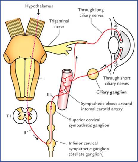
FIG. 20.8 Sympathetic pathway to the eye. I. First order neuron (hypothalamospinal fibres project) without interruption from hypothalamus to the ciliospinal centre in the intermediolateral cell column of T1 spinal segment, II. Second order neuron (preganglionic fibres), arise from T1 spinal segment and relay in the superior cervical sympathetic ganglion, III. Third order neuron (postganglionic fibres) arise from superior cervical sympathetic ganglion. The second and third order neurons form the peripheral part of the sympathetic motor pathway.
شکل 20.8 مسیر سمپاتیک به چشم. I. نورون مرتبه اول (فیبرهای هیپوتالاموس نخاعی) بدون وقفه از هیپوتالاموس به مرکز مژکی-نخاعی در ستون سلولی بین میانی-جانبی قطعه نخاعی T1، II. نورون مرتبه دوم (فیبرهای پیش عقدهای)، از قطعه نخاعی T1 منشأ میگیرند و در گانگلیون سمپاتیک گردنی فوقانی رله میکنند، III. نورون مرتبه سوم (فیبرهای پس عقدهای) از گانگلیون سمپاتیک گردنی فوقانی منشأ میگیرند. نورونهای مرتبه دوم و سوم بخش محیطی مسیر حرکتی سمپاتیک را تشکیل میدهند.
N.B. Sympathetic fibres also supply the orbitalis muscle in the floor of orbit, smooth muscle fibres of levator palpe-brae superioris (Muller’s muscle) and sudomotor and vaso-motor fibres to orbit, eyelid and face.
توجه: فیبرهای سمپاتیک همچنین عضله اربیتالیس در کف حدقه، فیبرهای عضله صاف عضله بالابرنده پلک فوقانی (عضله مولر) و فیبرهای سودوموتور و وازوموتور به حدقه، پلک و صورت را عصبدهی میکنند.
Clinical Correlation
Horner’s syndrome
It occurs due to interruption of sympathetic pathway to the head and neck (Fig. 20.8). The common sites of lesion are: brainstem, cervical part of spinal cord, or stellate ganglion.
همبستگی بالینی
سندرم هورنر
این سندرم به دلیل قطع مسیر سمپاتیک به سر و گردن رخ میدهد (شکل 20.8). محلهای شایع ضایعه عبارتند از: ساقه مغز، قسمت گردنی نخاع یا گانگلیون ستارهای.
Characteristic features
ویژگیهای مشخصه
• Miosis (i.e. constriction of pupil) | Due to paralysis of dilator pupillae and unopposed action of sphincter pupillae. |
• Partial ptosis (i.e. slight drooping of the upper eyelid) | Due to paralysis of smooth muscle fibres of levator palpebrae superioris called Muller’s muscle. |
• Anhidrosis (i.e. loss of sweating) | Due to involvement of sudomotor fibres which are concerned with sweating in the region of head and neck. |
• Enophthalmos (sunken eyeball) | It is often apparent but not real and is caused by ptosis. The paralysis of orbitalis muscle may also be responsible. |
• Flushing of the face | Due to involvement of vasoconstrictor fibres leading to vasodilation of skin arterioles in the region of head and neck. |
The Salivary Glands
Parotid gland
Parasympathetic innervation (secretomotor supply): The preganglionic fibres arise from inferior salivatory nucleus in the pons, pass successively through glossopharyngeal nerve and its tympanic branch, the tympanic plexus and the lesser petrosal nerve to terminate in the otic ganglion. The postganglionic fibres from the otic ganglion, pass through the auriculotemporal nerve to supply the parotid gland (Fig. 20.9).
غدد بزاقی
غده پاروتید
عصبدهی پاراسمپاتیک (تأمین ترشحی): فیبرهای پیشگانگلیونی از هسته بزاقی تحتانی در پل مغزی منشأ میگیرند، به ترتیب از طریق عصب زبانی-حلقی و شاخه تمپانیک آن، شبکه تمپانیک و عصب پتروزال کوچک عبور میکنند تا در گانگلیون گوش خاتمه یابند. فیبرهای پسگانگلیونی از گانگلیون گوش، از طریق عصب گوش-گیجگاهی عبور میکنند تا غده پاروتید را تأمین کنند (شکل 20.9).
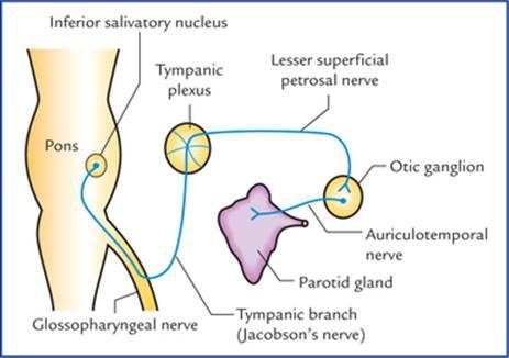
FIG. 20.9 Secretomotor pathway for parotid gland.
شکل 20.9 مسیر ترشحی-حرکتی غده پاروتید.
Submandibular and sublingual glands
Parasympathetic innervation (secretomotor nerve supply): The preganglionic fibres arise from superior salivatory nucleus in the pons, pass successively through facial, chorda tympani and lingual nerves to terminate in the submandibular ganglion. The postganglionic fibres from the submandibular ganglion supply the submandibular gland directly, and sublingual gland through the lingual nerve (Fig. 20.10).
غدد زیر فکی و زیر زبانی
عصب رسانی پاراسمپاتیک (عصب رسانی ترشحی-حرکتی): فیبرهای پیش عقدهای از هسته بزاقی فوقانی در پل مغزی منشأ میگیرند، به ترتیب از طریق اعصاب صورت، طناب صماخی و زبانی عبور میکنند تا در گانگلیون زیر فکی خاتمه یابند. فیبرهای پس عقدهای از گانگلیون زیر فکی مستقیماً به غده زیر فکی و از طریق عصب زبانی به غده زیر زبانی عصب میدهند (شکل 20.10).
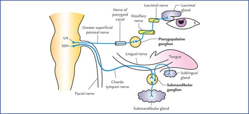
FIG. 20.10 Secretomotor pathways for lacrimal gland, and submandibular and sublingual salivary glands. (LN = lacrimatory nucleus, and SSN = superior salivatory nucleus.)
شکل 20.10 مسیرهای ترشحی-حرکتی برای غده اشکی و غدد بزاقی زیر فکی و زیر زبانی. (LN = هسته اشکی و SSN = هسته بزاقی فوقانی.)
Lacrimal gland
Parasympathetic innervation (secretomotor nerve supply): The preganglionic parasympathetic fibres arise from lacrimatory nucleus in the pons, and pass successively through nerves intermedius, geniculate ganglion of facial nerve, greater petrosal nerve and nerve of pterygoid canal to terminate in the pterygopalatine ganglion. The postganglionic fibres arise from pterygopalatine ganglion, and pass successively through maxillary nerve, zygomatic nerve and its zygomatico-temporal branch, and lacrimal nerve to supply the lacrimal gland (Fig. 20.10).
غده اشکی
عصب رسانی پاراسمپاتیک (عصب رسانی ترشحی-حرکتی): فیبرهای پاراسمپاتیک پیش عقدهای از هسته اشکی در پل مغزی منشأ میگیرند و به ترتیب از طریق اعصاب میانی، گانگلیون زانویی عصب صورتی، عصب پتروزال بزرگ و عصب کانال پتریگوئید عبور میکنند تا در گانگلیون پتریگوپالاتین خاتمه یابند. فیبرهای پس عقدهای از گانگلیون پتریگوپالاتین منشأ میگیرند و به ترتیب از طریق عصب فک بالا، عصب گونه و شاخه گونه-گیجگاهی آن و عصب اشکی عبور میکنند تا غده اشکی را تغذیه کنند (شکل 20.10).
Gastrointestinal Tract
Motor supply
Parasympathetic innervation
دستگاه گوارش
تغذیه حرکتی
عصب رسانی پاراسمپاتیک
• The most of GIT, i.e. from oesophagus to the junction of right two-third and left one-third of transverse colon, is supplied by vagus nerve. Preganglionic parasympathetic fibres arise from dorsal nucleus of vagus, and pass successively through vagus nerve coeliac, superior and inferior mesenteric plexuses, and then run along the coeliac, superior mesenteric and inferior mesenteric arteries and their branches to enter the GIT, where they relay in the ganglia within myenteric (Auerbach’s) and submucosal (Meissner’s) plexuses located in the wall of the gut.
• بخش عمدهای از دستگاه گوارش، یعنی از مری تا محل اتصال دو سوم راست و یک سوم چپ روده بزرگ عرضی، توسط عصب واگ خونرسانی میشود. فیبرهای پاراسمپاتیک پیش عقدهای از هسته پشتی واگ منشأ میگیرند و به ترتیب از شبکههای سلیاک، مزانتریک فوقانی و تحتانی عصب واگ عبور میکنند و سپس در امتداد شریانهای سلیاک، مزانتریک فوقانی و مزانتریک تحتانی و شاخههای آنها وارد دستگاه گوارش میشوند، جایی که در گانگلیونهای درون شبکههای میانتریک (اورباخ) و زیرمخاطی (مایسنر) واقع در دیواره روده رله میشوند.
• From junction of right two-third and left one-third of transverse colon to the junction of upper and lower halves of the anal canal the GIT is supplied by sacral parasympathetic outflow. The preganglionic parasympathetic fibres arise from anterolateral grey columns of S2, S3 and S4 spinal segments, pass successively through the ventral roots of spinal nerves, pelvic splanchnic nerves and superior and inferior hypogastric plexuses to terminate in the ganglia within myenteric (Auerbach’s) and submucosal (Meissner’s) plexuses located in the gut wall.
• از محل اتصال دو سوم راست و یک سوم چپ روده بزرگ عرضی تا محل اتصال نیمههای فوقانی و تحتانی کانال مقعد، دستگاه گوارش توسط جریان پاراسمپاتیک خارجی خونرسانی میشود. فیبرهای پاراسمپاتیک پیش عقدهای از ستونهای خاکستری قدامی-جانبی قطعات نخاعی S2، S3 و S4 منشأ میگیرند، به ترتیب از ریشههای شکمی اعصاب نخاعی، اعصاب اسپلانکنیک لگنی و شبکههای هیپوگاستریک فوقانی و تحتانی عبور میکنند تا در گانگلیونهای درون شبکههای میانتریک (اورباخ) و زیرمخاطی (مایسنر) واقع در دیواره روده خاتمه یابند.
The postganglionic fibres arise from parasympathetic ganglia located in the gut wall and supply the muscle and glands of GIT.
فیبرهای پس عقدهای از گانگلیونهای پاراسمپاتیک واقع در دیواره روده منشأ میگیرند و به عضله و غدد دستگاه گوارش خونرسانی میکنند.
Sympathetic innervation: The preganglionic sympathetic fibres arise from T5 to L2 spinal segments, pass through the sympathetic trunks without relay, travel through the splanchnic nerves to terminate in the ganglia related to the coeliac (coeliac ganglion), superior mesenteric (superior mesenteric ganglion) and inferior mesenteric (inferior mes-enteric ganglion) arteries. The postganglionic fibres arise from these ganglia and travel along these arteries to reach the gut which they supply.
عصبدهی سمپاتیک: فیبرهای سمپاتیک پیش عقدهای از بخشهای نخاعی T5 تا L2 منشأ میگیرند، بدون رله از تنههای سمپاتیک عبور میکنند، از طریق اعصاب احشایی حرکت میکنند تا در گانگلیونهای مربوط به شریانهای سلیاک (گانگلیون سلیاک)، مزانتریک فوقانی (گانگلیون مزانتریک فوقانی) و مزانتریک تحتانی (گانگلیون مزانتریک تحتانی) خاتمه یابند. فیبرهای پس عقدهای از این گانگلیونها منشأ میگیرند و در امتداد این شریانها حرکت میکنند تا به رودهای که آن را تغذیه میکنند، برسند.
Functional significance
The parasympathetic nerves stimulate peristalsis (gastrointestinal motility) and relax the sphincters. They also stimulate secretion from mucosal glands.
اهمیت عملکردی
اعصاب پاراسمپاتیک حرکات دودی (حرکات دستگاه گوارش) را تحریک کرده و اسفنکترها را شل میکنند. آنها همچنین ترشح غدد مخاطی را تحریک میکنند.
The sympathetic fibres inhibit peristalsis and cause contraction of the sphincters; they also inhibit secretion from mucosal glands.
فیبرهای سمپاتیک حرکات دودی را مهار کرده و باعث انقباض اسفنکترها میشوند. آنها همچنین ترشح غدد مخاطی را مهار میکنند.
Clinical Correlation
Congenital megacolon (Hirschsprung disease)
Sometimes, during embryonic period the neural crest cells fail to migrate into the wall of descending colon at its junction with the sigmoid colon, leading to lack of formation of parasympathetic ganglia in the myenteric (Auerbach’s) plexus. As a result the part of colon lacking ganglia shows marked decrease in its peristalsis leading to functional obstruction. Consequently the proximal part of descending colon becomes greatly distended due to accumulation of faeces, causing congenital megacolon (Hirschsprung disease).
همبستگی بالینی
مگاکولون مادرزادی (بیماری هیرشپرونگ)
گاهی اوقات، در دوران جنینی، سلولهای تاج عصبی نمیتوانند به دیواره روده بزرگ نزولی در محل اتصال آن با روده بزرگ سیگموئید مهاجرت کنند و منجر به عدم تشکیل گانگلیونهای پاراسمپاتیک در شبکه عصبی میانتریک (اورباخ) میشوند. در نتیجه، بخشی از روده بزرگ که فاقد گانگلیون است، کاهش قابل توجهی در حرکات دودی خود نشان میدهد که منجر به انسداد عملکردی میشود. در نتیجه، قسمت پروگزیمال روده بزرگ نزولی به دلیل تجمع مدفوع به شدت متسع میشود و باعث مگاکولون مادرزادی (بیماری هیرشپرونگ) میشود.
Sensory supply
The afferent fibres travel along both sympathetic and parasympathetic pathways.
تغذیه حسی
الیاف آوران در امتداد مسیرهای سمپاتیک و پاراسمپاتیک حرکت میکنند.
Pain from most of the GIT travel along the sympathetic nerves except from upper part (i.e. pharynx and oesophagus) and lower part (i.e. lower part of pelvic colon, rectum and upper part of anal canal) from where it is carried by parasympathetic fibres through vagus and pelvic splanchnic nerves respectively.
درد ناشی از بیشتر قسمتهای دستگاه گوارش، به جز قسمت فوقانی (یعنی حلق و مری) و قسمت تحتانی (یعنی قسمت تحتانی روده بزرگ لگنی، رکتوم و قسمت فوقانی کانال مقعدی) که از آنجا توسط الیاف پاراسمپاتیک به ترتیب از طریق اعصاب واگ و اسپلانکنیک لگنی منتقل میشود، در امتداد اعصاب سمپاتیک حرکت میکند.
Urinary Bladder
Motor supply (Fig. 20.11)
Parasympathetic innervation: The preganglionic parasympathetic fibres arise from S2, S3 and S4 spinal segments (spinal micturition centre), pass through the pelvic splanchnic nerves and inferior hypogastric plexus to terminate in the nerve cells located in the vesical plexus. The postganglionic fibres arise from vesical plexus and supply the urinary bladder.
مثانه ادراری
تغذیه حرکتی (شکل 20.11)
عصبدهی پاراسمپاتیک: فیبرهای پاراسمپاتیک پیش عقدهای از بخشهای نخاعی S2، S3 و S4 (مرکز ادرار نخاعی) منشأ میگیرند، از اعصاب اسپلانکنیک لگنی و شبکه هیپوگاستریک تحتانی عبور میکنند تا در سلولهای عصبی واقع در شبکه مثانهای خاتمه یابند. فیبرهای پس عقدهای از شبکه مثانهای منشأ میگیرند و مثانه را تغذیه میکنند.
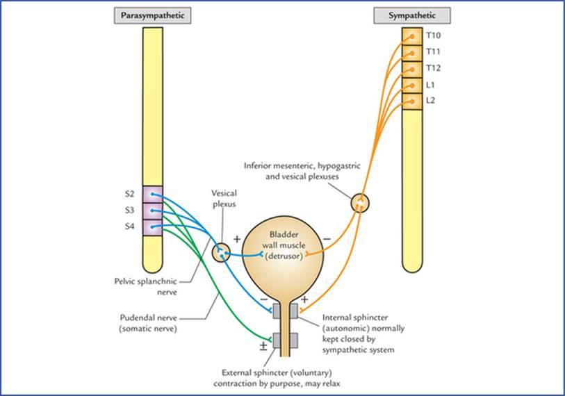
FIG. 20.11 Motor innervation of the urinary bladder, showing physiological and neurological principles of bladder function. (+) = contraction, (−) = relaxation, (±) = contraction and relaxation.
شکل 20.11 عصبدهی حرکتی مثانه، اصول فیزیولوژیکی و عصبی عملکرد مثانه را نشان میدهد. (+) = انقباض، (-) = شل شدن، (±) = انقباض و شل شدن.
Sympathetic innervation: The preganglionic sympathetic fibres arise from T10 to L2 spinal segments, and terminate in the cells of inferior mesenteric, superior hypogastric, inferior hypogastric and vesical plexuses. The postganglionic fibres arise from the cells within these plexuses and supply the urinary bladder.
عصبدهی سمپاتیک: فیبرهای سمپاتیک پیش عقدهای از قطعات نخاعی T10 تا L2 منشأ میگیرند و در سلولهای شبکه مزانتریک تحتانی، هیپوگاستریک فوقانی، هیپوگاستریک تحتانی و مثانهای خاتمه مییابند. فیبرهای پس عقدهای از سلولهای درون این شبکهها منشأ میگیرند و مثانه را تغذیه میکنند.
The parasympathetic fibres stimulate the contraction of the smooth muscle of the bladder wall (detrusor muscle) and relax the internal urethra! sphincter (sphincter vesicae).
فیبرهای پاراسمپاتیک انقباض عضله صاف دیواره مثانه (عضله دترسور) را تحریک کرده و اسفنکتر داخلی مجرای ادرار (اسفنکتر وزیکا) را شل میکنند.
The sympathetic fibres have little or no action on the detrusor muscle and play only a minor role in maintaining urinary continence by causing contraction of sphincter vesicae. However, in males during ejaculation (brought out by sympathetic action) the sympathetic stimulation causes active contraction of sphincter vesicae and prevents the seminal fluid from entering the bladder.
فیبرهای سمپاتیک تأثیر کمی یا هیچ تأثیری بر عضله دترسور ندارند و تنها با ایجاد انقباض در وزیکاهای اسفنکتر، نقش جزئی در حفظ اختیار ادرار ایفا میکنند. با این حال، در مردان هنگام انزال (که توسط عمل سمپاتیک انجام میشود)، تحریک سمپاتیک باعث انقباض فعال اسفنکتر وزیکا شده و از ورود مایع منی به مثانه جلوگیری میکند.
Sensory supply
Sensory fibres carry impulses of distension and pain from the bladder. They travel through both sympathetic and parasympathetic fibres to the CNS.
منبع حسی
فیبرهای حسی، تکانههای مربوط به اتساع و درد را از مثانه حمل میکنند. آنها از طریق فیبرهای سمپاتیک و پاراسمپاتیک به سیستم عصبی مرکزی (CNS) منتقل میشوند.
Clinical Correlation
• The fibres carrying sensations of distension (filling of the bladder) travel through the posterior white column of the spinal cord, while the fibres carrying pain travel through the anterior and lateral white columns of the spinal cord.
همبستگی بالینی
• فیبرهایی که حس اتساع (پر شدن مثانه) را منتقل میکنند، از طریق ستون سفید خلفی نخاع عبور میکنند، در حالی که فیبرهایی که حس درد را منتقل میکنند، از طریق ستونهای سفید قدامی و جانبی نخاع عبور میکنند.
The important point to note here is that sensory pathways carrying sensation of pain and distension take different routes in the spinal cord. Therefore, intractable bladder pain due to carcinoma bladder may be relieved by cutting anterior and lateral white columns of spinal cord on both the sides (bilateral anterolateral cordotomy).
نکته مهمی که باید در اینجا به آن توجه داشت این است که مسیرهای حسی که حس درد و اتساع را منتقل میکنند، مسیرهای متفاوتی را در نخاع طی میکنند. بنابراین، درد مثانه مقاوم به درمان ناشی از سرطان مثانه ممکن است با بریدن ستونهای سفید قدامی و جانبی نخاع در هر دو طرف (کوردوتومی قدامی-جانبی دو طرفه) تسکین یابد.
• Effects of spinal cord injuries on the bladder function
– The atonic bladder occurs during the phase of spinal shock (temporary loss/halt of all spinal functions) immediately after the injury. The muscle of bladder wall (detrusor muscle) is relaxed, the internal sphincter (sphincter vesicae) is tightly contracted (due to loss of inhibition from higher centres) and the external sphincter is relaxed. As a result the bladder becomes greatly distended with urine which finally overflows.
• اثرات آسیبهای نخاعی بر عملکرد مثانه
– مثانه آتونیک در طول مرحله شوک نخاعی (از دست دادن/توقف موقت تمام عملکردهای ستون فقرات) بلافاصله پس از آسیب رخ میدهد. عضله دیواره مثانه (عضله دترسور) شل میشود، اسفنکتر داخلی (اسفنکتر وزیکا) محکم منقبض میشود (به دلیل از دست دادن مهار از مراکز بالاتر) و اسفنکتر خارجی شل میشود. در نتیجه، مثانه با ادرار بسیار متسع میشود که در نهایت سرریز میشود.
– The automatic reflex bladder occurs after the patient has recovered from the spinal shock, and if the cord is injured above the level of segments responsible for the parasympathetic outflow, i.e. spinal micturition centre (S2, S3 and S4). Due to involvement of descending fibres from paracentral lobule, the cortical micturition centre, the voluntary control on bladder is lost. As a result, the bladder fills and empties reflexly. The stretch receptors in the bladder wall are stimulated as the bladder fills and the afferent impulses pass to the spinal cord (S2, S3 and S4). From spinal cord the efferent impulses pass to the bladder, causing detrusor muscle to contract and internal and external sphincter urethrae to relax leading to emptying of the bladder. This spinal reflex for emptying the bladder occurs every 1 to 4 hrs.
– مثانه رفلکسی خودکار پس از بهبودی بیمار از شوک نخاعی رخ میدهد و اگر نخاع در بالای سطح بخشهای مسئول خروج پاراسمپاتیک، یعنی مرکز ادرار نخاعی (S2، S3 و S4) آسیب ببیند. به دلیل درگیری فیبرهای نزولی از لوبول پاراسنترال، مرکز ادرار قشری، کنترل ارادی بر مثانه از بین میرود. در نتیجه، مثانه به صورت رفلکسی پر و خالی میشود. گیرندههای کششی در دیواره مثانه با پر شدن مثانه تحریک میشوند و تکانههای آوران به نخاع (S2، S3 و S4) منتقل میشوند. تکانههای وابران از نخاع به مثانه منتقل میشوند و باعث انقباض عضله دترسور و شل شدن مجرای ادرار اسفنکتر داخلی و خارجی میشوند که منجر به تخلیه مثانه میشود. این رفلکس نخاعی برای تخلیه مثانه هر 1 تا 4 ساعت یکبار رخ میدهد.
– The autonomous bladder occurs if the sacral segments (S2, S3 and S4) of the spinal cord are destroyed. In this condition, the bladder has neither reflex control nor voluntary control because both, reflex centre for bladder emptying (S2, S3 and S4) and descending fibres from voluntary cortical centre have become non-functional.
– مثانه خودمختار در صورتی رخ میدهد که بخشهای خاجی (S2، S3 و S4) نخاع از بین بروند. در این شرایط، مثانه نه کنترل رفلکسی و نه کنترل ارادی دارد زیرا هم مرکز رفلکس تخلیه مثانه (S2، S3 و S4) و هم فیبرهای نزولی از مرکز قشری ارادی غیرفعال شدهاند.
As a result the bladder wall becomes flaccid, and the capacity of the bladder is greatly increased. It fills to capacity arid overflows, causing continuous dribbling of urine.
در نتیجه، دیواره مثانه شل میشود و ظرفیت مثانه به میزان زیادی افزایش مییابد. مثانه تا حد ظرفیت پر میشود و سرریز میکند و باعث چکه کردن مداوم ادرار میشود.
Bladder function
The process of micturition requires proper integration of autonomic and voluntary nervous systems. The contraction of smooth muscle of bladder wall (the detrusor muscle) is initiated by parasympathetic activity, while the internal sphincter mechanism (which is normally closed by sympathetic activity) is allowed to relax by decreased sympathetic activity. The external sphincter mechanism, which can be voluntarily relaxed to allow micturition is controlled by pudendal nerves (S2, S3 and S4).
عملکرد مثانه
فرآیند ادرار کردن نیاز به ادغام مناسب سیستمهای عصبی خودکار و ارادی دارد. انقباض عضله صاف دیواره مثانه (عضله دترسور) توسط فعالیت پاراسمپاتیک آغاز میشود، در حالی که مکانیسم اسفنکتر داخلی (که معمولاً توسط فعالیت سمپاتیک بسته میشود) با کاهش فعالیت سمپاتیک شل میشود. مکانیسم اسفنکتر خارجی که میتواند به طور ارادی شل شود تا ادرار کردن را ممکن سازد، توسط اعصاب پودندال (S2، S3 و S4) کنترل میشود.
Heart
Motor supply
Parasympathetic innervation: The preganglionic parasympathetic fibres arise from dorsal nucleus of vagus in the medulla oblongata, run through vagus nerve and its cardiac branches to synapse on the cells or ganglia in superficial and deep cardiac plexuses. The postganglionic fibres arise from these cells or ganglia and supply the sinuatrial and atrioventricular nodes and coronary arteries.
قلب
تغذیه حرکتی
عصبدهی پاراسمپاتیک: فیبرهای پاراسمپاتیک پیش عقدهای از هسته پشتی واگ در بصل النخاع منشأ میگیرند، از طریق عصب واگ و شاخههای قلبی آن عبور میکنند تا روی سلولها یا گانگلیونهای موجود در شبکههای قلبی سطحی و عمقی سیناپس برقرار کنند. فیبرهای پس عقدهای از این سلولها یا گانگلیونها منشأ میگیرند و گرههای سینوسی-دهلیزی و دهلیزی-بطنی و شریانهای کرونری را عصبدهی میکنند.
Sympathetic innervation: The preganglionic sympathetic fibres arise from T1 to T5 spinal segments and terminate in the cells of upper 3 or 4 thoracic ganglia and cervical sympathetic ganglia.
عصبدهی سمپاتیک: فیبرهای سمپاتیک پیش عقدهای از قطعات نخاعی T1 تا T5 منشأ میگیرند و در سلولهای گانگلیونهای فوقانی 3 یا 4 قفسه سینه و گانگلیونهای سمپاتیک گردنی خاتمه مییابند.
The postganglionic sympathetic fibres arise from these ganglia, pass first through the cardiac branches of these ganglia and then through cardiac plexuses to supply sinuatrial and atrioventricular nodes, and coronary arteries of the heart.
فیبرهای سمپاتیک پس عقدهای از این گانگلیونها منشأ میگیرند، ابتدا از شاخههای قلبی این گانگلیونها و سپس از طریق شبکههای قلبی عبور میکنند تا گرههای سینوسی-دهلیزی و دهلیزی-بطنی و شریانهای کرونری قلب را خونرسانی کنند.
Functions
The sympathetic stimulation increases whereas the parasympathetic stimulation decreases the heart rate. During normal circumstances, the heart rate is normal because a balance is maintained between the inhibitory and the stimulatory effects of the parasympathetic and sympathetic innervation. The exertion and emotional emergencies tilt the balance towards the sympathetic tone, hence responsible for increased heart rate. The excessive vagal stimulation can bring the activity of the heart standstill.
عملکردها
تحریک سمپاتیک افزایش مییابد در حالی که تحریک پاراسمپاتیک ضربان قلب را کاهش میدهد. در شرایط عادی، ضربان قلب طبیعی است زیرا تعادلی بین اثرات مهاری و تحریکی عصبدهی پاراسمپاتیک و سمپاتیک حفظ میشود. فعالیت بدنی و فوریتهای عاطفی، تعادل را به سمت تون سمپاتیک متمایل میکنند، از این رو مسئول افزایش ضربان قلب هستند. تحریک بیش از حد عصب واگ میتواند فعالیت قلب را متوقف کند.
Further, the sympathetic stimulation causes dilatation, and parasympathetic stimulation causes constriction of the coronary arteries, thus increasing or decreasing blood supply to the heart respectively.
علاوه بر این، تحریک سمپاتیک باعث گشاد شدن و تحریک پاراسمپاتیک باعث انقباض عروق کرونر میشود، در نتیجه به ترتیب خونرسانی به قلب را افزایش یا کاهش میدهد.
Sensory supply
The afferent fibres from heart travel through both sympathetic and parasympathetic pathways. The sensations of pain from heart (due to anoxia, viz. angina) are carried by sympathetic fibres. They travel through cardiac branches of middle and inferior cervical sympathetic ganglia, pass successively through these ganglia, sympathetic trunks and spinal nerves to enter the spinal segments from T1 to T4/5.
تغذیه حسی
الیاف آوران قلب از طریق مسیرهای سمپاتیک و پاراسمپاتیک حرکت میکنند. احساس درد قلبی (به دلیل آنوکسی، یعنی آنژین صدری) توسط الیاف سمپاتیک منتقل میشود. آنها از طریق شاخههای قلبی گانگلیونهای سمپاتیک گردنی میانی و تحتانی حرکت میکنند، به طور متوالی از این گانگلیونها، تنههای سمپاتیک و اعصاب نخاعی عبور میکنند تا از T1 تا T4/5 وارد بخشهای نخاعی شوند.
Afferent fibres running along the cardiac branches of vagus nerves are concerned with reflexes controlling the activity of the heart.
الیاف آوران که در امتداد شاخههای قلبی اعصاب واگ قرار دارند، با رفلکسهایی که فعالیت قلب را کنترل میکنند، مرتبط هستند.
Arteries of the Upper Limb
The arteries of the upper limb are innervated by sympathetic fibres only.
شریانهای اندام فوقانی
شریانهای اندام فوقانی فقط توسط فیبرهای سمپاتیک عصبدهی میشوند.
The preganglionic fibres arise from T2 to T8 thoracic spinal segments, and pass to the sympathetic trunk through white rami communicantes. After entering the trunk they ascend to synapse with the cells within the stellate and middle cervical sympathetic ganglia. The postganglionic fibres arise from these ganglia, join the nerves forming brachial plexus and are distributed to the arteries through the branches of brachial plexus. The stimulation of sympathetic nerves causes vasoconstriction of cutaneous arteries and vasodilatation of arteries supplying the skeletal muscles.
فیبرهای پیشگانگلیونی از قطعات نخاعی قفسه سینه T2 تا T8 منشأ میگیرند و از طریق شاخههای سفید ارتباطی به تنه سمپاتیک میروند. پس از ورود به تنه، آنها به سمت سیناپس با سلولهای درون گانگلیونهای سمپاتیک ستارهای و گردنی میانی صعود میکنند. فیبرهای پسگانگلیونی از این گانگلیونها منشأ میگیرند، به اعصاب تشکیل دهنده شبکه بازویی میپیوندند و از طریق شاخههای شبکه بازویی به شریانها توزیع میشوند. تحریک اعصاب سمپاتیک باعث انقباض عروق شریانهای پوستی و گشاد شدن عروق شریانهای تغذیه کننده عضلات اسکلتی میشود.
Clinical Correlation
The Raynaud’s disease occurs due to vasospasm of the digital arteries of the upper limb following exposure to cold. It is common in women. There is pallor or cyanosis of the fingers associated with severe pain. The gangrene of the tips of the fingers may occur. It can be treated by preganglionic sympathectomy.
همبستگی بالینی
بیماری رینود به دلیل اسپاسم عروق انگشتان اندام فوقانی پس از قرار گرفتن در معرض سرما رخ میدهد. این بیماری در زنان شایع است. رنگپریدگی یا سیانوز انگشتان همراه با درد شدید وجود دارد. گانگرن نوک انگشتان ممکن است رخ دهد. این بیماری را میتوان با سمپاتکتومی پیش عقدهای درمان کرد.
The symptoms are relieved: (a) by avoiding exposure to cold, and stopping smoking (smoking causes vasoconstriction), and (b) by giving drugs that inhibit sympathetic activity to bring about arterial dilatation.
علائم به روشهای زیر تسکین مییابند: (الف) با اجتناب از قرار گرفتن در معرض سرما و ترک سیگار (سیگار کشیدن باعث انقباض عروق میشود) و (ب) با تجویز داروهایی که فعالیت سمپاتیک را مهار میکنند تا باعث گشاد شدن شریانها شوند.
The removal of stellate (cervicothoracic) ganglion (cervicothoracic preganglionic sympathectomy) has also been used in the past.
برداشتن گانگلیون ستارهای (سرویکوتوراسیک) (سرویکوتوراسیک پیش عقدهای سمپاتکتومی) نیز در گذشته مورد استفاده قرار گرفته است.
Arteries of the Lower Limb
The arteries of the lower limb are supplied by sympathetic nerves. The preganglionic fibres arise from lower three thoracic and upper two or three lumbar segments of the spinal cord (T10-L2), and pass to the lower thoracic and upper lumbar ganglia of the sympathetic chain. These fibres synapse in the lumbar and sacral ganglia. The postganglionic fibres arise from these ganglia and reach the arteries through the branches of lumbar and sacral plexuses.
شریانهای اندام تحتانی
شریانهای اندام تحتانی توسط اعصاب سمپاتیک تغذیه میشوند. فیبرهای پیشگانگلیونی از سه بخش پایینی قفسه سینه و دو یا سه بخش بالایی کمری نخاع (T10-L2) منشأ میگیرند و به گانگلیونهای پایینی قفسه سینه و بالایی کمری زنجیره سمپاتیک میروند. این فیبرها در گانگلیونهای کمری و خاجی سیناپس برقرار میکنند. فیبرهای پسگانگلیونی از این گانگلیونها منشأ میگیرند و از طریق شاخههای شبکههای کمری و خاجی به شریانها میرسند.
Clinical Correlation
Buerger’s disease is an arterial occlusive disease of the legs causing ischaemia of muscles. It is common in men, particularly those who smoke a lot. These individuals complain of intermittent claudication, i.e. appearance of pain on exercise which disappears after rest and reappears on exercise.
همبستگی بالینی
بیماری بورگر یک بیماری انسدادی شریانی پاها است که باعث ایسکمی عضلات میشود. این بیماری در مردان، به ویژه کسانی که زیاد سیگار میکشند، شایع است. این افراد از لنگش متناوب شکایت دارند، یعنی درد هنگام ورزش که پس از استراحت از بین میرود و دوباره هنگام ورزش ظاهر میشود.
They may suffer from ischaemia and dry gangrene of the toes.
آنها ممکن است از ایسکمی و گانگرن خشک انگشتان پا رنج ببرند.
The preganglionic lumbar sympathectomy (removal of upper three lumbar ganglia and intervening parts of the sympathetic chain) is advocated in order to bring about vasodilatation and increase in the blood flow through the collateral circulation.
سمپاتکتومی کمری پیش عقدهای (برداشتن سه عقده فوقانی کمری و بخشهای میانی زنجیره سمپاتیک) به منظور ایجاد گشاد شدن عروق و افزایش جریان خون از طریق گردش خون جانبی توصیه میشود.
Clinical problems
مشکلات بالینی
1. What is the embryological basis of congenital megacolon (Hirschsprung disease)?
1. اساس جنینی مگاکولون مادرزادی (بیماری هیرشپرونگ) چیست؟
2. What are the effects of spinal cord injury on the bladder function?
2. اثرات آسیب نخاعی بر عملکرد مثانه چیست؟
3. Explain, why there is enuresis (bed-wetting) during infancy.
3. توضیح دهید که چرا در دوران نوزادی شب ادراری (خیس کردن رختخواب) وجود دارد.
4. Pain arising from gall bladder is often referred to the tip of right shoulder. Mention the anatomical basis.
4. درد ناشی از کیسه صفرا اغلب به نوک شانه راست منتقل میشود. اساس آناتومیک را ذکر کنید.
5. Explain, why the pain of renal and ureteric colic is referred to loin, groin scrotum/labium majus and inner aspect of the upper thigh.
5. توضیح دهید که چرا درد قولنج کلیوی و حالبی به کمر، کشاله ران، کیسه بیضه/لب بزرگ و قسمت داخلی ران فوقانی منتقل میشود.
6. Pain from appendix, testis and ovary is often referred to the skin around umbilicus. Mention the anatomical basis.
6. درد آپاندیس، بیضه و تخمدان اغلب به پوست اطراف ناف منتقل میشود. اساس آناتومیک را ذکر کنید.
7. Explain, why the cardiac pain is felt over the mid-sternum in the precordium and often spreads to the medial side of arm and forearm.
7. توضیح دهید که چرا درد قلبی در قسمت میانی جناغ سینه در جلوی قلب احساس میشود و اغلب به سمت داخلی بازو و ساعد گسترش مییابد.
Clinical problem solving
1. Seepage 244.
2. Seepage 245.
3. Because there is no cortical (voluntary) control on the spinal micturition centre in infants (as the descending corticospinal fibres have yet not myelinated).
حل مسئله بالینی
۱. جستجو ۲۴۴.
۲. جستجو ۲۴۵.
۳. زیرا هیچ کنترل قشری (ارادی) بر روی مرکز ادرار نخاعی در نوزادان وجود ندارد (زیرا فیبرهای قشری نخاعی نزولی هنوز میلین دار نشدهاند).
N.B. The corticospinal fibres get myelinated and begin to function about the age of 3 to 6 years. Therefore bed-wetting after the age of 6 years attracts the parent’s concerns.
توجه: فیبرهای قشری-نخاعی میلیندار میشوند و حدود سن ۳ تا ۶ سالگی شروع به فعالیت میکنند. بنابراین، شب ادراری پس از ۶ سالگی نگرانی والدین را برانگیخته میکند.
4. Irritation of parietal peritoneum lining the diaphragm by diseased gall bladder (viz. cholecystitis) accounts for referred pain to the tip of right shoulder, because parietal diaphragmatic peritoneum and skin over tip of right shoulder are supplied by phrenic and supraclavicular nerves respectively. Both of which arise from same spinal segments, i.e. C3 and C4.
۴. تحریک صفاق جداری که دیافراگم را میپوشاند توسط کیسه صفرای بیمار (یعنی کوله سیستیت) باعث درد ارجاعی به نوک شانه راست میشود، زیرا صفاق جداری دیافراگم و پوست روی نوک شانه راست به ترتیب توسط اعصاب فرنیک و سوپراکلاویکولار عصبدهی میشوند. هر دو از بخشهای نخاعی یکسانی، یعنی C3 و C4، منشأ میگیرند.
5. The kidney and ureter are supplied by sympathetic fibres (responsible for carrying pain sensations) derived from T10 to L2 spinal segments. Therefore, the pain of renal and ureteric colic is referred to the regions of skin innervated by T10 to L2 spinal segments. It therefore commences in the back over the lower ribs (loin) and shoots downwards and forwards to the inguinal region (groin), scrotum/labium majus and upper inner aspect of the thigh (Fig. 20.7).
۵. کلیه و حالب توسط فیبرهای سمپاتیک (مسئول انتقال حس درد) مشتق شده از بخشهای نخاعی T10 تا L2 عصبدهی میشوند. بنابراین، درد قولنج کلیوی و حالب به نواحی پوستی که توسط بخشهای نخاعی T10 تا L2 عصبدهی میشوند، ارجاع داده میشود. بنابراین از پشت روی دندههای پایینی (کمر) شروع میشود و به سمت پایین و جلو به ناحیه کشاله ران، کیسه بیضه/لب بزرگ و قسمت داخلی فوقانی ران امتداد مییابد (شکل 20.7).
6. Pain from appendix, testis and ovary is referred to the skin around the umbilicus because all these organs and skin around the umbilicus are supplied by T10 spinal segment.
6. درد آپاندیس، بیضه و تخمدان به پوست اطراف ناف مربوط میشود زیرا تمام این اندامها و پوست اطراف ناف توسط قطعه نخاعی T10 خونرسانی میشوند.
7. The heart is supplied by upper four thoracic spinal segments (T1–T4). The skin over sternum and precordium is supplied by T4, T3 and T2 spinal segments, the skin on the medial aspect of arm by T2 spinal segment and that on the medial aspect of forearm by T1 spinal segment.
7. قلب توسط چهار قطعه نخاعی قفسه سینه فوقانی (T1-T4) خونرسانی میشود. پوست روی جناغ سینه و جلوی قلب توسط قطعات نخاعی T4، T3 و T2، پوست روی قسمت داخلی بازو توسط قطعه نخاعی T2 و پوست روی قسمت داخلی ساعد توسط قطعه نخاعی T1 خونرسانی میشود.
The cardiac pain therefore being visceral in nature, is not felt in the heart, rather it is referred to the skin areas supplied by the same spinal segments which supply the heart.
بنابراین، درد قلبی که ماهیت احشایی دارد، در قلب احساس نمیشود، بلکه به نواحی پوستی مربوط میشود که توسط همان قطعات نخاعی که به قلب خونرسانی میکنند، خونرسانی میشوند.
Textbook of Clinical Neuroanatomy, 2 ed
Chapter 1. Development of the Nervous System
Chapter 2. Organization and Functions of the Nervous System
Chapter 3. Peripheral Nerves and Ganglia
Chapter 4. Receptors and Effectors
Chapter 5. Dermatomes and Muscular Activity
Chapter 6. Central Nervous System: an Overview
Chapter 7. Spinal Cord
Chapter 8. Brainstem
Chapter 9. Nuclei, Functional Components and Distribution of Cranial Nerves
Chapter 10. Cerebellum and Fourth Ventricle
Chapter 11. Diencephalon and Third Ventricle
Chapter 12. Cerebrum
Chapter 13. Basal Nuclei (Basal Ganglia)
Chapter 14. White Matter of the Cerebrum and Lateral Ventricles
Chapter 15. Blood Supply of the Brain
Chapter 16. Meninges And Cerebrospinal Fluid
Chapter 17. Somatic Motor and Sensory Pathways
Chapter 18. Special Senses and their Neural Pathways
Chapter 19. Reticular Formation and Limbic System
Chapter 20. Autonomic Nervous System