پرسشهای مروری و مسائل بالینی مخ


دعای مطالعه [ نمایش ]
بِسْمِ الله الرَّحْمنِ الرَّحیمِ
اَللّهُمَّ اَخْرِجْنى مِنْ ظُلُماتِ الْوَهْمِ
خدايا مرا بيرون آور از تاريكىهاى وهم،
وَ اَكْرِمْنى بِنُورِ الْفَهْمِ
و به نور فهم گرامى ام بدار،
اَللّهُمَّ افْتَحْ عَلَيْنا اَبْوابَ رَحْمَتِكَ
خدايا درهاى رحمتت را به روى ما بگشا،
وَانْشُرْ عَلَيْنا خَزائِنَ عُلُومِكَ بِرَحْمَتِكَ يا اَرْحَمَ الرّاحِمينَ
و خزانههاى علومت را بر ما باز كن به امید رحمتت اى مهربانترين مهربانان.
آیندهنگاران مغز: «ما مغز را میشناسیم، تا آینده را بسازیم.»
کتاب «نوروآناتومی بالینی اسنل» اثر دکتر ریچارد اس. اسنل، با ویرایش جدید دکتر رایان اسپلیتگربر، همچنان بهعنوان مرجع طلایی این حوزه شناخته میشود. این اثر با تلفیق تازهترین پژوهشها و کاربردهای بالینی، پلی استوار میان دانش پایه و عمل پزشکی بنا میکند.
تلاشهای علمی و سالها تدریس ارزشمند استاد گرانقدر دکتر محمدتقی جغتایی الهامبخش ترجمه و گسترش این دانش در ایران بوده است.
ترجمه دقیق این کتاب توسط برند علمی آیندهنگاران مغز به مدیریت داریوش طاهری، امکان دسترسی فارسیزبانان به مرزهای نوین علوم اعصاب را فراهم کرده و رسالتی علمی در جهت شناخت عمیقتر مغز و ترسیم آیندهای روشنتر بر عهده گرفته است.
»» فصل ۷: مخ؛ بخش پرسشهای مروری و مسائل بالینی
»» Chapter 7: Cerebrum
? Clinical Problem Solving
1. A 53-year-old woman is admitted to an emergency department after she collapses in the street. Apart from being confused and disoriented, she exhibits violent, uncoordinated movements of her right arm and right leg and slight spontaneous movements on the right side of her face. The physician is able to ascertain from a friend that the patient had been perfectly fit that morning and has no previous history of this condition. On examination, the involuntary movements of the right limbs are mainly confined to the muscles of the proximal part of the limbs. One week later, the patient dies of cardiac failure. What is the medical term used to describe this condition? Which area of the brain is likely to be involved in the production of this condition?
؟ حل مسئله بالینی
۱. زنی 53 ساله پس از اینکه در خیابان از حال میرود، در اورژانس بستری میشود. او علاوه بر گیجی و عدم تعادل، حرکات خشن و ناهماهنگ دست و پای راست و حرکات خود به خودی خفیفی در سمت راست صورتش نشان میدهد. پزشک از طریق یکی از دوستانش میتواند تشخیص دهد که بیمار صبح آن روز کاملاً سالم بوده و سابقه قبلی این بیماری را نداشته است. در معاینه، حرکات غیرارادی اندامهای راست عمدتاً به عضلات قسمت پروگزیمال اندامها محدود میشود. یک هفته بعد، بیمار بر اثر نارسایی قلبی فوت میکند. اصطلاح پزشکی مورد استفاده برای توصیف این وضعیت چیست؟ کدام ناحیه از مغز احتمالاً در ایجاد این وضعیت نقش دارد؟
2. A 64-year-old man is admitted to a hospital on the suspicion that he has a cerebral tumor. One of the investigations asked for by the physician is a simple anteroposterior radiograph and lateral radiograph of the head. Using your knowledge of neuroanatomy, name the structure that would assist the radiologist in this case in determining whether lateral displacement of the brain had occurred within the skull.
۲. مردی ۶۴ ساله به دلیل مشکوک بودن به تومور مغزی در بیمارستان بستری میشود. یکی از بررسیهایی که پزشک درخواست میکند، رادیوگرافی ساده قدامی-خلفی و رادیوگرافی جانبی سر است. با استفاده از دانش خود در مورد نوروآناتومی، ساختاری را نام ببرید که در این مورد به رادیولوژیست در تعیین اینکه آیا جابجایی جانبی مغز در جمجمه رخ داده است یا خیر، کمک میکند.
3. A 12-year-old boy is seen by a pediatrician because his parents are concerned about his excessive weight and lack of development of the external genitalia. On examination, the child is seen to be tall for his age and very obese. The excessive fat is concentrated especially in the lower part of the anterior abdominal wall and the proximal parts of the limbs. His penis and testes are small. Is it possible that disease of the diencephalon might account for this condition?
۳. پسری ۱۲ ساله به دلیل نگرانی والدینش در مورد وزن بیش از حد و عدم رشد اندام تناسلی خارجی او، توسط متخصص اطفال ویزیت میشود. در معاینه، کودک نسبت به سنش قد بلند و بسیار چاق دیده میشود. چربی بیش از حد به ویژه در قسمت تحتانی دیواره قدامی شکم و قسمتهای پروگزیمال اندامها متمرکز است. آلت تناسلی و بیضههای او کوچک هستند. آیا ممکن است بیماری دیانسفالون عامل این وضعیت باشد؟
4. A neurosurgeon explains to her residents that she will attempt to remove a glioma located in the right middle frontal gyrus by turning back a flap of the scalp and removing a rectangular piece of the overlying skull. Where exactly is the right middle frontal gyrus in the brain? What are the names of the sulci that lie above and below this gyrus? Which skull bone overlays this gyrus?
۴. یک جراح مغز و اعصاب به رزیدنتهایش توضیح میدهد که سعی خواهد کرد با برگرداندن یک لایه از پوست سر و برداشتن یک قطعه مستطیل شکل از جمجمه روی آن، گلیوما واقع در شکنج پیشانی میانی راست را بردارد. شکنج پیشانی میانی راست دقیقاً در کجای مغز قرار دارد؟ نام شیارهایی که در بالا و پایین این شکنج قرار دارند چیست؟ کدام استخوان جمجمه این شکنج را پوشانده است؟
5. While performing an autopsy, a pathologist has great difficulty in finding the central sulcus in each cerebral hemisphere. Because finding this sulcus is the key to localizing many other sulci and gyri, what landmarks would you use to identify the central sulcus? Are the sulci and gyri in the two hemispheres similar in size and shape? Do they exhibit individual variations in their arrangement?
۵. هنگام انجام کالبدشکافی، یک آسیبشناس در یافتن شیار مرکزی در هر نیمکره مغزی با مشکل زیادی مواجه میشود. از آنجا که یافتن این شیار کلید تعیین محل بسیاری از شیارها و شکنجهای دیگر است، از چه نشانههایی برای شناسایی شیار مرکزی استفاده میکنید؟ آیا شیارها و شکنجها در دو نیمکره از نظر اندازه و شکل مشابه هستند؟ آیا آنها تغییرات فردی در چیدمان خود نشان میدهند؟
6. A fourth-year medical student is shown coronal and horizontal MRIs of the brain and is asked to comment on his observations. The patient is a 55-year-old man. The student responds by saying that the left lateral ventricle is larger than normal and that there is an area of low signal intensity close to the left interventricular foramen suggesting the presence of a brain tumor. On looking at a standard lateral radiograph of the skull and brain, he notes a small area of “calcification” situated in the region of the posterior part of the left ventricle. Using your knowledge of neuroanatomy, describe the location of the lateral ventricle in the brain. What are the different parts of the lateral ventricle? Where is the cerebrospinal fluid in the lateral ventricle produced, and what does it normally drain into? What is responsible for the calcification seen in the left lateral ventricle in this patient?
۶. به یک دانشجوی پزشکی سال چهارم، MRI های کرونالی و افقی مغز نشان داده میشود و از او خواسته میشود در مورد مشاهداتش اظهار نظر کند. بیمار مردی ۵۵ ساله است. دانشجو در پاسخ میگوید که بطن جانبی چپ بزرگتر از حد معمول است و ناحیهای با شدت سیگنال پایین در نزدیکی سوراخ بین بطنی چپ وجود دارد که نشاندهنده وجود تومور مغزی است. او با نگاه کردن به یک رادیوگرافی جانبی استاندارد از جمجمه و مغز، متوجه ناحیه کوچکی از “کلسیفیکاسیون” میشود که در ناحیه قسمت خلفی بطن چپ قرار دارد. با استفاده از دانش خود در مورد نوروآناتومی، محل بطن جانبی را در مغز شرح دهید. قسمتهای مختلف بطن جانبی چیست؟ مایع مغزی نخاعی در بطن جانبی در کجا تولید میشود و معمولاً به چه چیزی تخلیه میشود؟ چه چیزی مسئول کلسیفیکاسیون مشاهده شده در بطن جانبی چپ در این بیمار است؟
7. A medical student, while performing an autopsy, finds that the patient has no corpus callosum. On consulting the patient’s clinical notes, she is surprised to find no reference to a neurologic disorder. Are you surprised that this patient had no recorded neurologic signs and symptoms?
۷. یک دانشجوی پزشکی هنگام انجام کالبدشکافی متوجه میشود که بیمار جسم پینهای ندارد. با مراجعه به سوابق بالینی بیمار، او با کمال تعجب هیچ اشارهای به اختلال عصبی نمیبیند. آیا شما از اینکه این بیمار هیچ علامت و نشانه عصبی ثبت شدهای نداشته، تعجب میکنید؟
√ Answers and Explanations to Clinical Problem Solving
1. This woman exhibited continuous uncoordinated activity of the proximal musculature of the right arm and right leg, resulting in the limbs being flung violently about. The muscles of the right side of the face were also slightly affected. This condition is known as hemiballismus. It was caused by hemorrhage into the left subthalamic nucleus.
√ پاسخها و توضیحات مربوط به حل مسئله بالینی
۱. این زن فعالیت ناهماهنگ و مداومی را در عضلات پروگزیمال دست راست و پای راست نشان میداد که منجر به تکان خوردن شدید اندامها میشد. عضلات سمت راست صورت نیز کمی تحت تأثیر قرار گرفته بودند. این وضعیت به عنوان همیبالیسموس شناخته میشود. این بیماری در اثر خونریزی به هسته سابتالاموس چپ ایجاد شده بود.
2. During the third decade of life, calcareous concretions appear in the neuroglia and connective tissue of the pineal gland. This provides a useful midline landmark to the radiologist. A lateral displacement of such a landmark would indicate the presence of an intracranial mass. In this patient, the pineal gland shadow was in the midline, and all the other investigations, including CT, showed no evidence of a cerebral tumor.
۲. در طول دهه سوم زندگی، تودههای آهکی در نوروگلیا و بافت همبند غده پینهآل ظاهر میشوند. این یک نشانه مفید در خط میانی برای رادیولوژیست فراهم میکند. جابجایی جانبی چنین نشانه ای نشان دهنده وجود یک توده داخل جمجمهای است. در این بیمار، سایه غده پینهآل در خط میانی بود و تمام بررسیهای دیگر، از جمله سیتیاسکن، هیچ مدرکی دال بر تومور مغزی نشان ندادند.
3. Yes. Adiposity alone or associated with genital dystrophy can occur with disease of the hypothalamus.
۳. بله. چاقی به تنهایی یا همراه با دیستروفی تناسلی میتواند با بیماری هیپوتالاموس رخ دهد.
4. The right middle frontal gyrus is located on the lateral surface of the frontal lobe of the right cerebral hemisphere. It is bounded superiorly and inferiorly by the superior and inferior frontal sulci, respectively. The right middle frontal gyrus is overlaid by the frontal bone of the skull.
۴. شکنج پیشانی میانی راست در سطح جانبی لوب پیشانی نیمکره راست مغز قرار دارد. این شکنج از بالا و پایین به ترتیب توسط شیارهای پیشانی بالایی و پایینی محدود میشود. شکنج پیشانی میانی راست توسط استخوان پیشانی جمجمه پوشانده شده است.
5. The important central sulcus is large and runs downward and forward across the lateral aspect of each hemisphere. Superiorly, it indents the superior medial border of the hemisphere about 1 cm behind the midpoint; it lies between two parallel gyri. It is the only sulcus of any length that indents the superior medial border. The arrangement of the sulci and gyri is very similar on both sides of the brain. However, individual details of their arrangement vary greatly.
۵. شیار مرکزی مهم بزرگ است و در امتداد سطح جانبی هر نیمکره به سمت پایین و جلو امتداد دارد. در بالا، حدود ۱ سانتیمتر پشت نقطه میانی، مرز میانی بالایی نیمکره را فرو میکند؛ این شیار بین دو شکنج موازی قرار دارد. این تنها شیار با هر طولی است که مرز میانی بالایی را فرو میکند. چیدمان شیارها و شکنجها در هر دو طرف مغز بسیار مشابه است. با این حال، جزئیات جداگانه چیدمان آنها بسیار متفاوت است.
6. The lateral ventricle is a C-shaped cavity situated within each cerebral hemisphere. The lateral ventricle wraps itself around the thalamus, the lentiform nucleus, and the caudate nucleus. It is divided into a body that occupies the parietal lobe, an anterior horn that extends into the frontal lobe, a posterior horn that extends into the occipital lobe, and an inferior horn that runs forward and inferiorly into the temporal lobe. The cerebrospinal fluid is produced in the choroid plexus of the lateral ventricle and drains through the small interventricular foramen into the third ventricle. In later life, the choroid plexus, especially in its posterior part, sometimes shows calcified deposits, which are occasionally revealed on radiographs, as in this case. This patient later was found to have a cerebral tumor that was compressing the left interventricular foramen, hence the enlarged left ventricle.
۶. بطن جانبی یک حفره C شکل است که در هر نیمکره مغزی قرار دارد. بطن جانبی خود را به دور تالاموس، هسته عدسی شکل و هسته دمی میپیچد. بطن جانبی به یک بدنه که لوب جداری را اشغال میکند، یک شاخ قدامی که به لوب پیشانی امتداد مییابد، یک شاخ خلفی که به لوب پس سری امتداد مییابد و یک شاخ تحتانی که به جلو و پایین به لوب گیجگاهی میرود، تقسیم میشود. مایع مغزی نخاعی در شبکه مشیمیه بطن جانبی تولید میشود و از طریق سوراخ کوچک بین بطنی به بطن سوم تخلیه میشود. در مراحل بعدی زندگی، شبکه مشیمیه، به ویژه در قسمت خلفی آن، گاهی اوقات رسوبات کلسیفیه را نشان میدهد که گهگاه در رادیوگرافیها، مانند این مورد، آشکار میشوند. بعداً مشخص شد که این بیمار تومور مغزی دارد که سوراخ بین بطنی چپ را فشرده میکند، از این رو بطن چپ بزرگ شده است.
7. No. The corpus callosum occasionally fails to develop, and in those patients, no definite neurologic signs and symptoms appear. If, however, the corpus callosum is divided during a surgical procedure in the adult, the loss of interconnections between the two hemispheres becomes apparent (see pp. 267-268).
۷. خیر. جسم پینهای گاهی اوقات رشد نمیکند و در آن بیماران، هیچ علامت و نشانهای عصبی مشخصی ظاهر نمیشود. با این حال، اگر جسم پینهای در طول یک عمل جراحی در بزرگسالان تقسیم شود، از بین رفتن ارتباط بین دو نیمکره آشکار میشود (به صفحات ۲۶۷-۲۶۸ مراجعه کنید).
? Review Questions
Directions: Each of the numbered items in this section is followed by answers. Select the ONE lettered answer that is CORRECT.
؟ سوالات مروری
دستورالعمل: هر یک از موارد شمارهگذاری شده در این بخش با پاسخهایی دنبال میشوند. تنها پاسخ صحیح با یک حرف را انتخاب کنید.
1. The following statements concern the diencephalon:
a) It extends anteriorly as far as the optic chiasma.
(b) It is bounded laterally by the internal capsule.
(c) The thalamus is located in the medial wall of the third ventricle.
(d) The epithalamus is formed by the cranial end of the substantia nigra and the red nuclei.
(e) It extends posteriorly as far as the interthalamic connection.
۱. عبارات زیر مربوط به دیانسفالون است:
(الف) از جلو تا کیاسمای بینایی امتداد دارد.
(ب) از پهلو توسط کپسول داخلی محدود شده است.
(ج) تالاموس در دیواره داخلی بطن سوم قرار دارد.
(د) اپی تالاموس از انتهای جمجمهای جسم سیاه و هستههای قرمز تشکیل شده است.
(ه) از عقب تا اتصال بین تالاموس امتداد دارد.
(b) It contains high concentrations of melatonin.
(c) Melatonin stimulates the release of the gonadotrophic hormone from the anterior lobe of the pituitary gland.
(d) Production of secretions of the pineal gland decreases during darkness.
(e) The pinealocytes are inhibited by the sympa- thetic nerve endings.
۲. عبارات زیر مربوط به غده پینهآل است:
(الف) ترشحی تولید میکند که در برابر اشعه ایکس مات است.
(ب) حاوی غلظت بالایی از ملاتونین است.
(ج) ملاتونین ترشح هورمون گنادوتروپیک را از لوب قدامی غده هیپوفیز تحریک میکند.
(د) تولید ترشحات غده پینهآل در تاریکی کاهش مییابد.
(ه) پینهآلوسیتها توسط انتهای اعصاب سمپاتیک مهار میشوند.
(b) It is separated from the lentiform nucleus by the external capsule.
(c) It forms the anterior boundary of the interventricular foramen.
(d) It is completely separate from the thalamus on the opposite side.
(e) The thalamus is a small rectangular mass of gray matter.
۳. عبارات زیر مربوط به تالاموس است:
(الف) بزرگترین بخش دیانسفالون است و به عنوان یک ایستگاه رله برای تمام مسیرهای حسی اصلی (به جز مسیر بویایی) عمل میکند.
(ب) توسط کپسول خارجی از هسته عدسی شکل جدا میشود.
(ج) مرز قدامی سوراخ بین بطنی را تشکیل میدهد.
(د) در طرف مقابل کاملاً از تالاموس جدا است.
(ه) تالاموس یک توده مستطیلی کوچک از ماده خاکستری است.
(b) Caudally, the hypothalamus merges with the tectum of the midbrain.
(c) The nuclei are composed of groups of large nerve cells.
(d) Functionally, it plays a role in the release of pituitary hormones.
(e) The mammillary bodies are not part of the hypothalamus.
۴. عبارات زیر مربوط به هیپوتالاموس هستند:
(الف) توسط قسمت بالایی دیواره جانبی و سقف بطن سوم تشکیل میشود.
(ب) در قسمت خلفی، هیپوتالاموس با تکتوم مغز میانی ادغام میشود.
(ج) هستهها از گروههایی از سلولهای عصبی بزرگ تشکیل شدهاند.
(د) از نظر عملکردی، در آزادسازی هورمونهای هیپوفیز نقش دارد.
(ه) اجسام پستانی بخشی از هیپوتالاموس نیستند.
5. The following statements concern the hypothalamus:
(a) The hypothalamus has no influence on the activities of the autonomic and endocrine systems.
(b) It receives few afferent visceral and somatic sensory fibers.
(c) It gives off efferent fibers that pass to the sympathetic and parasympathetic outflows in the brain and spinal cord.
(d) It does not assist in the regulation of water metabolism.
(e) The hypothalamus plays no role in controlling emotional states.
۵. جملات زیر مربوط به هیپوتالاموس است:
(الف) هیپوتالاموس هیچ تاثیری بر فعالیتهای سیستمهای خودگردان و غدد درونریز ندارد.
(ب) تعداد کمی از فیبرهای حسی احشایی و سوماتیک آوران را دریافت میکند.
(ج) فیبرهای وابران را آزاد میکند که به خروجیهای سمپاتیک و پاراسمپاتیک در مغز و نخاع منتقل میشوند.
(د) در تنظیم متابولیسم آب کمکی نمیکند.
(ه) هیپوتالاموس هیچ نقشی در کنترل حالات عاطفی ندارد.
(b) It does not communicate directly with the lateral ventricles.
(c) The vascular tela choroidea projects from the floor to form the choroid plexus.
(d) Lying in the floor of the ventricle, from posterior to anterior, are the optic chiasma, the tuber cinereum, and the mammillary bodies.
(e) The wall of the ventricle is not lined with ependyma.
۶. عبارات زیر مربوط به بطن سوم است:
(الف) دیواره خلفی از طریق دهانهای به درون مجرای مغزی و فرورفتگی پینهآل تشکیل میشود.
(ب) مستقیماً با بطنهای جانبی ارتباط ندارد.
(ج) تلا کوروئیدای عروقی از کف بیرون زده و شبکه کوروئید را تشکیل میدهد.
(د) در کف بطن، از خلف به جلو، کیاسمای بینایی، توبر سینروم و اجسام پستانی قرار دارند.
(ه) دیواره بطن با اپاندیم پوشیده نشده است.
Matching Questions. Directions: The following questions apply to Figure 7-29. Match the numbers listed on the left with the appropriate lettered structure listed on the right. Each lettered option may be selected once, more than once, or not at all.
سوالات تطبیقی. دستورالعمل: سوالات زیر در مورد شکل 7-29 صدق میکند. اعداد ذکر شده در سمت چپ را با ساختار حروفی مناسب ذکر شده در سمت راست مطابقت دهید. هر گزینه حروفی را میتوان یک بار، بیش از یک بار یا اصلاً انتخاب نکرد.
7. Number 1 (a) Genu of corpus callosum
8. Number 2 (b) Interventricular foramen
9. Number 3 (c) Body of fornix
10. Number 4 (d) Anterior commissure
11. Number 5 (e) None of the above
12. Number 6
13. Number 7
۸. شماره ۲ (ب) سوراخ بین بطنی
۹. شماره ۳ (ج) بدنه فورنیکس
۱۰. شماره ۴ (د) رابط قدامی
۱۱. شماره ۵ (ه) هیچ یک از موارد فوق
۱۲. شماره ۶
۱۳. شماره ۷

(b) The fissure contains the middle cerebral arteries.
(c) The superior sagittal sinus lies below it.
(d) In the depths of the fissure, the corpus callosum crosses the midline.
(e) The inferior sagittal sinus lies above it.
۱۴. عبارات زیر مربوط به شکاف طولی مغز است:
(الف) این شکاف حاوی چین سختشامه، داس مخچه، است.
(ب) این شکاف حاوی شریانهای مغزی میانی است.
(ج) سینوس ساژیتال فوقانی در زیر آن قرار دارد.
(د) در اعماق شکاف، جسم پینهای از خط وسط عبور میکند.
(ه) سینوس ساژیتال تحتانی در بالای آن قرار دارد.
(b) The frontal lobe lies posterior to it.
(c) The parietal lobe lies anterior to it.
(d) The central sulcus is continuous inferiorly with the lateral sulcus.
(e) The arachnoid mater extends into the central sulcus.
۱۵. عبارات زیر مربوط به شیار مرکزی است:
(الف) شیار مرکزی تا سطح داخلی نیمکره مغزی امتداد دارد.
(ب) لوب پیشانی در خلف آن قرار دارد.
(ج) لوب آهیانه در قدام آن قرار دارد.
(د) شیار مرکزی در امتداد تحتانی شیار جانبی قرار دارد.
(ه) ماده عنکبوتیه به داخل شیار مرکزی امتداد دارد.
16. The following statements concern the lateral ventricle:
(a) Each ventricle is J shaped and filled with cerebrospinal fluid.
(b) It communicates with the third ventricle through the interventricular foramen.
(c) The body of the ventricle occupies the frontal lobe.
(d) The lateral ventricle does not possess a choroid plexus.
(e) The anterior horn occupies the parietal lobe.
۱۶. عبارات زیر مربوط به بطن جانبی است:
(الف) هر بطن به شکل J است و پر از مایع مغزی نخاعی است.
(ب) از طریق سوراخ بین بطنی با بطن سوم ارتباط برقرار میکند.
(ج) جسم بطن، لوب پیشانی را اشغال میکند.
(د) بطن جانبی شبکه مشیمیه ندارد.
(ه) شاخ قدامی، لوب آهیانه را اشغال میکند.
17. The following statements concern the corpus callosum:
(a) It is connected to the fornix by the lamina terminalis.
(b) The rostrum connects the genu to the septum pellucidum.
(c) Most of the fibers within the corpus callosum interconnect symmetrical areas of the cerebral cortex.
(d) The fibers of the genu curve forward into the frontal lobes as the forceps major.
(e) The corpus callosum is related inferiorly to the falx cerebri.
۱۷. عبارات زیر مربوط به جسم پینهای است:
(الف) توسط لایه انتهایی به فورنیکس متصل است.
(ب) روستروم، زانو را به سپتوم پلوسیدوم متصل میکند.
(ج) بیشتر فیبرهای درون جسم پینهای، نواحی متقارن قشر مغز را به هم متصل میکنند.
(د) فیبرهای زانو به صورت فورسپس ماژور به سمت لوبهای پیشانی خم میشوند.
(ه) جسم پینهای از پایین به داس مغزی مرتبط است.
18. The following statements concern the anterior commissure:
(a) It is embedded in the superior part of the septum pellucidum.
(b) When traced laterally, an anterior bundle of fibers curves forward to join the olfactory tract.
(c) Some of the fibers are concerned with the se sations of taste.
(d) It forms the anterior boundary of the interventricular foramen.
(e) It is formed by a large bundle of nerve fibers.
۱۸. عبارات زیر مربوط به رابط قدامی است:
(الف) در قسمت فوقانی سپتوم پلوسیدوم قرار دارد.
(ب) وقتی از پهلو ردیابی شود، یک دسته الیاف قدامی به سمت جلو خم میشوند تا به مجرای بویایی بپیوندند.
(ج) برخی از الیاف مربوط به حس چشایی هستند.
(د) مرز قدامی سوراخ بین بطنی را تشکیل میدهند.
(ه) توسط یک دسته بزرگ از الیاف عصبی تشکیل شدهاند.
19. The following statements concerning the internal capsule are correct except:
(b) It has an anterior limb and a posterior limb, which are in a straight line.
(c) The genu and the anterior part of the posterior limb contain the corticobulbar and corticospinal fibers.
(d) It is related medially to the lentiform nucleus.
(e) It is continuous below with the corona radiata.
(ب) دارای یک شاخه قدامی و یک شاخه خلفی است که در یک خط مستقیم قرار دارند.
(ج) زانو و قسمت قدامی شاخه خلفی حاوی الیاف قشری-بولبار و قشری-نخاعی هستند.
(د) از داخل به هسته عدسی شکل متصل است.
(ه) در پایین با تاج شعاعی ممتد است.
(b) The corpus striatum is concerned with muscular movement.
(c) The lentiform nucleus is related medially to the external capsule.
(d) The lentiform nucleus is oval shaped, as seen on horizontal section.
(e) The amygdaloid nucleus does not form one of the basal ganglia.
۲۰. عبارات زیر مربوط به عقدههای قاعدهای هستند:
(الف) هسته دمدار به هسته عدسی شکل متصل نیست.
(ب) جسم مخطط مربوط به حرکت عضلانی است.
(ج) هسته عدسی شکل از داخل به کپسول خارجی متصل است.
(د) هسته عدسی شکل بیضی شکل است، همانطور که در برش افقی دیده میشود.
(ه) هسته آمیگدالوئید یکی از عقدههای قاعدهای را تشکیل نمیدهد.
Matching Questions. Directions: The following questions apply to Figure 7-30. Match the numbers listed on the left with the appropriate lettered structure listed on the right. Each lettered option may be selected once, more than once, or not at all.
سوالات تطبیقی. دستورالعمل: سوالات زیر در مورد شکل 7-30 صدق میکند. اعداد ذکر شده در سمت چپ را با ساختار حروفی مناسب ذکر شده در سمت راست مطابقت دهید. هر گزینه حروفی را میتوان یک بار، بیش از یک بار یا اصلاً انتخاب نکرد.
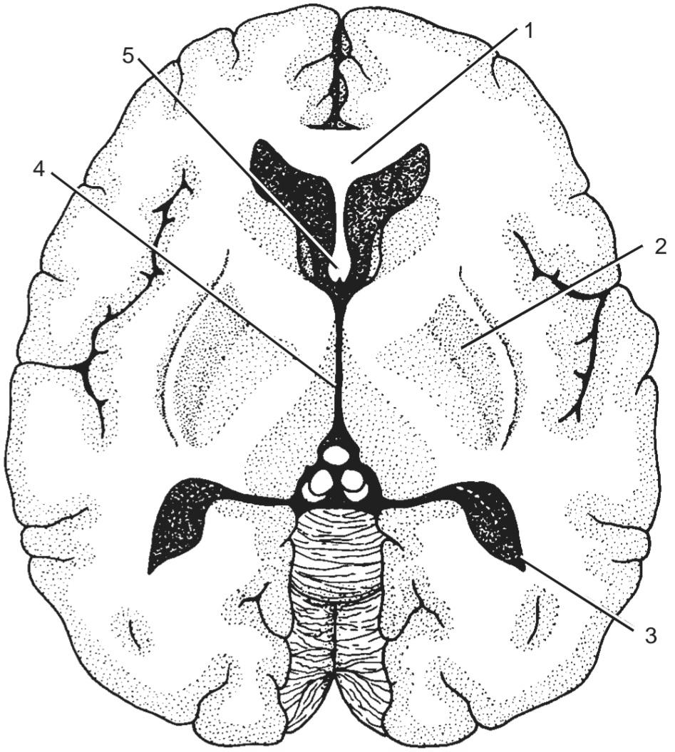
Figure 7-30 Horizontal section of the cerebrum, as seen from above.
شکل ۷-۳۰ برش افقی مخ، از نمای بالا.
21. Number 1 (a) Optic radiation
22. Number 2 (b) Lateral sulcus
23. Number 3 (c) Lentiform nucleus
24. Number 4 (d) Anterior horn of lateral ventricle
25. Number 5 (e) None of the above
۲۲. شماره ۲ (ب) شیار جانبی
۲۳. شماره ۳ (ج) هسته عدسی شکل
۲۴. شماره ۴ (د) شاخ قدامی بطن جانبی
۲۵. شماره ۵ (ه) هیچ یک از موارد فوق
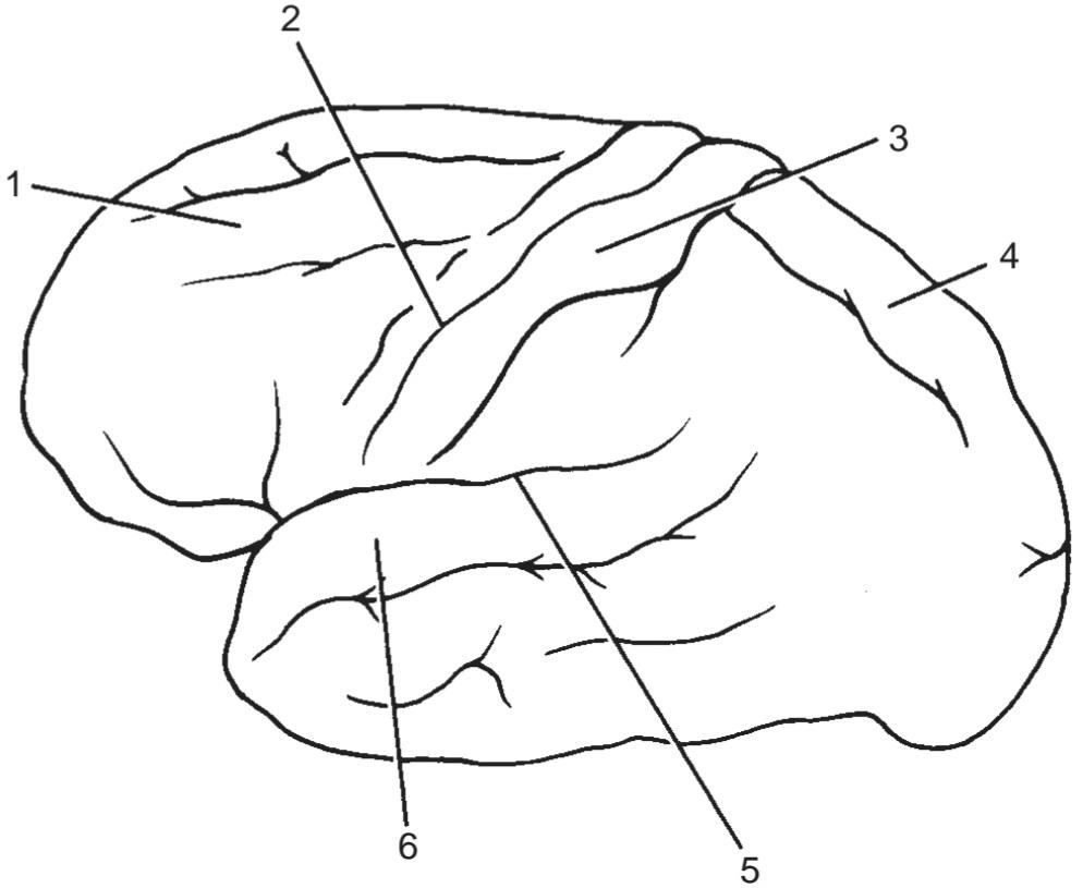
27. Number 2 (b) Postcentral gyrus
28. Number 3 (c) Superior temporal gyrus
29. Number 4 (d) Superior parietal lobule
30. Number 5 (e) None of the above
31. Number 6
۲۷. شماره ۲ (ب) شکنج پس مرکزی
۲۸. شماره ۳ (ج) شکنج گیجگاهی فوقانی
۲۹. شماره ۴ (د) لوبول جداری فوقانی
۳۰. شماره ۵ (ه) هیچکدام
۳۱. شماره ۶
(b) The hemorrhage into the thalamus extended laterally into the posterior limb of the left inter- nal capsule.
(c) The hemorrhage was small and confined to the thalamus on the left side.
(d) The hemorrhage was small and occurred in the lateral part of the left thalamus, producing tran- sient edema in the left internal capsule.
(e) The hemorrhage extended laterally into the left lateral ventricle.
۳۲. با استفاده از دانش خود در مورد روابط تالاموس چپ، عبارتی را انتخاب کنید که همی پارزی گذرای راست و بیحسی را توضیح میدهد.
(الف) خونریزی در بطن سوم رخ داده است.
(ب) خونریزی در تالاموس به صورت جانبی به اندام خلفی کپسول داخلی چپ گسترش یافته است.
(ج) خونریزی کوچک بوده و به تالاموس در سمت چپ محدود شده است.
(د) خونریزی کوچک بوده و در قسمت جانبی تالاموس چپ رخ داده و باعث ادم گذرا در کپسول داخلی چپ شده است.
(ه) خونریزی به صورت جانبی به بطن جانبی چپ گسترش یافته است.
33. This hypertensive patient had a small thalamic hemorrhage. Select the most likely cause for the hemorrhage
(a) One of the small diseased thalamic arteries may have ruptured.
(b) One of the small veins draining the thalamus may have ruptured.
(c) Vasoconstriction of the thalamic arteries could have occurred.
(d) Softening of the neuronal tissue around the thalamic arteries might have taken place.
(e) No relation exists between hypertension and the thalamic hemorrhage in this patient.
۳۳. این بیمار مبتلا به فشار خون بالا خونریزی کوچکی در تالاموس داشت. محتملترین علت خونریزی را انتخاب کنید.
(الف) ممکن است یکی از شریانهای کوچک تالاموس بیمار پاره شده باشد.
(ب) ممکن است یکی از رگهای کوچک تخلیه کننده تالاموس پاره شده باشد.
(ج) ممکن است انقباض عروق شریانهای تالاموس رخ داده باشد.
(د) ممکن است نرم شدن بافت عصبی اطراف شریانهای تالاموس رخ داده باشد.
(ه) هیچ ارتباطی بین فشار خون بالا و خونریزی تالاموس در این بیمار وجود ندارد.
An 8-year-old boy with a severe earache on the right side was taken to a pediatrician. The symptoms had started 7 days ago, and the pain had progressively worsened. On examination, the boy was found to have severe right-sided otitis media with acute mastoiditis. On being questioned, the boy admitted that his head hurt badly all over and that he felt sick. While he was being examined, he vomited. His body temperature was slightly elevated. In view of the severity of the headache and the presence of nausea and vomiting, the pediatrician decided to have an MRI performed. The result showed a small, well-defined, right cerebral abscess.
پسری ۸ ساله با درد شدید گوش در سمت راست، به متخصص اطفال برده شد. علائم از ۷ روز پیش شروع شده بود و درد به تدریج بدتر شده بود. در معاینه، مشخص شد که پسر مبتلا به اوتیت مدیای شدید سمت راست به همراه ماستوئیدیت حاد است. در بازجویی، پسر اعتراف کرد که تمام سرش به شدت درد میکند و احساس بیماری میکند. در حین معاینه، استفراغ کرد. دمای بدنش کمی بالا بود. با توجه به شدت سردرد و وجود حالت تهوع و استفراغ، متخصص اطفال تصمیم به انجام MRI گرفت. نتیجه، یک آبسه مغزی کوچک و مشخص در سمت راست مغز را نشان داد.
34. The cerebral abscess in this patient was most likely located at which site in the right cerebral hemisphere:
(a) Frontal lobe
(b) Thalamus
(c) Occipital lobe
(d) Temporal lobe
(e) Cuneus
۳۴. آبسه مغزی در این بیمار به احتمال زیاد در کدام قسمت از نیمکره راست مغز قرار داشته است:
(الف) لوب پیشانی
(ب) تالاموس
(ج) لوب پس سری
(د) لوب گیجگاهی
(ه) کونئوس
√ پاسخها و توضیحات برای مرور سوالات
۱. ب صحیح است. دیانسفالون از پهلو توسط کپسول داخلی محدود شده است (شکل ۷-۱ را ببینید). الف. دیانسفالون از جلو تا سوراخ بین بطنی امتداد دارد (شکل ۷-۳ را ببینید). ج. تالاموس روی دیواره جانبی بطن سوم قرار دارد (شکل ۷-۳ را ببینید). د. اپیتالاموس از هستههای هابنولار و اتصالات آنها و غده پینهآل تشکیل شده است. ه. دیانسفالون از عقب تا مجرای مغزی امتداد دارد (شکل ۷-۳ را ببینید).
۷. د صحیح است.
۸. الف صحیح است.
۹. هـ صحیح است. ساختار، سپتوم پلوسیدوم است.
۱۰. ب صحیح است.
۱۱. ج صحیح است.
۱۲. هـ صحیح است. ساختار، تالاموس است.
۲۱. E صحیح است. این ساختار، مفصل اصلی جسم پینهای است.
۲۲. C صحیح است.
۲۳. E صحیح است. این ساختار، شاخ خلفی بطن جانبی است.
۲۴. E صحیح است. این ساختار، بطن سوم است.
۲۵. E صحیح است. این ساختار، ستون قدامی فورنیکس است.
26. E is correct. The structure is the middle frontal gyrus.
۲۶. E صحیح است. این ساختار، شکنج پیشانی میانی است.
۲۷. A صحیح است.
۲۸. B صحیح است.
۲۹. D صحیح است.
۳۰. E صحیح است. این ساختار، شیار جانبی است.
۳۱. C صحیح است.
۳۲. D صحیح است.
۳۳. A صحیح است.
۳۴. D صحیح است.
«فصل هفتم کتاب نورواناتومی بالینی اسنل»
🚀 با ما همراه شوید!
تازهترین مطالب و آموزشهای مغز و اعصاب را از دست ندهید. با فالو کردن کانال تلگرام آیندهنگاران مغز، از ما حمایت کنید!