
یادداشتهای بالینی نوروآناتومی بالینی اسنل؛ مخ


دعای مطالعه [ نمایش ]
بِسْمِ الله الرَّحْمنِ الرَّحیمِ
اَللّهُمَّ اَخْرِجْنى مِنْ ظُلُماتِ الْوَهْمِ
خدايا مرا بيرون آور از تاريكىهاى وهم،
وَ اَكْرِمْنى بِنُورِ الْفَهْمِ
و به نور فهم گرامى ام بدار،
اَللّهُمَّ افْتَحْ عَلَيْنا اَبْوابَ رَحْمَتِكَ
خدايا درهاى رحمتت را به روى ما بگشا،
وَانْشُرْ عَلَيْنا خَزائِنَ عُلُومِكَ بِرَحْمَتِكَ يا اَرْحَمَ الرّاحِمينَ
و خزانههاى علومت را بر ما باز كن به امید رحمتت اى مهربانترين مهربانان.
آیندهنگاران مغز: «ما مغز را میشناسیم، تا آینده را بسازیم.»
کتاب «نوروآناتومی بالینی اسنل» اثر دکتر ریچارد اس. اسنل، با ویرایش جدید دکتر رایان اسپلیتگربر، همچنان بهعنوان مرجع طلایی این حوزه شناخته میشود. این اثر با تلفیق تازهترین پژوهشها و کاربردهای بالینی، پلی استوار میان دانش پایه و عمل پزشکی بنا میکند.
تلاشهای علمی و سالها تدریس ارزشمند استاد گرانقدر دکتر محمدتقی جغتایی الهامبخش ترجمه و گسترش این دانش در ایران بوده است.
ترجمه دقیق این کتاب توسط برند علمی آیندهنگاران مغز به مدیریت داریوش طاهری، امکان دسترسی فارسیزبانان به مرزهای نوین علوم اعصاب را فراهم کرده و رسالتی علمی در جهت شناخت عمیقتر مغز و ترسیم آیندهای روشنتر بر عهده گرفته است.
»» فصل ۷: مخ؛ بخش یادداشتهای بالینی
»» Chapter 7: Cerebrum
Clinical Notes
Thalamic Lesions
Lesions of the thalamus usually result from thrombosis or hemorrhage of one of the arteries that supply the thalamus. Because the thalamus is concerned with receiving sensory impulses from the opposite side of the body, the disability resulting from a lesion within it will be confined to the contralateral side of the body. There may be a major impairment of all forms of sensation, which could include light touch, tactile localization and discrimination, and loss of appreciation of joint movements.
یادداشتهای بالینی
ضایعات تالاموس
ضایعات تالاموس معمولاً ناشی از ترومبوز یا خونریزی یکی از شریانهایی است که تالاموس را تغذیه میکنند. از آنجا که تالاموس مسئول دریافت تکانههای حسی از طرف مقابل بدن است، ناتوانی ناشی از ضایعه در آن به سمت مقابل بدن محدود خواهد شد. ممکن است اختلال عمدهای در تمام اشکال حس وجود داشته باشد، که میتواند شامل لمس سبک، تشخیص و مکانیابی لمسی و از دست دادن درک حرکات مفاصل باشد.
Subthalamic Lesions
The subthalamus should be regarded as one of the extrapyramidal motor nuclei and has a large connection with the globus pallidus. Lesions of the subthalamus result in sudden, forceful involuntary movements in a contralateral extremity. The movements may be jerky (choreiform) or violent (ballistic).
ضایعات ساب تالاموس
ساب تالاموس را باید به عنوان یکی از هستههای حرکتی خارج هرمی در نظر گرفت و ارتباط زیادی با گلوبوس پالیدوس دارد. ضایعات ساب تالاموس منجر به حرکات ناگهانی، شدید و غیرارادی در اندام مقابل میشود. این حرکات ممکن است تند و تیز (کرهشکل) یا شدید (بالستیک) باشند.
Pineal Gland
The pineal gland consists essentially of pinealocytes and glial cells supported by a connective tissue framework. As the result of regressive changes that occur with age, calcareous concretions accumulate within the glial cells and connective tissue of the gland. These deposits are useful to the radiologist, since they serve as a landmark and assist in determining whether the pineal gland has been displaced laterally by a space-occupying lesion within the skull.
غده پینه آل
غده پینه آل اساساً از پینه آلوسیتها و سلولهای گلیال تشکیل شده است که توسط یک چارچوب بافت همبند پشتیبانی میشوند. در نتیجه تغییرات قهقرایی که با افزایش سن رخ میدهد، رسوبات آهکی در سلولهای گلیال و بافت همبند غده تجمع مییابند. این رسوبات برای رادیولوژیست مفید هستند، زیرا به عنوان یک نقطه عطف عمل میکنند و به تعیین اینکه آیا غده پینه آل توسط یک ضایعه فضاگیر در جمجمه به صورت جانبی جابجا شده است یا خیر، کمک میکنند.
The functions of the pineal gland are mainly inhibitory and have been shown to influence the pituitary gland, the islets of Langerhans, the parathyroids, the adrenals, and the gonads.
عملکرد غده پینهآل عمدتاً مهاری است و نشان داده شده است که بر غده هیپوفیز، جزایر لانگرهانس، پاراتیروئیدها، غدد فوق کلیوی و غدد جنسی تأثیر میگذارد.
Clinical observation of patients with pineal tumors or tumors of neighboring areas of nervous tissue that may press on the pineal gland has shown severe alteration of reproductive function.
مشاهدات بالینی بیماران مبتلا به تومورهای پینهآل یا تومورهای نواحی مجاور بافت عصبی که ممکن است به غده پینهآل فشار وارد کنند، تغییر شدید عملکرد تولید مثلی را نشان داده است.
Hypothalamus
The hypothalamus is an area of the nervous system that is of great functional importance. Not only does it control emotional states, but it also assists in the regulation of fat, carbohydrate, and water metabolism. Among its many other activities, it influences body temperature, genital functions, sleep, and food intake. The pituitary and the hypothalamus constitute a closely integrated unit, and the hypothalamus plays a role in the release of pituitary hormones.
هیپوتالاموس
هیپوتالاموس ناحیهای از سیستم عصبی است که از اهمیت عملکردی بالایی برخوردار است. نه تنها حالات عاطفی را کنترل میکند، بلکه به تنظیم متابولیسم چربی، کربوهیدرات و آب نیز کمک میکند. در میان بسیاری از فعالیتهای دیگرش، بر دمای بدن، عملکردهای تناسلی، خواب و مصرف غذا تأثیر میگذارد. هیپوفیز و هیپوتالاموس یک واحد کاملاً یکپارچه را تشکیل میدهند و هیپوتالاموس در آزادسازی هورمونهای هیپوفیز نقش دارد.
Hypothalamic Syndromes
Lesions of the hypothalamus may result from infection, trauma, or vascular disorders. Tumors, such as a craniopharyngioma or chromophobe adenoma of the pituitary and pineal tumors, may interfere with the function of the hypothalamus. The most common abnormalities include genital hypoplasia or atrophy, diabetes insipidus, obesity, disturbances of sleep, irregular pyrexia, and emaciation. Some of these disorders may occur together, such as in the adiposogenital dystrophy syndrome.
سندرمهای هیپوتالاموس
ضایعات هیپوتالاموس ممکن است در اثر عفونت، تروما یا اختلالات عروقی ایجاد شوند. تومورها، مانند کرانیوفارنژیوما یا آدنوم کروموفوب تومورهای هیپوفیز و پینهآل، ممکن است در عملکرد هیپوتالاموس اختلال ایجاد کنند. شایعترین ناهنجاریها شامل هیپوپلازی یا آتروفی دستگاه تناسلی، دیابت بیمزه، چاقی، اختلالات خواب، تب نامنظم و لاغری مفرط است. برخی از این اختلالات ممکن است با هم رخ دهند، مانند سندرم دیستروفی چربی-تناسلی.
Cerebral Cortex, Sulci, and Cerebral Hemisphere Lobes
The cerebral cortex is composed of gray matter. Only about a third lies on the exposed convexity of the gyri; the remaining two thirds form the walls of the sulci. Moreover, different areas of the cortex have different functions, and the anatomical division of the cortex into lobes and gyri by sulci enables the physician to localize loss of function or accurately place a brain lesion. For example, focal lesions of the precentral gyrus will produce contralateral hemiparesis, while lesions of the postcentral gyrus will result in contralateral hemisensory loss. More widespread lesions of the frontal lobe might cause symptoms and signs indicative of loss of attention span or change in social behavior. Widespread degeneration of the cerebral cortex gives rise to symptoms of dementia.
قشر مغز، شیارها و لوبهای نیمکره مغزی
قشر مغز از ماده خاکستری تشکیل شده است. تنها حدود یک سوم آن روی تحدب آشکار چینهای مغزی قرار دارد؛ دو سوم باقیمانده دیوارههای چینهای مغزی را تشکیل میدهند. علاوه بر این، نواحی مختلف قشر مغز عملکردهای متفاوتی دارند و تقسیم آناتومیکی قشر مغز به لوبها و چینهای مغزی توسط شیارها، پزشک را قادر میسازد تا از دست دادن عملکرد را محلیسازی کند یا ضایعه مغزی را به طور دقیق در محل مورد نظر قرار دهد. به عنوان مثال، ضایعات کانونی چینهای مغزی پیشمرکزی باعث فلج نیمهجانبی طرف مقابل میشود، در حالی که ضایعات چینهای مغزی پسمرکزی منجر به از دست دادن حس نیمهجانبی طرف مقابل میشود. ضایعات گستردهتر لوب پیشانی ممکن است علائم و نشانههایی را ایجاد کند که نشاندهنده از دست دادن دامنه توجه یا تغییر در رفتار اجتماعی است. انحطاط گسترده قشر مغز باعث ایجاد علائم زوال عقل میشود.
Lateral Ventricles
Each lateral ventricle contains about 7 to 10 mL of cerebrospinal fluid (CSF). This fluid is produced in the choroid plexus of the lateral ventricle and normally drains into the third ventricle through the interventricular foramen (foramen of Monro). Blockage of the foramen by a cerebral tumor would result in distention of the ventricle, thus producing a type of hydrocephalus.
بطنهای جانبی
هر بطن جانبی حاوی حدود ۷ تا ۱۰ میلیلیتر مایع مغزی نخاعی (CSF) است. این مایع در شبکه کوروئید بطن جانبی تولید میشود و معمولاً از طریق سوراخ بین بطنی (سوراخ مونرو) به بطن سوم تخلیه میشود. انسداد سوراخ توسط تومور مغزی منجر به اتساع بطن و در نتیجه ایجاد نوعی هیدروسفالی میشود.
The choroid plexus of the lateral ventricle is continuous with that of the third ventricle through the interventricular foramen. The choroid plexus is largest where the body and posterior and inferior horns join, where it may become calcified with age. Do not confuse this calcification of the choroid plexus, as seen on radiographs, with that of the pineal gland.
شبکه کوروئید بطن جانبی از طریق سوراخ بین بطنی با شبکه کوروئید بطن سوم در یک امتداد است. شبکه کوروئید در محل اتصال بدنه و شاخهای خلفی و تحتانی بزرگترین است، جایی که ممکن است با افزایش سن کلسیفیه شود. این کلسیفیه شدن شبکه کوروئید، همانطور که در رادیوگرافیها دیده میشود، را با کلسیفیه شدن غده پینهآل اشتباه نگیرید.
In the past, the size and shape of the lateral ventricle were investigated clinically by pneumoencephalography (Figs. 7-20 to 7-23). In this procedure, small amounts of air were introduced into the subarachnoid space by lumbar puncture with the patient in the sitting position. If the patient already had a raised intracranial pressure, this method was dangerous, and air or radiopaque fluid was injected directly into the lateral ventricles through a burr hole in the skull (this procedure was referred to as ventriculography). This procedure has now been replaced by CT and magnetic resonance imaging (MRI) (Figs. 7-24 to 7-27).
در گذشته، اندازه و شکل بطن جانبی به صورت بالینی توسط پنوموانسفالوگرافی بررسی میشد (شکلهای 7-20 تا 7-23). در این روش، مقادیر کمی هوا از طریق سوراخ کمری در بیمار در حالت نشسته وارد فضای زیر عنکبوتیه میشد. اگر بیمار از قبل فشار داخل جمجمهای بالایی داشت، این روش خطرناک بود و هوا یا مایع رادیواپک مستقیماً از طریق سوراخی در جمجمه به بطنهای جانبی تزریق میشد (این روش به عنوان ونتریکولوگرافی شناخته میشد). این روش اکنون با سیتیاسکن و تصویربرداری رزونانس مغناطیسی (MRI) جایگزین شده است (شکلهای 7-24 تا 7-27).
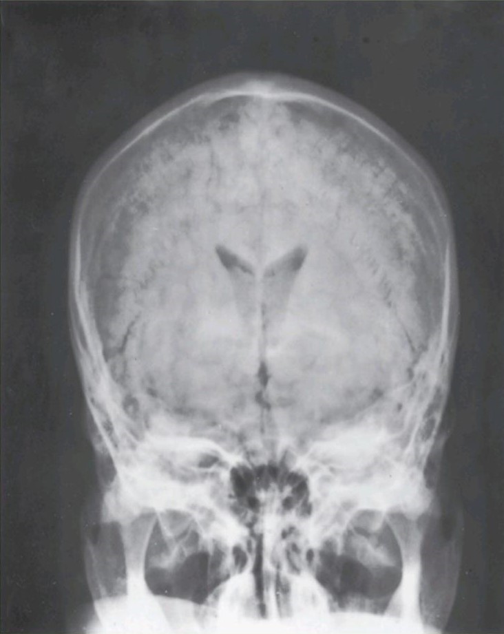
Figure 7-20 Anteroposterior pneumoencephalogram of a 28-year-old man.
شکل ۷-۲۰ پنوموانسفالوگرام قدامی-خلفی یک مرد 28 ساله.

Figure 7-21 Explanation of the radiograph seen in Figure 7-20. Note the position of the x-ray gun relative to the head and the film cassette.
شکل ۷-۲۱ توضیح رادیوگرافی که در شکل 7-20 دیده میشود. به موقعیت تفنگ اشعه ایکس نسبت به سر و کاست فیلم توجه کنید.

Figure 7-22 Lateral pneumoencephalogram ol a 28-yearold man.
شکل ۷-۲۲ پنوموانسفالوگرام جانبی یک مرد 28 ساله.

Figure 7-23 Explanation of the radiograph seen in Figure 7-22. Note the position of the x-ray gun relative to the head and the film cassette.
شکل ۷-۲۳ توضیح رادیوگرافی که در شکل 7-22 دیده میشود. به موقعیت تفنگ اشعه ایکس نسبت به سر و کاست فیلم توجه کنید.
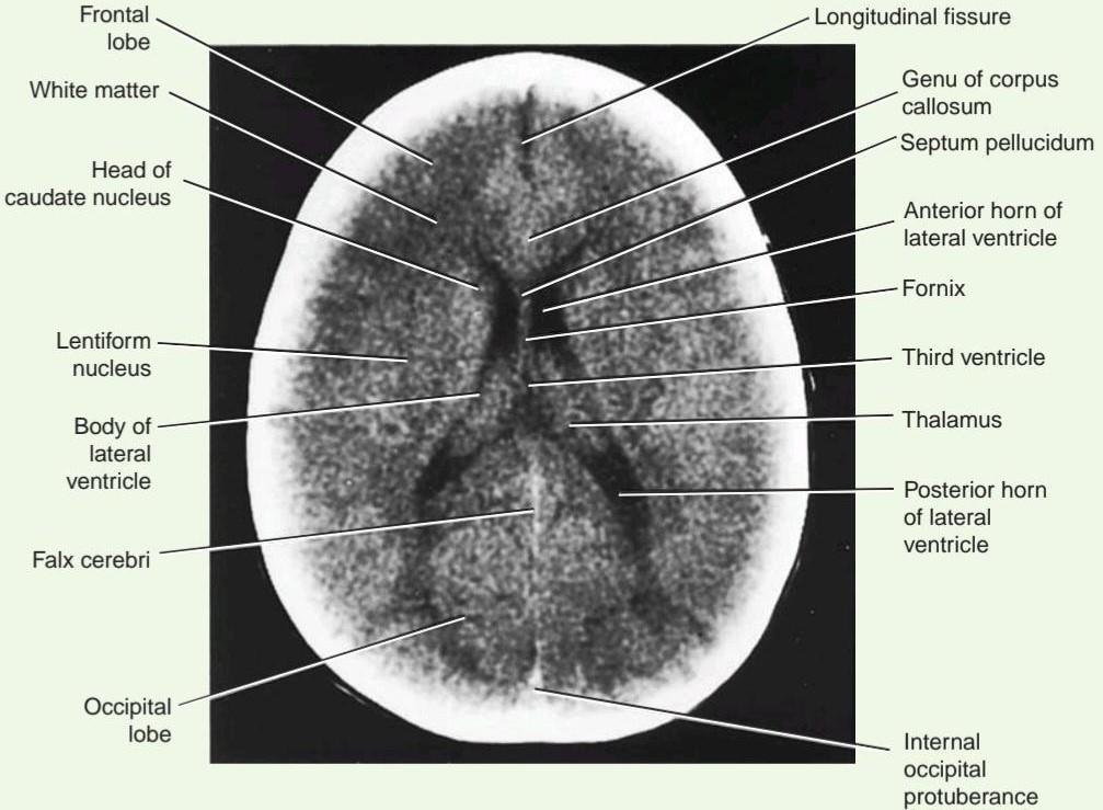
Figure 7-24 Horizontal (axial) CT scan of the brain.
شکل ۷-۲۴ سی تی اسکن افقی (محوری) مغز.
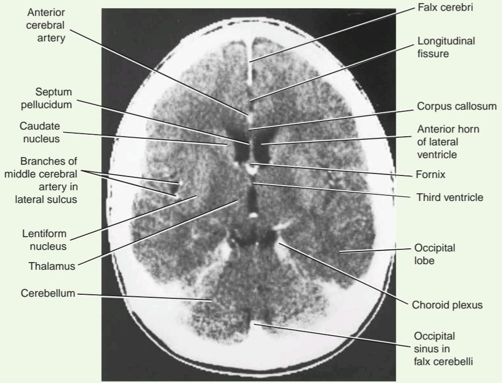
Figure 7-25 Horizontal (axial) CT scan of the brain (contrast enhanced).
شکل ۷-۲۵ سی تی اسکن افقی (محوری) مغز (با کنتراست تقویت شده).
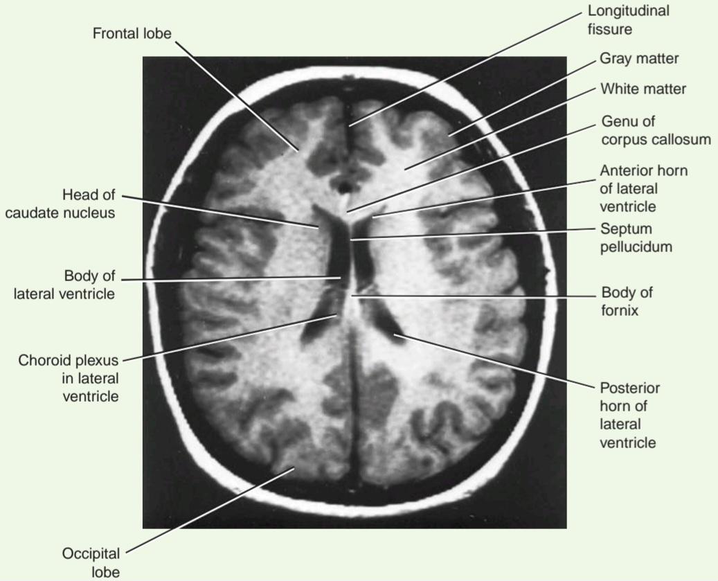
Figure 7-26 Horizontal (axial) MRI of the brain.
شکل ۷-۲۶ MRI افقی (محوری) مغز.
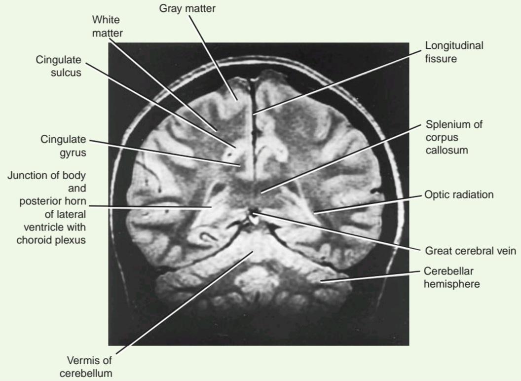
Figure 7-27 Coronal MRI of the brain.
شکل ۷-۲۷ MRI کرونال مغز.
Basal Nuclei
The basal nuclei, in this discussion, refers to the masses of gray matter that are deeply placed within the cerebrum. They include the caudate nucleus, the lentiform nucleus, the amygdaloid nucleus, and the claustrum.
هستههای قاعدهای
هستههای قاعدهای، در این بحث، به تودههایی از ماده خاکستری اشاره دارند که در اعماق مخ قرار دارند. این هستهها شامل هسته دمدار، هسته عدسی شکل، هسته آمیگدالوئید و کلاستروم هستند.
Because of the close relationship that exists between these nuclei and the internal capsule, tumors of the caudate or lentiform nuclei may cause severe motor or sensory symptoms on the opposite side of the body. Tumors pressing on the anterior two thirds of the posterior limb of the internal capsule will cause progressive spastic hemiplegia, while more posteriorly situated tumors will produce impairment of sensation on the opposite side.
به دلیل ارتباط نزدیکی که بین این هستهها و کپسول داخلی وجود دارد، تومورهای هسته دمدار یا عدسی شکل ممکن است باعث علائم شدید حرکتی یا حسی در طرف مقابل بدن شوند. تومورهایی که به دو سوم قدامی اندام خلفی کپسول داخلی فشار میآورند، باعث همی پلژی اسپاستیک پیشرونده میشوند، در حالی که تومورهایی که در قسمت خلفیتر قرار دارند، باعث اختلال در حس در طرف مقابل میشوند.
Disorders of function of the basal nuclei are considered after the connections of these nuclei are discussed in Chapter 10.
اختلالات عملکرد هستههای قاعدهای پس از بحث در مورد اتصالات این هستهها در فصل 10 بررسی میشوند.
Cerebral Commissures
The major commissure is the large corpus callosum. Most fibers within the corpus callosum interconnect symmet- rical areas of the cerebral cortex. Because it transfers information from one hemisphere to another, the corpus callosum is essential for learned discrimination, sensory experience, and memory.
رابطهای مغزی
رابطه اصلی، جسم پینهای بزرگ است. بیشتر فیبرهای درون جسم پینهای، نواحی متقارن قشر مغز را به هم متصل میکنند. از آنجا که جسم پینهای اطلاعات را از یک نیمکره به نیمکره دیگر منتقل میکند، برای تمایز آموختهشده، تجربه حسی و حافظه ضروری است.
Occasionally, the corpus callosum fails to develop, and in these individuals, no definite signs or symptoms appear. Should the corpus callosum be destroyed by disease in later life, however, each hemisphere becomes isolated, and the patient responds as if he or she has two separate brains. The patient’s general intelligence and behavior appear normal, since over the years both hemispheres have been trained to respond to different situations. If a pencil is placed in the patient’s right hand (with the eyes closed), he or she will recognize the object by touch and be able to describe it. If the pencil is placed in the left hand, the tactile information will pass to the right postcentral gyrus. This information will not be able to travel through the corpus callosum to the speech area in the left hemisphere; therefore, the patient will be unable to describe the object in his or her left hand.
گاهی اوقات، جسم پینهای رشد نمیکند و در این افراد، هیچ علامت یا نشانهای مشخص ظاهر نمیشود. با این حال، اگر جسم پینهای در اثر بیماری در مراحل بعدی زندگی از بین برود، هر نیمکره جدا میشود و بیمار طوری پاسخ میدهد که گویی دو مغز جداگانه دارد. هوش و رفتار کلی بیمار طبیعی به نظر میرسد، زیرا در طول سالها هر دو نیمکره برای پاسخ به موقعیتهای مختلف آموزش دیدهاند. اگر مدادی در دست راست بیمار (با چشمان بسته) قرار گیرد، او شیء را با لمس تشخیص میدهد و میتواند آن را توصیف کند. اگر مداد در دست چپ قرار گیرد، اطلاعات لمسی به شکنج پس مرکزی راست منتقل میشود. این اطلاعات قادر به عبور از جسم پینهای به ناحیه گفتار در نیمکره چپ نخواهند بود. بنابراین، بیمار قادر به توصیف شیء در دست چپ خود نخواهد بود.
Section of the corpus callosum has been attempted surgically, with some success, in order to prevent the spread of seizures from one hemisphere to the other.
برای جلوگیری از گسترش تشنج از یک نیمکره به نیمکره دیگر، تلاش شده است که بخشی از جسم پینهای را با جراحی برش دهند که با موفقیت نسبی همراه بوده است.
Internal Capsule Lesions
The internal capsule is an important compact band of white matter. It is composed of ascending and descending nerve fibers that connect the cerebral cortex to the brainstem and spinal cord. The internal capsule is flanked medially by the caudate nucleus and thalamus and laterally by the lentiform nucleus. The arrangement of the nerve fibers within the internal capsule is shown in Figure 7-18.
ضایعات کپسول داخلی
کپسول داخلی یک نوار فشرده مهم از ماده سفید است. این نوار از فیبرهای عصبی صعودی و نزولی تشکیل شده است که قشر مغز را به ساقه مغز و نخاع متصل میکند. کپسول داخلی از طرف داخل توسط هسته دمی و تالاموس و از طرف خارج توسط هسته عدسی شکل احاطه شده است. ترتیب قرارگیری فیبرهای عصبی درون کپسول داخلی در شکل 7-18 نشان داده شده است.
The internal capsule is frequently involved in vascular disorders of the brain. The most common cause of arterial hemorrhage is atheromatous degeneration in an artery in a patient with high blood pressure. Because of the high concentration of important nerve fibers within the internal capsule, even a small hemorrhage can cause widespread effects on the contralateral side of the body. Not only is the immediate neural tissue destroyed by the blood, which later clots, but also neighboring nerve fibers may be compressed or be edematous.
کپسول داخلی اغلب در اختلالات عروقی مغز دخیل است. شایعترین علت خونریزی شریانی، دژنراسیون آتروماتوز در یک شریان در بیمار مبتلا به فشار خون بالا است. به دلیل غلظت بالای فیبرهای عصبی مهم در کپسول داخلی، حتی یک خونریزی کوچک میتواند اثرات گستردهای در سمت مقابل بدن ایجاد کند. نه تنها بافت عصبی فوری توسط خون که بعداً لخته میشود، تخریب میشود، بلکه فیبرهای عصبی مجاور نیز ممکن است فشرده یا ادماتوز شوند.
Alzheimer Disease
Alzheimer disease is a degenerative disease of the brain occurring in middle to late life, but an early form of the disease is now well recognized. The disease affects more than 4 million people in the United States, resulting in over 100,000 deaths per year. The risk of the disease rises sharply with advancing years.
بیماری آلزایمر
بیماری آلزایمر یک بیماری تخریب کننده مغز است که در میانسالی تا اواخر عمر رخ میدهد، اما اکنون نوع اولیه این بیماری به خوبی شناخته شده است. این بیماری بیش از ۴ میلیون نفر را در ایالات متحده تحت تأثیر قرار میدهد و منجر به بیش از ۱۰۰۰۰۰ مرگ در سال میشود. خطر ابتلا به این بیماری با افزایش سن به شدت افزایش مییابد.
The cause of Alzheimer disease is unknown, but evidence suggests a genetic predisposition. Several abnormal genes have been found, each of which leads to a similar clinical and pathologic syndrome, with only variations in the age of onset and the rate of progression to suggest that the pathogenetic mechanisms differ. Some cases of familial Alzheimer disease, for example, have been shown to have mutations in several genes (App, presenilin 1, and presenilin 2).
علت بیماری آلزایمر ناشناخته است، اما شواهد نشان میدهد که یک استعداد ژنتیکی وجود دارد. چندین ژن غیرطبیعی یافت شده است که هر کدام منجر به یک سندرم بالینی و پاتولوژیک مشابه میشوند و تنها تفاوت در سن شروع و سرعت پیشرفت نشان میدهد که مکانیسمهای بیماریزا متفاوت هستند. به عنوان مثال، نشان داده شده است که برخی از موارد بیماری آلزایمر خانوادگی، جهشهایی در چندین ژن (App، presenilin 1 و presenilin 2) دارند.
Early memory loss, a disintegration of personality, complete disorientation, deterioration in speech, and restlessness are common signs. In the late stages, the patient may become mute, incontinent, and bedridden and usually dies of some other disease.
از دست دادن زودهنگام حافظه، فروپاشی شخصیت، گمگشتگی کامل، زوال گفتار و بیقراری از علائم رایج هستند. در مراحل پایانی، بیمار ممکن است لال، بیاختیار و بستری شود و معمولاً بر اثر بیماری دیگری میمیرد.
Microscopically, changes eventually occur throughout the cerebral cortex, but to begin with, certain regions of the brain are selectively involved. The early sites include the hippocampus, the entorhinal cortex, and the associated areas of the cerebral cortex. Many so-called senile plaques are found in the atrophic cortex. The plaques result from the accumulation of several proteins around deposits of beta amyloid. In the center of each plaque is an extracellular collection of degenerating nervous tissue; surrounding the core is a rim of large abnormal neuronal processes, probably presynaptic terminals, filled with an excess of intracellular neurofibrils that are tangled and twisted, forming neurofibrillary tangles. The neurofibrillary tangles are aggregations of the microtubular protein tau, which is hyperphosphorylated. There is a marked loss of choline acetyltransferase, the biosynthetic enzyme for acetylcholine, in the areas of the cortex in which the senile plaques occur. This is thought to be due to loss of the ascending projection fibers rather than a loss of cortical cells. As these cellular changes occur, the affected neurons die.
از نظر میکروسکوپی، در نهایت تغییراتی در سراسر قشر مغز رخ میدهد، اما در ابتدا، مناطق خاصی از مغز به صورت انتخابی درگیر میشوند. مکانهای اولیه شامل هیپوکامپ، قشر انتورینال و نواحی مرتبط با قشر مغز هستند. بسیاری از پلاکهای به اصطلاح پیری در قشر آتروفیک یافت میشوند. این پلاکها ناشی از تجمع چندین پروتئین در اطراف رسوبات بتا آمیلوئید هستند. در مرکز هر پلاک، مجموعهای خارج سلولی از بافت عصبی در حال تخریب وجود دارد. اطراف هسته، حاشیهای از زوائد عصبی غیرطبیعی بزرگ، احتمالاً پایانههای پیشسیناپسی، پر از نوروفیبریلهای درون سلولی اضافی است که درهمتنیده و پیچخورده هستند و گرههای نوروفیبریلاری را تشکیل میدهند. گرههای نوروفیبریلاری، تجمعاتی از پروتئین میکروتوبولی تائو هستند که بیش از حد فسفریله شدهاند. در مناطقی از قشر مغز که پلاکهای پیری در آنها ایجاد میشوند، کاهش قابل توجهی در کولین استیل ترانسفراز، آنزیم بیوسنتز کننده استیل کولین، وجود دارد. تصور میشود که این امر به دلیل از بین رفتن فیبرهای برآمدگی صعودی باشد تا از بین رفتن سلولهای قشر مغز. با وقوع این تغییرات سلولی، نورونهای آسیب دیده میمیرند.
At this time, there is no clinical test for making the definite diagnosis of Alzheimer disease. Reliance is placed on taking a careful history and carrying out numerous neurologic and psychiatric examinations spaced out over time. In this way, other causes of dementia can be excluded. Alterations in the levels of amyloid peptides or tau in the serum or CSF may be helpful. CT scans or MRIS are also used, and abnormalities in the medial part of the temporal lobe occur in this disease. In advanced cases, a thin, atrophied cerebral cortex and dilated lateral ventricles may be found. The recent use of positron emission tomography (PET) shows evidence of diminished cortical metabolism (Fig. 7-28).
در حال حاضر، هیچ آزمایش بالینی برای تشخیص قطعی بیماری آلزایمر وجود ندارد. تکیه بر گرفتن شرح حال دقیق و انجام معاینات عصبی و روانپزشکی متعدد با فاصله زمانی است. به این ترتیب، میتوان سایر علل زوال عقل را رد کرد. تغییرات در سطح پپتیدهای آمیلوئید یا تائو در سرم یا مایع مغزی نخاعی (CSF) ممکن است مفید باشد. سی تی اسکن یا MRI نیز استفاده میشود و ناهنجاریهایی در قسمت داخلی لوب گیجگاهی در این بیماری رخ میدهد. در موارد پیشرفته، ممکن است قشر مغز نازک و آتروفی شده و بطنهای جانبی گشاد شده یافت شود. استفاده اخیر از توموگرافی انتشار پوزیترون (PET) شواهدی از کاهش متابولیسم قشر مغز را نشان میدهد (شکل 7-28).
The use of cholinesterase inhibitors for the treatment of Alzheimer disease has been found to be helpful. These drugs probably act by increasing the presence of acetylcholine at the sites of the disease where there is a deficiency of this neurotransmitter.
استفاده از مهارکنندههای کولین استراز برای درمان بیماری آلزایمر مفید تشخیص داده شده است. این داروها احتمالاً با افزایش حضور استیل کولین در محلهای بیماری که کمبود این انتقالدهنده عصبی وجود دارد، عمل میکنند.

Figure 7-28 Axial (horizontal) PET scan of a male patient with Alzheimer disease, showing defects (arrowheads) in metabolism in the bitemporoparietal regions of the cerebral cortex, following the injection of 18-fluorodeoxyglucose. The yellow areas indicate regions of high metabolic activity. (Courtesy Dr. Holley Dey.)
شکل ۷-۲۸ اسکن PET محوری (افقی) از یک بیمار مرد مبتلا به آلزایمر، که نقص (نوک پیکانها) در متابولیسم در نواحی دو گیجگاهی-جداری قشر مغز، پس از تزریق 18-فلوئورودئوکسی گلوکز را نشان میدهد. نواحی زرد رنگ نشاندهنده مناطقی با فعالیت متابولیکی بالا هستند. (با احترام از دکتر هالی دی.)
Key Concepts
Cerebral Subdivisions
• The cerebrum is subdivided into the diencephalon (central core) and the telencephalon (cerebral hemispheres).
مفاهیم کلیدی
زیربخشهای مغزی
• مخ به دیانسفالون (هسته مرکزی) و تلهانسفالون (نیمکرههای مغزی) تقسیم میشود.
Diencephalon
• The diencephalon consists of the third ventricle and the structures that form its boundaries, including the thalamus, subthalamus, epithalamus, and hypothalamus.
دیانسفالون
• دیانسفالون از بطن سوم و ساختارهایی که مرزهای آن را تشکیل میدهند، از جمله تالاموس، ساب تالاموس، اپی تالاموس و هیپوتالاموس تشکیل شده است.
Thalamus
• The thalamus is a very important cell station that receives the main sensory tracts. Sensory information is integrated and relayed to the cerebral cortex and other subcortical regions.
تالاموس
• تالاموس یک ایستگاه سلولی بسیار مهم است که مسیرهای حسی اصلی را دریافت میکند. اطلاعات حسی ادغام شده و به قشر مغز و سایر نواحی زیر قشری منتقل میشوند.
Subthalamus
• The subthalamus consists of nerve cells associated with the red nuclei and substantia nigra and is involved in control of muscle activity.
ساب تالاموس
• ساب تالاموس از سلولهای عصبی مرتبط با هستههای قرمز و جسم سیاه تشکیل شده و در کنترل فعالیت عضلات نقش دارد.
Epithalamus
• The epithalamus consists of the habenular nuclei (integration of visceral and somatic pathways) and the pineal gland (endocrine gland and melatonin).
اپیتالاموس
• اپیتالاموس از هستههای هابنولار (ادغام مسیرهای احشایی و سوماتیک) و غده پینهآل (غده درونریز و ملاتونین) تشکیل شده است.
Hypothalamus
• The hypothalamus controls and integrates the functions of the autonomic nervous system and endocrine systems and plays a vital role in maintaining body homeostasis.
هیپوتالاموس
• هیپوتالاموس عملکردهای سیستم عصبی خودکار و سیستمهای غدد درونریز را کنترل و یکپارچه میکند و نقش حیاتی در حفظ هموستاز بدن ایفا میکند.
• Physiologically, hardly any activity in the body is not influenced by the hypothalamus.
• از نظر فیزیولوژیکی، تقریباً هیچ فعالیتی در بدن تحت تأثیر هیپوتالاموس نیست.
Main Sulci
• The central sulcus separates the frontal and parietal lobes. The gyrus anterior to it contains motor cells that initiate the movements of the contralateral side of the body; the gyrus posterior to it contains general sensory cortex that receives sensory information from the contralateral side of the body.
شیار اصلی
• شیار مرکزی، لوبهای پیشانی و آهیانه را از هم جدا میکند. شکنج قدامی آن حاوی سلولهای حرکتی است که حرکات سمت مقابل بدن را آغاز میکنند؛ شکنج خلفی آن حاوی قشر حسی عمومی است که اطلاعات حسی را از سمت مقابل بدن دریافت میکند.
• The lateral sulcus is a deep cleft on the inferior lateral cerebral hemisphere, between frontal and temporal lobes. The insula lies deep to the lateral sulcus.
• شیار جانبی یک شکاف عمیق در نیمکره مغزی جانبی تحتانی، بین لوبهای پیشانی و گیجگاهی است. اینسولا در عمق شیار جانبی قرار دارد.
• The parieto-occipital sulcus is located on the medial side of the hemisphere, running inferiorly to intersect the calcarine sulcus.
• شیار جداری-پسسری در سمت داخلی نیمکره قرار دارد و به سمت پایین امتداد مییابد تا شیار کالکارین را قطع کند.
• The calcarine sulcus is found on the medial surface of the hemispheres, in the occipital lobe. Primary visual cortex is located here.
• شیار کالکارین در سطح داخلی نیمکرهها، در لوب پسسری یافت میشود. قشر بینایی اولیه در اینجا قرار دارد.
Cerebral Hemisphere Lobes
• The frontal lobe occupies the area anterior to the central sulcus and is divided by three sulci into three gyri.
لوبهای نیمکره مغزی
• لوب پیشانی ناحیهای را در جلوی شیار مرکزی اشغال میکند و توسط سه شیار به سه شکنج تقسیم میشود.
• The temporal lobe occupies the area inferior to the lateral sulcus and is divided into three gyri by two sulci.
• لوب گیجگاهی ناحیهای را در پایین شیار جانبی اشغال میکند و توسط دو شیار به سه شکنج تقسیم میشود.
• The occipital lobe occupies the area behind the parieto-occipital sulcus.
• لوب پسسری ناحیهای را در پشت شیار جداری-پسسری اشغال میکند.
Internal Structures
• The two lateral ventricles are located one in each hemisphere. The lateral ventricles are large cavities that house CSF. Each are in communication with the third ventricle through the interventricular foramen.
ساختارهای داخلی
• دو بطن جانبی در هر نیمکره قرار دارند. بطنهای جانبی حفرههای بزرگی هستند که مایع مغزی نخاعی (CSF) را در خود جای دادهاند. هر کدام از طریق سوراخ بین بطنی با بطن سوم در ارتباط هستند.
• Basal nuclei are a collection of gray matter masses including the corpus striatum, amygdaloid nucleus, and the claustrum.
• هستههای قاعدهای مجموعهای از تودههای ماده خاکستری شامل جسم مخطط، هسته آمیگدالوئید و کلاستروم هستند.
• The corpus striatum is composed of the caudate nucleus and the lentiform nucleus, both separated by the internal capsule. These structures are involved in muscular movement control through communications with the cortex.
• جسم مخطط از هسته دمدار و هسته عدسی شکل تشکیل شده است که هر دو توسط کپسول داخلی از هم جدا شدهاند. این ساختارها از طریق ارتباط با قشر مغز در کنترل حرکت عضلانی نقش دارند.
• The claustrum is separated from the lentiform nucleus by the external capsule. The function of the claustrum is unknown.
• کلاستروم توسط کپسول خارجی از هسته عدسی شکل جدا میشود. عملکرد کلاستروم ناشناخته است.
• The amygdaloid nucleus is located in the temporal lobe adjacent to the anterior horn of the lateral ventricle.
• هسته آمیگدالوئید در لوب گیجگاهی مجاور شاخ قدامی بطن جانبی قرار دارد.
• Commissure fibers in the cerebrum are myelinated fibers connecting two corresponding regions of the two hemispheres.
• الیاف رابط در مخ، الیاف میلینداری هستند که دو ناحیه متناظر از دو نیمکره را به هم متصل میکنند.
• The largest commissure is the corpus callosum, which connects the two hemispheres. Other commissures include the fornix and the anterior and posterior commissures.
• بزرگترین رابط، جسم پینهای است که دو نیمکره را به هم متصل میکند. سایر رابطها شامل فورنیکس و رابطهای قدامی و خلفی هستند.
«فصل هفتم کتاب نورواناتومی بالینی اسنل»
🚀 با ما همراه شوید!
تازهترین مطالب و آموزشهای مغز و اعصاب را از دست ندهید. با فالو کردن کانال تلگرام آیندهنگاران مغز، از ما حمایت کنید!



