
ساختار و مناطق عملکردی قشر مغز؛ فصل هشتم نوروآناتومی بالینی اسنل


دعای مطالعه [ نمایش ]
بِسْمِ الله الرَّحْمنِ الرَّحیمِ
اَللّهُمَّ اَخْرِجْنى مِنْ ظُلُماتِ الْوَهْمِ
خدايا مرا بيرون آور از تاريكىهاى وهم،
وَ اَكْرِمْنى بِنُورِ الْفَهْمِ
و به نور فهم گرامى ام بدار،
اَللّهُمَّ افْتَحْ عَلَيْنا اَبْوابَ رَحْمَتِكَ
خدايا درهاى رحمتت را به روى ما بگشا،
وَانْشُرْ عَلَيْنا خَزائِنَ عُلُومِكَ بِرَحْمَتِكَ يا اَرْحَمَ الرّاحِمينَ
و خزانههاى علومت را بر ما باز كن به امید رحمتت اى مهربانترين مهربانان.
آیندهنگاران مغز: «ما مغز را میشناسیم، تا آینده را بسازیم.»
کتاب «نوروآناتومی بالینی اسنل» اثر دکتر ریچارد اس. اسنل، با ویرایش جدید دکتر رایان اسپلیتگربر، همچنان بهعنوان مرجع طلایی این حوزه شناخته میشود. این اثر با تلفیق تازهترین پژوهشها و کاربردهای بالینی، پلی استوار میان دانش پایه و عمل پزشکی بنا میکند.
تلاشهای علمی و سالها تدریس ارزشمند استاد گرانقدر آقای دکتر محمدتقی جغتایی الهامبخش ترجمه و گسترش این دانش در ایران بوده است.
ترجمه دقیق این کتاب توسط برند علمی آیندهنگاران مغز به مدیریت داریوش طاهری، امکان دسترسی فارسیزبانان به مرزهای نوین علوم اعصاب را فراهم کرده و رسالتی علمی در جهت شناخت عمیقتر مغز و ترسیم آیندهای روشنتر بر عهده گرفته است.
»» فصل ۸: ساختار و مناطق عملکردی قشر مغز
»» Chapter 8: The Structure and Functional Localization of the Cerebral Cortex
CHAPTER OBJECTIVES
• To describe the basic structure and functional localization of the highly complex cerebral cortex
• To describe behavioral consequences from regional damage of the different cortical regions
اهداف فصل
• توصیف ساختار اساسی و مناطق عملکردی قشر مغز بسیار پیچیده
• توصیف پیامدهای رفتاری ناشی از آسیب منطقهای نواحی مختلف قشر مغز

STRUCTURE
The cerebral cortex forms a complete covering of the cerebral hemisphere. It is composed of gray matter and has been estimated to contain approximately 10 billion neurons. The surface area of the cortex has been increased by throwing it into convolutions, or gyri, which are separated by fissures or sulci. The thickness of the cortex varies from 1.5 to 4.5 mm. The cortex is thickest over the crest of a gyrus and thinnest in the depth of a sulcus. The cerebral cortex, like gray matter elsewhere in the central nervous system, consists of a mixture of nerve cells, nerve fibers, neuroglia, and blood vessels. The following types of nerve cells are present in the cerebral cortex: (1) pyramidal cells, (2) stellate cells, (3) fusiform cells, (4) horizontal cells of Cajal, and (5) cells of Martinotti (Fig. 8-1).
ساختار
قشر مغز پوشش کاملی از نیمکره مغزی را تشکیل میدهد. این قشر از ماده خاکستری تشکیل شده است و تخمین زده میشود که تقریباً 10 میلیارد نورون داشته باشد. سطح قشر با قرار دادن آن در پیچ و خمها یا شکنجها که توسط شیارها یا شیارها از هم جدا میشوند، افزایش یافته است. ضخامت قشر از 1.5 تا 4.5 میلیمتر متغیر است. قشر مغز در بالای تاج شکنج ضخیمترین و در عمق شیار نازکترین است. قشر مغز، مانند ماده خاکستری در جاهای دیگر سیستم عصبی مرکزی، از مخلوطی از سلولهای عصبی، فیبرهای عصبی، نوروگلیا و رگهای خونی تشکیل شده است. انواع سلولهای عصبی زیر در قشر مغز وجود دارند: (1) سلولهای هرمی، (2) سلولهای ستارهای، (3) سلولهای دوکیشکل، (4) سلولهای افقی کاخال و (5) سلولهای مارتینوتی (شکل 8-1).
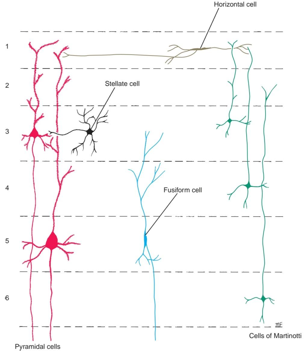
Figure 8-1 Main types of neurons found in the cerebral cortex.
شکل ۸-۱ انواع اصلی نورونهای موجود در قشر مغز.
Nerve Cells
The pyramidal cells are named from the shape of their cell bodies. Most of the cell bodies measure 10 to 50 μm long. However, giant pyramidal cells, also known as Betz cells, whose cell bodies measure as much as 120 μm, are found in the motor precentral gyrus of the frontal lobe.
سلولهای عصبی
سلولهای هرمی از شکل جسم سلولیشان نامگذاری شدهاند. طول بیشتر جسم سلولیها ۱۰ تا ۵۰ میکرومتر است. با این حال، سلولهای هرمی غولپیکر، که به سلولهای بتز نیز معروف هستند و جسم سلولی آنها تا ۱۲۰ میکرومتر میرسد، در شکنج حرکتی پیشمرکزی لوب پیشانی یافت میشوند.
The apices of the pyramidal cells are oriented toward the pial surface of the cortex. From the apex of each cell, a thick apical dendrite extends upward toward the pia, giving off collateral branches. From the basal angles, several basal dendrites pass laterally into the surrounding neuropil. Each dendrite possesses numerous dendritic spines for synaptic junctions with axons of other neurons. The axon arises from the base of the cell body and either terminates in the deeper cortical layers or, more commonly, enters the white matter of the cerebral hemisphere as a projection, association, or commissural fiber.
رأس سلولهای هرمی به سمت سطح پیال قشر مغز جهتگیری کردهاند. از رأس هر سلول، یک دندریت رأسی ضخیم به سمت بالا و به سمت نرمشامه امتداد مییابد و شاخههای جانبی را به خود اختصاص میدهد. از زوایای پایه، چندین دندریت پایه به صورت جانبی به داخل نوروپیل اطراف عبور میکنند. هر دندریت دارای خارهای دندریتی متعددی برای اتصالات سیناپسی با آکسونهای سایر نورونها است. آکسون از پایه جسم سلولی منشأ میگیرد و یا در لایههای عمیقتر قشر مغز خاتمه مییابد یا، معمولاً، به عنوان یک برآمدگی، ارتباط یا فیبر رابط وارد ماده سفید نیمکره مغزی میشود.
The stellate cells, sometimes called granule cells because of their small size, are polygonal in shape, and their cell bodies measure about 8 um in diameter. These cells have multiple branching dendrites and a relatively short axon, which terminates on a nearby neuron.
سلولهای ستارهای که گاهی به دلیل اندازه کوچکشان سلولهای گرانولی نامیده میشوند، شکل چندضلعی دارند و قطر جسم سلولی آنها حدود ۸ میکرومتر است. این سلولها دارای دندریتهای شاخهدار متعدد و یک آکسون نسبتاً کوتاه هستند که به یک نورون مجاور ختم میشود.
The fusiform cells have their long axis vertical to the surface and are concentrated mainly in the deepest cortical layers. Dendrites arise from each pole of the cell body. The inferior dendrite branches within the same cellular layer, while the superficial dendrite ascends toward the surface of the cortex and branches in the superficial layers. The axon arises from the inferior part of the cell body and enters the white matter as a projection, association, or commissural fiber.
سلولهای دوکیشکل محور طولی عمودی نسبت به سطح دارند و عمدتاً در عمیقترین لایههای قشری متمرکز شدهاند. دندریتها از هر قطب جسم سلولی منشأ میگیرند. دندریت تحتانی در همان لایه سلولی منشعب میشود، در حالی که دندریت سطحی به سمت سطح قشر بالا میرود و در لایههای سطحی منشعب میشود. آکسون از قسمت تحتانی جسم سلولی منشأ میگیرد و به عنوان یک برآمدگی، اتصال یا فیبر رابط وارد ماده سفید میشود.
The horizontal cells of Cajal are small, fusiform, horizontally oriented cells found in the most superficial layers of the cortex. A dendrite emerges from each end of the cell, and an axon runs parallel to the surface of the cortex, making contact with the dendrites of pyramidal cells.
سلولهای افقی کاخال، سلولهای کوچک، دوکیشکل و افقی هستند که در سطحیترین لایههای قشر مغز یافت میشوند. از هر انتهای سلول یک دندریت خارج میشود و یک آکسون به موازات سطح قشر مغز امتداد مییابد و با دندریتهای سلولهای هرمی تماس برقرار میکند.
The cells of Martinotti are small, multipolar cells that are present throughout the levels of the cortex. The cell has short dendrites, but the axon is directed toward the pial surface of the cortex, where it ends in a more superficial layer, commonly the most superficial layer. The axon gives origin to a few short collateral branches en route.
سلولهای مارتینوتی، سلولهای کوچک و چندقطبی هستند که در سراسر سطوح قشر مغز وجود دارند. این سلول دندریتهای کوتاهی دارد، اما آکسون به سمت سطح پیال قشر مغز هدایت میشود، جایی که در یک لایه سطحیتر، معمولاً سطحیترین لایه، به پایان میرسد. آکسون در طول مسیر، منشأ چند شاخه جانبی کوتاه است.
Nerve Fibers
The nerve fibers of the cerebral cortex are arranged both radially and tangentially (Figs. 8-2 and 8-3). The radial fibers run at right angles to the cortical surface.
رشتههای عصبی
رشتههای عصبی قشر مغز هم به صورت شعاعی و هم به صورت مماسی قرار گرفتهاند (شکلهای 8-2 و 8-3). رشتههای شعاعی با زاویه قائمه نسبت به سطح قشر مغز قرار دارند.

Figure 8-2 Neuronal connections of the cerebral cortex. Note the presence of the afferent and efferent fibers.
شکل ۸-۲ اتصالات نورونی قشر مغز. به وجود فیبرهای آوران و وابران توجه کنید.

Figure 8-3 Layers of the cerebral cortex showing the neurons on the left and the nerve fibers on the right.
شکل ۸-۳ لایههای قشر مغز که نورونها را در سمت چپ و رشتههای عصبی را در سمت راست نشان میدهد.
They include the afferent entering projection, association, and commissural fibers, which terminate within the cortex, and the axons of pyramidal, stellate, and fusiform cells, which leave the cortex to become projection, association, and commissural fibers of the white matter of the cerebral hemisphere.
آنها شامل فیبرهای آوران ورودی به بیرون زدگی، پیوندی و ارتباطی هستند که در داخل قشر مغز خاتمه مییابند و آکسونهای سلولهای هرمی، ستارهای و دوکیشکل که قشر مغز را ترک میکنند تا به فیبرهای برون زدگی، پیوندی و ارتباطی ماده سفید نیمکره مغزی تبدیل شوند.
The tangential fibers run parallel to the cortical surface and are, for the most part, collateral and terminal branches of afferent fibers. They also include the axons of horizontal and stellate cells and collateral branches of pyramidal and fusiform cells. The tangential fibers are most concentrated in layers 4 and 5, where they are referred to as the outer and inner bands of Baillarger, respectively. The bands of Baillarger are particularly well developed in the sensory areas due to the high concentration of the terminal parts of the thalamocortical fibers. In the visual cortex, the outer band of Baillarger, which is so thick that it can be seen with the naked eye, is known as the stria of Gennari. Because of this obvious band, or stria, the visual cortex in the walls of the calcarine sulcus is sometimes called the striate cortex.
فیبرهای مماسی به موازات سطح قشر مغز حرکت میکنند و عمدتاً شاخههای جانبی و انتهایی فیبرهای آوران هستند. آنها همچنین شامل آکسونهای سلولهای افقی و ستارهای و شاخههای جانبی سلولهای هرمی و دوکیشکل هستند. فیبرهای مماسی بیشتر در لایههای ۴ و ۵ متمرکز هستند، جایی که به ترتیب به عنوان نوارهای بیرونی و داخلی بایلاگر شناخته میشوند. نوارهای بایلاگر به دلیل غلظت بالای قسمتهای انتهایی فیبرهای تالاموکورتیکال، به ویژه در نواحی حسی به خوبی توسعه یافتهاند. در قشر بینایی، نوار بیرونی بایلاگر که آنقدر ضخیم است که با چشم غیرمسلح دیده میشود، به عنوان خطوط جناری شناخته میشود. به دلیل این نوار یا استریای آشکار، قشر بینایی در دیوارههای شیار کالکارین گاهی اوقات قشر مخطط نامیده میشود.
Layers
Dividing the cerebral cortex into layers that may be distinguished by the type, density, and arrangement of their cells is convenient for descriptive purposes (see Figs. 8-1 and 8-3). The names and characteristic features of the layers are described here; regional differences are discussed later.
لایهها
تقسیم قشر مغز به لایههایی که ممکن است بر اساس نوع، تراکم و چیدمان سلولهایشان از هم متمایز شوند، برای اهداف توصیفی مناسب است (شکلهای 8-1 و 8-3 را ببینید). نامها و ویژگیهای مشخصه لایهها در اینجا شرح داده شدهاند؛ تفاوتهای منطقهای بعداً مورد بحث قرار میگیرند.
1. Molecular layer (plexiform layer). This is the most superficial layer; it consists mainly of a dense network of tangentially oriented nerve fibers. These fibers are derived from the apical dendrites of the pyramidal cells and fusiform cells, the axons of the stellate cells, and the cells of Martinotti. Afferent fibers originating in the thalamus and in association with commissural fibers also are present. Scattered among these nerve fibers are occasional horizontal cells of Cajal. This most superficial layer of the cortex clearly is where large numbers of synapses between different neurons occur.
۱. لایه مولکولی (لایه شبکهای). این لایه سطحیترین لایه است؛ عمدتاً از شبکهای متراکم از فیبرهای عصبی با جهتگیری مماسی تشکیل شده است. این فیبرها از دندریتهای رأسی سلولهای هرمی و سلولهای دوکیشکل، آکسونهای سلولهای ستارهای و سلولهای مارتینوتی مشتق میشوند. فیبرهای آوران که از تالاموس منشأ میگیرند و با فیبرهای رابط مرتبط هستند نیز وجود دارند. در بین این فیبرهای عصبی، سلولهای افقی کاخال نیز پراکنده هستند. این سطحیترین لایه قشر مغز، به وضوح جایی است که تعداد زیادی سیناپس بین نورونهای مختلف رخ میدهد.
2. External granular layer. This layer contains large numbers of small pyramidal cells and stellate cells. The dendrites of these cells terminate in the molecular layer, and the axons enter deeper layers, where they terminate or pass on to enter the white matter of the cerebral hemisphere.
۲. لایه دانهدار خارجی. این لایه حاوی تعداد زیادی سلولهای هرمی کوچک و سلولهای ستارهای است. دندریتهای این سلولها به لایه مولکولی ختم میشوند و آکسونها وارد لایههای عمیقتر میشوند، جایی که به ماده سفید نیمکره مغزی ختم میشوند یا از آنجا عبور میکنند.
3. External pyramidal layer. This layer is composed of pyramidal cells, whose cell body size increases from the superficial to the deeper borders of the layer. The apical dendrites pass into the molecular layer, and the axons enter the white matter as projection, association, or commissural fibers.
۳. لایه هرمی خارجی. این لایه از سلولهای هرمی تشکیل شده است که اندازه جسم سلولی آنها از سطح به سمت مرزهای عمیقتر لایه افزایش مییابد. دندریتهای رأسی به لایه مولکولی وارد میشوند و آکسونها به صورت فیبرهای برآمدگی، اتصالی یا رابط وارد ماده سفید میشوند.
4. Internal granular layer. This layer is composed of closely packed stellate cells with a high concentration of horizontally arranged fibers known collectively as the external band of Baillarger.
۴. لایه دانهای داخلی. این لایه از سلولهای ستارهای فشرده با غلظت بالایی از فیبرهای افقی تشکیل شده است که در مجموع به عنوان نوار خارجی بایلاگر شناخته میشوند.
5. Ganglionic layer (internal pyramidal layer). This layer contains very large and medium-size pyramidal cells. Scattered among the pyramidal cells are stellate cells and cells of Martinotti. In addition, a large number of horizontally arranged fibers form the inner band of Baillarger (see Fig. 8-3). In the motor cortex of the precentral gyrus, the pyramidal cells of this layer are very large and are known as Betz cells. These cells account for about 3% of the projection fibers of the corticospinal or pyramidal tract.
۵. لایه گانگلیونی (لایه هرمی داخلی). این لایه حاوی سلولهای هرمی بسیار بزرگ و متوسط است. سلولهای ستارهای و سلولهای مارتینوتی در بین سلولهای هرمی پراکنده شدهاند. علاوه بر این، تعداد زیادی از فیبرهای افقی، نوار داخلی Baillarger را تشکیل میدهند (شکل ۸-۳ را ببینید). در قشر حرکتی شکنج پیشمرکزی، سلولهای هرمی این لایه بسیار بزرگ هستند و به عنوان سلولهای بتز شناخته میشوند. این سلولها حدود ۳٪ از فیبرهای برآمدگی راه قشری-نخاعی یا هرمی را تشکیل میدهند.
6. Multiform layer (layer of polymorphic cells). Although the majority of the cells are fusiform, many of the cells are modified pyramidal cells, whose cell bodies are triangular or ovoid. The cells of Martinotti also are conspicuous in this layer. Many nerve fibers are present that are entering or are leaving the underlying white matter.
۶. لایه چندشکلی (لایه سلولهای چندشکلی). اگرچه اکثر سلولها دوکیشکل هستند، بسیاری از سلولها سلولهای هرمی اصلاحشده هستند که جسم سلولی آنها مثلثی یا تخممرغی است. سلولهای مارتینوتی نیز در این لایه قابل مشاهده هستند. بسیاری از فیبرهای عصبی وجود دارند که وارد ماده سفید زیرین میشوند یا از آن خارج میشوند.
Cortical Structure Variations
The system of numbering and nomenclature of the cortical layers used above is similar to that distinguished by Brodmann (1909). However, recognize that not all areas of the cerebral cortex possess six layers (see Fig. 8-3). Those areas of the cortex in which the basic six layers cannot be recognized are referred to as heterotypical, as opposed to the majority, which are homotypical and possess six layers. Two heterotypical areas are described: the granular and the agranular type.
تنوعات ساختار قشری
سیستم شمارهگذاری و نامگذاری لایههای قشری که در بالا استفاده شد، مشابه سیستمی است که توسط برودمن (1909) مشخص شده است. با این حال، توجه داشته باشید که همه نواحی قشر مغز دارای شش لایه نیستند (شکل 8-3 را ببینید). آن دسته از نواحی قشر که در آنها شش لایه اصلی قابل تشخیص نیستند، به عنوان هتروتیپیک شناخته میشوند، برخلاف اکثر نواحی که هموتیپیک هستند و دارای شش لایه میباشند. دو ناحیه هتروتیپیک شرح داده شده است: نوع دانهدار و غیر دانهدار.
In the granular type, the granular layers are well developed and contain densely packed stellate cells. Thus, layers 2 and 4 are well developed, and layers 3 and 5 are poorly developed, so layers 2 through 5 merge into a single layer of predominantly granular cells. These cells receive thalamocortical fibers. The granular type of cortex is found in the postcentral gyrus, in the superior temporal gyrus, and in parts of the hippocampal gyrus.
در نوع دانهای، لایههای دانهای به خوبی توسعه یافتهاند و حاوی سلولهای ستارهای متراکم هستند. بنابراین، لایههای ۲ و ۴ به خوبی توسعه یافتهاند و لایههای ۳ و ۵ به طور ضعیفی توسعه یافتهاند، بنابراین لایههای ۲ تا ۵ در یک لایه واحد از سلولهای عمدتاً دانهای ادغام میشوند. این سلولها فیبرهای تالاموکورتیکال را دریافت میکنند. نوع دانهای قشر مغز در شکنج پس مرکزی، در شکنج گیجگاهی فوقانی و در بخشهایی از شکنج هیپوکامپ یافت میشود.
In the agranular type of cortex, the granular layers are poorly developed, so layers 2 and 4 are practically absent. The pyramidal cells in layers 3 and 5 are densely packed and are very large. The agranular type of cortex is found in the precentral gyrus and other areas in the frontal lobe. These areas give rise to large numbers of efferent fibers that are associated with motor function.
در نوع غیر دانهای قشر مغز، لایههای دانهای به طور ضعیفی توسعه یافتهاند، بنابراین لایههای ۲ و ۴ عملاً وجود ندارند. سلولهای هرمی در لایههای ۳ و ۵ به طور متراکمی فشرده شدهاند و بسیار بزرگ هستند. نوع غیر دانهای قشر مغز در شکنج پیشمرکزی و سایر نواحی لوب فرونتال یافت میشود. این نواحی تعداد زیادی فیبر وابران ایجاد میکنند که با عملکرد حرکتی مرتبط هستند.
CORTICAL MECHANISMS
Extensive research in recent years involving electrophysiology, histochemistry, immunocytochemistry, and other microscopic techniques has resulted in a vast increase in our knowledge of the connections of the neurons of the cerebral cortex. This information combined with new methods of studying the functions of the human cerebral cortex in the living using electroencephalograms (EEGS), positron emission tomography (PET), and magnetic resonance imaging (MRI) have led to a new understanding of the functions of the different areas and the different layers of the cerebral cortex. Much of the new information, however, is still merely factual data and cannot be used in the clinical setting.
مکانیسمهای قشری
تحقیقات گسترده در سالهای اخیر شامل الکتروفیزیولوژی، هیستوشیمی، ایمونوسیتوشیمی و سایر تکنیکهای میکروسکوپی منجر به افزایش چشمگیر دانش ما در مورد اتصالات نورونهای قشر مغز شده است. این اطلاعات همراه با روشهای جدید مطالعه عملکرد قشر مغز انسان در موجودات زنده با استفاده از الکتروانسفالوگرام (EEGS)، توموگرافی انتشار پوزیترون (PET) و تصویربرداری رزونانس مغناطیسی (MRI) منجر به درک جدیدی از عملکرد نواحی و لایههای مختلف قشر مغز شده است. با این حال، بسیاری از اطلاعات جدید هنوز صرفاً دادههای واقعی هستند و نمیتوان از آنها در محیط بالینی استفاده کرد.
The cerebral cortex is organized into vertical units or columns of functional activity (see Fig. 8-2) measuring about 300 to 600 μm wide. In the sensory cortex, for example, each column serves a single specific sensory function. Such a functional unit extends through all six layers from the cortical surface to the white matter. Each unit possesses afferent fibers, internuncial neurons, and efferent fibers. An afferent fiber may synapse directly with an efferent neuron or may involve vertical chains of internuncial neurons. A single vertical chain of neurons may be involved in isolation, or the wave of excitation may spread to adjacent vertical chains through short axon granular cells. The horizontal cells of Cajal permit activation of vertical units that lie some distance away from the incoming afferent fiber. The spread of incoming information serving one sensory modality laterally from one column to an adjacent column, or to columns some distance away, may permit the individual to start the pro- cess of understanding the nature of the sensory input.
قشر مغز به واحدهای عمودی یا ستونهایی از فعالیت عملکردی (شکل 8-2 را ببینید) با عرض حدود 300 تا 600 میکرومتر سازماندهی شده است. به عنوان مثال، در قشر حسی، هر ستون یک عملکرد حسی خاص را انجام میدهد. چنین واحد عملکردی در هر شش لایه از سطح قشر مغز تا ماده سفید امتداد دارد. هر واحد دارای فیبرهای آوران، نورونهای بینحلقهای و فیبرهای وابران است. یک فیبر آوران ممکن است مستقیماً با یک نورون وابران سیناپس برقرار کند یا ممکن است شامل زنجیرههای عمودی نورونهای بینحلقهای باشد. یک زنجیره عمودی واحد از نورونها ممکن است به صورت جداگانه درگیر باشد، یا موج تحریک ممکن است از طریق سلولهای دانهای کوتاه آکسون به زنجیرههای عمودی مجاور گسترش یابد. سلولهای افقی کاخال امکان فعالسازی واحدهای عمودی را که در فاصلهای دورتر از فیبر آوران ورودی قرار دارند، فراهم میکنند. گسترش اطلاعات ورودی که به یک روش حسی به صورت جانبی از یک ستون به ستون مجاور یا به ستونهایی در فاصلهای دورتر خدمت میکنند، ممکن است به فرد اجازه دهد فرآیند درک ماهیت ورودی حسی را آغاز کند.
«فصل هشتم کتاب نورواناتومی بالینی اسنل»
🚀 با ما همراه شوید!
تازهترین مطالب و آموزشهای مغز و اعصاب را از دست ندهید. با فالو کردن کانال تلگرام آیندهنگاران مغز، از ما حمایت کنید!



