
نواحی قشر مغز: لوب پیشانی، لوب آهیانه، لوب گیجگاهی و لوب پسسری


دعای مطالعه [ نمایش ]
بِسْمِ الله الرَّحْمنِ الرَّحیمِ
اَللّهُمَّ اَخْرِجْنى مِنْ ظُلُماتِ الْوَهْمِ
خدايا مرا بيرون آور از تاريكىهاى وهم،
وَ اَكْرِمْنى بِنُورِ الْفَهْمِ
و به نور فهم گرامى ام بدار،
اَللّهُمَّ افْتَحْ عَلَيْنا اَبْوابَ رَحْمَتِكَ
خدايا درهاى رحمتت را به روى ما بگشا،
وَانْشُرْ عَلَيْنا خَزائِنَ عُلُومِكَ بِرَحْمَتِكَ يا اَرْحَمَ الرّاحِمينَ
و خزانههاى علومت را بر ما باز كن به امید رحمتت اى مهربانترين مهربانان.
آیندهنگاران مغز: «ما مغز را میشناسیم، تا آینده را بسازیم.»
کتاب «نوروآناتومی بالینی اسنل» اثر دکتر ریچارد اس. اسنل، با ویرایش جدید دکتر رایان اسپلیتگربر، همچنان بهعنوان مرجع طلایی این حوزه شناخته میشود. این اثر با تلفیق تازهترین پژوهشها و کاربردهای بالینی، پلی استوار میان دانش پایه و عمل پزشکی بنا میکند.
تلاشهای علمی و سالها تدریس ارزشمند استاد گرانقدر آقای دکتر محمدتقی جغتایی الهامبخش ترجمه و گسترش این دانش در ایران بوده است.
ترجمه دقیق این کتاب توسط برند علمی آیندهنگاران مغز به مدیریت داریوش طاهری، امکان دسترسی فارسیزبانان به مرزهای نوین علوم اعصاب را فراهم کرده و رسالتی علمی در جهت شناخت عمیقتر مغز و ترسیم آیندهای روشنتر بر عهده گرفته است.
»» فصل ۸: ساختار و مناطق عملکردی قشر مغز
»» Chapter 8: The Structure and Functional Localization of the Cerebral Cortex
CORTICAL AREAS
Over the past century, clinicopathologic studies in humans and electrophysiologic and ablation studies in animals have produced evidence that different areas of the cerebral cortex are functionally specialized. However, the precise division of the cortex into different areas of specialization, as described by Brodmann, oversimplifies and misleads the reader. The simple division of cortical areas into motor and sensory is erroneous because many of the sensory areas are far more extensive than originally described and because motor responses can be obtained by stimulation of sensory areas. Until a satisfactory terminology has been devised to describe the various cortical areas, the main cortical areas will be named by their anatomical location.
نواحی قشری
در طول قرن گذشته، مطالعات کلینیکوپاتولوژیک در انسانها و مطالعات الکتروفیزیولوژیک و ابلیشن در حیوانات، شواهدی ارائه دادهاند که نشان میدهد نواحی مختلف قشر مغز از نظر عملکردی تخصصی هستند. با این حال، تقسیم دقیق قشر به نواحی مختلف تخصصی، همانطور که توسط برودمن توضیح داده شده است، خواننده را بیش از حد ساده کرده و گمراه میکند. تقسیم ساده نواحی قشری به حرکتی و حسی اشتباه است زیرا بسیاری از نواحی حسی بسیار گستردهتر از آن چیزی هستند که در ابتدا توصیف شده است و به این دلیل که پاسخهای حرکتی را میتوان با تحریک نواحی حسی به دست آورد. تا زمانی که اصطلاحات رضایتبخشی برای توصیف نواحی مختلف قشری ابداع نشده باشد، نواحی اصلی قشری بر اساس موقعیت آناتومیکی آنها نامگذاری خواهند شد.
Some of the main anatomical connections of the cerebral cortex are summarized in Table 8-1.
برخی از ارتباطات آناتومیکی اصلی قشر مغز در جدول 8-1 خلاصه شده است.
Table 8-1 Main Anatomical Connections of the Cerebral Cortex
جدول۸-۱ اتصالات آناتومیکی اصلی قشر مغز
 B, Brodmann area.
Frontal Lobe
The precentral area is situated in the precentral gyrus and includes the anterior wall of the central sulcus and the posterior parts of the superior, middle, and inferior frontal gyri; it extends over the superomedial border of the hemisphere into the paracentral lobule (Fig. 8-4). Histologically, the characteristic feature of this area is the almost complete absence of the granular layers and the prominence of the pyramidal nerve cells. The giant pyramidal cells of Betz, which can measure as much as 120 μm long and 60 μm wide, are concentrated most highly in the superior part of the precentral gyrus and the paracentral lobule; their numbers diminish as one passes anteriorly in the precentral gyrus or inferiorly toward the lateral fissure. The great majority of the corticospinal and corticobulbar fibers originate from the small pyramidal cells in this area. The number of Betz cells present is estimated to be between 25,000 and 30,000 and accounts for only about 3% of the corticospinal fibers. Notably, the postcentral gyrus and the second somatosensory areas, as well as the occipital and temporal lobes, give origin to descending tracts as well; they are involved in controlling the sensory input to the nervous system and are not involved in muscular movement.
لوب پیشانی
ناحیه پیشمرکزی در شکنج پیشمرکزی قرار دارد و شامل دیواره قدامی شیار مرکزی و قسمتهای خلفی شکنجهای پیشانی فوقانی، میانی و تحتانی است. این ناحیه از مرز فوقانی-میانی نیمکره به داخل لوبول پاراسنترال امتداد مییابد (شکل 8-4). از نظر بافتشناسی، ویژگی بارز این ناحیه، فقدان تقریباً کامل لایههای دانهای و برجستگی سلولهای عصبی هرمی است. سلولهای هرمی غولپیکر بتز، که میتوانند تا 120 میکرومتر طول و 60 میکرومتر عرض داشته باشند، بیشتر در قسمت فوقانی شکنج پیشمرکزی و لوبول پاراسنترال متمرکز هستند. تعداد آنها با عبور از جلوی شکنج پیشمرکزی یا پایینتر به سمت شیار جانبی کاهش مییابد. اکثریت قریب به اتفاق فیبرهای قشری-نخاعی و قشری-بولبار از سلولهای هرمی کوچک در این ناحیه منشأ میگیرند. تعداد سلولهای بتز موجود بین ۲۵۰۰۰ تا ۳۰۰۰۰ تخمین زده میشود و تنها حدود ۳٪ از فیبرهای قشری-نخاعی را تشکیل میدهند. نکته قابل توجه این است که شکنج پس مرکزی و نواحی حسی-پیکری دوم، و همچنین لوبهای پسسری و گیجگاهی، منشأ مسیرهای نزولی نیز هستند؛ آنها در کنترل ورودی حسی به سیستم عصبی نقش دارند و در حرکت عضلانی دخیل نیستند.
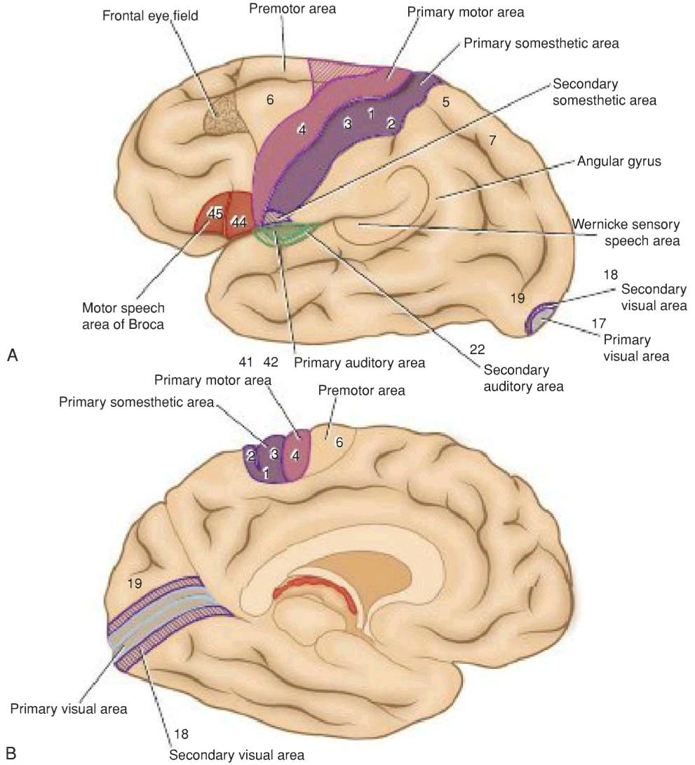
Figure 8-4 Functional localization of the cerebral cortex. A: Lateral view of the left cerebral hemisphere. B: Medial view of the left cerebral hemisphere.
شکل ۸-۴ موقعیتیابی عملکردی قشر مغز. الف: نمای جانبی نیمکره چپ مغز. ب: نمای میانی نیمکره چپ مغز.
The precentral area may be divided into posterior and anterior regions. The posterior region, which is referred to as the motor area, primary motor area, or Brodmann area 4, occupies the precentral gyrus extending over the superior border into the paracentral lobule. The anterior region is known as the premotor area, secondary motor area, or Brodmann area 6 and parts of areas 8, 44, and 45. It occupies the anterior part of the precentral gyrus and the posterior parts of the superior, middle, and inferior frontal gyri.
ناحیه پیشمرکزی را میتوان به نواحی خلفی و قدامی تقسیم کرد. ناحیه خلفی که به آن ناحیه حرکتی، ناحیه حرکتی اولیه یا ناحیه برودمن ۴ گفته میشود، شکنج پیشمرکزی را اشغال میکند که از مرز فوقانی به لوبول پاراسنترال امتداد دارد. ناحیه قدامی به عنوان ناحیه پیشحرکتی، ناحیه حرکتی ثانویه یا ناحیه برودمن ۶ و بخشهایی از نواحی ۸، ۴۴ و ۴۵ شناخته میشود. این ناحیه قسمت قدامی شکنج پیشمرکزی و قسمتهای خلفی شکنجهای پیشانی فوقانی، میانی و تحتانی را اشغال میکند.
The primary motor area, if electrically stimulated, produces isolated movements on the opposite side of the body as well as contraction of muscle groups concerned with the performance of a specific movement. Although isolated ipsilateral movements do not occur, bilateral movements of the extraocular muscles, the muscles of the upper part of the face, the tongue, and the mandible, and the larynx and the pharynx do occur.
ناحیه حرکتی اولیه، در صورت تحریک الکتریکی، حرکات مجزا در طرف مقابل بدن و همچنین انقباض گروههای عضلانی مربوط به انجام یک حرکت خاص را ایجاد میکند. اگرچه حرکات مجزای همان طرف رخ نمیدهد، اما حرکات دو طرفه عضلات خارج چشمی، عضلات قسمت فوقانی صورت، زبان و فک پایین و حنجره و حلق رخ میدهد.
The movement areas of the body are represented in inverted form in the precentral gyrus (Fig. 8-5). Starting from below and passing superiorly are structures involved in swallowing and the tongue, jaw, lips, larynx, eyelid, and brow. The next area is an extensive region for movements of the fingers, especially the thumb, hand, wrist, elbow, shoulder, and trunk. The movements of the hip, knee, and ankle are represented in the highest areas of the precentral gyrus; the movements of the toes are situated on the medial surface of the cerebral hemisphere in the paracentral lobule. The movements of the anal and vesical sphincters are also located in the paracentral lobule. The area of cortex controlling a particular movement is proportional to the skill involved in performing the movement and is unrelated to the mass of muscle participating in the movement.
نواحی حرکتی بدن به صورت معکوس در شکنج پیشمرکزی نشان داده شدهاند (شکل 8-5). از پایین شروع شده و به بالا ساختارهای دخیل در بلع و زبان، فک، لبها، حنجره، پلک و ابرو قرار دارند. ناحیه بعدی، ناحیه وسیعی برای حرکات انگشتان، به ویژه شست، دست، مچ دست، آرنج، شانه و تنه است. حرکات لگن، زانو و مچ پا در بالاترین نواحی شکنج پیشمرکزی نشان داده شدهاند. حرکات انگشتان پا در سطح داخلی نیمکره مغزی در لوبول پاراسنترال قرار دارند. حرکات اسفنکترهای مقعدی و مثانهای نیز در لوبول پاراسنترال قرار دارند. ناحیهای از قشر مغز که یک حرکت خاص را کنترل میکند، متناسب با مهارت لازم در انجام حرکت است و به توده عضلات شرکتکننده در حرکت ارتباطی ندارد.
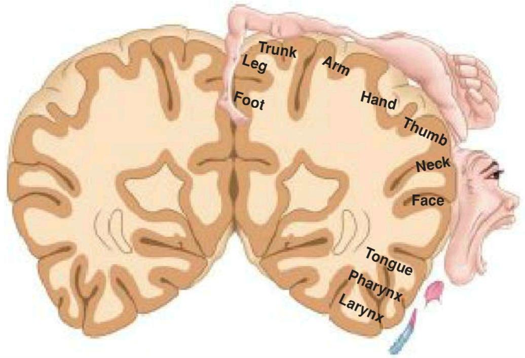
Figure 8-5 Motor homunculus on the precentral gyrus.
شکل ۸-۵ هومونکول حرکتی روی شکنج پیشمرکزی.
Thus, the function of the primary motor area is to carry out the individual movements of different parts of the body. To assist in this function, it receives numerous afferent fibers from the premotor area, the sensory cortex, the thalamus, the cerebellum, and the basal ganglia. The primary motor cortex is not responsible for the design of the pattern of movement but is the final station for conversion of the design into execution of the movement.
بنابراین، وظیفه ناحیه حرکتی اولیه، انجام حرکات جداگانه قسمتهای مختلف بدن است. برای کمک به این عملکرد، فیبرهای آوران متعددی از ناحیه پیش حرکتی، قشر حسی، تالاموس، مخچه و عقدههای قاعدهای دریافت میکند. قشر حرکتی اولیه مسئول طراحی الگوی حرکت نیست، بلکه ایستگاه نهایی برای تبدیل طرح به اجرای حرکت است.
The premotor area, which is wider superiorly than below and narrows down to be confined to the anterior part of the precentral gyrus, has no giant pyramidal cells of Betz. Electrical stimulation of the premotor area produces muscular movements similar to those obtained by stimulation of the primary motor area; however, stronger stimulation is necessary to produce the same degree of movement.
ناحیه پیش حرکتی که در بالا پهنتر از پایین است و به سمت پایین باریک میشود تا به قسمت قدامی شکنج پیشمرکزی محدود شود، سلولهای هرمی غولپیکر بتز ندارد. تحریک الکتریکی ناحیه پیش حرکتی، حرکات عضلانی مشابه حرکات حاصل از تحریک ناحیه حرکتی اولیه را ایجاد میکند. با این حال، تحریک قویتری برای تولید همان درجه از حرکت لازم است.
The premotor area receives numerous inputs from the sensory cortex, the thalamus, and the basal ganglia. The function of the premotor area is to store programs of motor activity assembled as the result of past experience. Thus, the premotor area programs the activity of the primary motor area. It is particularly involved in controlling coarse postural movements through its connections with the basal ganglia.
ناحیه پیش حرکتی ورودیهای متعددی را از قشر حسی، تالاموس و عقدههای قاعدهای دریافت میکند. وظیفه ناحیه پیش حرکتی ذخیره برنامههای فعالیت حرکتی است که در نتیجه تجربیات گذشته گردآوری شدهاند. بنابراین، ناحیه پیش حرکتی فعالیت ناحیه حرکتی اولیه را برنامهریزی میکند. این عصب به ویژه از طریق ارتباطاتش با عقدههای قاعدهای در کنترل حرکات زمخت وضعیتی نقش دارد.
The supplementary motor area is situated in the medial frontal gyrus on the medial surface of the hemisphere and anterior to the paracentral lobule. Stimulation of this area results in movements of the contralateral limbs, but a stronger stimulus is necessary than when the primary motor area is stimulated. Removal of the supplementary motor area produces no permanent loss of movement.
ناحیه حرکتی تکمیلی در شکنج پیشانی داخلی در سطح داخلی نیمکره و در جلوی لوبول پاراسنترال قرار دارد. تحریک این ناحیه منجر به حرکات اندامهای طرف مقابل میشود، اما محرک قویتری نسبت به زمانی که ناحیه حرکتی اولیه تحریک میشود، لازم است. حذف ناحیه حرکتی تکمیلی باعث از دست رفتن دائمی حرکت نمیشود.
The frontal eye field (see Fig. 8-4A) extends forward from the facial area of the precentral gyrus into the middle frontal gyrus (parts of Brodmann areas 6, 8, and 9). Electrical stimulation of this region causes conjugate movements of the eyes, especially toward the opposite side. The exact pathway taken by nerve fibers from this area is not known, but they are thought to pass to the superior colliculus of the midbrain. The superior colliculus is connected to the nuclei of the extraocular muscles by the reticular formation. The frontal eye field is considered to control voluntary scanning movements of the eye and is independent of visual stimuli. The involuntary following of moving objects by the eyes involves the visual area of the occipital cortex to which the frontal eye field is connected by association fibers.
میدان چشمی جلویی (به شکل 8-4A مراجعه کنید) از ناحیه صورت شکنج پیشانی به سمت جلو تا شکنج پیشانی میانی (بخشهایی از نواحی برودمن 6، 8 و 9) امتداد مییابد. تحریک الکتریکی این ناحیه باعث حرکات مزدوج چشمها، به ویژه به سمت طرف مقابل میشود. مسیر دقیق طی شده توسط فیبرهای عصبی از این ناحیه مشخص نیست، اما تصور میشود که آنها به کولیکولوس فوقانی مغز میانی میروند. کولیکولوس فوقانی توسط تشکیلات مشبک به هستههای عضلات خارج چشمی متصل است. میدان چشمی جلویی حرکات اسکن ارادی چشم را کنترل میکند و مستقل از محرکهای بینایی است. دنبال کردن غیرارادی اشیاء متحرک توسط چشمها، ناحیه بینایی قشر پسسری را درگیر میکند که میدان بینایی پیشانی توسط فیبرهای ارتباطی به آن متصل است.
The motor speech area of Broca is located in the inferior frontal gyrus between the anterior and ascending rami and the ascending and posterior rami of the lateral fissure (Brodmann areas 44 and 45). In the majority of individuals, this area is important on the left or dominant hemisphere, and ablation will result in paralysis of speech. In those individuals in whom the right hemisphere is dominant, the area on the right side is of importance. The ablation of this region in the nondominant hemisphere has no effect on speech.
ناحیه حرکتی گفتار بروکا در شکنج پیشانی تحتانی بین شاخههای قدامی و صعودی و شاخههای صعودی و خلفی شیار جانبی (نواحی برادمن ۴۴ و ۴۵) قرار دارد. در اکثر افراد، این ناحیه در نیمکره چپ یا غالب اهمیت دارد و تخریب آن منجر به فلج گفتار میشود. در افرادی که نیمکره راست در آنها غالب است، ناحیه سمت راست اهمیت دارد. تخریب این ناحیه در نیمکره غیرغالب هیچ تاثیری بر گفتار ندارد.
The Broca speech area brings about the formation of words by its connections with the adjacent primary motor areas; the muscles of the larynx, mouth, tongue, soft palate, and the respiratory muscles are appropriately stimulated.
ناحیه گفتار بروکا با ارتباط خود با نواحی حرکتی اولیه مجاور، باعث تشکیل کلمات میشود. عضلات حنجره، دهان، زبان، کام نرم و عضلات تنفسی به طور مناسب تحریک میشوند.
The prefrontal cortex is an extensive area that lies anterior to the precentral area. It includes the greater parts of the superior, middle, and inferior frontal gyri; the orbital gyri; most of the medial frontal gyrus; and the anterior half of the cingulate gyrus (Brodmann areas 9, 10, 11, and 12). Large numbers of afferent and efferent pathways connect the prefrontal area with other areas of the cerebral cortex, the thalamus, the hypothalamus, and the corpus striatum. The frontopontine fibers also connect this area to the cerebellum through the pontine nuclei. The commissural fibers of the forceps minor and genu of the corpus callosum unite these areas in both cerebral hemispheres.
قشر پیشپیشانی ناحیهی وسیعی است که در جلوی ناحیهی پیشمرکزی قرار دارد. این ناحیه شامل بخشهای بیشتری از شکنجهای پیشانی فوقانی، میانی و تحتانی؛ شکنجهای مداری؛ بیشتر شکنج پیشانی میانی؛ و نیمهی قدامی شکنج سینگولیت (نواحی برادمن ۹، ۱۰، ۱۱ و ۱۲) است. تعداد زیادی از مسیرهای آوران و وابران، ناحیهی پیشپیشانی را با سایر نواحی قشر مغز، تالاموس، هیپوتالاموس و جسم مخطط متصل میکنند. الیاف پیشانی-پل مغزی نیز این ناحیه را از طریق هستههای پل مغزی به مخچه متصل میکنند. الیاف رابط فورسپسهای کوچک و بزرگ جسم پینهای، این نواحی را در هر دو نیمکرهی مغزی به هم متصل میکنند.
The prefrontal area is concerned with the makeup of the individual’s personality. As the result of the input from many cortical and subcortical sources, this area plays a role as a regulator of the person’s depth of feeling. It also exerts its influence in determining the initiative and judgment of an individual.
ناحیهی پیشپیشانی مربوط به شکلگیری شخصیت فرد است. در نتیجهی ورودی از منابع قشری و زیرقشری متعدد، این ناحیه به عنوان تنظیمکنندهی عمق احساس فرد نقش ایفا میکند. همچنین تأثیر خود را در تعیین ابتکار عمل و قضاوت یک فرد اعمال میکند.
Parietal Lobe
The primary somesthetic area (primary somatic sensory cortex S1) occupies the postcentral gyrus (see Fig. 8-4) on the lateral surface of the hemisphere and the posterior part of the paracentral lobule on the medial surface (Brodmann areas 3, 1, and 2). Histologically, the anterior part of the postcentral gyrus is the area that borders the central sulcus (area 3), is granular in type, and contains only scattered pyramidal cells. The outer layer of Baillarger is broad and very obvious. The posterior part of the postcentral gyrus (areas 1 and 2) possesses fewer granular cells. The primary somesthetic areas of the cerebral cortex receive projection fibers from the ventral posterior lateral and ventral posterior medial nuclei of the thalamus. The opposite half of the body is represented as inverted. The pharyngeal region, tongue, and jaws are represented in the most inferior part of the postcentral gyrus; this is followed by the face, fingers, hand, arm, trunk, and thigh. The leg and the foot areas are found on the medial surface of the hemisphere in the posterior part of the paracentral lobule. The anal and genital regions are also found in this latter area. The apportioning of the cortex for a particular part of the body is related to its functional importance rather than to its size. The face, lips, thumb, and index finger have particularly large areas assigned to them. In fact, the size of the cortical area allocated to each part of the body is directly proportional to the number of sensory receptors present in that part of the body.
لوب آهیانه
ناحیه حسی-پیکری اولیه (قشر حسی-پیکری اولیه S1) در سطح جانبی نیمکره، شکنج پس مرکزی (شکل 8-4 را ببینید) و در سطح داخلی (نواحی برادمن 3، 1 و 2) در قسمت خلفی لوبول پاراسنترال قرار دارد. از نظر بافتشناسی، قسمت قدامی شکنج پس مرکزی، ناحیهای است که با شیار مرکزی (ناحیه 3) هممرز است، از نوع دانهدار است و فقط شامل سلولهای هرمی پراکنده است. لایه بیرونی بایلارگر پهن و بسیار واضح است. قسمت خلفی شکنج پس مرکزی (نواحی 1 و 2) دارای سلولهای دانهدار کمتری است. نواحی اولیه شکنج پس مرکزی، فیبرهای بیرونزده را از هستههای شکمی-خلفی-جانبی و شکمی-خلفی-میانی تالاموس دریافت میکنند. نیمه مقابل بدن به صورت معکوس نشان داده شده است. ناحیه حلق، زبان و فکها در پایینترین قسمت شکنج پس مرکزی نشان داده شدهاند. پس از آن صورت، انگشتان، دست، بازو، تنه و ران قرار دارند. نواحی ساق و پا در سطح داخلی نیمکره در قسمت خلفی لوبول پاراسنترال یافت میشوند. نواحی مقعدی و تناسلی نیز در این ناحیه اخیر یافت میشوند. تقسیم قشر مغز برای یک قسمت خاص از بدن به اهمیت عملکردی آن مربوط میشود نه به اندازه آن. صورت، لبها، شست و انگشت اشاره دارای نواحی به ویژه بزرگی هستند. در واقع، اندازه ناحیه قشری اختصاص داده شده به هر قسمت از بدن مستقیماً با تعداد گیرندههای حسی موجود در آن قسمت از بدن متناسب است.
Although most sensations reach the cortex from the contralateral side of the body, some from the oral region go to the same side, and those from the pharynx, larynx, and perineum go to both sides. On entering the cortex, the afferent fibers excite the neurons in layer IV, and then the signals spread toward the surface of the cerebral unit and toward the deeper layers. From layer VI, large numbers of axons leave the cortex and pass to lower sensory relay stations of the thalamus, medulla oblongata, and the spinal cord, providing feedback. This sensory feedback is largely inhibitory and serves to modulate the intensity of the sensory input.
اگرچه بیشتر حسها از سمت مقابل بدن به قشر مخ میرسند، برخی از حسها از ناحیه دهان به همان سمت میروند و حسهای حلق، حنجره و پرینه به هر دو طرف میروند. با ورود به قشر مخ، فیبرهای آوران، نورونهای لایه IV را تحریک میکنند و سپس سیگنالها به سمت سطح واحد مغزی و لایههای عمیقتر پخش میشوند. از لایه VI، تعداد زیادی آکسون قشر مخ را ترک کرده و به ایستگاههای رله حسی پایینتر تالاموس، بصل النخاع و نخاع میروند و بازخورد ایجاد میکنند. این بازخورد حسی تا حد زیادی مهاری است و برای تعدیل شدت ورودی حسی عمل میکند.
The anterior part of the postcentral gyrus situated in the central sulcus receives a large number of afferent fibers from muscle spindles, tendon organs, and joint receptors. This sensory information is analyzed in the vertical columns of the sensory cortex; it is then passed forward beneath the central sulcus to the primary motor cortex, where it greatly influences the control of skeletal muscle activity.
قسمت قدامی شکنج پس مرکزی واقع در شیار مرکزی، تعداد زیادی فیبر آوران از دوکهای عضلانی، اندامهای تاندونی و گیرندههای مفصلی دریافت میکند. این اطلاعات حسی در ستونهای عمودی قشر حسی تجزیه و تحلیل میشوند. سپس از زیر شیار مرکزی به قشر حرکتی اولیه منتقل میشوند، جایی که تأثیر زیادی بر کنترل فعالیت عضلات اسکلتی دارند.
The secondary somesthetic area (secondary somatic sensory cortex S2) is in the superior lip of the posterior limb of the lateral fissure. The secondary sensory area is much smaller and less important than the primary sensory area. The face area lies most anterior, and the leg area is posterior. The body is bilaterally represented with the contralateral side dominant. The detailed connections of this area are unknown. Many sensory impulses come from the primary area, and many signals are transmitted from the brainstem. The functional significance of this area is not understood. It has been shown that the neurons respond particularly to transient cutaneous stimuli, such as brush strokes or tapping of the skin.
ناحیه حسی-حرکتی ثانویه (قشر حسی-پیکری ثانویه S2) در لبه فوقانی اندام خلفی شیار جانبی قرار دارد. ناحیه حسی-حرکتی ثانویه بسیار کوچکتر و کم اهمیتتر از ناحیه حسی-حرکتی اولیه است. ناحیه صورت در قدامیترین قسمت و ناحیه پا در خلف قرار دارد. بدن به صورت دو طرفه نمایش داده میشود و سمت مقابل غالب است. ارتباطات دقیق این ناحیه ناشناخته است. بسیاری از تکانههای حسی از ناحیه اولیه میآیند و بسیاری از سیگنالها از ساقه مغز منتقل میشوند. اهمیت عملکردی این ناحیه مشخص نیست. نشان داده شده است که نورونها به ویژه به محرکهای پوستی گذرا، مانند ضربات قلم مو یا ضربه زدن به پوست پاسخ میدهند.
The somesthetic association area occupies the superior parietal lobule extending onto the medial surface of the hemisphere (Brodmann areas 5 and 7). This area has many connections with other sensory areas of the cortex, and its main function is probably to receive and integrate different sensory modalities. For example, it enables one to recognize objects placed in the hand without the help of vision. In other words, it not only receives information concerning the size and shape of an object but also relates this to past sensory experiences; thus, the information may be interpreted, and recognition may occur. A quarter placed in the hand can be distinguished from a dime or a nickel by the size, shape, and feel of the coin without having to use one’s eyes.
ناحیه ارتباط حسی-حرکتی، لوبول جداری فوقانی را اشغال میکند که تا سطح داخلی نیمکره امتداد دارد (نواحی 5 و 7 برودمن). این ناحیه ارتباطات زیادی با سایر نواحی حسی قشر مغز دارد و عملکرد اصلی آن احتمالاً دریافت و ادغام حالتهای حسی مختلف است. به عنوان مثال، فرد را قادر میسازد تا اشیاء قرار داده شده در دست را بدون کمک بینایی تشخیص دهد. به عبارت دیگر، نه تنها اطلاعات مربوط به اندازه و شکل یک شیء را دریافت میکند، بلکه این اطلاعات را به تجربیات حسی گذشته نیز مرتبط میسازد؛ بنابراین، اطلاعات ممکن است تفسیر شوند و تشخیص ممکن است رخ دهد. یک سکه بیست سنتی که در دست قرار میگیرد را میتوان از روی اندازه، شکل و لمس سکه، بدون نیاز به استفاده از چشم، از یک سکه ده سنتی یا پنج سنتی تشخیص داد.
Occipital Lobe
The primary visual area (Brodmann area 17) is situated in the walls of the posterior part of the calcarine sulcus and occasionally extends around the occipital pole onto the lateral surface of the hemisphere (see Fig. 8-4). Macroscopically, this area can be recognized by the thinness of the cortex and the visual stria; micro- scopically, it is seen to be a granular type of cortex with only a few pyramidal cells present.
لوب پسسری
ناحیه بینایی اولیه (ناحیه برادمن ۱۷) در دیوارههای قسمت خلفی شیار کالکارین قرار دارد و گاهی اوقات در اطراف قطب پسسری به سطح جانبی نیمکره امتداد مییابد (شکل ۸-۴ را ببینید). از نظر ماکروسکوپی، این ناحیه را میتوان با نازکی قشر و نوارهای بینایی تشخیص داد؛ از نظر میکروسکوپی، به صورت یک قشر دانهدار با تنها چند سلول هرمی دیده میشود.
The visual cortex receives afferent fibers from the lateral geniculate body. The fibers first pass forward in the white matter of the temporal lobe and then turn back to the primary visual cortex in the occipital lobe. The visual cortex receives fibers from the temporal half of the ipsilateral retina and the nasal half of the contralateral retina. The right half of the field of vision, therefore, is represented in the visual cortex of the left cerebral hemisphere and vice versa. Note that the superior retinal quadrants (inferior field of vision) pass to the superior wall of the calcarine sulcus, while the inferior retinal quadrants (superior field of vision) pass to the inferior wall of the calcarine sulcus.
قشر بینایی، فیبرهای آوران را از جسم زانویی جانبی دریافت میکند. فیبرها ابتدا در ماده سفید لوب گیجگاهی به جلو میروند و سپس در لوب پسسری به قشر بینایی اولیه برمیگردند. قشر بینایی، فیبرهایی را از نیمه گیجگاهی شبکیه همان طرف و نیمه بینی شبکیه طرف مقابل دریافت میکند. بنابراین، نیمه راست میدان بینایی در قشر بینایی نیمکره چپ مغز و برعکس نشان داده میشود. توجه داشته باشید که ربعهای فوقانی شبکیه (میدان دید تحتانی) به دیواره فوقانی شیار کالکارین و ربعهای تحتانی شبکیه (میدان دید فوقانی) به دیواره تحتانی شیار کالکارین منتهی میشوند.
The macula lutea, which is the central area of the retina and the area for most perfect vision, is represented on the cortex in the posterior part of area 17 and accounts for one-third of the visual cortex. The visual impulses from the peripheral parts of the retina terminate in concentric circles anterior to the occipital pole in the anterior part of area 17.
ماکولا لوتئا، که ناحیه مرکزی شبکیه و ناحیهای برای کاملترین بینایی است، در قشر مغز در قسمت خلفی ناحیه ۱۷ قرار دارد و یک سوم قشر بینایی را تشکیل میدهد. تکانههای بینایی از قسمتهای محیطی شبکیه در دایرههای متحدالمرکز قدامی قطب پسسری در قسمت قدامی ناحیه ۱۷ خاتمه مییابند.
The secondary visual area (Brodmann areas 18 and 19) surrounds the primary visual area on the medial and lateral surfaces of the hemisphere. This area receives afferent fibers from area 17 and other cortical areas as well as from the thalamus. The function of the secondary visual area is to relate the visual information received by the primary visual area to past visual experiences, thus enabling the individual to recognize and appreciate what he or she is seeing.
ناحیه بینایی ثانویه (نواحی ۱۸ و ۱۹ برودمن) ناحیه بینایی اولیه را در سطوح داخلی و جانبی نیمکره احاطه میکند. این ناحیه فیبرهای آوران را از ناحیه ۱۷ و سایر نواحی قشری و همچنین از تالاموس دریافت میکند. عملکرد ناحیه بینایی ثانویه، مرتبط کردن اطلاعات بصری دریافت شده توسط ناحیه بینایی اولیه با تجربیات بصری گذشته است، بنابراین فرد را قادر میسازد تا آنچه را که میبیند تشخیص داده و درک کند.
The occipital eye field is thought to exist in the secondary visual area in humans. Stimulation produces conjugate deviation of the eyes, especially to the opposite side. The function of this eye field is believed to be reflex and associated with movements of the eye when it is following an object. The occipital eye fields of both hemispheres are connected by nervous pathways and also are thought to be connected to the superior colliculus. By contrast, the frontal eye field controls voluntary scanning movements of the eye and is independent of visual stimuli.
تصور میشود که میدان بینایی پسسری در ناحیه بینایی ثانویه در انسان وجود دارد. تحریک باعث انحراف مزدوج چشمها، به ویژه به سمت مقابل میشود. اعتقاد بر این است که عملکرد این میدان چشم رفلکسی است و با حرکات چشم هنگام دنبال کردن یک شیء مرتبط است. میدانهای چشمی پسسری هر دو نیمکره توسط مسیرهای عصبی به هم متصل هستند و همچنین تصور میشود که به کولیکولوس فوقانی متصل هستند. در مقابل، میدان چشمی پیشانی حرکات اسکن ارادی چشم را کنترل میکند و مستقل از محرکهای بینایی است.
Temporal Lobe
The primary auditory area (Brodmann areas 41 and 42) includes the gyrus of Heschl and is situated in the inferior wall of the lateral sulcus (see Fig. 8-4A). Area 41 is a granular type of cortex; area 42 is homotypical and is mainly an auditory association area.
لوب گیجگاهی
ناحیه شنوایی اولیه (نواحی ۴۱ و ۴۲ برودمن) شامل شکنج هشل است و در دیواره تحتانی شیار جانبی قرار دارد (شکل ۸-۴A را ببینید). ناحیه ۴۱ یک نوع قشر دانهدار است؛ ناحیه ۴۲ هموتیپ است و عمدتاً یک ناحیه ارتباط شنوایی است.
Projection fibers to the auditory area arise principally in the medial geniculate body and form the auditory radiation of the internal capsule. The anterior part of the primary auditory area is concerned with the reception of sounds of low frequency, and the posterior part of the area is concerned with the sounds of high frequency. A unilateral lesion of the auditory area produces partial deafness in both ears, the greater loss being in the contralateral ear. This can be explained on the basis that the medial geniculate body receives fibers mainly from the organ of Corti of the opposite side as well as some fibers from the same side.
فیبرهای پرتابی به ناحیه شنوایی عمدتاً در جسم زانویی داخلی ایجاد میشوند و تشعشع شنوایی کپسول داخلی را تشکیل میدهند. قسمت قدامی ناحیه شنوایی اولیه مربوط به دریافت صداهای با فرکانس پایین و قسمت خلفی این ناحیه مربوط به صداهای با فرکانس بالا است. ضایعه یک طرفه ناحیه شنوایی باعث ناشنوایی جزئی در هر دو گوش میشود که بیشترین کاهش در گوش مقابل است. این را میتوان بر این اساس توضیح داد که جسم زانویی داخلی عمدتاً فیبرهایی را از اندام کورتی طرف مقابل و همچنین برخی از فیبرها را از همان طرف دریافت میکند.
The secondary auditory area (auditory association cortex) is situated posterior to the primary auditory area in the lateral sulcus and in the superior temporal gyrus (Brodmann area 22). It receives impulses from the primary auditory area and from the thalamus. The secondary auditory area is thought to be necessary for the interpretation of sounds and for the association of the auditory input with other sensory information.
ناحیه شنوایی ثانویه (قشر تداعی شنوایی) در خلف ناحیه شنوایی اولیه در شیار جانبی و در شکنج گیجگاهی فوقانی (ناحیه برودمن ۲۲) قرار دارد. این ناحیه تکانههایی را از ناحیه شنوایی اولیه و از تالاموس دریافت میکند. تصور میشود ناحیه شنوایی ثانویه برای تفسیر صداها و برای ارتباط ورودی شنوایی با سایر اطلاعات حسی ضروری است.
The sensory speech area of Wernicke is localized in the left dominant hemisphere, mainly in the superior temporal gyrus, with extensions around the posterior end of the lateral sulcus into the parietal region. The Wernicke area is connected to the Broca area by a bundle of nerve fibers called the arcuate fasciculus. It receives fibers from the visual cortex in the occipital lobe and the auditory cortex in the superior temporal gyrus. The Wernicke area permits understanding of written and spoken language and enables a person to read a sentence, understand it, and say it out loud (Figs. 8-6 and 8-7).
ناحیه حسی گفتار ورنیکه در نیمکره غالب چپ، عمدتاً در شکنج گیجگاهی فوقانی، با امتدادهایی در اطراف انتهای خلفی شیار جانبی به ناحیه آهیانه قرار دارد. ناحیه ورنیکه توسط دستهای از الیاف عصبی به نام رشته کمانی به ناحیه بروکا متصل میشود. این ناحیه الیافی را از قشر بینایی در لوب پس سری و قشر شنوایی در شکنج گیجگاهی فوقانی دریافت میکند. ناحیه ورنیکه امکان درک زبان نوشتاری و گفتاری را فراهم میکند و فرد را قادر میسازد جملهای را بخواند، آن را بفهمد و با صدای بلند بیان کند (شکلهای ۸-۶ و ۸-۷).
Because the Wernicke area represents the site on the cerebral cortex where somatic, visual, and auditory association areas all come together, it should be regarded as an area of very great importance.
از آنجا که ناحیه ورنیکه نشاندهنده محلی در قشر مغز است که در آن نواحی ارتباطی جسمی، بینایی و شنوایی همگی با هم جمع میشوند، باید به عنوان ناحیهای با اهمیت بسیار بالا در نظر گرفته شود.
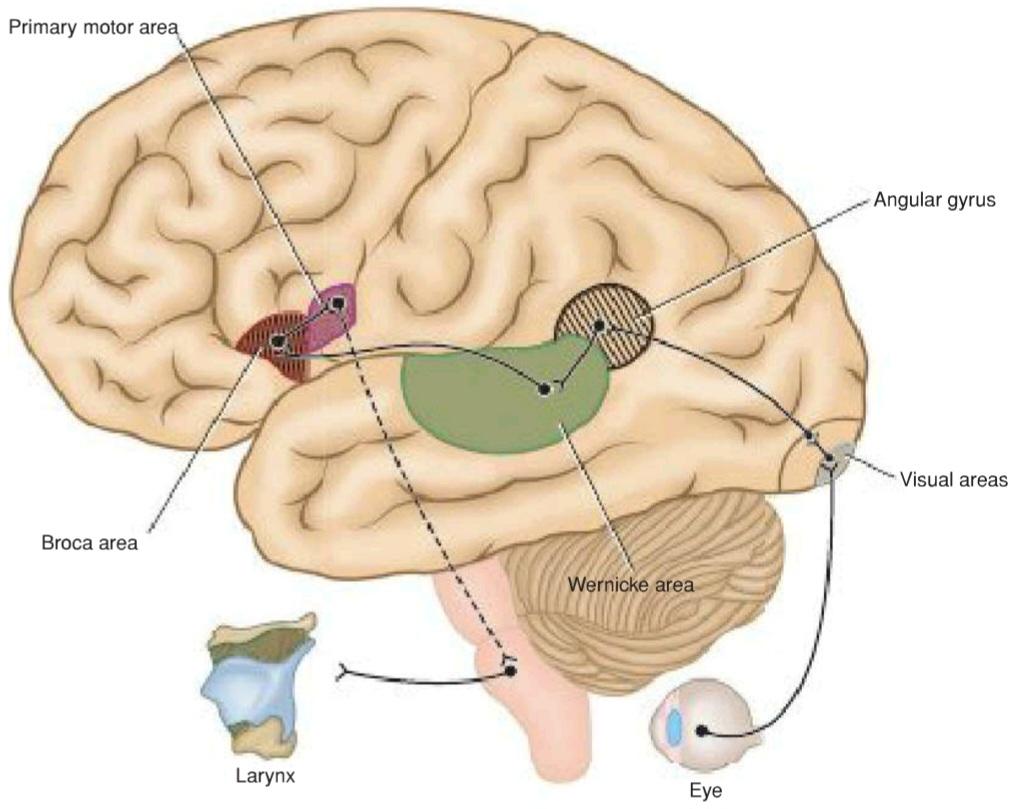
Figure 8-6 Probable nerve pathways involved in reading a sentence and repeating it out loud.
شکل ۸-۶ مسیرهای عصبی احتمالی دخیل در خواندن یک جمله و تکرار آن با صدای بلند.

Figure 8-7 Probable nerve pathways involved with hearing a question and answering it.
شکل ۸-۷ مسیرهای عصبی احتمالی درگیر در شنیدن یک سوال و پاسخ دادن به آن.
Other Cortical Areas
The taste area is situated at the lower end of the postcentral gyrus in the superior wall of the lateral sulcus and in the adjoining area of the insula (Brodmann area 43). Ascending fibers from the nucleus solitarius probably ascend to the ventral posteromedial nucleus of the thalamus, where they synapse on neurons that send fibers to the cortex.
سایر نواحی قشری
ناحیه چشایی در انتهای پایینی شکنج پس مرکزی در دیواره فوقانی شیار جانبی و در ناحیه مجاور اینسولا (ناحیه برودمن ۴۳) قرار دارد. فیبرهای صعودی از هسته سولیتاریوس احتمالاً به هسته خلفی میانی شکمی تالاموس صعود میکنند، جایی که با نورونهایی که فیبرها را به قشر مغز میفرستند، سیناپس برقرار میکنند.
The vestibular area is believed to be situated near the part of the postcentral gyrus concerned with sensations of the face. Its location lies opposite the auditory area in the superior temporal gyrus. The vestibular area and the vestibular part of the inner ear are concerned with appreciation of the positions and movements of the head in space. Through its nerve connections, the movements of the eyes and the muscles of the trunk and limbs are influenced in the maintenance of posture.
اعتقاد بر این است که ناحیه دهلیزی در نزدیکی بخشی از شکنج پس مرکزی که مربوط به احساسات چهره است، قرار دارد. محل آن در مقابل ناحیه شنوایی در شکنج گیجگاهی فوقانی قرار دارد. ناحیه دهلیزی و قسمت دهلیزی گوش داخلی مربوط به درک موقعیتها و حرکات سر در فضا هستند. از طریق ارتباطات عصبی آن، حرکات چشمها و عضلات تنه و اندامها در حفظ وضعیت بدن تأثیر میگذارند.
The insula is an area of the cortex that is buried within the lateral sulcus and forms its floor (see Fig. 7-9). It can be examined only when the lips of the lateral sulcus are separated widely. Histologically, the posterior part is granular and the anterior part is agranular, thus resembling the adjoining cortical areas. Its fiber connections are incompletely known. This area is thought to be important for planning or coordinating the articulatory movements necessary for speech.
اینسولا ناحیهای از قشر مغز است که در شیار جانبی قرار دارد و کف آن را تشکیل میدهد (شکل ۷-۹ را ببینید). تنها زمانی میتوان آن را بررسی کرد که لبهای شیار جانبی به طور گسترده از هم جدا شده باشند. از نظر بافتشناسی، قسمت خلفی دانهدار و قسمت قدامی غیردانهدار است، بنابراین شبیه نواحی قشری مجاور است. اتصالات فیبرهای آن به طور ناقص شناخته شده است. تصور میشود که این ناحیه برای برنامهریزی یا هماهنگی حرکات تلفظی لازم برای گفتار مهم است.
Association Cortex
The primary sensory areas with their granular cortex and the primary motor areas with their agranular cortex form only a small part of the total cortical surface area. The remaining areas have all six cellular layers and, therefore, are referred to as homotypical cortex. Classically, these large remaining areas were known as association areas, although precisely what they associate is not known. The original concept-that they receive information from the primary sensory areas that is to be integrated and analyzed in the association cortex and then fed to the motor areas-has not been established. As the result of clinical studies and animal experimentation, it has now become apparent that these areas of the cortex have multiple inputs and outputs and are very much concerned with behavior, discrimination, and interpretation of sensory experiences. Three main association areas are recognized: prefrontal, anterior temporal, and posterior parietal. The prefrontal cortex is discussed on page 291.
قشر ارتباطی
نواحی حسی اولیه با قشر دانهای خود و نواحی حرکتی اولیه با قشر غیر دانهای خود تنها بخش کوچکی از کل سطح قشر مغز را تشکیل میدهند. نواحی باقیمانده هر شش لایه سلولی را دارند و بنابراین، به عنوان قشر هموتیپیک شناخته میشوند. از نظر کلاسیک، این نواحی بزرگ باقیمانده به عنوان نواحی ارتباطی شناخته میشدند، اگرچه دقیقاً مشخص نیست که چه چیزی را مرتبط میکنند. مفهوم اصلی – اینکه آنها اطلاعاتی را از نواحی حسی اولیه دریافت میکنند که قرار است در قشر ارتباطی ادغام و تجزیه و تحلیل شوند و سپس به نواحی حرکتی تغذیه شوند – مشخص نشده است. در نتیجه مطالعات بالینی و آزمایشهای حیوانی، اکنون مشخص شده است که این نواحی قشر مغز ورودیها و خروجیهای متعددی دارند و بسیار با رفتار، تمایز و تفسیر تجربیات حسی مرتبط هستند. سه ناحیه ارتباطی اصلی شناخته شده است: پیشپیشانی، گیجگاهی قدامی و آهیانه خلفی. قشر پیشپیشانی در صفحه ۲۹۱ مورد بحث قرار گرفته است.
The anterior temporal cortex is thought to play a role in the storage of previous sensory experiences. Stimulation may cause the individual to recall objects seen or music heard in the past.
تصور میشود که قشر گیجگاهی قدامی در ذخیره تجربیات حسی قبلی نقش دارد. تحریک ممکن است باعث شود فرد اشیاء دیده شده یا موسیقی شنیده شده در گذشته را به یاد بیاورد.
In the posterior parietal cortex, visual information from the posterior occipital cortex and the sensory input of touch and pressure and proprioception from the anterior parietal cortex is integrated into concepts of size, form, and texture. This ability is known as stereognosis. A conscious appreciation of the body image is also assembled in the posterior parietal cortex. The brain knows at all times where each part of the body is located in relation to its environment. This information is so important when performing body movements. The right side of the body is represented in the left hemisphere, and the left side of the body is represented in the right hemisphere.
در قشر آهیانه خلفی، اطلاعات بصری از قشر پسسری خلفی و ورودی حسی لمس و فشار و حس عمقی از قشر آهیانه قدامی در مفاهیم اندازه، شکل و بافت ادغام میشوند. این توانایی به عنوان استریوگنوزیس شناخته میشود. درک آگاهانه از تصویر بدن نیز در قشر آهیانه خلفی گردآوری میشود. مغز در هر زمان میداند که هر قسمت از بدن در رابطه با محیط خود در کجا قرار دارد. این اطلاعات هنگام انجام حرکات بدن بسیار مهم است. سمت راست بدن در نیمکره چپ و سمت چپ بدن در نیمکره راست نشان داده میشود.
CEREBRAL DOMINANCE
Anatomical examination of the two cerebral hemispheres shows that the cortical gyri and fissures are almost identical. Moreover, nervous pathways projecting to the cortex do so largely contralaterally and equally to identical cortical areas. In addition, the cerebral commissures, especially the corpus callosum and the anterior commissure, provide a pathway for information that is received in one hemisphere to be transferred to the other. Nevertheless, certain nervous activity is predominantly performed by one of the two cerebral hemispheres. Handedness, perception of language, and speech are functional areas of behavior that are controlled by the dominant hemisphere in most individuals. By contrast, spatial perception, recognition of faces, and music are interpreted by the nondominant hemisphere (Fig. 8-8).
تسلط مغزی
بررسی آناتومیکی دو نیمکره مغزی نشان میدهد که شیارها و شیارهای قشری تقریباً یکسان هستند. علاوه بر این، مسیرهای عصبی که به قشر مغز منتهی میشوند، این کار را تا حد زیادی در جهت مخالف و به طور مساوی در نواحی قشری یکسان انجام میدهند. علاوه بر این، رابطهای مغزی، به ویژه جسم پینهای و رابط قدامی، مسیری را برای انتقال اطلاعات دریافتی در یک نیمکره به نیمکره دیگر فراهم میکنند. با این وجود، برخی از فعالیتهای عصبی عمدتاً توسط یکی از دو نیمکره مغزی انجام میشود. چپ دستی، درک زبان و گفتار، حوزههای عملکردی رفتار هستند که در اکثر افراد توسط نیمکره غالب کنترل میشوند. در مقابل، ادراک فضایی، تشخیص چهرهها و موسیقی توسط نیمکره غیرغالب تفسیر میشوند (شکل 8-8).

Figure 8-8 Nervous activities performed predominantly by dominant and nondominant hemispheres.
شکل ۸-۸ فعالیتهای عصبی که عمدتاً توسط نیمکرههای غالب و غیرغالب انجام میشوند.
More than 90% of the adult population is right handed and, therefore, is left-hemisphere dominant. About 96% of the adult population is left-hemisphere dominant for speech.
بیش از ۹۰٪ از جمعیت بزرگسالان راست دست هستند و بنابراین، نیمکره چپ غالب است. حدود ۹۶٪ از جمعیت بزرگسالان برای گفتار، نیمکره چپ غالب است.
In their work on human fetuses and neonates, Yakolev and Rakic have shown that more descending fibers in the left pyramid cross over the midline in the decussation than vice versa. This would suggest that the anterior horn cells on the right side of the spinal cord have a greater corticospinal innervation than those on the left side in most individuals, which might explain the dominance of the right hand.
یاکولف و راکیک در کار خود بر روی جنینها و نوزادان انسان نشان دادهاند که فیبرهای نزولی بیشتری در هرم چپ در تقاطع از خط میانی عبور میکنند تا برعکس. این نشان میدهد که سلولهای شاخ قدامی در سمت راست نخاع در بیشتر افراد، عصبدهی قشری-نخاعی بیشتری نسبت به سلولهای سمت چپ دارند، که ممکن است تسلط دست راست را توضیح دهد.
Other researchers have shown that the speech area of the adult cortex is larger on the left than on the right, whereas the two hemispheres of the newborn are believed to have equipotential capabilities. During childhood, one hemisphere slowly comes to dominate the other, and only after the first decade does the dominance become fixed. This would explain why a 5-year-old child with damage to the dominant hemisphere can easily learn to become left-handed and speak well, whereas this is almost impossible in the adult.
محققان دیگر نشان دادهاند که ناحیه گفتاری قشر مغز بزرگسالان در سمت چپ بزرگتر از سمت راست است، در حالی که اعتقاد بر این است که دو نیمکره نوزاد دارای قابلیتهای همپتانسیل هستند. در دوران کودکی، یک نیمکره به آرامی بر نیمکره دیگر غالب میشود و تنها پس از دهه اول، تسلط ثابت میشود. این توضیح میدهد که چرا یک کودک ۵ ساله با آسیب به نیمکره غالب میتواند به راحتی چپ دست شود و خوب صحبت کند، در حالی که این امر در یک بزرگسال تقریباً غیرممکن است.
«فصل هشتم کتاب نورواناتومی بالینی اسنل»
🚀 با ما همراه شوید!
تازهترین مطالب و آموزشهای مغز و اعصاب را از دست ندهید. با فالو کردن کانال تلگرام آیندهنگاران مغز، از ما حمایت کنید!



