پرسشهای مروری و مسائل بالینی ساختار و مناطق عملکردی قشر مغز


دعای مطالعه [ نمایش ]
بِسْمِ الله الرَّحْمنِ الرَّحیمِ
اَللّهُمَّ اَخْرِجْنى مِنْ ظُلُماتِ الْوَهْمِ
خدايا مرا بيرون آور از تاريكىهاى وهم،
وَ اَكْرِمْنى بِنُورِ الْفَهْمِ
و به نور فهم گرامى ام بدار،
اَللّهُمَّ افْتَحْ عَلَيْنا اَبْوابَ رَحْمَتِكَ
خدايا درهاى رحمتت را به روى ما بگشا،
وَانْشُرْ عَلَيْنا خَزائِنَ عُلُومِكَ بِرَحْمَتِكَ يا اَرْحَمَ الرّاحِمينَ
و خزانههاى علومت را بر ما باز كن به امید رحمتت اى مهربانترين مهربانان.
آیندهنگاران مغز: «ما مغز را میشناسیم، تا آینده را بسازیم.»
کتاب «نوروآناتومی بالینی اسنل» اثر دکتر ریچارد اس. اسنل، با ویرایش جدید دکتر رایان اسپلیتگربر، همچنان بهعنوان مرجع طلایی این حوزه شناخته میشود. این اثر با تلفیق تازهترین پژوهشها و کاربردهای بالینی، پلی استوار میان دانش پایه و عمل پزشکی بنا میکند.
تلاشهای علمی و سالها تدریس ارزشمند استاد گرانقدر دکتر محمدتقی جغتایی الهامبخش ترجمه و گسترش این دانش در ایران بوده است.
ترجمه دقیق این کتاب توسط برند علمی آیندهنگاران مغز به مدیریت داریوش طاهری، امکان دسترسی فارسیزبانان به مرزهای نوین علوم اعصاب را فراهم کرده و رسالتی علمی در جهت شناخت عمیقتر مغز و ترسیم آیندهای روشنتر بر عهده گرفته است.
»» فصل ۸: ساختار و مناطق عملکردی قشر مغز؛ بخش پرسشهای مروری و مسائل بالینی
»» Chapter 8: The Structure and Functional Localization of the Cerebral Cortex
? Clinical Problem Solving
1. During a pathology class, a student is shown a slide illustrating a particular form of cerebral tumor. A small area of the cerebral cortex is visible at the edge of the section. The instructor asks the student whether the tissue has been removed from a motor or sensory area of the cortex. What is the main difference in structure between the motor and sensory areas of the cerebral cortex?
؟ حل مسئله بالینی
۱. در طول کلاس پاتولوژی، به دانشجو اسلایدی نشان داده میشود که نوع خاصی از تومور مغزی را نشان میدهد. ناحیه کوچکی از قشر مغز در لبه برش قابل مشاهده است. استاد از دانشجو میپرسد که آیا بافت از ناحیه حرکتی یا حسی قشر مغز برداشته شده است. تفاوت اصلی در ساختار بین نواحی حرکتی و حسی قشر مغز چیست؟
2. A 43-year-old man is examined by a neurologist for a suspected brain tumor. The patient is tested for stereognosis, that is, the appreciation of form in three dimensions. With the patient’s eyes closed, a hairbrush is placed in his right hand, and he is asked to recognize the object. He is unable to recognize the brush even after the neurologist moves the brush about in the patient’s hand. On opening his eyes, the patient immediately recognizes the brush. (a) Name the area of the cerebral cortex likely to be diseased in this patient. (b) Do you think moving the object around in the patient’s hand is necessary?
۲. یک متخصص مغز و اعصاب، مردی ۴۳ ساله را به دلیل مشکوک بودن به تومور مغزی معاینه میکند. بیمار از نظر استریوگنوزیس، یعنی درک فرم در سه بعد، آزمایش میشود. در حالی که چشمان بیمار بسته است، یک برس مو در دست راست او قرار میگیرد و از او خواسته میشود که شیء را تشخیص دهد. حتی پس از اینکه متخصص مغز و اعصاب برس را در دست بیمار حرکت میدهد، او قادر به تشخیص برس نیست. با باز کردن چشمانش، بیمار بلافاصله برس را تشخیص میدهد. (الف) ناحیهای از قشر مغز که احتمالاً در این بیمار بیمار است را نام ببرید. (ب) آیا فکر میکنید حرکت دادن شیء در دست بیمار ضروری است؟
3. A 65-year-old man visits his physician because he notices that he has been dragging his right foot when walking for the past 3 weeks. On physical examination, he is found to have an increase in tone of the flexor muscles of the right arm, and, when he walks, he tends to hold his right arm adducted and flexed. He also holds his right fist tightly clenched. On study of the patient’s gait, he is seen to have difficulty in flexing his right hip and knee. Slight but definite weakness and increased tone of the muscles of the right leg is noted. As the patient walks, he moves his right leg in a semicircle and places his forefoot on the ground before the heel. Examination of the right shoe shows evidence of increased wear beneath the right toes. Given that this patient had a cerebrovascular lesion involving the cerebral cortex, which area of the cortex was involved to cause these symptoms?
۳. مردی ۶۵ ساله به پزشک مراجعه میکند زیرا متوجه میشود که در ۳ هفته گذشته هنگام راه رفتن، پای راست خود را روی زمین میکشد. در معاینه فیزیکی، افزایش تون عضلات خمکننده بازوی راست او مشاهده میشود و هنگام راه رفتن، تمایل دارد بازوی راست خود را به سمت داخل و خم نگه دارد. او همچنین مشت راستش را محکم گره میکند. در بررسی نحوه راه رفتن بیمار، مشاهده میشود که در خم کردن لگن و زانوی راست خود مشکل دارد. ضعف خفیف اما مشخص و افزایش تون عضلات پای راست مشاهده میشود. هنگام راه رفتن، پای راست خود را به صورت نیمدایره حرکت میدهد و جلوی پایش را قبل از پاشنه روی زمین قرار میدهد. معاینه کفش راست، شواهدی از افزایش ساییدگی زیر انگشتان پای راست را نشان میدهد. با توجه به اینکه این بیمار ضایعه عروقی مغزی در قشر مغز داشته است، کدام ناحیه از قشر مغز درگیر شده و باعث ایجاد این علائم شده است؟
4. While examining an unconscious patient, a clinician notes that when the patient’s head is gently rotated to the right, the two eyes deviated to the left. On rotation of the patient’s head to the left, the patient’s eyes still look to the left. Which area of the cortex is likely to be damaged in this patient?
۴. پزشک در حین معاینه یک بیمار بیهوش متوجه میشود که وقتی سر بیمار به آرامی به سمت راست چرخانده میشود، دو چشم او به سمت چپ منحرف میشوند. با چرخاندن سر بیمار به سمت چپ، چشمان بیمار همچنان به سمت چپ نگاه میکنند. کدام ناحیه از قشر مغز در این بیمار احتمالاً آسیب دیده است؟
5. A 25-year-old soldier is wounded in combat by an explosive device. A small piece of shrapnel enters the right side of his skull over the precentral gyrus. Five years later, he is examined by a physician during a routine physical checkup and is found to have weakness of the left leg. The physician cannot detect any increase in muscle tone in his left leg. Explain why most patients with damage to the motor area of the cerebral cortex have spastic muscle paralysis, while a few patients retain normal muscle tone.
۵. یک سرباز ۲۵ ساله در جنگ توسط یک وسیله انفجاری زخمی میشود. یک تکه کوچک ترکش به سمت راست جمجمه او روی شکنج پیشمرکزی وارد میشود. پنج سال بعد، او توسط یک پزشک در طی یک معاینه فیزیکی معمول معاینه میشود و مشخص میشود که دچار ضعف پای چپ است. پزشک نمیتواند هیچ افزایشی در تون عضلانی پای چپ او تشخیص دهد. توضیح دهید که چرا اکثر بیماران مبتلا به آسیب به ناحیه حرکتی قشر مغز، فلج عضلانی اسپاستیک دارند، در حالی که تعداد کمی از بیماران تون عضلانی طبیعی خود را حفظ میکنند.
6. A distinguished neurobiologist gives a lecture on the physiology of the cerebral cortex to the freshman medical student class. After reviewing the structure of the different areas of the cerebral cortex and the functional localization of the cerebral cortex, he states that our knowledge of the cytoarchitecture of the human cerebral cortex has contributed very little to our understanding of the normal functional activity of the cerebral cortex. Do you agree with his statement? What do you understand by the term vertical chain theory?
۶. یک متخصص مغز و اعصاب برجسته، در مورد فیزیولوژی قشر مغز برای دانشجویان سال اول پزشکی سخنرانی میکند. او پس از بررسی ساختار نواحی مختلف قشر مغز و محل قرارگیری عملکردی قشر مغز، اظهار میکند که دانش ما از ساختار سلولی قشر مغز انسان، کمک بسیار کمی به درک ما از فعالیت عملکردی طبیعی قشر مغز کرده است. آیا با گفته او موافقید؟ منظور شما از اصطلاح نظریه زنجیره عمودی چیست؟
7. An 18-year-old boy receives a gunshot wound that severely damages his left precentral gyrus. On recovering from the incident, he leaves the hospital with spastic paralysis of the right arm and leg. However, he still possesses some coarse voluntary movements of the right shoulder, hip, and knee. Explain the presence of these residual movements on the right side.
۷. یک پسر ۱۸ ساله بر اثر اصابت گلوله به شدت به شکنج پیشمرکزی چپ خود آسیب میزند. پس از بهبودی از این حادثه، او با فلج اسپاستیک دست و پای راست از بیمارستان مرخص میشود. با این حال، او هنوز برخی حرکات ارادی خشن شانه، لگن و زانوی راست را دارد. وجود این حرکات باقیمانده در سمت راست را توضیح دهید.
8. A 53-year-old professor and chairman of an anatomy department sustains a severe head injury while rock climbing. During the ascent of a crevasse, his companion’s ice axe falls from his belt and struck the professor’s head, causing a depressed fracture of the frontal bone. After convalescing from his accident, the professor returns to his position in the medical school, but faculty and the student body quickly notice that the professor’s social behavior has changed dramatically. His lectures, although amusing, no longer have direction. Although previously a smartly dressed man, he now has an unkempt appearance. The organization of the department started to deteriorate rapidly. Finally, he is removed from office after urinating into the trash basket in one of the classrooms. Use your knowledge of neuroanatomy to explain the professor’s altered behavior.
۸. یک استاد ۵۳ ساله و رئیس بخش آناتومی، هنگام صخرهنوردی دچار آسیب شدید سر میشود. در حین صعود از یک شکاف یخ، تبر یخ همراهش از کمربندش میافتد و به سر استاد برخورد میکند و باعث شکستگی فرورفته استخوان پیشانی میشود. پس از بهبودی از تصادف، استاد به سمت خود در دانشکده پزشکی بازمیگردد، اما اعضای هیئت علمی و دانشجویان به سرعت متوجه میشوند که رفتار اجتماعی استاد به طرز چشمگیری تغییر کرده است. سخنرانیهای او، اگرچه سرگرمکننده بود، اما دیگر جهت مشخصی نداشت. اگرچه قبلاً مردی شیکپوش بود، اما اکنون ظاهری نامرتب دارد. سازماندهی بخش به سرعت رو به وخامت گذاشت. سرانجام، او پس از ادرار کردن در سطل زباله یکی از کلاسها، از دفترش اخراج میشود. از دانش خود در مورد نوروآناتومی برای توضیح رفتار تغییر یافته استاد استفاده کنید.
9. A 50-year-old woman with a cerebrovascular lesion is found to experience difficulty in understanding spoken speech, although she fully understands written speech. Which area of the cerebral cortex was damaged?
۹. زنی ۵۰ ساله با ضایعه عروقی مغز، اگرچه گفتار نوشتاری را به طور کامل درک میکند، اما در درک گفتار شفاهی مشکل دارد. کدام ناحیه از قشر مغز آسیب دیده است؟
10. A 62-year-old man, on recovering from a stroke, is found to have difficulty in understanding written speech (alexia) but can easily understand spoken speech and written symbols. Which area of the cerebral cortex is damaged in this patient?
۱۰. مردی ۶۲ ساله، پس از بهبودی از سکته مغزی، در درک گفتار کتبی (آلکسی) مشکل دارد، اما میتواند به راحتی گفتار شفاهی و نمادهای نوشتاری را درک کند. کدام ناحیه از قشر مغز در این بیمار آسیب دیده است؟
11. What is understood by the following terms: (a) coma, (b) sleep, and (c) electroencephalogram? Name three neurologic conditions in which the diagnosis may be assisted by an electroencephalogram.
۱۱. منظور از اصطلاحات زیر چیست: (الف) کما، (ب) خواب و (ج) الکتروانسفالوگرام؟ سه بیماری عصبی را نام ببرید که در آنها الکتروانسفالوگرام میتواند به تشخیص کمک کند.
√ Answers and Explanations to Clinical Problem Solving
1. The cerebral cortex is made up of six identifiable layers. In the motor cortex in the precentral gyrus, the second and fourth layers lack of granular cells, and the third and fifth layers in the somesthetic cortex in the postcentral gyrus lack of pyramidal cells. The motor cortex is thicker than the sensory cortex.
√ پاسخها و توضیحات مربوط به حل مسئله بالینی
۱. قشر مغز از شش لایه قابل شناسایی تشکیل شده است. در قشر حرکتی در شکنج پیشمرکزی، لایههای دوم و چهارم فاقد سلولهای دانهای هستند و لایههای سوم و پنجم در قشر حسی در شکنج پسمرکزی فاقد سلولهای هرمی هستند. قشر حرکتی ضخیمتر از قشر حسی است.
2. (a) The area likely to be diseased is the left parietal lobe with advanced destruction of the superior parietal lobule. This is the somesthetic association area, where the sensations of touch, pressure, and proprioception are integrated. (b) Yes, the patient must be allowed to finger the object so that these different sensations can be appreciated.
۲. (الف) ناحیهای که احتمالاً بیمار است، لوب آهیانه چپ با تخریب پیشرفته لوبول آهیانه فوقانی است. این ناحیه، ناحیه ارتباط حسی-حرکتی است که در آن حسهای لمس، فشار و حس عمقی ادغام میشوند. (ب) بله، باید به بیمار اجازه داده شود که با انگشت خود شیء را لمس کند تا این حسهای مختلف درک شوند.
3. This patient had a cerebrovascular lesion involving the left precentral gyrus. The damage to the pyramidal cells that give origin to the corticospinal fibers was responsible for the right-sided paralysis. The increased tone of the paralyzed muscles was due to the loss of inhibition caused by involvement of the extrapyramidal fibers (see p. 290).
۳. این بیمار دچار ضایعه عروقی مغزی بود که شکنج پیشمرکزی چپ را درگیر میکرد. آسیب به سلولهای هرمی که منشأ فیبرهای قشری-نخاعی هستند، مسئول فلج سمت راست بود. افزایش تون عضلات فلج شده به دلیل از دست دادن مهار ناشی از درگیری فیبرهای خارج هرمی بود (به صفحه ۲۹۰ مراجعه کنید).
4. Destructive lesions of the frontal eye field of the left cerebral hemisphere caused the two eyes to deviate to the side of the lesion and an inability to turn the eyes to the opposite side. The frontal eye field is thought to control voluntary scanning movements of the eye and is independent of visual stimuli.
۴. ضایعات مخرب میدان دید جلویی نیمکره چپ مغز باعث انحراف دو چشم به سمت ضایعه و ناتوانی در چرخاندن چشمها به سمت مقابل شده است. تصور میشود میدان دید جلویی حرکات ارادی چشم را کنترل میکند و مستقل از محرکهای بینایی است.
5. A small discrete lesion of the primary motor cortex results in little change in muscle tone. Larger lesions involving the primary and secondary motor areas, which are the most common, result in muscle spasm. The explanation for this is given on page 290.
۵. یک ضایعه کوچک و مجزا در قشر حرکتی اولیه منجر به تغییر کمی در تون عضلانی میشود. ضایعات بزرگتر که نواحی حرکتی اولیه و ثانویه را درگیر میکنند، که شایعترین آنها هستند، منجر به اسپاسم عضلانی میشوند. توضیح این موضوع در صفحه ۲۹۰ آمده است.
6. The extensive histologic research of Brodmann, Campbell, Economo, and the Vogts has allowed the cerebral cortex to be divided into areas that have a different microscopic arrangement and different types of cells. These cortical maps are fundamentally similar, and the one proposed by Brodmann is used widely. Because the functional significance of many areas of the human cerebral cortex is not known, closely correlating structure with function has not been possible. In general, the motor cortices are thicker than the sensory cortices, and the motor cortex has less prominent second and fourth granular layers and has large pyramidal cells in the fifth layer. Other areas with a different structure may have similar functional roles. More recent studies using electrophysiologic techniques have indicated that dividing the cerebral cortex according to its thalamocortical projections is more accurate. The vertical chain mechanism of the cerebral cortex is fully described on page 283.
۶. تحقیقات بافتشناسی گستردهی برادمن، کمپبل، اکونومو و وگتس این امکان را فراهم کرده است که قشر مغز به مناطقی با آرایش میکروسکوپی متفاوت و انواع مختلف سلول تقسیم شود. این نقشههای قشری اساساً مشابه هستند و نقشهای که برادمن ارائه داده است، به طور گسترده مورد استفاده قرار میگیرد. از آنجا که اهمیت عملکردی بسیاری از نواحی قشر مغز انسان مشخص نیست، ارتباط دقیق ساختار با عملکرد امکانپذیر نبوده است. به طور کلی، قشرهای حرکتی ضخیمتر از قشرهای حسی هستند و قشر حرکتی لایههای دانهای دوم و چهارم کمتری دارد و در لایه پنجم سلولهای هرمی بزرگی دارد. سایر نواحی با ساختار متفاوت ممکن است نقشهای عملکردی مشابهی داشته باشند. مطالعات جدیدتر با استفاده از تکنیکهای الکتروفیزیولوژیک نشان دادهاند که تقسیم قشر مغز بر اساس برآمدگیهای تالاموکورتیکال آن دقیقتر است. مکانیسم زنجیره عمودی قشر مغز به طور کامل در صفحه ۲۸۳ شرح داده شده است.
7. In this patient, the persistence of coarse voluntary movements of the right shoulder, hip, and knee joints can be explained on the basis that coarse postural movements are controlled by the premotor area of the cortex and the basal ganglia, and these areas were spared in this patient.
۷. در این بیمار، تداوم حرکات ارادی خشن مفاصل شانه، ران و زانوی راست را میتوان بر این اساس توضیح داد که حرکات وضعیتی خشن توسط ناحیه پیش حرکتی قشر مغز و عقدههای قاعدهای کنترل میشوند و این نواحی در این بیمار سالم ماندهاند.
8. The professor’s altered behavior was due to a severe lesion involving both frontal lobes of the cerebrum secondary to the depressed fracture of the frontal bone. While destruction of the prefrontal cortex does not cause a marked loss of intelligence, it does result in the individual losing initiative and drive, and often the patient no longer conforms to the accepted modes of social behavior.
۸. تغییر رفتار استاد به دلیل ضایعه شدیدی بود که هر دو لوب پیشانی مغز را درگیر میکرد و ثانویه به شکستگی فرورفته استخوان پیشانی بود. اگرچه تخریب قشر جلوی مغز باعث از دست دادن قابل توجه هوش نمیشود، اما منجر به از دست دادن ابتکار و انگیزه فرد میشود و اغلب بیمار دیگر با شیوههای پذیرفته شده رفتار اجتماعی مطابقت ندارد.
9. The understanding of spoken speech requires the normal functioning of the secondary auditory area, which is situated posterior to the primary auditory area in the lateral sulcus and in the superior temporal gyrus. This area is believed to be necessary for the interpretation of sounds, and the information is passed on to the sensory speech area of Wernicke.
۹. درک گفتار شفاهی نیازمند عملکرد طبیعی ناحیه شنوایی ثانویه است که در خلف ناحیه شنوایی اولیه در شیار جانبی و در شکنج گیجگاهی فوقانی قرار دارد. اعتقاد بر این است که این ناحیه برای تفسیر صداها ضروری است و اطلاعات به ناحیه حسی گفتاری ورنیکه منتقل میشود.
10. The understanding of written speech requires the normal functioning of the secondary visual area of the cerebral cortex, which is situated in the walls of the posterior part of the calcarine sulcus on the medial and lateral surfaces of the cerebral hemisphere. The function of the secondary visual area is to relate visual information received by the primary visual area to past visual experiences. This information is then passed on to the dominant angular gyrus and relayed to the anterior part of the Wernicke speech area (see p. 283).
۱۰. درک گفتار نوشتاری نیازمند عملکرد طبیعی ناحیه بینایی ثانویه قشر مغز است که در دیوارههای قسمت خلفی شیار کالکارین روی سطوح داخلی و جانبی نیمکره مغزی قرار دارد. عملکرد ناحیه بینایی ثانویه، مرتبط کردن اطلاعات بصری دریافت شده توسط ناحیه بینایی اولیه با تجربیات بصری گذشته است. سپس این اطلاعات به شکنج زاویهای غالب منتقل شده و به قسمت قدامی ناحیه گفتاری ورنیکه منتقل میشود (به صفحه ۲۸۳ مراجعه کنید).
11. (a) Coma is the term applied to an unconscious patient. The patient will not speak and will respond only reflexly to painful stimuli. Deeply comatose individuals will make no response. The eyes are closed and do not move. (b) Sleep is a changed state of consciousness; it is discussed on page 292. (c) An electroencephalogram is a recording of the electrical activity of the cerebral cortex made by placing electrodes on the scalp. Detection of abnormalities of the alpha, beta, and delta rhythms may assist in the diagnosis of cerebral tumors, epilepsy, and cerebral abscesses.
۱۱. (الف) کما اصطلاحی است که برای بیمار بیهوش به کار میرود. بیمار صحبت نمیکند و فقط به صورت رفلکسی به محرکهای دردناک پاسخ میدهد. افراد در کمای عمیق هیچ پاسخی نمیدهند. چشمها بسته هستند و حرکت نمیکنند. (ب) خواب حالت تغییر یافتهای از هوشیاری است؛ در صفحه ۲۹۲ مورد بحث قرار گرفته است. (ج) الکتروانسفالوگرام ثبت فعالیت الکتریکی قشر مغز است که با قرار دادن الکترود روی پوست سر انجام میشود. تشخیص ناهنجاریهای ریتمهای آلفا، بتا و دلتا ممکن است به تشخیص تومورهای مغزی، صرع و آبسههای مغزی کمک کند.
? Review Questions
Directions: Each of the numbered items in this section is followed by answers. Select the ONE lettered answer that is CORRECT.
؟ سوالات مروری
دستورالعمل: هر یک از موارد شمارهگذاری شده در این بخش با پاسخهایی دنبال میشوند. تنها پاسخ صحیح با یک حرف را انتخاب کنید.
1. The following statements concern the cerebral cortex:
(a) The cerebral cortex is thinnest over the crest of a gyrus and thickest in the depth of a sulcus.
(b) The largest giant pyramidal cells are found in the postcentral gyrus.
(c) In the visual cortex, the outer band of Baillarger is thin and can only be seen under a microscope.
(d) The molecular layer is the most superficial layer of the cerebral cortex and is composed of the small cell bodies of the granular cells.
(e) From a functional point of view, the cerebral cortex is organized into vertical units of activity.
۱. عبارات زیر مربوط به قشر مغز است:
(الف) قشر مغز در بالای تاج یک شکنج نازکترین و در عمق یک شیار ضخیمترین است.
(ب) بزرگترین سلولهای هرمی غولپیکر در شکنج پس مرکزی یافت میشوند.
(ج) در قشر بینایی، نوار بیرونی Baillarger نازک است و فقط زیر میکروسکوپ قابل مشاهده است.
(د) لایه مولکولی سطحیترین لایه قشر مغز است و از اجسام سلولی کوچک سلولهای دانهای تشکیل شده است.
(ه) از دیدگاه عملکردی، قشر مغز به واحدهای عمودی فعالیت سازماندهی شده است.
2. The following statements concern the precentral area of the frontal lobe of the cerebral cortex
(a) The anterior region is known as the primary motor area.
(b) The primary motor area is responsible for skilled movements on the opposite side of the body.
(c) The function of the primary motor area is to store programs of motor activity, which are. conveyed to the premotor area for the execution of movements.
(d) Individual skeletal muscles are represented in the primary motor area.
(e) The area of cortex controlling a particular movement is not proportional to the skill involved.
۲. عبارات زیر مربوط به ناحیه پیشمرکزی لوب پیشانی قشر مغز است.
(الف) ناحیه قدامی به عنوان ناحیه حرکتی اولیه شناخته میشود.
(ب) ناحیه حرکتی اولیه مسئول حرکات ماهرانه در طرف مقابل بدن است.
(ج) وظیفه ناحیه حرکتی اولیه ذخیره برنامههای فعالیت حرکتی است که برای اجرای حرکات به ناحیه پیشحرکتی منتقل میشوند.
(د) عضلات اسکلتی منفرد در ناحیه حرکتی اولیه نمایش داده میشوند.
(ه) ناحیهای از قشر که یک حرکت خاص را کنترل میکند، متناسب با مهارت مربوطه نیست.
(b) The Broca speech area brings about the formation of words by its connections with the secondary motor area.
(c) It is not connected to the sensory speech area of Wernicke.
(d) It is located in the superior frontal gyrus between the anterior and ascending rami and the ascending and posterior rami of the lateral fissure.
(e) Brodmann areas 34 and 35 represent the motor speech area.
۳. عبارات زیر مربوط به ناحیه حرکتی گفتار بروکا است:
(الف) در بیشتر افراد، این ناحیه در نیمکره چپ یا غالب اهمیت دارد.
(ب) ناحیه گفتار بروکا با ارتباط با ناحیه حرکتی ثانویه، کلمات را تشکیل میدهد.
(ج) به ناحیه حسی گفتار ورنیکه متصل نیست.
(د) در شکنج پیشانی فوقانی بین شاخههای قدامی و صعودی و شاخههای صعودی و خلفی شیار جانبی قرار دارد.
(ه) نواحی ۳۴ و ۳۵ برادمن نشان دهنده ناحیه حرکتی گفتار هستند.
4. The following statements concern the primary somesthetic area:
(a) It occupies the lower part of the precentral gyrus.
(b) Histologically, it contains large numbers of pyramidal cells and few granular cells.
(c) The opposite half of the body is represented inverted.
(d) Although most sensations reach the cortex from the contralateral side of the body, sensations from the hand go to both sides.
(e) The area extends onto the anterior part of the paracentral lobule.
۴. عبارات زیر مربوط به ناحیه اولیه سمستتیک است:
(الف) این ناحیه قسمت پایینی شکنج پیشمرکزی را اشغال میکند.
(ب) از نظر بافتشناسی، حاوی تعداد زیادی سلول هرمی و تعداد کمی سلول دانهای است.
(ج) نیمه مقابل بدن به صورت معکوس نمایش داده شده است.
(د) اگرچه بیشتر حسها از سمت مقابل بدن به قشر مخ میرسند، حسهای دست به هر دو طرف میروند.
(ه) این ناحیه تا قسمت قدامی لوبول پاراسنترال امتداد مییابد.
(b) The visual cortex receives afferent fibers from the medial geniculate body.
(c) The right half of the visual field is represented in the visual cortex of the right cerebral hemisphere.
(d) The superior retinal quadrants pass to the inferior portion of the visual cortex.
(e) The secondary visual area (Brodmann areas 18 and 19) is surrounded by the primary visual area on the medial and lateral surfaces of the hemisphere.
۵. عبارات زیر مربوط به نواحی بینایی قشر مغز است:
(الف) ناحیه بینایی اولیه در دیوارههای شیار آهیانه-پسسری قرار دارد.
(ب) قشر بینایی فیبرهای آوران را از جسم زانویی میانی دریافت میکند.
(ج) نیمه راست میدان بینایی در قشر بینایی نیمکره راست مغز نشان داده شده است.
(د) ربعهای فوقانی شبکیه به قسمت تحتانی قشر بینایی منتقل میشوند.
(ه) ناحیه بینایی ثانویه (نواحی ۱۸ و ۱۹ برودمن) توسط ناحیه بینایی اولیه در سطوح داخلی و جانبی نیمکره احاطه شده است.
6. The following statements concern the superior temporal gyrus:
(a) The primary auditory area is situated in the inferior wall of the lateral sulcus.
(b) The main projection fibers to the primary auditory area arise from the thalamus.
(c) The sensory speech area of Wernicke is localized in the inferior temporal gyrus in the dominant hemisphere. (d) A unilateral lesion of the auditory area produces complete deafness in both ears.
(e) The secondary auditory area is sometimes referred to as Brodmann areas 41 and 42.
(ب) فیبرهای اصلی به ناحیه شنوایی اولیه از تالاموس منشأ میگیرند.
(ج) ناحیه حسی گفتاری ورنیکه در شکنج گیجگاهی تحتانی در نیمکره غالب قرار دارد.
(د) ضایعه یک طرفه ناحیه شنوایی باعث ناشنوایی کامل در هر دو گوش میشود.
(ه) ناحیه شنوایی ثانویه گاهی اوقات به عنوان نواحی ۴۱ و ۴۲ برودمن شناخته میشود.
7. The following statements concern the association areas of the cerebral cortex:
(a) They form a small area of the cortical surface.
(b) The prefrontal area is concerned with the makeup of the individual’s personality.
(c) They are concerned with the interpretation of motor experiences.
(d) Appreciation of the body image is assembled in the anterior parietal cortex, and the right side of the body is represented in the left hemisphere.
(e) The association areas have only four layers of cortex.
۷. عبارات زیر مربوط به نواحی ارتباطی قشر مغز هستند:
(الف) آنها ناحیه کوچکی از سطح قشر مغز را تشکیل میدهند.
(ب) ناحیه پیشپیشانی مربوط به ساختار شخصیت فرد است.
(ج) آنها مربوط به تفسیر تجربیات حرکتی هستند.
(د) درک تصویر بدن در قشر آهیانه قدامی جمع میشود و سمت راست بدن در نیمکره چپ نشان داده میشود.
(ه) نواحی ارتباطی فقط چهار لایه قشر دارند.
(b) More than 90% of the adult population is righthanded and, therefore, is left-hemisphere dominant.
(c) About 96% of the adult population is right-hemisphere dominant for speech.
(d) The nondominant hemisphere interprets handedness, perception of language, and speech.
(e) After puberty, the dominance of the cerebral hemispheres becomes fixed.
۸. عبارات زیر مربوط به تسلط مغزی هستند:
(الف) چینش قشر مغز در نیمکرههای غالب و غیرغالب به طور متفاوتی مرتب شدهاند.
(ب) بیش از ۹۰٪ از جمعیت بزرگسال راست دست هستند و بنابراین نیمکره چپ غالب است.
(ج) حدود ۹۶٪ از جمعیت بزرگسال نیمکره راست غالب برای گفتار است.
(د) نیمکره غیرغالب، چپ دستی، درک زبان و گفتار را تفسیر میکند.
(ه) پس از بلوغ، تسلط نیمکرههای مغزی ثابت میشود.
Matching Questions. Directions: The following questions apply to Figure 8-9. Match the numbers listed on the left with the most likely words designating lettered functional areas of the cerebral cortex listed on the right. Each lettered option may be selected once, more than once, or not at all.
سوالات تطبیقی. دستورالعمل: سوالات زیر در مورد شکل ۸-۹ صدق میکند. اعداد ذکر شده در سمت چپ را با محتملترین کلماتی که نواحی عملکردی حروفدار قشر مغز را که در سمت راست فهرست شدهاند، مشخص میکنند، مطابقت دهید. هر گزینه حروفدار میتواند یک بار، بیش از یک بار یا اصلاً انتخاب نشود.

10. Number 2 (b) Secondary auditory area
11. Number 3 (c) Frontal eye field
12. Number 4 (d) Primary somesthetic area
(e) None of the above
۱۰. شماره ۲ (ب) ناحیه شنوایی ثانویه
۱۱. شماره ۳ (ج) میدان دید جلویی
۱۲. شماره ۴ (د) ناحیه حسی اولیه
(ه) هیچ یک از موارد فوق
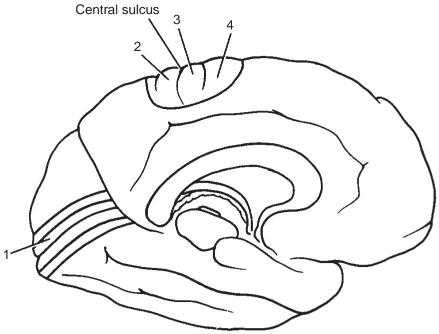
14. Number 1 (b) Primary somesthetic area
15. Number 1 (c) Primary visual area
16. Number 1 (d) Primary motor area None of the above
۱۴. شماره ۱ (ب) ناحیه حسی-حرکتی اولیه
۱۵. شماره ۱ (ج) ناحیه بینایی اولیه
۱۶. شماره ۱ (د) ناحیه حرکتی اولیه هیچ یک از موارد فوق
(b) She readily reacted to sensory stimulation of her skin on the left side.
(c) On being asked to move her left leg, she promptly did so.
(d) Muscular weakness of the upper and lower limbs on the left side was evident.
(e) On being asked to walk across the examining room, she tended not to use her left leg as much as her right leg.
۱۷. متخصص مغز و اعصاب بیمار را معاینه کرد و علائم محتمل زیر را یافت، به جز:
(الف) بیمار به سمت چپ خود نگاه نمیکرد.
(ب) او به راحتی به تحریک حسی پوست خود در سمت چپ واکنش نشان میداد.
(ج) وقتی از او خواسته شد پای چپ خود را حرکت دهد، او فوراً این کار را انجام داد.
(د) ضعف عضلانی اندام فوقانی و تحتانی در سمت چپ مشهود بود.
(ه) وقتی از او خواسته شد در اتاق معاینه راه برود، تمایل داشت از پای چپ خود به اندازه پای راستش استفاده نکند.
18. The neurologist made the following likely conclusions except:
(a) The diagnosis of left hemiasomatognosia (loss of appreciation of the left side of the body) was made.
(b) This condition probably resulted from a lesion of the left parietal lobe.
(c) In addition, the patient exhibited left hemiakinesia (unilateral motor neglect).
(d) A lesion was likely in areas 6 and 8 of the medial and lateral premotor regions of the right frontal lobe.
(e) The failure to look toward the left side (visual extinction) suggested a lesion existed in the right parietooccipital lobes.
(ب) این وضعیت احتمالاً ناشی از ضایعهای در لوب آهیانه چپ بود.
(ج) علاوه بر این، بیمار همیآکینزی چپ (ناتوانی حرکتی یک طرفه) را نشان میداد.
(د) احتمالاً ضایعهای در نواحی ۶ و ۸ از نواحی پیشحرکتی داخلی و خارجی لوب پیشانی راست وجود داشت.
(ه) عدم نگاه به سمت چپ (خاموشی بینایی) نشان دهنده وجود ضایعه در لوبهای آهیانه پسسری راست بود.
√ Answers and Explanations to Review Questions
1. E is correct. From a functional standpoint, the cerebral cortex is organized into vertical units of activity. A. The cerebral cortex is thickest over the crest of a gyrus and thinnest in the depth of a sulcus. B. The largest giant pyramidal cells are found in the precentral gyrus (see Fig. 8-1). C. In the visual cortex, the outer band of Baillarger is so thick that it can be seen with the naked eye (see Fig. 8-3). D. The molecular layer is the most superficial layer of the cerebral cortex and is composed mainly of a dense network of tangentially oriented nerve fibers (see Fig. 8-2).
√ پاسخها و توضیحات برای مرور سوالات
۱. ه صحیح است. از دیدگاه عملکردی، قشر مغز به واحدهای عمودی فعالیت سازماندهی شده است. الف. قشر مغز در بالای تاج یک شکنج ضخیمترین و در عمق یک شیار نازکترین است. ب. بزرگترین سلولهای هرمی غولپیکر در شکنج پیشمرکزی یافت میشوند (شکل ۸-۱ را ببینید). ج. در قشر بینایی، نوار بیرونی Baillarger آنقدر ضخیم است که میتوان آن را با چشم غیرمسلح دید (شکل ۸-۳ را ببینید). د. لایه مولکولی سطحیترین لایه قشر مغز است و عمدتاً از یک شبکه متراکم از الیاف عصبی مماس تشکیل شده است (شکل ۸-۲ را ببینید).
2. B is correct. The primary motor area of the frontal lobe is responsible for skilled movements on the opposite side of the body. A. In the frontal lobe of the cerebral hemisphere, the posterior region is known as the primary motor area (see Fig. 8-4). C. The function of the premotor area is to store programs of motor activity, which are conveyed to the primary motor area for the execution of movements. D. The individual skeletal muscles are not represented in the primary motor area. E. The area of cerebral cortex controlling a particular move- ment is proportional to the skill of the movement.
۲. ب صحیح است. ناحیه حرکتی اولیه لوب پیشانی مسئول حرکات ماهرانه در طرف مقابل بدن است. الف. در لوب پیشانی نیمکره مغزی، ناحیه خلفی به عنوان ناحیه حرکتی اولیه شناخته میشود (شکل ۸-۴ را ببینید). ج. وظیفه ناحیه پیش حرکتی ذخیره برنامههای فعالیت حرکتی است که برای اجرای حرکات به ناحیه حرکتی اولیه منتقل میشوند. د. عضلات اسکلتی منفرد در ناحیه حرکتی اولیه نمایش داده نمیشوند. ه. ناحیهای از قشر مغز که یک حرکت خاص را کنترل میکند، متناسب با مهارت آن حرکت است.
3. A is correct. In most individuals, the speech area of Broca is important on the left or dominant hemi- sphere. B. The Broca speech area brings about the formation of words by its connections with the primary motor area. C. The Broca speech area is connected to the sensory speech area of Wernicke. D. The speech area of Broca is in the inferior frontal gyrus between the anterior and ascending rami and the ascending and posterior rami of the lateral fissure (see Fig. 8-4). E. Brodmann areas 44 and 45 represent the motor speech area (see Fig. 8-4).
۳. الف صحیح است. در بیشتر افراد، ناحیه گفتاری بروکا در نیمکره چپ یا غالب اهمیت دارد. ب. ناحیه گفتاری بروکا با ارتباطاتش با ناحیه حرکتی اولیه، باعث شکلگیری کلمات میشود. ج. ناحیه گفتاری بروکا به ناحیه گفتاری حسی ورنیکه متصل است. د. ناحیه گفتاری بروکا در شکنج پیشانی تحتانی بین شاخههای قدامی و صعودی و شاخههای صعودی و خلفی شیار جانبی قرار دارد (شکل ۸-۴ را ببینید). ه. نواحی ۴۴ و ۴۵ برادمن نشان دهنده ناحیه گفتاری حرکتی هستند (شکل ۸-۴ را ببینید).
4. C is correct. In the primary somesthetic area, the opposite half of the body is represented inverted. A. The primary somesthetic area occupies the postcentral gyrus (see Fig. 8-4). B. Histologically, the primary somesthetic area contains large numbers of granular cells and few pyramidal cells. D. Most sensations from different parts of the body reach the cortex from the contralateral side of the body; those from the hand also only go to the contralateral side. E. The primary somesthetic area extends onto the posterior part of the paracentral lobule (see Fig. 8-4).
۴. ج صحیح است. در ناحیهی اولیهی حس شنوایی، نیمهی مقابل بدن به صورت معکوس نمایش داده میشود. الف. ناحیهی اولیهی حس شنوایی، شکنج پسمرکزی را اشغال میکند (شکل ۸-۴ را ببینید). ب. از نظر بافتشناسی، ناحیهی اولیهی حس شنوایی شامل تعداد زیادی سلول دانهدار و تعداد کمی سلول هرمی است. د. بیشتر حسها از قسمتهای مختلف بدن از سمت مقابل بدن به قشر مغز میرسند؛ حسهای دست نیز فقط به سمت مقابل میروند. ه. ناحیهی اولیهی حس شنوایی تا قسمت خلفی لوبول پاراسنترال امتداد مییابد (شکل ۸-۴ را ببینید).
5. D is correct. The superior retinal quadrants pass to the inferior portion of the visual cortex. A. The primary visual cortex is located in the walls of the posterior part of the calcarine sulcus (see Fig. 8-4). B. The visual cortex receives afferent fibers from the lateral geniculate body. C. The right half of the visual field is represented in the visual cortex of the left cerebral hemisphere. E. The secondary visual area (Brodmann areas 18 and 19) surrounds the primary visual area on the medial and lateral surfaces of the hemisphere (see Fig. 8-4).
۵. د صحیح است. ربعهای فوقانی شبکیه به قسمت تحتانی قشر بینایی منتقل میشوند. الف. قشر بینایی اولیه در دیوارههای قسمت خلفی شیار کالکارین قرار دارد (شکل ۸-۴ را ببینید). ب. قشر بینایی فیبرهای آوران را از جسم زانویی جانبی دریافت میکند. ج. نیمه راست میدان بینایی در قشر بینایی نیمکره چپ مغز نشان داده شده است. ه. ناحیه بینایی ثانویه (نواحی ۱۸ و ۱۹ برودمن) ناحیه بینایی اولیه را در سطوح داخلی و جانبی نیمکره احاطه کرده است (شکل ۸-۴ را ببینید).
6. A is correct. The primary auditory area is situated in the inferior wall of the lateral sulcus (see Fig. 8-4). B. The main projection fibers to the primary auditory area arise from the medial geniculate body. C. The sensory speech area of Wernicke is localized in the superior temporal gyrus in the dominant hemisphere (see Fig. 8-4). D. A unilateral lesion of the auditory area produces partial deafness in both ears. E. The primary auditory area is sometimes referred to as Brodmann areas 41 and 42.
۶. الف صحیح است. ناحیه شنوایی اولیه در دیواره تحتانی شیار جانبی قرار دارد (شکل ۸-۴ را ببینید). ب. فیبرهای اصلی به ناحیه شنوایی اولیه از جسم زانویی داخلی منشأ میگیرند. ج. ناحیه حسی گفتاری ورنیکه در شکنج گیجگاهی فوقانی در نیمکره غالب قرار دارد (شکل ۸-۴ را ببینید). د. ضایعه یک طرفه ناحیه شنوایی باعث ناشنوایی نسبی در هر دو گوش میشود. ه. ناحیه شنوایی اولیه گاهی اوقات به عنوان نواحی ۴۱ و ۴۲ برودمن شناخته میشود.
7. B is correct. The prefrontal area is concerned with the makeup of the individual’s personality. A. The association areas of the cerebral cortex form a large area of the cortical surface. C. The association areas are concerned with the interpretations of sensory experiences. D. Appreciation of the body image is assembled in the posterior parietal cortex, and the right side of the body is represented in the left hemisphere. E. The association areas have all six cellular layers and are referred to as homotypical cortex.
۷. ب صحیح است. ناحیه پیشپیشانی مربوط به ساختار شخصیت فرد است. الف. نواحی ارتباطی قشر مغز، ناحیه بزرگی از سطح قشری را تشکیل میدهند. ج. نواحی ارتباطی مربوط به تفسیر تجربیات حسی هستند. د. درک تصویر بدن در قشر آهیانه خلفی جمع میشود و سمت راست بدن در نیمکره چپ نشان داده میشود. ه. نواحی ارتباطی هر شش لایه سلولی را دارند و به عنوان قشر هموتیپیک شناخته میشوند.
8. B is correct. More than 90% of the adult population is right-handed and, therefore, is left-hemisphere dominant. A. The cortical gyri of the dominant and nondominant hemispheres are arranged in the same way. C. About 96% of the adult population is left-hemisphere dominant for speech. D. The nondominant hemisphere interprets spatial perception, recognition of faces, and music. E. After the first decade of life, the dominance of the cerebral hemispheres becomes fixed.
۸. ب صحیح است. بیش از ۹۰٪ از جمعیت بزرگسال راست دست هستند و بنابراین، نیمکره چپ غالب است. الف) چینش چینههای قشری نیمکرههای غالب و غیرغالب به یک شکل قرار گرفتهاند. ج) حدود ۹۶٪ از جمعیت بزرگسال نیمکره چپ غالب برای گفتار است. د) نیمکره غیرغالب ادراک فضایی، تشخیص چهرهها و موسیقی را تفسیر میکند. ه) پس از دهه اول زندگی، تسلط نیمکرههای مغزی ثابت میشود.
The answers for Figure 8-9, which shows the lateral view of the left cerebral hemisphere, are as follows:
پاسخهای شکل ۸-۹، که نمای جانبی نیمکره چپ مغز را نشان میدهد، به شرح زیر است:
9. C is correct; 1 is the frontal eye field.
۹. ج صحیح است؛ ۱ میدان دید جلویی است.
10. E is correct; 2 is the secondary somesthetic area (see Fig. 8-4).
۱۰. ه صحیح است؛ ۲ ناحیه حسی-حسی ثانویه است (شکل ۸-۴ را ببینید).
11. E is correct; 3 is the Wernicke sensory speech area (see Fig. 8-4).
۱۱. ه صحیح است؛ ۳ ناحیه گفتار حسی ورنیکه است (شکل ۸-۴ را ببینید).
12. B is correct; 4 is the secondary auditory area (see Fig. 8-4).
۱۲. ب صحیح است؛ ۴ ناحیه شنوایی ثانویه است (شکل ۸-۴ را ببینید).
The answers for Figure 8-10, which shows the medial view of the left cerebral hemisphere, are as follows:
پاسخهای شکل ۸-۱۰، که نمای داخلی نیمکره چپ مغز را نشان میدهد، به شرح زیر است:
13. C is correct; 1 is the primary visual area (see Fig. 8-4).
۱۳. ج صحیح است؛ ۱ ناحیه بینایی اولیه است (شکل ۸-۴ را ببینید).
۱۶. الف صحیح است؛ ۴ ناحیه پیش حرکتی است (شکل ۸-۴ را ببینید).
17. D is correct. The patient exhibited no weakness of her muscles on the left side despite the fact that her sister stated that she tended not to use her left leg.
۱۷. د صحیح است. بیمار هیچ ضعفی در عضلات سمت چپ خود نشان نداد، با وجود اینکه خواهرش اظهار داشت که تمایل به استفاده از پای چپ خود ندارد.
18. B is correct. An MRI revealed a tumor in the right parieto-occipital lobes; a further lesion was present in the right frontal lobe.
۱۸. ب صحیح است. MRI توموری را در لوبهای آهیانه-پسسری راست نشان داد؛ ضایعه دیگری در لوب پیشانی راست وجود داشت.
«فصل هشتم کتاب نورواناتومی بالینی اسنل»
🚀 با ما همراه شوید!
تازهترین مطالب و آموزشهای مغز و اعصاب را از دست ندهید. با فالو کردن کانال تلگرام آیندهنگاران مغز، از ما حمایت کنید!