
فیزیولوژی پزشکی گایتون و هال؛ عضله قلب؛ قلب به عنوان یک پمپ و عملکرد دریچه های قلب


دعای مطالعه [ نمایش ]
بِسْمِ الله الرَّحْمنِ الرَّحیمِ
اَللّهُمَّ اَخْرِجْنى مِنْ ظُلُماتِ الْوَهْمِ
خدايا مرا بيرون آور از تاريكىهاى وهم،
وَ اَكْرِمْنى بِنُورِ الْفَهْمِ
و به نور فهم گرامى ام بدار،
اَللّهُمَّ افْتَحْ عَلَيْنا اَبْوابَ رَحْمَتِكَ
خدايا درهاى رحمتت را به روى ما بگشا،
وَانْشُرْ عَلَيْنا خَزائِنَ عُلُومِكَ بِرَحْمَتِكَ يا اَرْحَمَ الرّاحِمينَ
و خزانههاى علومت را بر ما باز كن به امید رحمتت اى مهربانترين مهربانان.
کتاب «فیزیولوژی پزشکی گایتون وهال» بهعنوان یکی از جامعترین و معتبرترین منابع در حوزه علوم پزشکی، همچنان مرجع کلیدی برای درک عملکرد پیچیده بدن انسان است. این اثر با تکیه بر تازهترین پژوهشها و توضیحات دقیق از سازوکارهای فیزیولوژیک، پلی میان علوم پایه پزشکی و کاربردهای بالینی ایجاد میکند و نقشی بیبدیل در آموزش، پژوهش و ارتقای دانش سلامت ایفا مینماید.
ترجمه دقیق و علمیاین شاهکار توسط برند علمیآیندهنگاران مغز به مدیریت داریوش طاهری، دسترسی فارسیزبانان به مرزهای نوین دانش فیزیولوژی را ممکن ساخته و رسالتی علمیبرای ارتقای آموزش پزشکی، فهم عمیقتر سازوکارهای بدن و توسعه روشهای نوین در حوزه سلامت فراهم آورده است.
» کتاب فیزیولوژی پزشکی گایتون وهال
» » فصل ۹: عضله قلب؛ قلب به عنوان یک پمپ و عملکرد دریچههای قلب
در حال ویرایش
» Guyton and Hall Textbook of Medical Physiology
»» CHAPTER 9: Cardiac Muscle; The Heart as a Pump and Function of the Heart Valves
The heart, shown in Figure 9-1, is actually two separate pumps, a right heart that pumps blood through the lungs and a left heart that pumps blood through the systemic circulation that provides blood flow to the other organs and tissues of the body. Each of these is a pulsatile, two- chamber pump composed of an atrium and a ventricle. Each atrium is a weak primer pump for the ventricle, helping to move blood into the ventricle. The ventricles then supply the main pumping force that propels the blood either (1) through the pulmonary circulation by the right ventricle or (2) through the systemic circulation by the left ventricle. The heart is surrounded by a two-layer sac called the pericardium, which protects the heart and holds it in place.
قلب، که در شکل 9-1 نشان داده شده است، در واقع دو پمپ مجزا است، یک قلب راست که خون را از طریق ریهها پمپ میکند و یک قلب چپ که خون را از طریق گردش خون سیستمیک که جریان خون را به سایر اندامها و بافتهای بدن تامین میکند، پمپ میکند. هر یک از اینها یک پمپ ضربانی و دو محفظه است که از یک دهلیز و یک بطن تشکیل شده است. هر دهلیز یک پمپ پرایمر ضعیف برای بطن است که به حرکت خون به داخل بطن کمک میکند. سپس بطنها نیروی پمپاژ اصلی را تامین میکنند که خون را یا (1) از طریق گردش خون ریوی توسط بطن راست یا (2) از طریق گردش خون سیستمیک توسط بطن چپ به حرکت در میآورد. قلب توسط یک کیسه دو لایه به نام پریکارد احاطه شده است که از قلب محافظت میکند و آن را در جای خود نگه میدارد.
Special mechanisms in the heart cause a continuing succession of contractions called cardiac rhythmicity, transmitting action potentials throughout the cardiac muscle to cause the heart’s rhythmical beat. This rhythmical control system is discussed in Chapter 10. In this chapter, we explain how the heart operates as a pump, beginning with the special features of cardiac muscle (Video 9-1).
مکانیسمهای خاصی در قلب باعث میشود که انقباضات متوالی به نام ریتمیک قلبی وجود داشته باشد و پتانسیلهای عمل را در سراسر عضله قلب منتقل میکند تا باعث ضربان ریتمیک قلب شود. این سیستم کنترل ریتمیک در فصل 10 مورد بحث قرار گرفته است. در این فصل، نحوه عملکرد قلب به عنوان یک پمپ را توضیح میدهیم که با ویژگیهای خاص عضله قلب شروع میشود (ویدئو 9-1).
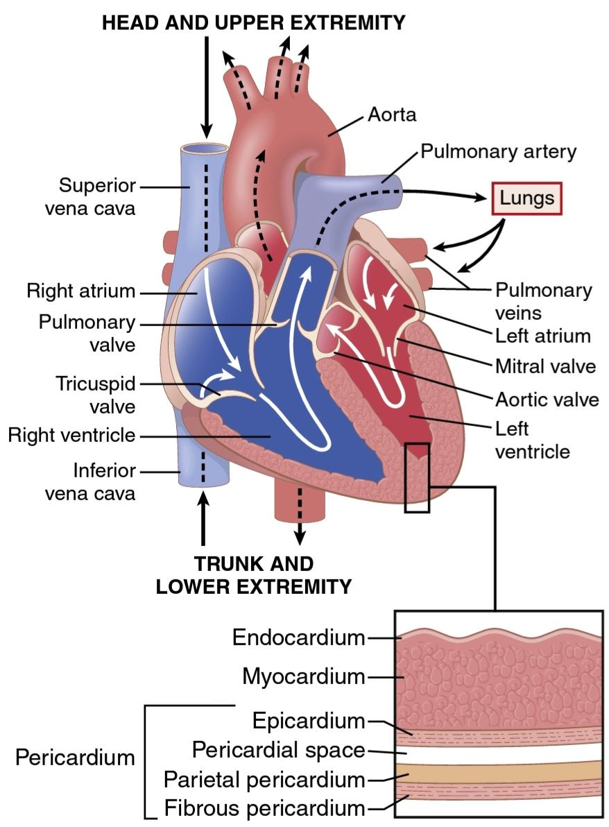
Figure 9-1.
Structure of the heart and course of blood flow through the heart chambers and heart valves. The heart consists of multiple layers, including the inner endocardium, myocardium, and more out- ward epicardium and pericardium layers.
شکل 9-1.
ساختار قلب و جریان خون از طریق حفرههای قلب و دریچههای قلب. قلب از لایههای متعددی تشکیل شده است که شامل اندوکارد داخلی، میوکارد و لایههای بیرونی اپی کاردیوم و پریکارد میشود.
PHYSIOLOGY OF CARDIAC MUSCLE
The heart is composed of three major types of cardiac muscle-atrial muscle, ventricular muscle, and specialized excitatory and conductive muscle fibers. The atrial and ventricular types of muscle contract in much the same way as skeletal muscle, except that the duration of contraction is much longer. The specialized excitatory and conductive fibers of the heart, however, contract feebly because they contain few contractile fibrils; instead, they exhibit automatic rhythmical electrical discharge in the form of action potentials or conduction of the action potentials through the heart, providing an excitatory system that controls the rhythmical beating of the heart.
فیزیولوژی عضله قلب
قلب از سه نوع اصلی عضله قلبی-عضله دهلیزی، عضله بطنی و فیبرهای عضلانی تحریک کننده و رسانا تشکیل شده است. ماهیچههای دهلیزی و بطنی تقریباً مانند ماهیچههای اسکلتی منقبض میشوند، با این تفاوت که مدت زمان انقباض بسیار طولانی تر است. فیبرهای تحریکی و رسانای تخصصی قلب، به دلیل اینکه حاوی فیبرهای انقباضی کمیهستند، به شدت منقبض میشوند. در عوض، آنها تخلیه الکتریکی ریتمیک خودکار را به شکل پتانسیلهای عمل یا هدایت پتانسیلهای عمل از طریق قلب نشان میدهند و یک سیستم تحریکی را فراهم میکنند که ضربان ریتمیک قلب را کنترل میکند.
CARDIAC MUSCLE ANATOMY
Figure 9-2 shows the cardiac muscle histology, demonstrating cardiac muscle fibers arranged in a latticework, with the fibers dividing, recombining, and then spreading again. Note that cardiac muscle is striated in the same manner as in skeletal muscle. Furthermore, cardiac muscle has typical myofibrils that contain actin and myosin filaments almost identical to those found in skeletal muscle; these filaments lie side by side and slide during con- traction in the same manner as occurs in skeletal muscle (see Chapter 6). In other ways, however, cardiac muscle is quite different from skeletal muscle, as we shall see.
آناتومیعضله قلب
شکل 9-2 بافت شناسی عضله قلب را نشان میدهد که فیبرهای عضلانی قلبی را که به صورت شبکه ای چیده شده اند، نشان میدهد که فیبرها تقسیم میشوند، دوباره ترکیب میشوند و سپس دوباره پخش میشوند. توجه داشته باشید که عضله قلب مانند ماهیچههای اسکلتی مخطط است. علاوه بر این، عضله قلب دارای میوفیبریلهای معمولی است که حاوی رشتههای اکتین و میوزین تقریباً مشابه با رشتههای موجود در ماهیچههای اسکلتی است. این رشتهها در کنار هم قرار میگیرند و در طول انقباض به همان شیوه ای که در ماهیچههای اسکلتی رخ میدهد میلغزند (به فصل 6 مراجعه کنید). با این حال، از جهات دیگر، همانطور که خواهیم دید، عضله قلب کاملاً با ماهیچه اسکلتی متفاوت است.

Figure 9-2.
Syncytial interconnecting nature of cardiac muscle fibers.
شکل 9-2.
ماهیت به هم پیوستگی سنسیتیال فیبرهای عضلانی قلب
Left Ventricular Rotation (Twist) Aids Left Ventricular Ejection and Relaxation. The left ventricle is organized into complex muscle fiber layers that run in different di- rections and allow the heart to contract in a twisting mo- tion during systole. The subepicardial (outer) layer spirals in a leftward direction, and the subendocardial (inner) layer spirals in the opposite direction (rightward), causing clockwise rotation of the apex of the heart and counter- clockwise rotation of the base of the left ventricle (Figure 9-3). This causes a wringing motion of the left ventricle, pulling the base downward toward the apex during sys- tole (contraction). At the end of systole, the left ventricle is similar to a loaded spring and recoils or untwists during diastole (relaxation) to allow blood to enter the pumping chambers rapidly.
چرخش بطن چپ (پیچش) به تخلیه و آرامش بطن چپ کمک میکند. بطن چپ به صورت لایههای فیبر ماهیچهای پیچیده سازماندهی شده است که در جهتهای مختلف قرار دارند و به قلب اجازه میدهند در طول سیستول در یک حرکت پیچشی منقبض شود. لایه ساب اپیکاردیال (خارجی) در جهت چپ و لایه ساب اندوکاردیال (داخلی) در جهت مخالف (راست) مارپیچ میشود که باعث چرخش در جهت عقربههای ساعت راس قلب و چرخش خلاف جهت عقربههای ساعت پایه بطن چپ میشود (شکل 9-3). این باعث حرکت انقباض بطن چپ میشود و در طول سیستول (انقباض) پایه را به سمت راس به سمت پایین میکشد. در انتهای سیستول، بطن چپ شبیه به فنر بارگذاری شده است و در طی دیاستول (آرامش) پس میزند یا باز میشود تا خون به سرعت وارد اتاقکهای پمپاژ شود.

Figure 9-3. A, The left ventricular inner subendocardial fibers (laven- der shade) run obliquely to the outer subepicardial fibers (red shade). B. The subepicardial muscle fibers are wrapped in a left-handed helix and subendocardial fibers are arranged in a right-handed helix.
شکل 9-3. الف، فیبرهای ساب اندوکارد داخلی بطن چپ (سایههای اسطوخودوس) بهطور مایل به رشتههای زیر اپیکارد خارجی (سایه قرمز) کشیده میشوند. ب- رشتههای عضلانی ساب اپیکارد در یک مارپیچ سمت چپ پیچیده شده اند و رشتههای ساب اندوکاردیال در یک مارپیچ سمت راست قرار گرفته اند.
Cardiac Muscle Is a Syncytium. The dark areas crossing the cardiac muscle fibers in Figure 9-2 are called interca- lated discs; they are actually cell membranes that separate individual cardiac muscle cells from one another. That is, cardiac muscle fibers are made up of many individual cells connected in series and in parallel with one another.
ماهیچه قلب یک سیسیتیوم است. نواحی تیره ای که در شکل 9-2 از فیبرهای عضلانی قلب عبور میکنند، دیسکهای درونی نامیده میشوند. آنها در واقع غشای سلولی هستند که سلولهای عضله قلب را از یکدیگر جدا میکنند. یعنی تارهای عضلانی قلب از تعداد زیادی سلول مجزا تشکیل شده اند که به صورت متوالی و موازی با یکدیگر متصل شده اند.
At each intercalated disc, the cell membranes fuse with one another to form permeable communicating junctions (gap junctions) that allow rapid diffusion of ions. There- fore, from a functional point of view, ions move with ease in the intracellular fluid along the longitudinal axes of the cardiac muscle fibers so that action potentials travel easily from one cardiac muscle cell to the next, past the interca- lated discs. Thus, cardiac muscle is a syncytium of many heart muscle cells in which the cardiac cells are so inter- connected that when one cell becomes excited, the action potential rapidly spreads to all of them.
در هر دیسک درهم، غشای سلولی با یکدیگر ترکیب میشوند تا اتصالات ارتباطی نفوذپذیر (اتصالات شکاف) را تشکیل دهند که امکان انتشار سریع یونها را فراهم میکند. بنابراین، از نقطه نظر عملکردی، یونها به راحتی در مایع درون سلولی در امتداد محورهای طولی رشتههای عضلانی قلب حرکت میکنند، به طوری که پتانسیلهای عمل به راحتی از یک سلول عضلانی قلبی به سلول دیگر منتقل میشوند و از دیسکهای درهم تنیده میگذرند. بنابراین، عضله قلب یک سی سی سیتیوم از بسیاری از سلولهای عضله قلب است که در آن سلولهای قلبی به قدری به هم متصل هستند که وقتی یک سلول برانگیخته میشود، پتانسیل عمل به سرعت به همه آنها گسترش مییابد.
The heart actually is composed of two syncytia; the atrial syncytium, which constitutes the walls of the two atria; and the ventricular syncytium, which constitutes the walls of the two ventricles. The atria are separated from the ventricles by fibrous tissue that surrounds the atrioventricular (A-V) valvular openings between the atria and ventricles. Normally, potentials are not con- ducted from the atrial syncytium into the ventricular syncytium directly through this fibrous tissue. Instead, they are only conducted by way of a specialized conduc- tive system called the A-V bundle, a bundle of conductive fibers several millimeters in diameter that is discussed in Chapter 10.
قلب در واقع از دو سنسیتی تشکیل شده است. سنسیتیوم دهلیزی، که دیوارههای دو دهلیز را تشکیل میدهد. و سینسیتیوم بطنی که دیوارههای دو بطن را تشکیل میدهد. دهلیزها توسط بافت فیبری که دهانه دریچه دهلیزی (A-V) بین دهلیزها و بطنها را احاطه کرده است از بطنها جدا میشوند. به طور معمول، پتانسیلها از سنسیتیوم دهلیزی به طور مستقیم از طریق این بافت فیبری به سینسیتیوم بطنی هدایت نمیشوند. در عوض، آنها فقط از طریق یک سیستم رسانای تخصصی به نام بسته A-V هدایت میشوند، دسته ای از الیاف رسانا به قطر چند میلی متر که در فصل 10 مورد بحث قرار گرفته است.
This division of the muscle of the heart into two func- tional syncytia allows the atria to contract a short time ahead of ventricular contraction, which is important for the effectiveness of heart pumping.
این تقسیم عضله قلب به دو سنسیشیا عملکردی به دهلیز اجازه میدهد تا مدت کوتاهی قبل از انقباض بطنی منقبض شود، که برای اثربخشی پمپاژ قلب مهم است.
ACTION POTENTIALS IN CARDIAC MUSCLE
The action potential recorded in a ventricular muscle fiber, shown in Figure 9-4, averages about 105 millivolts, which means that the intracellular potential rises from a very negative value between beats, about -85 millivolts, to a slightly positive value, about +20 millivolts, during each beat. After the initial spike, the membrane remains depolarized for about 0.2 second, exhibiting a plateau, fol- lowed at the end of the plateau by abrupt repolarization. The presence of this plateau in the action potential causes ventricular contraction to last as much as 15 times longer in cardiac muscle than in skeletal muscle.
پتانسیلهای عمل در عضله قلب
پتانسیل عمل ثبت شده در یک فیبر عضلانی بطنی، که در شکل 9-4 نشان داده شده است، به طور متوسط حدود 105 میلی ولت است، به این معنی که پتانسیل درون سلولی از یک مقدار بسیار منفی بین ضربانها، حدود 85- میلی ولت، به مقدار کمیمثبت، حدود 20 میلی ولت، در طول هر ضربه افزایش مییابد. پس از سنبله اولیه، غشاء برای حدود 0.2 ثانیه دپلاریزه باقی میماند و یک پلاتو را نشان میدهد که در انتهای فلات با رپلاریزاسیون ناگهانی دنبال میشود. وجود این فلات در پتانسیل عمل باعث میشود که انقباض بطنی در عضله قلب 15 برابر بیشتر از ماهیچه اسکلتی طول بکشد.
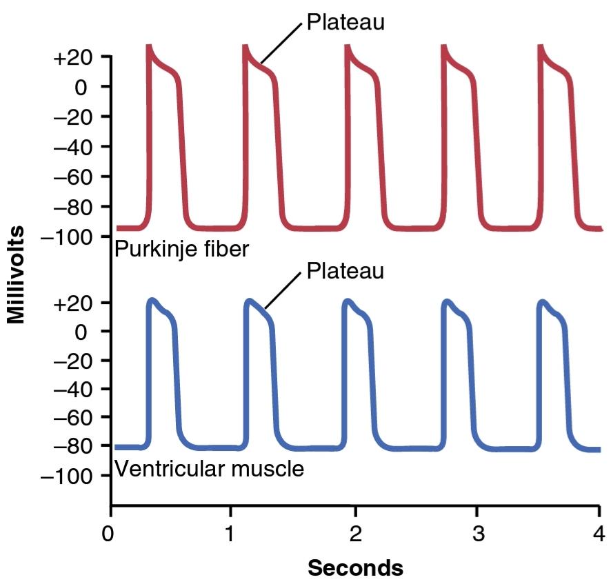
Figure 9-4. Rhythmical action potentials (in millivolts) from a Purkin- je fiber and from a ventricular muscle fiber, recorded by microelec- trodes.
شکل 9-4. پتانسیل عمل ریتمیک (بر حسب میلی ولت) از یک فیبر پورکینژ و از یک فیبر عضلانی بطنی، ثبت شده توسط میکروالکترودها.
What Causes the Long Action Potential and Plateau in Cardiac Muscle? At least two major differences between the membrane properties of cardiac and skeletal muscle ac- count for the prolonged action potential and the plateau in cardiac muscle. First, the action potential of skeletal muscle is caused almost entirely by the sudden opening of large numbers of fast sodium channels that allow tremendous numbers of sodium ions to enter the skeletal muscle fiber from the extracellular fluid. These channels are called fast channels because they remain open for only a few thou- sandths of a second and then abruptly close. At the end of this closure, repolarization occurs, and the action potential is over within about another thousandth of a second.
چه چیزی باعث پتانسیل عمل طولانی و پلاتو در عضله قلب میشود؟ حداقل دو تفاوت عمده بین خصوصیات غشایی عضله قلبی و اسکلتی مربوط به پتانسیل عمل طولانی مدت و فلات در عضله قلبی است. اول، پتانسیل عمل عضله اسکلتی تقریباً به طور کامل ناشی از باز شدن ناگهانی تعداد زیادی کانال سدیم سریع است که به تعداد زیادی یون سدیم اجازه میدهد تا از مایع خارج سلولی وارد فیبر عضلانی اسکلتی شوند. این کانالها کانالهای سریع نامیده میشوند زیرا فقط چند هزارم ثانیه باز میمانند و سپس به طور ناگهانی بسته میشوند. در پایان این بسته شدن، رپولاریزاسیون اتفاق میافتد و پتانسیل عمل در حدود هزارم ثانیه دیگر به پایان میرسد.
In cardiac muscle, the action potential is caused by opening of two types of channels: (1) the same voltage- activated fast sodium channels as those in skeletal muscle; and (2) another entirely different population of L-type calcium channels (slow calcium channels), which are also called calcium-sodium channels. This second population of channels differs from the fast sodium channels in that they are slower to open and, even more importantly, remain open for several tenths of a second. During this time, a large quantity of both calcium and sodium ions flows through these channels to the interior of the cardiac muscle fiber, and this activity maintains a prolonged period of depolar- ization, causing the plateau in the action potential. Fur- thermore, the calcium ions that enter during this plateau phase activate the muscle contractile process, whereas the calcium ions that cause skeletal muscle contraction are derived from the intracellular sarcoplasmic reticulum.
در عضله قلب، پتانسیل عمل با باز کردن دو نوع کانال ایجاد میشود: (1) کانالهای سدیم سریع فعال شده با ولتاژ مشابه کانالهای عضله اسکلتی. و (2) جمعیت کاملاً متفاوت دیگری از کانالهای کلسیم نوع L (کانالهای کلسیم آهسته)، که به آنها کانالهای کلسیم-سدیم نیز میگویند. این گروه دوم از کانالها با کانالهای سدیم سریع متفاوت است، زیرا کندتر باز میشوند و مهمتر از آن، برای چند دهم ثانیه باز میمانند. در طول این مدت، مقدار زیادی از یونهای کلسیم و سدیم از طریق این کانالها به داخل فیبر عضلانی قلب جریان مییابد، و این فعالیت یک دوره طولانی دپلاریزاسیون را حفظ میکند و باعث ایجاد فلات در پتانسیل عمل میشود. علاوه بر این، یونهای کلسیمیکه در این فاز فلات وارد میشوند، فرآیند انقباض عضلانی را فعال میکنند، در حالی که یونهای کلسیم که باعث انقباض ماهیچههای اسکلتی میشوند، از شبکه سارکوپلاسمیداخل سلولی مشتق میشوند.
The second major functional difference between car- diac muscle and skeletal muscle that helps account for both the prolonged action potential and its plateau is that immediately after the onset of the action potential, the permeability of the cardiac muscle membrane for potas- sium ions decreases about fivefold, an effect that does not occur in skeletal muscle. This decreased potassium perme- ability may result from the excess calcium influx through the calcium channels just noted. Regardless of the cause, the decreased potassium permeability greatly decreases the efflux of positively charged potassium ions during the action potential plateau and thereby prevents early return of the action potential voltage to its resting level. When the slow calcium-sodium channels do close at the end of 0.2 to 0.3 second, and the influx of calcium and sodium ions ceases, the membrane permeability for potassium ions also increases rapidly. This rapid loss of potassium from the fiber immediately returns the membrane potential to its resting level, thus ending the action potential.
دومین تفاوت عملکردی عمده بین عضله قلبی و عضله اسکلتی که به پتانسیل عمل طولانی مدت و فلات آن کمک میکند این است که بلافاصله پس از شروع پتانسیل عمل، نفوذپذیری غشای عضله قلب برای یونهای پتاسیم حدود پنج برابر کاهش مییابد، اثری که در عضله اسکلتی رخ نمیدهد. این کاهش نفوذپذیری پتاسیم ممکن است ناشی از هجوم کلسیم اضافی از طریق کانالهای کلسیمیباشد. صرف نظر از علت، کاهش نفوذپذیری پتاسیم تا حد زیادی جریان یونهای پتاسیم با بار مثبت را در طول فلات پتانسیل عمل کاهش میدهد و در نتیجه از بازگشت زودهنگام ولتاژ پتانسیل عمل به سطح استراحت خود جلوگیری میکند. هنگامیکه کانالهای آهسته کلسیم-سدیم در پایان 0.2 تا 0.3 ثانیه بسته میشوند و هجوم یونهای کلسیم و سدیم متوقف میشود، نفوذپذیری غشاء برای یونهای پتاسیم نیز به سرعت افزایش مییابد. این از دست دادن سریع پتاسیم از فیبر بلافاصله پتانسیل غشا را به سطح استراحت خود باز میگرداند و در نتیجه پتانسیل عمل را پایان میدهد.
Phases of Cardiac Muscle Action Potential. Figure 9-5 summarizes the phases of the action potential in cardiac muscle and the ion flows that occur during each phase.
مراحل پتانسیل عمل عضله قلبی. شکل 9-5 مراحل پتانسیل عمل در عضله قلب و جریانهای یونی را که در طول هر فاز رخ میدهد، خلاصه میکند.

Figure 9-5.
Phases of action potential of cardiac ventricular muscle cell and associated ionic currents for sodium (Na+), calcium (Ca2+), and potassium (K+).
شکل 9-5.
مراحل پتانسیل عمل سلول عضلانی بطن قلب و جریانهای یونی مرتبط برای سدیم (Na+)، کلسیم (Ca2+) و پتاسیم (K+).
Phase 0 (Depolarization): Fast Sodium Channels Open. When the cardiac cell is stimulated and depolar- izes, the membrane potential becomes more positive. Voltage-gated sodium channels (fast sodium channels) open and permit sodium to rapidly flow into the cell and depolarize it. The membrane potential reaches about +20 millivolts before the sodium channels close.
فاز 0 (دپلاریزاسیون): کانالهای سدیم سریع باز میشوند. هنگامیکه سلول قلبی تحریک میشود و دپلاریزه میشود، پتانسیل غشاء مثبت تر میشود. کانالهای سدیم دارای ولتاژ (کانالهای سدیم سریع) باز میشوند و به سدیم اجازه میدهند تا به سرعت به داخل سلول جریان یابد و آن را دپولاریزه کند. پتانسیل غشا قبل از بسته شدن کانالهای سدیم به حدود 20+ میلی ولت میرسد.
Phase 1 (Initial Repolarization): Fast Sodium Chan- nels Close. The sodium channels close, the cell begins to repolarize, and potassium ions leave the cell through open potassium channels.
فاز 1 (بازپلاریزاسیون اولیه): کانالهای سدیم سریع بسته میشوند. کانالهای سدیم بسته میشوند، سلول شروع به قطبش مجدد میکند و یونهای پتاسیم از طریق کانالهای پتاسیم باز سلول را ترک میکنند.
Phase 2 (Plateau): Calcium Channels Open and Fast Potassium Channels Close. A brief initial repolarization occurs and the action potential then plateaus as a result of increased calcium ion permeability and decreased po- tassium ion permeability. The voltage-gated calcium ion channels open slowly during phases 1 and 0, and calcium enters the cell. Potassium channels then close, and the combination of decreased potassium ion efflux and in- creased calcium ion influx causes the action potential to plateau.
فاز 2 (فلات): کانالهای کلسیم باز و کانالهای پتاسیم سریع بسته میشوند. یک رپلاریزاسیون اولیه مختصر اتفاق میافتد و پتانسیل عمل در نتیجه افزایش نفوذپذیری یون کلسیم و کاهش نفوذپذیری یون پتاسیم فلات میشود. کانالهای یون کلسیم دارای ولتاژ در فازهای 1 و 0 به آرامیباز میشوند و کلسیم وارد سلول میشود. سپس کانالهای پتاسیم بسته میشوند و ترکیبی از کاهش جریان یون پتاسیم و افزایش هجوم یون کلسیم باعث میشود پتانسیل عمل فلاتآمیز شود.
Phase 3 (Rapid Repolarization): Calcium Channels Close and Slow Potassium Channels Open. The closure of calcium ion channels and increased potassium ion permeability, permitting potassium ions to exit the cell rapidly, ends the plateau and returns the cell membrane potential to its resting level.
فاز 3 (بازپلاریزاسیون سریع): کانالهای کلسیم بسته میشوند و کانالهای پتاسیم آهسته باز میشوند. بسته شدن کانالهای یون کلسیم و افزایش نفوذپذیری یون پتاسیم، اجازه خروج سریع یونهای پتاسیم از سلول را میدهد و به فلات پایان میدهد و پتانسیل غشای سلولی را به سطح استراحت خود باز میگرداند.
Phase 4 (Resting Membrane Potential): This averages about-80 to -90 millivolts.
فاز 4 (پتانسیل غشاء در حالت استراحت): این میانگین حدود 80- تا 90- میلی ولت است.
Velocity of Signal Conduction in Cardiac Muscle. The velocity of conduction of the excitatory action potential signal along both atrial and ventricular muscle fibers is about 0.3 to 0.5 m/sec, or about 1/250 the velocity in very large nerve fibers and about 1/10 the velocity in skeletal muscle fibers. The velocity of conduction in the special- ized heart conductive system-in the Purkinje fibers-is as high as 4 m/sec in most parts of the system, allowing rapid conduction of the excitatory signal to the different parts of the heart, as explained in Chapter 10.
سرعت هدایت سیگنال در عضله قلب. سرعت انتقال سیگنال پتانسیل عمل تحریکی در امتداد فیبرهای عضلانی دهلیزی و بطنی حدود 0.3 تا 0.5 متر بر ثانیه یا حدود 1/250 سرعت در رشتههای عصبی بسیار بزرگ و حدود 1/10 سرعت در فیبرهای عضلانی اسکلتی است. همانطور که در فصل 10 توضیح داده شد، سرعت رسانش در سیستم رسانای ویژه قلب – در فیبرهای پورکنژ – در بیشتر قسمتهای سیستم به 4 متر بر ثانیه میرسد و امکان هدایت سریع سیگنال تحریکی به قسمتهای مختلف قلب را فراهم میکند.
Refractory Period of Cardiac Muscle. Cardiac muscle, like all excitable tissue, is refractory to restimulation dur- ing the action potential. Therefore, the refractory period of the heart is the interval of time, as shown to the left in Figure 9-6, during which a normal cardiac impulse can- not re-excite an already excited area of cardiac muscle. The normal refractory period of the ventricle is 0.25 to 0.30 second, which is about the duration of the prolonged plateau action potential. There is an additional relative refractory period of about 0.05 second during which the muscle is more difficult to excite than normal but can be excited by a very strong excitatory signal, as demonstrated by the early premature contraction in the second example of Figure 9-6. The refractory period of atrial muscle is much shorter than that for the ventricles (about 0.15 sec- ond for the atria compared with 0.25 to 0.30 second for the ventricles).
دوره نسوز عضله قلب. عضله قلب، مانند تمام بافتهای تحریک پذیر، در طول پتانسیل عمل نسبت به تحریک مجدد مقاوم است. بنابراین، دوره نسوز قلب همان فاصله زمانی است، همانطور که در شکل 9-6 در سمت چپ نشان داده شده است، که طی آن یک تکانه قلبی طبیعی نمیتواند یک ناحیه از قبل برانگیخته شده از عضله قلب را دوباره تحریک کند. دوره نسوز طبیعی بطن 0.25 تا 0.30 ثانیه است که تقریباً مدت زمان پتانسیل عمل طولانی مدت فلات است. یک دوره نسبی نسبی اضافی در حدود 0.05 ثانیه وجود دارد که طی آن تحریک عضله دشوارتر از حالت عادی است، اما میتواند توسط یک سیگنال تحریکی بسیار قوی تحریک شود، همانطور که با انقباض زودرس اولیه در مثال دوم شکل 9-6 نشان داده شده است. دوره نسوز عضله دهلیزی بسیار کوتاهتر از بطنها است (حدود 0.15 ثانیه برای دهلیزها در مقایسه با 0.25 تا 0.30 ثانیه برای بطنها).
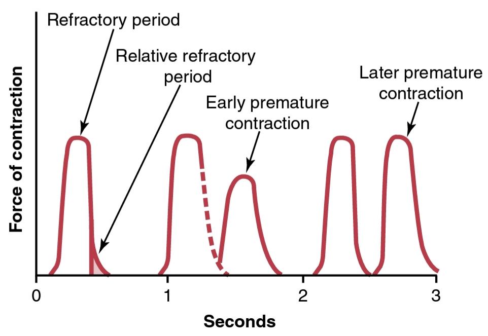
Figure 9-6.
Force of ventricular heart muscle contraction, showing also the duration of the refractory period and relative refractory pe- riod, plus the effect of premature contraction. Note that premature contractions do not cause wave summation, as occurs in skeletal muscle.
شکل 9-6.
نیروی انقباض عضله قلب بطنی، همچنین نشان دهنده طول دوره نسوز و دوره نسبی نسبی، به علاوه اثر انقباض زودرس است. توجه داشته باشید که انقباضات زودرس باعث جمع شدن موج نمیشود، همانطور که در عضلات اسکلتی اتفاق میافتد.
EXCITATION-CONTRACTION COUPLING- FUNCTION OF CALCIUM IONS AND THE TRANSVERSE TUBULES
The term excitation-contraction coupling refers to the mechanism whereby the action potential causes the myofibrils of muscle to contract. This mechanism was discussed for skeletal muscle in Chapter 7. Again, there are differences in this mechanism in cardiac muscle that have important effects on the characteristics of heart muscle contraction.
جفت برانگیختگی – انقباض – عملکرد یونهای کلسیم و لولههای عرضی
اصطلاح کوپلینگ تحریک-انقباض به مکانیزمیاشاره دارد که در آن پتانسیل عمل باعث انقباض میوفیبریلهای عضله میشود. این مکانیسم برای ماهیچههای اسکلتی در فصل 7 مورد بحث قرار گرفت. باز هم تفاوتهایی در این مکانیسم در عضله قلب وجود دارد که تأثیرات مهمیبر ویژگیهای انقباض عضله قلب دارد.
As is true for skeletal muscle, when an action poten- tial passes over the cardiac muscle membrane, the action potential spreads to the interior of the cardiac muscle fiber along the membranes of the transverse (T) tubules. The T tubule action potentials then act on the membranes of the longitudinal sarcoplasmic tubules to cause release of calcium ions into the muscle sarcoplasm from the sar- coplasmic reticulum. In another few thousandths of a second, these calcium ions diffuse into the myofibrils and catalyze the chemical reactions that promote sliding of the actin and myosin filaments along one another, which produces the muscle contraction.
همانطور که در مورد عضله اسکلتی صدق میکند، هنگامیکه یک پتانسیل عمل از غشای عضله قلب عبور میکند، پتانسیل عمل به داخل فیبر عضله قلب در امتداد غشای لولههای عرضی (T) گسترش مییابد. پتانسیل عمل توبول T سپس بر روی غشای لولههای سارکوپلاسمیطولی عمل میکند تا باعث آزاد شدن یونهای کلسیم به داخل سارکوپلاسم عضلانی از شبکه سارکوپلاسمیشود. در چند هزارم ثانیه دیگر، این یونهای کلسیم به داخل میوفیبریلها منتشر میشوند و واکنشهای شیمیایی را کاتالیز میکنند که باعث لغزش رشتههای اکتین و میوزین در امتداد یکدیگر میشود که باعث انقباض عضلانی میشود.
Thus far, this mechanism of excitation-contraction coupling is the same as that for skeletal muscle, but there is a second effect that is quite different. In addition to the calcium ions that are released into the sarcoplasm from the cisternae of the sarcoplasmic reticulum, cal- cium ions also diffuse into the sarcoplasm from the T tubules at the time of the action potential, which opens voltage-dependent calcium channels in the membrane of the T tubule (Figure 9-7). Calcium entering the cell then activates calcium release channels, also called ryanodine receptor channels, in the sarcoplasmic reticulum mem- brane, triggering the release of calcium into the sarco- plasm. Calcium ions in the sarcoplasm then interact with troponin to initiate cross-bridge formation and contrac- tion by the same basic mechanism as that described for skeletal muscle in Chapter 6.
تا اینجای کار، این مکانیسم جفت شدن تحریک-انقباض همان است که برای ماهیچههای اسکلتی انجام میشود، اما اثر دومیوجود دارد که کاملاً متفاوت است. علاوه بر یونهای کلسیمیکه از مخزن شبکه سارکوپلاسمیبه داخل سارکوپلاسم آزاد میشوند، یونهای کلسیم نیز در زمان پتانسیل عمل به داخل سارکوپلاسم از لولههای T منتشر میشوند که کانالهای کلسیم وابسته به ولتاژ را در غشای لوله T باز میکند (شکل 99). کلسیمیکه وارد سلول میشود، کانالهای آزادسازی کلسیم را که کانالهای گیرنده رایانودین نیز نامیده میشوند، در غشای شبکه سارکوپلاسمیفعال میکند و باعث آزاد شدن کلسیم در سارکوپلاسم میشود. سپس یونهای کلسیم در سارکوپلاسم با تروپونین برهمکنش میکنند تا تشکیل پل متقاطع و انقباض را با همان مکانیسمیکه برای ماهیچههای اسکلتی در فصل 6 توضیح داده شد، آغاز کنند.
Without the calcium from the T tubules, the strength of cardiac muscle contraction would be reduced considerably because the sarcoplasmic reticu- lum of cardiac muscle is less well developed than that of skeletal muscle and does not store enough calcium to provide full contraction. The T tubules of cardiac muscle, however, have a diameter five times as great as that of the skeletal muscle tubules, which means a volume 25 times as great. Also, inside the T tubules is a large quantity of mucopolysaccharides that are elec- tronegatively charged and bind an abundant store of calcium ions, keeping them available for diffusion to the interior of the cardiac muscle fiber when a T tubule action potential appears.
بدون کلسیم لولههای T، قدرت انقباض عضله قلب به طور قابل توجهی کاهش مییابد زیرا شبکه سارکوپلاسمیعضله قلب نسبت به عضلات اسکلتی توسعه یافته کمتری دارد و کلسیم کافی برای ایجاد انقباض کامل ذخیره نمیکند. با این حال، لولههای T عضله قلب، قطری پنج برابر قطر لولههای عضله اسکلتی دارند، که به معنای حجم 25 برابر بزرگتر است. همچنین، در داخل لولههای T مقدار زیادی موکوپلی ساکارید وجود دارد که به صورت الکتریکی باردار میشوند و ذخیره فراوانی از یونهای کلسیم را به هم متصل میکنند و آنها را برای انتشار در داخل فیبر عضلانی قلب در هنگام ظهور پتانسیل عمل توبول T در دسترس نگه میدارند.
The strength of contraction of cardiac muscle depends to a great extent on the concentration of calcium ions in the extracellular fluids. In fact, a heart placed in a calcium- free solution will quickly stop beating. The reason for this response is that the openings of the T tubules pass directly through the cardiac muscle cell membrane into the extra- cellular spaces surrounding the cells, allowing the same extracellular fluid that is in the cardiac muscle intersti- tium to percolate through the T tubules. Consequently, the quantity of calcium ions in the T tubule system (i.e., the availability of calcium ions to cause cardiac muscle contraction) depends to a great extent on the extracellular fluid calcium ion concentration.
قدرت انقباض عضله قلب تا حد زیادی به غلظت یونهای کلسیم در مایعات خارج سلولی بستگی دارد. در واقع، قلب قرار داده شده در محلول بدون کلسیم به سرعت از تپش باز میایستد. دلیل این پاسخ این است که منافذ لولههای T مستقیماً از غشای سلولی عضله قلب به فضاهای خارج سلولی اطراف سلولها میگذرد و به همان مایع خارج سلولی که در بینابینی عضله قلب است اجازه میدهد از طریق لولههای T نفوذ کند. در نتیجه، مقدار یونهای کلسیم در سیستم توبول T (به عنوان مثال، در دسترس بودن یونهای کلسیم برای ایجاد انقباض عضله قلب) تا حد زیادی به غلظت یون کلسیم مایع خارج سلولی بستگی دارد.
In contrast, the strength of skeletal muscle contrac- tion is hardly affected by moderate changes in extracel- lular fluid calcium concentration. This is because skeletal muscle contraction is caused almost entirely by calcium ions released from the sarcoplasmic reticulum inside the skeletal muscle fiber.
در مقابل، قدرت انقباض عضلات اسکلتی به سختی تحت تاثیر تغییرات متوسط در غلظت کلسیم مایع خارج سلولی قرار میگیرد. این به این دلیل است که انقباض ماهیچههای اسکلتی تقریباً به طور کامل توسط یونهای کلسیم آزاد شده از شبکه سارکوپلاسمیداخل فیبر عضلانی اسکلتی ایجاد میشود.
At the end of the plateau of the cardiac action poten- tial, the influx of calcium ions to the interior of the muscle fiber is suddenly cut off, and calcium ions in the sarco- plasm are rapidly pumped back out of the muscle fibers into the sarcoplasmic reticulum and T tubule-extra- cellular fluid space. Transport of calcium back into the sarcoplasmic reticulum is achieved with the help of a calcium-adenosine triphosphatase (ATPase) pump (the sarcoplasmic endoplasmic reticulum calcium ATPase, SERCA2; see Figure 9-7). Calcium ions are also removed from the cell by a sodium-calcium exchanger. The sodium that enters the cell during this exchange is then trans- ported out of the cell by the sodium-potassium ATPase pump. As a result, the contraction ceases until a new action potential comes along.
در انتهای پلاتوی پتانسیل عمل قلبی، هجوم یونهای کلسیم به داخل فیبر عضلانی ناگهان قطع میشود و یونهای کلسیم در سارکوپلاسم به سرعت به بیرون از رشتههای عضلانی به داخل شبکه سارکوپلاسمیو فضای توبولهای T خارج میشوند. انتقال کلسیم به شبکه سارکوپلاسمیبا کمک یک پمپ کلسیم آدنوزین تری فسفاتاز (ATPase) انجام میشود (شبکه آندوپلاسمیکلسیم ATPase سارکوپلاسمی، SERCA2؛ به شکل 9-7 مراجعه کنید). یونهای کلسیم نیز توسط مبدل سدیم-کلسیم از سلول خارج میشوند. سپس سدیمیکه در طی این تبادل وارد سلول میشود توسط پمپ سدیم پتاسیم ATPase به خارج از سلول منتقل میشود. در نتیجه، انقباض متوقف میشود تا زمانی که یک پتانسیل عمل جدید ظاهر شود.

Figure 9-7.
Mechanisms of excitation-contraction coupling and relaxation in cardiac muscle. ATP, Adenosine triphosphate. RyR, ryanodine receptor Ca2+ release channel; SERCA, sarcoplasmic reticulum Ca2+-ATPase
شکل 9-7.
مکانیسمهای جفت شدن تحریک-انقباض و آرامش در عضله قلب. ATP، آدنوزین تری فسفات. RyR، گیرنده رایانودین Ca2 کانال انتشار. SERCA، شبکه سارکوپلاسمیCa2+-ATPase
Duration of Contraction. Cardiac muscle begins to con- tract a few milliseconds after the action potential begins and continues to contract until a few milliseconds af- ter the action potential ends. Therefore, the duration of contraction of cardiac muscle is mainly a function of the duration of the action potential, including the plateau- about 0.2 second in atrial muscle and 0.3 second in ven- tricular muscle.
مدت زمان انقباض. عضله قلب چند میلی ثانیه پس از شروع پتانسیل عمل شروع به انقباض میکند و تا چند میلی ثانیه پس از پایان پتانسیل عمل به انقباض ادامه میدهد. بنابراین، مدت زمان انقباض عضله قلب عمدتاً تابعی از مدت زمان پتانسیل عمل است، از جمله فلات – حدود 0.2 ثانیه در عضله دهلیزی و 0.3 ثانیه در عضله بطنی.
CARDIAC CYCLE
The cardiac events that occur from the beginning of one heartbeat to the beginning of the next are called the cardiac cycle. Each cycle is initiated by the spontane- ous generation of an action potential in the sinus node, as explained in Chapter 10. This node is located in the superior lateral wall of the right atrium near the open- ing of the superior vena cava, and the action potential travels from here rapidly through both atria and then through the A-V bundle into the ventricles. Because of this special arrangement of the conducting system from the atria into the ventricles, there is a delay of more than 0.1 second during passage of the cardiac impulse from the atria into the ventricles. This delay allows the atria to contract ahead of ventricular contraction, thereby pumping blood into the ventricles before the strong ven- tricular contraction begins. Thus, the atria act as primer pumps for the ventricles, and the ventricles in turn provide the major source of power for moving blood through the body’s vascular system.
چرخه قلبی
رویدادهای قلبی که از ابتدای یک ضربان قلب تا شروع ضربان بعدی رخ میدهد، چرخه قلبی نامیده میشود. همانطور که در فصل 10 توضیح داده شد، هر چرخه با تولید خود به خود یک پتانسیل عمل در گره سینوسی آغاز میشود. این گره در دیواره جانبی فوقانی دهلیز راست در نزدیکی دهانه ورید اجوف فوقانی قرار دارد و پتانسیل عمل از اینجا به سرعت از طریق هر دو دهلیز و سپس از طریق بسته نرم افزاری A-V عبور میکند. به دلیل این ترتیب خاص سیستم هدایت از دهلیزها به بطنها، تاخیر بیش از 0.1 ثانیه در طول عبور ضربه قلبی از دهلیزها به بطنها وجود دارد. این تأخیر به دهلیز اجازه میدهد تا قبل از انقباض بطنی منقبض شود، در نتیجه خون را قبل از شروع انقباض قوی بطن به داخل بطن پمپ میکند. بنابراین، دهلیزها به عنوان پمپهای آغازگر برای بطنها عمل میکنند و بطنها به نوبه خود منبع اصلی قدرت برای حرکت خون در سیستم عروقی بدن هستند.
Diastole and Systole
The total duration of the cardiac cycle, including systole and diastole, is the reciprocal of the heart rate. For exam- ple, if the heart rate is 72 beats/min, the duration of the cardiac cycle is 1/72 min/beat-about 0.0139 min/beat, or 0.833 sec/beat.
دیاستول و سیستول
کل مدت چرخه قلبی، از جمله سیستول و دیاستول، متقابل ضربان قلب است. به عنوان مثال، اگر ضربان قلب 72 ضربه در دقیقه باشد، مدت چرخه قلبی 1/72 دقیقه / ضربان – حدود 0.0139 دقیقه / ضربان یا 0.833 ثانیه در ضربان است.
Figure 9-8 shows the different events during the car- diac cycle for the left side of the heart. The top three curves show the pressure changes in the aorta, left ventri- cle, and left atrium, respectively. The fourth curve depicts the changes in left ventricular volume, the fifth depicts the electrocardiogram, and the sixth depicts a phonocar- diogram, which is a recording of the sounds produced by the heart-mainly by the heart valves-as it pumps. It is especially important that the reader study this figure in detail and understand the causes of all the events shown.
شکل 9-8 رویدادهای مختلف در طول چرخه قلبی برای سمت چپ قلب را نشان میدهد. سه منحنی بالا به ترتیب تغییرات فشار در آئورت، بطن چپ و دهلیز چپ را نشان میدهند. منحنی چهارم تغییرات حجم بطن چپ را نشان میدهد، منحنی پنجم الکتروکاردیوگرام را نشان میدهد و منحنی ششم فونوکاردیوگرام را نشان میدهد که ضبط صداهایی است که توسط قلب تولید میشود – عمدتاً توسط دریچههای قلب – در حین پمپاژ. به ویژه مهم است که خواننده این رقم را با جزئیات مطالعه کند و علل همه وقایع نشان داده شده را درک کند.
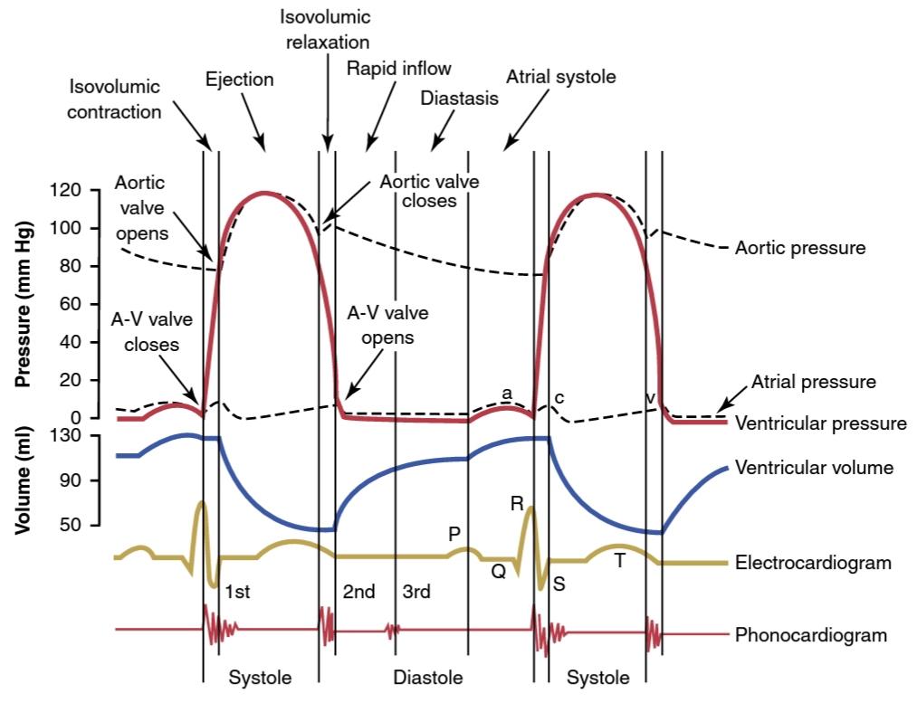
Figure 9-8.
Events of the cardiac cycle for left ventricular function, showing changes in left atrial pres- sure, left ventricular pressure, aortic pressure, ventricular volume, the electrocardiogram, and the phono- cardiogram. A-V, Atrioventricular.
شکل 9-8.
رویدادهای چرخه قلبی برای عملکرد بطن چپ، نشان دهنده تغییرات در فشار دهلیز چپ، فشار بطن چپ، فشار آئورت، حجم بطن، الکتروکاردیوگرام، و فونوکاردیوگرام. A-V، دهلیزی بطنی.
Increasing Heart Rate Decreases Duration of Cardiac Cycle. When heart rate increases, the duration of each cardiac cycle decreases, including the contraction and relaxation phases. The duration of the action potential and systole also decrease, but not by as great a percent- age as diastole. At a normal heart rate of 72 beats/min, systole comprises about 0.4 of the entire cardiac cycle. At three times the normal heart rate, systole is about 0.65 of the entire cardiac cycle. This means that the heart beat- ing very rapidly does not remain relaxed long enough to allow complete filling of the cardiac chambers before the next contraction.
افزایش ضربان قلب مدت چرخه قلبی را کاهش میدهد. هنگامیکه ضربان قلب افزایش مییابد، مدت زمان هر چرخه قلبی کاهش مییابد، از جمله مراحل انقباض و آرامش. مدت زمان پتانسیل عمل و سیستول نیز کاهش مییابد، اما نه به اندازه دیاستول. در ضربان قلب طبیعی 72 ضربه در دقیقه، سیستول حدود 0.4 از کل چرخه قلبی را شامل میشود. در سه برابر ضربان قلب طبیعی، سیستول حدود 0.65 کل چرخه قلبی است. این بدان معنی است که ضربان قلب بسیار سریع به اندازه کافی آرام نمیماند تا اجازه پر شدن کامل اتاقهای قلب را قبل از انقباض بعدی بدهد.
Relationship of the Electrocardiogram to the Cardiac Cycle
The electrocardiogram in Figure 9-8 shows the P, Q, R, S, and T waves, discussed in Chapters 11 and 12. These are electrical voltages generated by the heart and recorded by the electrocardiogram from the surface of the body.
رابطه الکتروکاردیوگرام با چرخه قلبی
الکتروکاردیوگرام در شکل 9-8 امواج P، Q، R، S و T را نشان میدهد که در فصلهای 11 و 12 مورد بحث قرار گرفتهاند. اینها ولتاژهای الکتریکی هستند که توسط قلب تولید میشوند و توسط الکتروکاردیوگرام از سطح بدن ثبت میشوند.
The P wave is caused by the spread of depolarization through the atria and is followed by atrial contraction, which causes a slight rise in the atrial pressure curve immediately after the electrocardiographic P wave.
موج P در اثر گسترش دپلاریزاسیون از طریق دهلیزها ایجاد میشود و به دنبال آن انقباض دهلیزی ایجاد میشود که بلافاصله پس از موج P الکتروکاردیوگرافی باعث افزایش خفیف منحنی فشار دهلیزی میشود.
About 0.16 second after the onset of the P wave, the QRS waves appear as a result of electrical depolarization of the ventricles, which initiates contraction of the ven- tricles and causes the ventricular pressure to begin ris- ing. Therefore, the QRS complex begins slightly before the onset of ventricular systole.
حدود 0.16 ثانیه پس از شروع موج P، امواج QRS در نتیجه دپلاریزاسیون الکتریکی بطنها ظاهر میشوند که انقباض بطنها را آغاز میکند و باعث میشود فشار بطنی شروع به افزایش کند. بنابراین، کمپلکس QRS کمیقبل از شروع سیستول بطنی شروع میشود.
Finally, the ventricular T wave represents the stage of repolarization of the ventricles when the ventricular muscle fibers begin to relax. Therefore, the T wave occurs slightly before the end of ventricular contraction.
در نهایت، موج T بطنی نشان دهنده مرحله رپلاریزاسیون بطنها است که فیبرهای عضلانی بطنی شروع به شل شدن میکنند. بنابراین، موج T کمیقبل از پایان انقباض بطن رخ میدهد.
The Atria Function as Primer Pumps for the Ventricles
Blood normally flows continually from the great veins into the atria; about 80% of the blood flows directly through the atria into the ventricles, even before the atria contract. Then, atrial contraction usually causes an additional 20% filling of the ventricles. Therefore, the atria function as primer pumps that increase the ventric- ular pumping effectiveness as much as 20%. However, the heart can continue to operate under most condi- tions even without this extra 20% effectiveness because it normally has the capability of pumping 300% to 400% more blood than is required by the resting body. There- fore, when the atria fail to function, the difference is unlikely to be noticed unless a person exercises; then, symptoms of heart failure occasionally develop, espe- cially shortness of breath.
عملکرد دهلیز به عنوان پمپهای آغازگر برای بطنها
خون به طور معمول به طور مداوم از وریدهای بزرگ به دهلیزها جریان مییابد. حدود 80 درصد خون مستقیماً از طریق دهلیزها به داخل بطنها جریان مییابد، حتی قبل از انقباض دهلیزها. سپس، انقباض دهلیزی معمولاً باعث پر شدن 20٪ اضافی بطنها میشود. بنابراین، دهلیزها به عنوان پمپهای آغازگر عمل میکنند که اثربخشی پمپاژ بطنی را تا 20٪ افزایش میدهد. با این حال، قلب میتواند تحت اکثر شرایط حتی بدون این 20 درصد اثر اضافی به کار خود ادامه دهد، زیرا به طور معمول توانایی پمپاژ 300 تا 400 درصد خون بیشتری نسبت به بدن در حال استراحت را دارد. بنابراین، هنگامیکه دهلیزها از کار بیفتند، بعید است که تفاوت متوجه شود، مگر اینکه فرد ورزش کند. سپس علائم نارسایی قلبی گهگاه ایجاد میشود، به ویژه تنگی نفس.
Pressure Changes in the Atria-a, c, and v Waves. In the atrial pressure curve of Figure 9-8, three minor pressure el- evations, called a, c, and v atrial pressure waves, are shown.
تغییرات فشار در امواج دهلیز a، c و v. در منحنی فشار دهلیزی شکل 9-8، سه افزایش فشار جزئی به نام امواج فشار دهلیزی a، c و v نشان داده شده است.
The a wave is caused by atrial contraction. Ordinarily, the right atrial pressure increases 4 to 6 mm Hg during atri- al contraction, and the left atrial pressure increases about 7 to 8 mm Hg.
موج a در اثر انقباض دهلیزی ایجاد میشود. به طور معمول، فشار دهلیز راست 4 تا 6 میلی متر جیوه در طول انقباض دهلیزی افزایش مییابد و فشار دهلیز چپ حدود 7 تا 8 میلی متر جیوه افزایش مییابد.
The c wave occurs when the ventricles begin to contract; it is caused partly by slight backflow of blood into the atria at the onset of ventricular contraction, but mainly by bulg- ing of the A-V valves backward toward the atria because of increasing pressure in the ventricles.
موج c زمانی رخ میدهد که بطنها شروع به انقباض میکنند. تا حدی به دلیل برگشت خفیف خون به دهلیزها در شروع انقباض بطنی ایجاد میشود، اما عمدتاً به دلیل برآمدگی دریچههای A-V به سمت دهلیزها به دلیل افزایش فشار در بطنها ایجاد میشود.
The v wave occurs toward the end of ventricular con- traction; it results from slow flow of blood into the atria from the veins while the A-V valves are closed during ven- tricular contraction. Then, when ventricular contraction is over, the A-V valves open, allowing this stored atrial blood to flow rapidly into the ventricles, causing the v wave to disappear.
موج v در انتهای انقباض بطن رخ میدهد. این ناشی از جریان آهسته خون به دهلیزها از وریدها است در حالی که دریچههای A-V در طول انقباض بطن بسته میشوند. سپس، هنگامیکه انقباض بطن به پایان میرسد، دریچههای A-V باز میشوند و به این خون دهلیزی ذخیره شده اجازه میدهند تا به سرعت به داخل بطنها جریان یابد و باعث ناپدید شدن موج v میشود.
FUNCTION OF THE VENTRICLES AS PUMPS
عملکرد بطنها به عنوان پمپ
The Ventricles Fill with Blood During Diastole. During ventricular systole, large amounts of blood accumulate in the right and left atria because of the closed A-V valves. Therefore, as soon as systole is over, and the ventricular pressures fall again to their low diastolic values, the moderately increased pres- sures that have developed in the atria during ven- tricular systole immediately push the A-V valves open and allow blood to flow rapidly into the ventricles, as shown by the rise of the left ventricular volume curve in Figure 9-8. This period is called the period of rapid filling of the ventricles.
بطنها در طول دیاستول با خون پر میشوند. در طی سیستول بطنی، به دلیل بسته بودن دریچههای A-V، مقادیر زیادی خون در دهلیز راست و چپ جمع میشود. بنابراین، به محض پایان یافتن سیستول، و فشارهای بطنی دوباره به مقادیر پایین دیاستولیک خود کاهش مییابد، فشارهای نسبتاً افزایش یافتهای که در طی سیستول بطنی در دهلیز ایجاد میشود، بلافاصله دریچههای A-V را باز میکنند و اجازه میدهند خون به سرعت به داخل بطنها جریان یابد، همانطور که با افزایش حجم بطن چپ نشان داده شده است. این دوره را دوره پر شدن سریع بطنها مینامند.
In a healthy heart, the period of rapid filling lasts for about the first third of diastole. During the middle third of diastole, only a small amount of blood normally flows into the ventricles. This is blood that continues to empty into the atria from the veins and passes through the atria directly into the ventricles. During the last third of diastole, the atria contract and give an additional thrust to the inflow of blood into the ventricles. This mechanism accounts for about 20% of the filling of the ventricles during each heart cycle.
در یک قلب سالم، دوره پر شدن سریع حدود یک سوم اول دیاستول طول میکشد. در طول یک سوم میانی دیاستول، تنها مقدار کمیاز خون به طور معمول به داخل بطنها جریان مییابد. این خونی است که از وریدها به دهلیزها تخلیه میشود و مستقیماً از دهلیزها به بطنها میگذرد. در یک سوم آخر دیاستول، دهلیزها منقبض میشوند و به جریان خون وارد بطنها فشار بیشتری میدهند. این مکانیسم حدود 20 درصد از پر شدن بطنها را در طول هر چرخه قلب تشکیل میدهد.
The ventricles stiffen with aging or diseases that cause cardiac fibrosis such as high blood pressure or diabetes mellitus. This causes less blood to fill the ventricles in the early portion of diastole and requires more volume (preload; discussed later) or more filling from the later atrial contraction to maintain adequate cardiac output.
بطنها با افزایش سن یا بیماریهایی که باعث فیبروز قلبی میشوند، مانند فشار خون بالا یا دیابت، سفت میشوند. این باعث میشود که خون کمتری بطنها را در قسمت اولیه دیاستول پر کند و به حجم بیشتری (پیش بارگذاری؛ در ادامه بحث شده) یا پر شدن بیشتر از انقباض دهلیزی بعدی برای حفظ برون ده قلبی کافی نیاز دارد.
Outflow of Blood from the Ventricles During Systole
خروج خون از بطنها در طول سیستول
Period of Isovolumic (Isometric) Contraction. Immedi- ately after ventricular contraction begins, the ventricular pressure rises abruptly, as shown in Figure 9-8, causing the A-V valves to close. Then, an additional 0.02 to 0.03 second is required for the ventricle to build up sufficient pressure to push the semilunar (aortic and pulmonary) valves open against the pressures in the aorta and pulmo- nary artery. Therefore, during this period, contraction is occurring in the ventricles, but no emptying occurs. This period is called the period of isovolumic or isometric con- traction, meaning that cardiac muscle tension is increas- ing but little or no shortening of the muscle fibers is oc- curring.
دوره انقباض ایزوولومیک (ایزومتریک). بلافاصله پس از شروع انقباض بطن، فشار بطن به طور ناگهانی افزایش مییابد، همانطور که در شکل 9-8 نشان داده شده است و باعث بسته شدن دریچههای A-V میشود. سپس، 0.02 تا 0.03 ثانیه اضافی لازم است تا بطن فشار کافی ایجاد کند تا دریچههای نیمه ماه (آئورت و ریوی) را در برابر فشارهای آئورت و شریان ریوی باز کند. بنابراین، در این دوره، انقباض در بطنها رخ میدهد، اما تخلیه رخ نمیدهد. این دوره دوره انقباض ایزوولومیک یا ایزومتریک نامیده میشود، به این معنی که تنش عضله قلب در حال افزایش است اما کوتاه شدن رشتههای عضلانی کم یا بدون کوتاهی رخ میدهد.
Period of Ejection. When the left ventricular pressure rises slightly above 80 mm Hg (and the right ventricular pressure rises slightly above 8 mm Hg), the ventricular pressures push the semilunar valves open. Immediately, blood is ejected out of the ventricles into the aorta and pulmonary artery. Approximately 60% of the blood in the ventricles at the end of diastole is ejected during systole; about 70% of this portion flows out during the first third of the ejection period, with the remaining 30% emptying during the next two thirds. Therefore, the first third is called the period of rapid ejection, and the last two thirds is called the period of slow ejection.
دوره دفع. هنگامیکه فشار بطن چپ کمیاز 80 میلی متر جیوه بالا میرود (و فشار بطن راست کمیبالاتر از 8 میلی متر جیوه است)، فشارهای بطنی دریچههای نیمه قمری را باز میکنند. بلافاصله خون از بطنها به داخل آئورت و شریان ریوی خارج میشود. تقریباً 60 درصد خون در بطنها در انتهای دیاستول در طی سیستول خارج میشود. حدود 70 درصد از این بخش در یک سوم اول دوره تخلیه خارج میشود و 30 درصد باقی مانده در دو سوم بعدی تخلیه میشود. بنابراین، ثلث اول را دوره پرتاب سریع و دو سوم آخر را دوره تخلیه آهسته مینامند.
Period of Isovolumic (Isometric) Relaxation. At the end of systole, ventricular relaxation begins suddenly, al- lowing both the right and left intraventricular pressures to decrease rapidly. The elevated pressures in the distended large arteries that have just been filled with blood from the contracted ventricles immediately push blood back toward the ventricles, which snaps the aortic and pulmo- nary valves closed. For another 0.03 to 0.06 second, the ventricular muscle continues to relax, even though the ventricular volume does not change, giving rise to the pe- riod of isovolumic or isometric relaxation. During this pe- riod, the intraventricular pressures rapidly decrease back to their low diastolic levels. Then, the A-V valves open to begin a new cycle of ventricular pumping.
دوره آرامش ایزوولومیک (ایزومتریک). در پایان سیستول، شل شدن بطن به طور ناگهانی شروع میشود و به فشار داخل بطنی راست و چپ اجازه میدهد تا به سرعت کاهش یابد. فشارهای بالا در شریانهای بزرگ متسع شده که به تازگی با خون از بطنهای منقبض پر شدهاند، بلافاصله خون را به سمت بطنها به عقب میرانند، که دریچههای آئورت و ریوی را به هم میزند. برای 0.03 تا 0.06 ثانیه دیگر، عضله بطنی به شل شدن ادامه میدهد، حتی اگر حجم بطن تغییر نمیکند و باعث ایجاد دوره آرامش ایزوولومیک یا ایزومتریک میشود. در طی این دوره، فشارهای داخل بطنی به سرعت به سطوح پایین دیاستولیک خود کاهش مییابد. سپس، دریچههای A-V باز میشوند تا چرخه جدیدی از پمپاژ بطنی آغاز شود.
End-Diastolic Volume, End-Systolic Volume, and Stroke Volume Output. During diastole, normal filling of the ventricles increases the volume of each ventricle to about 110 to 120 ml. This volume is called the end- diastolic volume. Then, as the ventricles empty during sys- tole, the volume decreases by about 70 ml, which is called the stroke volume output. The remaining volume in each ventricle, about 40 to 50 ml, is called the end-systolic volume. The fraction of the end-diastolic volume that is eject- ed is called the ejection fraction, usually equal to about 0.6 (or 60%). The ejection fraction percentage is often used clinically to assess cardiac systolic (pumping) capability.
حجم پایان دیاستولیک، حجم پایان سیستولیک و خروجی حجم سکته مغزی. در طی دیاستول، پر شدن طبیعی بطنها، حجم هر بطن را به حدود 110 تا 120 میلی لیتر افزایش میدهد. این حجم را حجم پایان دیاستولیک مینامند. سپس، با خالی شدن بطنها در طول سیستول، حجم آن حدود 70 میلی لیتر کاهش مییابد که به آن خروجی حجم ضربه ای میگویند. حجم باقی مانده در هر بطن، حدود 40 تا 50 میلی لیتر، حجم سیستولیک انتهایی نامیده میشود. کسری از حجم انتهای دیاستولی که خارج میشود، کسر جهشی نامیده میشود که معمولاً برابر با 0.6 (یا 60٪) است. درصد کسر جهشی اغلب به صورت بالینی برای ارزیابی قابلیت سیستولیک (پمپ کردن) قلب استفاده میشود.
When the heart contracts strongly, the end-systolic volume may decrease to as little as 10 to 20 ml. Con- versely, when large amounts of blood flow into the ventri- cles during diastole, the ventricular end-diastolic volumes can become as much as 150 to 180 ml in the healthy heart. By both increasing the end-diastolic volume and decreas- ing the end-systolic volume, the stroke volume output can be increased to more than double that which is normal.
هنگامیکه قلب به شدت منقبض میشود، حجم سیستولیک انتهایی ممکن است به 10 تا 20 میلی لیتر کاهش یابد. برعکس، هنگامیکه مقادیر زیادی خون در طول دیاستول به داخل بطنها جریان مییابد، حجم انتهای دیاستولیک بطنی در قلب سالم به 150 تا 180 میلیلیتر میرسد. با افزایش حجم پایان دیاستولیک و کاهش حجم پایان سیستولیک، میتوان خروجی حجم ضربه ای را به بیش از دو برابر مقدار طبیعی افزایش داد.
THE HEART VALVES PREVENT BACKFLOW OF BLOOD DURING SYSTOLE
Atrioventricular Valves. The A-V valves (i.e., the tricus- pid and mitral valves) prevent backflow of blood from the ventricles to the atria during systole, and the semilu- nar valves (ie., the aortic and pulmonary artery valves) prevent backflow from the aorta and pulmonary arteries into the ventricles during diastole. These valves, shown in Figure 9-9 for the left ventricle, close and open passively. That is, they close when a backward pressure gradient pushes blood backward, and they open when a forward pressure gradient forces blood in the forward direction. For anatomical reasons, the thin A-V valves require al- most no backflow to cause closure, whereas the much heavier semilunar valves require rather rapid backflow for a few milliseconds.
دریچههای قلب از برگشت خون در طول سیستول جلوگیری میکنند
دریچههای دهلیزی. دریچههای A-V (یعنی دریچههای سه لتی و میترال) از برگشت خون از بطنها به دهلیزها در طول سیستول جلوگیری میکنند و دریچههای نیمه قمری (یعنی دریچههای آئورت و شریان ریوی) از جریان برگشتی از آئورت و شریانهای ریوی به داخل آئورت جلوگیری میکنند. این دریچهها که در شکل 9-9 برای بطن چپ نشان داده شده اند به صورت غیر فعال بسته و باز میشوند. به این معنا که وقتی گرادیان فشار به سمت عقب خون را به سمت عقب میراند، بسته میشوند و زمانی که گرادیان فشار رو به جلو، خون را به سمت جلو فشار میدهد، باز میشوند. به دلایل تشریحی، دریچههای A-V نازک برای ایجاد بسته شدن تقریباً به جریان برگشتی نیاز ندارند، در حالی که دریچههای نیمه قمری بسیار سنگین تر به جریان برگشتی نسبتاً سریع برای چند میلی ثانیه نیاز دارند.
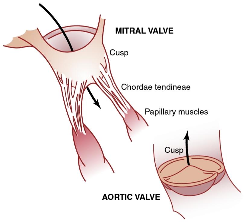
Figure 9-9.
Mitral and aortic valves (the left ventricular valves).
شکل 9-9.
دریچههای میترال و آئورت (دریچههای بطن چپ).
Function of the Papillary Muscles. Figure 9-9 also shows papillary muscles that attach to the vanes of the A-V valves by the chordae tendineae. The papillary mus- cles contract when the ventricular walls contract but, contrary to what might be expected, they do not help the valves to close. Instead, they pull the vanes of the valves inward toward the ventricles to prevent their bulging too far backward toward the atria during ventricular contrac- tion. If a chorda tendina becomes ruptured, or if one of the papillary muscles becomes paralyzed due to low blood flow from a myocardial infarction, the valve bulges far backward during ventricular contraction, sometimes so far that it leaks severely and results in severe or even lethal cardiac incapacity.
عملکرد عضلات پاپیلاری. شکل 9-9 نیز ماهیچههای پاپیلاری را نشان میدهد که به پرههای دریچههای A-V توسط تاندینههای chordae متصل میشوند. عضلات پاپیلاری زمانی که دیوارههای بطن منقبض میشوند منقبض میشوند، اما بر خلاف آنچه ممکن است انتظار میرود، به بسته شدن دریچهها کمکی نمیکنند. در عوض، پرههای دریچهها را به سمت داخل و به سمت بطنها میکشند تا از برآمدگی بیش از حد آنها به سمت دهلیزها در طول انقباض بطن جلوگیری کنند. اگر تاندینا چوردا پاره شود، یا اگر یکی از عضلات پاپیلاری به دلیل جریان خون کم ناشی از انفارکتوس میوکارد فلج شود، دریچه در طول انقباض بطن بسیار به سمت عقب برآمده میشود، گاهی اوقات به حدی که به شدت نشت میکند و منجر به ناتوانی قلبی شدید یا حتی کشنده میشود.
Aortic and Pulmonary Artery Valves. The aortic and pulmonary artery semilunar valves function quite dif- ferently from the A-V valves. First, the high pressures in the arteries at the end of systole cause the semilunar valves to snap closed, in contrast to the much softer closure of the A-V valves. Second, because of smaller openings, the velocity of blood ejection through the aortic and pulmonary valves is much greater than that through the much larger A-V valves. Also, because of the rapid closure and rapid ejection, the edges of the aortic and pulmonary valves are subjected to much greater mechanical abrasion than the A-V valves. Fi- nally, the A-V valves are supported by the chordae tendineae, which is not true for the semilunar valves. It is obvious from the anatomy of the aortic and pul- monary valves (as shown for the aortic valve at the bot- tom of Figure 9-9) that they must be constructed with an especially strong, yet very pliable, fibrous tissue to withstand the extra physical stresses.
دریچههای شریان آئورت و ریوی. دریچههای نیمه قمری شریان آئورت و ریوی کاملاً متفاوت از دریچههای A-V عمل میکنند. اول، فشار زیاد در شریانها در انتهای سیستول باعث میشود که دریچههای نیمه قمری بهسرعت بسته شوند، برخلاف بسته شدن بسیار نرمتر دریچههای A-V. دوم، به دلیل منافذ کوچکتر، سرعت خروج خون از طریق دریچههای آئورت و ریوی بسیار بیشتر از دریچههای A-V بسیار بزرگتر است. همچنین به دلیل بسته شدن سریع و خروج سریع، لبههای دریچههای آئورت و ریوی نسبت به دریچههای A-V در معرض ساییدگی مکانیکی بسیار بیشتری قرار میگیرند. در نهایت، دریچههای A-V توسط chordae tendineae پشتیبانی میشوند، که برای دریچههای نیمه قمری صادق نیست. از آناتومیدریچههای آئورت و ریوی (همانطور که برای دریچه آئورت در پایین شکل 9-9 نشان داده شده است) واضح است که آنها باید با بافت فیبری به خصوص قوی و در عین حال بسیار انعطاف پذیر ساخته شوند تا در برابر فشارهای فیزیکی اضافی مقاومت کنند.
AORTIC PRESSURE CURVE
When the left ventricle contracts, the ventricular pressure increases rapidly until the aortic valve opens. Then, after the valve opens, the pressure in the ventricle rises much less rapidly, as shown in Figure 9-7, because blood imme- diately flows out of the ventricle into the aorta and then into the systemic distribution arteries.
منحنی فشار آئورت
هنگامیکه بطن چپ منقبض میشود، فشار بطن به سرعت افزایش مییابد تا زمانی که دریچه آئورت باز شود. سپس، پس از باز شدن دریچه، فشار در بطن با سرعت بسیار کمتری افزایش مییابد، همانطور که در شکل 9-7 نشان داده شده است، زیرا خون بلافاصله از بطن به داخل آئورت و سپس به شریانهای توزیع سیستمیک جریان مییابد.
The entry of blood into the arteries during systole causes the walls of these arteries to stretch and the pres- sure to increase to about 120 mm Hg. Next, at the end of systole, after the left ventricle stops ejecting blood and the aortic valve closes, the elastic walls of the arter- ies maintain a high pressure in the arteries, even during diastole.
ورود خون به سرخرگها در طی سیستول باعث میشود که دیواره این شریانها کشیده شده و فشار تا حدود 120 میلی متر جیوه افزایش یابد. در مرحله بعد، در پایان سیستول، پس از اینکه بطن چپ خروج خون را متوقف کرد و دریچه آئورت بسته شد، دیوارههای الاستیک شریانها فشار بالایی را در شریانها حفظ میکنند، حتی در زمان دیاستول.
An incisura occurs in the aortic pressure curve when the aortic valve closes. This is caused by a short period of backward flow of blood immediately before closure of the valve, followed by the sudden cessation of backflow.
هنگامیکه دریچه آئورت بسته میشود، انسیزورا در منحنی فشار آئورت ایجاد میشود. این به دلیل یک دوره کوتاه جریان برگشتی خون بلافاصله قبل از بسته شدن دریچه و به دنبال آن قطع ناگهانی جریان برگشتی ایجاد میشود.
After the aortic valve closes, pressure in the aorta decreases slowly throughout diastole because the blood stored in the distended elastic arteries flows continually through the peripheral vessels back to the veins. Before the ventricle contracts again, the aortic pressure usually has fallen to about 80 mm Hg (diastolic pressure), which is two thirds the maximal pressure of 120 mm Hg (sys- tolic pressure) that occurs in the aorta during ventricular contraction.
پس از بسته شدن دریچه آئورت، فشار در آئورت به آرامیدر طول دیاستول کاهش مییابد زیرا خون ذخیره شده در شریانهای الاستیک متسع شده به طور مداوم از طریق عروق محیطی به وریدها باز میگردد. قبل از انقباض مجدد بطن، فشار آئورت معمولاً به حدود 80 میلیمتر جیوه (فشار دیاستولیک) کاهش مییابد که دو سوم فشار حداکثری 120 میلیمتر جیوه (فشار سیستولیک) است که در آئورت در طول انقباض بطن ایجاد میشود.
The pressure curves in the right ventricle and pulmo- nary artery are similar to those in the aorta, except that the pressures are only about one-sixth as great, as dis- cussed in Chapter 14.
منحنیهای فشار در بطن راست و شریان ریوی شبیه به آئورت است، با این تفاوت که فشارها تنها حدود یک ششم بیشتر است، همانطور که در فصل 14 بحث شد.
Relationship of the Heart Sounds to Heart Pumping
When listening to the heart with a stethoscope, one does not hear the opening of the valves because this is a relatively slow process that normally makes no noise. However, when the valves close, the vanes of the valves and the surrounding fluids vibrate under the influence of sudden pressure changes, giving off sound that travels in all directions through the chest.
رابطه صداهای قلب با پمپاژ قلب
هنگام گوش دادن به قلب با گوشیپزشکی، باز شدن دریچهها را نمیشنوید، زیرا این فرآیند نسبتاً آهسته است و معمولاً صدا ایجاد نمیکند. با این حال، هنگامیکه دریچهها بسته میشوند، پرههای دریچهها و سیالهای اطراف تحت تأثیر تغییرات فشار ناگهانی به ارتعاش در میآیند و صدایی تولید میکنند که در تمام جهات از قفسه سینه عبور میکند.
When the ventricles contract, one first hears a sound caused by closure of the A-V valves. The vibration pitch is low and relatively long-lasting and is known as the first heart sound (S1). When the aortic and pulmonary valves close at the end of systole, one hears a rapid snap because these valves close rapidly, and the surroundings vibrate for a short period. This sound is called the second heart sound (S2). The precise causes of the heart sounds are discussed more fully in Chapter 23 in relation to listening to the sounds with the stethoscope.
هنگامیکه بطنها منقبض میشوند، ابتدا صدایی را میشنوند که ناشی از بسته شدن دریچههای A-V است. گام ارتعاش پایین و نسبتاً طولانی است و به عنوان اولین صدای قلب (S1) شناخته میشود. هنگامیکه دریچههای آئورت و ریوی در انتهای سیستول بسته میشوند، صدای ضربه سریعی شنیده میشود، زیرا این دریچهها به سرعت بسته میشوند و محیط اطراف برای مدت کوتاهی میلرزد. این صدا را صدای قلب دوم (S2) مینامند. علل دقیق صداهای قلب در فصل 23 در رابطه با گوش دادن به صداها با گوشی پزشکی به طور کامل مورد بحث قرار گرفته است.
Work Output of the Heart
The stroke work output of the heart is the amount of energy that the heart converts to work during each heartbeat while pumping blood into the arteries. Work output of the heart is in two forms. First, the major proportion is used to move the blood from the low-pressure veins to the high-pressure arteries. This is called volume-pressure work or external work. Second, a minor proportion of the energy is used to accelerate the blood to its velocity of ejection through the aortic and pulmonary valves, which is the kinetic energy of blood flow component of the work output.
خروجی کار قلب
خروجی کار سکته مغزی مقدار انرژی است که قلب در طی هر ضربان قلب در حالی که خون را به داخل رگها پمپاژ میکند به کار تبدیل میکند. برون ده کار قلب به دو صورت است. اول، نسبت عمده برای انتقال خون از وریدهای کم فشار به شریانهای فشار بالا استفاده میشود. به این کار حجم فشار یا کار خارجی میگویند. دوم، بخش کوچکی از انرژی برای تسریع خون به سرعت خروج آن از طریق دریچههای آئورت و ریوی استفاده میشود، که انرژی جنبشی جزء جریان خون در خروجی کار است.
Right ventricular external work output is normally about one-sixth the work output of the left ventricle be- cause of the sixfold difference in systolic pressures pumped by the two ventricles. The additional work output of each ventricle required to create kinetic energy of blood flow is proportional to the mass of blood ejected times the square of velocity of ejection.
خروجی کار خارجی بطن راست به طور معمول حدود یک ششم خروجی کار بطن چپ است به دلیل اختلاف شش برابری در فشار سیستولیک پمپ شده توسط دو بطن. خروجی کار اضافی هر بطن مورد نیاز برای ایجاد انرژی جنبشی جریان خون متناسب با جرم خون خارج شده ضربدر مجذور سرعت تخلیه است.
Ordinarily, the work output of the left ventricle required to create kinetic energy of blood flow is only about 1% of the total work output of the ventricle and therefore is ig- nored in the calculation of the total stroke work output. In certain abnormal conditions, however, such as aortic ste- nosis, in which blood flows with great velocity through the stenosed valve, more than 50% of the total work output may be required to create kinetic energy of blood flow.
به طور معمول، خروجی کار بطن چپ مورد نیاز برای ایجاد انرژی جنبشی جریان خون تنها حدود 1٪ از کل خروجی کار بطن است و بنابراین در محاسبه کل خروجی کار سکته مغزی نادیده گرفته میشود. با این حال، در برخی شرایط غیر طبیعی، مانند تنگی آئورت، که در آن خون با سرعت زیادی از دریچه تنگ شده جریان مییابد، ممکن است بیش از 50 درصد از کل خروجی کار برای ایجاد انرژی جنبشی جریان خون مورد نیاز باشد.
GRAPHIC ANALYSIS OF VENTRICULAR PUMPING
Figure 9-10 shows a diagram that is especially useful in explaining the pumping mechanics of the left ventricle. The most important components of the diagram are the two curves labeled “diastolic pressure” and “systolic pres- sure.” These curves are volume-pressure curves.
تجزیه و تحلیل گرافیکی پمپاژ بطنی
شکل 9-10 نموداری را نشان میدهد که به ویژه در توضیح مکانیک پمپاژ بطن چپ مفید است. مهمترین اجزای نمودار دو منحنی با عنوان “فشار دیاستولیک” و “فشار سیستولیک” هستند. این منحنیها منحنیهای حجم-فشار هستند.
The diastolic pressure curve is determined by filling the heart with progressively greater volumes of blood and then measuring the diastolic pressure immediately before ventricular contraction occurs, which is the end-diastolic pressure of the ventricle.
منحنی فشار دیاستولیک با پر کردن قلب با حجمهای بیشتر خون و سپس اندازه گیری فشار دیاستولیک بلافاصله قبل از وقوع انقباض بطنی تعیین میشود که فشار پایان دیاستولیک بطن است.
The systolic pressure curve is determined by recording the systolic pressure achieved during ventricular contrac- tion at each volume of filling.
منحنی فشار سیستولیک با ثبت فشار سیستولیک حاصل از انقباض بطن در هر حجم پر شدن تعیین میشود.
Until the volume of the noncontracting ventricle rises above about 150 ml, the diastolic pressure does not increase much. Therefore, up to this volume, blood can flow easily into the ventricle from the atrium. Above 150 ml, the ventricular diastolic pressure increases rap- idly, partly because of fibrous tissue in the heart that will stretch no more, and partly because the pericardium that surrounds the heart becomes filled nearly to its limit.
تا زمانی که حجم بطن غیر انقباضی از حدود 150 میلی لیتر بالاتر برود، فشار دیاستولیک زیاد افزایش نمییابد. بنابراین تا این حجم خون میتواند به راحتی از دهلیز به داخل بطن جریان یابد. بیش از 150 میلیلیتر، فشار دیاستولیک بطنی به سرعت افزایش مییابد، تا حدی به دلیل بافت فیبری در قلب که دیگر کشیده نمیشود، و تا حدی به این دلیل که پریکاردی که قلب را احاطه کرده است تقریباً تا حد خود پر میشود.
During ventricular contraction, the systolic pressure increases, even at low ventricular volumes, and reaches a maximum at a ventricular volume of 150 to 170 ml. Then, as the volume increases further, the systolic pressure actu- ally decreases under some conditions, as demonstrated by the falling systolic pressure curve in Figure 9-10. This occurs because at these great volumes, the actin and myo- sin filaments of the cardiac muscle fibers are pulled apart far enough that the strength of each cardiac fiber contrac- tion becomes less than optimal.
در حین انقباض بطن، فشار سیستولیک حتی در حجمهای کم بطن افزایش مییابد و در حجم بطنی 150 تا 170 میلی لیتر به حداکثر میرسد. سپس، با افزایش بیشتر حجم، فشار سیستولیک در برخی شرایط در واقع کاهش مییابد، همانطور که با کاهش منحنی فشار سیستولیک در شکل 9-10 نشان داده شده است. این امر به این دلیل اتفاق میافتد که در این حجمهای زیاد، رشتههای اکتین و میوسین فیبرهای عضلانی قلب به اندازهای از هم جدا میشوند که قدرت هر انقباض فیبر قلبی کمتر از حد مطلوب میشود.
Note especially in the figure that the maximum sys- tolic pressure for the normal left ventricle is between 250 and 300 mm Hg, but this varies widely with each person’s heart strength and degree of heart stimulation by cardiac nerves. For the normal right ventricle, the maximum sys- tolic pressure is between 60 and 80 mm Hg.
به خصوص در شکل توجه داشته باشید که حداکثر فشار سیستولیک برای بطن چپ طبیعی بین 250 تا 300 میلیمتر جیوه است، اما این میزان با قدرت قلب و میزان تحریک قلب توسط اعصاب قلب به طور گستردهای متفاوت است. برای بطن راست طبیعی، حداکثر فشار سیستولیک بین 60 تا 80 میلی متر جیوه است.

Figure 9-10.
Relationship between left ventricular volume and intra- ventricular pressure during diastole and systole. Also shown by the red lines is the “volume-pressure diagram,” demonstrating changes in intraventricular volume and pressure during the normal cardiac cy- cle. EW, Net external work; PE, potential energy.
شکل 9-10.
رابطه بین حجم بطن چپ و فشار داخل بطن در طول دیاستول و سیستول. همچنین با خطوط قرمز “نمودار فشار حجم” نشان داده شده است که تغییرات در حجم و فشار داخل بطنی را در طول چرخه طبیعی قلب نشان میدهد. EW، کار خارجی خالص؛ پلی اتیلن، انرژی پتانسیل
Volume-Pressure Diagram During the Cardiac Cycle; Cardiac Work Output. The red lines in Figure 9-10 form a loop called the volume-pressure diagram of the cardiac cycle for normal function of the left ventricle. A more de- tailed version of this loop is shown in Figure 9-11. It is divided into four phases.
نمودار حجم-فشار در طول چرخه قلبی. برون ده کار قلبی خطوط قرمز در شکل 9-10 حلقه ای به نام نمودار حجم-فشار چرخه قلبی را برای عملکرد طبیعی بطن چپ تشکیل میدهند. یک نسخه دقیق تر از این حلقه در شکل 9-11 نشان داده شده است. به چهار فاز تقسیم میشود.
Phase I: Period of Filling. Phase I in the volume- pressure diagram begins at a ventricular volume of about 50 ml and a diastolic pressure of 2 to 3 mm Hg. The amount of blood that remains in the ventricle after the previous heartbeat, 50 ml, is called the end-systolic vol- ume. As venous blood flows into the ventricle from the left atrium, the ventricular volume normally increases to about 120 ml, called the end-diastolic volume, an increase of 70 ml. Therefore, the volume-pressure diagram during phase I extends along the line in Figure 9-10 labeled “I” and from point A to point B in Figure 9-11, with the vol- ume increasing to 120 ml and the diastolic pressure rising to about 5 to 7 mm Hg.
فاز اول: دوره پر کردن. فاز اول در نمودار حجم- فشار با حجم بطنی حدود 50 میلی لیتر و فشار دیاستولیک 2 تا 3 میلی متر جیوه شروع میشود. مقدار خونی که پس از ضربان قلب قبلی 50 میلی لیتر در بطن باقی میماند، حجم سیستولیک انتهایی نامیده میشود. هنگامیکه خون وریدی از دهلیز چپ به داخل بطن جریان مییابد، حجم بطن به طور معمول به حدود 120 میلی لیتر افزایش مییابد که به آن حجم پایان دیاستولیک میگویند، یعنی افزایش 70 میلی لیتری. بنابراین، نمودار حجم فشار در طول فاز I در امتداد خط در شکل 9-10 با برچسب “I” و از نقطه A به نقطه B در شکل 9-11 گسترش مییابد، با افزایش حجم به 120 میلی لیتر و افزایش فشار دیاستولیک به حدود 5 تا 7 میلی متر جیوه.
Phase II: Period of Isovolumic Contraction. During isovolumic contraction, the volume of the ventricle does not change because all valves are closed. However, the pressure inside the ventricle increases to equal the pres- sure in the aorta, at a pressure value of about 80 mm Hg, as depicted by point C (see Figure 9-11).
فاز دوم: دوره انقباض ایزوولومیک. در طول انقباض ایزوولومیک، حجم بطن تغییر نمیکند زیرا همه دریچهها بسته هستند. با این حال، فشار داخل بطن به اندازه فشار در آئورت، در مقدار فشار حدود 80 میلیمتر جیوه، همانطور که در نقطه C نشان داده شده است، افزایش مییابد (شکل 9-11 را ببینید).
Phase III: Period of Ejection. During ejection, the systolic pressure rises even higher because of still more contraction of the ventricle. At the same time, the volume of the ventricle decreases because the aortic valve has now opened, and blood flows out of the ventricle into the aorta. Therefore, in Figure 9-10, the curve labeled “III,” or “period of ejection,” traces the changes in volume and systolic pressure during this period of ejection.
فاز III: دوره تخلیه. در طول جهش، فشار سیستولیک به دلیل انقباض بیشتر بطن حتی بیشتر میشود. در همان زمان، حجم بطن کاهش مییابد زیرا دریچه آئورت اکنون باز شده است و خون از بطن به داخل آئورت جریان مییابد. بنابراین، در شکل 9-10، منحنی با برچسب “III” یا “دوره جهش”، تغییرات حجم و فشار سیستولیک را در طول این دوره جهش نشان میدهد.
Phase IV: Period of Isovolumic Relaxation. At the end of the period of ejection (point D, Figure 9-11), the aortic valve closes, and the ventricular pressure falls back to the diastolic pressure level. The line labeled “IV” (Figure 9- 10) traces this decrease in intraventricular pressure with- out any change in volume. Thus, the ventricle returns to its starting point, with about 50 ml of blood left in the ventricle at an atrial pressure of 2 to 3 mm Hg.
فاز IV: دوره آرامش ایزوولومیک. در پایان دوره جهش (نقطه D، شکل 9-11)، دریچه آئورت بسته میشود و فشار بطنی به سطح فشار دیاستولیک باز میگردد. خط با برچسب “IV” (شکل 9-10) این کاهش فشار داخل بطنی را بدون تغییر در حجم نشان میدهد. بنابراین، بطن به نقطه شروع خود باز میگردد و حدود 50 میلی لیتر خون در بطن با فشار دهلیزی 2 تا 3 میلی متر جیوه باقی میماند.
The area subtended by this functional volume- pressure diagram (the shaded area, labeled “EW”) repre- sents the net external work output of the ventricle during its contraction cycle. In experimental studies of cardiac contraction, this diagram is used for calculating cardiac work output.
ناحیه ای که توسط این نمودار عملکردی حجم- فشار (ناحیه سایه دار با برچسب “EW”) تحت تاثیر قرار میگیرد، خروجی خالص کار خارجی بطن را در طول چرخه انقباض آن نشان میدهد. در مطالعات تجربی انقباض قلب، از این نمودار برای محاسبه برون ده کار قلبی استفاده میشود.
When the heart pumps large quantities of blood, the area of the work diagram becomes much larger. That is, it extends far to the right because the ventricle fills with more blood during diastole, it rises much higher because the ventricle contracts with greater pressure, and it usu- ally extends farther to the left because the ventricle con- tracts to a smaller volume-especially if the ventricle is stimulated to increased activity by the sympathetic ner- vous system.
وقتی قلب مقادیر زیادی خون پمپاژ میکند، مساحت نمودار کار بسیار بزرگتر میشود. به این معنا که به سمت راست امتداد مییابد زیرا بطن در طول دیاستول با خون بیشتری پر میشود، بسیار بالاتر میرود زیرا بطن با فشار بیشتر منقبض میشود، و معمولاً به سمت چپ بیشتر میشود زیرا بطن به حجم کمتری منقبض میشود – به خصوص اگر بطن برای افزایش فعالیت توسط سیستم سمپاتیک تحریک شود.

Figure 9-11.
The volume-pressure diagram dem- onstrating changes in intraventricular volume and pressure during a single cardiac cycle (red line). The shaded area represents the net external work (EW) output by the left ventricle during the cardiac cycle.
شکل 9-11.
نمودار حجم-فشار که تغییرات حجم و فشار داخل بطنی را در طول یک سیکل قلبی منفرد نشان میدهد (خط قرمز). ناحیه سایهدار نشاندهنده خروجی خالص کار خارجی (EW) توسط بطن چپ در طول چرخه قلبی است.
Concepts of Preload and Afterload. In assessing the contractile properties of muscle, it is important to specify the degree of tension on the muscle when it begins to con- tract, called the preload, and to specify the load against which the muscle exerts its contractile force, called the afterload.
مفاهیم پیش از بارگیری و پس از بارگذاری. در ارزیابی ویژگیهای انقباضی عضله، تعیین درجه کشش روی ماهیچه هنگام شروع انقباض، به نام پیشبار، و مشخص کردن باری که عضله بر آن نیروی انقباضی خود را اعمال میکند، مشخص میشود که به آن پس بار میگویند.
For cardiac contraction, the preload is usually con- sidered to be the end-diastolic pressure when the ven- tricle has become filled. The afterload of the ventricle is the pressure in the aorta leading from the ventricle. In Figure 9-10, this corresponds to the systolic pressure described by the phase III curve of the volume-pressure diagram. (Sometimes the afterload is loosely considered to be the resistance in the circulation rather than the pressure.)
برای انقباض قلب، پیشبار معمولاً فشار پایان دیاستولیک زمانی در نظر گرفته میشود که بطن پر شده باشد. پس بار بطن فشار در آئورت است که از بطن منتهی میشود. در شکل 9-10، این مربوط به فشار سیستولیک توصیف شده توسط منحنی فاز III نمودار حجم فشار است. (گاهی اوقات پس بار به جای فشار به عنوان مقاومت در گردش در نظر گرفته میشود.)
The importance of the concepts of preload and after- load is that in many abnormal functional states of the heart or circulation, the pressure during filling of the ven- tricle (the preload), the arterial pressure against which the ventricle must contract (the afterload), or both are altered from normal to a severe degree.
اهمیت مفاهیم پیش بار و پس بار در این است که در بسیاری از حالات عملکردی غیرطبیعی قلب یا گردش خون، فشار در حین پر شدن بطن (پیش بار)، فشار شریانی که بطن باید در برابر آن منقبض شود (پس بار)، یا هر دو از نرمال به درجه شدیدی تغییر میکنند.
Chemical Energy Required for Cardiac Contraction: Oxygen Utilization by the Heart
Heart muscle, like skeletal muscle, uses chemical energy to provide the work of contraction. Approximately 70% to 90% of this energy is normally derived from oxidative metabolism of fatty acids, with about 10% to 30% com- ing from other nutrients, especially glucose and lactate. Therefore, the rate of oxygen consumption by the heart is an excellent measure of the chemical energy liberated while the heart performs its work. The different chemi- cal reactions that liberate this energy are discussed in Chapters 68 and 69.
انرژی شیمیایی مورد نیاز برای انقباض قلب: استفاده از اکسیژن توسط قلب
عضله قلب مانند ماهیچههای اسکلتی از انرژی شیمیایی برای انجام کار انقباض استفاده میکند. تقریباً 70 تا 90 درصد این انرژی معمولاً از متابولیسم اکسیداتیو اسیدهای چرب به دست میآید که حدود 10 تا 30 درصد آن از سایر مواد مغذی به ویژه گلوکز و لاکتات تأمین میشود. بنابراین، میزان مصرف اکسیژن توسط قلب، معیار عالی انرژی شیمیایی آزاد شده در حین انجام کار قلب است. واکنشهای شیمیایی مختلف که این انرژی را آزاد میکنند در فصلهای 68 و 69 مورد بحث قرار گرفتهاند.
Experimental studies have shown that oxygen consump- tion of the heart and the chemical energy expended during contraction are directly related to the total shaded area in Figure 9-10. This shaded portion consists of the external work (EW), as explained earlier, and an additional portion called the potential energy, labeled “PE” The potential ener- gy represents additional work that could be accomplished by contraction of the ventricle if the ventricle could completely empty all the blood in its chamber with each contraction.
مطالعات تجربی نشان داده اند که مصرف اکسیژن قلب و انرژی شیمیایی صرف شده در طول انقباض مستقیماً با کل منطقه سایه دار در شکل 9-10 مرتبط است. این بخش سایه دار از کار خارجی (EW) تشکیل شده است، همانطور که قبلا توضیح داده شد، و یک بخش اضافی به نام انرژی پتانسیل، با برچسب “PE” انرژی بالقوه نشان دهنده کار اضافی است که میتواند با انقباض بطن انجام شود، اگر بطن بتواند تمام خون در محفظه خود را با هر انقباض به طور کامل تخلیه کند.
Oxygen consumption has also been shown to be nearly proportional to the tension that occurs in the heart mus- cle during contraction multiplied by the duration of time that the contraction persists; this is called the tension-time index. According to the law of Laplace, ventricular wall ten- sion (T) is related to the left ventricular pressure (P) and the radius (r): T P x r.
همچنین نشان داده شده است که مصرف اکسیژن تقریباً متناسب با تنشی است که در طول انقباض در عضله قلب ایجاد میشود ضربدر مدت زمانی که انقباض ادامه دارد. به این شاخص زمان تنش میگویند. طبق قانون لاپلاس، کشش دیواره بطن (T) مربوط به فشار بطن چپ (P) و شعاع (r): T P x r است.
Because tension is high when systolic pressure (and therefore left ventricular pressure) is high, correspondingly more oxygen is used. When systolic pressure is chroni- cally elevated, wall stress and cardiac workload are also in- creased, inducing thickening of the left ventricular walls, which can reduce the ventricular chamber radius (concen- tric hypertrophy) and at least partially relieve the increased wall tension. Also, much more chemical energy is expend- ed, even at normal systolic pressures, when the ventricle is abnormally dilated (eccentric hypertrophy) because the heart muscle tension during contraction is proportional to pressure times the radius of the ventricle. This becomes es- pecially important in heart failure when the heart ventricle is dilated and, paradoxically, the amount of chemical en- ergy required for a given amount of work output is greater than normal, even though the heart is already failing.
از آنجایی که وقتی فشار سیستولیک (و در نتیجه فشار بطن چپ) بالا باشد، تنش زیاد است، به همین ترتیب اکسیژن بیشتری مصرف میشود. هنگامیکه فشار سیستولیک به طور مزمن افزایش مییابد، استرس دیواره و بار کاری قلبی نیز افزایش مییابد و باعث ضخیم شدن دیوارههای بطن چپ میشود که میتواند شعاع محفظه بطنی را کاهش دهد (هیپرتروفی متمرکز) و حداقل تا حدی افزایش کشش دیواره را کاهش دهد. همچنین، انرژی شیمیایی بسیار بیشتری صرف میشود، حتی در فشار سیستولیک طبیعی، زمانی که بطن به طور غیر طبیعی متسع میشود (هیپرتروفی خارج از مرکز)، زیرا تنش عضله قلب در طول انقباض متناسب با فشار ضربدر شعاع بطن است. این امر به ویژه در نارسایی قلبی زمانی که بطن قلب گشاد شده است اهمیت پیدا میکند و به طور متناقض، مقدار انرژی شیمیایی مورد نیاز برای مقدار مشخصی از کار بیشتر از حد طبیعی است، حتی اگر قلب در حال از کار افتادن باشد.
Cardiac Efficiency. During heart muscle contraction, most of the expended chemical energy is converted into heat, and a much smaller portion is converted into work output. Cardiac efficiency is the ratio of work output to total chemi- cal energy used to perform the work. Maximum efficiency of the normal heart is between 20% and 25%. In persons with heart failure, this efficiency can decrease to as low as 5%.
کارایی قلبی. در طول انقباض عضله قلب، بیشتر انرژی شیمیایی مصرف شده به گرما و بخش بسیار کمتری به خروجی کار تبدیل میشود. راندمان قلبی نسبت خروجی کار به کل انرژی شیمیایی مورد استفاده برای انجام کار است. حداکثر کارایی قلب طبیعی بین 20 تا 25 درصد است. در افراد مبتلا به نارسایی قلبی، این بازده میتواند تا 5 درصد کاهش یابد.
REGULATION OF HEART PUMPING
When a person is at rest, the heart pumps only 4 to 6 liters of blood each minute. During strenuous exercise, the heart may pump four to seven times this amount. The basic mechanisms for regulating heart pumping are as fol- lows: (1) intrinsic cardiac pumping regulation in response to changes in volume of blood flowing into the heart; and (2) control of heart rate and heart strength by the auto- nomic nervous system.
تنظیم پمپاژ قلب
زمانی که فرد در حال استراحت است، قلب در هر دقیقه تنها 4 تا 6 لیتر خون پمپاژ میکند. در طول ورزش شدید، قلب ممکن است چهار تا هفت برابر این مقدار پمپاژ کند. مکانیسمهای اساسی برای تنظیم پمپاژ قلب به شرح زیر است: (1) تنظیم درونی پمپاژ قلب در پاسخ به تغییرات در حجم خون جریان یافته به قلب. و (2) کنترل ضربان قلب و قدرت قلب توسط سیستم عصبی خودکار.
INTRINSIC REGULATION OF HEART PUMPING THE FRANK-STARLING MECHANISM
In Chapter 20, we will learn that under most conditions, the amount of blood pumped by the heart each minute is normally determined almost entirely by the rate of blood flow into the heart from the veins, which is called venous return. That is, each peripheral tissue of the body controls its own local blood flow, and all the local tissue flows com- bine and return by way of the veins to the right atrium. The heart, in turn, automatically pumps this incoming blood into the arteries so that it can flow around the circuit again.
تنظیم درونی پمپاژ قلب مکانیسم فرانک-ستارلینگ
در فصل 20، خواهیم آموخت که در اکثر شرایط، مقدار خون پمپاژ شده توسط قلب در هر دقیقه به طور معمول تقریباً به طور کامل توسط سرعت جریان خون از وریدها به قلب تعیین میشود که به آن بازگشت وریدی میگویند. یعنی هر بافت محیطی بدن جریان خون موضعی خود را کنترل میکند و تمام جریانهای بافت محلی با هم ترکیب میشوند و از طریق سیاهرگها به دهلیز راست باز میگردند. قلب نیز به نوبه خود، به طور خودکار این خون ورودی را به داخل رگها پمپ میکند تا بتواند دوباره در اطراف مدار جریان یابد.
This intrinsic ability of the heart to adapt to increasing volumes of inflowing blood is called the Frank-Starling mechanism of the heart, named in honor of Otto Frank and Ernest Starling, two great physiologists. Basically, the Frank-Starling mechanism means that the more the heart muscle is stretched during filling, the greater is the force of contraction, and the greater is the quantity of blood pumped into the aorta. Or, stated another way-within physiological limits, the heart pumps all the blood that returns to it by way of the veins.
این توانایی ذاتی قلب برای انطباق با حجم فزاینده خون ورودی، مکانیسم قلب فرانک استارلینگ نامیده میشود که به افتخار اتو فرانک و ارنست استارلینگ، دو فیزیولوژیست بزرگ، نامگذاری شده است. اساساً مکانیسم فرانک-استارلینگ به این معنی است که هرچه عضله قلب در حین پر شدن بیشتر کشیده شود، نیروی انقباض بیشتر است و مقدار خون پمپاژ شده به آئورت بیشتر است. یا به روش دیگری گفته شد – در محدوده فیزیولوژیکی، قلب تمام خونی را که از طریق وریدها به آن باز میگردد پمپ میکند.
What Is the Explanation of the Frank-Starling Mechanism? When an extra amount of blood flows into the ventricles, the cardiac muscle is stretched to a greater length. This stretching causes the muscle to contract with increased force because the actin and myosin filaments are brought to a more nearly optimal degree of overlap for force generation. Therefore, the ventricle, because of its increased pumping, automatically pumps the extra blood into the arteries. This ability of stretched muscle, up to an optimal length, to contract with increased work output is characteristic of all striated muscle, as explained in Chapter 6, and is not simply a characteristic of cardiac muscle.
توضیح مکانیسم فرانک استارلینگ چیست؟ هنگامیکه مقدار اضافی خون به داخل بطن جریان مییابد، عضله قلب به طول بیشتری کشیده میشود. این کشش باعث انقباض عضله با افزایش نیرو میشود، زیرا رشتههای اکتین و میوزین برای تولید نیرو به درجه بهینهتری از همپوشانی میرسند. بنابراین، بطن به دلیل افزایش پمپاژ خود، به طور خودکار خون اضافی را به داخل شریانها پمپ میکند. این توانایی عضله کشیده شده، تا یک طول بهینه، برای انقباض با افزایش برون ده کار، مشخصه تمام عضلات مخطط است، همانطور که در فصل 6 توضیح داده شد، و صرفاً یک ویژگی عضله قلبی نیست.
In addition to the important effect of lengthening the heart muscle, another factor increases heart pumping when its volume is increased. Stretch of the right atrial wall directly increases the heart rate by 10% to 20%, which also helps increase the amount of blood pumped each minute, although its contribution is much less than that of the Frank-Starling mechanism. As discussed in Chapter 18, stretch of the atrium also activates stretch receptors and a nervous reflex, the Bainbridge reflex, that is trans- mitted by the vagus nerve and may increase heart rate an additional 40% to 60%.
علاوه بر تاثیر مهم افزایش طول عضله قلب، عامل دیگری باعث افزایش پمپاژ قلب در هنگام افزایش حجم آن میشود. کشش دیواره دهلیز راست مستقیماً ضربان قلب را بین 10 تا 20 درصد افزایش میدهد که به افزایش میزان خون پمپاژ شده در هر دقیقه کمک میکند، اگرچه سهم آن بسیار کمتر از مکانیسم فرانک استارلینگ است. همانطور که در فصل 18 بحث شد، کشش دهلیز همچنین گیرندههای کششی و یک رفلکس عصبی را فعال میکند، رفلکس بینبریج، که توسط عصب واگ منتقل میشود و ممکن است ضربان قلب را 40% تا 60% افزایش دهد.
Ventricular Function Curves
One of the best ways to express the functional ability of the ventricles to pump blood is by ventricular function curves. Figure 9-12 shows a type of ventricular function curve called the stroke work output curve. Note that as atrial pressure for each side of the heart increases, stroke work output for that side increases until it reaches the limit of the ventricle’s pumping ability.
منحنیهای عملکرد بطنی
یکی از بهترین راهها برای بیان توانایی عملکردی بطنها در پمپاژ خون، منحنیهای عملکرد بطن است. شکل 9-12 نوعی منحنی عملکرد بطنی به نام منحنی خروجی کار ضربه ای را نشان میدهد. توجه داشته باشید که با افزایش فشار دهلیزی برای هر طرف قلب، برون ده کار سکته مغزی برای آن سمت افزایش مییابد تا زمانی که به حد توانایی پمپاژ بطن برسد.
Figure 9-13 shows another type of ventricular func- tion curve called the ventricular volume output curve. The two curves of this figure represent function of the two ventricles of the human heart based on data extrapolated from experimental animal studies. As the right and left atrial pressures increase, the respective ventricular vol- ume outputs per minute also increase.
شکل 9-13 نوع دیگری از منحنی عملکرد بطنی به نام منحنی خروجی حجم بطن را نشان میدهد. دو منحنی این شکل نشان دهنده عملکرد دو بطن قلب انسان بر اساس دادههای برون یابی شده از مطالعات حیوانی تجربی است. با افزایش فشار دهلیز راست و چپ، خروجی حجم بطن مربوطه در دقیقه نیز افزایش مییابد.
Thus, ventricular function curves are another way of expressing the Frank-Starling mechanism of the heart. That is, as the ventricles fill in response to higher atrial pressures, each ventricular volume and strength of car- diac muscle contraction increase, causing the heart to pump increased quantities of blood into the arteries.
بنابراین، منحنیهای عملکرد بطن راه دیگری برای بیان مکانیسم فرانک-استارلینگ قلب است. یعنی با پر شدن بطنها در پاسخ به فشارهای دهلیزی بالاتر، هر حجم بطنی و قدرت انقباض عضله قلب افزایش مییابد و باعث میشود که قلب مقادیر بیشتری خون را به داخل رگها پمپاژ کند.

Figure 9-12.
Left and right ventricular function curves recorded from dogs, depicting ventricular stroke work output as a function of left and right mean atrial pressures. (Data from Sarnoff SJ: Myocardial contractility as described by ventricular function curves. Physiol Rev 35:107, 1955.)
شکل 9-12.
منحنیهای عملکرد بطن چپ و راست ثبتشده از سگها، خروجی کار سکته مغزی بطن را به عنوان تابعی از فشار متوسط دهلیزی چپ و راست نشان میدهد. (دادههای Sarnoff SJ: انقباض میوکارد همانطور که توسط منحنیهای عملکرد بطنی توصیف شده است. Physiol Rev 35:107، 1955.)

Figure 9-13.
Approximate normal right and left ventricular volume output curves for the normal resting human heart as extrapolated from data obtained in dogs and data from humans.
شکل 9-13.
منحنیهای خروجی حجم بطن راست و چپ طبیعی تقریبی برای قلب طبیعی انسان در حال استراحت همانطور که از دادههای به دست آمده در سگها و دادههای انسان برون یابی شده است.
Control of the Heart by the Sympathetic and Parasympathetic Nerves
The pumping effectiveness of the heart also is controlled by the sympathetic and parasympathetic (vagus) nerves, which abundantly supply the heart, as shown in Figure 9-14. For given levels of atrial pressure, the amount of blood pumped each minute (cardiac output) often can be increased more than 100% by sympathetic stimulation. By contrast, the output can be decreased to almost zero by vagal (parasympathetic) stimulation.
کنترل قلب توسط اعصاب سمپاتیک و پاراسمپاتیک
کارایی پمپاژ قلب نیز توسط اعصاب سمپاتیک و پاراسمپاتیک (واگ) کنترل میشود که به وفور قلب را تامین میکنند، همانطور که در شکل 9-14 نشان داده شده است. برای سطوح معین فشار دهلیزی، مقدار خون پمپاژ شده در هر دقیقه (برون ده قلبی) اغلب میتواند بیش از 100٪ با تحریک سمپاتیک افزایش یابد. در مقابل، خروجی را میتوان با تحریک واگ (پاراسمپاتیک) تقریباً به صفر رساند.

Figure 9-14.
Cardiac sympathetic and parasympathetic nerves. (The vagus nerves to the heart are parasympathetic nerves.) A-V, Atrioven- tricular; S-A, sinoatrial.
شکل 9-14.
اعصاب سمپاتیک و پاراسمپاتیک قلبی. (اعصاب واگ به قلب اعصاب پاراسمپاتیک هستند.) A-V، دهلیزی بطنی. S-A، سینوسی دهلیزی.
Mechanisms of Excitation of the Heart by the Sym- pathetic Nerves. Strong sympathetic stimulation can increase the heart rate in young adult humans from the normal rate of 70 beats/min up to 180 to 200 beats/min and, rarely, even 250 beats/min. Also, sympathetic stimu- lation may double the force of heart contraction, thereby increasing the volume of blood pumped and increasing the ejection pressure. Thus, sympathetic stimulation often can increase the maximum cardiac output as much as twofold to threefold, in addition to the increased output caused by the Frank-Starling mechanism already discussed.
مکانیسمهای تحریک قلب توسط اعصاب سمپاتیک. تحریک شدید سمپاتیک میتواند ضربان قلب را در انسان بالغ جوان از نرخ طبیعی 70 ضربه در دقیقه به 180 تا 200 ضربه در دقیقه و به ندرت حتی 250 ضربه در دقیقه افزایش دهد. همچنین، تحریک سمپاتیک ممکن است نیروی انقباض قلب را دو برابر کند، در نتیجه حجم خون پمپ شده را افزایش داده و فشار بیرون را افزایش میدهد. بنابراین، تحریک سمپاتیک اغلب میتواند حداکثر برون ده قلبی را دو برابر تا سه برابر افزایش دهد، علاوه بر افزایش برون ده ناشی از مکانیسم فرانک-استارلینگ که قبلاً بحث شد.
Conversely, inhibition of the sympathetic nerves to the heart can decrease cardiac pumping to a moderate extent. Under normal conditions, the sympathetic nerve fibers to the heart discharge continuously at a slow rate that maintains pumping at about 30% above that with no sym- pathetic stimulation. Therefore, when sympathetic ner- vous system activity is depressed below normal, both the heart rate and strength of ventricular muscle contraction decrease, thereby decreasing the level of cardiac pumping by as much as 30% below normal.
برعکس، مهار اعصاب سمپاتیک به قلب میتواند پمپاژ قلب را تا حد متوسطی کاهش دهد. در شرایط عادی، رشتههای عصبی سمپاتیک به طور پیوسته با سرعت آهسته به سمت قلب تخلیه میشوند که پمپاژ آن در حدود 30 درصد بالاتر از آن بدون تحریک سمپاتیک حفظ میشود. بنابراین، زمانی که فعالیت سیستم عصبی سمپاتیک کمتر از حد طبیعی باشد، هم ضربان قلب و هم قدرت انقباض عضله بطنی کاهش مییابد و در نتیجه سطح پمپاژ قلبی را تا 30 درصد کمتر از حد طبیعی کاهش میدهد.
Parasympathetic (Vagal) Stimulation Reduces Heart Rate and Strength of Contraction. Strong stimulation of the parasympathetic nerve fibers in the vagus nerves to the heart can stop the heartbeat for a few seconds, but then the heart usually “escapes” and beats at a rate of 20 to 40 beats/min as long as the parasympathetic stimula- tion continues. In addition, strong vagal stimulation can decrease the strength of heart muscle contraction by 20% to 30%.
تحریک پاراسمپاتیک (واگال) ضربان قلب و قدرت انقباض را کاهش میدهد. تحریک شدید رشتههای عصبی پاراسمپاتیک در اعصاب واگ به قلب میتواند ضربان قلب را برای چند ثانیه متوقف کند، اما پس از آن قلب معمولاً “فرار” میکند و تا زمانی که تحریک پاراسمپاتیک ادامه دارد، با سرعت 20 تا 40 ضربه در دقیقه میزند. علاوه بر این، تحریک قوی واگ میتواند قدرت انقباض عضله قلب را بین 20 تا 30 درصد کاهش دهد.
The vagal fibers are distributed mainly to the atria and not much to the ventricles, where the power contrac- tion of the heart occurs. This distribution explains why the effect of vagal stimulation is mainly to decrease the heart rate rather than to decrease greatly the strength of heart contraction. Nevertheless, the great decrease in heart rate, combined with a slight decrease in heart con- traction strength, can decrease ventricular pumping by 50% or more.
فیبرهای واگ عمدتاً در دهلیزها و نه چندان به بطنها، جایی که انقباض قدرت قلب رخ میدهد، توزیع میشوند. این توزیع توضیح میدهد که چرا اثر تحریک واگ عمدتاً به جای کاهش شدید قدرت انقباض قلب، کاهش ضربان قلب است. با این وجود، کاهش شدید ضربان قلب، همراه با کاهش جزئی در قدرت انقباض قلب، میتواند پمپاژ بطنی را تا 50 درصد یا بیشتر کاهش دهد.
Effect of Sympathetic or Parasympathetic Stimula- tion on the Cardiac Function Curve. Figure 9-15 shows four cardiac function curves. These curves are similar to the ventricular function curves of Figure 9-13. However, they represent function of the entire heart rather than that of a single ventricle. They show the relationship be- tween right atrial pressure at the input of the right heart and cardiac output from the left ventricle into the aorta.
اثر تحریک سمپاتیک یا پاراسمپاتیک بر منحنی عملکرد قلب. شکل 9-15 چهار منحنی عملکرد قلب را نشان میدهد. این منحنیها مشابه منحنیهای عملکرد بطنی شکل 9-13 هستند. با این حال، آنها عملکرد کل قلب را به جای یک بطن منفرد نشان میدهند. آنها رابطه بین فشار دهلیز راست در ورودی قلب راست و برون ده قلبی از بطن چپ به آئورت را نشان میدهند.
The curves of Figure 9-15 demonstrate that at any given right atrial pressure, the cardiac output increases during increased sympathetic stimulation and decreases during increased parasympathetic stimulation. These changes in output caused by autonomic nervous system stimulation result from changes in heart rate and from changes in contractile strength of the heart.
منحنیهای شکل 9-15 نشان میدهد که در هر فشار دهلیز راست مشخص، برون ده قلبی در طول افزایش تحریک سمپاتیک افزایش مییابد و در طول افزایش تحریک پاراسمپاتیک کاهش مییابد. این تغییرات در خروجی ناشی از تحریک سیستم عصبی خودمختار ناشی از تغییرات در ضربان قلب و تغییر در قدرت انقباضی قلب است.

Figure 9-15.
Effect on the cardiac output curve of different degrees of sympathetic or parasympathetic stimulation.
شکل 9-15.
تأثیر درجات مختلف تحریک سمپاتیک یا پاراسمپاتیک بر منحنی برون ده قلبی.
EFFECT OF POTASSIUM AND CALCIUM IONS ON HEART FUNCTION
In our discussion of membrane potentials in Chapter 5, we pointed out that potassium ions have a marked effect on membrane potentials, and in Chapter 6 we noted that calcium ions play an especially important role in activat- ing the muscle contractile process. Therefore, it is not surprising that the concentrations of each of these two ions in the extracellular fluids have important effects on cardiac pumping.
تاثیر یونهای پتاسیم و کلسیم بر عملکرد قلب
در بحث ما در مورد پتانسیلهای غشایی در فصل 5، اشاره کردیم که یونهای پتاسیم تأثیر قابل توجهی بر پتانسیلهای غشا دارند و در فصل 6 اشاره کردیم که یونهای کلسیم نقش مهمیدر فعال کردن فرآیند انقباض عضلانی ایفا میکنند. بنابراین، جای تعجب نیست که غلظت هر یک از این دو یون در مایعات خارج سلولی اثرات مهمیبر پمپاژ قلب داشته باشد.
Effect of Potassium lons. Excess potassium in the ex- tracellular fluids causes the heart to become dilated and flaccid and also slows the heart rate. Large quantities of potassium also can block conduction of the cardiac im- pulse from the atria to the ventricles through the A-V bundle. Elevation of potassium concentration to only 8 to 12 mEq/L-two to three times the normal value-can cause severe weakness of the heart, abnormal rhythm, and death.
اثر لانهای پتاسیم. پتاسیم اضافی در مایعات خارج سلولی باعث گشاد شدن و شل شدن قلب و همچنین کاهش ضربان قلب میشود. مقادیر زیاد پتاسیم همچنین میتواند هدایت ضربان قلب از دهلیزها به بطنها را از طریق بسته A-V مسدود کند. افزایش غلظت پتاسیم به تنها 8 تا 12 میلی اکیوالان در لیتر – دو تا سه برابر مقدار طبیعی – میتواند باعث ضعف شدید قلب، ریتم غیر طبیعی و مرگ شود.
These effects result partially from the fact that a high potassium concentration in the extracellular fluids. decreases the resting membrane potential in the car- diac muscle fibers, as explained in Chapter 5. That is, a high extracellular fluid potassium concentration partially depolarizes the cell membrane, causing the membrane potential to be less negative. As the membrane poten- tial decreases, the intensity of the action potential also decreases, which makes contraction of the heart progres- sively weaker.
این اثرات تا حدی ناشی از این واقعیت است که غلظت بالای پتاسیم در مایعات خارج سلولی وجود دارد. همانطور که در فصل 5 توضیح داده شد، پتانسیل غشای استراحت در فیبرهای عضلانی قلبی را کاهش میدهد. یعنی غلظت بالای پتاسیم مایع خارج سلولی تا حدی غشای سلولی را دپولاریزه میکند و باعث میشود پتانسیل غشاء منفی کمتر شود. با کاهش پتانسیل غشاء، شدت پتانسیل عمل نیز کاهش مییابد، که انقباض قلب را به تدریج ضعیف میکند.
Effect of Calcium lons. Excess calcium ions cause effects almost exactly opposite to those of potassium ions, caus- ing the heart to move toward spastic contraction. This ef- fect is caused by a direct effect of calcium ions to initiate the cardiac contractile process, as explained earlier in this chapter.
تاثیر کلسیم lons. یونهای کلسیم اضافی، اثراتی تقریباً برخلاف یونهای پتاسیم ایجاد میکنند و باعث میشوند قلب به سمت انقباض اسپاستیک حرکت کند. همانطور که قبلا در این فصل توضیح داده شد، این اثر ناشی از اثر مستقیم یونهای کلسیم برای شروع فرآیند انقباض قلبی است.
Conversely, deficiency of calcium ions causes cardiac weakness, similar to the effect of high potassium. For- tunately, calcium ion levels in the blood normally are regulated within a very narrow range. Therefore, cardiac effects of abnormal calcium concentrations are seldom of clinical concern.
برعکس، کمبود یونهای کلسیم باعث ضعف قلب میشود، مانند اثر پتاسیم بالا. خوشبختانه، سطح یون کلسیم در خون به طور معمول در یک محدوده بسیار باریک تنظیم میشود. بنابراین، اثرات قلبی غلظت غیر طبیعی کلسیم به ندرت مورد توجه بالینی است.
EFFECT OF TEMPERATURE ON HEART FUNCTION
Increased body temperature, such as that which occurs during fever, greatly increases the heart rate, sometimes to double the normal rate. Decreased temperature greatly decreases the heart rate, which may fall to as low as a few beats per minute when a person is near death from hypothermia in the body temperature range of 60° to 70°F (15.5°-21°C). These effects presumably result from the fact that heat increases the permeability of the cardiac muscle membrane to ions that control heart rate, result- ing in acceleration of the self-excitation process.
اثر دما بر عملکرد قلب
افزایش دمای بدن، مانند دمایی که در هنگام تب رخ میدهد، ضربان قلب را به شدت افزایش میدهد، گاهی اوقات تا دو برابر ضربان طبیعی. کاهش دما ضربان قلب را تا حد زیادی کاهش میدهد، که ممکن است زمانی که فرد به دلیل هیپوترمیدر محدوده دمای بدن 60 تا 70 درجه فارنهایت (15.5-21 درجه سانتیگراد) نزدیک به مرگ است، به چند ضربه در دقیقه کاهش یابد. این اثرات احتمالاً ناشی از این واقعیت است که گرما نفوذپذیری غشای عضله قلب را به یونهایی که ضربان قلب را کنترل میکنند، افزایش میدهد و در نتیجه فرآیند خود تحریکی را تسریع میکند.
Contractile strength of the heart often is enhanced temporarily by a moderate increase in temperature, such as that which occurs during body exercise, but prolonged temperature elevation exhausts the metabolic systems of the heart and eventually causes weakness. Therefore, opti- mal heart function depends greatly on proper control of body temperature by the control mechanisms explained in Chapter 74.
قدرت انقباض قلب اغلب با افزایش متوسط دما، مانند آنچه در طول ورزش بدن رخ میدهد، به طور موقت افزایش مییابد، اما افزایش طولانی مدت دما سیستمهای متابولیک قلب را خسته میکند و در نهایت باعث ضعف میشود. بنابراین، عملکرد مطلوب قلب تا حد زیادی به کنترل مناسب دمای بدن توسط مکانیسمهای کنترلی توضیح داده شده در فصل 74 بستگی دارد.
INCREASING THE ARTERIAL PRESSURE LOAD (UP TO A LIMIT) DOES NOT DECREASE CARDIAC OUTPUT
Note in Figure 9-16 that increasing the arterial pressure in the aorta does not decrease cardiac output until the mean arterial pressure rises above about 160 mm Hg. In other words, during normal heart function at normal sys- tolic arterial pressures (80-140 mm Hg), cardiac output is determined almost entirely by the ease of blood flow through the body’s tissues, which in turn controls venous return of blood to the heart. This mechanism is the prin- cipal subject of Chapter 20.
افزایش بار فشار شریانی (تا حدی) برون ده قلبی را کاهش نمیدهد
در شکل 9-16 توجه داشته باشید که افزایش فشار شریانی در آئورت برون ده قلبی را کاهش نمیدهد تا زمانی که فشار متوسط شریانی از حدود 160 میلی متر جیوه بالاتر برود. به عبارت دیگر، در طول عملکرد طبیعی قلب در فشارهای شریانی سیستولیک نرمال (80-140 میلی متر جیوه)، برون ده قلبی تقریباً به طور کامل توسط سهولت جریان خون در بافتهای بدن تعیین میشود که به نوبه خود بازگشت وریدی خون به قلب را کنترل میکند. این مکانیسم موضوع اصلی فصل 20 است.

Figure 9-16.
Constancy of cardiac output up to a pressure level of 160 mm Hg. Only when the arterial pressure rises above this normal limit does the increasing pressure load cause the cardiac output to fall significantly.
شکل 9-16.
پایداری برون ده قلبی تا سطح فشار 160 میلی متر جیوه. تنها زمانی که فشار شریانی از این حد طبیعی بالاتر میرود، بار فشار فزاینده باعث کاهش قابل توجه برون ده قلبی میشود.
کلیک کنید «Bibliography: فهرست کتب مربوطه»
Bell V, Mitchell GF: Influence of vascular function and pulsatile hemodynamics on cardiac function. Curr Hypertens Rep 17: 580, 2015.
Bertero E, Maack C: Calcium signaling and reactive oxygen species in mitochondria. Circ Res 122: 1460, 2018
Cingolani HE, Pérez NG, Cingolani OH, Ennis IL: The Anrep effect: 100 years later. Am J Physiol Heart Circ Physiol 304:H175, 2013.
Dewenter M, von der Lieth A, Katus HA, Backs J: Calcium signal- ing and transcriptional regulation in cardiomyocytes. Circ Res 121:1000, 2017.
Doenst T, Nguyen TD, Abel ED: Cardiac metabolism in heart failure: implications beyond ATP production. Circ Res 113:709, 2013.
Eisner DA, Caldwell JL, Kistamás K, Trafford AW: Calcium and excitation-contraction coupling in the heart. Circ Res 121:181,
2017.
Finkel T, Menazza S, Holmström KM, et al: The ins and outs of mito- chondrial calcium. Circ Res 116:1810, 2015.
Guyton AC, Jones CE, Coleman TG: Circulatory Physiology: Cardiac Output and Its Regulation, 2nd ed. Philadelphia: WB Saunders, 1973.
Kho C, Lee A, Hajjar RJ: Altered sarcoplasmic reticulum calcium cy- cling targets for heart failure therapy. Nat Rev Cardiol 9:717, 2012.
Lewis GA, Schelbert EB, Williams SG, et al: Biological phenotypes of heart failure with preserved ejection fraction. J Am Coll Cardiol 70:2186, 2017.
Luo M, Anderson ME: Mechanisms of altered Ca2+ handling in heart failure. Circ Res 113:690, 2013.
Mangoni ME, Nargeot J: Genesis and regulation of the heart automa- ticity. Physiol Rev 88:919, 2008.
Marks AR: Calcium cycling proteins and heart failure: mechanisms and therapeutics. J Clin Invest 123:46, 2013.
Mayourian J, Ceholski DK, Gonzalez DM, et al: Physiologic, patho- logic, and therapeutic paracrine modulation of cardiac excitation- contraction coupling. Circ Res 122:167, 2018.
Omar AM, Vallabhajosyula S, Sengupta PP: Left ventricular twist and torsion: research observations and clinical applications. Circ Car- diovasc Imaging 8:74, 2015.
Puglisi JL, Negroni JA, Chen-Izu Y, Bers DM: The force-frequency re- lationship: insights from mathematical modeling. Adv Physiol Educ 37:28, 2013.
Sarnoff SJ: Myocardial contractility as described by ventricular function curves. Physiol Rev 35:107, 1955.
Starling EH: The Linacre Lecture on the Law of the Heart. London: Longmans Green, 1918.
ter Keurs HE: The interaction of Ca2+ with sarcomeric proteins: role in function and dysfunction of the heart. Am J Physiol Heart Circ Physiol 302:H38, 2012.
Triposkiadis F, Pieske B, Butler J, et al: Global left atrial failure in heart failure. Eur J Heart Fail 18:1307, 2016.
Vega RB, Kelly DP: Cardiac nuclear receptors: architects of mitochon- drial structure and function. J Clin Invest 127:1155, 2017.
🚀 با ما همراه شوید!
تازهترین مطالب و آموزشهای مغز و اعصاب را از دست ندهید. با فالو کردن کانال تلگرام آیندهنگاران مغز، از ما حمایت کنید!


