
فیزیولوژی پزشکی گایتون و هال؛ تحریک ریتمیک قلب


دعای مطالعه [ نمایش ]
بِسْمِ الله الرَّحْمنِ الرَّحیمِ
اَللّهُمَّ اَخْرِجْنى مِنْ ظُلُماتِ الْوَهْمِ
خدايا مرا بيرون آور از تاريكىهاى وهم،
وَ اَكْرِمْنى بِنُورِ الْفَهْمِ
و به نور فهم گرامى ام بدار،
اَللّهُمَّ افْتَحْ عَلَيْنا اَبْوابَ رَحْمَتِكَ
خدايا درهاى رحمتت را به روى ما بگشا،
وَانْشُرْ عَلَيْنا خَزائِنَ عُلُومِكَ بِرَحْمَتِكَ يا اَرْحَمَ الرّاحِمينَ
و خزانههاى علومت را بر ما باز كن به امید رحمتت اى مهربانترين مهربانان.
کتاب «فیزیولوژی پزشکی گایتون وهال» بهعنوان یکی از جامعترین و معتبرترین منابع در حوزه علوم پزشکی، همچنان مرجع کلیدی برای درک عملکرد پیچیده بدن انسان است. این اثر با تکیه بر تازهترین پژوهشها و توضیحات دقیق از سازوکارهای فیزیولوژیک، پلی میان علوم پایه پزشکی و کاربردهای بالینی ایجاد میکند و نقشی بیبدیل در آموزش، پژوهش و ارتقای دانش سلامت ایفا مینماید.
ترجمه دقیق و علمیاین شاهکار توسط برند علمیآیندهنگاران مغز به مدیریت داریوش طاهری، دسترسی فارسیزبانان به مرزهای نوین دانش فیزیولوژی را ممکن ساخته و رسالتی علمیبرای ارتقای آموزش پزشکی، فهم عمیقتر سازوکارهای بدن و توسعه روشهای نوین در حوزه سلامت فراهم آورده است.
» کتاب فیزیولوژی پزشکی گایتون وهال
» » فصل ۱۰: تحریک ریتمیک قلب
در حال ویرایش
» Guyton and Hall Textbook of Medical Physiology
»» CHAPTER 10: Rhythmical Excitation of the Heart

The human heart has a special system for rhythmic self-excitation and repetitive contraction approximately 100,000 times each day or 3 billion times in the average human lifetime. This impressive feat is performed by a system that does the following: (1) generates electrical impulses to initiate rhythmical contraction of the heart muscle; and (2) conducts these impulses rapidly through the heart. When this system functions normally, the atria contract about one-sixth of a second ahead of ventricular contraction, which allows filling of the ventricles before they pump blood through the lungs and peripheral circulation. Another especially important feature of the system is that it allows all portions of the ventricles to contract almost simultaneously, which is essential for the most effective pressure generation in the ventricular chambers.
قلب انسان یک سیستم ویژه برای خود تحریکی ریتمیک و انقباضات تکراری تقریباً 100000 بار در روز یا 3 میلیارد بار در طول عمر متوسط انسان دارد. این شاهکار چشمگیر توسط سیستمیانجام میشود که کارهای زیر را انجام میدهد: (1) تکانههای الکتریکی برای شروع انقباض ریتمیک عضله قلب ایجاد میکند. و (2) این تکانهها را به سرعت از طریق قلب هدایت میکند. هنگامیکه این سیستم به طور طبیعی کار میکند، دهلیزها حدود یک ششم ثانیه قبل از انقباض بطنی منقبض میشوند، که اجازه میدهد بطنها را قبل از پمپاژ خون از طریق ریهها و گردش خون محیطی پر کنند. یکی دیگر از ویژگیهای مهم این سیستم این است که به تمام قسمتهای بطنها اجازه میدهد تقریباً به طور همزمان منقبض شوند، که برای موثرترین تولید فشار در اتاقکهای بطنی ضروری است.
This rhythmical and conductive system of the heart is susceptible to damage by heart disease, especially by ischemia resulting from inadequate coronary blood flow. The effect is often a bizarre heart rhythm or an abnormal sequence of contraction of the heart chambers, and the pumping effectiveness of the heart can be affected severely, even to the extent of causing death.
این سیستم ریتمیک و هدایت کننده قلب در معرض آسیب ناشی از بیماری قلبی، به ویژه ایسکمیناشی از جریان ناکافی خون کرونر است. این اثر اغلب یک ریتم قلب عجیب یا یک توالی غیر طبیعی انقباض حفرههای قلب است و اثر پمپاژ قلب میتواند به شدت تحت تاثیر قرار گیرد، حتی تا حدی که باعث مرگ شود.
SPECIALIZED EXCITATORY AND CONDUCTIVE SYSTEM OF THE HEART
Figure 10-1 shows the specialized excitatory and conductive system of the heart that controls cardiac con- tractions. The figure shows the sinus node (also called sinoatrial [S-A] node), in which the normal rhythmical impulses are generated; the internodal pathways that conduct impulses from the sinus node to the atrioventricular (A-V) node; the A-V node in which impulses from the atria are delayed before passing into the ventricles; the A-V bundle, which conducts impulses from the atria into the ventricles; and the left and right bundle branches of Purkinje fibers, which conduct the cardiac impulses to all parts of the ventricles.
سیستم تحریکی و رسانای تخصصی قلب
شکل 10-1 سیستم تحریکی و هدایتی تخصصی قلب را نشان میدهد که انقباضات قلبی را کنترل میکند. شکل گره سینوسی را نشان میدهد (که به آن گره سینوسی دهلیزی [S-A] نیز گفته میشود)، که در آن تکانههای ریتمیک طبیعی ایجاد میشود. مسیرهای بین گره ای که تکانهها را از گره سینوسی به گره دهلیزی (A-V) هدایت میکنند. گره A-V که در آن تکانههای دهلیزها قبل از عبور به بطنها به تعویق میافتد. بسته A-V، که تکانهها را از دهلیزها به بطنها هدایت میکند. و شاخههای چپ و راست رشتههای پورکنژ که تکانههای قلبی را به تمام قسمتهای بطن هدایت میکنند.

Figure 10-1. Sinus node and the purkinje system of the hear, showing also the atrioventricular (A-B) node, atral internodal pathways, and ventricular bundle branches.
شکل 10-1. گره سینوسی و سیستم پورکنژ قلب، همچنین گره دهلیزی-بطنی (A-B)، مسیرهای بین گرهای دهلیزی و شاخههای دسته بطنی را نشان میدهد.
SINUS (SINOATRIAL) NODE
The sinus node is a small, flattened, ellipsoid strip of specialized cardiac muscle about 3 mm wide, 15 mm long, and 1 mm thick. It is located in the superior posterolateral wall of the right atrium immediately below and slightly lateral to the opening of the superior vena cava. The fibers of this node have almost no contractile muscle filaments and are each only 3 to 5 micrometers (um) in diameter, in contrast to a diameter of 10 to 15 um for the surrounding atrial muscle fibers. However, the sinus nodal fibers connect directly with the atrial muscle fibers, so that any action potential that begins in the sinus node spreads immediately into the atrial muscle wall.
گره سینوسی (سینوسی دهلیزی).
گره سینوسی یک نوار کوچک، مسطح و بیضی شکل از عضله قلب تخصصی است که حدود 3 میلی متر عرض، 15 میلی متر طول و 1 میلی متر ضخامت دارد. در دیواره خلفی جانبی فوقانی دهلیز راست بلافاصله در زیر و کمیجانبی دهانه ورید اجوف فوقانی قرار دارد. فیبرهای این گره تقریباً هیچ رشته ماهیچه ای انقباضی ندارند و هر کدام فقط 3 تا 5 میکرومتر قطر دارند، برخلاف قطر 10 تا 15 میکرومتر برای فیبرهای عضلانی دهلیزی اطراف. با این حال، فیبرهای گره سینوسی مستقیماً با فیبرهای عضلانی دهلیزی متصل میشوند، به طوری که هر پتانسیل عملی که در گره سینوسی شروع میشود، بلافاصله به دیواره عضلانی دهلیزی گسترش مییابد.
AUTOMATIC ELECTRICAL RHYTHMICITY OF THE SINUS FIBERS
Some cardiac fibers have the capability of self-excitation, a process that can cause automatic rhythmical discharge and contraction. This capability is especially true of the heart’s specialized conducting system, including fibers of the sinus node. For this reason, the sinus node ordinarily controls the beat rate of the entire heart, as discussed in detail later in this chapter. First, let us describe this automatic rhythmicity.
ریتم الکتریکی خودکار فیبرهای سینوسی
برخی از فیبرهای قلبی دارای قابلیت خود تحریکی هستند، فرآیندی که میتواند باعث ترشحات ریتمیک و انقباض خودکار شود. این قابلیت به ویژه در مورد سیستم هدایت تخصصی قلب، از جمله فیبرهای گره سینوسی صادق است. به همین دلیل، گره سینوسی معمولاً ضربان کل قلب را کنترل میکند، همانطور که بعداً در این فصل به تفصیل مورد بحث قرار گرفت. ابتدا اجازه دهید این ریتمیک خودکار را شرح دهیم.
Mechanism of Sinus Nodal Rhythmicity. Figure 10- 2 shows action potentials recorded from inside a sinus nodal fiber for three heartbeats and, by comparison, a single ventricular muscle fiber action potential. Note that the resting membrane potential of the sinus nodal fiber between discharges is about -55 to -60 millivolts, in comparison with -85 to -90 millivolts for the ventricular muscle fiber. The cause of this lower negativity is that the cell membranes of the sinus fibers are naturally leaky to sodium and calcium ions, and positive charges of the entering sodium and calcium ions neutralize some of the intracellular negativity.
مکانیسم ریتمیک گره سینوسی. شکل 10-2 پتانسیل عمل ثبت شده از داخل یک فیبر گره سینوسی را برای سه ضربان قلب و در مقایسه، یک پتانسیل عمل فیبر عضلانی بطنی منفرد را نشان میدهد. توجه داشته باشید که پتانسیل استراحت غشای فیبر گره سینوسی بین تخلیهها در حدود 55- تا 60- میلی ولت است، در مقایسه با 85- تا 90- میلی ولت برای فیبر عضلانی بطنی. علت این منفی بودن کمتر این است که غشای سلولی رشتههای سینوسی به طور طبیعی به یونهای سدیم و کلسیم نشت میکنند و بارهای مثبت یونهای سدیم و کلسیم ورودی مقداری از منفی بودن داخل سلولی را خنثی میکند.
Before we explain the rhythmicity of the sinus nodal fibers, first recall from the discussions of Chapters 5 and 9 that cardiac muscle has three main types of membrane ion channels that play important roles in causing the voltage changes of the action potential. They are (1) fast sodium channels, (2) calcium channels (particularly L-type or “slow” calcium channels), and (3) potassium channels (see Figure 9-5).
قبل از توضیح ریتمیک فیبرهای گره سینوسی، ابتدا از بحثهای فصل 5 و 9 به یاد بیاورید که عضله قلبی دارای سه نوع کانال یونی غشایی است که نقش مهمیدر ایجاد تغییرات ولتاژ پتانسیل عمل دارند. آنها (1) کانالهای سدیم سریع، (2) کانالهای کلسیمی(به ویژه کانالهای کلسیم نوع L یا “کند”) و (3) کانالهای پتاسیم هستند (شکل 9-5 را ببینید).
Opening of the fast sodium channels for a few 10,000ths of a second is responsible for the rapid upstroke spike of the action potential observed in ventricular muscle because of rapid influx of positive sodium ions to the interior of the fiber. Then, the plateau of the ventricular action potential is caused primarily by slower opening of the slow sodium-calcium channels, which lasts for about 0.3 second. Finally, opening of potassium channels allows for the diffusion of large amounts of positive potassium ions in the outward direction through the fiber membrane and returns the membrane potential to its resting level.
باز کردن کانالهای سدیم سریع برای چند ده هزارم ثانیه مسئول افزایش سریع پتانسیل عمل مشاهده شده در عضله بطنی به دلیل هجوم سریع یونهای مثبت سدیم به داخل فیبر است. سپس، فلات پتانسیل عمل بطنی در درجه اول به دلیل باز شدن آهسته کانالهای سدیم-کلسیمیایجاد میشود که حدود 0.3 ثانیه طول میکشد. در نهایت، باز کردن کانالهای پتاسیم امکان انتشار مقادیر زیادی از یونهای مثبت پتاسیم را در جهت بیرونی از طریق غشای فیبر فراهم میکند و پتانسیل غشا را به سطح استراحت خود باز میگرداند.
However, there is a difference in the function of these channels in the sinus nodal fiber because the resting potential is much less negative-only -55 millivolts in the nodal fiber instead of the -90 millivolts in the ventricular muscle fiber. At this level of -55 millivolts, the fast sodium channels mainly have already become inactivated, or blocked. This is because any time the membrane potential remains less negative than about -55 millivolts for more than a few milliseconds, the inactivation gates on the inside of the cell membrane that close the fast sodium channels become closed and remain so. Therefore, only the slow sodium-calcium channels can open (i.e., can become activated) and thereby cause the action potential. As a result, the atrial nodal action potential is slower to develop than the action potential of the ventricular muscle. Also, after the action potential does occur, return of the potential to its negative state occurs slowly as well, rather than the abrupt return that occurs for the ventricular fiber.
با این حال، تفاوتی در عملکرد این کانالها در فیبر گره سینوسی وجود دارد زیرا پتانسیل استراحت بسیار کمتر منفی است – فقط 55- میلی ولت در فیبر گرهی به جای 90- میلی ولت در فیبر عضلانی بطنی. در این سطح 55- میلی ولت، کانالهای سدیم سریع عمدتاً قبلاً غیرفعال یا مسدود شده اند. این به این دلیل است که هر زمانی که پتانسیل غشاء کمتر از حدود 55- میلی ولت منفی برای بیش از چند میلی ثانیه باقی بماند، دروازههای غیرفعال در داخل غشای سلولی که کانالهای سدیم سریع را میبندند بسته میشوند و به همین شکل باقی میمانند. بنابراین، تنها کانالهای سدیم-کلسیم آهسته میتوانند باز شوند (یعنی فعال شوند) و در نتیجه باعث ایجاد پتانسیل عمل شوند. در نتیجه، پتانسیل عمل گره دهلیزی کندتر از پتانسیل عمل عضله بطنی رشد میکند. همچنین، پس از وقوع پتانسیل عمل، بازگشت پتانسیل به حالت منفی نیز به آرامیرخ میدهد، نه بازگشت ناگهانی که برای فیبر بطنی رخ میدهد.

Figure 10-2 Rhythmical discharge of a sinus nodal fiber. Also, the sinus nodal action potential is compared with that of a ventricular muscle fiber.
شکل 10-2 ترشحات ریتمیک فیبر گره سینوسی. همچنین، پتانسیل عمل گره سینوسی با فیبر عضلانی بطنی مقایسه میشود.
Leakiness of Sinus Nodal Fibers to Sodium and Calcium Causes Self-Excitation. Because of the high sodium ion concentration in the extracellular fluid outside the nodal fiber, as well as a moderate number of already open sodium channels, positive sodium ions from outside the fibers normally tend to leak to the inside through inward, “funny” currents. Therefore, between heartbeats, the influx of positively charged sodium ions causes a slow rise in the resting membrane potential in the positive direction. Thus, as shown in Figure 10-2, the resting potential gradually rises and becomes less negative between each two heartbeats. When the potential reaches a threshold voltage of about -40 millivolts, the L-type calcium channels become activated, thus causing the action potential. Therefore, basically, the inherent leakiness of the sinus nodal fibers to sodium and calcium ions causes their self- excitation.
نشتی فیبرهای گره سینوسی به سدیم و کلسیم باعث خود تحریکی میشود. به دلیل غلظت بالای یون سدیم در مایع خارج سلولی خارج از فیبر گرهی، و همچنین تعداد متوسطی از کانالهای سدیمیکه از قبل باز شده اند، یونهای مثبت سدیم از خارج الیاف معمولاً تمایل دارند از طریق جریانهای «خنده دار» به داخل نشت کنند. بنابراین، بین ضربانهای قلب، هجوم یونهای سدیم با بار مثبت باعث افزایش آهسته پتانسیل غشای استراحت در جهت مثبت میشود. بنابراین، همانطور که در شکل 10-2 نشان داده شده است، پتانسیل استراحت به تدریج افزایش مییابد و بین هر دو ضربان قلب کمتر منفی میشود. هنگامیکه پتانسیل به ولتاژ آستانه ای در حدود -40 میلی ولت میرسد، کانالهای کلسیم نوع L فعال میشوند و در نتیجه باعث ایجاد پتانسیل عمل میشوند. بنابراین، اساساً نشتی ذاتی فیبرهای گره سینوسی به یونهای سدیم و کلسیم باعث خود تحریکی آنها میشود.
Why does this leakiness to sodium and calcium ions not cause the sinus nodal fibers to remain depolarized all the time? Two events occur during the course of the action potential to prevent such a constant state of depolarization. First, the L-type calcium channels become inactivated (i.e., they close) within about 100 to 150 milliseconds after opening; and second, at about the same time, greatly increased numbers of potassium channels open. Therefore, influx of positive calcium and sodium ions through the L-type calcium channels ceases, while at the same time large quantities of positive potassium ions diffuse out of the fiber. Both these effects reduce the intracellular potential back to its negative resting level and therefore terminate the action potential. Furthermore, the potassium channels remain open for another few tenths of a second, temporarily continuing movement of positive charges out of the cell, with resultant excess negativity inside the fiber; this process is called hyperpolarization. The hyperpolarization state initially carries the resting membrane potential down to about -55 to -60 millivolts at the termination of the action potential.
چرا این نشتی به یونهای سدیم و کلسیم باعث نمیشود که رشتههای گره سینوسی همیشه دپلاریزه بمانند؟ دو رویداد در طول پتانسیل عمل برای جلوگیری از چنین حالت ثابت دپولاریزاسیون رخ میدهد. اول، کانالهای کلسیم نوع L در حدود 100 تا 150 میلی ثانیه پس از باز شدن غیرفعال میشوند (یعنی بسته میشوند). و دوم، تقریباً در همان زمان، تعداد کانالهای پتاسیم به میزان قابل توجهی باز شد. بنابراین، هجوم یونهای کلسیم و سدیم مثبت از طریق کانالهای کلسیم نوع L متوقف میشود، در حالی که در همان زمان مقادیر زیادی از یونهای مثبت پتاسیم از فیبر پخش میشود. هر دو این اثرات پتانسیل درون سلولی را به سطح استراحت منفی خود کاهش میدهند و بنابراین پتانسیل عمل را خاتمه میدهند. علاوه بر این، کانالهای پتاسیم برای چند دهم ثانیه دیگر باز میمانند، به طور موقت حرکت بارهای مثبت به خارج از سلول ادامه مییابد و در نتیجه منفی بیش از حد در داخل فیبر ایجاد میشود. این فرآیند هیپرپلاریزه شدن نامیده میشود. حالتهایپرپلاریزاسیون در ابتدا پتانسیل غشاء در حال استراحت را تا حدود 55- تا 60- میلی ولت در پایان پتانسیل عمل کاهش میدهد.
Why is this new state of hyperpolarization not maintained forever? The reason is that during the next few tenths of a second after the action potential is over, progressively more and more potassium channels close. The inward-leaking sodium (“funny” current) and calcium ions once again overbalance the outward flux of potassium ions, which causes the resting potential to drift upward once more, finally reaching the threshold level for discharge at a potential of about -40 millivolts. Then, the entire process begins again: self-excitation to cause the action potential, recovery from the action potential, hyperpolarization after the action potential is over, drift of the resting potential to threshold, and finally re-excitation to elicit another cycle. This process continues throughout a person’s life.
چرا این حالت جدید ابرقطبی شدن برای همیشه حفظ نمیشود؟ دلیل آن این است که طی چند دهم ثانیه بعد از پایان پتانسیل عمل، کانالهای پتاسیم به تدریج بسته میشوند. یونهای سدیم (جریان «خندهدار») و کلسیم بار دیگر باعث تعادل بیش از حد شار بیرونی یونهای پتاسیم میشوند که باعث میشود پتانسیل استراحت یک بار دیگر به سمت بالا حرکت کند و در نهایت به سطح آستانه تخلیه با پتانسیل حدود 40 میلیولت برسد. سپس، کل فرآیند دوباره آغاز میشود: خود تحریکی برای ایجاد پتانسیل عمل، بازیابی از پتانسیل عمل،هایپر قطبی شدن پس از پایان پتانسیل عمل، رانش پتانسیل استراحت به آستانه، و در نهایت تحریک مجدد برای برانگیختن یک چرخه دیگر. این روند در طول زندگی فرد ادامه دارد.

Figure 10-3 Organization of the atrioventricular (A-V) node. The numbers represent the interval of time from the origin of the impulse in the sinus node. The values have been extrapolated to humans.
شکل 10-3 سازمان دهی دهلیزی (A-V). اعداد نشان دهنده فاصله زمانی از مبدا ضربه در گره سینوسی است. ارزشها به انسان تعمیم داده شده است.
INTERNODAL AND INTERATRIAL PATHWAYS TRANSMIT CARDIAC IMPULSES THROUGH THE ATRIA
The ends of the sinus nodal fibers connect directly with the surrounding atrial muscle fibers. Therefore, action potentials originating in the sinus node travel outward into these atrial muscle fibers. In this way, the action potential spreads through the entire atrial muscle mass and, eventually, to the A-V node. The velocity of conduction in most atrial muscle is about 0.3 m/sec, but conduction is more rapid, about 1 m/sec, in several small bands of atrial fibers. One of these bands, called the anterior interatrial band (also called Bachman’s bundle), passes through the anterior walls of the atria to the left atrium. In addition, three other small bands curve through the anterior, lateral, and posterior atrial walls and terminate in the A-V node, shown in Figure 10-1 and Figure 10-3; these are called, respectively, the anterior, middle, and posterior internodal pathways. The cause of more rapid velocity of conduction in these bands is the presence of specialized conduction fibers. These fibers are similar to even more Chapter 10 Rhythmical Excitation of the Heart rapidly conducting Purkinje fibers of the ventricles, discussed below.
مسیرهای درون گره ای و بین دهلیزی تکانههای قلبی را از طریق دهلیز منتقل میکنند
انتهای فیبرهای گره سینوسی مستقیماً با فیبرهای عضلانی دهلیزی اطراف ارتباط دارند. بنابراین، پتانسیلهای عملی که از گره سینوسی منشا میگیرند، به سمت بیرون به داخل این رشتههای عضلانی دهلیزی حرکت میکنند. به این ترتیب پتانسیل عمل در کل توده عضلانی دهلیزی و در نهایت به گره A-V گسترش مییابد. سرعت رسانش در بیشتر عضله دهلیزی حدود 0.3 متر بر ثانیه است، اما در چندین نوار کوچک از فیبرهای دهلیزی، رسانایی سریعتر، حدود 1 متر بر ثانیه است. یکی از این نوارها به نام نوار بین دهلیزی قدامی(که باندل باخمن نیز نامیده میشود) از دیوارههای قدامیدهلیز به دهلیز چپ میگذرد. علاوه بر این، سه نوار کوچک دیگر از طریق دیوارههای دهلیز قدامی، جانبی و خلفی منحنی میشوند و به گره A-V ختم میشوند که در شکل 10-1 و شکل 10-3 نشان داده شده است. اینها به ترتیب مسیرهای بین گرهی قدامی، میانی و خلفی نامیده میشوند. علت سرعت بیشتر رسانش در این باندها وجود الیاف رسانای تخصصی است. این فیبرها مشابه بیشتر فصل 10 تحریک ریتمیک قلب که الیاف پورکنژ بطنها را به سرعت هدایت میکنند، هستند که در زیر بحث شده است.
THE ATRIOVENTRICULAR NODE DELAYS IMPULSE CONDUCTION FROM THE ATRIA TO THE VENTRICLES
The atrial conductive system is organized so that the cardiac impulse does not travel from the atria into the ventricles too rapidly; this delay allows time for the atria to empty their blood into the ventricles before ventricular contraction begins. It is primarily the A-V node and its adjacent conductive fibers that delay this transmission into the ventricles.
گره دهلیزی – بطنی، هدایت تکانه را از دهلیزها به بطنها به تاخیر میاندازد
سیستم رسانای دهلیزی به گونه ای سازماندهی شده است که تکانه قلبی از دهلیزها به داخل بطنها با سرعت زیاد حرکت نکند. این تاخیر به دهلیزها اجازه میدهد تا قبل از شروع انقباض بطن، خون خود را به داخل بطن تخلیه کنند. در درجه اول گره A-V و فیبرهای رسانای مجاور آن هستند که این انتقال را به بطنها به تاخیر میاندازند.
The A-V node is located in the posterior wall of the right atrium, immediately behind the tricuspid valve, as shown in Figure 10-1. Figure 10-3 diagrams the different parts of this node, plus its connections with the entering atrial internodal pathway fibers and the exiting A-V bundle. This figure also shows the approximate intervals of time (in fractions of a second) between the initial onset of the cardiac impulse in the sinus node and its subsequent appearance in the A-V nodal system. Note that the impulse, after traveling through the internodal pathways, reaches the A-V node about 0.03 second after its origin in the sinus node. Then, there is a delay of another 0.09 second in the A-V node itself before the impulse enters the penetrating portion of the A-V bundle, where it passes into the ventricles. A final delay of another 0.04 second occurs mainly in this penetrating A-V bundle, which is composed of multiple small fascicles passing through the fibrous tissue separating the atria from the ventricles.
همانطور که در شکل 10-1 نشان داده شده است، گره A-V در دیواره خلفی دهلیز راست، بلافاصله در پشت دریچه تریکوسپید قرار دارد. شکل 10-3 نمودار قسمتهای مختلف این گره، به علاوه اتصالات آن با فیبرهای مسیر بین گره ای دهلیزی ورودی و بسته A-V خروجی را نشان میدهد. این شکل همچنین فواصل زمانی تقریبی (در کسری از ثانیه) را بین شروع اولیه تکانه قلبی در گره سینوسی و ظهور بعدی آن در سیستم گرهی A-V نشان میدهد. توجه داشته باشید که ایمپالس پس از عبور از مسیرهای بین گرهی، حدود 0.03 ثانیه پس از مبدأ در گره سینوسی به گره A-V میرسد. سپس، 0.09 ثانیه دیگر در خود گره A-V قبل از ورود ضربه به بخش نفوذی بسته A-V، جایی که به داخل بطنها میرود، تاخیر وجود دارد. تأخیر نهایی 0.04 ثانیه دیگر عمدتاً در این بسته نافذ A-V رخ میدهد که از فاسیکلهای کوچک متعددی تشکیل شده است که از بافت فیبری که دهلیزها را از بطنها جدا میکند عبور میکنند.
Thus, the total delay in the A-V nodal and A-V bundle system is about 0.13 second. This delay, in addition to the initial conduction delay of 0.03 second from the sinus node to the A-V node, makes a total delay of 0.16 second before the excitatory signal finally reaches the contracting muscle of the ventricles.
بنابراین، کل تاخیر در سیستم گرهی A-V و بسته نرم افزاری A-V حدود 0.13 ثانیه است. این تأخیر، علاوه بر تأخیر رسانایی اولیه 0.03 ثانیه از گره سینوسی به گره A-V، یک تأخیر کلی 0.16 ثانیه قبل از رسیدن سیگنال تحریکی به عضله منقبض بطنها ایجاد میکند.
Cause of the Slow Conduction. The slow conduction in the transitional, nodal, and penetrating A-V bundle fibers is caused mainly by diminished numbers of gap junctions between successive cells in the conducting pathways, so there is great resistance to conduction of excitatory ions from one conducting fiber to the next. Therefore, it is easy to see why each succeeding cell is slow to be excited.
علت کندی هدایت رسانش آهسته. در الیاف دسته A-V انتقالی، گرهای و نافذ عمدتاً به دلیل کاهش تعداد اتصالات شکاف بین سلولهای متوالی در مسیرهای رسانا ایجاد میشود، بنابراین مقاومت زیادی در برابر هدایت یونهای تحریکی از یک فیبر رسانا به فیبر دیگر وجود دارد. بنابراین، به راحتی میتوان فهمید که چرا هر سلول بعدی به کندی هیجان زده میشود.
RAPID TRANSMISSION OF THE CARDIAC IMPULSE IN THE VENTRICULAR PURKINJE SYSTEM
Special Purkinje fibers lead from the A-V node through the A-V bundle into the ventricles. Except for the initial portion of these fibers, where they penetrate the A-V fibrous barrier, they have functional characteristics that are the opposite of those of the A-V nodal fibers. They are very large fibers, even larger than the normal ventricular muscle fibers, and they transmit action potentials at a velocity of 1.5 to 4.0 m/sec, a velocity about six times that in the usual ventricular muscle and 150 times that in some of the A-V nodal fibers. This velocity allows almost instantaneous transmission of the cardiac impulse throughout the entire remainder of the ventricular muscle.
انتقال سریع ایمپالس قلبی در سیستم پورکینژ بطنی
فیبرهای خاص پورکنژ از گره A-V از طریق بسته نرم افزاری A-V به بطنها هدایت میشوند. به جز بخش اولیه این الیاف، جایی که آنها به مانع فیبری A-V نفوذ میکنند، دارای ویژگیهای عملکردی هستند که برعکس الیاف گرهی A-V است. آنها فیبرهای بسیار بزرگی هستند، حتی بزرگتر از فیبرهای عضلانی بطنی معمولی، و پتانسیل عمل را با سرعت 1.5 تا 4.0 متر بر ثانیه منتقل میکنند، سرعتی در حدود شش برابر عضله بطنی معمولی و 150 برابر در برخی از فیبرهای گرهی A-V. این سرعت امکان انتقال تقریباً آنی تکانه قلبی را در کل باقیمانده عضله بطنی فراهم میکند.
The rapid transmission of action potentials by Purkinje fibers is believed to be caused by a very high level of permeability of the gap junctions at the intercalated discs between the successive cells that make up the Purkinje fibers. Therefore, ions are transmitted easily from one cell to the next, thus enhancing the velocity of transmission. The Purkinje fibers also have very few myofibrils, which means that they contract little or not at all during the course of impulse transmission.
اعتقاد بر این است که انتقال سریع پتانسیل عمل توسط الیاف پورکنژ ناشی از سطح بسیار بالایی از نفوذپذیری اتصالات شکاف در دیسکهای بین سلولی متوالی است که الیاف پورکنژ را تشکیل میدهند. بنابراین، یونها به راحتی از یک سلول به سلول دیگر منتقل میشوند و در نتیجه سرعت انتقال افزایش مییابد. فیبرهای پورکنژ همچنین دارای میوفیبریلهای بسیار کمیهستند، به این معنی که در طول انتقال تکانه، انقباض کمیدارند یا اصلا منقبض نمیشوند.
The A-V Bundle Is Normally a One-Way Conduction Path. A special characteristic of the A-V bundle is the inability, except in abnormal states, of action potentials to travel backward from the ventricles to the atria. This characteristic prevents re-entry of cardiac impulses by this route from the ventricles to the atria, allowing only forward conduction from the atria to the ventricles.
بسته A-V معمولاً یک مسیر هدایت یک طرفه است. ویژگی خاص بسته A-V عدم توانایی، به جز در حالتهای غیر طبیعی، پتانسیل عمل در حرکت به سمت عقب از بطنها به دهلیزها است. این مشخصه از ورود مجدد تکانههای قلبی از این مسیر از بطنها به دهلیزها جلوگیری میکند و تنها امکان هدایت رو به جلو از دهلیزها به بطنها را فراهم میکند.
Furthermore, it should be recalled that everywhere, except at the A-V bundle, the atrial muscle is separated from the ventricular muscle by a continuous fibrous barrier, a portion of which is shown in Figure 10-3. This barrier normally acts as an insulator to prevent passage of the cardiac impulse between atrial and ventricular muscle through any other route besides forward conduction through the A-V bundle. In rare cases, an abnormal muscle bridge, or accessory pathway, does penetrate the fibrous barrier elsewhere besides at the A-V bundle. Under such conditions, the cardiac impulse can re-enter the atria from the ventricles and cause serious cardiac arrhythmias.
علاوه بر این، باید به خاطر داشت که در همه جا، به جز در بسته A-V، عضله دهلیزی توسط یک مانع فیبری پیوسته از عضله بطنی جدا میشود که بخشی از آن در شکل 10-3 نشان داده شده است. این سد معمولاً به عنوان یک عایق عمل میکند تا از عبور تکانه قلبی بین عضله دهلیزی و بطنی از طریق هر مسیر دیگری به غیر از هدایت به جلو از طریق بسته A-V جلوگیری کند. در موارد نادر، یک پل عضلانی غیرطبیعی، یا مسیر جانبی، در جای دیگری علاوه بر بسته A-V، به مانع فیبری نفوذ میکند. در چنین شرایطی، تکانه قلبی میتواند دوباره از بطنها وارد دهلیز شود و باعث آریتمیهای قلبی جدی شود.
Distribution of the Purkinje Fibers in the Ventricles- Left and Right Bundle Branches. After penetrating the fibrous tissue between the atrial and ventricular muscle, the distal portion of the A-V bundle passes downward in the ventricular septum for 5 to 15 mm toward the apex of the heart, as shown in Figures 10-1 and 10-3. Then, the bundle divides into left and right bundle branches that lie beneath the endocardium on the two respective sides of the ventricular septum. Each branch spreads downward toward the apex of the ventricle, progressively dividing into smaller branches. These branches, in turn, course sidewise around each ventricular chamber and back toward the base of the heart. The ends of the Purkinje fibers penetrate about one-third of the way into the muscle mass and finally become continuous with the cardiac muscle fibers.
توزیع فیبرهای پورکنژ در بطن – شاخههای چپ و راست. همانطور که در شکلهای 10-1 و 10-3 نشان داده شده است، پس از نفوذ به بافت فیبری بین عضله دهلیزی و بطنی، قسمت انتهایی بسته نرم افزاری A-V در سپتوم بطنی به مدت 5 تا 15 میلی متر به سمت پایین به سمت راس قلب عبور میکند. سپس، بسته نرم افزاری به شاخههای چپ و راست تقسیم میشود که در زیر آندوکارد در دو طرف سپتوم بطنی قرار دارند. هر شاخه به سمت پایین به سمت راس بطن گسترش مییابد و به تدریج به شاخههای کوچکتر تقسیم میشود. این شاخهها به نوبه خود به صورت جانبی در اطراف هر حفره بطنی و به سمت قاعده قلب حرکت میکنند. انتهای فیبرهای پورکنژ تقریباً یک سوم راه به داخل توده عضلانی نفوذ میکند و در نهایت با رشتههای عضلانی قلبی پیوسته میشود.
The total elapsed time averages only 0.03 second from the time the cardiac impulse enters the bundle branches in the ventricular septum until it reaches the terminations of the Purkinje fibers. Therefore, once the cardiac impulse enters the ventricular Purkinje conductive system, it spreads almost immediately to the entire ventricular muscle mass.
کل زمان سپری شده از زمانی که تکانه قلبی به شاخههای بسته در سپتوم بطنی وارد میشود تا زمانی که به انتهای رشتههای پورکنژ برسد، به طور متوسط تنها 0.03 ثانیه است. بنابراین، هنگامیکه تکانه قلبی وارد سیستم رسانای پورکنژ بطنی میشود، تقریباً بلافاصله به کل توده عضلانی بطنی گسترش مییابد.
TRANSMISSION OF THE CARDIAC IMPULSE IN THE VENTRICULAR MUSCLE
Once the impulse reaches the ends of the Purkinje fibers, it is transmitted through the ventricular muscle mass by the ventricular muscle fibers themselves. The velocity of transmission is now only 0.3 to 0.5 m/sec, one-sixth that in the Purkinje fibers.
انتقال ایمپالس قلبی در عضله بطنی
هنگامیکه تکانه به انتهای فیبرهای پورکنژ میرسد، از طریق توده عضلانی بطنی توسط خود فیبرهای عضلانی بطنی منتقل میشود. سرعت انتقال در حال حاضر تنها 0.3 تا 0.5 متر بر ثانیه است که یک ششم آن در الیاف پورکنژ است.
The cardiac muscle wraps around the heart in a double spiral, with fibrous septa between the spiraling layers; there- fore, the cardiac impulse does not necessarily travel directly outward toward the surface of the heart but, instead, angulates toward the surface along the directions of the spirals. Because of this angulation, transmission from the endocardial surface to the epicardial surface of the ventricle requires as much as another 0.03 second, approximately equal to the time required for transmission through the entire ventricular portion of the Purkinje system. Thus, the total time for transmission of the cardiac impulse from the initial bundle branches to the last of the ventricular muscle fibers in the normal heart is about 0.06 second.
عضله قلب در یک مارپیچ دوتایی به دور قلب میپیچد که بین لایههای مارپیچی سپتومهای فیبری وجود دارد. بنابراین، تکانه قلبی لزوماً مستقیماً به سمت بیرون به سمت سطح قلب حرکت نمیکند، بلکه در امتداد جهت مارپیچها به سمت سطح حرکت میکند. به دلیل این زاویهبندی، انتقال از سطح اندوکارد به سطح اپیکاردی بطن به 0.03 ثانیه دیگر، تقریباً برابر با زمان لازم برای انتقال از طریق کل بخش بطنی سیستم پورکنژ است. بنابراین، کل زمان برای انتقال تکانه قلبی از شاخههای بسته اولیه به آخرین رشتههای عضلانی بطنی در قلب طبیعی حدود 0.06 ثانیه است.
SUMMARY OF THE SPREAD OF THE CARDIAC IMPULSE THROUGH THE HEART
Figure 10-4 summarizes the transmission of the cardiac impulse through the human heart. The numbers on the figure represent the intervals of time, in fractions of a second, that lapse between the origin of the cardiac impulse in the sinus node and its appearance at each respective point in the heart. Note that the impulse spreads at moderate velocity through the atria but is delayed more than 0.1 second in the A-V nodal region before appearing in the ventricular septal A-V bundle. Once it has entered this bundle, it spreads very rapidly through the Purkinje fibers to the entire endocardial surfaces of the ventricles. Then, the impulse once again spreads slightly less rapidly through the ventricular muscle to the epicardial surfaces.
خلاصه ای از انتشار تکانههای قلبی از طریق قلب
شکل 10-4 انتقال تکانه قلبی از طریق قلب انسان را خلاصه میکند. اعداد روی شکل نشان دهنده فواصل زمانی، در کسری از ثانیه، است که بین مبدأ تکانه قلبی در گره سینوسی و ظاهر آن در هر نقطه مربوطه در قلب میگذرد. توجه داشته باشید که تکانه با سرعت متوسطی در دهلیزها پخش میشود اما قبل از ظاهر شدن در بسته A-V سپتوم بطنی بیش از 0.1 ثانیه در ناحیه گرهی A-V به تأخیر میافتد. هنگامیکه وارد این بسته میشود، به سرعت از طریق رشتههای پورکنژ به تمام سطوح اندوکاردی بطنها پخش میشود. سپس، تکانه یک بار دیگر با کمیسرعت کمتر از طریق عضله بطنی به سطوح اپیکارد گسترش مییابد.
It is important that the student learn in detail the course of the cardiac impulse through the heart and the precise times of its appearance in each separate part of the heart. A thorough quantitative knowledge of this process is essential for understanding electrocardiography, which is discussed in Chapters 11 through 13.
مهم است که دانش آموز به طور مفصل سیر تکانه قلبی را از طریق قلب و زمان دقیق ظهور آن در هر قسمت جداگانه از قلب یاد بگیرد. دانش کمیکامل از این فرآیند برای درک الکتروکاردیوگرافی ضروری است که در فصلهای 11 تا 13 مورد بحث قرار گرفته است.
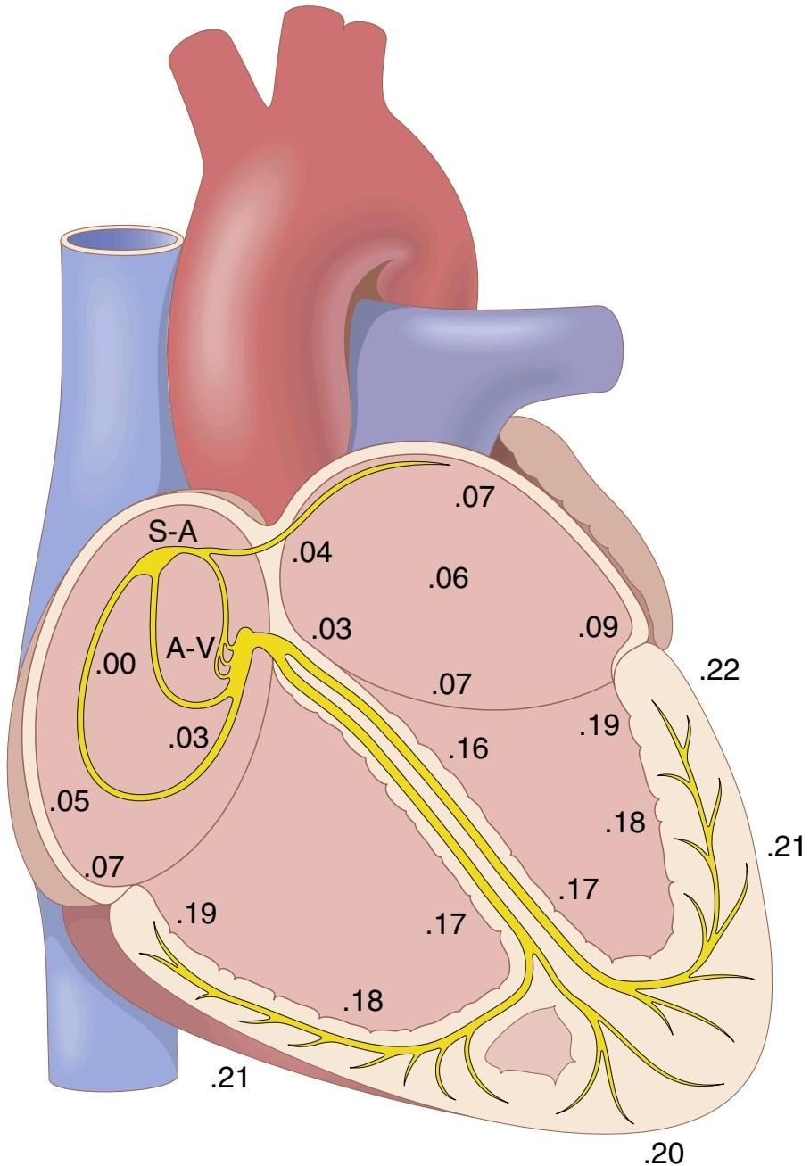
Figure 10-4 Transmission of the cardiac impulse through the heart, showing the time of appearance (in fractions of a second after initial appearance at the sinoatrial node) in different parts of the heart. A-V, Atrioventricular; S-A, sinoatrial.
شکل 10-4 انتقال تکانه قلبی از طریق قلب، نشان دادن زمان ظهور (در کسری از ثانیه پس از ظهور اولیه در گره سینوسی دهلیزی) در قسمتهای مختلف قلب. A-V، دهلیزی. S-A، سینوسی دهلیزی.
CONTROL OF EXCITATION AND CONDUCTION IN THE HEART
کنترل برانگیختگی و هدایت در قلب
THE SINUS NODE IS THE NORMAL PACE- MAKER OF THE HEART
In discussing the genesis and transmission of the cardiac impulse through the heart, we have noted that the impulse normally arises in the sinus node. In some abnormal conditions, this is not the case. Other parts of the heart can also exhibit intrinsic rhythmical excitation in the same way as the sinus nodal fibers; this is particularly true of the A-V nodal and Purkinje fibers.
گره سینوسی ضربان ساز طبیعی قلب است
در بحث پیدایش و انتقال تکانه قلبی از طریق قلب، به این نکته اشاره کردیم که تکانه معمولاً در گره سینوسی ایجاد میشود. در برخی از شرایط غیر طبیعی، این مورد نیست. سایر قسمتهای قلب نیز میتوانند تحریک ریتمیک ذاتی را مانند رشتههای گره سینوسی نشان دهند. این به ویژه در مورد الیاف A-V گره و پورکنژ صادق است.
The A-V nodal fibers, when not stimulated from some outside source, discharge at an intrinsic rhythmical rate of 40 to 60 times per minute, and the Purkinje fibers discharge at a rate somewhere between 15 and 40 times per minute. These rates are in contrast to the normal rate of the sinus node of 70 to 80 times per minute.
فیبرهای گره ای A-V، هنگامیکه از منبع خارجی تحریک نمیشوند، با سرعت ریتمیک ذاتی 40 تا 60 بار در دقیقه تخلیه میشوند و الیاف پورکنژ با سرعتی بین 15 تا 40 بار در دقیقه تخلیه میشوند. این نرخها برخلاف نرخ طبیعی گره سینوسی 70 تا 80 بار در دقیقه است.
Why then does the sinus node rather than the A-V node or the Purkinje fibers control the heart’s rhythmicity? The answer derives from the fact that the discharge rate of the sinus node is considerably faster than the natural self-excitatory discharge rate of either the A-V node or the Purkinje fibers. Each time the sinus node discharges, its impulse is conducted into both the A-V node and Purkinje fibers, also discharging their excitable membranes. How- ever, the sinus node discharges again before either the A-V node or Purkinje fibers can reach their own thresholds for self-excitation. Therefore, the new impulse from the sinus node discharges both the A-V node and Purkinje fibers before self-excitation can occur in either of these sites.
پس چرا گره سینوسی به جای گره A-V یا فیبرهای پورکنژ ریتمیک قلب را کنترل میکند؟ پاسخ از این واقعیت ناشی میشود که سرعت تخلیه گره سینوسی به طور قابل توجهی سریعتر از نرخ تخلیه طبیعی خود تحریکی گره A-V یا فیبرهای پورکنژ است. هر بار که گره سینوسی تخلیه میشود، تکانه آن به هر دو گره A-V و فیبرهای پورکنژ هدایت میشود و غشاهای تحریک پذیر آنها را نیز تخلیه میکند. با این حال، گره سینوسی قبل از اینکه گره A-V یا فیبرهای پورکنژ بتوانند به آستانه خود برای تحریک خود برسند، دوباره تخلیه میشود. بنابراین، تکانه جدید از گره سینوسی، گره A-V و فیبرهای پورکنژ را قبل از اینکه خود تحریکی در هر یک از این مکانها رخ دهد، تخلیه میکند.
Thus, the sinus node controls the beat of the heart because its rate of rhythmical discharge is faster than that of any other part of the heart. Therefore, the sinus node is almost always the pacemaker of the normal heart.
بنابراین، گره سینوسی ضربان قلب را کنترل میکند زیرا سرعت ترشح ریتمیک آن از هر قسمت دیگر قلب سریعتر است. بنابراین، گره سینوسی تقریباً همیشه ضربان ساز قلب طبیعی است.
Abnormal Pacemakers-Ectopic Pacemaker. Occasionally, some other part of the heart develops a rhythmical discharge rate that is more rapid than that of the sinus node. For example, this development sometimes occurs in the A-V node or in the Purkinje fibers when one of these becomes abnormal. In either case, the pacemaker of the heart shifts from the sinus node to the A-V node or to the excited Purkinje fibers. Under rarer conditions, a place in the atrial or ventricular muscle develops excessive excitability and becomes the pacemaker.
ضربان ساز غیر طبیعی-پیس میکر نابجا. گاهی اوقات، در برخی از قسمتهای دیگر قلب، سرعت ترشح ریتمیکی ایجاد میشود که سریعتر از گره سینوسی است. به عنوان مثال، گاهی اوقات این توسعه در گره A-V یا در فیبرهای پورکنژ رخ میدهد که یکی از آنها غیر طبیعی میشود. در هر صورت، ضربان ساز قلب از گره سینوسی به گره A-V یا فیبرهای پورکنژ برانگیخته میشود. در شرایط نادرتر، مکانی در عضله دهلیزی یا بطنی تحریک پذیری بیش از حد ایجاد میکند و به ضربان ساز تبدیل میشود.
A pacemaker elsewhere than the sinus node is called an ectopic pacemaker. An ectopic pacemaker causes an abnormal sequence of contraction of the different parts of the heart and can cause significant weakening of heart pumping.
ضربان ساز در جایی غیر از گره سینوسی ضربان ساز نابجا نامیده میشود. ضربان ساز نابجا باعث انقباض غیرطبیعی قسمتهای مختلف قلب میشود و میتواند باعث ضعیف شدن قابل توجه پمپاژ قلب شود.
Another cause of shift of the pacemaker is blockage of transmission of the cardiac impulse from the sinus node to the other parts of the heart. The new pacemaker then usually occurs at the A-V node or in the penetrating portion of the A-V bundle on the way to the ventricles.
یکی دیگر از دلایل جابجایی ضربان ساز، انسداد انتقال تکانه قلبی از گره سینوسی به سایر قسمتهای قلب است. سپس ضربان ساز جدید معمولاً در گره A-V یا در قسمت نفوذی بسته A-V در مسیر بطنها ایجاد میشود.
When A-V block occurs-that is, when the cardiac impulse fails to pass from the atria into the ventricles through the A-V nodal and bundle system-the atria continue to beat at the normal rate of rhythm of the sinus node while a new pacemaker usually develops in the Pur- kinje system of the ventricles and drives the ventricular muscle at a new rate, somewhere between 15 and 40 beats per minute. After sudden A-V bundle block, the Purkinje system does not begin to emit its intrinsic rhythmical impulses until 5 to 20 seconds later because, before the blockage, the Purkinje fibers had been “overdriven” by the rapid sinus impulses and, consequently, are in a suppressed state. During these 5 to 20 seconds, the ventricles fail to pump blood, and the person faints after the first 4 to 5 seconds because of lack of blood flow to the brain. This delayed pickup of the heartbeat is called Stokes- Adams syndrome. If the delay period is too long, it can lead to death.
هنگامیکه بلوک A-V رخ میدهد – یعنی هنگامیکه تکانه قلبی از دهلیزها به داخل بطنها از طریق سیستم گرهی و باندل A-V عبور نمیکند – دهلیزها با سرعت طبیعی ریتم گره سینوسی به ضربان خود ادامه میدهند در حالی که معمولاً یک ضربان ساز جدید در سیستم پورکنتریک و سرعت حرکت عضلانی جدید در ناحیه سینوسی ایجاد میشود. 15 و 40 ضربه در دقیقه. پس از بلوک ناگهانی باندل A-V، سیستم پورکنژ تا 5 تا 20 ثانیه بعد شروع به انتشار تکانههای ریتمیک ذاتی خود نمیکند، زیرا قبل از انسداد، الیاف پورکنژ توسط تکانههای سریع سینوسی “بیش از حد رانده” شده بودند و در نتیجه در حالت فشار قرار میگیرند. در طی این 5 تا 20 ثانیه، بطنها قادر به پمپاژ خون نیستند و فرد پس از 4 تا 5 ثانیه اول به دلیل عدم جریان خون به مغز، غش میکند. این دریافت تاخیری ضربان قلب، سندرم استوکس-آدامز نامیده میشود. اگر دوره تاخیر بیش از حد طولانی باشد، میتواند منجر به مرگ شود.
ROLE OF THE PURKINJE SYSTEM IN CAUSING SYNCHRONOUS CONTRACTION OF THE VENTRICULAR MUSCLE
The rapid conduction of the Purkinje system normally permits the cardiac impulse to arrive at almost all portions of the ventricles within a narrow span of time, exciting the first ventricular muscle fiber only 0.03 to 0.06 second ahead of excitation of the last ventricular muscle fiber. This timing causes all portions of the ventricular muscle in both ventricles to begin contracting at almost the same time and then to continue contracting for about another 0.3 second.
نقش سیستم پورکینژ در ایجاد انقباض همزمان عضله بطنی
رسانش سریع سیستم پورکنژ به طور معمول به تکانه قلبی اجازه میدهد تا تقریباً در تمام بخشهای بطن در یک بازه زمانی باریک برسد و اولین فیبر عضلانی بطنی را فقط 0.03 تا 0.06 ثانیه جلوتر از تحریک آخرین فیبر عضلانی بطن تحریک کند. این زمانبندی باعث میشود که تمام بخشهای عضله بطنی در هر دو بطن تقریباً همزمان شروع به انقباض کنند و سپس حدود 0.3 ثانیه دیگر به انقباض ادامه دهند.
Effective pumping by the two ventricular chambers requires this synchronous type of contraction. If the cardiac impulse should travel through the ventricles slowly, much of the ventricular mass would contract before contraction of the remainder, in which case the overall pumping effect would be greatly depressed. Indeed, in some types of cardiac dysfunction, several of which are discussed in Chapters 12 and 13, slow transmission does occur, and the pumping effectiveness of the ventricles is decreased as much as 20% to 30%. Implantable cardiac resynchronization devices are types of pacemakers using electrical wires or leads that can be inserted into the cardiac chambers to restore appropriate timing between the atria and both ventricles to improve pumping effectiveness in patients with enlarged and weakened hearts.
پمپاژ موثر توسط دو محفظه بطنی به این نوع انقباض همزمان نیاز دارد. اگر ضربان قلب به آرامیاز طریق بطنها عبور کند، بیشتر توده بطنی قبل از انقباض باقیمانده منقبض میشود، در این صورت اثر پمپاژ کلی به شدت کاهش مییابد. در واقع، در برخی از انواع اختلالات قلبی، که چندین مورد از آنها در فصلهای 12 و 13 مورد بحث قرار گرفتهاند، انتقال آهسته رخ میدهد و اثر پمپاژ بطنها به میزان 20% تا 30% کاهش مییابد. دستگاههای همگامسازی مجدد قلب قابل کاشت، انواعی از ضربانسازها هستند که از سیمهای الکتریکی یا سیمهایی استفاده میکنند که میتوانند برای بازگرداندن زمان مناسب بین دهلیزها و هر دو بطن برای بهبود کارایی پمپاژ در بیماران با قلبهای بزرگ و ضعیف، وارد اتاقکهای قلبی شوند.
SYMPATHETIC AND PARASYMPATHETIC NERVES CONTROL HEART RHYTHMICITY AND IMPULSE CONDUCTION BY THE CARDIAC NERVES
The heart is supplied with both sympathetic and parasympathetic nerves, as shown in Figure 9-14 of Chapter 9. The parasympathetic nerves (the vagi) are distributed mainly to the S-A and A-V nodes, to a lesser extent to the muscle of the two atria, and very little directly to the ventricular muscle. The sympathetic nerves, conversely, are distributed to all parts of the heart, with strong representation in the ventricular muscle, as well as in all the other areas.
اعصاب سمپاتیک و پاراسمپاتیک ریتمیک قلب و هدایت تکانه توسط اعصاب قلبی را کنترل میکنند
همانطور که در شکل 9-14 از فصل 9 نشان داده شده است، قلب دارای هر دو اعصاب سمپاتیک و پاراسمپاتیک است. برعکس، اعصاب سمپاتیک در تمام قسمتهای قلب پخش میشوند و بازنمایی قوی در عضله بطنی و همچنین در تمام نواحی دیگر وجود دارد.
Parasympathetic (Vagal) Stimulation Slows the Cardiac Rhythm and Conduction. Stimulation of the parasympathetic nerves to the heart (the vagi) causes acetylcholine to be released at the vagal endings. This neurotransmitter has two major effects on the heart. First, it decreases the rate of rhythm of the sinus node, and second, it decreases the excitability of the A-V junctional fibers between the atrial musculature and the A-V node, thereby slowing transmission of the cardiac impulse into the ventricles.
تحریک پاراسمپاتیک (واگال) ریتم و هدایت قلبی را کند میکند. تحریک اعصاب پاراسمپاتیک به قلب (واگی) باعث ترشح استیل کولین در انتهای واگ میشود. این انتقال دهنده عصبی دو اثر عمده بر قلب دارد. اول، سرعت ریتم گره سینوسی را کاهش میدهد، و دوم، تحریک پذیری فیبرهای اتصالی A-V بین عضله دهلیزی و گره A-V را کاهش میدهد، در نتیجه انتقال ضربه قلبی به بطنها را کند میکند.
Weak to moderate vagal stimulation slows the rate of heart pumping, often to as little as one-half normal. Furthermore, strong stimulation of the vagi can completely stop the rhythmical excitation by the sinus node or completely block transmission of the cardiac impulse from the atria into the ventricles through the A-V node. In either case, rhythmical excitatory signals are no longer transmitted into the ventricles. The ventricles may stop beating for 5 to 20 seconds, but then some small area in the Purkinje fibers, usually in the ventricular septal portion of the A-V bundle, develops a rhythm of its own and causes ventricular contraction at a rate of 15 to 40 beats per minute. This phenomenon is called ventricular escape.
تحریک ضعیف تا متوسط واگ، سرعت پمپاژ قلب را کاهش میدهد، که اغلب به نصف نرمال میرسد. علاوه بر این، تحریک قوی واگی میتواند تحریک ریتمیک توسط گره سینوسی را به طور کامل متوقف کند یا انتقال تکانه قلبی از دهلیزها به بطنها را از طریق گره A-V به طور کامل مسدود کند. در هر صورت، سیگنالهای ریتمیک تحریکی دیگر به بطنها منتقل نمیشوند. بطنها ممکن است به مدت 5 تا 20 ثانیه از تپش باز بمانند، اما پس از آن مقداری ناحیه کوچک در رشتههای پورکنژ، معمولاً در قسمت سپتوم بطنی بسته A-V، ریتم خود را ایجاد میکند و باعث انقباض بطن با سرعت 15 تا 40 ضربه در دقیقه میشود. این پدیده را فرار بطنی مینامند.
Mechanism of the Vagal Effects. The acetylcholine released at the vagal nerve endings greatly increases the permeability of the fiber membranes to potassium ions, which allows rapid leakage of potassium out of the conductive fibers. This process causes increased negativity inside the fibers, an effect called hyperpolarization, which makes this excitable tissue much less excitable, as explained in Chapter 5.
مکانیسم اثرات واگال. استیل کولین آزاد شده در انتهای عصب واگ، نفوذپذیری غشاهای فیبر را به یونهای پتاسیم بسیار افزایش میدهد، که امکان نشت سریع پتاسیم را از الیاف رسانا فراهم میکند. این فرآیند باعث افزایش منفی در داخل الیاف میشود، اثری به نامهایپرپلاریزاسیون، که باعث میشود این بافت تحریک پذیر بسیار کمتر تحریک شود، همانطور که در فصل 5 توضیح داده شد.
In the sinus node, the state of hyperpolarization makes the resting membrane potential of the sinus nodal fibers considerably more negative than usual-that is, -65 to-75 millivolts rather than the normal level of -55 to -60 millivolts. Therefore, the initial rise of the sinus nodal membrane potential caused by inward sodium and calcium leakage requires much longer to reach the threshold potential for excitation. This requirement greatly slows the rate of rhythmicity of these nodal fibers. If the vagal stimulation is strong enough, it is possible to stop the rhythmical self-excitation of this node entirely.
در گره سینوسی، حالتهایپرپلاریزاسیون باعث میشود که پتانسیل غشای استراحت الیاف گره سینوسی به طور قابل توجهی منفی تر از حد معمول باشد – یعنی 65- تا 75 میلی ولت به جای سطح نرمال 55- تا 60- میلی ولت. بنابراین، افزایش اولیه پتانسیل غشای گره سینوسی ناشی از نشت سدیم و کلسیم به داخل، برای رسیدن به پتانسیل آستانه برای تحریک به زمان بیشتری نیاز دارد. این نیاز تا حد زیادی سرعت ریتمیک این الیاف گرهی را کاهش میدهد. اگر تحریک واگ به اندازه کافی قوی باشد، میتوان خود تحریک ریتمیک این گره را به طور کامل متوقف کرد.
In the A-V node, a state of hyperpolarization caused by vagal stimulation makes it difficult for the small atrial fibers entering the node to generate enough electricity to excite the nodal fibers. Therefore, the safety factor for transmission of the cardiac impulse through the transitional fibers into the A-V nodal fibers decreases. A moderate decrease simply delays conduction of the impulse, but a large decrease blocks conduction entirely.
در گره A-V، حالتهایپرپلاریزاسیون ناشی از تحریک واگ، فیبرهای دهلیزی کوچکی که وارد گره میشوند، تولید الکتریسیته کافی برای تحریک فیبرهای گره را دشوار میکند. بنابراین، ضریب ایمنی برای انتقال تکانه قلبی از طریق الیاف انتقالی به فیبرهای گرهی A-V کاهش مییابد. کاهش متوسط به سادگی هدایت ضربه را به تاخیر میاندازد، اما کاهش زیاد هدایت را به طور کامل مسدود میکند.
Sympathetic Stimulation Increases the Cardiac Rhythm and Conduction. Sympathetic stimulation causes essentially the opposite effects on the heart as those caused by vagal stimulation, as follows.
تحریک سمپاتیک ریتم و هدایت قلبی را افزایش میدهد. تحریک سمپاتیک اساساً اثراتی متضاد بر روی قلب ایجاد میکند که در اثر تحریک واگ به شرح زیر است.
1. It increases the rate of sinus nodal discharge.
2. It increases the rate of conduction, as well as the level of excitability in all portions of the heart.
3. It increases greatly the force of contraction of all the cardiac musculature, both atrial and ventricular, as discussed in Chapter 9.
1. سرعت ترشح گره سینوسی را افزایش میدهد.
2. سرعت هدایت و همچنین سطح تحریک پذیری را در تمام قسمتهای قلب افزایش میدهد.
3. نیروی انقباض تمام عضلههای قلب، اعم از دهلیزی و بطنی را بسیار افزایش میدهد، همانطور که در فصل 9 بحث شد.
In short, sympathetic stimulation increases the overall activity of the heart. Maximal stimulation can almost triple the heartbeat frequency and can increase the strength of heart contraction as much as twofold.
به طور خلاصه، تحریک سمپاتیک فعالیت کلی قلب را افزایش میدهد. حداکثر تحریک میتواند فرکانس ضربان قلب را تقریباً سه برابر کند و میتواند قدرت انقباض قلب را تا دو برابر افزایش دهد.
Mechanism of the Sympathetic Effect. Stimulation of the sympathetic nerves releases norepinephrine at the sympathetic nerve endings. Norepinephrine, in turn, stimulates beta-1 adrenergic receptors, which mediate the effects on heart rate. The precise mechanism whereby beta-1 adrenergic stimulation acts on cardiac muscle fibers is somewhat unclear, but is thought to increase the permeability of the fiber membrane to sodium and calcium ions. In the sinus node, an increase of sodium-calcium permeability causes a more positive resting potential. It also causes an increased rate of upward drift of the diastolic membrane potential toward the threshold level for self-excitation, thus accelerating self-excitation and, therefore, increasing the heart rate.
مکانیسم اثر سمپاتیک. تحریک اعصاب سمپاتیک باعث آزاد شدن نوراپی نفرین در انتهای عصب سمپاتیک میشود. نوراپی نفرین به نوبه خود گیرندههای بتا-1 آدرنرژیک را تحریک میکند که واسطه تأثیرات بر ضربان قلب است. مکانیسم دقیق اثر تحریک بتا-1 آدرنرژیک بر روی فیبرهای عضلانی قلب تا حدودی نامشخص است، اما تصور میشود که نفوذپذیری غشای فیبر به یونهای سدیم و کلسیم را افزایش میدهد. در گره سینوسی، افزایش نفوذپذیری سدیم-کلسیم باعث پتانسیل استراحت مثبت تری میشود. همچنین باعث افزایش سرعت رانش رو به بالا پتانسیل غشای دیاستولیک به سمت آستانه برای خود تحریکی میشود، بنابراین خود تحریکی را تسریع میکند و در نتیجه ضربان قلب را افزایش میدهد.
In the A-V node and A-V bundles, increased sodium- calcium permeability makes it easier for the action potential to excite each succeeding portion of the conducting fiber bundles, thereby decreasing the conduction time from the atria to the ventricles.
در گره A-V و بستههای A-V، افزایش نفوذپذیری سدیم- کلسیم، پتانسیل عمل را برای تحریک هر بخش بعدی از بستههای فیبر رسانا آسانتر میکند، در نتیجه زمان انتقال از دهلیزها به بطنها را کاهش میدهد.
The increase in permeability to calcium ions is at least partially responsible for the increase in contractile strength of the cardiac muscle under the influence of sympathetic stimulation. This is because calcium ions play a powerful role in exciting the contractile process of the myofibrils.
افزایش نفوذپذیری به یونهای کلسیم حداقل تا حدی مسئول افزایش قدرت انقباضی عضله قلب تحت تأثیر تحریک سمپاتیک است. این به این دلیل است که یونهای کلسیم نقش قدرتمندی در تحریک فرآیند انقباضی میوفیبریلها دارند.
کلیک کنید «Bibliography: فهرست کتب مربوطه»
Abriel H, Rougier JS, Jalife J: lon channel macromolecular complexes in cardiomyocytes: roles in sudden cardiac death. Circ Res 116:1971, 2015.
Anderson RH, Boyett MR, Dobrzynski H, Moorman AF: The anatomy of the conduction system: implications for the clinical cardiologist. J Cardiovasc Transl Res 6:187, 2013.
Barbuti A, DiFrancesco D: Control of cardiac rate by “funny” channels in health and disease. Ann NY Acad Sci 1123:213, 2008.
Fedorov VV, Glukhov AV, Chang R: Conduction barriers and path- ways of the sinoatrial pacemaker complex: their role in normal rhythm and atrial arrhythmias. Am J Physiol Heart Circ Physiol 302:H1773, 2012.
Fukada K, Kanazawa H, Aizawa Y, et al: Cardiac innervation and sudden cardiac death. Circ Res 116: 2005, 2015.
Kléber AG, Rudy Y: Basic mechanisms of cardiac impulse propagation and associated arrhythmias. Physiol Rev 84:431, 2004.
John RM, Kumar S: Sinus node and atrial arrhythmias. Circulation 133:1892, 2016.
Leyva F, Nisam S, Auricchio: 20 years of cardiac resynchronization therapy. J Am Coll Cardiol. 64:1047, 2014.
Mangoni ME, Nargeot J: Genesis and regulation of the heart automaticity. Physiol Rev 88:919, 2008.
Monfredi O, Maltsev VA, Lakatta EG: Modern concepts concerning the origin of the heartbeat. Physiology (Bethesda) 28:74, 2013.
Murphy C, Lazzara R: Current concepts of anatomy and electrophysiology of the sinus node. J Interv Card Electrophysiol 46:9, 2016.
Roubille F, Tardif JC: New therapeutic targets in cardiology: heart failure and arrhythmia: HCN channels. Circulation 127:1986, 2013.
Smaill BH, Zhao J, Trew ML: Three-dimensional impulse propagation in myocardium: arrhythmogenic mechanisms at the tissue level. Circ Res 112:834, 2013.
Wickramasinghe SR, Patel VV: Local innervation and atrial fibrillation. Circulation 128:1566, 2013.
Willis BC, Ponce-Balbuena D, Jaliffe J: Protein assemblies of sodium and inward rectifier potassium channels control cardiac excitability and arrhythmogenesis. Am J Physiol Heart Circ Physiol 308:H1463, 2015.
🚀 با ما همراه شوید!
تازهترین مطالب و آموزشهای مغز و اعصاب را از دست ندهید. با فالو کردن کانال تلگرام آیندهنگاران مغز، از ما حمایت کنید!



