
فیزیولوژی پزشکی گایتون و هال؛ انبساط عروقی و عملکرد سیستمهای شریانی و وریدی


دعای مطالعه [ نمایش ]
بِسْمِ الله الرَّحْمنِ الرَّحیمِ
اَللّهُمَّ اَخْرِجْنى مِنْ ظُلُماتِ الْوَهْمِ
خدايا مرا بيرون آور از تاريكىهاى وهم،
وَ اَكْرِمْنى بِنُورِ الْفَهْمِ
و به نور فهم گرامى ام بدار،
اَللّهُمَّ افْتَحْ عَلَيْنا اَبْوابَ رَحْمَتِكَ
خدايا درهاى رحمتت را به روى ما بگشا،
وَانْشُرْ عَلَيْنا خَزائِنَ عُلُومِكَ بِرَحْمَتِكَ يا اَرْحَمَ الرّاحِمينَ
و خزانههاى علومت را بر ما باز كن به امید رحمتت اى مهربانترين مهربانان.
کتاب «فیزیولوژی پزشکی گایتون وهال» بهعنوان یکی از جامعترین و معتبرترین منابع در حوزه علوم پزشکی، همچنان مرجع کلیدی برای درک عملکرد پیچیده بدن انسان است. این اثر با تکیه بر تازهترین پژوهشها و توضیحات دقیق از سازوکارهای فیزیولوژیک، پلی میان علوم پایه پزشکی و کاربردهای بالینی ایجاد میکند و نقشی بیبدیل در آموزش، پژوهش و ارتقای دانش سلامت ایفا مینماید.
ترجمه دقیق و علمیاین شاهکار توسط برند علمیآیندهنگاران مغز به مدیریت داریوش طاهری، دسترسی فارسیزبانان به مرزهای نوین دانش فیزیولوژی را ممکن ساخته و رسالتی علمیبرای ارتقای آموزش پزشکی، فهم عمیقتر سازوکارهای بدن و توسعه روشهای نوین در حوزه سلامت فراهم آورده است.
» کتاب فیزیولوژی پزشکی گایتون وهال
» » فصل ۱۵: انبساط عروقی و عملکرد سیستمهای شریانی و وریدی
در حال ویرایش
» Guyton and Hall Textbook of Medical Physiology
»» CHAPTER 15: Vascular Distensibility and Functions of the Arterial and Venous Systems

VASCULAR DISTENSIBILITY
A valuable characteristic of the vascular system is that all blood vessels are distensible. The distensible nature of the arteries allows them to accommodate the pulsatile output of the heart and to average out the pressure pulsations. This capability provides smooth continuous flow of blood through the very small blood vessels of the tissues.
انبساط عروقی
ویژگی ارزشمند سیستم عروقی این است که همه رگهای خونی قابل انبساط هستند. ماهیت انبساط پذیر سرخرگها به آنها اجازه میدهد تا برون ده ضربانی قلب را تطبیق دهند و نبضهای فشار را به طور متوسط محاسبه کنند. این قابلیت جریان مداوم خون را از طریق رگهای خونی بسیار کوچک بافتها فراهم میکند.
The most distensible of all the vessels are the veins. Even slight increases in venous pressure cause the veins to store 0.5 to 1.0 liter of extra blood. Therefore, the veins provide a reservoir for storing large quantities of extra blood that can be called into use whenever blood is required elsewhere in the circulation.
قابل اتساع ترین رگها رگها هستند. حتی افزایش جزئی در فشار وریدی باعث میشود که وریدها 0.5 تا 1.0 لیتر خون اضافی ذخیره کنند. بنابراین، وریدها مخزنی را برای ذخیره مقادیر زیادی خون اضافی فراهم میکنند که میتواند هر زمان که خون در جای دیگری در گردش خون مورد نیاز باشد، مورد استفاده قرار گیرد.
Units of Vascular Distensibility. Vascular distensibility normally is expressed as the fractional increase in volume for each millimeter of mercury rise in pressure, in accordance with the following formula:
واحدهای انبساط عروقی. انبساط عروقی معمولاً به صورت افزایش جزئی حجم برای هر میلی متر افزایش فشار جیوه مطابق با فرمول زیر بیان میشود:

That is, if 1 mm Hg causes a vessel that originally contained 10 ml of blood to increase its volume by 1 ml, the distensibility would be 0.1 per mm Hg, or 10% per mm Hg.
یعنی اگر 1 میلیمتر جیوه باعث شود رگی که در ابتدا حاوی 10 میلیلیتر خون بود، حجم خود را به میزان 1 میلیلیتر افزایش دهد، انبساط آن 0.1 در میلیمتر جیوه یا 10 درصد در میلیمتر جیوه خواهد بود.
The Veins Are Much More Distensible Than the Arteries. The walls of the arteries are thicker and far stronger than those of the veins. Consequently, the veins, on average, are about eight times more distensible than the arteries. That is, a given increase in pressure causes about eight times as much increase in blood in a vein as in an artery of comparable size.
رگها بسیار بیشتر از شریانها قابل انبساط هستند. دیواره رگها ضخیم تر و به مراتب قوی تر از دیواره رگها هستند. در نتیجه، وریدها، به طور متوسط، حدود هشت برابر بیشتر از شریانها قابل انبساط هستند. یعنی افزایش معین فشار باعث افزایش خون در ورید حدود هشت برابر بیشتر از یک سرخرگ با اندازه مشابه میشود.
In the pulmonary circulation, the pulmonary vein distensibilities are similar to those of the systemic circulation. However, the pulmonary arteries normally operate under pressures about one sixth of those in the systemic arterial system, and their distensibilities are correspondingly greater-about six times the distensibility of systemic arteries.
در گردش خون ریوی، انقباضات ورید ریوی شبیه به گردش خون سیستمیک است. با این حال، شریانهای ریوی معمولاً تحت فشارهایی در حدود یک ششم فشارهای موجود در سیستم شریانی سیستمیک عمل میکنند و انبساط آنها تقریباً شش برابر بیشتر از انبساط شریانهای سیستمیک است.
VASCULAR COMPLIANCE (OR VASCULAR CAPACITANCE)
In hemodynamic studies, it usually is much more important to know the total quantity of blood that can be stored in a given portion of the circulation for each mm Hg pressure rise than to know the distensibilities of the individual vessels. This value is called the compliance or capacitance of the respective vascular bed; that is:
انطباق عروقی (یا ظرفیت عروقی)
در مطالعات همودینامیک، معمولاً دانستن مقدار کل خونی که میتوان در قسمت مشخصی از گردش خون برای هر افزایش فشار میلیمتر جیوه ذخیره کرد، بسیار مهمتر از دانستن انبساط رگهای منفرد است. این مقدار انطباق یا ظرفیت بستر عروقی مربوطه نامیده میشود. یعنی:

Compliance and distensibility are quite different. A highly distensible vessel that has a small volume may have far less compliance than a much less distensible vessel that has a large volume because compliance is equal to distensibility times volume.
انطباق و انبساط کاملاً متفاوت است. یک ظرف بسیار قابل انبساط که حجم کمیدارد ممکن است انطباق بسیار کمتری نسبت به ظرف بسیار کمتر قابل انبساط داشته باشد که حجم زیادی دارد زیرا انطباق برابر با انبساط بار حجم است.
The compliance of a systemic vein is about 24 times that of its corresponding artery because it is about 8 times as distensible and has a volume about 3 times as great (8 × 3 = 24).
انطباق یک ورید سیستمیک حدود 24 برابر شریان متناظر آن است زیرا حدود 8 برابر قابل انبساط است و حجم آن حدود 3 برابر بزرگتر است (8 × 3 = 24).
VOLUME-PRESSURE CURVES OF THE ARTERIAL AND VENOUS CIRCULATIONS
A convenient method for expressing the relationship of pressure to volume in a vessel or in any portion of the circulation is to use a volume-pressure curve. The red and blue solid curves in Figure 15-1 represent, respectively, the volumepressure curves of the normal systemic arterial system and venous system, showing that when the arterial system of the average adult person (including all the large arteries, small arteries, and arterioles) is filled with about 700 ml of blood, the mean arterial pressure is 100 mm Hg but, when it is filled with only 400 ml of blood, the pressure falls to zero.
منحنیهای حجم-فشار گردش خون شریانی و وریدی
یک روش مناسب برای بیان رابطه فشار به حجم در یک ظرف یا در هر قسمت از گردش، استفاده از منحنی فشار حجم است. منحنیهای جامد قرمز و آبی در شکل 1-15 به ترتیب منحنیهای حجم فشار سیستم شریانی سیستمیک و سیستم وریدی را نشان میدهند و نشان میدهند که وقتی سیستم شریانی یک فرد بزرگسال متوسط (شامل همه شریانهای بزرگ، سرخرگهای کوچک و شریانها) با حدود 700 میلیلیتر خون پر میشود، میانگین H فقط با فشار شریانی 0 پر میشود. 400 میلی لیتر خون، فشار به صفر میرسد.
In the entire systemic venous system, the volume normally ranges from 2000 to 3500 ml, and a change of several hundred ml in this volume is required to change the venous pressure by only 3 to 5 mm Hg. This requirement mainly explains why as much as one-half liter of blood can be transfused into a healthy person in only a few minutes without greatly altering the function of the circulation.
در کل سیستم وریدی سیستمیک، حجم به طور معمول از 2000 تا 3500 میلیلیتر متغیر است و برای تغییر فشار وریدی تنها به میزان 3 تا 5 میلیمتر جیوه، صدها میلیلیتر در این حجم تغییر میکند. این نیاز عمدتاً توضیح میدهد که چرا میتوان تنها در چند دقیقه نیم لیتر خون به یک فرد سالم تزریق کرد بدون اینکه عملکرد گردش خون را به شدت تغییر دهد.

Figure 15-1. Volume-pressure curves of the systemic arterial and venous systems, showing the effects of stimulation or inhibition of the sympathetic nerves to the circulatory system.
شکل 15-1. منحنیهای حجم فشار سیستمهای شریانی و وریدی سیستمیک، که اثرات تحریک یا مهار اعصاب سمپاتیک را برای سیستم گردش خون نشان میدهد.
Effect of Sympathetic Stimulation or Sympathetic Inhibition on the Volume-Pressure Relationships of the Arterial and Venous Systems. Also shown in Figure 15-1 are the effects on the volume-pressure curves when the vascular sympathetic nerves are excited or inhibited. It is evident that an increase in vascular smooth muscle tone caused by sympathetic stimulation increases the pressure at each volume of the arteries or veins, whereas sympathetic inhibition decreases the pressure at each volume. Control of the vessels by the sympathetics in this manner is a valuable means for diminishing the dimensions of one segment of the circulation, thus transferring blood to other segments. For example, an increase in vascular tone throughout the systemic circulation can cause large volumes of blood to shift into the heart, which is one of the principal methods that the body uses to rapidly increase heart pumping.
اثر تحریک سمپاتیک یا مهار سمپاتیک بر روابط حجمی- فشار سیستمهای شریانی و وریدی. همچنین در شکل 15-1، اثرات روی منحنیهای فشار حجم نشان داده شده است، زمانی که اعصاب سمپاتیک عروقی برانگیخته یا مهار میشوند. بدیهی است که افزایش تون عضلات صاف عروق ناشی از تحریک سمپاتیک باعث افزایش فشار در هر حجم از شریانها یا وریدها میشود، در حالی که مهار سمپاتیک باعث کاهش فشار در هر حجم میشود. کنترل رگها توسط سمپاتیکها به این روش وسیله ای ارزشمند برای کاهش ابعاد یک بخش از گردش خون و در نتیجه انتقال خون به بخشهای دیگر است. به عنوان مثال، افزایش تون عروقی در سراسر گردش خون سیستمیک میتواند باعث شود حجم زیادی از خون به قلب منتقل شود، که یکی از روشهای اصلی است که بدن برای افزایش سریع پمپاژ قلب استفاده میکند.
Sympathetic control of vascular capacitance is also highly important during hemorrhage. Enhancement of sympathetic tone, especially to the veins, reduces the vessel sizes enough that the circulation continues to operate almost normally, even when as much as 25% of the total blood volume has been lost.
کنترل سمپاتیک ظرفیت عروقی نیز در هنگام خونریزی بسیار مهم است. افزایش صدای سمپاتیک، بهویژه رگها، اندازه رگها را به اندازهای کاهش میدهد که گردش خون تقریباً به طور معمول به کار خود ادامه میدهد، حتی زمانی که 25 درصد از کل حجم خون از بین رفته باشد.
Delayed Compliance (Stress-Relaxation) of Vessels. The term delayed compliance means that a vessel exposed to increased volume at first exhibits a large increase in pressure, but progressive delayed stretching of smooth muscle in the vessel wall allows the pressure to return toward normal over a period of minutes to hours. This effect is shown in Figure 15-2. In this figure, the pressure is recorded in a small segment of a vein that is occluded at both ends. An extra volume of blood is suddenly injected until the pressure rises from 5 to 12 mm Hg. Even though none of the blood is removed after it is injected, the pressure begins to decrease immediately and approaches about 9 mm Hg after several minutes. Thus, the volume of blood injected causes immediate elastic distention of the vein, but then the smooth muscle fibers of the vein begin to creep to longer lengths, and their tensions correspondingly decrease. This effect is a characteristic of all smooth muscle and is called stress-relaxation, as explained in Chapter 8.
تاخیر در انطباق (استرس-آرامش) عروق. اصطلاح انطباق تاخیری به این معنی است که رگ در معرض افزایش حجم در ابتدا افزایش زیادی در فشار نشان میدهد، اما کشش تدریجی تاخیری عضله صاف در دیواره رگ باعث میشود فشار طی چند دقیقه تا چند ساعت به حالت عادی بازگردد. این اثر در شکل 15-2 نشان داده شده است. در این شکل، فشار در بخش کوچکی از ورید که در دو انتها مسدود شده است، ثبت میشود. حجم اضافی خون به طور ناگهانی تزریق میشود تا زمانی که فشار از 5 به 12 میلی متر جیوه افزایش یابد. حتی اگر هیچ یک از خون پس از تزریق خارج نشود، فشار بلافاصله شروع به کاهش میکند و پس از چند دقیقه به حدود 9 میلی متر جیوه میرسد. بنابراین، حجم خون تزریق شده باعث اتساع الاستیک فوری ورید میشود، اما پس از آن رشتههای عضلانی صاف سیاهرگ شروع به خزش به طولهای طولانیتر میکنند و تنش آنها به ترتیب کاهش مییابد. این اثر مشخصه تمام عضلات صاف است و همانطور که در فصل 8 توضیح داده شد، استرس-آرامش نامیده میشود.
Delayed compliance is a valuable mechanism whereby the circulation can accommodate extra blood when necessary, such as after a large transfusion. Delayed compliance in the reverse direction is one way in which the circulation automatically adjusts itself over a period of minutes or hours to diminished blood volume after serious hemorrhage.
انطباق با تأخیر مکانیسم ارزشمندی است که به موجب آن گردش خون میتواند در صورت لزوم، مانند پس از یک تزریق بزرگ، خون اضافی را در خود جای دهد. انطباق تاخیری در جهت معکوس یکی از راههایی است که در آن گردش خون به طور خودکار در طی چند دقیقه یا چند ساعت تنظیم میشود تا حجم خون پس از خونریزی جدی کاهش یابد.

Figure 15-2. Effect on the intravascular pressure of injecting a volume of blood into a venous segment and later removing the excess blood, demonstrating the principle of delayed compliance.
شکل 15-2. تأثیر بر فشار داخل عروقی تزریق حجمیاز خون به یک بخش وریدی و بعداً برداشتن خون اضافی، که اصل انطباق با تاخیر را نشان میدهد.
ARTERIAL PRESSURE PULSATIONS
With each beat of the heart, a new surge of blood fills the arteries. Were it not for distensibility of the arterial system, all this new blood would have to flow through the peripheral blood vessels almost instantaneously, only during cardiac systole, and no flow would occur during diastole. However, the compliance of the arterial tree normally reduces the pressure pulsations to almost no pulsations by the time the blood reaches the capillaries; therefore, tissue blood flow is mainly continuous with very little pulsation.
ضربانهای فشار شریانی
با هر ضربان قلب، موج جدیدی از خون سرخرگها را پر میکند. اگر انبساط سیستم شریانی نبود، تمام این خون جدید باید تقریباً بلافاصله و فقط در طول سیستول قلبی از طریق رگهای خونی محیطی جریان مییابد، و هیچ جریانی در طول دیاستول رخ نمیدهد. با این حال، انطباق درخت شریانی معمولاً ضربانهای فشار را تا زمانی که خون به مویرگها میرسد تقریباً به هیچ ضربان کاهش میدهد. بنابراین، جریان خون بافت عمدتاً با ضربان بسیار کم ادامه دارد.
The pressure pulsations at the root of the aorta are illustrated in Figure 15-3. In a healthy young adult, the pressure at the top of each pulse, called the systolic pres- sure, is about 120 mm Hg. At the lowest point of each pulse, called the diastolic pressure, it is about 80 mm Hg. The difference between these two pressures, about 40 mm Hg, is called the pulse pressure.
ضربان فشار در ریشه آئورت در شکل 15-3 نشان داده شده است. در یک بالغ جوان سالم، فشار بالای هر نبض که فشار سیستولیک نامیده میشود، حدود 120 میلی متر جیوه است. در پایین ترین نقطه هر نبض که فشار دیاستولیک نامیده میشود، حدود 80 میلی متر جیوه است. تفاوت بین این دو فشار، حدود 40 میلی متر جیوه، فشار پالس نامیده میشود.
Two major factors affect the pulse pressure: (1) the stroke volume output of the heart; and (2) the compliance (total distensibility) of the arterial tree. A third less important factor is the character of ejection from the heart during systole.
دو عامل اصلی بر فشار نبض تأثیر میگذارند: (1) حجم ضربهای خروجی قلب. و (2) انطباق (انبساط کامل) درخت شریانی. سومین عاملی که کمتر مهم است، ویژگی بیرون راندن از قلب در طول سیستول است.

Figure 15-3. pressure pulse contour in the ascending aorta.
شکل 15-3. نمودار نبض فشار در آئورت صعودی.
In general, the greater the stroke volume output, the greater the amount of blood that must be accommodated in the arterial tree with each heartbeat and, therefore, the greater the pressure rise and fall during systole and diastole, thus causing a greater pulse pressure. Conversely, the less the compliance of the arterial system, the greater the rise in pressure for a given stroke volume of blood pumped into the arteries. For example, as demonstrated by the middle top curves in Figure 15-4, the pulse pressure in old age sometimes rises to twice normal because the arteries have stiffened with arteriosclerosis and therefore are relatively noncompliant.
به طور کلی، هرچه میزان خروجی حجم ضربه بیشتر باشد، مقدار خون بیشتری باید در درخت شریانی با هر ضربان قلب جای گیرد و بنابراین، افزایش و کاهش فشار در طول سیستول و دیاستول بیشتر میشود و در نتیجه باعث افزایش فشار نبض میشود. برعکس، هرچه انطباق سیستم شریانی کمتر باشد، افزایش فشار برای یک حجم ضربه ای معین از خون پمپ شده به شریانها بیشتر میشود. به عنوان مثال، همانطور که در شکل 15-4 با منحنیهای بالای وسط نشان داده شده است، فشار نبض در سنین بالا گاهی اوقات تا دو برابر طبیعی افزایش مییابد زیرا شریانها با تصلب شرایین سفت شده اند و بنابراین نسبتاً ناسازگار هستند.
In effect, pulse pressure is determined approximately by the ratio of stroke volume output to compliance of the arterial tree. Any condition of the circulation that affects either of these two factors also affects the pulse pressure: Pulse pressure = Stroke volume/arterial compliance
در واقع، فشار پالس تقریباً با نسبت خروجی حجم ضربه به انطباق درخت شریانی تعیین میشود. هر شرایطی از گردش خون که بر هر یک از این دو عامل تأثیر بگذارد بر فشار نبض نیز تأثیر میگذارد: فشار نبض = حجم ضربه / انطباق شریانی

Figure 15-4. Aortic pressure pulse contours in arteriosclerosis, aortic stenosis, patent ductus arteriosus, and aortic regurgitation.
شکل 15-4. خطوط نبض فشار آئورت در تصلب شرایین، تنگی آئورت، مجرای شریانی باز، و نارسایی آئورت.
ABNORMAL PRESSURE PULSE CONTOURS
Some pathophysiological conditions of the circulation cause abnormal contours of the pressure pulse wave in addition to altering the pulse pressure. Especially distinctive among these conditions are aortic stenosis, patent ductus arteriosus, and aortic regurgitation, each of which is shown in Figure 15-4.
کانتورهای پالس فشار غیرعادی
برخی از شرایط پاتوفیزیولوژیک گردش خون علاوه بر تغییر فشار پالس باعث ایجاد خطوط غیرطبیعی موج پالس فشار میشود. به خصوص در میان این شرایط، تنگی آئورت، مجرای شریانی باز، و نارسایی آئورت مشخص است که هر کدام در شکل 15-4 نشان داده شده است.
In persons with aortic valve stenosis, the diameter of the aortic valve opening is reduced significantly, and the aortic pressure pulse is decreased significantly because of diminished blood flow outward through the stenotic valve.
در افراد مبتلا به تنگی دریچه آئورت، قطر دهانه دریچه آئورت به طور قابل توجهی کاهش مییابد و نبض فشار آئورت به دلیل کاهش جریان خون به بیرون از طریق دریچه تنگی به طور قابل توجهی کاهش مییابد.
In persons with patent ductus arteriosus, 50% or more of the blood pumped into the aorta by the left ventricle flows immediately backward through the wide open ductus into the pulmonary artery and lung blood vessels, thus allowing the diastolic pressure to fall very low before the next heartbeat and increasing the pulse pressure.
در افراد مبتلا به مجرای شریانی باز، 50٪ یا بیشتر از خون پمپ شده به آئورت توسط بطن چپ بلافاصله از طریق مجرای باز به سمت شریان ریوی و عروق خونی ریه جریان مییابد، بنابراین فشار دیاستولیک قبل از ضربان قلب بعدی بسیار پایین میآید و فشار نبض افزایش مییابد.
In persons with aortic regurgitation, the aortic valve is absent or does not close completely. Therefore, after each heartbeat, the blood that has just been pumped into the aorta flows immediately backward into the left ventricle. As a result, the aortic pressure can fall all the way to zero between heartbeats. Also, there is no incisura in the aortic pulse contour because there is no aortic valve to close.
در افراد مبتلا به نارسایی آئورت، دریچه آئورت وجود ندارد یا به طور کامل بسته نمیشود. بنابراین، پس از هر ضربان قلب، خونی که به تازگی به آئورت پمپاژ شده است، بلافاصله به سمت عقب به سمت بطن چپ جریان مییابد. در نتیجه، فشار آئورت میتواند بین ضربانهای قلب به صفر برسد. همچنین، هیچ برشی در کانتور نبض آئورت وجود ندارد زیرا دریچه آئورت برای بسته شدن وجود ندارد.

Figure 15-5. Progressive stages in transmission of the pressure pulse along the aorta.
شکل 15-5. مراحل پیشرونده در انتقال پالس فشار در امتداد آئورت.
TRANSMISSION OF PRESSURE PULSES TO THE PERIPHERAL ARTERIES
When the heart ejects blood into the aorta during systole, only the proximal portion of the aorta initially becomes distended because the inertia of the blood prevents sudden blood movement all the way to the periphery. However, the rising pressure in the proximal aorta rapidly overcomes this inertia, and the wavefront of distention spreads farther and farther along the aorta, as shown in Figure 15-5. This phenomenon is called transmission of the pressure pulse in the arteries.
انتقال پالسهای فشاری به شریانهای محیطی
هنگامیکه قلب در طی سیستول خون را به داخل آئورت پرتاب میکند، در ابتدا فقط قسمت پروگزیمال آئورت متسع میشود زیرا اینرسی خون مانع از حرکت ناگهانی خون تا سراسر محیط میشود. با این حال، افزایش فشار در آئورت پروگزیمال به سرعت بر این اینرسی غلبه میکند و جبهه موج اتساع دورتر و دورتر در امتداد آئورت گسترش مییابد، همانطور که در شکل 15-5 نشان داده شده است. این پدیده را انتقال پالس فشار در شریانها مینامند.
The velocity of pressure pulse transmission is 3 to 5 m/ sec in the normal aorta, 7 to 10 m/sec in the large arterial branches, and 15 to 35 m/sec in the small arteries. In general, the greater the compliance of each vascular segment, the slower the velocity, which explains the slow transmission in the aorta and the much faster transmission in the much less compliant small distal arteries. In the aorta, the velocity of transmission of the pressure pulse is 15 or more times the velocity of blood flow because the pressure pulse is simply a moving wave of pressure that involves little forward total movement of blood volume.
سرعت انتقال پالس فشار در آئورت طبیعی 3 تا 5 متر بر ثانیه، در شاخههای بزرگ شریانی 7 تا 10 متر بر ثانیه و در شریانهای کوچک 15 تا 35 متر بر ثانیه است. به طور کلی، هرچه انطباق هر بخش عروقی بیشتر باشد، سرعت آن کمتر است، که انتقال آهسته در آئورت و انتقال بسیار سریعتر در شریانهای دیستال کوچک بسیار کمتر سازگار را توضیح میدهد. در آئورت، سرعت انتقال پالس فشار 15 یا بیشتر برابر سرعت جریان خون است زیرا نبض فشار صرفاً یک موج متحرک فشار است که شامل حرکت کمیبه جلو حجم خون است.

Figure 15-6. Changes in the pulse pressure contour as the pulse wave travels toward the smaller vessels.
شکل 15-6. با حرکت موج پالس به سمت عروق کوچکتر، کانتور فشار پالس تغییر میکند.
Pressure Pulses Are Damped in the Smaller Arteries, Arterioles, and Capillaries. Figure 15-6 shows typical changes in the pressure pulse contours as the pulse travels into the peripheral vessels. Note especially in the three lower curves that the intensity of pulsation becomes progressively less in the smaller arteries, arterioles and, especially, capillaries. In fact, only when the aortic pulsations are extremely large or the arterioles are greatly dilated can pulsations be observed in the capillaries.
پالسهای فشار در شریانهای کوچکتر، شریانها و مویرگها کاهش مییابند. شکل 15-6 تغییرات معمولی در خطوط پالس فشار را با حرکت پالس به داخل عروق محیطی نشان میدهد. به ویژه در سه منحنی پایینتر توجه داشته باشید که شدت ضربان در شریانهای کوچکتر، شریانها و بهویژه مویرگها به تدریج کمتر میشود. در واقع، تنها زمانی که ضربانهای آئورت بسیار بزرگ هستند یا شریانها بسیار متسع شدهاند، میتوان نبضها را در مویرگها مشاهده کرد.
This progressive diminution of the pulsations in the periphery is called damping of the pressure pulses. The cause of this damping is twofold: (1) resistance to blood movement in the vessels; and (2) compliance of the vessels. The resistance damps the pulsations because a small amount of blood must flow forward at the pulse wave front to distend the next segment of the vessel; the greater the resistance, the more difficult it is for this to occur. The compliance damps the pulsations because the more com- pliant a vessel, the greater the quantity of blood required at the pulse wave front to cause an increase in pressure.
این کاهش تدریجی ضربانها در حاشیه را میرایی پالسهای فشار میگویند. علت این میرایی دو چیز است: (1) مقاومت در برابر حرکت خون در عروق. و (2) انطباق کشتیها. مقاومت ضربانها را کاهش میدهد زیرا مقدار کمیخون باید در جلوی موج نبض به سمت جلو جریان یابد تا بخش بعدی رگ متسع شود. هرچه مقاومت بیشتر باشد، وقوع آن دشوارتر است. انطباق ضربانها را کاهش میدهد، زیرا هرچه یک رگ سازگارتر باشد، مقدار خون بیشتری در جبهه موج پالس برای افزایش فشار لازم است.
Therefore, the degree of damping is almost directly proportional to the product of resistance times compliance.
بنابراین، درجه میرایی تقریباً مستقیماً با محصول انطباق زمان مقاومت متناسب است.
CLINICAL METHODS FOR MEASURING SYSTOLIC AND DIASTOLIC PRESSURES
It is not practical to use pressure recorders that require needle insertion into an artery for making routine arterial pressure measurements in human patients, although these types of recorders are used on occasion when special studies are necessary. Instead, the clinician determines systolic and diastolic pressures through indirect means, usually by the auscultatory method.
روشهای بالینی برای اندازه گیری فشار سیستولیک و دیاستولیک
استفاده از دستگاههای ثبت فشار که نیاز به وارد کردن سوزن در شریان دارند، برای انجام اندازهگیری فشار شریانی معمول در بیماران انسانی، عملی نیست، اگرچه این نوع ضبطکنندهها در مواردی که مطالعات خاصی لازم است استفاده میشوند. درعوض، پزشک فشار سیستولیک و دیاستولیک را از طریق روشهای غیرمستقیم، معمولاً با روش سمعی، تعیین میکند.
Auscultatory Method. Figure 15-7 shows the auscultatory method for determining systolic and diastolic arterial pressures. A stethoscope is placed over the antecubital artery, and a blood pressure cuff is inflated around the upper arm. As long as the cuff continues to compress the arm with too little pressure to close the brachial artery, no sounds are heard from the antecubital artery with the stethoscope. However, when the cuff pressure is great enough to close the artery during part of the arterial pressure cycle, a sound is then heard with each pulsation. These sounds are called Korotkoff sounds, named after Nikolai Korotkoff, a Russian physician, who described them in 1905.
روش شنیداری. شکل 7-15 روش شنوایی برای تعیین فشار شریانی سیستولیک و دیاستولیک را نشان میدهد. یک گوشی پزشکی روی شریان جلویی قرار داده میشود و یک کاف فشار خون در اطراف بازو باد میشود. تا زمانی که کاف بازو را با فشار بسیار کمیبرای بستن شریان بازویی فشرده میکند، هیچ صدایی از شریان جلویی با گوشی پزشکی شنیده نمیشود. با این حال، زمانی که فشار کاف به اندازه کافی برای بستن شریان در بخشی از چرخه فشار شریانی زیاد باشد، سپس با هر ضربان صدایی شنیده میشود. این صداها صداهای کوروتکف نامیده میشوند که به افتخار نیکولای کوروتکف، پزشک روسی که در سال 1905 آنها را توصیف کرد، نامگذاری شده است.
Korotkoff sounds are believed to be caused mainly by blood jetting through the partly occluded vessel and by vibrations of the vessel wall. The jet causes turbulence in the vessel beyond the cuff, and this turbulence sets up the vibrations heard through the stethoscope.
اعتقاد بر این است که صداهای کوروتکف عمدتاً در اثر جهش خون از طریق رگ تا حدی مسدود شده و ارتعاشات دیواره رگ ایجاد میشود. جت باعث ایجاد تلاطم در رگ فراتر از کاف میشود و این تلاطم باعث ایجاد ارتعاشات شنیده شده از طریق گوشی پزشکی میشود.
In determining blood pressure by the auscultatory method, the pressure in the cuff is first elevated well above arterial systolic pressure. As long as this cuff pressure is higher than systolic pressure, the brachial artery remains collapsed so that no blood jets into the lower artery during any part of the pressure cycle. Therefore, no Korotkoff sounds are heard in the lower artery. Then the cuff pressure gradually is reduced. Just as soon as the pressure in the cuff falls below systolic pressure (point B, Figure 15-7), blood begins to flow through the artery beneath the cuff during the peak of systolic pressure, and one begins to hear tapping sounds from the antecubital artery in synchrony with the heartbeat. As soon as these sounds begin to be heard, the pressure level indicated by the manometer connected to the cuff is about equal to the systolic pressure.
در تعیین فشار خون به روش سمعی، فشار در کاف ابتدا بسیار بالاتر از فشار سیستولیک شریانی است. تا زمانی که این فشار کاف بالاتر از فشار سیستولیک باشد، شریان بازویی فرو ریخته میماند به طوری که در هیچ بخشی از چرخه فشار، هیچ جت خونی به شریان پایینی وارد نمیشود. بنابراین هیچ صدای کوروتکف در شریان تحتانی شنیده نمیشود. سپس فشار کاف به تدریج کاهش مییابد. درست به محض اینکه فشار در کاف به زیر فشار سیستولیک میرسد (نقطه B، شکل 15-7)، خون در طول اوج فشار سیستولیک از طریق شریان زیر کاف شروع به جریان میکند و فرد شروع به شنیدن صداهای ضربه زدن از شریان انتکوبیتال همزمان با ضربان قلب میکند. به محض اینکه این صداها شروع به شنیدن میکنند، سطح فشار نشان داده شده توسط مانومتر متصل به کاف تقریباً برابر با فشار سیستولیک است.
As the pressure in the cuff is lowered still more, the Korotkoff sounds change in quality, having less of the tapping quality and more of a rhythmical and harsher quality. Then, finally, when the pressure in the cuff falls near diastolic pressure, the sounds suddenly change to a muffled quality (point C, Figure 15-7). One notes the manometer pressure when the Korotkoff sounds change to the muffled quality, and this pressure is about equal to the diastolic pressure, although it slightly overestimates the diastolic pressure determined by direct intraarterial catheter. As the cuff pressure falls a few mm Hg further, the artery no longer closes during diastole, which means that the basic factor causing the sounds (the jetting of blood through a squeezed artery) is no longer present. Therefore, the sounds disappear entirely. Many clinicians believe that the pressure at which the Korotkoff sounds completely disappear should be used as the diastolic pres- sure, except in situations in which the disappearance of sounds cannot reliably be determined because sounds are audible, even after complete deflation of the cuff. For example, in patients with arteriovenous fistulas for hemodialysis or with aortic insufficiency, Korotkoff sounds may be heard after complete deflation of the cuff.
با کاهش بیشتر فشار در کاف، صداهای Korotkoff کیفیت خود را تغییر میدهند و کیفیت ضربه کمتر و کیفیت ریتمیک و خشنتری دارند. سپس، در نهایت، هنگامیکه فشار در کاف نزدیک به فشار دیاستولیک کاهش مییابد، صداها ناگهان به کیفیتی خفه میشوند (نقطه C، شکل 15-7). هنگامیکه صداهای Korotkoff به کیفیت خفه میشوند، فشار مانومتر را یادداشت میکنید، و این فشار تقریباً برابر با فشار دیاستولیک است، اگرچه فشار دیاستولیک تعیینشده توسط کاتتر مستقیم داخل شریانی را کمیبیش از حد تخمین میزند. از آنجایی که فشار کاف چند میلی متر جیوه بیشتر میشود، شریان دیگر در طول دیاستول بسته نمیشود، به این معنی که عامل اصلی ایجاد صداها (جوش خون از طریق شریان فشرده) دیگر وجود ندارد. بنابراین، صداها به طور کامل ناپدید میشوند. بسیاری از پزشکان بر این باورند که فشاری که در آن صداهای کوروتکف به طور کامل ناپدید میشوند باید به عنوان فشار دیاستولیک استفاده شود، مگر در شرایطی که در آن ناپدید شدن صداها به طور قابل اعتمادی قابل تشخیص نیست زیرا صداها حتی پس از تخلیه کامل کاف قابل شنیدن هستند. به عنوان مثال، در بیماران مبتلا به فیستول شریانی وریدی برای همودیالیز یا با نارسایی آئورت، صداهای کوروتکف ممکن است پس از تخلیه کامل کاف شنیده شود.
The auscultatory method for determining systolic and diastolic pressures is not entirely accurate, but it usually gives values within 10% of those determined by direct catheter measurement from inside the arteries.
روش شنوایی برای تعیین فشار سیستولیک و دیاستولیک کاملاً دقیق نیست، اما معمولاً مقادیری در 10٪ از مقادیر تعیین شده توسط اندازه گیری مستقیم کاتتر از داخل شریانها را ارائه میدهد.

Figure 15-7. Auscultatory method for measuring systolic and diastolic arterial pressures.
شکل 15-7. روش شنوایی برای اندازه گیری فشار شریانی سیستولیک و دیاستولیک.
Automated Oscillometric Method. Systolic and diastolic arterial pressures are often measured using automated oscillometric devices. These devices use a sphygmoma- nometer cuff, like the auscultatory method, but with an electronic pressure sensor to detect cuff pressure oscillations that occur when blood flows through an artery, often the brachial artery. Oscillometric arterial pressure devices use specific electronic algorithms to inflate and deflate the cuff automatically and interpret the cuff pres- sure oscillations. When the cuff is inflated, and its pressure exceeds systolic pressure, there is no blood flow in the artery and no oscillation of the cuff pressure. As the cuff is slowly deflated, blood begins to spurt through the artery, and the cuff pressure then oscillates in synchrony with the cyclic expansion and contraction of the artery. As the cuff pressure declines, the oscillations increase in amplitude to a maximum, which corresponds to the mean arterial pressure. The oscillation amplitude then declines as the cuff pressure falls below the patient’s diastolic pres- sure and blood flows smoothly through the artery. Using device-specific algorithms, the cuff pressure oscillations are automatically converted into digital systolic and diastolic pressures signals, as well as heart rate, and displayed.
روش اسیلومتری خودکار. فشار شریانی سیستولیک و دیاستولیک اغلب با استفاده از دستگاههای اسیلومتری خودکار اندازه گیری میشود. این دستگاهها از کاف فشارسنج مانند روش سمعی استفاده میکنند، اما با یک سنسور فشار الکترونیکی برای تشخیص نوسانات فشار کاف که هنگام جریان خون در یک سرخرگ، اغلب در شریان بازویی، رخ میدهد. دستگاههای فشار شریانی نوسان سنجی از الگوریتمهای الکترونیکی خاصی برای باد کردن و تخلیه خودکار کاف و تفسیر نوسانات فشار کاف استفاده میکنند. هنگامیکه کاف باد میشود و فشار آن از فشار سیستولیک بیشتر میشود، جریان خون در شریان وجود ندارد و فشار کاف نوسانی ندارد. همانطور که کاف به آرامیتخلیه میشود، خون شروع به جهش در شریان میکند و سپس فشار کاف همزمان با انبساط و انقباض چرخه ای شریان در نوسان است. با کاهش فشار کاف، دامنه نوسانات به حداکثر افزایش مییابد که با فشار متوسط شریانی مطابقت دارد. سپس دامنه نوسان کاهش مییابد زیرا فشار کاف به زیر فشار دیاستولیک بیمار میرسد و خون به آرامیدر شریان جریان مییابد. با استفاده از الگوریتمهای خاص دستگاه، نوسانات فشار کاف به طور خودکار به سیگنالهای دیجیتال فشار سیستولیک و دیاستولیک و همچنین ضربان قلب تبدیل شده و نمایش داده میشود.
Oscillometric arterial pressure monitors require less skill than the auscultatory technique and can be used by the patient at home, avoiding the so-called white-coat effect that raises blood pressure in some patients when a health care professional is present. These devices, however, must be calibrated for accuracy and can yield unreliable measurements in patients when the cuff size is inappropriate or in some abnormal circulatory conditions, such as severe arteriosclerosis, which increases stiffness of the artery wall.
مانیتورهای فشار شریانی نوسانسنجی به مهارت کمتری نسبت به تکنیک سمعشناختی نیاز دارند و میتوانند توسط بیمار در خانه مورد استفاده قرار گیرند و از به اصطلاح اثر پوشش سفید که باعث افزایش فشار خون در برخی بیماران با حضور متخصص مراقبتهای بهداشتی میشود، اجتناب میکنند. با این حال، این دستگاهها باید برای دقت کالیبره شوند و میتوانند در بیمارانی که اندازه کاف نامناسب است یا در برخی شرایط گردش خون غیرطبیعی، مانند تصلب شرایین شدید، که سفتی دیواره شریان را افزایش میدهد، اندازهگیریهای غیرقابل اعتمادی را ارائه دهند.
Normal Arterial Pressures as Measured by the Auscultatory and Oscillatory Methods. Figure 15-8 shows the approximate normal systolic and diastolic arterial pressures at different ages. The progressive increase in pressure with age results from the effects of aging on the blood pressure control mechanisms. We shall see in Chapter 19 that the kidneys are primarily responsible for this long-term regulation of arterial pressure; it is well known that the kidneys exhibit definitive changes with age, especially after the age of 50 years.
فشارهای عادی شریانی که با روشهای شنیداری و نوسانی اندازهگیری میشوند. شکل 15-8 فشار شریانی سیستولیک و دیاستولیک طبیعی تقریبی را در سنین مختلف نشان میدهد. افزایش تدریجی فشار با افزایش سن ناشی از تأثیرات پیری بر مکانیسمهای کنترل فشار خون است. در فصل 19 خواهیم دید که کلیهها مسئول اصلی تنظیم طولانی مدت فشار شریانی هستند. به خوبی شناخته شده است که کلیهها با افزایش سن تغییرات قطعی نشان میدهند، به خصوص بعد از سن 50 سالگی.
A slight extra increase in systolic pressure usually occurs beyond the age of 60 years. This increase results from decreasing distensibility, or hardening, of the arteries, which is often a result of atherosclerosis. The final effect is a higher systolic pressure with considerable increase in pulse pressure, as previously explained.
افزایش خفیف اضافی در فشار سیستولیک معمولاً پس از سن 60 سالگی رخ میدهد. این افزایش ناشی از کاهش انبساط یا سخت شدن شریانها است که اغلب در نتیجه آترواسکلروز است. اثر نهایی فشار سیستولیک بالاتر با افزایش قابل توجه فشار پالس است، همانطور که قبلا توضیح داده شد.
Mean Arterial Pressure. The mean arterial pressure is the average of the arterial pressures measured millisecond by millisecond over a period of time. It is not equal to the average of the systolic and diastolic pressures because at normal heart rates, a greater fraction of the cardiac cycle is spent in diastole than in systole. Thus, the arterial pressure remains closer to diastolic pressure than to systolic pressure during the greater part of the cardiac cycle. The mean arterial pressure is therefore determined about 60% by the diastolic pressure and 40% by the systolic pressure. Note in Figure 15-8 that the mean pressure (solid green line) at all ages is nearer to the diastolic pressure than to the systolic pressure. However, at very high heart rates, diastole comprises a smaller fraction of the cardiac cycle, and the mean arterial pressure is more closely approximated as the average of systolic and diastolic pressures.
فشار متوسط شریانی. فشار متوسط شریانی میانگین فشارهای شریانی است که میلی ثانیه به میلی ثانیه در یک دوره زمانی اندازه گیری میشود. این با میانگین فشار سیستولیک و دیاستولیک برابر نیست زیرا در ضربان قلب طبیعی، بخش بیشتری از چرخه قلبی در دیاستول سپری میشود تا در سیستول. بنابراین، فشار شریانی به فشار دیاستولیک نزدیکتر از فشار سیستولیک در بخش بیشتری از چرخه قلبی باقی میماند. بنابراین فشار متوسط شریانی حدود 60٪ توسط فشار دیاستولیک و 40٪ توسط فشار سیستولیک تعیین میشود. در شکل 15-8 توجه داشته باشید که فشار متوسط (خط سبز جامد) در تمام سنین به فشار دیاستولیک نزدیکتر از فشار سیستولیک است. با این حال، در ضربان قلب بسیار بالا، دیاستول بخش کوچک تری از چرخه قلبی را شامل میشود و فشار متوسط شریانی با میانگین فشار سیستولیک و دیاستولیک تقریب بیشتری دارد.

Figure 15-8. Changes in systolic, diastolic, and mean arterial pressures with age. The shaded areas show the approximate normal ranges.
شکل 15-8. تغییرات در فشار سیستولیک، دیاستولیک و متوسط شریانی با افزایش سن. مناطق سایه دار محدودههای طبیعی تقریبی را نشان میدهند.
VEINS AND THEIR FUNCTIONS
The veins provide passageways for flow of blood to the heart, but they also perform other special functions that are necessary for operation of the circulation. Of special importance is that they are capable of constricting and enlarging and thereby storing small or large quantities of blood and making this blood available when required by the remainder of the circulation. The peripheral veins can also propel blood forward by means of a so-called venous pump, and they even help regulate cardiac output, an exceedingly important function described in detail in Chapter 20.
وریدها و عملکرد آنها
وریدها گذرگاهی را برای جریان خون به قلب فراهم میکنند، اما عملکردهای ویژه دیگری را نیز انجام میدهند که برای عملکرد گردش خون ضروری است. اهمیت ویژه این است که آنها میتوانند منقبض و بزرگ شوند و در نتیجه مقادیر کم یا زیاد خون را ذخیره کنند و در صورت نیاز باقیمانده گردش خون، این خون را در دسترس قرار دهند. وریدهای محیطی همچنین میتوانند خون را با استفاده از یک پمپ وریدی به جلو برانند و حتی به تنظیم برون ده قلبی کمک میکنند، عملکرد بسیار مهمیکه در فصل 20 به تفصیل شرح داده شده است.
VENOUS PRESSURES-RIGHT ATRIAL PRESSURE (CENTRAL VENOUS PRESSURE) AND PERIPHERAL VENOUS PRESSURES
Blood from all the systemic veins flows into the right atrium of the heart. Therefore, the pressure in the right atrium is called the central venous pressure.
فشارهای وریدی – فشار دهلیزی راست (فشار ورید مرکزی) و فشارهای ورید محیطی
خون از تمام وریدهای سیستمیک به دهلیز راست قلب جریان مییابد. بنابراین فشار موجود در دهلیز راست را فشار ورید مرکزی مینامند.
Right atrial pressure is regulated by a balance between (1) the ability of the heart to pump blood out of the right atrium and ventricle into the lungs and (2) the tendency for blood to flow from the peripheral veins into the right atrium. If the right heart is pumping strongly, the right atrial pressure decreases. Conversely, weakness of the heart elevates the right atrial pressure. Also, any effect that causes rapid inflow of blood into the right atrium from the peripheral veins elevates the right atrial pressure. Some factors that can increase this venous return and thereby increase the right atrial pressure are as follows: (1) increased blood volume; (2) increased large vessel tone throughout the body with resultant increased peripheral venous pressures; and (3) dilation of the arterioles, which decreases the peripheral resistance and allows rapid flow of blood from the arteries into the veins.
فشار دهلیز راست با تعادل بین (1) توانایی قلب برای پمپاژ خون از دهلیز و بطن راست به ریهها و (2) تمایل به جریان خون از وریدهای محیطی به دهلیز راست تنظیم میشود. اگر قلب راست به شدت پمپاژ میکند، فشار دهلیز راست کاهش مییابد. برعکس، ضعف قلب باعث افزایش فشار دهلیز راست میشود. همچنین، هر اثری که باعث ورود سریع خون به دهلیز راست از سیاهرگهای محیطی شود، فشار دهلیز راست را بالا میبرد. برخی از عواملی که میتوانند این بازگشت وریدی را افزایش دهند و در نتیجه فشار دهلیز راست را افزایش دهند به شرح زیر است: (1) افزایش حجم خون. (2) افزایش تون عروق بزرگ در سراسر بدن و در نتیجه افزایش فشار وریدهای محیطی. و (3) اتساع شریانها، که مقاومت محیطی را کاهش میدهد و جریان سریع خون را از شریانها به داخل سیاهرگها میدهد.
The same factors that regulate right atrial pressure also contribute to the regulation of cardiac output because the amount of blood pumped by the heart depends on both the ability of the heart to pump and the tendency for blood to flow into the heart from the peripheral vessels. Therefore, we discuss regulation of right atrial pressure in much more depth in Chapter 20 in connection with cardiac output regulation.
همان عواملی که فشار دهلیز راست را تنظیم میکنند به تنظیم برون ده قلبی نیز کمک میکنند زیرا مقدار خون پمپ شده توسط قلب هم به توانایی قلب برای پمپاژ و هم به تمایل جریان خون به قلب از رگهای محیطی بستگی دارد. بنابراین، تنظیم فشار دهلیز راست را با عمق بیشتری در فصل 20 در ارتباط با تنظیم برون ده قلبی مورد بحث قرار میدهیم.
The normal right atrial pressure is about 0 mm Hg, which is equal to the atmospheric pressure around the body. It can increase to 20 to 30 mm Hg under very abnormal conditions, such as the following: (1) serious heart failure; or (2) after massive transfusion of blood, which greatly increases the total blood volume and causes excessive quantities of blood to attempt to flow into the heart from the peripheral vessels.
فشار طبیعی دهلیز راست حدود 0 میلی متر جیوه است که برابر با فشار اتمسفر اطراف بدن است. در شرایط بسیار غیر طبیعی، مانند موارد زیر، میتواند به 20 تا 30 میلی متر جیوه افزایش یابد: (1) نارسایی جدی قلبی. یا (2) پس از تزریق انبوه خون، که حجم کل خون را تا حد زیادی افزایش میدهد و باعث میشود که مقادیر بیش از حد خون از عروق محیطی به قلب جریان یابد.
The lower limit to the right atrial pressure is usually about -3 to -5 mm Hg below atmospheric pressure, which is also the pressure in the chest cavity that surrounds the heart. The right atrial pressure approaches these low values when the heart pumps with exceptional vigor or when blood flow into the heart from the peripheral vessels is greatly depressed, such as after severe hemorrhage.
حد پایین فشار دهلیز راست معمولاً در حدود 3- تا 5- میلی متر جیوه زیر فشار اتمسفر است، که همچنین فشار در حفره قفسه سینه است که قلب را احاطه کرده است. فشار دهلیز راست زمانی به این مقادیر پایین نزدیک میشود که قلب با قدرت استثنایی پمپاژ میکند یا زمانی که جریان خون از عروق محیطی به قلب به شدت کاهش مییابد، مانند پس از خونریزی شدید.
Venous Resistance and Peripheral Venous Pressure
Large veins have so little resistance to blood flow when they are distended that the resistance then is almost zero. However, as shown in Figure 15-9, most of the large veins that enter the thorax are compressed at many points by the surrounding tissues, so that blood flow is impeded at these points. For example, the veins from the arms are compressed by their sharp angulations over the first rib. Also, the pressure in the neck veins often falls so low that the atmospheric pressure on the outside of the neck causes these veins to collapse. Finally, veins coursing through the abdomen are often compressed by different organs and by the intra-abdominal pressure, so they usually are at least partially collapsed to an ovoid or slitlike state. For these reasons, the large veins do usu- ally offer some resistance to blood flow, and thus the pressure in the more peripheral small veins in a person lying down is usually +4 to +6 mm Hg greater than the right atrial pressure.
مقاومت وریدی و فشار وریدی محیطی
وریدهای بزرگ زمانی که متسع میشوند مقاومت کمیدر برابر جریان خون دارند که در این صورت مقاومت تقریباً به صفر میرسد. با این حال، همانطور که در شکل 15-9 نشان داده شده است، بیشتر وریدهای بزرگی که وارد قفسه سینه میشوند در بسیاری از نقاط توسط بافتهای اطراف فشرده میشوند، به طوری که جریان خون در این نقاط با مشکل مواجه میشود. برای مثال، رگهای بازوها با زاویههای تیزشان بر روی دنده اول فشرده میشوند. همچنین فشار در وریدهای گردن اغلب به حدی پایین میآید که فشار اتمسفر در قسمت بیرونی گردن باعث فروپاشی این رگها میشود. در نهایت، وریدهایی که از شکم عبور میکنند اغلب توسط اندامهای مختلف و فشار داخل شکمیفشرده میشوند، بنابراین معمولاً حداقل تا حدی به حالت تخمییا شکافی فرو میروند. به این دلایل، وریدهای بزرگ معمولاً مقداری مقاومت در برابر جریان خون ایجاد میکنند و بنابراین فشار در وریدهای کوچک محیطیتر در فردی که دراز کشیده است معمولاً 4+ تا 6+ میلیمتر جیوه بیشتر از فشار دهلیز راست است.

Figure 15-9. Compressing points that tend to collapse the veins entering the thorax.
شکل 15-9. نقاط فشارندهای که تمایل به کلاپس وریدهای ورودی به قفسه سینه دارند.
Effect of High Right Atrial Pressure on Peripheral Venous Pressure. When the right atrial pressure rises above its normal value of 0 mm Hg, blood begins to backup in the large veins. This backup of blood enlarges the veins, and even the collapse points in the veins open up when the right atrial pressure rises above +4 to +6 mm Hg. Then, as the right atrial pressure rises further, the additional increase causes a corresponding rise in peripheral venous pressure in the limbs and elsewhere. Because heart function must be impaired significantly to cause a rise in right atrial pressure as high as +4 to +6 mm Hg, the peripheral venous pressure is not noticeably elevated, even in the early stages of heart failure, as long as the per- son is at rest.
تأثیر فشار بالای دهلیز راست بر فشار ورید محیطی. هنگامیکه فشار دهلیز راست از مقدار طبیعی 0 میلی متر جیوه بالاتر میرود، خون در سیاهرگهای بزرگ شروع به پشتیبان گیری میکند. این پشتیبان خون وریدها را بزرگ میکند و حتی نقاط فروپاشی وریدها زمانی که فشار دهلیز راست به بالای 4+ تا 6+ میلی متر جیوه میرسد باز میشوند. سپس، با افزایش بیشتر فشار دهلیز راست، افزایش اضافی باعث افزایش متناظر در فشار ورید محیطی در اندامها و جاهای دیگر میشود. از آنجا که عملکرد قلب باید به طور قابل توجهی مختل شود تا باعث افزایش فشار دهلیز راست تا 4+ تا 6+ میلیمتر جیوه شود، فشار ورید محیطی حتی در مراحل اولیه نارسایی قلبی تا زمانی که فرد در حالت استراحت است، بهطور محسوسی افزایش نمییابد.
Effect of Intra-abdominal Pressure on Venous Pressures of the Leg. The pressure in the abdominal cavity of a recumbent person normally averages about +6 mm Hg, but it can rise to +15 to +30 mm Hg as a result of pregnancy, large tumors, abdominal obesity, or excessive fluid (called ascites) in the abdominal cavity. When the intra-abdominal pressure rises, the pressure in the veins of the legs must rise above the abdominal pressure before the abdominal veins will open and allow the blood to flow from the legs to the heart. Thus, if the intra-abdominal pressure is +20 mm Hg, the low- est possible pressure in the femoral veins is also about +20 mm Hg.
تأثیر فشار داخل شکمیبر فشارهای وریدی ساق پا. فشار در حفره شکمییک فرد دراز کشیده به طور معمول حدود 6+ میلیمتر جیوه است، اما میتواند در نتیجه بارداری، تومورهای بزرگ، چاقی شکمییا مایع بیش از حد (به نام آسیت) در حفره شکمیبه 15+ تا 30+ میلیمتر جیوه برسد. هنگامیکه فشار داخل شکمیافزایش مییابد، قبل از اینکه وریدهای شکمیباز شوند و اجازه دهند خون از پاها به سمت قلب برود، فشار در وریدهای پا باید از فشار شکمیبالاتر برود. بنابراین، اگر فشار داخل شکمی20+ میلیمتر جیوه باشد، کمترین فشار ممکن در وریدهای فمورال نیز حدود 20+ میلیمتر جیوه است.
Effect of Gravitational Pressure on Venous Pressure
In any body of water that is exposed to air, the pressure at the surface of the water is equal to atmospheric pressure, but the pressure rises 1 mm Hg for each 13.6 millimeters of distance below the surface. This pressure results from the weight of the water and therefore is called gravitational pressure or hydrostatic pressure.
تاثیر فشار گرانشی بر فشار وریدی
در هر توده آبی که در معرض هوا باشد، فشار سطح آب برابر با فشار اتمسفر است، اما فشار به ازای هر 13.6 میلی متر فاصله از سطح، 1 میلی متر جیوه افزایش مییابد. این فشار از وزن آب ناشی میشود و بنابراین فشار گرانشی یا فشار هیدرواستاتیکی نامیده میشود.
Gravitational pressure also occurs in the vascular system because of the weight of the blood in the vessels, as shown in Figure 15-10. When a person is standing, the pressure in the right atrium remains about 0 mm Hg because the heart pumps any excess blood that attempts to accumulate at this point into the arteries. However, in an adult who is standing absolutely still, the pressure in the veins of the feet is about +90 mm Hg simply because of the gravitational weight of the blood in the veins between the heart and the feet. The venous pressures at other levels of the body are proportionately between 0 and 90 mm Hg.
همانطور که در شکل 15-10 نشان داده شده است، فشار گرانشی نیز به دلیل وزن خون در عروق در سیستم عروقی رخ میدهد. هنگامیکه یک فرد ایستاده است، فشار در دهلیز راست حدود 0 میلی متر جیوه باقی میماند، زیرا قلب خون اضافی را که در این نقطه تجمع میکند به داخل شریانها پمپ میکند. با این حال، در بزرگسالانی که کاملاً ثابت میایستند، فشار وریدهای پا حدود 90+ میلیمتر جیوه است که صرفاً به دلیل وزن گرانشی خون در سیاهرگهای بین قلب و پا است. فشار وریدی در سایر سطوح بدن به نسبت 0 تا 90 میلی متر جیوه است.
In the arm veins, the pressure at the level of the top rib is usually about +6 mm Hg because of compression of the subclavian vein as it passes over this rib. The gravitational pressure down the length of the arm is then determined by the distance below the level of this rib. Thus, if the gravitational difference between the level of the rib and the hand is +29 mm Hg, this gravitational pressure is added to the +6 mm Hg pressure caused by compression of the vein as it crosses the rib, making a total of +35 mm Hg pressure in the veins of the hand.
در وریدهای بازو، فشار در سطح دنده بالایی معمولاً حدود 6+ میلیمتر جیوه است، زیرا ورید ساب ترقوه هنگام عبور از روی این دنده فشرده میشود. سپس فشار گرانشی در طول بازو با فاصله زیر سطح این دنده تعیین میشود. بنابراین، اگر اختلاف گرانشی بین سطح دنده و دست 29+ میلیمتر جیوه باشد، این فشار گرانشی به فشار 6+ میلیمتر جیوه ناشی از فشردگی ورید هنگام عبور از دنده اضافه میشود و در مجموع فشار 35+ میلیمتر جیوه در رگهای دست ایجاد میشود.
The neck veins of a person standing upright collapse almost completely all the way to the skull because of atmospheric pressure on the outside of the neck. This collapse causes the pressure in these veins to remain at zero along their entire extent. Any tendency for the pressure to rise above this level opens the veins and allows the pressure to fall back to zero because of flow of the blood. Conversely, any tendency for the neck vein pressure to fall below zero collapses the veins still more, which further increases their resistance and again returns the pressure back to zero.
رگهای گردن فردی که به صورت عمودی ایستاده است تقریباً به طور کامل تا جمجمه به دلیل فشار اتمسفر در قسمت بیرونی گردن فرو میریزد. این فروپاشی باعث میشود که فشار در این وریدها در تمام وسعت آنها در حد صفر باقی بماند. هر گونه تمایل به افزایش فشار از این سطح، رگها را باز میکند و به دلیل جریان خون، فشار به صفر میرسد. برعکس، هرگونه تمایل به کاهش فشار ورید گردن به زیر صفر، وریدها را بیشتر فرو میریزد، که مقاومت آنها را بیشتر میکند و دوباره فشار را به صفر برمیگرداند.
The veins inside the skull, on the other hand, are in a chamber (the skull cavity) that cannot collapse. Consequently, negative pressure can exist in the dural sinuses of the head; in the standing position, the venous pressure in the sagittal sinus at the top of the brain is about -10 mm Hg because of the hydrostatic “suction” between the top of the skull and the base of the skull. Therefore, if the sagittal sinus is opened during surgery, air can be sucked immediately into the venous system; the air may even pass downward to cause air embolism in the heart and death.
از طرف دیگر وریدهای داخل جمجمه در یک محفظه (حفره جمجمه) قرار دارند که نمیتوانند فرو بریزند. در نتیجه، فشار منفی میتواند در سینوسهای دورال سر وجود داشته باشد. در حالت ایستاده، فشار وریدی در سینوس ساژیتال در بالای مغز حدود 10- میلی متر جیوه است، زیرا به دلیل “مکش” هیدرواستاتیک بین بالای جمجمه و پایه جمجمه است. بنابراین، اگر سینوس ساژیتال در حین جراحی باز شود، هوا میتواند بلافاصله به سیستم وریدی مکیده شود. حتی ممکن است هوا به سمت پایین حرکت کند و باعث آمبولی هوا در قلب و مرگ شود.

Figure 15-10. Effect of gravitational pressure on the venous pressures throughout the body in the standing person.
شکل 15-10. تأثیر فشار گرانشی بر فشارهای وریدی در سراسر بدن در فرد ایستاده.
Effect of the Gravitational Factor on Arterial and Other Pressures. The gravitational factor also affects pressures in the peripheral arteries and capillaries. For example, a standing person who has a mean arterial pressure of 100 mm Hg at the level of the heart has an arterial pressure in the feet of about 190 mm Hg. Therefore, when the arterial pressure is stated to be 100 mm Hg, this generally means that 100 mm Hg is the pressure at the gravitational level of the heart but not necessarily elsewhere in the arterial vessels.
تأثیر عامل گرانشی بر فشارهای شریانی و سایر فشارها. عامل گرانشی نیز بر فشار در شریانهای محیطی و مویرگها تأثیر میگذارد. برای مثال، فردی ایستاده که فشار شریانی متوسط 100 میلیمتر جیوه در سطح قلب دارد، فشار شریانی در پاها حدود 190 میلیمتر جیوه دارد. بنابراین، وقتی فشار شریانی 100 میلیمتر جیوه است، این به طور کلی به این معنی است که 100 میلیمتر جیوه فشاری است که در سطح گرانشی قلب است، اما نه لزوماً در جای دیگری در عروق شریانی.
Venous Valves and the Venous Pump: Their Effects on Venous Pressure
Were it not for valves in the veins, the gravitational pressure effect would cause the venous pressure in the feet always to be about +90 mm Hg in a standing adult. However, every time the legs move, the muscles tighten and compress the veins in or adjacent to the muscles, which squeezes the blood out of the veins. However, the valves in the veins, shown in Figure 15-11, are arranged so that the direction of venous blood flow can only be toward the heart. Consequently, every time a person moves the legs or even tenses the leg muscles, a certain amount of venous blood is propelled toward the heart. This pumping system is known as the venous pump or muscle pump, and it is efficient enough that under ordinary circumstances, the venous pressure in the feet of a walking adult remains less than +20 mm Hg.
دریچههای وریدی و پمپ وریدی: اثرات آنها بر فشار وریدی
اگر دریچههای موجود در رگها نبود، اثر فشار گرانشی باعث میشد که فشار وریدی در پا همیشه در حدود 90+ میلیمتر جیوه در بزرگسالان ایستاده باشد. با این حال، هر بار که پاها حرکت میکنند، ماهیچهها سفت میشوند و رگهای داخل یا مجاور آنها را فشرده میکنند، که خون را از رگها خارج میکند. با این حال، دریچههای موجود در وریدها، که در شکل 15-11 نشان داده شده است، به گونه ای چیده شده اند که جهت جریان خون وریدی فقط میتواند به سمت قلب باشد. در نتیجه، هر بار که فرد پاها را حرکت میدهد یا حتی ماهیچههای ساق پا را منقبض میکند، مقدار مشخصی خون وریدی به سمت قلب پیش میرود. این سیستم پمپاژ به پمپ وریدی یا پمپ عضلانی معروف است و به اندازه ای کارآمد است که در شرایط معمولی، فشار وریدی در پای یک فرد بالغ راه رفتن کمتر از 20+ میلی متر جیوه باقی بماند.
If a person stands perfectly still, the venous pump does not work, and the venous pressures in the lower legs increase to the full gravitational value of 90 mm Hg in about 30 seconds. The pressures in the capillaries also increase greatly, causing fluid to leak from the circulatory system into the tissue spaces. As a result, the legs swell, and the blood volume diminishes; 10% to 20% of the blood volume can be lost from the circulatory system within the 15 to 30 minutes of standing absolutely still, which may lead to fainting, as sometimes occurs when a soldier is made to stand at attention. This situation can be avoided by simply flexing the leg muscles periodically and slightly bending the knees, thus permitting the venous pump to work.
اگر فردی کاملاً بی حرکت بایستد، پمپ وریدی کار نمیکند و فشارهای وریدی در ساق پاها به مقدار گرانشی کامل 90 میلی متر جیوه در حدود 30 ثانیه افزایش مییابد. فشار در مویرگها نیز به شدت افزایش مییابد و باعث نشت مایع از سیستم گردش خون به فضاهای بافتی میشود. در نتیجه، پاها متورم میشوند و حجم خون کاهش مییابد. 10 تا 20 درصد از حجم خون را میتوان در عرض 15 تا 30 دقیقه پس از ایستادن کاملاً ثابت از سیستم گردش خون از دست داد، که ممکن است منجر به غش شود، همانطور که گاهی اوقات زمانی رخ میدهد که سرباز را وادار میکنند تا در معرض توجه بایستد. این وضعیت را میتوان با خم کردن ماهیچههای پا به صورت دوره ای و کمیخم کردن زانوها اجتناب کرد و به پمپ وریدی اجازه کار داد.
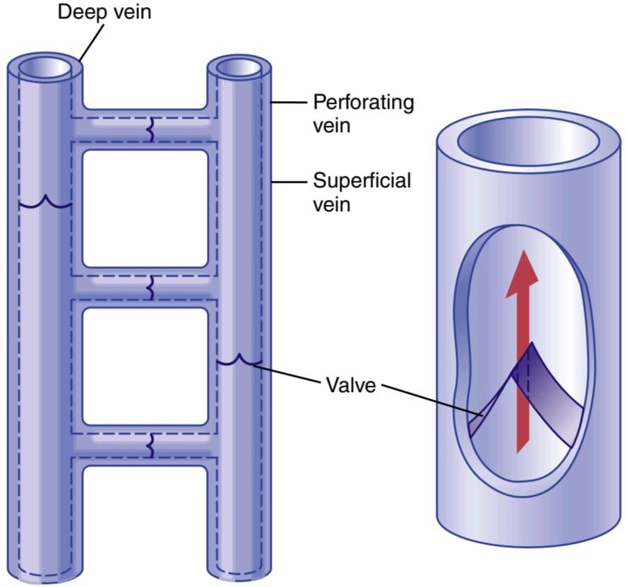
Figure 15-11. Venous valves of the leg.
شکل 15-11. دریچههای وریدی ساق پا.
Venous Valve Incompetence Causes Varicose Veins. The valves of the venous system may become incompetent or even be destroyed when the veins have been overstretched by excess venous pressure lasting weeks or months, which can occur in pregnancy or when a person stands most of the time. Stretching the veins increases their cross-sectional areas, but the leaflets of the valves do not increase in size. Therefore, the leaflets of the valves no longer close completely. With this lack of complete closure, the pressure in the veins of the legs increases greatly because of failure of the venous pump, which further increases the sizes of the veins and finally destroys the function of the valves entirely. Thus, the person develops what are called varicose veins, which are characterized by large bulbous protrusions of the veins beneath the skin of the entire leg, particularly the lower leg.
نارسایی دریچه وریدی باعث واریس میشود. دریچههای سیستم وریدی ممکن است زمانی که رگها به دلیل فشار بیش از حد وریدی هفتهها یا ماهها کشیده شدهاند، ناکارآمد شوند یا حتی از بین بروند، که میتواند در بارداری یا زمانی که فرد بیشتر اوقات میایستد رخ دهد. کشش وریدها سطح مقطع آنها را افزایش میدهد، اما اندازه لتهای دریچهها افزایش نمییابد. بنابراین، لتهای دریچهها دیگر به طور کامل بسته نمیشوند. با این عدم بسته شدن کامل، فشار در وریدهای پاها به دلیل از کار افتادن پمپ وریدی به شدت افزایش مییابد که باعث افزایش بیشتر اندازه وریدها و در نهایت از بین رفتن عملکرد دریچهها میشود. بنابراین، فرد دچار رگهای واریسی میشود که با برآمدگیهای پیازی بزرگ سیاهرگها در زیر پوست کل ساق پا، بهویژه ساق پا مشخص میشود.
Whenever people with varicose veins stand for more than a few minutes, the venous and capillary pressures become very high, and leakage of fluid from the capillaries causes constant edema in the legs. The edema, in turn, prevents adequate diffusion of nutritional materials from the capillaries to the muscle and skin cells, so the muscles become painful and weak, and the skin may even become gangrenous and ulcerate. The best treatment for such a condition is continual elevation of the legs to a level at least as high as the heart. Tight binders or long compression stockings on the legs also can be of considerable assistance in preventing the edema and its sequelae.
هرگاه افراد مبتلا به واریس بیش از چند دقیقه بایستند، فشار وریدی و مویرگی بسیار بالا میرود و نشت مایع از مویرگها باعث ادم مداوم در پاها میشود. ادم به نوبه خود از انتشار کافی مواد غذایی از مویرگها به سلولهای عضلانی و پوست جلوگیری میکند، بنابراین ماهیچهها دردناک و ضعیف میشوند و حتی ممکن است پوست قانقاریا و زخم شود. بهترین درمان برای چنین وضعیتی بالا بردن مداوم پاها به سطحی حداقل به اندازه قلب است. چسبهای تنگ یا جورابهای بلند فشاری روی پاها نیز میتوانند کمک قابل توجهی در پیشگیری از ادم و عواقب آن داشته باشند.
Clinical Estimation of Venous Pressure Venous pressure often can be estimated by simply observing the degree of distention of the peripheral veins, especially of the neck veins. For example, in the sitting position, the neck veins are never distended in the normal, quietly resting person. However, when the right atrial pressure becomes increased to as much as +10 mm Hg, the lower veins of the neck begin to protrude and, at +15 mm Hg atrial pressure, essentially all the veins in the neck become distended.
تخمین بالینی فشار وریدی فشار وریدی را اغلب میتوان با مشاهده میزان اتساع وریدهای محیطی، به ویژه وریدهای گردن، تخمین زد. به عنوان مثال، در حالت نشسته، رگهای گردن در افراد عادی و آرام هرگز متسع نمیشوند. با این حال، زمانی که فشار دهلیز راست به 10+ میلیمتر جیوه افزایش مییابد، سیاهرگهای پایینی گردن شروع به بیرون زدگی میکنند و در فشار دهلیزی +15 میلیمتر جیوه، اساساً تمام سیاهرگهای گردن متسع میشوند.
Direct Measurement of Venous Pressure and Right Atrial Pressure Venous pressure can be measured easily by inserting a needle directly into a vein and connecting it to a pressure recorder. The only means whereby right atrial pressure can be measured accurately is by inserting a catheter through the peripheral veins and into the right atrium. Pressures measured through such central venous catheters are often used in some types of hospitalized cardiac patients to provide a constant assessment of the heart-pumping ability.
اندازه گیری مستقیم فشار وریدی و فشار دهلیز راست فشار وریدی را میتوان به راحتی با وارد کردن یک سوزن به طور مستقیم در ورید و اتصال آن به یک دستگاه ثبت فشار اندازه گیری کرد. تنها وسیله ای که از طریق آن میتوان فشار دهلیز راست را با دقت اندازه گیری کرد، قرار دادن کاتتر از طریق وریدهای محیطی و داخل دهلیز راست است. فشارهای اندازه گیری شده از طریق چنین کاتترهای ورید مرکزی اغلب در برخی از انواع بیماران قلبی بستری در بیمارستان استفاده میشود تا ارزیابی ثابتی از توانایی پمپاژ قلب ارائه شود.
Pressure Reference Level for Measuring Venous and Other Circulatory Pressures Although we have spoken of right atrial pressure as being 0 mm Hg and arterial pressure as being 100 mm Hg, we have not stated the gravitational level in the circulatory system to which this pressure is referred. There is one point in the circulatory system at which gravitational pressure factors caused by changes in body position of a healthy person usually do not affect the pressure measurement by more than 1 to 2 mm Hg. This is at or near the level of the tricuspid valve, as shown by the crossed axes in Figure 15-12. Therefore, all circulatory pressure measurements discussed in this text are referred to this level, which is called the reference level for pressure measurement.
سطح مرجع فشار برای اندازهگیری فشارهای وریدی و سایر فشارهای گردش خون اگرچه ما فشار دهلیز راست را 0 میلیمتر جیوه و فشار شریانی را 100 میلیمتر جیوه گفتهایم، اما سطح گرانشی در سیستم گردش خون را که این فشار به آن اشاره میشود، بیان نکردهایم. یک نقطه در سیستم گردش خون وجود دارد که در آن عوامل فشار گرانشی ناشی از تغییر در وضعیت بدن یک فرد سالم معمولاً بیش از 1 تا 2 میلی متر جیوه بر اندازه گیری فشار تأثیر نمیگذارد. همانطور که توسط محورهای متقاطع در شکل 15-12 نشان داده شده است، در سطح دریچه سه لتی یا نزدیک به آن است. بنابراین، تمام اندازه گیریهای فشار گردش خون مورد بحث در این متن به این سطح اشاره میشود که به آن سطح مرجع برای اندازه گیری فشار میگویند.
The reason for the lack of gravitational effects at the tricuspid valve is that the heart automatically prevents significant gravitational changes in pressure at this point in the following way. If the pressure at the tricuspid valve rises slightly above normal, the right ventricle fills to a greater extent than usual, causing the heart to pump blood more rapidly and therefore decreasing the pressure at the tricuspid valve back toward the normal mean value. Conversely, if the pressure falls, the right ventricle fails to fill adequately, its pumping decreases, and blood dams up in the venous system until the pressure at the tricuspid level again rises to the normal value. In other words, the heart acts as a feed- back regulator of pressure at the tricuspid valve.
دلیل فقدان اثرات گرانشی در دریچه سه لتی این است که قلب به صورت خودکار از تغییرات گرانشی قابل توجه فشار در این نقطه به روش زیر جلوگیری میکند. اگر فشار دریچه سه لتی کمیبالاتر از حد طبیعی باشد، بطن راست به میزان بیشتری از حد معمول پر میشود و باعث میشود قلب خون را با سرعت بیشتری پمپ کند و در نتیجه فشار در دریچه سه لتی را به سمت مقدار متوسط طبیعی کاهش میدهد. برعکس، اگر فشار کاهش یابد، بطن راست به اندازه کافی پر نمیشود، پمپاژ آن کاهش مییابد و خون در سیستم وریدی میریزد تا زمانی که فشار در سطح سه لتی دوباره به مقدار طبیعی افزایش یابد. به عبارت دیگر، قلب به عنوان یک تنظیم کننده بازخورد فشار در دریچه سه لتی عمل میکند.
When a person is lying on his or her back, the tricuspid valve is located at almost exactly 60% of the chest thickness in front of the back. This is the zero pressure reference level for a person lying down.
هنگامیکه فردی به پشت دراز کشیده است، دریچه سه لتی تقریباً دقیقاً در 60 درصد ضخامت قفسه سینه در جلوی پشت قرار دارد. این سطح مرجع فشار صفر برای فردی است که دراز کشیده است.
BLOOD RESERVOIR FUNCTION OF THE VEINS
We indicated in Chapter 14 that more than 60% of all the blood in the circulatory system is usually in the veins. For this reason, and also because the veins are so compliant, the venous system serves as a blood reservoir for the circulation.
عملکرد مخزن خون وریدها
ما در فصل 14 اشاره کردیم که بیش از 60 درصد از کل خون در سیستم گردش خون معمولاً در وریدها است. به همین دلیل، و همچنین به دلیل سازگاری وریدها، سیستم وریدی به عنوان یک مخزن خون برای گردش خون عمل میکند.
When blood is lost from the body, and the arterial pres- sure begins to fall, nervous signals are elicited from the carotid sinuses and other pressure-sensitive areas of the circulation, as discussed in Chapter 18. These signals, in turn, elicit nerve signals from the brain and spinal cord, mainly through sympathetic nerves to the veins, causing them to constrict. This process takes up much of the slack in the circulatory system caused by the lost blood. Even after as much as 20% of the total blood volume has been lost, the circulatory system often functions almost normally because of this variable reservoir function of the veins.
هنگامیکه خون از بدن از دست میرود و فشار شریانی شروع به کاهش میکند، سیگنالهای عصبی از سینوسهای کاروتید و سایر نواحی حساس به فشار گردش خون خارج میشوند، همانطور که در فصل 18 بحث شد. این فرآیند بسیاری از سستی در سیستم گردش خون ناشی از خون از دست رفته را میگیرد. حتی پس از از بین رفتن 20 درصد از کل حجم خون، سیستم گردش خون اغلب به دلیل این عملکرد مخزن متغیر وریدها تقریباً طبیعی عمل میکند.

Figure 15-12. Reference point for circulatory pressure measurement (located near the tricuspid valve).
شکل 15-12. نقطه مرجع برای اندازه گیری فشار گردش خون (واقع در نزدیکی دریچه سه لتی).
SPECIFIC BLOOD RESERVOIRS
Certain portions of the circulatory system are so extensive and/or so compliant that they are called specific blood reservoirs. These reservoirs include the following: (1) the spleen, which sometimes can decrease in size sufficiently to release as much as 100 ml of blood into other areas of the circulation; (2) the liver, the sinuses of which can release several hundred milliliters of blood into the remainder of the circulation; (3) the large abdominal veins, which can contribute as much as 300 ml; and (4) the venous plexus beneath the skin, which also can contribute several hundred milliliters. The heart and lungs, although not parts of the systemic venous reservoir system, may also be considered blood reservoirs. The heart, for example, shrinks during sympathetic stimulation and in this way can contribute some 50 to 100 ml of blood; the lungs can contribute another 100 to 200 ml when the pulmonary pressures decrease to low values.
مخازن خون خاص
بخشهای خاصی از سیستم گردش خون آنقدر گسترده و/یا آنقدر سازگار هستند که به آنها مخازن خون خاص میگویند. این مخازن شامل موارد زیر است: (1) طحال، که گاهی اوقات میتواند اندازه آن به اندازه ای کاهش یابد که تا 100 میلی لیتر خون در سایر نواحی گردش خون آزاد شود. (2) کبد، سینوسهای آن میتوانند چند صد میلیلیتر خون را در بقیه گردش خون آزاد کنند. (3) وریدهای بزرگ شکمی، که میتواند تا 300 میلی لیتر کمک کند. و (4) شبکه وریدی در زیر پوست، که همچنین میتواند چند صد میلی لیتر کمک کند. قلب و ریهها، اگرچه بخشی از سیستم مخزن وریدی سیستمیک نیستند، ممکن است به عنوان مخزن خون نیز در نظر گرفته شوند. به عنوان مثال، قلب در هنگام تحریک سمپاتیک منقبض میشود و از این طریق میتواند حدود 50 تا 100 میلی لیتر خون را به بدن برساند. ریهها میتوانند 100 تا 200 میلی لیتر دیگر را در زمانی که فشارهای ریوی به مقادیر پایین کاهش میدهد، کمک کنند.
THE SPLEEN IS A RESERVOIR FOR RED BLOOD CELLS
Figure 15-13 shows that the spleen has two separate areas for storing blood, the venous sinuses and the pulp. The sinuses can swell in the same way as any other part of the venous system and store whole blood.
طحال یک مخزن برای گلبولهای قرمز خون است
شکل 15-13 نشان میدهد که طحال دارای دو ناحیه مجزا برای ذخیره خون است، سینوسهای وریدی و پالپ. سینوسها میتوانند مانند هر قسمت دیگر از سیستم وریدی متورم شوند و خون کامل را ذخیره کنند.
In the splenic pulp, the capillaries are so permeable that whole blood, including the red blood cells, oozes through the capillary walls into a trabecular mesh, forming the red pulp. The red cells are trapped by the trabeculae while the plasma flows on into the venous sinuses and then into the general circulation. As a consequence, the red pulp of the spleen is a special reservoir that contains large quantities of concentrated red blood cells. These concentrated red blood cells can then be expelled into the general circulation whenever the sympathetic nervous system becomes excited and causes the spleen and its vessels to contract. As much as 50 ml of concentrated red blood cells can be released into the circulation, raising the hematocrit by 1% to 2%.
در پالپ طحال، مویرگها چنان نفوذپذیر هستند که خون کامل، از جمله گلبولهای قرمز خون، از طریق دیوارههای مویرگ به یک شبکه ترابکولار میریزد و پالپ قرمز را تشکیل میدهد. گلبولهای قرمز توسط ترابکولاها به دام میافتند در حالی که پلاسما به سینوسهای وریدی و سپس به گردش خون عمومیجریان مییابد. در نتیجه، پالپ قرمز طحال یک مخزن ویژه است که حاوی مقادیر زیادی گلبول قرمز متمرکز است. هر زمان که سیستم عصبی سمپاتیک تحریک شده و باعث انقباض طحال و عروق آن میشود، این گلبولهای قرمز متمرکز را میتوان به گردش عمومیخارج کرد. 50 میلیلیتر گلبول قرمز غلیظ میتواند در گردش خون آزاد شود که هماتوکریت را 1 تا 2 درصد افزایش میدهد.
In other areas of the splenic pulp are islands of white blood cells, which collectively are called the white pulp. Here, lymphoid cells are manufactured that are similar to those manufactured in the lymph nodes. They are part of the body’s immune system, described in Chapter 35.
در سایر نواحی پالپ طحال جزایری از گلبولهای سفید وجود دارد که در مجموع به آنها پالپ سفید میگویند. در اینجا، سلولهای لنفاوی ساخته میشوند که شبیه به سلولهای ساخته شده در غدد لنفاوی هستند. آنها بخشی از سیستم ایمنی بدن هستند که در فصل 35 توضیح داده شده است.
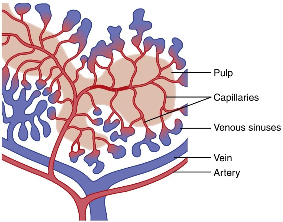
Figure 15-13. Functional structures of the spleen.
شکل 15-13. ساختارهای عملکردی طحال.
Blood-Cleansing Function of the Spleen-Removal of Old Cells Blood cells passing through the splenic pulp before entering the sinuses undergo thorough squeezing. Therefore, it is to be expected that fragile red blood cells would not withstand the trauma. For this reason, many of the red blood cells are destroyed in the spleen. After the cells rupture, the released hemoglobin and cell stroma are digested by the reticuloendothelial cells of the spleen, and the products of digestion are mainly reused by the body as nutrients, often used for making new blood cells.
عملکرد پاکسازی خون طحال – حذف سلولهای قدیمیسلولهای خونی که از پالپ طحال عبور میکنند قبل از ورود به سینوسها تحت فشار کامل قرار میگیرند. بنابراین، میتوان انتظار داشت که گلبولهای قرمز شکننده در برابر ضربه مقاومت نکنند. به همین دلیل، بسیاری از گلبولهای قرمز خون در طحال از بین میروند. پس از پاره شدن سلولها، هموگلوبین آزاد شده و استرومای سلولی توسط سلولهای رتیکولواندوتلیال طحال هضم میشوند و محصولات هضم عمدتاً توسط بدن به عنوان مواد مغذی مورد استفاده مجدد قرار میگیرند که اغلب برای ساخت سلولهای خونی جدید استفاده میشود.
Reticuloendothelial Cells of the Spleen. The pulp of the spleen contains many large phagocytic reticuloendothelial cells, and the venous sinuses are lined with similar cells. These cells function as part of a cleansing system for the blood, acting in concert with a similar system of reticulo- endothelial cells in the venous sinuses of the liver. When the blood is invaded by infectious agents, the reticuloendothelial cells of the spleen rapidly remove substances such as debris, bacteria, and parasites. Also, in many chronic infections, the spleen enlarges in the same manner as lymph nodes and then performs its cleansing function even more avidly.
سلولهای رتیکولواندوتلیال طحال. پالپ طحال حاوی بسیاری از سلولهای رتیکولواندوتلیال فاگوسیتی بزرگ است و سینوسهای وریدی با سلولهای مشابه پوشیده شده اند. این سلولها به عنوان بخشی از یک سیستم پاکسازی خون عمل میکنند و در هماهنگی با سیستم مشابهی از سلولهای رتیکولو اندوتلیال در سینوسهای وریدی کبد عمل میکنند. هنگامیکه خون توسط عوامل عفونی مورد هجوم قرار میگیرد، سلولهای رتیکولواندوتلیال طحال به سرعت موادی مانند زبالهها، باکتریها و انگلها را حذف میکنند. همچنین، در بسیاری از عفونتهای مزمن، طحال مانند غدد لنفاوی بزرگ میشود و سپس عملکرد پاکسازی خود را با اشتیاق بیشتری انجام میدهد.
کلیک کنید «Bibliography: فهرست کتب مربوطه»
Badeer HS: Hemodynamics for medical students. Am J Physiol (Adv Physiol Educ) 25:44, 2001.
Bazigou E, Makinen T: Flow control in our vessels: vascular valves make sure there is no way back. Cell Mol Life Sci 70:1055, 2013.
Hall JE: Integration and regulation of cardiovascular function. Am J Physiol (Adv Physiol Educ) 22:s174, 1999.
Hicks JW, Badeer HS: Gravity and the circulation: “open” vs. “closed” systems. Am J Physiol 262:R725, 1992.
Lacolley P, Regnault V, Segers P, Laurent S: Vascular smooth muscle cells and arterial stiffening: Relevance in development, aging, and disease. Physiol Rev 97:1555, 2017.
Min E, Schwartz MA: Translocating transcription factors in fluid shear stress-mediated vascular remodeling and disease. Exp Cell Res 376:92, 2019.
O’Rourke MF, Adji A: Noninvasive studies of central aortic pressure. Curr Hypertens Rep 14:8, 2012.
Pickering TG, Hall JE, Appel U, et al: Recommendations for blood pressure measurement in humans and experimental animals: Part 1: blood pressure measurement in humans: a statement for professionals from the Subcommittee of Professional and Public Education of the American Heart Association Council on High Blood Pressure Research. Hypertension 45:142, 2005.
Stergiou GS, Alpert B, Mieke S, Asmar R, et. al: A Universal Stand- ard for the Validation of Blood Pressure Measuring Devices: Association for the Advancement of Medical Instrumentation/European Society of Hypertension/International Organization for Standardi- zation (AAMI/ESH/ISO) Collaboration Statement. Hypertension 71:368, 2018.
Whelton PK, Carey RM, Aronow WS, Casey DE Jr, et. al: Guideline for the Prevention, Detection, Evaluation, and Management of High Blood Pressure in Adults: Executive Summary: A Report of the American College of Cardiology/American Heart Association Task Force on Clinical Practice Guidelines. Hypertension. 71:1269, 2018.
🚀 با ما همراه شوید!
تازهترین مطالب و آموزشهای مغز و اعصاب را از دست ندهید. با فالو کردن کانال تلگرام آیندهنگاران مغز، از ما حمایت کنید!



