
فیزیولوژی پزشکی گایتون و هال؛ مروری بر گردش: فشار، جریان و مقاومت


دعای مطالعه [ نمایش ]
بِسْمِ الله الرَّحْمنِ الرَّحیمِ
اَللّهُمَّ اَخْرِجْنى مِنْ ظُلُماتِ الْوَهْمِ
خدايا مرا بيرون آور از تاريكىهاى وهم،
وَ اَكْرِمْنى بِنُورِ الْفَهْمِ
و به نور فهم گرامى ام بدار،
اَللّهُمَّ افْتَحْ عَلَيْنا اَبْوابَ رَحْمَتِكَ
خدايا درهاى رحمتت را به روى ما بگشا،
وَانْشُرْ عَلَيْنا خَزائِنَ عُلُومِكَ بِرَحْمَتِكَ يا اَرْحَمَ الرّاحِمينَ
و خزانههاى علومت را بر ما باز كن به امید رحمتت اى مهربانترين مهربانان.
کتاب «فیزیولوژی پزشکی گایتون وهال» بهعنوان یکی از جامعترین و معتبرترین منابع در حوزه علوم پزشکی، همچنان مرجع کلیدی برای درک عملکرد پیچیده بدن انسان است. این اثر با تکیه بر تازهترین پژوهشها و توضیحات دقیق از سازوکارهای فیزیولوژیک، پلی میان علوم پایه پزشکی و کاربردهای بالینی ایجاد میکند و نقشی بیبدیل در آموزش، پژوهش و ارتقای دانش سلامت ایفا مینماید.
ترجمه دقیق و علمیاین شاهکار توسط برند علمیآیندهنگاران مغز به مدیریت داریوش طاهری، دسترسی فارسیزبانان به مرزهای نوین دانش فیزیولوژی را ممکن ساخته و رسالتی علمیبرای ارتقای آموزش پزشکی، فهم عمیقتر سازوکارهای بدن و توسعه روشهای نوین در حوزه سلامت فراهم آورده است.
» Guyton and Hall Textbook of Medical Physiology
»» CHAPTER 14: Overview of the Circulation: Pressure, Flow, and Resistance
 The function of the circulation is to serve the needs of the body tissues-to transport nutrients to the tissues, to transport waste products away, transport hormones from one part of the body to another and, in general, to maintain an appropriate environment in all the tissue fluids for survival and optimal function of the cells.
عملکرد گردش خون تامین نیازهای بافتهای بدن است – انتقال مواد مغذی به بافتها، انتقال مواد زائد به دور، انتقال هورمونها از یک قسمت از بدن به قسمت دیگر و به طور کلی، حفظ یک محیط مناسب در تمام مایعات بافتی برای بقا و عملکرد بهینه سلولها.
The rate of blood flow through many tissues is controlled mainly in response to their need for nutrients and removal of waste products of metabolism. In some organs, such as the kidneys, the circulation serves additional functions. Blood flow to the kidney, for example, is far in excess of its metabolic requirements and is related to its excretory function, which requires that a large volume of blood be filtered each minute.
سرعت جریان خون در بسیاری از بافتها عمدتاً در پاسخ به نیاز آنها به مواد مغذی و حذف مواد زائد متابولیسم کنترل میشود. در برخی از اندامها، مانند کلیهها، گردش خون عملکردهای بیشتری را انجام میدهد. برای مثال، جریان خون به کلیه بیش از نیازهای متابولیکی آن است و به عملکرد دفعی آن مربوط میشود، که مستلزم آن است که حجم زیادی از خون در هر دقیقه فیلتر شود.
The heart and blood vessels, in turn, are controlled to provide the cardiac output and arterial pressure needed to supply adequate tissue blood flow. What are the mechanisms for controlling blood volume and blood flow, and how does this process relate to the other functions of the circulation? These are some of the topics and questions that we discuss in this section on the circulation.
قلب و عروق خونی به نوبه خود برای تامین برون ده قلبی و فشار شریانی مورد نیاز برای تامین جریان خون کافی در بافت کنترل میشوند. مکانیسمهای کنترل حجم خون و جریان خون چیست و این فرآیند چگونه با سایر عملکردهای گردش خون ارتباط دارد؟ اینها برخی از موضوعات و سوالاتی است که در این بخش در مورد تیراژ بحث میکنیم.
PHYSICAL CHARACTERISTICS OF THE CIRCULATION
The circulation, shown in Figure 14-1, is divided into the systemic circulation and the pulmonary circulation. Because the systemic circulation supplies blood flow to all the tissues of the body except the lungs, it is also called the greater circulation or peripheral circulation.
ویژگیهای فیزیکی گردش خون
گردش خون، که در شکل 14-1 نشان داده شده است، به گردش خون سیستمیک و گردش خون ریوی تقسیم میشود. از آنجایی که گردش خون سیستمیک جریان خون را به تمام بافتهای بدن به جز ریهها تامین میکند، به آن گردش خون بیشتر یا گردش محیطی نیز میگویند.
Functional Parts of the Circulation. Before discussing the details of circulatory function, it is important to understand the role of each part of the circulation.
بخشهای عملکردی گردش خون. قبل از بحث در مورد جزئیات عملکرد گردش خون، مهم است که نقش هر قسمت از گردش خون را درک کنید.
The function of the arteries is to transport blood under high pressure to the tissues. For this reason, the arteries have strong vascular walls, and blood flows at a high velocity in the arteries.
وظیفه شریانها انتقال خون تحت فشار بالا به بافتها است. به همین دلیل، شریانها دارای دیوارههای عروقی قوی هستند و خون با سرعت بالایی در شریانها جریان دارد.
The arterioles are the last small branches of the arterial system; they act as control conduits through which blood is released into the capillaries. Arterioles have strong muscular walls that can close the arterioles completely or, by relaxing, can dilate the vessels severalfold; thus, the arterioles can vastly alter blood flow in each tissue in response to its needs.
شریانها آخرین شاخههای کوچک سیستم شریانی هستند. آنها به عنوان مجرای کنترلی عمل میکنند که از طریق آن خون به داخل مویرگها آزاد میشود. سرخرگها دارای دیوارههای عضلانی قوی هستند که میتواند شریانها را به طور کامل ببندد یا با شل کردن عروق، رگها را چندین برابر کند. بنابراین، شریانها میتوانند به میزان زیادی جریان خون را در هر بافت در پاسخ به نیاز آن تغییر دهند.
The function of the capillaries is to exchange fluid, nutrients, electrolytes, hormones, and other substances between the blood and interstitial fluid. To serve this role, the capillary walls are thin and have numerous minute capillary pores permeable to water and other small molecular substances.
وظیفه مویرگها تبادل مایعات، مواد مغذی، الکترولیتها، هورمونها و سایر مواد بین خون و مایع بینابینی است. برای ایفای این نقش، دیوارههای مویرگی نازک هستند و دارای منافذ مویرگی ریز متعدد و قابل نفوذ به آب و سایر مواد مولکولی کوچک هستند.
The venules collect blood from the capillaries and gradually coalesce into progressively larger veins.
ونولها خون را از مویرگها جمع آوری میکنند و به تدریج به وریدهای بزرگتر تبدیل میشوند.
The veins function as conduits for transport of blood from the venules back to the heart. The veins also serve as a major reservoir of extra blood. Because the pressure in the venous system is low, the venous walls are thin. Even so, they are muscular enough to contract or expand and thereby serve as a controllable reservoir for the extra blood, either a small or a large amount, depending on the needs of the circulation.
سیاهرگها به عنوان مجرای انتقال خون از وریدها به قلب عمل میکنند. سیاهرگها همچنین به عنوان مخزن اصلی خون اضافی عمل میکنند. از آنجایی که فشار در سیستم وریدی کم است، دیوارههای وریدی نازک هستند. با این حال، آنها به اندازه کافی عضلانی هستند که منقبض یا منبسط شوند و در نتیجه بسته به نیاز گردش خون، به عنوان یک مخزن قابل کنترل برای خون اضافی، چه مقدار کمییا زیاد، عمل میکنند.

Figure 14-1. Distribution of blood (in percentage of total blood) in the different parts of the circulatory system.
شکل 14-1. توزیع خون (بر حسب درصد کل خون) در قسمتهای مختلف دستگاه گردش خون.
Volumes of Blood in the Different Parts of the Circulation. Figure 14-1 provides an overview of the circulation and lists the percentages of total blood volume in major segments of the circulation. For example, about 84% of the entire blood volume of the body is in the systemic circulation, and 16% is in the heart and lungs. Of the 84% in the systemic circulation, approximately 64% is in the veins, 13% is in the arteries, and 7% is in the systemic arterioles and capillaries. The heart contains 7% of the blood, and the pulmonary vessels contain 9%.
حجم خون در قسمتهای مختلف گردش خون. شکل 14-1 یک نمای کلی از گردش خون را ارائه میدهد و درصدهای حجم کل خون را در بخشهای اصلی گردش خون فهرست میکند. به عنوان مثال، حدود 84٪ از کل حجم خون بدن در گردش سیستمیو 16٪ در قلب و ریهها است. از 84 درصد در گردش خون سیستمیک، تقریباً 64 درصد در وریدها، 13 درصد در شریانها و 7 درصد در شریانها و مویرگهای سیستمیک است. قلب دارای 7 درصد خون و عروق ریوی 9 درصد است.
Most surprising is the low blood volume in the capillaries. It is here, however, that the most important function of the circulation occurs-diffusion of substances back and forth between the blood and tissues, as discussed in Chapter 16.
شگفت آورترین حجم خون کم در مویرگها است. با این حال، در اینجا است که مهمترین عملکرد گردش خون رخ میدهد – انتشار مواد به عقب و جلو بین خون و بافتها، همانطور که در فصل 16 بحث شد.
Cross-Sectional Areas and Velocities of Blood Flow. If all the systemic vessels of each type were put side by side, their approximate total cross-sectional areas for the average human would be as follows:
سطح مقطع و سرعت جریان خون. اگر تمام عروق سیستمیک هر نوع در کنار هم قرار گیرند، مجموع سطح مقطع تقریبی آنها برای انسان متوسط به شرح زیر خواهد بود:
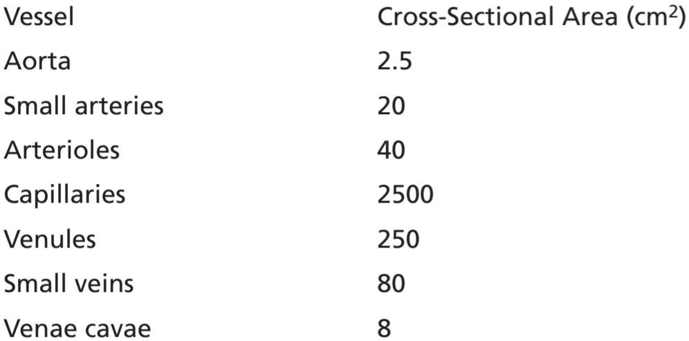
Note particularly that the cross-sectional areas of the veins are much larger than those of the arteries, averaging about four times those of the corresponding arteries. This difference explains the large blood storage capacity of the venous system in comparison with the arterial system.
به ویژه توجه داشته باشید که سطح مقطع وریدها بسیار بزرگتر از سطح شریانها است و به طور متوسط حدود چهار برابر شریانهای مربوطه است. این تفاوت ظرفیت ذخیره خون زیاد سیستم وریدی را در مقایسه با سیستم شریانی توضیح میدهد.
Because the same volume of blood flow (F) must pass through each segment of the circulation each minute, the velocity of blood flow (v) is inversely proportional to the vascular cross-sectional area (A):
از آنجایی که حجم یکسانی از جریان خون (F) باید در هر دقیقه از هر بخش از گردش خون عبور کند، سرعت جریان خون (v) با سطح مقطع عروقی (A) نسبت معکوس دارد:

Thus, under resting conditions, the velocity averages about 33 cm/sec in the aorta but is only 1/1000 as rapid in the capillaries-about 0.3 mm/sec. However, because the capillaries have a typical length of only 0.3 to 1 millimeter, the blood remains in the capillaries for only 1 to 3 seconds, which is surprising because all diffusion of nutrient food substances and electrolytes that occurs through the capillary walls must be performed in this short time.
بنابراین، در شرایط استراحت، سرعت در آئورت به طور متوسط حدود 33 سانتیمتر بر ثانیه است، اما در مویرگها تنها 1000/1 است – حدود 0.3 میلیمتر بر ثانیه. با این حال، از آنجایی که مویرگها طول معمولی 0.3 تا 1 میلیمتر دارند، خون تنها 1 تا 3 ثانیه در مویرگها باقی میماند، که جای تعجب دارد زیرا همه انتشار مواد غذایی مغذی و الکترولیتها که از طریق دیوارههای مویرگها انجام میشود باید در این زمان کوتاه انجام شود.
Pressures in the Various Portions of the Circulation. Because the heart pumps blood continually into the aorta, the mean pressure in the aorta is high, averaging about 100 mm Hg. Also, because heart pumping is pulsatile, the arterial pressure normally alternates between an average systolic pressure level of 120 mm Hg and a diastolic pressure level of 80 mm Hg under resting conditions, as shown on the left side of Figure 14-2.
فشار در بخشهای مختلف گردش خون. از آنجایی که قلب به طور مداوم خون را به داخل آئورت پمپ میکند، فشار متوسط در آئورت بالا است و به طور متوسط حدود 100 میلی متر جیوه است. همچنین، از آنجا که پمپاژ قلب ضربان دار است، فشار شریانی به طور معمول بین سطح فشار سیستولیک متوسط 120 میلی متر جیوه و سطح فشار دیاستولیک 80 میلی متر جیوه در شرایط استراحت متناوب است، همانطور که در سمت چپ شکل 14-2 نشان داده شده است.
As the blood flows through the systemic circulation, its mean pressure falls progressively to about 0 mm Hg by the time it reaches the termination of the superior and inferior venae cavae where they empty into the right atrium of the heart.
همانطور که خون از طریق گردش خون سیستمیک جریان مییابد، فشار متوسط آن به تدریج تا حدود 0 میلی متر جیوه کاهش مییابد تا زمانی که به انتهای ورید اجوف فوقانی و تحتانی میرسد، جایی که آنها به دهلیز راست قلب تخلیه میشوند.
The pressure in many of the systemic capillaries varies from as high as 35 mm Hg near the arteriolar ends to as low as 10 mm Hg near the venous ends, but their aver- age functional pressure in most vascular beds is about 17 mm Hg, a pressure low enough that little of the plasma leaks through the minute pores of the capillary walls, even though nutrients can diffuse easily through these same pores to the outlying tissue cells. In some capillaries, such as the glomerular capillaries of the kidneys, the pressure is considerably higher, averaging about 60 mm Hg and causing much higher rates of fluid filtration.
فشار در بسیاری از مویرگهای سیستمیک از 35 میلیمتر جیوه در نزدیکی انتهای شریانی تا 10 میلیمتر جیوه در نزدیکی انتهای وریدی متغیر است، اما فشار عملکردی متوسط آنها در بیشتر بسترهای عروقی حدود 17 میلیمتر جیوه است، فشاری که به اندازهای کم است که مقدار کمیاز پلاسما به راحتی از طریق این منافذ ریز درپوش نشت میکند. منافذ به سلولهای بافت بیرونی. در برخی از مویرگها، مانند مویرگهای گلومرولی کلیهها، فشار به طور قابل توجهی بالاتر است و به طور متوسط حدود 60 میلیمتر جیوه است و باعث افزایش نرخ فیلتراسیون مایع میشود.
At the far-right side of Figure 14-2, note the respective pressures in the different parts of the pulmonary circulation. In the pulmonary arteries, the pressure is pulsatile, just as in the aorta, but the pressure is far less; pulmonary artery systolic pressure averages about 25 mm Hg and diastolic pressure averages about 8 mm Hg, with a mean pulmonary arterial pressure of only 16 mm Hg. The mean pulmonary capillary pressure averages only 7 mm Hg. Yet, the total blood flow through the lungs each minute is the same as through the systemic circulation. The low pressures of the pulmonary system are in accord with the needs of the lungs because all that is required is to expose the blood in the pulmonary capillaries to oxygen and other gases in the pulmonary alveoli.
در سمت راست شکل 14-2، به فشارهای مربوطه در قسمتهای مختلف گردش خون ریوی توجه کنید. در شریانهای ریوی، فشار مانند آئورت ضربان دار است، اما فشار به مراتب کمتر است. فشار سیستولیک شریان ریوی به طور متوسط حدود 25 میلی متر جیوه و فشار دیاستولیک به طور متوسط حدود 8 میلی متر جیوه است، با فشار متوسط شریان ریوی تنها 16 میلی متر جیوه. میانگین فشار مویرگی ریوی به طور متوسط تنها 7 میلی متر جیوه است. با این حال، کل جریان خون از طریق ریهها در هر دقیقه مانند گردش خون سیستمیک است. فشارهای پایین سیستم ریوی با نیازهای ریهها مطابقت دارد زیرا تنها چیزی که لازم است قرار دادن خون در مویرگهای ریوی در معرض اکسیژن و سایر گازهای موجود در آلوئولهای ریوی است.
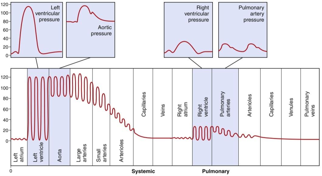
Figure 14-2. Normal blood pressures (in mm Hg) in the different portions of the circulatory system when a person is lying in the horizontal position.
شکل 14-2. فشار خون طبیعی (بر حسب میلی متر جیوه) در قسمتهای مختلف سیستم گردش خون زمانی که فرد در حالت افقی دراز میکشد.
BASIC PRINCIPLES OF CIRCULATORY FUNCTION
Although the details of circulatory function are complex, three basic principles underlie all functions of the system.
اصول اساسی عملکرد گردش خون
اگرچه جزئیات عملکرد گردش خون پیچیده است، سه اصل اساسی زیربنای همه عملکردهای سیستم است.
1. Blood flow to most tissues is controlled according to the tissue needs. When tissues are active, they need an increased supply of nutrients and therefore more blood flow than when at rest, occasionally as much as 20 to 30 times the resting level. However, the heart normally cannot increase its cardiac output more than four to seven times higher than resting levels. Therefore, it is not possible simply to increase blood flow everywhere in the body when a particular tissue demands increased flow. Instead, the microvessels of each tissue, especially the arterioles, continuously monitor tissue needs, such as the availability of oxygen and other nutrients and the accumulation of carbon dioxide and other tissue waste products. These microvessels, in turn, dilate or constrict to control local blood flow at the level required for the tissue activity. Also, nervous control of the circulation from the central nervous system and hormones provides additional help in controlling tissue blood flow.
1. جریان خون در اکثر بافتها با توجه به نیاز بافت کنترل میشود. زمانی که بافتها فعال هستند، نسبت به زمانی که در حالت استراحت هستند، به میزان بیشتری از مواد مغذی و در نتیجه جریان خون بیشتری نیاز دارند، گاهی اوقات تا 20 تا 30 برابر سطح استراحت. با این حال، قلب به طور معمول نمیتواند برون ده قلبی خود را بیش از چهار تا هفت برابر بیشتر از سطوح استراحت افزایش دهد. بنابراین، هنگامیکه یک بافت خاص نیاز به افزایش جریان دارد، نمیتوان به سادگی جریان خون را در همه جای بدن افزایش داد. درعوض، رگهای کوچک هر بافت، بهویژه شریانها، نیازهای بافت مانند در دسترس بودن اکسیژن و سایر مواد مغذی و تجمع دیاکسید کربن و سایر مواد زائد بافتی را بهطور مداوم کنترل میکنند. این ریزرگها به نوبه خود منبسط یا منقبض میشوند تا جریان خون موضعی را در سطح مورد نیاز برای فعالیت بافت کنترل کنند. همچنین، کنترل عصبی گردش خون از سیستم عصبی مرکزی و هورمونها کمک بیشتری به کنترل جریان خون بافت میکند.
2. Cardiac output is the sum of all the local tissue flows. When blood flows through a tissue, it immediately returns by way of the veins to the heart. The heart responds automatically to this increased in- flow of blood by pumping it immediately back into the arteries. Thus, as long as the heart is functioning normally, it acts as an automaton, responding to the demands of the tissues. The heart, however, often needs help in the form of special nerve signals to make it pump the required amounts of blood flow.
2. برون ده قلبی مجموع تمام جریانهای بافت محلی است. هنگامیکه خون در بافتی جریان مییابد، بلافاصله از طریق سیاهرگها به قلب باز میگردد. قلب به طور خودکار به این افزایش جریان خون با پمپاژ فوری آن به شریانها پاسخ میدهد. بنابراین، تا زمانی که قلب به طور طبیعی کار میکند، به عنوان یک خودکار عمل میکند و به نیازهای بافتها پاسخ میدهد. با این حال، قلب اغلب به کمک سیگنالهای عصبی ویژه نیاز دارد تا بتواند مقدار مورد نیاز جریان خون را پمپاژ کند.
3. Arterial pressure regulation is generally independent of either local blood flow control or cardiac output control. The circulatory system is provided with an extensive system for controlling the arterial blood pressure. For example, if at any time the pressure falls significantly below the normal level of about 100 mm Hg, a barrage of nervous reflexes elicits a series of circulatory changes to raise the pressure back toward normal within seconds. The nervous signals especially do the following: (a) increase the force of heart pumping; (b) cause contraction of the large venous reservoirs to provide more blood to the heart; and (c) cause generalized constriction of the arterioles in many tissues so that more blood accumulates in the large arteries to increase the arterial pressure. Then, over more prolonged periods-hours and days-the kidneys play an additional major role in pressure control by secreting pressure-controlling hormones and regulating blood volume.
3. تنظیم فشار شریانی به طور کلی مستقل از کنترل جریان خون موضعی یا کنترل برون ده قلبی است. سیستم گردش خون با یک سیستم گسترده برای کنترل فشار خون شریانی ارائه شده است. به عنوان مثال، اگر در هر زمان فشار به طور قابل توجهی به زیر سطح طبیعی حدود 100 میلی متر جیوه کاهش یابد، رگباری از رفلکسهای عصبی باعث ایجاد یک سری تغییرات در گردش خون میشود تا فشار را در عرض چند ثانیه به حالت عادی برگرداند. سیگنالهای عصبی به ویژه موارد زیر را انجام میدهند: (الف) نیروی پمپاژ قلب را افزایش میدهند. (ب) باعث انقباض مخازن بزرگ وریدی برای رساندن خون بیشتری به قلب شود. و (ج) باعث انقباض عمومیشریانها در بسیاری از بافتها میشود به طوری که خون بیشتری در شریانهای بزرگ جمع میشود تا فشار شریانی را افزایش دهد. سپس، در مدت طولانیتر – ساعتها و روزها – کلیهها با ترشح هورمونهای کنترلکننده فشار و تنظیم حجم خون، نقش مهمیدر کنترل فشار دارند.
Thus, the needs of the individual tissues are served specifically by the circulation. In the remainder of this chapter, we begin to discuss the basic control of tissue blood flow, cardiac output, and arterial pressure.
بنابراین، نیازهای بافتهای فردی به طور خاص توسط گردش خون تامین میشود. در ادامه این فصل، شروع به بحث در مورد کنترل اساسی جریان خون بافت، برون ده قلبی و فشار شریانی میکنیم.
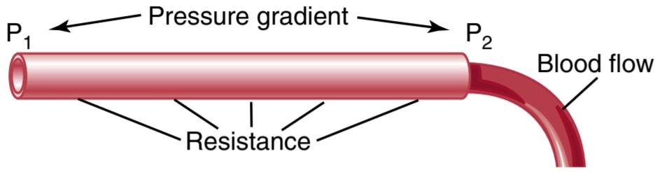
Figure 14-3. Interrelationships of pressure, resistance, and blood flow. P1, Pressure at the origin of the vessel; P2, pressure at the other end of the vessel.
شکل 14-3. روابط متقابل فشار، مقاومت و جریان خون. P1، فشار در مبدا کشتی. P2، فشار در انتهای دیگر ظرف.
INTERRELATIONSHIPS OF PRESSURE, FLOW, AND RESISTANCE
Blood flow through a blood vessel is determined by two factors: (1) pressure difference of the blood between the two ends of the vessel, also sometimes called the pressure gradient along the vessel, which pushes the blood through the vessel; and (2) the impediment to blood flow through the vessel, which is called vascular resistance. Figure 14-3 demonstrates these relationships, showing a blood vessel segment located anywhere in the circulatory system.
روابط متقابل فشار، جریان و مقاومت
جریان خون از طریق رگ خونی توسط دو عامل تعیین میشود: (1) اختلاف فشار خون بین دو انتهای رگ، که گاهی به آن گرادیان فشار در امتداد رگ نیز میگویند، که خون را از طریق رگ هل میدهد. و (2) مانع جریان خون در رگ که مقاومت عروقی نامیده میشود. شکل 14-3 این روابط را نشان میدهد و یک قطعه رگ خونی را نشان میدهد که در هر نقطه از سیستم گردش خون قرار دارد.
P1 represents the pressure at the origin of the vessel and P2 is the pressure at the other end. Resistance occurs as a result of friction between the flowing blood and the intravascular endothelium all along the inside of the vessel. The flow through the vessel can be calculated by the following formula, which is called Ohm’s law:
P1 نشان دهنده فشار در مبدا ظرف و P2 فشار در انتهای دیگر است. مقاومت در نتیجه اصطکاک بین خون جاری و اندوتلیوم داخل عروقی در سراسر داخل رگ ایجاد میشود. جریان عبوری از ظرف را میتوان با فرمول زیر محاسبه کرد که به آن قانون اهم میگویند:
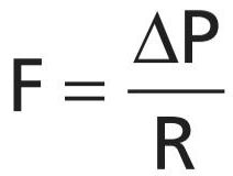
in which F is blood flow, AP is the pressure difference (P1 -P2) between the two ends of the vessel, and R is the resistance. This formula states that the blood flow is directly proportional to the pressure difference but inversely proportional to the resistance.
که در آن F جریان خون است، AP اختلاف فشار (P1 -P2) بین دو انتهای رگ و R مقاومت است. این فرمول بیان میکند که جریان خون با اختلاف فشار نسبت مستقیم دارد اما با مقاومت نسبت معکوس دارد.
Note that it is the difference in pressure between the two ends of the vessel, not the absolute pressure in the vessel, that determines flow rate. For example, if the pressure at both ends of a vessel is 100 mm Hg and no difference exists between the two ends, there will be no flow, despite the presence of 100 mm Hg pressure.
توجه داشته باشید که این اختلاف فشار بین دو انتهای ظرف است، نه فشار مطلق در ظرف، که نرخ جریان را تعیین میکند. به عنوان مثال، اگر فشار در هر دو انتهای یک ظرف 100 میلی متر جیوه باشد و هیچ تفاوتی بین دو انتها وجود نداشته باشد، با وجود وجود فشار 100 میلی متر جیوه، جریانی وجود نخواهد داشت.
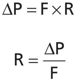
Ohm’s law, illustrated in the preceding formula, expresses one of the most important of all the relationships that the reader needs to understand to comprehend the hemodynamics of the circulation. Because of the extreme importance of this formula, the reader should also become familiar with its other algebraic forms:
قانون اهم، که در فرمول قبلی نشان داده شده است، یکی از مهمترین روابطی را بیان میکند که خواننده برای درک همودینامیک گردش خون باید بداند. به دلیل اهمیت فوق العاده این فرمول، خواننده باید با سایر اشکال جبری آن نیز آشنا شود:
 BLOOD FLOW
Blood flow rate means the quantity of blood that passes a given point in the circulation in a given period of time. Ordinarily, blood flow is expressed in milliliters per minute or liters per minute, but it can be expressed in milliliters per second or in any other units of flow and time.
جریان خون
نرخ جریان خون به معنای مقدار خونی است که در یک دوره زمانی معین از نقطه مشخصی از گردش خون عبور میکند. به طور معمول، جریان خون بر حسب میلی لیتر در دقیقه یا لیتر در دقیقه بیان میشود، اما میتوان آن را بر حسب میلی لیتر در ثانیه یا هر واحد دیگر جریان و زمان بیان کرد.
The overall blood flow in the total circulation of an adult person at rest is about 5000 ml/min. This is called the cardiac output because it is the amount of blood pumped into the aorta by the heart each minute.
جریان کلی خون در کل گردش خون یک فرد بالغ در حالت استراحت حدود 5000 میلی لیتر در دقیقه است. این برون ده قلبی نامیده میشود زیرا مقدار خونی است که در هر دقیقه توسط قلب به آئورت پمپاژ میشود.
Methods for Measuring Blood Flow. Many mechanical and mechanoelectrical flowmeter devices can be inserted in series with a blood vessel or, in some cases, applied to the outside of the vessel to measure blood flow.
روشهای اندازه گیری جریان خون. بسیاری از دستگاههای فلومتر مکانیکی و مکانیکی را میتوان به صورت متوالی با یک رگ خونی وارد کرد یا در برخی موارد در قسمت بیرونی رگ برای اندازه گیری جریان خون اعمال کرد.
Electromagnetic Flowmeter. An electromagnetic flow- meter, the principles of which are illustrated in Figure 14-4, can be used to measure blood flow experimentally without opening the blood vessel. Figure 14-4A shows the generation of electromotive force (electrical voltage) in a wire that is moved rapidly in a cross-wise direction through a magnetic field. This is the well-known principle for production of electricity by the electric generator. Figure 14-4B shows that the same principle applies for generation of electromotive force in blood that is moving through a magnetic field. In this case, a blood vessel is placed between the poles of a strong magnet, and electrodes are placed on the two sides of the vessel perpendicular to the magnetic lines of force. When blood flows through the vessel, an electrical voltage proportional to the rate of blood flow is generated between the two electrodes, and this voltage is recorded using an appropriate voltmeter or electronic recording apparatus. Figure 14-4C shows an actual probe that is placed on a large blood vessel to record its blood flow. The probe contains both the strong magnet and the electrodes.
فلومتر الکترومغناطیسی. یک جریان سنج الکترومغناطیسی، که اصول آن در شکل 14-4 نشان داده شده است، میتواند برای اندازه گیری جریان خون به صورت تجربی بدون باز کردن رگ خونی استفاده شود. شکل 14-44 تولید نیروی الکتروموتور (ولتاژ الکتریکی) در سیمیرا نشان میدهد که به سرعت در جهت متقاطع از طریق میدان مغناطیسی حرکت میکند. این اصل شناخته شده برای تولید برق توسط ژنراتور الکتریکی است. شکل 14-4B نشان میدهد که همین اصل برای تولید نیروی حرکتی الکتریکی در خونی که در یک میدان مغناطیسی در حال حرکت است، اعمال میشود. در این حالت، یک رگ خونی بین قطبهای یک آهنربای قوی قرار میگیرد و الکترودها در دو طرف رگ عمود بر خطوط مغناطیسی نیرو قرار میگیرند. هنگامیکه خون در رگ جریان مییابد، یک ولتاژ الکتریکی متناسب با سرعت جریان خون بین دو الکترود ایجاد میشود و این ولتاژ با استفاده از یک ولت متر یا دستگاه ثبت الکترونیکی مناسب ثبت میشود. شکل 14-4C یک کاوشگر واقعی را نشان میدهد که روی یک رگ خونی بزرگ قرار داده شده است تا جریان خون آن را ثبت کند. پروب حاوی آهنربای قوی و الکترود است.
A special advantage of the electromagnetic flowmeter is that it can record changes in flow in less than 1/100 of a second, allowing for the accurate recording of pulsatile changes in flow, as well as steady flow.
مزیت ویژه فلومتر الکترومغناطیسی این است که میتواند تغییرات جریان را در کمتر از 1/100 ثانیه ثبت کند و امکان ثبت دقیق تغییرات ضربانی در جریان و همچنین جریان ثابت را فراهم میکند.
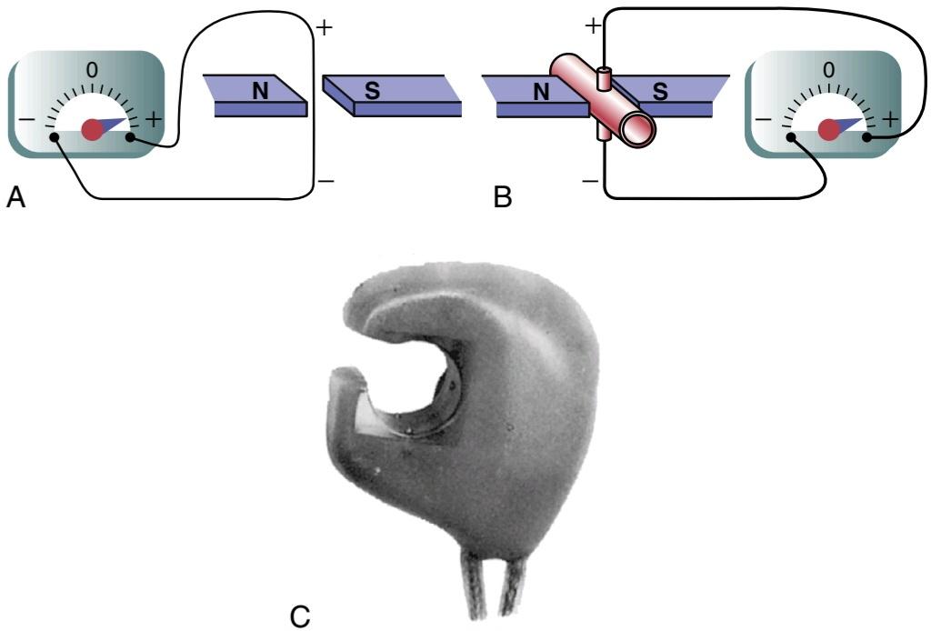
Figure 14-4. A, Electromagnetic flowmeter showing generation of an electrical voltage in a wire as it passes through an electromagnetic field. B, Generation of an electrical voltage in electrodes on a blood vessel when the vessel is placed in a strong magnetic field, and blood flows through the vessel. C, Modern electromagnetic flowmeter probe for chronic implantation around blood vessels. N and S refer to the mag- net’s north and south poles.
شکل 14-4. الف، فلومتر الکترومغناطیسی که تولید ولتاژ الکتریکی را در سیم هنگام عبور از میدان الکترومغناطیسی نشان میدهد. ب، ایجاد ولتاژ الکتریکی در الکترودهای روی رگ خونی هنگامیکه رگ در یک میدان مغناطیسی قوی قرار میگیرد و خون در رگ جریان مییابد. C، پروب جریان سنج الکترومغناطیسی مدرن برای کاشت مزمن در اطراف عروق خونی. N و S به قطب شمال و جنوب آهنربا اشاره دارد.
Ultrasonic Doppler Flowmeter. Another type of flow- meter that can be applied to the outside of the vessel and that has many of the same advantages as the electromagnetic flowmeter is the ultrasonic Doppler flowmeter, shown in Figure 14-5. A minute piezoelectric crystal is mounted at one end in the wall of the device. This crystal, when energized with an appropriate electronic apparatus, transmits ultrasound at a frequency of several hundred thousand cycles per second downstream along the flowing blood. A portion of the sound is reflected by the red blood cells in the flowing blood. The reflected ultrasound waves then travel backward from the blood cells toward the crystal. These reflected waves have a lower frequency than the transmitted wave because the red blood cells are moving away from the transmitter crystal. This effect is called the Doppler effect. (It is the same effect that one experiences when a train approaches and passes by while blowing its whistle. Once the whistle has passed by the person, the pitch of the sound from the whistle suddenly becomes much lower than when the train is approaching.)
فلومتر داپلر اولتراسونیک. نوع دیگری از فلومتر که میتواند در خارج از ظرف اعمال شود و دارای مزایای مشابهی با فلومتر الکترومغناطیسی است، فلومتر داپلر اولتراسونیک است که در شکل 14-5 نشان داده شده است. یک کریستال پیزوالکتریک دقیقه ای در یک انتها در دیواره دستگاه نصب شده است. این کریستال، هنگامیکه با یک دستگاه الکترونیکی مناسب انرژی میگیرد، فراصوت را با فرکانس چند صد هزار سیکل در ثانیه به پایین دست در امتداد خون جاری منتقل میکند. بخشی از صدا توسط گلبولهای قرمز خون در جریان خون منعکس میشود. سپس امواج اولتراسوند منعکس شده از سلولهای خونی به سمت کریستال حرکت میکنند. این امواج منعکس شده فرکانس کمتری نسبت به موج ارسالی دارند زیرا گلبولهای قرمز خون از کریستال فرستنده دور میشوند. این اثر را اثر داپلر مینامند. (این همان تأثیری است که فرد هنگام نزدیک شدن قطار و عبور از آن در هنگام سوت زدن تجربه میکند. هنگامیکه سوت از کنار شخص عبور میکند، صدای سوت ناگهان بسیار کمتر از زمانی است که قطار در حال نزدیک شدن است.)
For the flowmeter shown in Figure 14-5, the high- frequency ultrasound wave is intermittently cut off, and the reflected wave is received back onto the crystal and greatly amplified by the electronic apparatus. Another portion of the electronic apparatus determines the frequency difference between the transmitted wave and the reflected wave, thus determining the velocity of blood flow. As long as the diameter of a blood vessel does not change, changes in blood flow in the vessel are directly related to changes in flow velocity.
برای فلومتر نشان داده شده در شکل 14-5، موج اولتراسوند با فرکانس بالا به طور متناوب قطع میشود و موج منعکس شده به کریستال برمیگردد و توسط دستگاه الکترونیکی تا حد زیادی تقویت میشود. بخش دیگری از دستگاه الکترونیکی تفاوت فرکانس بین موج ارسالی و موج بازتابی را تعیین میکند، بنابراین سرعت جریان خون را تعیین میکند. تا زمانی که قطر رگ خونی تغییر نکند، تغییرات در جریان خون در رگ ارتباط مستقیمیبا تغییرات سرعت جریان دارد.
Like the electromagnetic flowmeter, the ultrasonic Doppler flowmeter is capable of recording rapid pulsatile changes in flow, as well as steady flow.
مانند فلومتر الکترومغناطیسی، فلومتر داپلر اولتراسونیک قادر است تغییرات ضربانی سریع در جریان و همچنین جریان ثابت را ثبت کند.
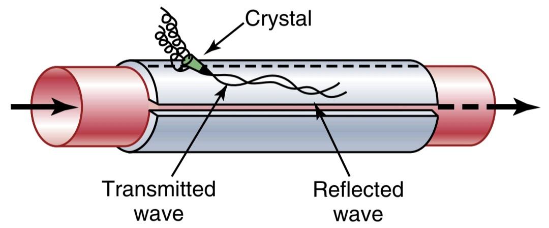
Figure 14-5. Ultrasonic Doppler flowmeter.
شکل 14-5. فلومتر داپلر اولتراسونیک.
Laminar Flow of Blood in Vessels. When blood flows at a steady rate through a long smooth blood vessel, it flows in streamlines, with each layer of blood remaining the same distance from the vessel wall. Also, the centralmost portion of the blood stays in the center of the vessel. This type of flow is called laminar flow or streamline flow, in contrast to turbulent flow, which is blood flowing in all directions in the vessel and continually mixing in the vessel, as discussed subsequently.
جریان آرام خون در عروق. هنگامیکه خون با سرعت ثابتی از طریق یک رگ خونی صاف و طولانی جریان مییابد، در خطوط جریانی جریان مییابد و هر لایه خون در همان فاصله از دیواره رگ باقی میماند. همچنین، مرکزی ترین قسمت خون در مرکز رگ باقی میماند. این نوع جریان، جریان آرام یا جریان جریان نامیده میشود، برخلاف جریان آشفته، که خون در همه جهات در رگ جریان دارد و به طور مداوم در رگ مخلوط میشود، همانطور که در ادامه بحث شد.
Parabolic Velocity Profile During Laminar Flow. When laminar flow occurs, the velocity of flow in the center of the vessel is far greater than that toward the outer edges. This phenomenon is demonstrated in Figure 14-6. In Figure 14-6A, a vessel contains two fluids, the one at the left colored by a dye and the one at the right a clear fluid, but there is no flow in the vessel. When the fluids are made to flow, a parabolic interface develops between them, as shown 1 second later in Figure 14-6B. The portion of fluid adjacent to the vessel wall has hardly moved, the portion slightly away from the wall has moved a small distance, and the portion in the center of the vessel has moved a long distance. This effect is called the parabolic profile for velocity of blood flow.
مشخصات سرعت سهموی در طول جریان آرام. هنگامیکه جریان آرام رخ میدهد، سرعت جریان در مرکز ظرف بسیار بیشتر از سرعت جریان به سمت لبههای بیرونی است. این پدیده در شکل 14-6 نشان داده شده است. در شکل 14-6A، یک ظرف حاوی دو سیال است، یکی در سمت چپ با رنگ و دیگری در سمت راست یک مایع شفاف است، اما هیچ جریانی در ظرف وجود ندارد. هنگامیکه سیالها به جریان میافتند، یک رابط سهموی بین آنها ایجاد میشود، همانطور که 1 ثانیه بعد در شکل 14-6B نشان داده شده است. قسمت سیال مجاور دیواره رگ به سختی جابجا شده است، قسمتی که کمیاز دیواره فاصله دارد فاصله کمیدارد و قسمتی که در مرکز رگ قرار دارد مسافت زیادی را جابجا کرده است. این اثر، مشخصات سهموی برای سرعت جریان خون نامیده میشود.
The cause of the parabolic profile is as follows. The fluid molecules touching the wall move slowly because of adherence to the vessel wall. The next layer of molecules slips over these, the third layer over the second, the fourth layer over the third, and so forth. Therefore, the fluid in the middle of the vessel can move rapidly because many layers of slipping molecules exist between the middle of the vessel and the vessel wall. Thus, each layer toward the center flows progressively more rapidly than the outer layers.
علت نیمرخ سهموی به شرح زیر است. مولکولهای مایعی که دیواره را لمس میکنند به دلیل چسبیدن به دیواره رگ به آرامیحرکت میکنند. لایه بعدی مولکولها روی آنها میلغزد، لایه سوم روی دوم، لایه چهارم روی سوم و غیره. بنابراین، سیال در وسط رگ میتواند به سرعت حرکت کند زیرا لایههای زیادی از مولکولهای لغزنده بین وسط رگ و دیواره رگ وجود دارد. بنابراین، هر لایه به سمت مرکز به تدریج سریعتر از لایههای بیرونی جریان مییابد.
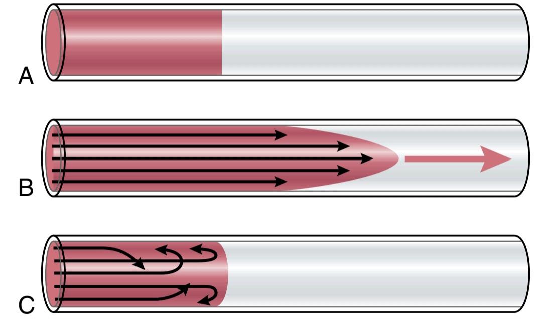
Figure 14-6. A, Two fluids (one dyed red, and the other clear) before flow begins. B. The same fluids 1 second after flow begins. C, Turbulent flow, with elements of the fluid moving in a disorderly pattern.
شکل 14-6. A، دو مایع (یکی به رنگ قرمز و دیگری شفاف) قبل از شروع جریان. ب– همان سیالات 1 ثانیه پس از شروع جریان. C، جریان آشفته، با عناصر سیال در یک الگوی نامنظم حرکت میکنند.
Turbulent Flow of Blood Under Some Conditions. When the rate of blood flow becomes too great, when it passes by an obstruction in a vessel, when it makes a sharp turn, or when it passes over a rough surface, the flow may then become turbulent, or disorderly, rather than streamlined (Figure 14-6C). Turbulent flow means that the blood flows crosswise in the vessel and along the vessel, usually forming whorls in the blood, called eddy currents. These eddy currents are similar to the whirlpools that can be seen in a rapidly flowing river at a point of obstruction. When eddy currents are present, the blood flows with much greater resistance than when the flow is streamlined because eddies add to the overall friction of flow in the vessel tremendously.
جریان آشفته خون تحت برخی شرایط. هنگامیکه سرعت جریان خون خیلی زیاد میشود، هنگامیکه از یک انسداد در یک رگ عبور میکند، هنگامیکه یک چرخش شدید ایجاد میکند، یا زمانی که از روی یک سطح ناهموار عبور میکند، جریان ممکن است متلاطم یا نامنظم شود، نه اینکه جریان داشته باشد (شکل 14-6C). جریان آشفته به این معنی است که خون به صورت متقاطع در رگ و در امتداد رگ جریان مییابد و معمولاً حلقههایی در خون ایجاد میکند که به آنها جریان گردابی میگویند. این جریانهای گردابی شبیه گردابهایی هستند که در رودخانهای با جریان سریع در نقطهای از انسداد دیده میشوند. هنگامیکه جریانهای گردابی وجود دارند، خون با مقاومت بسیار بیشتری نسبت به زمانی که جریان جریان دارد جریان مییابد، زیرا گردابها به شدت به اصطکاک کلی جریان در رگ میافزایند.
The tendency for turbulent flow increases in direct pro- portion to the velocity of blood flow, the diameter of the blood vessel, and the density of the blood and is inversely proportional to the viscosity of the blood, in accordance with the following equation:
گرایش جریان آشفته به نسبت مستقیم با سرعت جریان خون، قطر رگ خونی و چگالی خون افزایش مییابد و مطابق با معادله زیر با ویسکوزیته خون نسبت معکوس دارد:
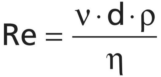
where Re is Reynolds’ number, the measure of the tendency for turbulence to occur, v is the mean velocity of blood flow (in cm/sec), d is the vessel diameter (in centimeters), p is density (in grams/ml), and n is the viscosity (in poise). The viscosity of blood is normally about 1/30 poise, and the density is only slightly greater than 1. When Reynolds’ number rises above 200 to 400, turbulent flow will occur at some branches of vessels but will die out along the smooth portions of the vessels. However, when Reynolds’ number rises above approximately 2000, turbulence will usually occur, even in a straight, smooth vessel.
جایی که Re عدد رینولدز است، میزان تمایل به وقوع تلاطم، v میانگین سرعت جریان خون (بر حسب سانتی متر بر ثانیه)، d قطر رگ (بر حسب سانتی متر)، p چگالی (بر حسب گرم بر میلی لیتر)، و n ویسکوزیته (بر حسب پویز) است. ویسکوزیته خون معمولاً حدود 1/30 پویز است و چگالی آن فقط کمیبیشتر از 1 است. وقتی عدد رینولدز از 200 به 400 میرسد، جریان آشفته در برخی از شاخههای رگها رخ میدهد اما در امتداد بخشهای صاف رگها از بین میرود. با این حال، هنگامیکه عدد رینولدز از حدود 2000 بالاتر میرود، معمولاً تلاطم حتی در یک رگ صاف و صاف رخ میدهد.
Reynolds’ number for flow in the vascular system normally rises to 200 to 400, even in large arteries. As a result, there is almost always some flow turbulence at the branches of these vessels. In the proximal portions of the aorta and pulmonary artery, Reynolds’ number can rise to several thousand during the rapid phase of ejection by the ventricles, which causes considerable turbulence in the proximal aorta and pulmonary artery, where many conditions are appropriate for turbulence, such as the following: (1) high velocity of blood flow; (2) pulsatile nature of the flow; (3) sudden change in vessel diameter; and (4) large vessel diameter. However, in small vessels, Reynolds’ number is almost never high enough to cause turbulence.
تعداد رینولدز برای جریان در سیستم عروقی به طور معمول به 200 تا 400 میرسد، حتی در شریانهای بزرگ. در نتیجه تقریباً همیشه مقداری تلاطم جریان در شاخههای این کشتیها وجود دارد. در بخشهای پروگزیمال آئورت و شریان ریوی، تعداد رینولدز میتواند در طول فاز سریع جهش توسط بطنها به چندین هزار برسد، که باعث آشفتگی قابلتوجهی در آئورت پروگزیمال و شریان ریوی میشود، جایی که بسیاری از شرایط برای تلاطم مناسب هستند، مانند موارد زیر: (1) سرعت جریان خون بالا؛ (2) ماهیت ضربانی جریان. (3) تغییر ناگهانی در قطر رگ؛ و (4) قطر کشتی بزرگ. با این حال، در کشتیهای کوچک، تعداد رینولدز تقریباً هرگز آنقدر زیاد نیست که باعث آشفتگی شود.
BLOOD PRESSURE
فشار خون
Standard Units of Pressure. Blood pressure almost al- ways is measured in millimeters of mercury (mm Hg) because the mercury manometer has been used as the standard reference for measuring pressure since its invention in 1846 by Poiseuille. Actually, blood pressure means the force exerted by the blood against any unit area of the vessel wall. If the pressure in a vessel is 100 mm Hg, this means that the force exerted is sufficient to push a column of mercury against gravity up to a level 50 millimeters high.
واحدهای استاندارد فشار. فشار خون تقریباً همیشه بر حسب میلیمتر جیوه (میلیمتر جیوه) اندازهگیری میشود، زیرا مانومتر جیوهای از زمان اختراع آن در سال 1846 توسط Poiseuille به عنوان مرجع استاندارد برای اندازهگیری فشار استفاده میشود. در واقع فشار خون به معنای نیرویی است که خون بر هر واحد سطح دیواره عروق وارد میکند. اگر فشار در یک ظرف 100 میلی متر جیوه باشد، به این معنی است که نیروی اعمال شده برای فشار دادن یک ستون جیوه در برابر گرانش تا سطح 50 میلی متر کافی است.
Occasionally, pressure is measured in centimeters of water (cm H2O). A pressure of 10 cm H2O means a pres- sure sufficient to raise a column of water against gravity to a height of 10 centimeters. One millimeter of mercury pressure equals 1.36 centimeters of water pressure because the specific gravity of mercury is 13.6 times that of water, and 1 centimeter is 10 times as great as 1 millimeter.
گاهی اوقات فشار بر حسب سانتی متر آب (سانتی متر H2O) اندازه گیری میشود. فشار 10 سانتی متر H2O به معنای فشار کافی برای بالا بردن یک ستون آب در برابر گرانش تا ارتفاع 10 سانتی متر است. یک میلی متر فشار جیوه برابر با 1.36 سانتی متر فشار آب است زیرا وزن مخصوص جیوه 13.6 برابر آب است و 1 سانتی متر 10 برابر 1 میلی متر است.
High-Fidelity Methods for Measuring Blood Pressure. The mercury in a manometer has so much inertia that it cannot rise and fall rapidly. For this reason, the mercury manometer, although excellent for recording steady pressures, cannot respond to pressure changes that occur more rapidly than about one cycle every 2 to 3 seconds. Whenever it is desired to record rapidly changing pressures, some other type of pressure recorder is necessary. Figure 14-7 demonstrates the basic principles of three electronic pressure transducers commonly used for converting blood pressure and/ or rapid changes in pressure into electrical signals and then recording the electrical signals on a high-speed electrical recorder. Each of these transducers uses a very thin, highly stretched metal membrane that forms one wall of the fluid chamber. The fluid chamber, in turn, is connected through a needle or catheter inserted into the blood vessel in which the pressure is to be measured. When the pressure is high, the membrane bulges slightly and, when it is low, it returns toward its resting position.
روشهای با وفاداری بالا برای اندازهگیری فشار خون. جیوه در یک مانومتر دارای اینرسی زیادی است که نمیتواند به سرعت بالا و پایین بیاید. به همین دلیل، مانومتر جیوه ای، اگرچه برای ثبت فشارهای ثابت عالی است، اما نمیتواند به تغییرات فشاری که سریعتر از یک چرخه در هر 2 تا 3 ثانیه رخ میدهد، پاسخ دهد. هر زمان که بخواهید فشارهای با تغییر سریع را ثبت کنید، نوع دیگری از ضبط کننده فشار ضروری است. شکل 14-7 اصول اولیه سه مبدل فشار الکترونیکی را نشان میدهد که معمولاً برای تبدیل فشار خون و/یا تغییرات سریع فشار به سیگنالهای الکتریکی و سپس ثبت سیگنالهای الکتریکی روی یک ضبط کننده الکتریکی پرسرعت استفاده میشوند. هر یک از این مبدلها از یک غشای فلزی بسیار نازک و بسیار کشیده استفاده میکنند که یک دیواره محفظه سیال را تشکیل میدهد. محفظه مایع به نوبه خود از طریق یک سوزن یا کاتتر وارد رگ خونی میشود که فشار در آن اندازه گیری میشود. هنگامیکه فشار زیاد است، غشاء اندکی برآمده میشود و وقتی کم است، به حالت استراحت خود باز میگردد.
In Figure 14-74, a simple metal plate is placed a few hundredths of a centimeter above the membrane. When the membrane bulges, the membrane comes closer to the plate, which increases the electrical capacitance between these two, and this change in capacitance can be recorded using an appropriate electronic system.
در شکل 14-74 یک صفحه فلزی ساده در چند صدم سانتیمتر بالای غشا قرار داده شده است. هنگامیکه غشا برآمده میشود، غشا به صفحه نزدیک میشود که باعث افزایش ظرفیت الکتریکی بین این دو میشود و با استفاده از یک سیستم الکترونیکی مناسب میتوان این تغییر ظرفیت را ثبت کرد.
In Figure 14-7B, a small iron slug rests on the membrane, and this slug can be displaced upward into a center space inside an electrical wire coil. Movement of the iron into the coil increases the inductance of the coil, and this too can be recorded electronically.
در شکل 14-7B، یک حلزون کوچک آهنی بر روی غشاء قرار میگیرد، و این راب میتواند به سمت بالا به فضای مرکزی در داخل یک سیم پیچ سیم برق جابجا شود. حرکت آهن به داخل سیم پیچ باعث افزایش اندوکتانس سیم پیچ میشود و این نیز میتواند به صورت الکترونیکی ثبت شود.
Finally, in Figure 14-7C, a very thin, stretched resistance wire is connected to the membrane. When this wire is stretched greatly, its resistance increases; when it is stretched less, its resistance decreases. These changes can also be recorded by an electronic system.
در نهایت، در شکل 14-7C، یک سیم مقاومتی بسیار نازک و کشیده به غشاء وصل شده است. هنگامیکه این سیم به شدت کشیده میشود، مقاومت آن افزایش مییابد. وقتی کمتر کشیده شود مقاومت آن کاهش مییابد. این تغییرات توسط یک سیستم الکترونیکی نیز قابل ثبت است.
The electrical signals from the transducer are sent to an amplifier and then to an appropriate recording device. With some of these high-fidelity types of recording systems, pres- sure cycles up to 500 cycles/sec have been recorded accurately. In common use are recorders capable of registering pressure changes that occur as rapidly as 20 to 100 cycles/ sec in the manner shown on the recorder in Figure 14-7C.
سیگنالهای الکتریکی از مبدل به یک تقویت کننده و سپس به یک دستگاه ضبط مناسب ارسال میشود. با برخی از این انواع سیستمهای ضبط با وفاداری بالا، چرخههای فشار تا 500 سیکل در ثانیه به دقت ثبت شده است. معمولاً ضبط کنندههایی هستند که میتوانند تغییرات فشار را که به سرعت 20 تا 100 سیکل در ثانیه رخ میدهد، به روشی که روی ضبط کننده در شکل 14-7C نشان داده شده است، ثبت کنند.

Figure 14-7. A-C, Principles of three types of electronic transducers for recording rapidly changing blood pressures (see text).
شکل 14-7. A-C، اصول سه نوع مبدل الکترونیکی برای ثبت فشار خون با تغییر سریع (به متن مراجعه کنید).
RESISTANCE TO BLOOD FLOW
مقاومت در برابر جریان خون
Units of Resistance. Resistance is the impediment to blood flow in a vessel, but it cannot be measured by any direct means. Instead, resistance must be calculated from measurements of blood flow and pressure difference be- tween two points in the vessel. If the pressure difference between two points is 1 mm Hg and the flow is 1 ml/sec, the resistance is said to be 1 peripheral resistance unit, usually abbreviated PRU.
واحدهای مقاومت. مقاومت مانع جریان خون در رگ است، اما نمیتوان آن را با هیچ وسیله مستقیم اندازه گیری کرد. در عوض، مقاومت باید از اندازه گیری جریان خون و اختلاف فشار بین دو نقطه در رگ محاسبه شود. اگر اختلاف فشار بین دو نقطه 1 میلیمتر جیوه و جریان 1 میلیلیتر بر ثانیه باشد، به مقاومت 1 واحد مقاومت محیطی گفته میشود که معمولاً به اختصار PRU میگویند.
Expression of Resistance in CGS Units. Occasionally, a basic physical unit called the CGS (centimeters, grams, seconds) unit is used to express resistance. This unit is dyne sec/cm3. Resistance in these units can be calculated by the following formula:
بیان مقاومت در واحدهای CGS. گاهی اوقات، یک واحد فیزیکی پایه به نام واحد CGS (سانتی متر، گرم، ثانیه) برای بیان مقاومت استفاده میشود. این واحد dyne sec/cm3 است. مقاومت در این واحدها را میتوان با فرمول زیر محاسبه کرد:

Total Peripheral Vascular Resistance and Total Pulmonary Vascular Resistance. The rate of blood flow through the entire circulatory system is equal to the rate of blood pumping by the heart-that is, it is equal to the cardiac output. In an adult human, this averages approximately 100 ml/sec. The pressure difference from the systemic arteries to the systemic veins is about 100 mm Hg. Therefore, the resistance of the entire systemic circulation, called the total peripheral resistance, is about 100/100, or 1 PRU.
مقاومت کلی عروق محیطی و مقاومت عروقی کلی ریوی. سرعت جریان خون در کل سیستم گردش خون برابر است با سرعت پمپاژ خون توسط قلب – یعنی برابر با برون ده قلبی است. در یک انسان بالغ، این میانگین تقریباً 100 میلی لیتر در ثانیه است. اختلاف فشار از شریانهای سیستمیک به سیاهرگهای سیستمیک حدود 100 میلی متر جیوه است. بنابراین، مقاومت کل گردش خون، که مقاومت محیطی کل نامیده میشود، حدود 100/100 یا 1 PRU است.
In conditions in which all the blood vessels throughout the body become strongly constricted, the total peripheral resistance occasionally rises to as high as 4 PRU. Conversely, when the vessels become greatly dilated, the resistance can fall to as little as 0.2 PRU.
در شرایطی که تمام رگهای خونی بدن به شدت منقبض میشوند، مقاومت کلی محیطی گاهی تا 4 PRU افزایش مییابد. برعکس، زمانی که عروق بسیار گشاد میشوند، مقاومت میتواند تا 0.2 PRU کاهش یابد.
In the pulmonary system, the mean pulmonary arterial pressure averages 16 mm Hg and the mean left atrial pres- sure averages 2 mm Hg, giving a net pressure difference of 14 mm. Therefore, when the cardiac output is normal at about 100 ml/sec, the total pulmonary vascular resistance calculates to be about 0.14 PRU (about one seventh that in the systemic circulation).
در سیستم ریوی، میانگین فشار شریانی ریوی به طور متوسط 16 میلی متر جیوه و میانگین فشار دهلیز چپ به طور متوسط 2 میلی متر جیوه است که اختلاف فشار خالص 14 میلی متر را نشان میدهد. بنابراین، زمانی که برون ده قلبی در حدود 100 میلی لیتر در ثانیه نرمال باشد، مقاومت کلی عروق ریوی حدود 0.14 PRU (حدود یک هفتم آن در گردش خون سیستمیک) محاسبه میشود.
Conductance of Blood in a Vessel Is the Reciprocal of Resistance. Conductance is a measure of the blood flow through a vessel for a given pressure difference. This measurement is generally expressed in terms of ml/sec per mm Hg pressure, but it can also be expressed in terms of L/sec per mm Hg or in any other units of blood flow and pressure.
هدایت خون در یک رگ، متقابل مقاومت است. رسانایی اندازه گیری جریان خون در یک رگ برای اختلاف فشار معین است. این اندازهگیری عموماً بر حسب میلیلیتر بر ثانیه فشار بر میلیمتر جیوه بیان میشود، اما میتوان آن را بر حسب L/sec بر میلیمتر جیوه یا هر واحد دیگری از جریان خون و فشار نیز بیان کرد.
It is evident that conductance is the exact reciprocal of resistance in accord with the following equation:
بدیهی است که رسانایی مطابق با معادله زیر دقیقاً متقابل مقاومت است:

Small Changes in Vessel Diameter Markedly Change Its Conductance. Slight changes in the diameter of a vessel cause tremendous changes in the vessel’s ability to conduct blood when the blood flow is streamlined. This phenomenon is illustrated in Figure 14-84, which shows three vessels with relative diameters of 1, 2, and 4 but with the same pressure difference of 100 mm Hg between the two ends of the vessels. Although the diameters of these vessels increase only fourfold, the respective flows are 1, 16, and 256 ml/ min, which is a 256-fold increase in flow. Thus, the conductance of the vessel increases in proportion to the fourth pow- er of the diameter, in accordance with the following formula:
تغییرات کوچک در قطر کشتی به طور قابل توجهی هدایت آن را تغییر میدهد. تغییرات جزئی در قطر رگ باعث تغییرات فوق العاده ای در توانایی رگ برای هدایت خون در زمانی که جریان خون جریان دارد، میشود. این پدیده در شکل 14-84 نشان داده شده است که سه رگ را با قطرهای نسبی 1، 2 و 4 اما با اختلاف فشار یکسان 100 میلی متر جیوه بین دو انتهای رگ نشان میدهد. اگرچه قطر این مخازن تنها چهار برابر افزایش مییابد، جریانهای مربوطه 1، 16 و 256 میلی لیتر در دقیقه است که افزایش 256 برابری در جریان است. بنابراین، رسانایی ظرف متناسب با چهارمین قدرت قطر، مطابق با فرمول زیر افزایش مییابد:

Poiseuille’s Law. The cause of this great increase in conductance when the diameter increases can be explained by referring to Figure 14-8B, which shows cross sections of a large and small vessel. The concentric rings inside the vessels indicate that the velocity of flow in each ring is different from that in the adjacent rings because of laminar flow, which was discussed earlier in the chapter. That is, the blood in the ring touching the wall of the vessel is barely flowing because of its adherence to the vascular endothelium. The next ring of blood toward the center of the vessel slips past the first ring and, therefore, flows more rapidly. Likewise, the third, fourth, fifth, and sixth rings flow at progressively increasing velocities. Thus, the blood that is near the wall of the vessel flows slowly, whereas that in the middle of the vessel flows much more rapidly.
قانون پوازوی. علت این افزایش بزرگ در رسانایی در هنگام افزایش قطر را میتوان با مراجعه به شکل 14-8B توضیح داد که سطح مقطع یک کشتی بزرگ و کوچک را نشان میدهد. حلقههای متحدالمرکز داخل رگها نشان میدهند که سرعت جریان در هر حلقه با حلقههای مجاور به دلیل جریان آرام که قبلاً در فصل مورد بحث قرار گرفت متفاوت است. یعنی خون در حلقهای که دیواره رگ را لمس میکند به دلیل چسبیدن آن به اندوتلیوم عروقی به سختی در جریان است. حلقه بعدی خون به سمت مرکز رگ از روی حلقه اول میلغزد و بنابراین با سرعت بیشتری جریان مییابد. به همین ترتیب، حلقههای سوم، چهارم، پنجم و ششم با سرعتهای فزاینده ای در جریان هستند. بنابراین، خونی که در نزدیکی دیواره رگ قرار دارد به کندی جریان مییابد، در حالی که خون در وسط رگ بسیار سریعتر جریان مییابد.

Figure 14-8. A, Demonstration of the effect of vessel diameter on blood flow. B, Concentric rings of blood flowing at different velocities; the farther away from the vessel wall, the faster the flow. d, diameter; P, pressure difference between the two ends of the vessels.
شکل 14-8. الف، نشان دادن تأثیر قطر رگ بر جریان خون. ب، حلقههای متحدالمرکز خون که با سرعتهای مختلف جریان دارند. هرچه از دیواره رگ دورتر باشد، جریان سریعتر است. d، قطر؛ P، اختلاف فشار بین دو انتهای رگها.
In the small vessel, essentially all the blood is near the wall, so the extremely rapidly flowing central stream of blood simply does not exist. By integrating the velocities of all the concentric rings of flowing blood and multiplying them by the areas of the rings, one can derive the following formula, known as Poiseuille’s law:
در رگ کوچک، اساساً تمام خون نزدیک دیواره است، بنابراین جریان مرکزی خون با سرعت بسیار بالا به سادگی وجود ندارد. با ادغام سرعت همه حلقههای متحدالمرکز خون جاری و ضرب آنها در مساحت حلقهها، میتوان فرمول زیر را به دست آورد که به قانون پوازوی معروف است:
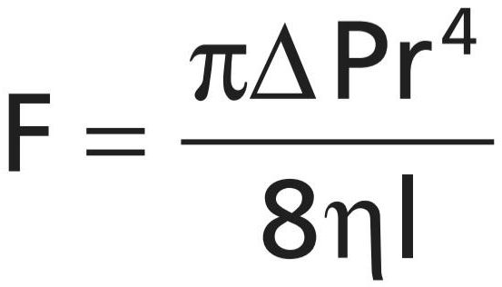
in which F is the rate of blood flow, AP is the pressure difference between the ends of the vessel, r is the radius of the vessel, I is length of the vessel, and n is viscosity of the blood.
که در آن F سرعت جریان خون، AP اختلاف فشار بین انتهای رگ، r شعاع رگ، I طول رگ و n ویسکوزیته خون است.
Note particularly in this equation that the rate of blood flow is directly proportional to the fourth power of the radius of the vessel, which demonstrates once again that the diameter of a blood vessel (which is equal to twice the radius) plays the greatest role of all factors in determining the rate of blood flow through a vessel.
به ویژه در این معادله توجه داشته باشید که سرعت جریان خون با توان چهارم شعاع رگ نسبت مستقیم دارد، که یک بار دیگر نشان میدهد که قطر رگ خونی (که برابر با دو برابر شعاع است) بیشترین نقش را در بین همه عوامل در تعیین سرعت جریان خون در یک رگ دارد.
Importance of the Vessel Diameter Fourth Power Law in Determining Arteriolar Resistance. In the systemic circulation, about two thirds of the total systemic resistance to blood flow is resistance in the small arterioles. The internal diameters of the arterioles range from as little as 4 micrometers to as much as 25 micrometers. However, their strong vascular walls allow the internal diameters to change tremendously, often as much as fourfold. From the fourth power law discussed earlier, which relates blood flow to diameter of the vessel, one can see that a fourfold increase in vessel diameter can increase the flow as much as 256-fold. Thus, this fourth power law makes it possible for the arterioles, responding with only small changes in diameter to nervous signals or local tissue chemical signals, either to turn off the blood flow to the tissue almost completely or, at the other extreme, to cause a vast increase in flow. Ranges of blood flow of more than 100-fold in separate tissue areas have been recorded between the limits of maximum arteriolar constriction and maximum arteriolar dilation.
اهمیت قانون چهارم قدرت قطر عروق در تعیین مقاومت شریانی. در گردش خون سیستمیک، حدود دو سوم کل مقاومت سیستمیدر برابر جریان خون، مقاومت در شریانهای کوچک است. قطر داخلی شریانها از 4 میکرومتر تا 25 میکرومتر متغیر است. با این حال، دیوارههای عروقی قوی آنها اجازه میدهد تا قطر داخلی آنها به شدت تغییر کند، اغلب تا چهار برابر. از قانون چهارم قدرتی که قبلاً بحث شد، که جریان خون را به قطر رگ مرتبط میکند، میتوان دریافت که افزایش چهار برابری در قطر رگ میتواند جریان را تا 256 برابر افزایش دهد. بنابراین، این قانون چهارم قدرت این امکان را برای شریانها فراهم میکند که تنها با تغییرات کوچکی در قطر به سیگنالهای عصبی یا سیگنالهای شیمیایی بافت محلی واکنش نشان میدهند، یا جریان خون به بافت را تقریباً به طور کامل قطع میکنند یا در نهایت باعث افزایش شدید جریان میشوند. محدوده جریان خون بیش از 100 برابر در نواحی بافتی مجزا بین حدود حداکثر انقباض شریانی و حداکثر اتساع شریانی ثبت شده است.
Resistance to Blood Flow in Series and Parallel Vascular Circuits. Blood pumped by the heart flows from the high-pressure part of the systemic circulation (i.e., aorta) to the low-pressure side (i.e., vena cava) through many miles of blood vessels arranged in series and in parallel. The arteries, arterioles, capillaries, venules, and veins are collectively arranged in series. When blood vessels are arranged in series, flow through each blood vessel is the same, and the total resistance to blood flow (Rtotal) is equal to the sum of the resistances of each vessel:
مقاومت در برابر جریان خون در مدارهای عروقی سری و موازی. خون پمپ شده توسط قلب از قسمت پرفشار گردش خون سیستمیک (یعنی آئورت) به سمت کم فشار (یعنی ورید اجوف) از طریق چندین مایل رگهای خونی که به صورت متوالی و موازی مرتب شده اند جریان مییابد. شریانها، شریانها، مویرگها، وریدها و وریدها در مجموع به صورت سری قرار گرفته اند. هنگامیکه رگهای خونی به صورت سری قرار میگیرند، جریان از طریق هر رگ یکسان است و مقاومت کل در برابر جریان خون (Rtotal) برابر است با مجموع مقاومتهای هر رگ:

The total peripheral vascular resistance is therefore equal to the sum of resistances of the arteries, arterioles, capillaries, venules, and veins. In the example shown in Figure 14-94, the total vascular resistance is equal to the sum of R1 and R2.
بنابراین، کل مقاومت عروق محیطی برابر است با مجموع مقاومتهای سرخرگها، شریانها، مویرگها، وریدها و سیاهرگها. در مثالی که در شکل 14-94 نشان داده شده است، کل مقاومت عروقی برابر با مجموع R1 و R2 است.
Blood vessels branch extensively to form parallel circuits that supply blood to the many organs and tissues of the body. This parallel arrangement permits each tissue to regulate its own blood flow, to a great extent, independently of flow to other tissues.
رگهای خونی به طور گسترده ای منشعب میشوند تا مدارهای موازی تشکیل دهند که خون را به بسیاری از اندامها و بافتهای بدن میرسانند. این آرایش موازی به هر بافت اجازه میدهد تا جریان خون خود را تا حد زیادی مستقل از جریان به سایر بافتها تنظیم کند.
For blood vessels arranged in parallel (Figure 14-9B), the total resistance to blood flow is expressed as follows:
برای رگهای خونی که به صورت موازی قرار گرفته اند (شکل 14-9B)، مقاومت کل در برابر جریان خون به صورت زیر بیان میشود:
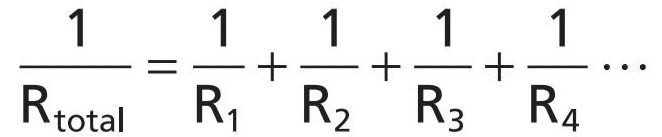
It is obvious that for a given pressure gradient, far greater amounts of blood will flow through this parallel system than through any of the individual blood vessels. Therefore, the total resistance is far less than the resistance of any single blood vessel. Flow through each of the parallel vessels in Figure 14-9B is determined by the pressure gradient and its own resistance, not the resistance of the other parallel blood vessels. However, increasing the resistance of any of the blood vessels increases the total vascular resistance.
بدیهی است که برای یک گرادیان فشار معین، مقادیر بسیار بیشتری از خون در این سیستم موازی نسبت به هر یک از رگهای خونی منفرد جریان مییابد. بنابراین، مقاومت کل بسیار کمتر از مقاومت هر رگ خونی است. جریان از طریق هر یک از رگهای موازی در شکل 14-9B توسط گرادیان فشار و مقاومت خود تعیین میشود، نه مقاومت رگهای خونی موازی دیگر. با این حال، افزایش مقاومت هر یک از رگهای خونی، مقاومت کلی عروق را افزایش میدهد.
It may seem paradoxical that adding more blood vessels to a circuit reduces the total vascular resistance. Many parallel blood vessels, however, make it easier for blood to flow through the circuit because each parallel vessel provides another pathway, or conductance, for blood flow. The total conductance (Ctotal) for blood flow is the sum of the conductance of each parallel pathway:
ممکن است متناقض به نظر برسد که افزودن رگهای خونی بیشتر به یک مدار باعث کاهش مقاومت کلی عروق میشود. با این حال، بسیاری از رگهای خونی موازی، جریان خون را در مدار آسانتر میکنند، زیرا هر رگ موازی مسیر یا رسانایی دیگری را برای جریان خون فراهم میکند. رسانایی کل (Ctotal) برای جریان خون مجموع رسانایی هر مسیر موازی است:

For example, brain, kidney, muscle, gastrointestinal, skin, and coronary circulations are arranged in parallel, and each tissue contributes to the overall conductance of the systemic circulation. Blood flow through each tissue is a fraction of the total blood flow (cardiac output) and is determined by the resistance (the reciprocal of conductance) for blood flow in the tissue, as well as the pressure gradient. Therefore, amputation of a limb or surgical removal of a kidney also removes a parallel circuit and reduces the total vascular conductance and total blood flow (i.e., cardiac output) while increasing the total peripheral vascular resistance.
به عنوان مثال، گردش خون مغز، کلیه، ماهیچه، دستگاه گوارش، پوست و کرونری به صورت موازی مرتب شده اند و هر بافت به هدایت کلی گردش خون سیستمیک کمک میکند. جریان خون در هر بافت کسری از کل جریان خون (برون ده قلبی) است و با مقاومت (رسانایی متقابل) برای جریان خون در بافت و همچنین گرادیان فشار تعیین میشود. بنابراین، قطع عضو یا برداشتن کلیه از طریق جراحی، یک مدار موازی را از بین میبرد و هدایت عروقی و جریان خون کل (یعنی برون ده قلبی) را کاهش میدهد و در عین حال مقاومت کلی عروق محیطی را افزایش میدهد.
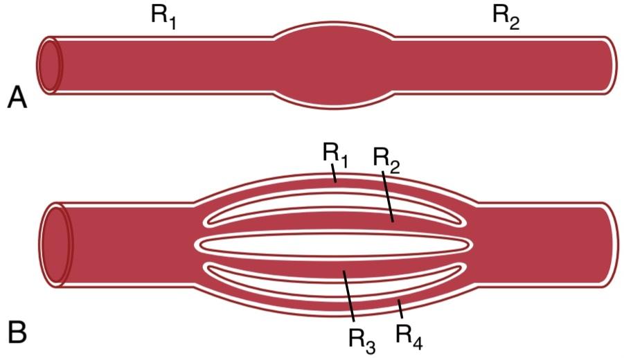
Figure 14-9. Vascular resistance (R). A, In series. B, In parallel.
شکل 14-9. مقاومت عروقی (R). الف، در سری. ب، به طور موازی
Effect of Blood Hematocrit and Blood Viscosity on Vascular Resistance and Blood Flow
Note that another important factor in Poiseuille’s equation is the viscosity of the blood. The greater the viscosity, the lower the flow in a vessel if all other factors are constant. Furthermore, the viscosity of normal blood is about three times as great as the viscosity of water.
تأثیر هماتوکریت و ویسکوزیته خون بر مقاومت عروقی و جریان خون
توجه داشته باشید که یکی دیگر از عوامل مهم در معادله Poiseuille ویسکوزیته خون است. هرچه ویسکوزیته بیشتر باشد، در صورتی که سایر عوامل ثابت باشند، جریان در یک ظرف کمتر است. علاوه بر این، ویسکوزیته خون طبیعی حدود سه برابر ویسکوزیته آب است.
What makes the blood so viscous? It is mainly the large numbers of suspended red cells in the blood, each of which exerts frictional drag against adjacent cells and against the wall of the blood vessel.
چه چیزی خون را اینقدر چسبناک میکند؟ این عمدتاً تعداد زیادی گلبول قرمز معلق در خون است که هر یک از آنها کشش اصطکاکی بر روی سلولهای مجاور و دیواره رگ خونی اعمال میکنند.
Hematocrit the Proportion of Blood That Is Red Blood Cells. If a person has a hematocrit of 40, this means that 40% of the blood volume is cells, and the remainder is plasma. The hematocrit of adult men aver- ages about 42, whereas that of women averages about 38. These values can vary greatly, depending on whether the person has anemia, the degree of bodily activity, and the altitude at which the person resides. These changes in hematocrit are discussed in relationship to the red blood cells and their oxygen transport function in Chapter 33.
هماتوکریت نسبت خون که گلبولهای قرمز است. اگر فردی دارای هماتوکریت 40 باشد، به این معنی است که 40 درصد از حجم خون را سلولها تشکیل میدهد و بقیه را پلاسما تشکیل میدهد. هماتوکریت مردان بالغ به طور متوسط حدود 42 سال است، در حالی که میانگین هماتوکریت در زنان حدود 38 است. این مقادیر بسته به اینکه فرد مبتلا به کم خونی است، درجه فعالیت بدنی و ارتفاعی که فرد در آن زندگی میکند میتواند بسیار متفاوت باشد. این تغییرات در هماتوکریت در رابطه با گلبولهای قرمز خون و عملکرد انتقال اکسیژن آنها در فصل 33 مورد بحث قرار گرفته است.
Hematocrit is determined by centrifuging blood in a calibrated tube, as shown in Figure 14-10. The calibration allows direct reading of the percentage of cells.
همانطور که در شکل 14-10 نشان داده شده است، هماتوکریت با سانتریفیوژ کردن خون در یک لوله مدرج تعیین میشود. کالیبراسیون امکان خواندن مستقیم درصد سلولها را فراهم میکند.

Figure 14-10. Hematocrit values in a healthy (normal) person and in patients with anemia and polycythemia. The numbers refer to the percentage of the blood composed of red blood cells.
شکل 14-10. مقادیر هماتوکریت در یک فرد سالم (طبیعی) و در بیماران مبتلا به کم خونی و پلی سیتمی. اعداد مربوط به درصد خونی است که از گلبولهای قرمز تشکیل شده است.
Increasing Hematocrit Markedly Increases Blood Viscosity. The viscosity of blood increases markedly as the hematocrit increases, as shown in Figure 14-11. The viscosity of whole blood at a normal hematocrit is about 3 to 4, which means that three to four times as much pressure is required to force whole blood as to force water through the same blood vessel. When the hematocrit rises to 60 or 70, which it often does in persons with polycythemia, the blood viscosity can become as great as 10 times that of water, and its flow through blood vessels is greatly retarded.
افزایش هماتوکریت به طور قابل توجهی ویسکوزیته خون را افزایش میدهد. همانطور که در شکل 14-11 نشان داده شده است، با افزایش هماتوکریت ویسکوزیته خون به طور قابل توجهی افزایش مییابد. ویسکوزیته خون کامل در یک هماتوکریت طبیعی حدود 3 تا 4 است، به این معنی که برای فشار دادن خون کامل به فشار 3 تا 4 برابر فشار لازم است تا آب وارد همان رگ شود. هنگامیکه هماتوکریت به 60 یا 70 میرسد، که اغلب در افراد مبتلا به پلی سیتمیانجام میشود، ویسکوزیته خون میتواند 10 برابر آب باشد و جریان آن از طریق رگهای خونی تا حد زیادی به تاخیر میافتد.
Other factors that affect blood viscosity are the plasma protein concentration and types of proteins in the plasma, but these effects are so much less than the effect of hematocrit that they are not significant considerations in most hemodynamic studies. The viscosity of blood plasma is about 1.5 times that of water.
عوامل دیگری که بر ویسکوزیته خون تأثیر میگذارند، غلظت پروتئین پلاسما و انواع پروتئینهای پلاسما است، اما این تأثیرات به قدری کمتر از تأثیر هماتوکریت است که در اکثر مطالعات همودینامیک مورد توجه قرار نمیگیرد. ویسکوزیته پلاسمای خون حدود 1.5 برابر آب است.
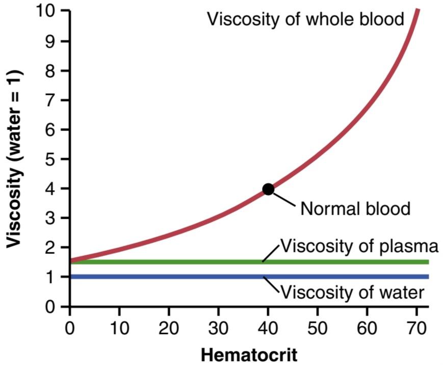
Figure 14-11. Effect of hematocrit on blood viscosity (water viscosity = 1).
شکل 14-11. اثر هماتوکریت بر ویسکوزیته خون (ویسکوزیته آب = 1).
EFFECTS OF PRESSURE ON VASCULAR RESISTANCE AND TISSUE BLOOD FLOW
اثرات فشار بر مقاومت عروقی و جریان خون بافت
Autoregulation Attenuates the Effect of Arterial Pressure on Tissue Blood Flow. From the discussion thus far, one might expect an increase in arterial pressure to cause a proportionate increase in blood flow through the body’s tissues. However, the effect of arterial pressure on blood flow in many tissues is usually far less than one might expect, as shown in Figure 14-12. This is because an increase in arterial pressure not only increases the force that pushes blood through the vessels, but also initiates compensatory increases in vascular resistance within a few seconds through activation of the local control mechanisms, discussed in Chapter 17. Conversely, with reductions in arterial pressure, vascular resistance is promptly reduced in most tissues, and blood flow is maintained at a relatively constant rate. The ability of each tissue to adjust its vascular resistance and to maintain normal blood flow during changes in arterial pressure between approximately 70 and 175 mm Hg is called blood flow autoregulation.
تنظیم خودکار اثر فشار شریانی بر جریان خون بافت را کاهش میدهد. از بحث تا کنون، میتوان انتظار داشت که افزایش فشار شریانی باعث افزایش متناسب در جریان خون در بافتهای بدن شود. با این حال، همانطور که در شکل 14-12 نشان داده شده است، تأثیر فشار شریانی بر جریان خون در بسیاری از بافتها معمولاً بسیار کمتر از آن چیزی است که انتظار میرود. این به این دلیل است که افزایش فشار شریانی نه تنها نیرویی را که خون را از طریق رگها رانده میکند، افزایش میدهد، بلکه باعث افزایش جبرانی مقاومت عروقی در عرض چند ثانیه از طریق فعالسازی مکانیسمهای کنترل موضعی، مورد بحث در فصل 17 میشود. برعکس، با کاهش فشار شریانی، مقاومت عروقی بهسرعت حفظ میشود و در اکثر بافتها سرعت نسبی خون ثابت میشود. توانایی هر بافت برای تنظیم مقاومت عروقی خود و حفظ جریان خون طبیعی در طول تغییرات فشار شریانی بین 70 تا 175 میلیمتر جیوه، خود تنظیم جریان خون نامیده میشود.
Note in Figure 14-12 that changes in blood flow can be caused by strong sympathetic stimulation, which constricts the blood vessels. Likewise, hormonal vasoconstrictors, such as norepinephrine, angiotensin II, vasopressin, or endothelin, can also reduce blood flow, at least transiently.
در شکل 14-12 توجه داشته باشید که تغییرات در جریان خون میتواند ناشی از تحریک شدید سمپاتیک باشد که رگهای خونی را منقبض میکند. به همین ترتیب، منقبض کنندههای عروقی هورمونی، مانند نوراپی نفرین، آنژیوتانسین II، وازوپرسین، یا اندوتلین نیز میتوانند جریان خون را حداقل به طور موقت کاهش دهند.
Blood flow changes rarely last for more than a few hours in most tissues, even when increases in arterial pressure or increased levels of vasoconstrictors are sustained. The reason for the relative constancy of blood flow is that each tissue’s local autoregulatory mechanisms eventually override most of the effects of vasoconstrictors to provide a blood flow that is appropriate for the needs of the tissue.
تغییرات جریان خون به ندرت در بیشتر بافتها بیش از چند ساعت طول میکشد، حتی زمانی که افزایش فشار شریانی یا افزایش سطوح منقبضکنندههای عروقی ادامه داشته باشد. دلیل ثبات نسبی جریان خون این است که مکانیسمهای خودتنظیمیموضعی هر بافت در نهایت بر بیشتر اثرات منقبضکنندههای عروقی غلبه میکنند تا جریان خونی مناسب برای نیازهای بافت فراهم کنند.

Figure 14-12. Effect of changes in arterial pressure over a period of several minutes on blood flow in a tissue such as skeletal muscle. Note that between pressures of 70 and 175 mm Hg, blood flow is autoregulated. The blue line shows the effect of sympathetic nerve stimulation or vasoconstriction by hormones such as norepinephrine, angiotensin II, vasopressin, or endothelin on this relationship. Reduced tissue blood flow is rarely maintained for more than a few hours because of the activation of local autoregulatory mechanisms that eventually return blood flow toward normal.
شکل 14-12. تأثیر تغییرات فشار شریانی در یک دوره چند دقیقه ای بر جریان خون در بافتی مانند عضله اسکلتی. توجه داشته باشید که بین فشارهای 70 تا 175 میلی متر جیوه، جریان خون به صورت خودکار تنظیم میشود. خط آبی اثر تحریک عصب سمپاتیک یا انقباض عروق توسط هورمونهایی مانند نوراپی نفرین، آنژیوتانسین II، وازوپرسین یا اندوتلین را بر این رابطه نشان میدهد. کاهش جریان خون بافتی به ندرت بیش از چند ساعت حفظ میشود زیرا مکانیسمهای خودتنظیمیمحلی فعال میشود که در نهایت جریان خون را به حالت عادی برمیگرداند.
Pressure-Flow Relationship in Passive Vascular Beds. In isolated blood vessels or in tissues that do not exhibit autoregulation, changes in arterial pressure may have important effects on blood flow. The effect of pressure on blood flow may be greater than that predicted by Poiseuille’s equation, as shown by the upward curving lines in Figure 14-13. The reason for this is that increased arterial pressure not only increases the force that pushes blood through the vessels, but also distends the elastic vessels, actually decreasing vascular resistance. Conversely, decreased arterial pressure in passive blood vessels increases resistance as the elastic vessels gradually collapse due to reduced distending pressure. When pressure falls below a critical level, called the critical closing pressure, flow ceases because the blood vessels are completely collapsed.
رابطه فشار-جریان در بسترهای عروقی غیر فعال. در رگهای خونی جدا شده یا در بافتهایی که خودتنظیمینشان نمیدهند، تغییرات فشار شریانی ممکن است اثرات مهمیبر جریان خون داشته باشد. همانطور که توسط خطوط انحنای رو به بالا در شکل 14-13 نشان داده شده است، تأثیر فشار بر جریان خون ممکن است بیشتر از آن چیزی باشد که معادله پویسوی پیش بینی میکند. دلیل این امر این است که افزایش فشار شریانی نه تنها نیرویی را افزایش میدهد که خون را از طریق رگها فشار میدهد، بلکه عروق کشسان را نیز متسع میکند و در واقع مقاومت عروقی را کاهش میدهد. برعکس، کاهش فشار شریانی در رگهای خونی غیرفعال، مقاومت را افزایش میدهد، زیرا رگهای الاستیک بهتدریج به دلیل کاهش فشار متسع میشوند. هنگامیکه فشار به زیر سطح بحرانی که فشار بسته شدن بحرانی نامیده میشود، کاهش مییابد، جریان متوقف میشود زیرا رگهای خونی کاملاً فرو میریزند.
Sympathetic stimulation and other vasoconstrictors can alter the passive pressure-flow relationship shown in Figure 14-13. Thus, inhibition of sympathetic activity greatly dilates the vessels and can increase the blood flow twofold or more. Conversely, very strong sympathetic stimulation can constrict the vessels so much that blood flow occasionally decreases to as low as zero for a few seconds, despite high arterial pressure.
تحریک سمپاتیک و سایر منقبض کنندههای عروقی میتوانند رابطه فشار-جریان غیرفعال نشان داده شده در شکل 14-13 را تغییر دهند. بنابراین، مهار فعالیت سمپاتیک رگها را تا حد زیادی گشاد میکند و میتواند جریان خون را دو برابر یا بیشتر افزایش دهد. برعکس، تحریک سمپاتیک بسیار قوی میتواند رگها را چنان منقبض کند که با وجود فشار شریانی بالا، جریان خون گهگاه برای چند ثانیه به صفر میرسد.
In reality, there are few physiological conditions in which tissues display the passive pressure-flow relationship shown in Figure 14-13. Even in tissues that do not effectively autoregulate blood flow during acute changes in arterial pressure, blood flow is regulated according to the needs of the tissue when the pressure changes are sustained, as discussed in Chapter 17.
در واقع، شرایط فیزیولوژیکی کمیوجود دارد که در آن بافتها رابطه فشار-جریان غیرفعال نشان داده شده در شکل 14-13 را نشان میدهند. حتی در بافتهایی که به طور موثری جریان خون را در طول تغییرات حاد فشار شریانی تنظیم نمیکنند، جریان خون بر اساس نیازهای بافت تنظیم میشود که تغییرات فشار ادامه یابد، همانطور که در فصل 17 بحث شد.
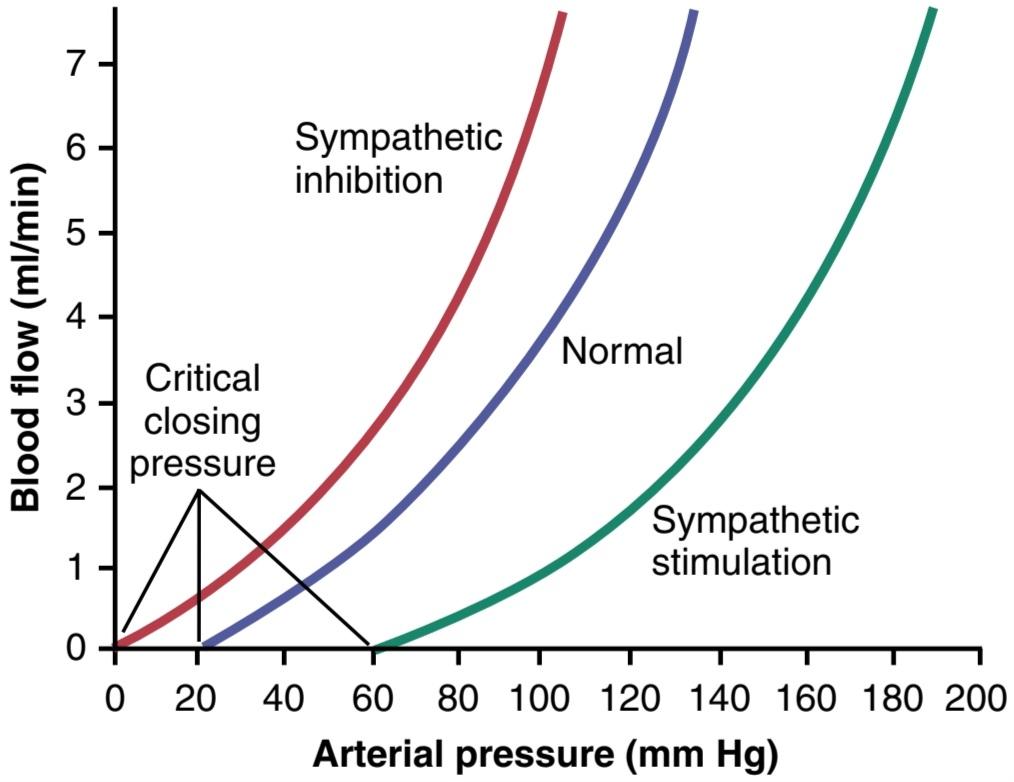
Figure 14-13. Effect of arterial pressure on blood flow through a passive blood vessel at different degrees of vascular tone caused by increased or decreased sympathetic stimulation of the vessel.
شکل 14-13. تأثیر فشار شریانی بر جریان خون از طریق یک رگ خونی غیرفعال در درجات مختلف تون عروق ناشی از افزایش یا کاهش تحریک سمپاتیک رگ.
Vascular Wall Tension. Tension on the blood vessel wall develops in response to transmural pressure gradients and causes vascular smooth muscle and endothelial cells to stretch in all directions (Figure 14-14A). According to the law of Laplace, wall tension (T) for a thin-walled tube is proportional to the transmural pressure gradient (AP) times the radius (r) of the blood vessel divided by its wall thickness (h):
کشش دیواره عروقی. کشش روی دیواره رگ خونی در پاسخ به گرادیانهای فشار transmural ایجاد میشود و باعث میشود عضلات صاف عروق و سلولهای اندوتلیال در همه جهات کشیده شوند (شکل 14-14A). طبق قانون لاپلاس، کشش دیواره (T) برای یک لوله با دیواره نازک، متناسب با گرادیان فشار بین دیواری (AP) برابر شعاع (r) رگ خونی تقسیم بر ضخامت دیواره آن (h) است:

Thus, larger blood vessels exposed to high pressures, such as the aorta, must have stronger walls to withstand higher levels of tension and are generally reinforced with fibrous bands of collagen. In contrast, capillaries have a much smaller radii and therefore are exposed to much lower wall tension, permitting them to withstand pressures as high as 65 to 70 mm Hg in some organs such as the kidneys. As discussed in Chapter 17, chronic changes in blood pressure lead to remodeling of blood vessels to accommodate the associated changes in wall tension.
بنابراین، رگهای خونی بزرگتری که در معرض فشارهای بالا قرار دارند، مانند آئورت، باید دیوارههای قویتری داشته باشند تا بتوانند سطوح بالاتری از کشش را تحمل کنند و عموماً با نوارهای فیبری کلاژن تقویت میشوند. در مقابل، مویرگها شعاع بسیار کوچکتری دارند و بنابراین در معرض کشش دیواره بسیار کمتری هستند و به آنها اجازه میدهد تا فشارهای بالای 65 تا 70 میلی متر جیوه را در برخی از اندامها مانند کلیهها تحمل کنند. همانطور که در فصل 17 مورد بحث قرار گرفت، تغییرات مزمن در فشار خون منجر به بازسازی عروق خونی برای تطبیق با تغییرات مرتبط در کشش دیواره میشود.
Vascular Shear Stress. As blood flows it creates a frictional force, or drag, on the endothelial cells lining the blood vessels (see Figure 14-14B). This force, called shear stress, is proportional to the flow velocity and viscosity of the blood, inversely related to the radius cubed, and generally is expressed in force/unit area (e.g., dynes/cm2). In clinical practice, there is no single commonly used method for measuring shear stress. However, despite its relatively low magnitude compared to contractile forces or wall stretch from blood pressure, shear stress is important in the development and adaptation of the vascular system to accommodate the blood flow requirements of the tissues.Endothelial cells contain multiple proteins that together serve as mechanical sensors and regulate signaling pathways that shape the vasculature during embryonic development and continue altering blood vessel morphology to optimize delivery of blood to tissues in adult life, as discussed further in Chapter 17.
استرس برشی عروقی. با جریان یافتن خون، نیروی اصطکاکی یا کشش بر روی سلولهای اندوتلیال پوشاننده رگهای خونی ایجاد میکند (شکل 14-14B را ببینید). این نیرو که تنش برشی نامیده میشود، متناسب با سرعت جریان و ویسکوزیته خون است و با شعاع مکعبی رابطه معکوس دارد و عموماً بر حسب نیرو/واحد مساحت بیان میشود (به عنوان مثال، dynes/cm2). در عمل بالینی، هیچ روش متداول واحدی برای اندازه گیری تنش برشی وجود ندارد. با این حال، علیرغم قدر نسبتاً کم آن در مقایسه با نیروهای انقباضی یا کشش دیوار ناشی از فشار خون، تنش برشی در توسعه و انطباق سیستم عروقی برای انطباق با جریان خون مورد نیاز بافتها مهم است. سلولهای اندوتلیال حاوی پروتئینهای متعددی هستند که با هم به عنوان حسگرهای مکانیکی عمل میکنند و در طول مسیر تکاملی سیگنالینگ خونی را تنظیم میکنند. مورفولوژی عروق برای بهینهسازی انتقال خون به بافتها در بزرگسالی، همانطور که در فصل 17 بیشتر بحث شد.

Figure 14-14. Illustration of the effects of vessel wall tension and shear stress on blood vessels. Wall tension develops in response to transmural pressure gradients and causes stretch of endothelial and vascular smooth muscle cells in all directions. Shear stress is the frictional force or drag on endothelial cells caused by flowing blood. Shear stress results in unidirectional endothelial cell deformation.
شکل 14-14. تصویری از اثرات کشش دیواره عروق و تنش برشی بر روی عروق خونی. کشش دیواره در پاسخ به گرادیانهای فشار بین دیواری ایجاد میشود و باعث کشیدگی سلولهای عضلات صاف اندوتلیال و عروق در همه جهات میشود. تنش برشی نیروی اصطکاکی یا کشش روی سلولهای اندوتلیال است که در اثر جریان خون ایجاد میشود. تنش برشی منجر به تغییر شکل سلولهای اندوتلیال یک طرفه میشود.
کلیک کنید «Bibliography: فهرست کتب مربوطه»
see the bibliography for chapter 15.
🚀 با ما همراه شوید!
تازهترین مطالب و آموزشهای مغز و اعصاب را از دست ندهید. با فالو کردن کانال تلگرام آیندهنگاران مغز، از ما حمایت کنید!


