
فیزیولوژی پزشکی گایتون و هال؛ آریتمیهای قلبی و تفسیر الکتروکاردیوگرافی آنها


دعای مطالعه [ نمایش ]
بِسْمِ الله الرَّحْمنِ الرَّحیمِ
اَللّهُمَّ اَخْرِجْنى مِنْ ظُلُماتِ الْوَهْمِ
خدايا مرا بيرون آور از تاريكىهاى وهم،
وَ اَكْرِمْنى بِنُورِ الْفَهْمِ
و به نور فهم گرامى ام بدار،
اَللّهُمَّ افْتَحْ عَلَيْنا اَبْوابَ رَحْمَتِكَ
خدايا درهاى رحمتت را به روى ما بگشا،
وَانْشُرْ عَلَيْنا خَزائِنَ عُلُومِكَ بِرَحْمَتِكَ يا اَرْحَمَ الرّاحِمينَ
و خزانههاى علومت را بر ما باز كن به امید رحمتت اى مهربانترين مهربانان.
کتاب «فیزیولوژی پزشکی گایتون وهال» بهعنوان یکی از جامعترین و معتبرترین منابع در حوزه علوم پزشکی، همچنان مرجع کلیدی برای درک عملکرد پیچیده بدن انسان است. این اثر با تکیه بر تازهترین پژوهشها و توضیحات دقیق از سازوکارهای فیزیولوژیک، پلی میان علوم پایه پزشکی و کاربردهای بالینی ایجاد میکند و نقشی بیبدیل در آموزش، پژوهش و ارتقای دانش سلامت ایفا مینماید.
ترجمه دقیق و علمیاین شاهکار توسط برند علمیآیندهنگاران مغز به مدیریت داریوش طاهری، دسترسی فارسیزبانان به مرزهای نوین دانش فیزیولوژی را ممکن ساخته و رسالتی علمیبرای ارتقای آموزش پزشکی، فهم عمیقتر سازوکارهای بدن و توسعه روشهای نوین در حوزه سلامت فراهم آورده است.
» کتاب فیزیولوژی پزشکی گایتون وهال
» » فصل ۱۳: آریتمیهای قلبی و تفسیر الکتروکاردیوگرافی آنها
در حال ویرایش
» Guyton and Hall Textbook of Medical Physiology
»» CHAPTER 13: Cardiac Arrhythmias and Their Electrocardiographic Interpretation
 Some of the most distressing types of heart malfunction occur because of abnormal rhythm of the heart. For example, sometimes the beat of the atria is not coordinated with the beat of the ventricles, so the atria no longer function to optimize ventricular filling.
برخی از ناراحت کننده ترین انواع نارسایی قلبی به دلیل ریتم غیر طبیعی قلب رخ میدهد. به عنوان مثال، گاهی اوقات ضربان دهلیزها با ضربان بطنها هماهنگ نیست، بنابراین دهلیزها دیگر برای بهینه سازی پر شدن بطن عمل نمیکنند.
The purpose of this chapter is to discuss the physiology of common cardiac arrhythmias and their effects on heart pumping, as well as their diagnosis by electrocardiography. The causes of the cardiac arrhythmias are usually one or a combination of the following abnormalities in the rhythmicity-conduction system of the heart:
هدف از این فصل بحث در مورد فیزیولوژی آریتمیهای شایع قلبی و اثرات آنها بر پمپاژ قلب و همچنین تشخیص آنها توسط الکتروکاردیوگرافی است. علل آریتمیهای قلبی معمولاً یکی یا ترکیبی از ناهنجاریهای زیر در سیستم هدایت ریتم قلب است:
• Abnormal rhythmicity of the pacemaker
• Shift of the pacemaker from the sinus node to an- other place in the heart
• Blocks at different points in the spread of the impulse through the heart
• Abnormal pathways of impulse transmission through the heart
• Spontaneous generation of spurious impulses in al- most any part of the heart
• ریتمیک غیر طبیعی پیس میکر
• جابجایی ضربان ساز از گره سینوسی به جای دیگری در قلب
• در نقاط مختلف انتشار تکانه از طریق قلب مسدود میشود
• مسیرهای غیر طبیعی انتقال تکانه از طریق قلب
• تولید خودبهخودی تکانههای کاذب تقریباً در هر قسمت از قلب
ABNORMAL SINUS RHYTHMS
ریتمهای سینوسی غیر طبیعی
TACHYCARDIA
The term tachycardia means fast heart rate, which usu- ally is defined as faster than 100 beats/min in an adult. An electrocardiogram (ECG) recorded from a patient with tachycardia is shown in Figure 13-1. This ECG is normal except that the heart rate, as determined from the time intervals between QRS complexes, is about 150 beats/ min instead of the normal 72 beats/min. Some causes of tachycardia include increased body temperature, dehydration, blood loss anemia, stimulation of the heart by the sympathetic nerves, and toxic conditions of the heart.
تاکی کاردی
اصطلاح تاکی کاردی به معنای ضربان قلب سریع است که معمولاً به صورت تندتر از 100 ضربه در دقیقه در بزرگسالان تعریف میشود. یک الکتروکاردیوگرام (ECG) ثبت شده از یک بیمار مبتلا به تاکی کاردی در شکل 13-1 نشان داده شده است. این ECG طبیعی است با این تفاوت که ضربان قلب، همانطور که از فواصل زمانی بین کمپلکسهای QRS تعیین میشود، به جای 72 ضربه در دقیقه طبیعی، حدود 150 ضربه در دقیقه است. برخی از علل تاکی کاردی عبارتند از افزایش دمای بدن، کم آبی، کم خونی از دست دادن خون، تحریک قلب توسط اعصاب سمپاتیک و شرایط سمیقلب.
The heart rate usually increases about 10 beats/min for each degree Fahrenheit increase in body temperature (with an increase of 18 beats/min for each degree Celsius), up to a body temperature of about 105°F (40.5°C). Beyond this, the heart rate may decrease because of progressive debility of the heart muscle as a result of the fever. Fever causes tachycardia because an increased temperature increases the rate of metabolism of the sinus node, which in turn directly increases its excitability and rate of rhythm.
ضربان قلب معمولاً به ازای هر درجه فارنهایت افزایش دمای بدن (با افزایش 18 ضربه در دقیقه برای هر درجه سانتیگراد)، تا دمای بدن حدود 105 درجه فارنهایت (40.5 درجه سانتیگراد) حدود 10 ضربه در دقیقه افزایش مییابد. فراتر از این، ضربان قلب ممکن است به دلیل ناتوانی پیشرونده عضله قلب در نتیجه تب کاهش یابد. تب باعث تاکی کاردی میشود زیرا افزایش دما باعث افزایش سرعت متابولیسم گره سینوسی میشود که به نوبه خود مستقیماً تحریک پذیری و سرعت ریتم آن را افزایش میدهد.
Many factors can cause the sympathetic nervous system to excite the heart, as discussed in this text. For example, when a patient sustains severe blood loss, sympathetic reflex stimulation of the heart may increase the heart rate to 150 to 180 beats/min. Simple weakening of the myocardium usually increases the heart rate because the weakened heart does not pump blood into the arterial tree to a normal extent, causing reductions in blood pressure and eliciting sympathetic reflexes to increase the heart rate.
عوامل زیادی میتوانند باعث تحریک سیستم عصبی سمپاتیک قلب شوند، همانطور که در این متن مورد بحث قرار گرفت. به عنوان مثال، هنگامیکه بیمار دچار از دست دادن شدید خون میشود، تحریک رفلکس سمپاتیک قلب ممکن است ضربان قلب را به 150 تا 180 ضربه در دقیقه افزایش دهد. تضعیف ساده میوکارد معمولا ضربان قلب را افزایش میدهد زیرا قلب ضعیف شده خون را به میزان طبیعی به درخت شریانی پمپ نمیکند و باعث کاهش فشار خون و ایجاد رفلکسهای سمپاتیک برای افزایش ضربان قلب میشود.
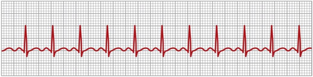
Figure 13-1. Sinus tachycardia (lead I).
شکل 13-1. تاکی کاردی سینوسی (لید I).
BRADYCARDIA
The term bradycardia means a slow heart rate, usually defined as fewer than 60 beats/min. Bradycardia is shown by the ECG in Figure 13-2.
برادی کاردی
اصطلاح برادی کاردی به معنای ضربان قلب آهسته است که معمولاً کمتر از 60 ضربه در دقیقه تعریف میشود. برادی کاردی توسط ECG در شکل 13-2 نشان داده شده است.
Bradycardia in Athletes. The well-trained athlete’s heart is often larger and considerably stronger than that of a nor- mal person, which allows the athlete’s heart to pump a large stroke volume output per beat, even during periods of rest. When the athlete is at rest, increased quantities of blood pumped into the arterial tree with each beat initiate feedback circulatory reflexes or other effects that cause bradycardia.
برادی کاردی در ورزشکاران. قلب ورزشکارانی که به خوبی تمرین کرده اند، اغلب بزرگتر و به طور قابل توجهی قویتر از قلب یک فرد عادی است، که به قلب ورزشکار اجازه میدهد تا حجم ضربه ای زیادی را در هر ضربان، حتی در دورههای استراحت، پمپاژ کند. هنگامیکه ورزشکار در حال استراحت است، مقادیر بیشتری از خون پمپاژ شده به درخت شریانی با هر ضربان باعث شروع بازخوردهای بازخوردی در گردش خون یا سایر اثراتی میشود که باعث برادی کاردی میشود.
Vagal Stimulation Causes Bradycardia. Any circulatory reflex that stimulates the vagus nerves causes release of acetylcholine at the vagal endings in the heart, resulting in a parasympathetic effect. Perhaps the most striking ex- ample of this phenomenon occurs in patients with carotid sinus syndrome. In these patients, the pressure receptors (baroreceptors) in the carotid sinus region of the carotid artery walls are excessively sensitive. Therefore, even mild external pressure on the neck elicits a strong baroreceptor reflex, causing intense vagal-acetylcholine effects on the heart, including extreme bradycardia. Sometimes this reflex is so powerful that it actually stops the heart for 5 to 10 seconds, leading to loss of consciousness (syncope).
تحریک واگ باعث برادی کاردی میشود. هر رفلکس گردش خون که اعصاب واگ را تحریک میکند باعث آزاد شدن استیل کولین در انتهای واگ در قلب میشود و در نتیجه اثر پاراسمپاتیک ایجاد میشود. شاید بارزترین مثال از این پدیده در بیماران مبتلا به سندرم سینوس کاروتید رخ دهد. در این بیماران، گیرندههای فشار (بارورسپتورها) در ناحیه سینوس کاروتید دیواره شریان کاروتید بیش از حد حساس هستند. بنابراین، حتی فشار خارجی خفیف بر روی گردن، یک رفلکس بارورسپتور قوی ایجاد میکند، که باعث اثرات شدید واگ-استیل کولین بر قلب، از جمله برادی کاردی شدید میشود. گاهی اوقات این رفلکس آنقدر قوی است که در واقع قلب را برای 5 تا 10 ثانیه متوقف میکند و منجر به از دست دادن هوشیاری (سنکوپ) میشود.

Figure 13-2. Sinus bradycardia (lead III).
شکل 13-2. برادی کاردی سینوسی (لید III).
SINUS ARRHYTHMIA
Figure 13-3 shows a cardiotachometer recording of the heart rate, at first during normal respiration and then, in the second half of the record, during deep respiration. A cardiotachometer is an instrument that records the duration of the interval between the successive QRS complexes in the ECG by the height of successive spikes. Note from this record that the heart rate increased and decreased no more than 5% during quiet respiration (shown on the left half of the record). Then, during deep respiration, the heart rate increased and decreased with each respiratory cycle by as much as 30%.
آریتمیسینوسی
شکل 13-3 ضبط ضربان قلب توسط کاردیوتاکومتر را در ابتدا در طول تنفس طبیعی و سپس در نیمه دوم ثبت در طی تنفس عمیق نشان میدهد. کاردیوتاکومتر ابزاری است که مدت زمان فاصله بین کمپلکسهای QRS متوالی در ECG را با ارتفاع اسپکهای متوالی ثبت میکند. از این رکورد توجه داشته باشید که ضربان قلب در طول تنفس آرام بیش از 5٪ افزایش و کاهش نمییابد (نشان داده شده در نیمه سمت چپ رکورد). سپس، در طول تنفس عمیق، ضربان قلب با هر چرخه تنفسی تا 30٪ افزایش و کاهش مییابد.
Sinus arrhythmia can result from any one of many circulatory conditions that alter the strengths of the sympathetic and parasympathetic nerve signals to the heart sinus node. The respiratory type of sinus arrhythmia results mainly from the spillover of signals from the medullary respiratory center into the adjacent vasomotor center during inspiratory and expiratory cycles of respiration. The spillover signals cause alternate increases and decreases in the number of impulses transmitted through the sympathetic and vagus nerves to the heart.
آریتمیسینوسی میتواند ناشی از هر یک از شرایط گردش خون باشد که قدرت سیگنالهای عصبی سمپاتیک و پاراسمپاتیک را به گره سینوسی قلب تغییر میدهد. نوع تنفسی آریتمیسینوسی عمدتاً از سرریز سیگنالها از مرکز تنفسی مدولاری به مرکز وازوموتور مجاور در طی چرخههای دمیو بازدمیتنفس ناشی میشود. سیگنالهای سرریز باعث افزایش و کاهش متناوب تعداد تکانههای منتقل شده از طریق اعصاب سمپاتیک و واگ به قلب میشوند.

Figure 13-3. Sinus arrhythmia as recorded by a cardiotachometer.To the left is the record when the subject was breathing normally; to the right, when the subject was breathing deeply.
شکل 13-3. آریتمیسینوسی که توسط یک کاردیوتاکومتر ثبت شده است. در سمت چپ رکورد زمانی است که سوژه به طور طبیعی نفس میکشید. سمت راست، زمانی که سوژه عمیقاً نفس میکشید.
HEART BLOCK WITHIN THE INTRACARDIAC CONDUCTION PATHWAYS
بلوک قلب در مسیرهای هدایت داخل قلب
SINOATRIAL BLOCK
In rare cases, the impulse from the sinus node is blocked before it enters the atrial muscle. This phenomenon is demonstrated in Figure 13-4, which shows sudden cessation of P waves, with resultant standstill of the atria. However, the ventricles pick up a new rhythm, with the impulse usually originating spontaneously in the atrioventricular (A-V) node, so the rate of the ventricular QRS-T complex is slowed but not otherwise altered. Sinoatrial block can be due to myocardial ischemia affecting the sinus node, inflammation or infection of the heart, or side effects from certain medications, and it can be observed in well-trained athletes.
بلوک سینوسی
در موارد نادر، تکانه از گره سینوسی قبل از ورود به عضله دهلیزی مسدود میشود. این پدیده در شکل 13-4 نشان داده شده است، که توقف ناگهانی امواج P را نشان میدهد و در نتیجه دهلیزها متوقف میشوند. با این حال، بطنها ریتم جدیدی میگیرند، با تکانه معمولاً خود به خود در گره دهلیزی بطنی (A-V) منشا میگیرد، بنابراین سرعت کمپلکس QRS-T بطنی کند میشود، اما تغییری در آن ایجاد نمیشود. بلوک سینوسی دهلیزی میتواند به دلیل ایسکمیمیوکارد که بر غده سینوسی تأثیر میگذارد، التهاب یا عفونت قلب یا عوارض جانبی برخی داروها باشد و در ورزشکارانی که به خوبی تمرین کرده اند دیده میشود.

Figure 13-4. Sinoatrial (SA) nodal block, with atrioventricular nodal rhythm during the block period (lead III).
شکل 13-4. بلوک گره سینوسی دهلیزی (SA)، با ریتم گره دهلیزی بطنی در طول دوره بلوک (لید III).
ATRIOVENTRICULAR BLOCK
The only means whereby impulses ordinarily can pass from the atria into the ventricles is through the A-V bundle, also known as the bundle of His. Conditions that can either decrease the rate of impulse conduction in this bundle or block the impulse entirely are as follows:
بلوک دهلیزی بطنی
تنها وسیله ای که از طریق آن تکانهها معمولاً میتوانند از دهلیزها به بطنها منتقل شوند، از طریق بسته A-V است که به عنوان بسته His نیز شناخته میشود. شرایطی که میتواند سرعت هدایت ضربه را در این بسته کاهش دهد یا ضربه را به طور کامل مسدود کند به شرح زیر است:
1. Ischemia of the A-V node or A-V bundle fibers of- ten delays or blocks conduction from the atria to the ventricles. Coronary insufficiency can cause ischemia of the A-V node and bundle in the same way that it can cause ischemia of the myocardium.
1. ایسکمیگره A-V یا فیبرهای دسته A-V اغلب هدایت را از دهلیزها به بطنها به تاخیر میاندازد یا مسدود میکند. نارسایی عروق کرونر میتواند باعث ایسکمیگره و بسته A-V شود، همان طور که میتواند باعث ایسکمیمیوکارد شود.
2. Compression of the A-V bundle by scar tissue or by calcified portions of the heart can depress or block conduction from the atria to the ventricles.
2. فشردگی بسته A-V توسط بافت اسکار یا بخشهای کلسیفیه قلب میتواند باعث کاهش یا انسداد هدایت از دهلیزها به بطنها شود.
3. Inflammation of the A-V node or A-V bundle can depress conduction from the atria to the ventricles. Inflammation results frequently from different types of myocarditis that are caused, for example, by diphtheria or rheumatic fever.
3. التهاب گره A-V یا دسته A-V میتواند هدایت را از دهلیزها به بطنها کاهش دهد. التهاب اغلب ناشی از انواع مختلف میوکاردیت است که به عنوان مثال در اثر دیفتری یا تب روماتیسمیایجاد میشود.
4. Extreme stimulation of the heart by the vagus nerves in rare cases blocks impulse conduction through the A-V node. Such vagal excitation occasionally results from strong stimulation of the baroreceptors in people with carotid sinus syndrome, discussed earlier in relationship to bradycardia.
4. تحريك شديد قلب توسط اعصاب واگ در موارد نادري هدايت ضربه از طريق گره A-V را مسدود مي كند. چنین برانگیختگی واگ گاهی از تحریک قوی گیرندههای فشاری در افراد مبتلا به سندرم سینوس کاروتید ناشی میشود که قبلاً در رابطه با برادی کاردی بحث شد.
5. Degeneration of the A-V conduction system, which is sometimes seen in older patients.
5. دژنراسیون سیستم هدایت A-V که گاهی در بیماران مسن دیده میشود.
6. Medications such as digitalis or beta-adrenergic antagonists can, in some cases, impair A-V conduction.
6. داروهایی مانند دیژیتال یا آنتاگونیستهای بتا آدرنرژیک، در برخی موارد میتوانند هدایت A-V را مختل کنند.
INCOMPLETE ATRIOVENTRICULAR BLOCK
بلوک دهلیزی بطنی ناقص
First-Degree Block-Prolonged P-R Interval. The usual lapse of time between the beginning of the P wave and the beginning of the QRS complex is about 0.16 second when the heart is beating at a normal rate. This so-called P-R interval usually decreases in length with a faster heartbeat and increases with a slower heartbeat. In general, when the P-R interval increases to more than 0.20 second, the P-R interval is said to be prolonged, and the patient is said to have first-degree incomplete heart block.
بازه P-R طولانی مدت بلوک درجه یک. فاصله زمانی معمول بین شروع موج P و شروع کمپلکس QRS حدود 0.16 ثانیه است که قلب با سرعت طبیعی میتپد. این فاصله P-R که اصطلاحاً P-R نامیده میشود، معمولاً با ضربان قلب سریعتر از طول کاهش مییابد و با ضربان قلب آهستهتر افزایش مییابد. به طور کلی، زمانی که فاصله P-R به بیش از 0.20 ثانیه افزایش مییابد، گفته میشود که فاصله P-R طولانی شده است و گفته میشود که بیمار دارای بلوک قلبی ناقص درجه یک است.
Figure 13-5 shows an ECG with a prolonged P-R interval; the interval in this case is about 0.30 second instead of the normal 0.20 second or less. Thus, first-degree block is defined as a delay of conduction from the atria to the ventricles but not actual blockage of conduction. The P-R interval seldom increases above 0.35 to 0.45 second because, by that time, conduction through the A-V bundle is depressed so much that conduction stops entirely. One means for determining the severity of some heart diseases, such as acute rheumatic heart disease, for example, is to measure the P-R interval.
شکل 13-5 یک ECG را با فاصله P-R طولانی نشان میدهد. فاصله در این مورد حدود 0.30 ثانیه به جای 0.20 ثانیه معمولی یا کمتر است. بنابراین، بلوک درجه یک به عنوان تاخیر در رسانش از دهلیزها به بطنها تعریف میشود اما انسداد واقعی هدایت نیست. فاصله P-R به ندرت از 0.35 تا 0.45 ثانیه افزایش مییابد، زیرا در آن زمان، هدایت از طریق بسته A-V به قدری کاهش مییابد که هدایت به طور کامل متوقف میشود. یکی از ابزارهای تعیین شدت برخی از بیماریهای قلبی، مانند بیماری روماتیسمیحاد قلبی، برای مثال، اندازه گیری فاصله P-R است.

Figure 13-5. Prolonged P-R interval caused by first-degree atrioventricular heart block (lead II).
شکل 13-5. طولانی شدن فاصله P-R ناشی از بلوک قلب دهلیزی بطنی درجه یک (لید II).
Second-Degree Block. When conduction through the A-V bundle is slowed enough to increase the P-R interval to 0.25 to 0.45 second, the action potential is sometimes strong enough to pass through the bundle into the ventricles and sometimes not strong enough to do so. In this case, there will be an atrial P wave but no QRS-T wave, and it is said that there are “dropped beats” of the ventricles. This condition is called second-degree heart block.
بلوک درجه دو. هنگامیکه رسانش از طریق بسته A-V به اندازه ای کند میشود که فاصله P-R را به 0.25 تا 0.45 ثانیه افزایش میدهد، پتانسیل عمل گاهی به اندازه کافی قوی است که از طریق بسته نرم افزاری به داخل بطنها عبور کند و گاهی اوقات برای انجام این کار به اندازه کافی قوی نیست. در این حالت، یک موج P دهلیزی وجود خواهد داشت اما موج QRS-T وجود ندارد و گفته میشود که “ضربههای افت” بطنها وجود دارد. به این حالت بلوک قلبی درجه دو میگویند.
There are two types of second-degree A-V blockMobitz type I (also known as Wenckebach periodicity) and Mobitz type II. Type I block is characterized by progressive prolongation of the P-R interval until a ventricular beat is dropped and is then followed by resetting of the P-R interval and repeating of the abnormal cycle. A type I block is almost always caused by abnormality of the A-V node. In most cases, this type of block is benign, and no specific treatment is needed.
دو نوع بلوک A-V درجه دوم وجود دارد – موبیتز نوع I (همچنین به عنوان دوره تناوب ونک باخ شناخته میشود) و موبیتز نوع II. بلوک نوع I با طولانی شدن پیشرونده فاصله P-R تا زمانی که ضربان بطنی کاهش یابد مشخص میشود و سپس با تنظیم مجدد فاصله P-R و تکرار چرخه غیر طبیعی دنبال میشود. بلوک نوع I تقریباً همیشه به دلیل غیر طبیعی بودن گره A-V ایجاد میشود. در بیشتر موارد، این نوع بلوک خوش خیم است و نیازی به درمان خاصی نیست.
In type II block, there is usually a fixed number of non- conducted P waves for every QRS complex. For example, a 2:1 block implies that there are two P waves for every QRS complex. At other times, rhythms of 3:2 or 3:1 may develop. In contrast to type I block, with type II block the P-R interval does not change before the dropped beat; it remains fixed. Type II block is generally caused by an abnormality of the bundle of His-Purkinje system and may require implantation of a pacemaker to prevent progression to complete heart block and cardiac arrest.
در بلوک نوع II، معمولاً تعداد ثابتی از امواج P غیر رسانا برای هر کمپلکس QRS وجود دارد. به عنوان مثال، یک بلوک 2:1 نشان میدهد که دو موج P برای هر کمپلکس QRS وجود دارد. در مواقع دیگر، ریتمهای 3:2 یا 3:1 ممکن است ایجاد شود. برخلاف بلوک نوع I، با بلوک نوع II فاصله P-R قبل از ضربان افت شده تغییر نمیکند. ثابت میماند بلوک نوع II عموماً به دلیل ناهنجاری در بسته سیستم His-Purkinje ایجاد میشود و ممکن است نیاز به کاشت ضربان ساز برای جلوگیری از پیشرفت به بلوک کامل قلب و ایست قلبی داشته باشد.
Figure 13-6 shows progressive P-R interval prolongation typical of type I (Wenckebach) block. Note prolongation of the P-R interval preceding the dropped beat, followed by a shortened P-R interval after the dropped beat.
شکل 13-6 افزایش تدریجی فاصله P-R را نشان میدهد که نمونه بلوک نوع I (ونک باخ) است. به طولانی شدن فاصله P-R قبل از ضربان افت شده، و به دنبال آن یک فاصله P-R کوتاه شده بعد از ضربان افت شده توجه کنید.
Complete A-V Block (Third-Degree Block). When the condition causing poor conduction in the A-V node or A-V bundle becomes severe, complete block of the impulse from the atria into the ventricles occurs. In this case, the ventricles spontaneously establish their own signal, usually originating in the A-V node or A-V bundle distal to the block. Therefore, the P waves become dissociated from the QRS-T complexes, as shown in Figure 13-7. Note that the rate of rhythm of the atria in this ECG is about 100 beats/min, whereas the rate of ventricular beat is less than 40 beats/min. Furthermore, there is no relationship between the rhythm of the P waves and that of the QRS- T complexes because the ventricles have “escaped” from control by the atria and are beating at their own natural rate, controlled most often by rhythmical signals generated distal to the A-V node or A-V bundle where the block occurs.
بلوک A-V کامل (بلوک درجه سوم). هنگامیکه وضعیتی که باعث هدایت ضعیف در گره A-V یا بسته نرم افزاری A-V میشود، شدید میشود، بلوک کامل تکانه از دهلیزها به بطنها رخ میدهد. در این مورد، بطنها به طور خود به خود سیگنال خود را ایجاد میکنند، که معمولاً از گره A-V یا بسته نرم افزاری A-V در انتهای بلوک منشا میگیرد. بنابراین، همانطور که در شکل 13-7 نشان داده شده است، امواج P از کمپلکسهای QRS-T جدا میشوند. توجه داشته باشید که سرعت ریتم دهلیزها در این ECG حدود 100 ضربه در دقیقه است، در حالی که میزان ضربان بطنی کمتر از 40 ضربه در دقیقه است. علاوه بر این، هیچ رابطهای بین ریتم امواج P و کمپلکسهای QRS-T وجود ندارد، زیرا بطنها توسط دهلیزها از کنترل خارج شدهاند و با سرعت طبیعی خود میکوبند، که اغلب توسط سیگنالهای ریتمیک تولید شده در دیستال از گره A-V یا دسته A-V که در آن بلوک ایجاد میشود، کنترل میشود.
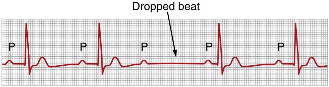
Figure 13-6. Type I second-degree atrioventricular block showing progressive P-R prolongation prior to the dropped beat.
شکل 13-6. بلوک دهلیزی بطنی درجه دوم نوع I که نشان دهنده طولانی شدن پیشرونده P-R قبل از ضربان افت شده است.
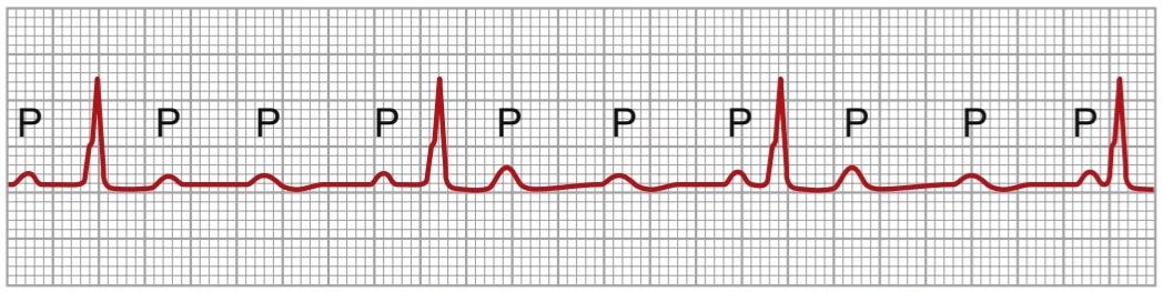
Figure 13-7. Complete atrioventricular block (lead II).
شکل 13-7. بلوک دهلیزی بطنی کامل (لید II).
Stokes-Adams Syndrome-Ventricular Escape. In some patients with A-V block, the total block comes and goes; that is, impulses are conducted from the atria into the ventricles for a period of time and then, suddenly, impulses are not conducted. The duration of block may be a few seconds, a few minutes, a few hours, or even weeks or longer before conduction returns. This condition occurs in hearts with borderline ischemia of the conductive system.
سندرم استوکس-آدامز-فرار بطنی. در برخی از بیماران با بلوک A-V، بلوک کل میآید و میرود. یعنی تکانهها برای مدتی از دهلیزها به داخل بطنها هدایت میشوند و سپس ناگهان تکانهها انجام نمیشود. مدت زمان بلوک ممکن است چند ثانیه، چند دقیقه، چند ساعت یا حتی هفتهها یا بیشتر قبل از بازگشت هدایت باشد. این وضعیت در قلبهای مبتلا به ایسکمیمرزی سیستم رسانا رخ میدهد.
Each time A-V conduction ceases, the ventricles often do not start their own beating until after a delay of 5 to 30 seconds. This delay results from the phenomenon called overdrive suppression. Overdrive suppression means that ventricular excitability is at first suppressed because the ventricles have been driven by the atria at a rate greater than their natural rate of rhythm. However, after a few seconds, some part of the Purkinje system beyond the block, usually in the distal part of the A-V node beyond the blocked point in the node, or in the A-V bundle, begins discharging rhythmically at a rate of 15 to 40 times/min, acting as the pacemaker of the ventricles. This phenomenon is called ventricular escape.
هر بار که رسانش A-V متوقف میشود، بطنها اغلب تا پس از تأخیر 5 تا 30 ثانیه ای شروع به ضربان خود نمیکنند. این تاخیر ناشی از پدیده ای به نام سرکوب بیش از حد است. سرکوب Overdrive به این معنی است که تحریک پذیری بطن در ابتدا سرکوب میشود زیرا بطنها توسط دهلیزها با سرعتی بیشتر از نرخ طبیعی ریتم خود هدایت میشوند. با این حال، پس از چند ثانیه، بخشی از سیستم پورکنژ فراتر از بلوک، معمولاً در قسمت انتهایی گره A-V فراتر از نقطه مسدود شده در گره، یا در بسته A-V، شروع به تخلیه ریتمیک با سرعت 15 تا 40 بار در دقیقه میکند و به عنوان ضربان ساز بطنها عمل میکند. این پدیده فرار بطنی نامیده میشود.
Because the brain cannot remain active for more than 4 to 7 seconds without blood supply, most people faint a few seconds after complete block occurs because the heart does not pump any blood for 5 to 30 seconds, until the ventricles “escape.” After escape, however, the slowly beating ventricles (typically beating less than 40 beats/ min) usually pump enough blood to allow rapid recovery from the faint and then to sustain the person. These periodic fainting spells (syncope) are known as the Stokes- Adams syndrome.
از آنجایی که مغز نمیتواند بیش از 4 تا 7 ثانیه بدون تامین خون فعال بماند، اکثر افراد چند ثانیه پس از بلوک کامل غش میکنند زیرا قلب به مدت 5 تا 30 ثانیه هیچ خونی را پمپ نمیکند تا زمانی که بطنها “فرار” کنند. با این حال، پس از فرار، بطنهای آهسته ضربان (معمولاً کمتر از 40 ضربه در دقیقه) معمولاً خون کافی پمپاژ میکنند تا امکان بهبودی سریع از غش را فراهم کند و سپس فرد را نگه دارد. این غشهای دوره ای (سنکوپ) به عنوان سندرم استوکس-آدامز شناخته میشود.
Occasionally, the interval of ventricular standstill at the onset of complete block is so long that it becomes detrimental to the patient’s health or even causes death. Consequently, most of these patients are provided with an artificial pacemaker, a small battery-operated electrical stimulator planted beneath the skin, with electrodes usually connected to the right ventricle. The pacemaker provides continued rhythmical impulses to the ventricles.
گاهی اوقات، فاصله توقف بطن در شروع بلوک کامل آنقدر طولانی است که برای سلامتی بیمار مضر است یا حتی باعث مرگ میشود. در نتیجه، برای اکثر این بیماران یک ضربانساز مصنوعی، یک محرک الکتریکی کوچک که با باتری کار میکند، در زیر پوست، با الکترودهایی که معمولاً به بطن راست متصل هستند، ارائه میشوند. پیس میکر تکانههای ریتمیک مداوم را به بطنها میدهد.

Figure 13-8. Partial intraventricular block-electrical alternans (lead I).
شکل 13-8. آلترنانسهای الکتریکی درون بطنی جزئی (لید I).
INCOMPLETE INTRAVENTRICULAR BLOCK-ELECTRICAL ALTERNANS
Most of the same factors that can cause A-V block can also block impulse conduction in the peripheral ventricular Pur- kinje system. Figure 13-8 shows the condition known as electrical alternans, which results from partial intraventricular block every other heartbeat. This ECG also shows tachycardia (rapid heart rate), which is probably the reason the block has occurred. This is because when the rate of the heart is rapid, it may be impossible for some portions of the Pur- kinje system to recover from the previous refractory period quickly enough to respond during every succeeding heart- beat. Also, many conditions that depress the heart, such as ischemia, myocarditis, or digitalis toxicity, can cause incomplete intraventricular block, resulting in electrical alternans.
جایگزینهای الکتریکی داخل بطنی ناقص
بسیاری از عوامل مشابهی که میتوانند باعث بلوک A-V شوند، میتوانند هدایت ضربه را در سیستم پورکنژ بطن محیطی نیز مسدود کنند. شکل 13-8 وضعیتی را نشان میدهد که به عنوان جایگزینهای الکتریکی شناخته میشود، که از انسداد جزئی داخل بطنی هر ضربان قلب دیگر ناشی میشود. این ECG همچنین تاکی کاردی (ضربان قلب سریع) را نشان میدهد که احتمالاً دلیل بروز بلوک است. این به این دلیل است که وقتی ضربان قلب سریع است، ممکن است برای برخی از بخشهای سیستم پورکنژ غیرممکن باشد که دوره نسوز قبلی را بهاندازه کافی سریع بازیابی کنند تا در هر ضربان قلب بعدی پاسخ دهند. همچنین، بسیاری از بیماریهایی که قلب را تحت فشار قرار میدهند، مانند ایسکمی، میوکاردیت، یا مسمومیت با دیژیتال، میتوانند باعث بلوک داخل بطنی ناقص و در نتیجه آلترنانسهای الکتریکی شوند.
PREMATURE CONTRACTIONS
A premature contraction is a contraction of the heart before the time that normal contraction would have been expected. This condition is also called extrasystole, premature beat, or ectopic beat.
انقباضات زودرس
انقباض زودرس انقباض قلب قبل از زمانی است که انتظار میرود انقباض طبیعی باشد. به این وضعیت اکستراسیستول، ضربان زودرس یا ضربان نابجا نیز گفته میشود.
CAUSES OF PREMATURE CONTRACTIONS
Most premature contractions result from ectopic foci in the heart, which emit abnormal impulses at odd times during the cardiac rhythm. Possible causes of ectopic foci are as follows: (1) local areas of ischemia; (2) small calcified plaques at different points in the heart, which press against the adjacent cardiac muscle so that some of the fibers are irritated; and (3) toxic irritation of the A-V node, Purkinje system, or myocardium caused by infection, drugs, nicotine, or caffeine. The mechanical initiation of premature contractions is also frequent during cardiac catheterization; large numbers of premature contractions often occur when the catheter enters the ventricle and presses against the endocardium.
علل انقباضات زودرس
اکثر انقباضات زودرس ناشی از کانونهای نابجا در قلب هستند که در زمانهای عجیب و غریب در طول ریتم قلبی، تکانههای غیرطبیعی از خود ساطع میکنند. علل احتمالی کانونهای نابجا به شرح زیر است: (1) مناطق محلی ایسکمی. (2) پلاکهای کوچک کلسیفیه در نقاط مختلف قلب، که به عضله قلب مجاور فشار میآورند تا برخی از رشتهها تحریک شوند. و (3) تحریک سمیگره A-V، سیستم پورکنژ، یا میوکارد ناشی از عفونت، داروها، نیکوتین یا کافئین. شروع مکانیکی انقباضات زودرس نیز در طی کاتتریزاسیون قلبی مکرر است. تعداد زیادی از انقباضات زودرس اغلب زمانی رخ میدهد که کاتتر وارد بطن میشود و به اندوکارد فشار میآورد.
PREMATURE ATRIAL CONTRACTIONS
Figure 13-9 shows a single premature atrial contraction (PAC). The P wave of this beat occurred too soon in the heart cycle; the P-R interval is shortened, indicating that the ectopic origin of the beat is in the atria near the A-V node. Also, the interval between the premature contraction and the next succeeding contraction is slightly prolonged, which is called a compensatory pause. One of the reasons for this compensatory pause is that the premature contraction originated in the atrium some distance from the sinus node, and the impulse had to travel through a considerable amount of atrial muscle before it discharged the sinus node. Consequently, the sinus node discharged late in the premature cycle, which made the succeeding sinus node discharge also late in appearing.
انقباضات زودرس دهلیزی
شکل 13-9 یک انقباض زودرس دهلیزی (PAC) را نشان میدهد. موج P این ضربان خیلی زود در چرخه قلب اتفاق افتاد. فاصله P-R کوتاه شده است، که نشان میدهد منشاء نابجای ضربان در دهلیزهای نزدیک گره A-V است. همچنین فاصله بین انقباض زودرس و انقباض بعدی کمیطولانی میشود که به آن مکث جبرانی میگویند. یکی از دلایل این مکث جبرانی این است که انقباض زودرس از دهلیز در فاصله ای از گره سینوسی منشأ میگیرد و تکانه باید قبل از تخلیه گره سینوسی از مقدار قابل توجهی از عضله دهلیزی عبور کند. در نتیجه، گره سینوسی دیرتر در سیکل نارس تخلیه شد، که باعث شد ترشحات سینوسی بعدی نیز دیر ظاهر شود.
PACs occur frequently in otherwise healthy people. They often occur in athletes whose hearts are in a very healthy condition. Mild toxic conditions resulting from such factors as smoking, lack of sleep, ingestion of too much coffee, alcoholism, and use of various drugs can also initiate such contractions.
PACs اغلب در افراد سالم رخ میدهد. آنها اغلب در ورزشکارانی که قلبشان در وضعیت بسیار سالمیاست رخ میدهد. شرایط سمیخفیف ناشی از عواملی مانند سیگار کشیدن، کم خوابی، مصرف بیش از حد قهوه، اعتیاد به الکل و استفاده از داروهای مختلف نیز میتواند شروع کننده چنین انقباضاتی باشد.
Pulse Deficit. When the heart contracts ahead of schedule, the ventricles will not have filled with blood normally, and the stroke volume output during that contraction is depressed or is almost absent. Therefore, the pulse wave passing to the peripheral arteries after a premature contraction may be so weak that it cannot be felt in the radial artery. Thus, a deficit in the number of radial pulses occurs when compared with the actual number of contractions of the heart.
کمبود نبض. وقتی قلب زودتر از موعد مقرر منقبض میشود، بطنها به طور طبیعی با خون پر نمیشوند و خروجی حجم ضربه ای در طول آن انقباض کاهش مییابد یا تقریباً وجود ندارد. بنابراین، موج پالسی که پس از یک انقباض زودرس به شریانهای محیطی میرود، ممکن است آنقدر ضعیف باشد که در شریان رادیال احساس نشود. بنابراین، در مقایسه با تعداد واقعی انقباضات قلب، کسری در تعداد پالسهای شعاعی رخ میدهد.

Figure 13-9. Atrial premature beat (lead I).
شکل 13-9. ضربان زودرس دهلیزی (لید I).
A-V NODAL OR A-V BUNDLE PREMATURE CONTRACTIONS
Figure 13-10 shows a premature contraction that originated in the A-V node or A-V bundle. The P wave is missing from the electrocardiographic record of the premature contraction. Instead, the P wave is superimposed onto the QRS-T complex because the cardiac impulse traveled backward into the atria at the same time that it traveled forward into the ventricles. This P wave slightly distorts the QRS-T complex, but the P wave itself can- not be discerned as such. In general, A-V nodal premature contractions have the same significance and causes as atrial premature contractions.
انقباضات زودرس گره ای یا A-V باندل
شکل 13-10 یک انقباض زودرس را نشان میدهد که از گره A-V یا بسته نرم افزاری A-V منشأ میگیرد. موج P در رکورد الکتروکاردیوگرافی انقباض زودرس وجود ندارد. در عوض، موج P بر روی کمپلکس QRS-T قرار میگیرد، زیرا تکانههای قلبی همزمان با حرکت به سمت جلو به داخل بطنها به سمت عقب به دهلیزها حرکت میکنند. این موج P کمیکمپلکس QRS-T را تحریف میکند، اما خود موج P را نمیتوان به این صورت تشخیص داد. به طور کلی، انقباضات زودرس گرهی A-V دارای اهمیت و علل مشابه با انقباضات زودرس دهلیزی هستند.

Figure 13-10. Atrioventricular nodal premature contraction (lead III).
شکل 13-10. انقباض زودرس گره دهلیزی بطنی (لید III).
PREMATURE VENTRICULAR CONTRACTIONS
The ECG in Figure 13-11 shows a series of premature ventricular contractions (PVCs) alternating with normal contractions in a pattern known as bigeminy. PVCs cause specific effects in the ECG, as follows:
انقباضات بطنی زودرس
ECG در شکل 13-11 مجموعه ای از انقباضات بطنی زودرس (PVCs) را نشان میدهد که متناوب با انقباضات طبیعی در الگویی به نام bigeminy شناخته میشوند. پی وی سیها اثرات خاصی در ECG ایجاد میکنند که عبارتند از:
1. The QRS complex is usually considerably prolonged. The reason for this prolongation is that the impulse is conducted mainly through slowly conducting muscle of the ventricles rather than through the Purkinje system.
1. کمپلکس QRS معمولاً به طور قابل توجهی طولانی است. دلیل این طولانی شدن این است که تکانه عمدتاً از طریق عضله آهسته هدایت کننده بطنها انجام میشود تا از طریق سیستم پورکنژ.
2. The QRS complex has a high voltage. When the normal impulse passes through the heart, it passes through both ventricles nearly simultaneously. Consequently, in the normal heart, the depolarization waves of the two sides of the heart-mainly of opposite polarity to each other-partially neutralize. each other in the ECG. When a PVC occurs, the impulse almost always travels in only one direction, so there is no such neutralization effect, and one entire side or end of the ventricles is depolarized ahead of the other, which causes large electrical potentials, as shown for the PVCs in Figure 13-11.
2. مجتمع QRS دارای ولتاژ بالا است. هنگامیکه تکانه طبیعی از قلب عبور میکند، تقریباً به طور همزمان از هر دو بطن عبور میکند. در نتیجه، در قلب طبیعی، امواج دپلاریزاسیون دو طرف قلب – عمدتاً دارای قطب مخالف یکدیگر – تا حدی خنثی میشوند. یکدیگر در ECG هنگامیکه یک پی وی سی رخ میدهد، تکانه تقریباً همیشه فقط در یک جهت حرکت میکند، بنابراین چنین اثر خنثی سازی وجود ندارد و کل یک طرف یا انتهای بطنها جلوتر از دیگری دپلاریزه میشود که باعث پتانسیلهای الکتریکی بزرگ میشود، همانطور که برای PVCها در شکل 13-11 نشان داده شده است.
3. After almost all PVCs, the T wave has an electrical potential polarity exactly opposite to that of the QRS complex because the slow conduction of the impulse through the cardiac muscle causes the muscle fibers that depolarize first also to repolarize first.
3. تقریباً بعد از تمام PVCها، موج T دارای قطبیت پتانسیل الکتریکی دقیقاً مخالف با کمپلکس QRS است، زیرا رسانش آهسته تکانه از طریق عضله قلب باعث میشود تا فیبرهای عضلانی که ابتدا دپلاریزه میشوند نیز ابتدا مجدداً قطبی شوند.
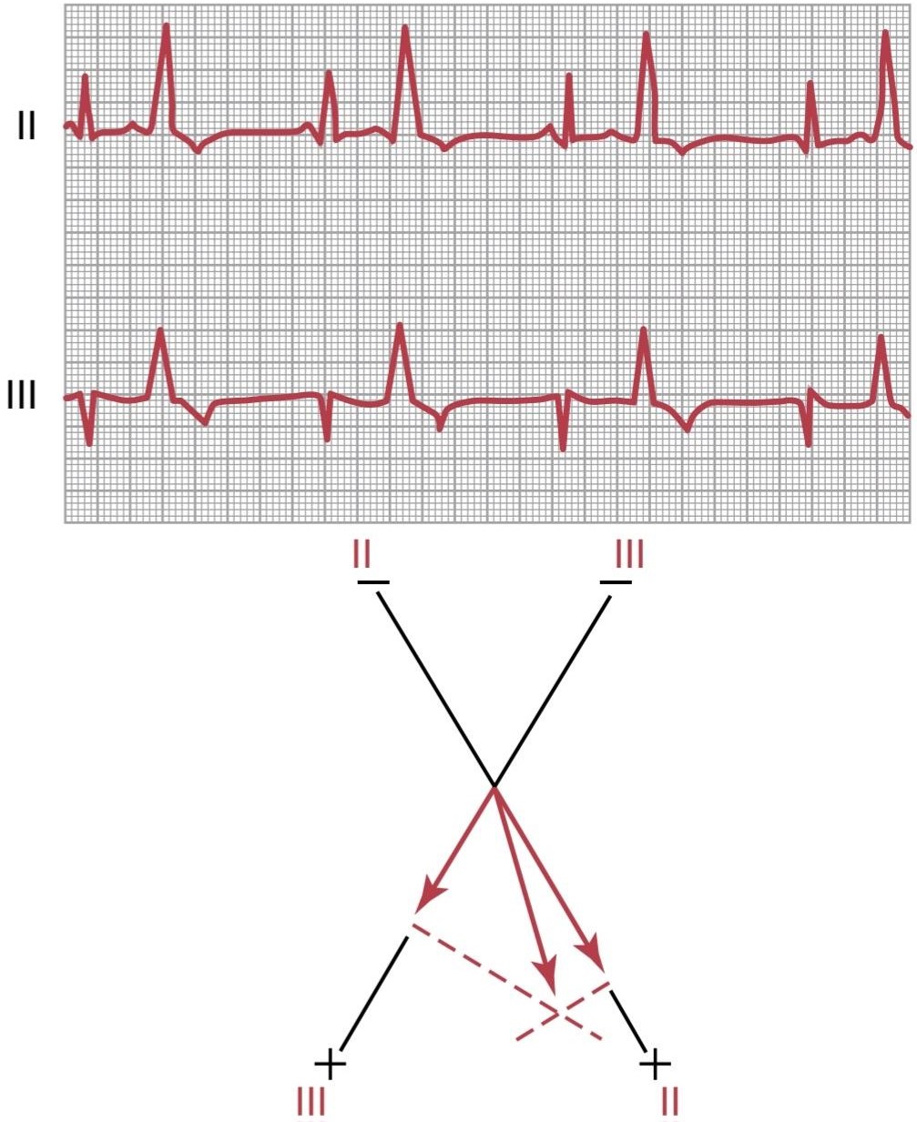
Figure 13-11. Premature ventricular contractions (PVCs) demonstrated by the large abnormal QRS-T complexes (leads II and III). The axis of the premature contractions is plotted in accordance with the principles of vectorial analysis explained in Chapter 12 and shows the origin of the PVC to be near the base of the ventricles.
شکل 13-11. انقباضات زودرس بطنی (PVCs) که توسط کمپلکسهای غیرطبیعی QRS-T (سرنخ II و III) نشان داده میشود. محور انقباضات زودرس مطابق با اصول آنالیز برداری توضیح داده شده در فصل 12 ترسیم شده است و نشان میدهد که منشاء PVC نزدیک پایه بطنها است.
Some PVCs are relatively benign in their effects on overall pumping by the heart; they can result from such factors as cigarettes, excessive intake of coffee, lack of sleep, various mild toxic states, and even emotional irritability. Conversely, many other PVCs result from stray impulses or re-entrant signals that originate around the borders of infarcted or ischemic areas of the heart. The presence of such PVCs is not to be taken lightly. People with significant numbers of PVCs often have a much higher than normal risk of developing spontaneous lethal ventricular fibrillation, presumably initiated by one of the PVCs. This development is especially true when the PVCs occur during the vulnerable period for causing fibrillation, just at the end of the T wave, when the ventricles are coming out of refractoriness, as explained later in this chapter.
برخی از پی وی سیها در تأثیراتشان بر پمپاژ کلی قلب نسبتاً خوش خیم هستند. آنها میتوانند ناشی از عواملی مانند سیگار، مصرف بیش از حد قهوه، کمبود خواب، حالتهای مختلف سمیخفیف و حتی تحریک پذیری عاطفی باشند. برعکس، بسیاری از PVCهای دیگر از تکانههای سرگردان یا سیگنالهای بازگشتی ناشی میشوند که در اطراف مرزهای نواحی انفارکتوس یا ایسکمیک قلب منشا میگیرند. وجود چنین پی وی سیهایی را نباید به سادگی گرفت. افرادی که تعداد قابل توجهی PVC دارند، اغلب در معرض خطر بسیار بالاتر از حد معمول ابتلا به فیبریلاسیون بطنی کشنده خود به خودی هستند که احتمالاً توسط یکی از PVCها آغاز شده است. این پیشرفت به ویژه زمانی صادق است که PVCها در طول دوره آسیب پذیر برای ایجاد فیبریلاسیون، درست در انتهای موج T، زمانی که بطنها از حالت نسوز خارج میشوند، رخ میدهد، همانطور که در ادامه این فصل توضیح داده شد.
Vector Analysis of the Origin of an Ectopic Premature Ventricular Contraction. In Chapter 12, the principles of vectorial analysis are explained. By applying these principles, one can determine from the ECG in Figure 13-11 the point of origin of the PVC, as follows. Note that the potentials of the premature contractions in leads II and III are both strongly positive. On plotting these potentials on the axes of leads II and III and solving by vectorial analysis for the mean QRS vector in the heart, one finds that the vector of this premature contraction has its negative end (origin) at the base of the heart and its positive end toward the apex. Thus, the first portion of the heart to become depolarized during this premature contraction is near the base of the ventricles, which therefore is the origin of the ectopic focus.
تجزیه و تحلیل برداری منشأ یک انقباض زودرس بطنی خارج از رحم. در فصل 12 اصول تحلیل برداری توضیح داده شده است. با اعمال این اصول، میتوان از ECG در شکل 13-11، نقطه مبدا PVC را به شرح زیر تعیین کرد. توجه داشته باشید که پتانسیل انقباضات زودرس در لیدهای II و III هر دو به شدت مثبت هستند. با ترسیم این پتانسیلها روی محورهای لیدهای II و III و حل با تجزیه و تحلیل برداری برای میانگین بردار QRS در قلب، متوجه میشوید که بردار این انقباض زودرس انتهای (منشا) منفی آن در قاعده قلب و انتهای مثبت آن به سمت راس است. بنابراین، اولین قسمتی از قلب که در طول این انقباض زودرس دپلاریزه میشود، در نزدیکی قاعده بطنها است، که بنابراین منشأ کانون خارج از رحم است.
Disorders of Cardiac Repolarization-the Long QT Syndromes. Recall that the Q wave corresponds to ventricular depolarization, whereas the T wave corresponds to ventricular repolarization. The Q-T interval is the time from the Q point to the end of the T wave. Disorders that delay repolarization of ventricular muscle after the action potential cause prolonged ventricular action potentials and therefore excessively long Q-T intervals on the ECG, a condition called long QT syndrome (LQTS).
اختلالات رپلاریزاسیون قلبی – سندرم QT طولانی. به یاد بیاورید که موج Q مربوط به دپلاریزاسیون بطنی است، در حالی که موج T مربوط به رپلاریزاسیون بطنی است. فاصله Q-T زمان از نقطه Q تا پایان موج T است. اختلالاتی که پس از پتانسیل عمل، رپولاریزاسیون عضله بطنی را به تاخیر میاندازد، باعث طولانی شدن پتانسیلهای عمل بطنی و بنابراین فواصل Q-T بیش از حد طولانی در ECG میشود، وضعیتی که به آن سندرم QT طولانی (LQTS) میگویند.
The major reason that LQTS is of concern is that delayed repolarization of ventricular muscle increases a person’s susceptibility to developing ventricular arrhythmias called torsades de pointes, which literally means “twisting of the points.” This type of arrhythmia has the features shown in Figure 13-12. The shape of the QRS complex may change over time, with the onset of arrhythmia usually following a premature beat, a pause, and then another beat with a long Q-T interval, which may trigger arrhythmias, tachycardia and, in some cases, ventricular fibrillation.
دلیل اصلی نگرانی LQTS این است که رپولاریزاسیون تاخیری عضله بطنی استعداد فرد را برای ایجاد آریتمیهای بطنی به نام torsades de pointes افزایش میدهد که به معنای واقعی کلمه به معنای “پیچش نقاط” است. این نوع آریتمیدارای ویژگیهایی است که در شکل 13-12 نشان داده شده است. شکل کمپلکس QRS ممکن است در طول زمان تغییر کند، با شروع آریتمیمعمولاً به دنبال یک ضربان زودرس، یک مکث، و سپس ضربان دیگری با فاصله Q-T طولانی، که ممکن است باعث آریتمی، تاکی کاردی و در برخی موارد فیبریلاسیون بطنی شود.
Disorders of cardiac repolarization that lead to LQTS may be inherited or acquired. The congenital forms of LQTS are rare disorders caused by mutations of sodium or potassium channel genes. At least 17 different mutations of these genes causing variable degrees of Q-T prolongation have been identified.
اختلالات رپلاریزاسیون قلبی که منجر به LQTS میشود ممکن است ارثی یا اکتسابی باشد. اشکال مادرزادی LQTS اختلالات نادری هستند که در اثر جهش ژنهای کانال سدیم یا پتاسیم ایجاد میشوند. حداقل 17 جهش مختلف از این ژنها که باعث درجات متغیر طولانی شدن Q-T میشوند، شناسایی شده است.
More common are the acquired forms of LQTS that are associated with plasma electrolyte disturbances, such as hypomagnesemia, hypokalemia, or hypocalcemia, or with the administration of excess amounts of antiarrhythmic drugs such as quinidine or some antibiotics such as fluoroquinolones or erythromycin, which prolong the Q-T interval.
اشکال اکتسابی LQTS که با اختلالات الکترولیت پلاسما همراه است، مانند هیپومنیزیمی، هیپوکالمییا هیپوکلسمی، یا با تجویز مقادیر بیش از حد داروهای ضد آریتمیمانند کینیدین یا برخی از آنتی بیوتیکها مانند فلوروکینولونها یا اریترومایسین که باعث طولانی شدن Q-T میشوند، شایع تر است.
Although some people with LQTS exhibit no major symptoms (other than the prolonged Q-T interval), other people exhibit fainting and experience ventricular arrhythmias that may be precipitated by physical exercise, intense emotions such as fright or anger, or being startled by a noise. The ventricular arrhythmias associated with LQTS can, in some cases, deteriorate into ventricular fibrillation and sudden death.
اگرچه برخی از افراد مبتلا به LQTS هیچ علامت عمده ای نشان نمیدهند (غیر از فاصله طولانی Q-T)، برخی دیگر از افراد غش میکنند و آریتمیهای بطنی را تجربه میکنند که ممکن است با ورزش بدنی، احساسات شدید مانند ترس یا عصبانیت، یا مبهوت شدن در اثر صدا ایجاد شود. آریتمیهای بطنی مرتبط با LQTS میتواند در برخی موارد به فیبریلاسیون بطنی و مرگ ناگهانی بدتر شود.
Treatment may include magnesium sulfate for acute LQTS and antiarrhythmic medications such as beta- adrenergic blockers or surgical implantation of a cardiac defibrillator for long-term LQTS.
درمان ممکن است شامل سولفات منیزیم برای LQTS حاد و داروهای ضد آریتمیمانند مسدود کنندههای بتا آدرنرژیک یا کاشت دفیبریلاتور قلبی برای طولانی مدت LQTS باشد.

Figure 13-12. Development of arrhythmias in long QT syndrome (LQTS). When the ventricular muscle fiber action potential is prolonged as a result of delayed repolarization, a premature depolarization (dashed line in top left figure) may occur before complete repolarization. Repetitive premature depolarizations (top right figure) may lead to multiple depolarizations under certain conditions. In torsades de pointes (bottom figure), premature ventricular beats lead pauses, postpause prolongation of the Q-T interval, and arrhythmias. (Modified from Murray KT, Roden DM:Disorders of cardiac repolarization: the long QT syndromes. In: Crawford MG, DiMarco JP (eds): Cardiology. London: Mosby, 2001.)
شکل 13-12. ایجاد آریتمیدر سندرم QT طولانی (LQTS). هنگامیکه پتانسیل عمل فیبر عضلانی بطنی در نتیجه رپلاریزاسیون تاخیری طولانی شود، دپلاریزاسیون زودرس (خط چین در شکل بالا سمت چپ) ممکن است قبل از رپلاریزاسیون کامل رخ دهد. دپلاریزاسیونهای زودرس مکرر (شکل بالا سمت راست) ممکن است تحت شرایط خاصی منجر به دپلاریزاسیونهای متعدد شود. در torsades de pointes (شکل پایین)، ضربانهای زودرس بطنی، مکثهای لید، طولانی شدن فاصله Q-T پس از مکث و آریتمیها. (اصلاح شده از Murray KT، Roden DM: Disorders of cardiac repolarization: the long QT syndromes. In: Crawford MG, DiMarco JP (eds): Cardiology. لندن: Mosby، 2001.)
PAROXYSMAL TACHYCARDIA
Some abnormalities in different portions of the heart, including the atria, Purkinje system, or ventricles, can occasionally cause rapid rhythmical discharge of impulses that spread in all directions throughout the heart. This phenomenon is believed to be caused most frequently by re-entrant circus movement feedback pathways that set up local repeated self-re-excitation. Because of the rapid rhythm in the irritable focus, this focus becomes the pace- maker of the heart.
تاکی کاردی پاروکسیزمال
برخی از ناهنجاریها در بخشهای مختلف قلب، از جمله دهلیز، سیستم پورکنژ، یا بطنها، گاهی اوقات میتوانند باعث تخلیه ریتمیک سریع تکانهها شوند که در تمام جهات در سراسر قلب پخش میشوند. اعتقاد بر این است که این پدیده اغلب توسط مسیرهای بازخورد حرکت سیرک که مجدداً وارد میشوند ایجاد میشود که تحریک مجدد محلی مکرر را ایجاد میکند. به دلیل ریتم سریع در کانون تحریک پذیر، این تمرکز به ضربان ساز قلب تبدیل میشود.
The term paroxysmal means that the heart rate becomes rapid in paroxysms, with the paroxysm beginning suddenly and lasting for a few seconds, a few minutes, a few hours, or much longer. The paroxysm usually ends as suddenly as it began, with the pacemaker of the heart instantly shifting back to the sinus node.
اصطلاح پراکسیسمال به این معنی است که ضربان قلب در پاروکسیسم سریع میشود، به طوری که حمله ناگهانی شروع میشود و برای چند ثانیه، چند دقیقه، چند ساعت یا خیلی بیشتر طول میکشد. پاراکسیسم معمولاً به همان اندازه که شروع شد ناگهانی به پایان میرسد و ضربان ساز قلب فوراً به گره سینوسی برمیگردد.
Paroxysmal tachycardia often can be stopped by eliciting a vagal reflex. A type of vagal reflex sometimes elicited for this purpose is to press on the neck in the regions of the carotid sinuses, which may cause enough of a vagal reflex to stop the paroxysm. Antiarrhythmic drugs may also be used to slow conduction or prolong the refractory period in cardiac tissues.
تاکی کاردی حمله ای اغلب میتواند با ایجاد رفلکس واگ متوقف شود. یک نوع رفلکس واگ که گاهی اوقات برای این منظور ایجاد میشود فشار دادن روی گردن در نواحی سینوسهای کاروتید است که ممکن است باعث شود که رفلکس واگ به اندازه کافی برای متوقف کردن حمله قلبی ایجاد شود. داروهای ضد آریتمینیز ممکن است برای کند کردن رسانش یا طولانی کردن دوره مقاوم سازی در بافتهای قلبی استفاده شوند.
PAROXYSMAL ATRIAL TACHYCARDIA
Figure 13-13 demonstrates a sudden increase in the heart rate from about 95 to about 150 beats/min in the middle of the record. On close study of the ECG, an inverted P wave is seen during the rapid heartbeat before each QRS-T complex, and this P wave is partially superimposed onto the normal T wave of the preceding beat. This finding indicates that the origin of this paroxysmal tachycardia is in the atrium but, because the P wave is abnormal in shape, the origin is not near the sinus node.
تاکی کاردی دهلیزی پاروکسیسمال
شکل 13-13 افزایش ناگهانی ضربان قلب را از حدود 95 به حدود 150 ضربه در دقیقه در میانه رکورد نشان میدهد. در مطالعه دقیق ECG، یک موج P معکوس در طول ضربان قلب سریع قبل از هر کمپلکس QRS-T مشاهده میشود، و این موج P تا حدی بر روی موج T طبیعی ضربان قبلی قرار میگیرد. این یافته نشان میدهد که منشأ این تاکی کاردی حملهای در دهلیز است، اما به دلیل غیرعادی بودن شکل موج P، منشاء نزدیک گره سینوسی نیست.

Figure 13-13. Paroxysmal atrial tachycardia-onset in the middle of the record (lead I).
شکل 13-13. تاکی کاردی دهلیزی حمله ای – شروع در وسط رکورد (لید I).
A-V Nodal Paroxysmal Tachycardia. Paroxysmal tachy- cardia often results from an aberrant rhythm involving the A-V node that usually causes almost normal QRS-T complexes but totally missing or obscured P waves.
تاکی کاردی پاروکسیسمال گرهی A-V. تاکی کاردی حمله ای اغلب ناشی از یک ریتم نابجای گره A-V است که معمولاً باعث ایجاد کمپلکسهای QRS-T تقریباً طبیعی میشود اما امواج P کاملاً از بین رفته یا مبهم است.
Atrial or A-V nodal paroxysmal tachycardia, both of which are referred to as supraventricular tachycardias, usually occur in young, otherwise healthy people, and they generally grow out of the predisposition to tachycardia after adolescence. In general, supraventricular tachycardia frightens a person tremendously and may cause weakness during the paroxysm, but it usually does not cause permanent harm from the attack.
تاکی کاردی پاروکسیسمال دهلیزی یا گرهی A-V، که هر دو به عنوان تاکی کاردی فوق بطنی شناخته میشوند، معمولاً در افراد جوان و در غیر این صورت سالم رخ میدهد و معمولاً بعد از نوجوانی از استعداد ابتلا به تاکی کاردی خارج میشوند. به طور کلی، تاکی کاردی فوق بطنی فرد را به شدت میترساند و ممکن است در حین حمله قلبی باعث ضعف شود، اما معمولاً آسیب دائمیاز حمله وارد نمیکند.
VENTRICULAR TACHYCARDIA
Figure 13-14 shows a typical short paroxysm of ventricular tachycardia. The ECG of ventricular tachycardia has the appearance of a series of ventricular premature beats occurring one after another, without any normal beats interspersed.
تاکی کاردی بطنی
شکل 13-14 یک حمله کوتاه معمولی تاکی کاردی بطنی را نشان میدهد. ECG تاکی کاردی بطنی به شکل یک سری ضربان زودرس بطنی ظاهر میشود که یکی پس از دیگری رخ میدهد، بدون اینکه هیچ ضربان طبیعی در هم قرار گیرد.
Ventricular tachycardia is usually a serious condition for two reasons. First, this type of tachycardia usually does not occur unless considerable ischemic damage is present in the ventricles. Second, ventricular tachycardia frequently initiates the lethal condition of ventricular fibrillation because of rapid repeated stimulation of the ventricular muscle, as discussed in the next section.
تاکی کاردی بطنی معمولاً به دو دلیل یک بیماری جدی است. اولاً، این نوع تاکی کاردی معمولاً رخ نمیدهد مگر اینکه آسیب ایسکمیک قابل توجهی در بطنها وجود داشته باشد. دوم، تاکی کاردی بطنی به دلیل تحریک مکرر سریع عضله بطنی، همانطور که در بخش بعدی بحث شد، اغلب شرایط کشنده فیبریلاسیون بطنی را آغاز میکند.
Sometimes, intoxication from the heart failure treatment drug digitalis causes irritable foci that lead to ventricular tachycardia. Antiarrhythmic drugs such as amiodarone or lidocaine can be used to treat ventricular tachycardia. Lidocaine depresses the normal increase in sodium permeability of the cardiac muscle membrane during generation of the action potential, thereby often blocking the rhythmical discharge of the focal point that has been causing the paroxysmal attack. Amiodarone has multiple actions, such as prolonging the action potential and refractory period in cardiac muscle and slowing A-V conduction. In some cases, cardioversion with an electric shock to the heart is needed for restoration of normal heart rhythm.
گاهی اوقات مسمومیت ناشی از داروی دیژیتالیس برای درمان نارسایی قلبی باعث ایجاد کانونهای تحریک پذیر میشود که منجر به تاکی کاردی بطنی میشود. برای درمان تاکی کاردی بطنی میتوان از داروهای ضد آریتمیمانند آمیودارون یا لیدوکائین استفاده کرد. لیدوکائین افزایش طبیعی نفوذپذیری سدیم غشای عضله قلب را در طول تولید پتانسیل عمل کاهش میدهد، در نتیجه اغلب ترشحات ریتمیک نقطه کانونی را که باعث حمله حمله شده مسدود میکند. آمیودارون اثرات متعددی دارد، مانند طولانی کردن پتانسیل عمل و دوره نسوز در عضله قلب و کند کردن هدایت A-V. در برخی موارد، کاردیوورژن همراه با شوک الکتریکی به قلب برای بازگرداندن ریتم طبیعی قلب مورد نیاز است.
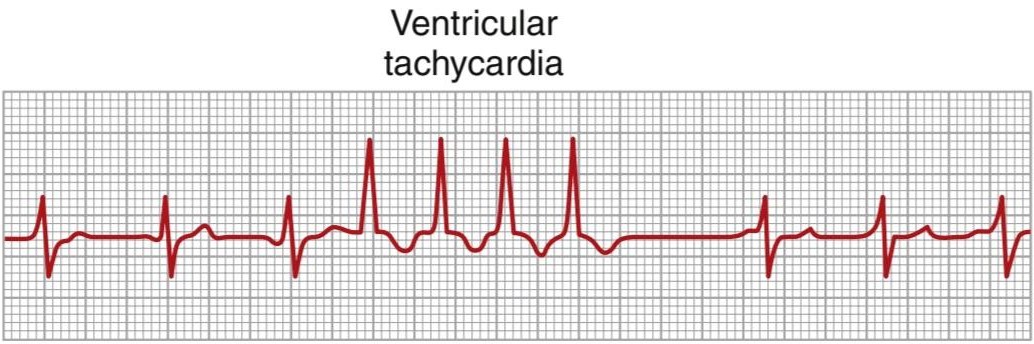
Figure 13-14. Ventricular paroxysmal tachycardia (lead III).
شکل 13-14. تاکی کاردی حمله ای بطنی (لید III).
VENTRICULAR FIBRILLATION
The most serious of all cardiac arrhythmias is ventricular fibrillation, which, if not stopped within 1 to 3 minutes, is almost invariably fatal. Ventricular fibrillation results from cardiac impulses that have gone berserk within the ventricular muscle mass, stimulating first one portion of the ventricular muscle, then another portion, then another, and eventually feeding back onto itself to re- excite the same ventricular muscle over and over, never stopping. When this phenomenon occurs, many small portions of the ventricular muscle will be contracting at the same time, while equally as many other portions will be relaxing. Thus, there is never a coordinated contraction of all the ventricular muscle at once, which is required for a pumping cycle of the heart. Despite massive movement of stimulatory signals throughout the ventricles, the ventricular chambers neither enlarge nor contract but remain in an indeterminate stage of partial contraction, pumping either no blood or negligible amounts. Therefore, after fibrillation begins, unconsciousness occurs within 4 to 5 seconds because of lack of blood flow to the brain, and irretrievable death of tissues begins to occur throughout the body within a few minutes.
فیبریلاسیون بطنی
جدی ترین آریتمیقلبی، فیبریلاسیون بطنی است که اگر در عرض 1 تا 3 دقیقه متوقف نشود، تقریباً همیشه کشنده است. فیبریلاسیون بطنی ناشی از تکانههای قلبی است که در داخل توده عضلانی بطنی از بین رفتهاند و ابتدا بخشی از عضله بطنی را تحریک میکنند، سپس بخشی دیگر و سپس قسمتی دیگر را تحریک میکنند و در نهایت به خود باز میگردند تا همان عضله بطنی را بارها و بارها تحریک کند و هرگز متوقف نشود. هنگامیکه این پدیده رخ میدهد، بسیاری از بخشهای کوچک عضله بطنی به طور همزمان منقبض میشوند، در حالی که به همان اندازه بخشهای دیگر آرام میشوند. بنابراین، هرگز یک انقباض هماهنگ تمام عضله بطنی که برای چرخه پمپاژ قلب لازم است وجود ندارد. علیرغم حرکت عظیم سیگنالهای تحریکی در سراسر بطنها، حفرههای بطنی نه بزرگ میشوند و نه منقبض میشوند، بلکه در مرحله نامشخصی از انقباض جزئی باقی میمانند و یا خونی را پمپاژ نمیکنند یا مقادیر ناچیزی را پمپاژ میکنند. بنابراین، پس از شروع فیبریلاسیون، بیهوشی در عرض 4 تا 5 ثانیه به دلیل عدم جریان خون به مغز رخ میدهد و مرگ غیرقابل برگشت بافتها در عرض چند دقیقه در سراسر بدن شروع میشود.
Multiple factors can spark the beginning of ventricular fibrillation; a person may have a normal heartbeat one moment, but 1 second later, the ventricles are in fibrillation. Especially likely to initiate fibrillation are sudden electrical shock of the heart, ischemia of the heart muscle, or ischemia of the specialized conducting system.
عوامل متعددی میتوانند جرقه شروع فیبریلاسیون بطنی را ایجاد کنند. یک فرد ممکن است یک لحظه ضربان قلب طبیعی داشته باشد، اما 1 ثانیه بعد، بطنها در فیبریلاسیون هستند. شوک الکتریکی ناگهانی قلب، ایسکمیعضله قلب یا ایسکمیسیستم رسانای تخصصی به ویژه برای شروع فیبریلاسیون احتمال دارد.
PHENOMENON OF RE-ENTRY-CIRCUS MOVEMENTS AS THE BASIS FOR VENTRICULAR FIBRILLATION
When the normal cardiac impulse in the normal heart has traveled through the extent of the ventricles, it has no place to go because all the ventricular muscle is refractory and cannot conduct the impulse farther. Therefore, that impulse dies, and the heart awaits a new action potential to begin in the sinus node.
پدیده جنبشهای ورود مجدد به سیرک به عنوان پایه ای برای فیبریلاسیون بطنی
وقتی تکانه قلبی طبیعی در قلب طبیعی از وسعت بطنها عبور کرده باشد، جایی برای رفتن ندارد زیرا تمام عضله بطنی نسوز است و نمیتواند تکانه را دورتر هدایت کند. بنابراین، آن تکانه میمیرد و قلب منتظر یک پتانسیل عمل جدید برای شروع در گره سینوسی است.
Under some circumstances, however, this normal sequence of events does not occur. Therefore, the following is a more complete explanation of the background conditions that can initiate re-entry and lead to what is referred to as circus movements, which in turn cause ventricular fibrillation.
با این حال، در برخی شرایط، این توالی طبیعی از رویدادها رخ نمیدهد. بنابراین، موارد زیر توضیح کاملتری از شرایط پسزمینهای است که میتواند باعث ورود مجدد شود و به آنچه به عنوان حرکات سیرک گفته میشود، منجر شود، که به نوبه خود باعث فیبریلاسیون بطنی میشود.
Figure 13-15 shows several small cardiac muscle strips cut in the form of circles. If such a strip is stimulated at the 12 o’clock position so that the impulse travels in only one direction, the impulse spreads progressively around the circle until it returns to the 12 o’clock position. If the originally stimulated muscle fibers are still in a refractory state, the impulse then dies out because refractory muscle cannot transmit a second impulse. However, three different conditions can cause this impulse to continue to travel around the circle-that is, cause re-entry of the impulse into muscle that has already been excited (circus movement):
شکل 13-15 چندین نوار کوچک عضله قلب را نشان میدهد که به شکل دایره بریده شده اند. اگر چنین نواری در موقعیت ساعت 12 تحریک شود به طوری که تکانه فقط در یک جهت حرکت کند، ضربه به تدریج در اطراف دایره پخش میشود تا زمانی که به موقعیت ساعت 12 باز میگردد. اگر فیبرهای عضلانی تحریک شده اولیه هنوز در حالت نسوز باشند، تکانه از بین میرود زیرا عضله مقاوم نمیتواند تکانه دوم را منتقل کند. با این حال، سه شرایط مختلف میتوانند باعث شوند که این تکانه به حرکت در اطراف دایره ادامه دهد – یعنی باعث وارد شدن مجدد تکانه به عضله ای شود که قبلاً برانگیخته شده است (حرکت سیرک):
1. If the pathway around the circle is much longer than normal, by the time the impulse returns to the 12 o’clock position, the originally stimulated muscle will no longer be refractory, and the impulse will continue around the circle again and again.
1.اگر مسیر اطراف دایره بسیار طولانی تر از حد طبیعی باشد، تا زمانی که ضربه به موقعیت ساعت 12 برگردد، عضله تحریک شده اولیه دیگر نسوز نخواهد بود و تکانه بارها و بارها در اطراف دایره ادامه خواهد داشت.
2. If the length of the pathway remains constant but the velocity of conduction becomes decreased enough, an increased interval of time will elapse be- fore the impulse returns to the 12 o’clock position. By this time, the originally stimulated muscle might be out of the refractory state, and the impulse can continue around the circle again and again.
2. اگر طول مسیر ثابت بماند اما سرعت رسانش به اندازه کافی کاهش یابد، فاصله زمانی افزایش یافته قبل از بازگشت ضربه به موقعیت ساعت 12 میگذرد. در این زمان، عضله تحریک شده اولیه ممکن است از حالت نسوز خارج شود و تکانه میتواند بارها و بارها در اطراف دایره ادامه یابد.
3. The refractory period of the muscle might become greatly shortened. In this case, the impulse could also continue around and around the circle.
3. دوره نسوز عضله ممکن است تا حد زیادی کوتاه شود. در این حالت، تکانه میتواند در اطراف و اطراف دایره نیز ادامه یابد.
All these conditions occur in different pathological states of the human heart: (1) a long pathway typically occurs in dilated hearts; (2) a decreased rate of conduction frequently results from blockage of the Purkinje system, ischemia of the muscle, high blood potassium levels, or many other factors; and (3) a shortened refractory period commonly occurs in response to various drugs, such as epinephrine, or after repetitive electrical stimulation. Thus, in many cardiac disturbances, re-entry can cause abnormal patterns of cardiac contraction or abnormal cardiac rhythms that ignore the pace-setting effects of the sinus node.
همه این شرایط در حالات پاتولوژیک مختلف قلب انسان رخ میدهد: (1) یک مسیر طولانی معمولاً در قلبهای متسع رخ میدهد. (2) کاهش سرعت انتقال اغلب ناشی از انسداد سیستم پورکنژ، ایسکمیعضله، سطح بالای پتاسیم خون، یا بسیاری از عوامل دیگر است. و (3) دوره نسوز کوتاه شده معمولاً در پاسخ به داروهای مختلف مانند اپی نفرین یا پس از تحریک الکتریکی مکرر رخ میدهد. بنابراین، در بسیاری از اختلالات قلبی، ورود مجدد میتواند باعث الگوهای غیرطبیعی انقباضات قلبی یا ریتمهای غیرطبیعی قلبی شود که اثرات تنظیمکننده سرعت گره سینوسی را نادیده میگیرد.

Figure 13-15. Circus movement, showing annihilation of the impulse in the short pathway and continued propagation of the impulse in the long pathway.
شکل 13-15. حرکت سیرک، نشان دهنده نابودی تکانه در مسیر کوتاه و ادامه انتشار تکانه در مسیر طولانی است.
CHAIN REACTION MECHANISM OF FIBRILLATION
In ventricular fibrillation, one sees many separate and small contractile waves spreading at the same time in different directions over the cardiac muscle. The re-entrant impulses in fibrillation are not simply a single impulse moving in a circle, as shown in Figure 13-15. Instead, they have degenerated into a series of multiple wave fronts that have the appearance of a chain reaction. One of the best ways to explain this process in fibrillation is to describe the initiation of fibrillation by electric shock with a 60-cycle alternating electric current.
مکانیسم واکنش زنجیره ای فیبریلاسیون
در فیبریلاسیون بطنی، امواج انقباضی مجزا و کوچک زیادی را میبینیم که همزمان در جهات مختلف روی عضله قلب پخش میشوند. همانطور که در شکل 13-15 نشان داده شده است، تکانههای بازگشتی در فیبریلاسیون صرفاً یک تکانه نیستند که در یک دایره حرکت میکنند. در عوض، آنها به مجموعه ای از جبهههای موجی متعدد که ظاهر یک واکنش زنجیره ای را دارند، انحطاط یافته اند. یکی از بهترین راهها برای توضیح این فرآیند در فیبریلاسیون، توصیف شروع فیبریلاسیون توسط شوک الکتریکی با جریان الکتریکی متناوب 60 سیکلی است.
Fibrillation Caused by 60-Cycle Alternating Current. At a central point in the ventricles of heart A in Figure 13-16, a 60-cycle electrical stimulus is applied through a stimulating electrode. The first cycle of the electrical stimulus causes a depolarization wave to spread in all directions, leaving all the muscle beneath the electrode in a refractory state. After about 0.25 second, part of this muscle begins to come out of the refractory state. Some portions come out of refractoriness before other portions. This state of events is depicted in heart A by many lighter patches, which represent excitable cardiac muscle, and dark patches, which represent muscle that is still refractory. Now, continuing 60-cycle stimuli from the electrode can cause impulses to travel only in certain directions through the heart but not in all directions. Thus, in heart A, certain impulses travel for short distances until they reach refractory areas of the heart, and then they are blocked. However, other impulses pass between the refractory areas and continue to travel in the excitable areas. Then, several events transpire in rapid succession, all occurring simultaneously and eventuating in a state of fibrillation.
فیبریلاسیون ناشی از جریان متناوب 60 سیکل. در یک نقطه مرکزی در بطن قلب A در شکل 13-16، یک محرک الکتریکی 60 سیکلی از طریق یک الکترود تحریک کننده اعمال میشود. اولین چرخه محرک الکتریکی باعث میشود که یک موج دپلاریزاسیون در همه جهات پخش شود و تمام ماهیچههای زیر الکترود در حالت نسوز باقی بماند. پس از حدود 0.25 ثانیه، بخشی از این عضله شروع به خارج شدن از حالت نسوز میکند. برخی از بخشها قبل از سایر بخشها از حالت نسوز خارج میشوند. این حالت در قلب A با تعداد زیادی تکههای روشنتر، که نشاندهنده ماهیچههای تحریکپذیر قلب هستند، و تکههای تیره، که نشاندهنده ماهیچهای است که هنوز نسوز هستند، نشان داده میشود. اکنون، ادامه محرکهای 60 چرخه از الکترود میتواند باعث شود که تکانهها فقط در جهتهای خاصی از قلب حرکت کنند، اما نه در همه جهات. بنابراین، در قلب A، تکانههای خاصی برای مسافتهای کوتاه حرکت میکنند تا زمانی که به مناطق نسوز قلب میرسند و سپس مسدود میشوند. با این حال، سایر تکانهها از بین مناطق نسوز عبور میکنند و در مناطق تحریک پذیر به حرکت خود ادامه میدهند. سپس، چندین رویداد بهسرعت متوالی رخ میدهند که همه بهطور همزمان اتفاق میافتند و در حالت فیبریلاسیون قرار میگیرند.
First, block of the impulses in some directions but successful transmission in other directions creates one of the necessary conditions for a re-entrant signal to developthat is, transmission of some of the depolarization waves around the heart in only some directions but not in other directions.
اول، مسدود کردن تکانهها در برخی جهات، اما انتقال موفقیت آمیز در جهات دیگر، یکی از شرایط لازم را برای ایجاد سیگنال ورودی مجدد ایجاد میکند، یعنی انتقال برخی از امواج دپلاریزاسیون در اطراف قلب تنها در برخی جهات، اما نه در جهات دیگر.
Second, the rapid stimulation of the heart causes two changes in the cardiac muscle, both of which predispose to circus movement: (1) the velocity of conduction through the heart muscle decreases, which allows a longer time interval for the impulses to travel around the heart; and (2) the refractory period of the muscle is shortened, allowing re-entry of the impulse into previously excited heart muscle within a much shorter time than normal.
دوم، تحریک سریع قلب باعث دو تغییر در عضله قلب میشود که هر دو مستعد حرکت سیرک هستند: (1) سرعت انتقال از طریق عضله قلب کاهش مییابد، که باعث میشود فاصله زمانی طولانی تری برای حرکت تکانهها در اطراف قلب وجود داشته باشد. و (2) دوره نسوز عضله کوتاه میشود و اجازه میدهد تا در مدت زمان بسیار کمتری نسبت به حالت عادی، تکانه به عضله قلب قبلاً برانگیخته شود.
Third, one of the most important features of ventricular fibrillation is the division of impulses, as demonstrated in heart A in Figure 13-16. When a depolarization wave reaches a refractory area in the heart, it travels to both sides around the refractory area. Thus, a single impulse becomes two impulses. Then, when each of these impulses reaches another refractory area, it divides to form two more impulses. In this way, many new wave fronts are continually being formed in the heart by progressive chain reactions until, finally, many small depolarization waves are traveling in many directions at the same time. Furthermore, this irregular pattern of impulse travel causes many circuitous routes for the impulses to travel, greatly lengthening the conductive pathway, which is one of the conditions that sustains the fibrillation. It also results in a continual irregular pattern of patchy refractory areas in the heart.
سوم، یکی از مهم ترین ویژگیهای فیبریلاسیون بطنی، تقسیم تکانهها است، همانطور که در قلب A در شکل 13-16 نشان داده شده است. هنگامیکه یک موج دپلاریزاسیون به یک ناحیه نسوز در قلب میرسد، به دو طرف اطراف ناحیه نسوز حرکت میکند. بنابراین، یک تکانه به دو تکانه تبدیل میشود. سپس، هنگامیکه هر یک از این تکانهها به ناحیه نسوز دیگری رسید، تقسیم میشود تا دو تکانه دیگر تشکیل شود. به این ترتیب، بسیاری از جبهههای موج جدید به طور مداوم در قلب توسط واکنشهای زنجیرهای پیشرونده تشکیل میشوند تا اینکه در نهایت، بسیاری از امواج کوچک دپولاریزاسیون به طور همزمان در جهات مختلف حرکت میکنند. علاوه بر این، این الگوی نامنظم حرکت ضربه ای باعث میشود که مسیرهای مداری زیادی برای حرکت تکانهها ایجاد شود و مسیر رسانا را تا حد زیادی طولانی تر کند، که یکی از شرایطی است که فیبریلاسیون را حفظ میکند. همچنین منجر به یک الگوی نامنظم مداوم از مناطق نسوز تکهای در قلب میشود.
One can readily see when a vicious circle has been initiated. More and more impulses are formed; these impulses cause more and more patches of refractory muscle, and the refractory patches cause more and more division of the impulses. Therefore, whenever a single area of cardiac muscle comes out of refractoriness, an impulse is close at hand to re-enter the area.
به راحتی میتوان دید که چه زمانی یک دور باطل آغاز شده است. تکانههای بیشتر و بیشتری شکل میگیرند. این تکانهها باعث ایجاد تکههای بیشتر و بیشتر عضله نسوز میشوند و تکههای نسوز باعث تقسیم بیشتر و بیشتر تکانهها میشوند. بنابراین، هرگاه یک ناحیه از عضله قلب از حالت نسوز خارج شود، یک تکانه برای ورود مجدد به آن ناحیه نزدیک است.
Heart B in Figure 13-16 demonstrates the final state that develops in ventricular fibrillation. Here, one can see many impulses traveling in all directions, with some dividing and increasing the number of impulses and others blocked by refractory areas. A single electric shock during this vulnerable period frequently can lead to an odd pattern of impulses spreading multidirectionally around refractory areas of muscle, which will lead to ventricular fibrillation.
قلب B در شکل 16-13 حالت نهایی را نشان میدهد که در فیبریلاسیون بطنی ایجاد میشود. در اینجا، میتوان بسیاری از تکانهها را دید که در همه جهات حرکت میکنند، برخی از آنها تعداد تکانهها را تقسیم و افزایش میدهند و برخی دیگر توسط نواحی نسوز مسدود میشوند. یک شوک الکتریکی منفرد در طول این دوره آسیبپذیر اغلب میتواند منجر به الگوی عجیبی از تکانهها شود که به صورت چند جهته در اطراف مناطق مقاوم عضلانی پخش میشوند که منجر به فیبریلاسیون بطنی میشود.
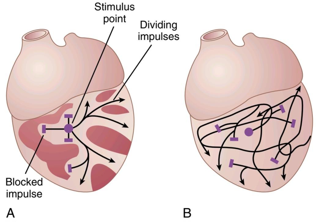
Figure 13-16. A, Initiation of fibrillation in a heart when patches of refractory musculature are present. B, Continued propagation of fibrillatory impulses in the fibrillating ventricle.
شکل 13-16. الف، شروع فیبریلاسیون در قلب زمانی که لکههای عضلانی مقاوم وجود داشته باشد. ب، ادامه انتشار تکانههای فیبریلاتور در بطن فیبریلاتور.
ELECTROCARDIOGRAM IN VENTRICULAR FIBRILLATION
In ventricular fibrillation, the ECG is bizarre (Figure 13-17) and ordinarily shows no tendency toward a regular rhythm of any type. During the first few seconds of ventricular fibrillation, relatively large masses of muscle contract simultaneously, which causes coarse irregular waves in the ECG. After another few seconds, the coarse contractions of the ventricles disappear, and the ECG changes into a new pattern of low-voltage, very irregular waves. Thus, no repetitive electrocardiographic pattern can be ascribed to ventricular fibrillation. Instead, the ventricular muscle contracts at as many as 30 to 50 small patches of muscle at a time, and electrocardiographic potentials change constantly and spasmodically because the electrical currents in the heart flow first in one direction and then in another and seldom repeat any specific cycle.
الکتروکاردیوگرام در بطن فیبریلاسیون
در فیبریلاسیون بطنی، ECG عجیب است (شکل 13-17) و معمولاً تمایلی به ریتم منظم از هر نوع نشان نمیدهد. در طی چند ثانیه اول فیبریلاسیون بطنی، تودههای نسبتاً بزرگی از عضله به طور همزمان منقبض میشوند که باعث ایجاد امواج نامنظم درشت در ECG میشود. پس از چند ثانیه دیگر، انقباضات درشت بطنها ناپدید میشوند و ECG به الگوی جدیدی از امواج کم ولتاژ و بسیار نامنظم تبدیل میشود. بنابراین، هیچ الگوی الکتروکاردیوگرافی تکراری را نمیتوان به فیبریلاسیون بطنی نسبت داد. درعوض، عضله بطنی در هر بار 30 تا 50 تکه کوچک عضلانی منقبض میشود و پتانسیلهای الکتروکاردیوگرافی دائماً و بهطور اسپاسم تغییر میکنند، زیرا جریانهای الکتریکی در قلب ابتدا در یک جهت و سپس در جهت دیگر جریان مییابند و به ندرت هر چرخه خاصی را تکرار میکنند.
The voltages of the waves in the ECG in ventricular fibrillation are usually about 0.5 millivolt when ventricular fibrillation first begins, but they decay rapidly; thus, after 20 to 30 seconds, they are usually only 0.2 to 0.3 millivolt. Minute voltages of 0.1 millivolt or less may be recorded for 10 minutes or longer after ventricular fibrillation begins. As already noted, because no pumping of blood occurs during ventricular fibrillation, this state is lethal unless stopped by successful therapy, such as an immediate electroshock (defibrillation) through the heart, as explained in the next section.
ولتاژ امواج در ECG در فیبریلاسیون بطنی معمولاً حدود 0.5 میلی ولت زمانی است که فیبریلاسیون بطنی برای اولین بار شروع میشود، اما آنها به سرعت تحلیل میروند. بنابراین، پس از 20 تا 30 ثانیه، آنها معمولا تنها 0.2 تا 0.3 میلی ولت هستند. ولتاژهای دقیقه ای 0.1 میلی ولت یا کمتر ممکن است به مدت 10 دقیقه یا بیشتر پس از شروع فیبریلاسیون بطنی ثبت شود. همانطور که قبلا ذکر شد، از آنجایی که هیچ پمپاژ خون در طول فیبریلاسیون بطنی رخ نمیدهد، این حالت کشنده است مگر اینکه با درمان موفقیت آمیز، مانند الکتروشوک فوری (دفیبریلاسیون) در قلب، همانطور که در بخش بعدی توضیح داده شد، متوقف شود.

Figure 13-17. Ventricular fibrillation (lead II).
شکل 13-17. فیبریلاسیون بطنی (لید II).
VENTRICULAR DEFIBRILLATION
Although a moderate alternating current voltage applied directly to the ventricles almost invariably throws the ventricles into fibrillation, a strong high-voltage electrical cur- rent passed through the ventricles for a fraction of a second can stop fibrillation by throwing all the ventricular muscle into refractoriness simultaneously. This is accomplished by passing intense current through large electrodes placed on two sides of the heart. The current penetrates most of the fibers of the ventricles at the same time, thus stimulating essentially all parts of the ventricles simultaneously and causing them all to become refractory. All action potentials stop, and the heart remains quiescent for 3 to 5 seconds, after which it begins to beat again, usually with the sinus node or some other part of the heart becoming the pace- maker. However, if the same re-entrant focus that had originally thrown the ventricles into fibrillation is still present, fibrillation may begin again immediately.
دفیبریلاسیون بطنی
اگرچه یک ولتاژ جریان متناوب متوسط که مستقیماً به بطنها اعمال میشود تقریباً همیشه بطنها را به سمت فیبریلاسیون میاندازد، یک جریان الکتریکی با ولتاژ بالا که برای کسری از ثانیه از بطنها عبور میکند میتواند فیبریلاسیون را با پرتاب همزمان تمام عضله بطنی به حالت نسوز متوقف کند. این کار با عبور جریان شدید از الکترودهای بزرگی که در دو طرف قلب قرار گرفته اند انجام میشود. جریان به طور همزمان به بیشتر الیاف بطنها نفوذ میکند، بنابراین اساساً تمام قسمتهای بطنها را به طور همزمان تحریک میکند و باعث میشود که همه آنها نسوز شوند. تمام پتانسیلهای عمل متوقف میشوند و قلب به مدت 3 تا 5 ثانیه ساکن میماند، پس از آن دوباره شروع به تپیدن میکند، معمولاً با گره سینوسی یا قسمت دیگری از قلب که ضربانساز میشود. با این حال، اگر همان کانون بازگشتی که در ابتدا بطنها را به سمت فیبریلاسیون پرتاب کرده بود، همچنان وجود داشته باشد، فیبریلاسیون ممکن است بلافاصله دوباره شروع شود.
When electrodes are applied directly to the two sides of the heart, fibrillation can usually be stopped using 1000 volts of direct current applied for a few thousandths of a second. When applied through two electrodes on the chest wall, as shown in Figure 13-18, the usual procedure is to charge a large electrical capacitor up to several thou- sand volts and then to cause the capacitor to discharge for a few thousandths of a second through the electrodes and through the heart.
هنگامیکه الکترودها مستقیماً به دو طرف قلب اعمال میشوند، فیبریلاسیون را معمولاً میتوان با استفاده از 1000 ولت جریان مستقیم برای چند هزارم ثانیه متوقف کرد. هنگامیکه از طریق دو الکترود روی دیواره قفسه سینه اعمال میشود، همانطور که در شکل 13-18 نشان داده شده است، روش معمول این است که یک خازن الکتریکی بزرگ را تا چندین هزار ولت شارژ میکنیم و سپس باعث میشود که خازن برای چند هزارم ثانیه از طریق الکترودها و از طریق قلب تخلیه شود.
In most cases, defibrillation current is delivered to the heart in biphasic waveforms, alternating the direction of the current pulse through the heart. This form of delivery substantially reduces the energy needed for successful defibrillation, thereby decreasing the risk for burns and cardiac damage.
در بیشتر موارد، جریان دفیبریلاسیون به شکل موج دو فازی به قلب منتقل میشود و جهت پالس جریان از طریق قلب متناوب میشود. این نوع زایمان انرژی مورد نیاز برای دفیبریلاسیون موفقیت آمیز را به میزان قابل توجهی کاهش میدهد و در نتیجه خطر سوختگی و آسیب قلبی را کاهش میدهد.
In patients with a high risk for ventricular fibrillation, a small, battery-powered, implantable cardioverter- defibrillator (ICD) with electrode wires lodged in the right ventricle may be implanted. The device is programmed to detect ventricular fibrillation and revert it by delivering a brief electrical impulse to the heart. Advances in electronics and batteries have permitted the development of ICDs that can deliver enough electrical current to defibrillate the heart through electrode wires implanted subcutaneously, outside the rib cage near the heart rather than in or on the heart itself. These devices can be implanted with a minor surgical procedure.
در بیمارانی که در معرض خطر بالای فیبریلاسیون بطنی قرار دارند، ممکن است یک دفیبریلاتور کوچک با باتری قابل کاشت (ICD) با سیمهای الکترود در بطن راست کاشته شود. این دستگاه برای تشخیص فیبریلاسیون بطنی و بازگرداندن آن با ارسال یک تکانه الکتریکی کوتاه به قلب برنامه ریزی شده است. پیشرفتها در الکترونیک و باتریها امکان توسعه ICDهایی را فراهم کرده است که میتوانند جریان الکتریکی کافی برای دفیبریلاسیون قلب را از طریق سیمهای الکترودی که به صورت زیر جلدی، خارج از قفسه سینه نزدیک قلب به جای درون یا روی قلب، کاشته میشوند، ایجاد کنند. این دستگاهها را میتوان با یک روش جراحی جزئی کاشت.

Figure 13-18. Application of electrical current to the chest to stop ventricular fibrillation.
شکل 13-18. اعمال جریان الکتریکی به قفسه سینه برای توقف فیبریلاسیون بطنی.
HAND PUMPING OF THE HEART (CARDIOPULMONARY RESUSCITATION) AS AN AID TO DEFIBRILLATION
Unless defibrillated within 1 minute after ventricular fibrillation begins, the heart is usually too weak to be revived by defibrillation because of the lack of nutrition from coronary blood flow. However, it is still possible to revive the heart by preliminarily pumping the heart by hand (intermittent hand squeezing) and then defibrillating the heart later. In this way, small quantities of blood are delivered into the aorta, and a renewed coronary blood supply develops. Then, after a few minutes of hand pumping, electrical defibrillation often becomes possible. Fibrillating hearts have been pumped by hand for as long as 90 minutes followed by successful defibrillation.
پمپاژ دستی قلب (احیای قلبی ریوی) به عنوان کمکی به دفیبریلاسیون
مگر اینکه در عرض 1 دقیقه پس از شروع فیبریلاسیون بطنی دفیبریلاسیون شود، قلب معمولاً ضعیف تر از آن است که با دفیبریلاسیون دوباره احیا شود زیرا جریان خون کرونری تغذیه نمیشود. با این حال، هنوز هم میتوان با پمپاژ اولیه قلب با دست (فشار متناوب دست) و سپس دفیبریلاسیون قلب، قلب را احیا کرد. به این ترتیب، مقدار کمیخون به آئورت وارد میشود و یک منبع خون مجدد کرونری ایجاد میشود. سپس، پس از چند دقیقه پمپاژ دستی، دفیبریلاسیون الکتریکی اغلب امکان پذیر میشود. قلبهای فیبریلاسیون به مدت 90 دقیقه با دست پمپاژ میشوند و سپس دفیبریلاسیون موفقیت آمیز انجام میشود.
A technique for pumping the heart without opening the chest consists of intermittent thrusts of pressure on the chest wall along with artificial respiration. This process, plus defibrillation, is called cardiopulmonary resuscitation (CPR).
یک تکنیک برای پمپاژ قلب بدون باز کردن قفسه سینه شامل فشارهای متناوب بر روی دیواره قفسه سینه همراه با تنفس مصنوعی است. این فرآیند، به علاوه دفیبریلاسیون، احیای قلبی ریوی (CPR) نامیده میشود.
Lack of blood flow to the brain for more than 5 to 8 minutes usually causes permanent mental impairment or even destruction of brain tissue. Even if the heart is revived, the person may die from the effects of brain damage or may live with permanent mental impairment.
فقدان جریان خون به مغز برای بیش از 5 تا 8 دقیقه معمولاً باعث اختلال ذهنی دائمییا حتی تخریب بافت مغز میشود. حتی اگر قلب احیا شود، ممکن است فرد در اثر آسیب مغزی بمیرد یا با اختلال روانی دائمیزندگی کند.
ATRIAL FIBRILLATION
Remember that except for the conducting pathway through the A-V bundle, the atrial muscle mass is separated from the ventricular muscle mass by fibrous tissue. Therefore, ventricular fibrillation often occurs without atrial fibrillation. Likewise, fibrillation often occurs in the atria without ventricular fibrillation (shown on the right in Figure 13-20).
فیبریلاسیون دهلیزی
به یاد داشته باشید که به جز مسیر هدایت از طریق بسته A-V، توده عضلانی دهلیزی توسط بافت فیبری از توده عضلانی بطنی جدا میشود. بنابراین، فیبریلاسیون بطنی اغلب بدون فیبریلاسیون دهلیزی رخ میدهد. به همین ترتیب، فیبریلاسیون اغلب در دهلیزها بدون فیبریلاسیون بطنی رخ میدهد (در شکل 13-20 در سمت راست نشان داده شده است).
The mechanism of atrial fibrillation is identical to that of ventricular fibrillation, except that the process occurs only in the atrial muscle mass instead of the ventricular mass. A frequent cause of atrial fibrillation is atrial enlargement, which can result, for example, from heart valve lesions that prevent the atria from emptying adequately into the ventricles or from ventricular failure with excess damming of blood in the atria. The dilated atrial walls provide ideal conditions of a long conductive path- way, as well as slow conduction, both of which predispose to atrial fibrillation.
مکانیسم فیبریلاسیون دهلیزی با فیبریلاسیون بطنی یکسان است، با این تفاوت که این فرآیند به جای توده بطنی، فقط در توده عضلانی دهلیزی رخ میدهد. یکی از علل شایع فیبریلاسیون دهلیزی بزرگ شدن دهلیزی است که میتواند به عنوان مثال از ضایعات دریچه قلب که از تخلیه مناسب دهلیزها به داخل بطنها جلوگیری میکند یا ناشی از نارسایی بطنی همراه با سد کردن بیش از حد خون در دهلیزها باشد. دیوارههای دهلیزی گشاد شده شرایط ایده آلی را برای یک مسیر رسانای طولانی و همچنین هدایت آهسته فراهم میکند که هر دو مستعد فیبریلاسیون دهلیزی هستند.
Impaired Pumping of the Atria During Atrial Fibrillation. For the same reasons that the ventricles will not pump blood during ventricular fibrillation, neither do the atria pump blood in atrial fibrillation. Therefore, the atria become useless as primer pumps for the ventricles. Even so, blood flows passively through the atria into the ventricles, and the efficiency of ventricular pumping is decreased by only 20% to 30%. Therefore, in contrast to the lethality of ventricular fibrillation, a person can live for years with atrial fibrillation, although at reduced efficiency of overall heart pumping. However, due to the reduced atrial contractile function, blood can stagnate, allowing blood clots to form in the atrial appendage. These blood clots can dislodge and travel to the brain, causing stroke, or to other parts of the body. Therefore, patients with atrial fibrillation are often placed on blood thinner medications (anticoagulants) to reduce the risk of embolism.
اختلال در پمپاژ دهلیزها در طول فیبریلاسیون دهلیزی. به همان دلایلی که بطنها در جریان فیبریلاسیون بطنی خون را پمپ نمیکنند، دهلیزها نیز در فیبریلاسیون دهلیزی خون را پمپاژ نمیکنند. بنابراین دهلیزها به عنوان پمپهای آغازگر برای بطنها بی فایده میشوند. با این حال، خون به طور غیر فعال از طریق دهلیزها به داخل بطنها جریان مییابد و بازده پمپاژ بطنی تنها 20 تا 30 درصد کاهش مییابد. بنابراین، بر خلاف کشنده بودن فیبریلاسیون بطنی، یک فرد میتواند سالها با فیبریلاسیون دهلیزی زندگی کند، اگرچه با کاهش راندمان پمپاژ کلی قلب. با این حال، به دلیل کاهش عملکرد انقباضی دهلیزی، خون میتواند راکد شود و به لخته شدن خون در زائده دهلیزی اجازه تشکیل دهد. این لختههای خون میتوانند از جای خود خارج شده و به مغز بروند و باعث سکته مغزی یا سایر قسمتهای بدن شوند. بنابراین، بیماران مبتلا به فیبریلاسیون دهلیزی اغلب از داروهای رقیق کننده خون (ضد انعقادها) استفاده میکنند تا خطر آمبولی کاهش یابد.
ELECTROCARDIOGRAM IN ATRIAL FIBRILLATION
Figure 13-19 shows the ECG during atrial fibrillation. Numerous small depolarization waves spread in all directions through the atria during atrial fibrillation. Because the waves are weak, and many of them are of opposite polarity at any given time, they usually almost completely electrically neutralize one another. Therefore, in the ECG, one can see either no P waves from the atria or only a fine, high-frequency, very low voltage wave record. Conversely, the QRS-T complexes are normal unless there is some pathology of the ventricles, but their timing is irregular, as explained next.
الکتروکاردیوگرام در دهلیز فیبریلاسیون
شکل 13-19 ECG را در طول فیبریلاسیون دهلیزی نشان میدهد. امواج دپلاریزاسیون کوچک متعددی در طول فیبریلاسیون دهلیزی در تمام جهات از دهلیزها پخش میشوند. از آنجایی که امواج ضعیف هستند و بسیاری از آنها در هر زمان دارای قطب مخالف هستند، معمولاً به طور کامل یکدیگر را خنثی میکنند. بنابراین، در ECG، میتوان امواج P را از دهلیزها مشاهده نکرد یا فقط یک رکورد موج با فرکانس بالا و ولتاژ بسیار پایین مشاهده کرد. برعکس، کمپلکسهای QRS-T طبیعی هستند، مگر اینکه پاتولوژی بطنها وجود داشته باشد، اما زمانبندی آنها نامنظم است، همانطور که در ادامه توضیح داده شد.
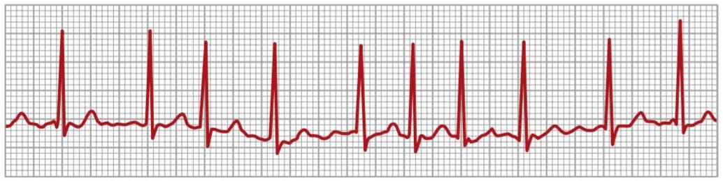
Figure 13-19. Atrial fibrillation (lead II). The waves that can be seen are ventricular QRS and T waves.
شکل 13-19. فیبریلاسیون دهلیزی (لید II). امواجی که قابل مشاهده هستند، امواج QRS و T بطنی هستند.
IRREGULARITY OF VENTRICULAR RHYTHM DURING ATRIAL FIBRILLATION
When the atria are fibrillating, impulses arrive from the atrial muscle at the A-V node rapidly but also irregularly. Because the A-V node will not pass a second impulse for about 0.35 second after a previous one, at least 0.35 second must elapse between one ventricular contraction and the next. Then, an additional but variable interval of 0 to 0.6 second occurs before one of the irregular atrial fibrillatory impulses happens to arrive at the A-V node. Thus, the interval between successive ventricular contractions varies from a minimum of about 0.35 second to a maximum of about 0.95 second, causing a very irregular heartbeat. In fact, this irregularity, demonstrated by the variable spacing of the heartbeats in the ECG shown in Figure 13-19, is one of the clinical findings used to diagnose the condition. Also, because of the rapid rate of the fibrillatory impulses in the atria, the ventricle is driven at a fast heart rate, usually between 125 and 150 beats/min.
بی نظمیریتم بطنی در حین فیبریلاسیون دهلیزی
هنگامیکه دهلیزها در حال فیبریلاسیون هستند، تکانهها از عضله دهلیزی به سرعت اما به طور نامنظم به گره A-V میرسند. از آنجایی که گره A-V یک ضربه دوم را برای حدود 0.35 ثانیه پس از ضربه قبلی ارسال نمیکند، حداقل 0.35 ثانیه باید بین یک انقباض بطنی و بعدی انقباض بگذرد. سپس، قبل از اینکه یکی از تکانههای فیبریلاتوری دهلیزی نامنظم به گره A-V برسد، یک فاصله اضافی اما متغیر بین 0 تا 0.6 ثانیه رخ میدهد. بنابراین، فاصله بین انقباضات متوالی بطن از حداقل حدود 0.35 ثانیه تا حداکثر حدود 0.95 ثانیه متغیر است و باعث ضربان قلب بسیار نامنظم میشود. در واقع، این بی نظمیکه با فاصله متغیر ضربان قلب در ECG نشان داده شده در شکل 13-19 نشان داده شده است، یکی از یافتههای بالینی مورد استفاده برای تشخیص این بیماری است. همچنین، به دلیل سرعت سریع تکانههای فیبریلاتوری در دهلیزها، بطن با ضربان قلب سریع، معمولا بین 125 تا 150 ضربه در دقیقه رانده میشود.
ELECTROSHOCK TREATMENT OF ATRIAL FIBRILLATION
Similar to ventricular fibrillation being converted back to a normal rhythm by electroshock, so can atrial fibrillation be converted by electroshock. The procedure is similar as that for ventricular fibrillation conversion, except the single electric shock is programmed (or synchronized) to fire only during the QRS complex when the ventricles are refractory to stimulation. A normal rhythm often follows if the heart is capable of generating a normal rhythm. This procedure is called synchronized cardioversion instead of defibrillation in the setting of ventricular fibrillation.
درمان فیبریلاسیون دهلیزی با الکتروشوک
مانند فیبریلاسیون بطنی که توسط الکتروشوک به ریتم طبیعی تبدیل میشود، بنابراین میتوان فیبریلاسیون دهلیزی را با الکتروشوک تبدیل کرد. این روش مانند تبدیل فیبریلاسیون بطنی است، به جز اینکه شوک الکتریکی منفرد برای شلیک فقط در طول مجتمع QRS زمانی که بطنها نسبت به تحریک مقاوم هستند، برنامهریزی شده (یا هماهنگ میشود). اگر قلب قادر به ایجاد یک ریتم طبیعی باشد، اغلب یک ریتم طبیعی به دنبال دارد. این روش به جای دفیبریلاسیون در شرایط فیبریلاسیون بطنی، کاردیوورژن همزمان نامیده میشود.
ATRIAL FLUTTER
Atrial flutter is another condition caused by a circus movement in the atria. Atrial flutter is different from atrial fibrillation in that the electrical signal travels as a single large wave, always in one direction, around and around the atrial muscle mass, as shown to the left in Figure 13-20. Atrial flutter causes a rapid rate of contraction of the atria, usually between 200 and 350 beats/min. How- ever, because one side of the atria is contracting while the other side is relaxing, the amount of blood pumped by the atria is reduced. Furthermore, the signals reach the A-V node too rapidly for all of them to be passed into the ventricles because the refractory periods of the A-V node and A-V bundle are too long to pass more than a fraction of the atrial signals. Therefore, there are usually two to three beats of the atria for every single beat of the ventricles.
فلاتر دهلیزی
فلوتر دهلیزی یکی دیگر از بیماریهای ناشی از حرکت سیرک در دهلیز است. فلوتر دهلیزی با فیبریلاسیون دهلیزی متفاوت است، زیرا سیگنال الکتریکی به صورت یک موج بزرگ، همیشه در یک جهت، در اطراف و اطراف توده عضلانی دهلیزی حرکت میکند، همانطور که در شکل 13-20 در سمت چپ نشان داده شده است. فلوتر دهلیزی باعث انقباض سریع دهلیزها میشود که معمولاً بین 200 تا 350 ضربه در دقیقه است. با این حال، به دلیل انقباض یک طرف دهلیز در حالی که طرف دیگر در حال استراحت است، مقدار خون پمپ شده توسط دهلیز کاهش مییابد. علاوه بر این، سیگنالها خیلی سریع به گره A-V میرسند تا همه آنها به بطن منتقل شوند، زیرا دورههای نسوز گره A-V و بسته A-V برای عبور بیش از کسری از سیگنالهای دهلیزی بسیار طولانی است. بنابراین، معمولاً دو تا سه ضربه دهلیز برای هر ضربان بطن وجود دارد.

Figure 13-20. Pathways of impulses in atrial flutter and atrial fibrillation.
شکل 13-20. مسیرهای تکانه در فلوتر دهلیزی و فیبریلاسیون دهلیزی.
Figure 13-21 shows a typical ECG in atrial flutter. The P waves are strong because of the contraction of semicoordinated masses of muscle. However, note that a QRS-T complex follows an atrial P wave only once for every two beats of the atria, giving a 2:1 rhythm.
شکل 13-21 یک ECG معمولی را در فلوتر دهلیزی نشان میدهد. امواج P به دلیل انقباض تودههای نیمه هماهنگ عضلانی قوی هستند. با این حال، توجه داشته باشید که یک کمپلکس QRS-T یک موج P دهلیزی را تنها یک بار برای هر دو ضربان دهلیز دنبال میکند و ریتم 2:1 را ارائه میدهد.

Figure 13-21. Atrial flutter at 250 beats/min with a 2:1 atrial to ventricular rhythm at 125 beats/min (lead II).
شکل 13-21. فلاتر دهلیزی با 250 ضربه در دقیقه با ریتم دهلیزی به بطنی 2:1 با 125 ضربه در دقیقه (لید II).
CARDIAC ARREST
A final serious abnormality of the cardiac rhythmicityconduction system is cardiac arrest, which results from cessation of all electrical control signals in the heart. That is, no spontaneous rhythm remains.
ایست قلبی
آخرین ناهنجاری جدی در سیستم هدایت ریتم قلبی، ایست قلبی است که از قطع تمام سیگنالهای کنترل الکتریکی در قلب ناشی میشود. یعنی هیچ ریتم خود به خودی باقی نمیماند.
Cardiac arrest may occur during deep anesthesia, when severe hypoxia may develop because of inadequate respiration. The hypoxia prevents the muscle fibers and conductive fibers from maintaining normal electrolyte concentration differentials across their membranes, and their excitability may be so affected that the automatic rhythmicity disappears.
ایست قلبی ممکن است در طول بیهوشی عمیق رخ دهد، زمانی که هیپوکسی شدید ممکن است به دلیل تنفس ناکافی ایجاد شود. هیپوکسی باعث میشود تا فیبرهای عضلانی و الیاف رسانا از حفظ اختلاف غلظت الکترولیت طبیعی در سراسر غشاء خود جلوگیری کنند و تحریک پذیری آنها ممکن است چنان تحت تأثیر قرار گیرد که ریتمیک خودکار از بین میرود.
In many cases of cardiac arrest from anesthesia, prolonged CPR (for many minutes or even hours) is quite successful in re-establishing a normal heart rhythm. In some patients, severe myocardial disease can cause permanent or semipermanent cardiac arrest, which can cause death. To treat the condition, rhythmical electrical impulses from an implanted electronic cardiac pacemaker have been used successfully to keep patients alive for months to years.
در بسیاری از موارد ایست قلبی ناشی از بیهوشی، CPR طولانی مدت (برای چندین دقیقه یا حتی ساعتها) در برقراری مجدد ریتم طبیعی قلب کاملاً موفق است. در برخی از بیماران، بیماری شدید میوکارد میتواند باعث ایست قلبی دائمییا نیمه دائمیشود که میتواند منجر به مرگ شود. برای درمان این بیماری، تکانههای الکتریکی ریتمیک از یک ضربان ساز الکترونیکی قلبی کاشته شده با موفقیت برای زنده نگه داشتن بیماران برای ماهها تا سالها استفاده شده است.
کلیک کنید «Bibliography: فهرست کتب مربوطه»
Ackerman M, Atkins DL, Triedman JK: Sudden cardiac death in the young, Circulation 133:1006, 2016.
Adler A, Rosso R, Viskin D, et al.: What do we know about the “malignant form” of early repolarization? J Am Coll Cardiol 62:863, 2013.
Borne RT, Katz D, Betz J, et al.: Implantable cardioverter-defibrillators for secondary prevention of sudden cardiac death: a review, J Am Heart Assoc 6:e005515, 2017.
Darby AE, DiMarco JP: Management of atrial fibrillation in patients with structural heart disease, Circulation 125:945, 2012.
Dobrzynski H, Boyett MR, Anderson RH: New insights into pacemaker activity: promoting understanding of sick sinus syndrome, Circulation 115:1921, 2007.
Fukuda K, Kanazawa H, Aizawa Y, et al.: Cardiac innervation and sudden cardiac death, Circ Res 116:2015, 2005.
Giudicessi JR, Roden DM, Wilde AAM, et al.: Classification and re- porting of potentially proarrhythmic common genetic variation in long QT syndrome genetic testing, Circulation 137:619, 2018.
Guasch E, Mont L: Diagnosis, pathophysiology, and management of exercise-induced arrhythmias, Nat Rev Cardiol 14:88, 2017.
John RM, Tedrow UB, Koplan BA, et al.: Ventricular arrhythmias and sudden cardiac death, Lancet 380:1520, 2012.
Koruth JS, Lala A, Pinney S, et al.: The clinical use of ivabradine, J Am Coll Cardiol 70:1777, 2017.
Lee G, Sanders P, Kalman JM: Catheter ablation of atrial arrhythmias: state of the art, Lancet 380:1509, 2012.
Macfarlane PW, Antzelevitch C, Haissaguerre M, et al.: The early repolar- ization pattern: A consensus paper, J Am Coll Cardiol 66:470, 2015. Morita H, Wu J, Zipes DP: The QT syndromes: long and short, Lancet 372:750, 2008.
Olshansky B, Sullivan RM: Inappropriate sinus tachycardia, J Am Coll Cardiol 61:793, 2013.
Park DS, Fishman Gl: The cardiac conduction system, Circulation 123:904, 2011.
Passman R, Kadish A: Sudden death prevention with implantable de- vices, Circulation 116:561, 2007.
Prystowsky EN, Padanilam BJ, Joshi S, Fogel RI: Ventricular arrhyth- mias in the absence of structural heart disease, J Am Coll Cardiol 59:1733, 2012.
Reed GW, Rossi JE, Cannon CP: Acute myocardial infarction, Lancet 389:197, 2017.
Rienstra M, Lubitz SA, Mahida S, et al.: Symptoms and functional status of patients with atrial fibrillation: state of the art and future research opportunities, Circulation 125:2933, 2012.
Roden DM: Drug-induced prolongation of the QT interval, N Engl J Med 350:1013, 2004.
Schlapfer J, Wellens HJ: Computer-interpreted electrocardiograms. Benefits and limitations, J Am Coll Cardiol 70:1183, 2017.
Schwartz PJ, Ackerman MJ, George Jr AL, Wilde AA: Impact of genetics on the clinical management of channelopathies, J Am Coll Cardiol 62:169, 2013.
Schwartz PJ, Woosley RL: Predicting the unpredictable: Drug-induced QT prolongation and Torsades de Pointes, J Am Coll Cardiol 67:1639, 2016.
Shen MJ, Zipes DP: Role of the autonomic nervous system in modulating cardiac arrhythmias, Circ Res 114:1004, 2014.
Staerk L, Sherer JA, Ko D, Benjamin EJ, Helm RH: Atrial fibrillation: epidemiology, pathophysiology, and clinical outcomes, Circ Res 120:1501, 2017.
Vijayaraman P, Chung MK, Dandamudi G, et al.: His bundle pacing, J Am Coll Cardiol 72:927, 2018.
🚀 با ما همراه شوید!
تازهترین مطالب و آموزشهای مغز و اعصاب را از دست ندهید. با فالو کردن کانال تلگرام آیندهنگاران مغز، از ما حمایت کنید!


