
فیزیولوژی پزشکی گانونگ؛ گردش از طریق مناطق ویژه


دعای مطالعه [ نمایش ]
بِسْمِ الله الرَّحْمنِ الرَّحیمِ
اَللّهُمَّ اَخْرِجْنى مِنْ ظُلُماتِ الْوَهْمِ
خدايا مرا بيرون آور از تاريكىهاى وهم،
وَ اَكْرِمْنى بِنُورِ الْفَهْمِ
و به نور فهم گرامى ام بدار،
اَللّهُمَّ افْتَحْ عَلَيْنا اَبْوابَ رَحْمَتِكَ
خدايا درهاى رحمتت را به روى ما بگشا،
وَانْشُرْ عَلَيْنا خَزائِنَ عُلُومِكَ بِرَحْمَتِكَ يا اَرْحَمَ الرّاحِمينَ
و خزانههاى علومت را بر ما باز كن به امید رحمتت اى مهربانترين مهربانان.
کتاب «فیزیولوژی پزشکی گانونگ» بهعنوان یکی از جامعترین و معتبرترین منابع در حوزه علوم پزشکی، همچنان مرجع کلیدی برای درک عملکرد پیچیده بدن انسان است. این اثر با تکیه بر تازهترین پژوهشها و توضیحات دقیق از سازوکارهای فیزیولوژیک، پلی میان علوم پایه پزشکی و کاربردهای بالینی ایجاد میکند و نقشی بیبدیل در آموزش، پژوهش و ارتقای دانش سلامت ایفا مینماید.
ترجمه دقیق و علمی این شاهکار توسط برند علمی آیندهنگاران مغز به مدیریت داریوش طاهری، دسترسی فارسیزبانان به مرزهای نوین دانش فیزیولوژی را ممکن ساخته و رسالتی علمی برای ارتقای آموزش پزشکی، فهم عمیقتر سازوکارهای بدن و توسعه روشهای نوین در حوزه سلامت فراهم آورده است.
» کتاب فیزیولوژی پزشکی گانونگ
» » فصل ۳۳: گردش از طریق مناطق ویژه
در حال ویرایش
» Ganong’s Review of Medical Physiology
»» CHAPTER 33: Circulation Through Special Regions
OBJECTIVES
After studying his chapter, you should be able to:
• Define the special features of the circulation in the brain. Describe how cerebrospinal fluid (CSF) is formed and reabsorbed, and its role in protecting the brain from injury. Understand how the blood-brain barrier impedes the entry of specific substances into the brain. Explain how cerebral blood flow can be measured, how it is regulated by intracranial pressure, and how it protects the supply of oxygen to the brain.
• Understand the anatomy of the coronary circulation of the heart and how the cardiac cycle regulates flow in the coronary blood vessels. Delineate how the oxygen needs of the contracting myocardium are met by the coronary arteries, and the consequences of their occlusion.
• Describe how the cutaneous circulation participates in heat loss from the body and how this function is regulated. List the vascular reactions of the skin and the reflexes that mediate them.
• Describe the uterine circulation and how it changes during the course of pregnancy and parturition. Understand how the fetus is supplied with oxygen and nutrients in utero, and the circulatory events required for a transition to independent life after birth.
اهداف
پس از مطالعه این فصل، باید بتوانید:
• ویژگی های خاص گردش خون در مغز را تعریف کنید. نحوه تشکیل و بازجذب مایع مغزی نخاعی (CSF) و نقش آن در محافظت از مغز در برابر آسیب را شرح دهید. درک کنید که چگونه سد خونی مغز مانع ورود مواد خاص به مغز می شود. توضیح دهید که چگونه جریان خون مغزی را می توان اندازه گیری کرد، چگونه با فشار داخل جمجمه تنظیم می شود، و چگونه از اکسیژن رسانی به مغز محافظت می کند.
• آناتومی گردش خون کرونر قلب و چگونگی تنظیم چرخه قلبی جریان در رگ های خونی کرونری را بدانید. نحوه تامین اکسیژن مورد نیاز میوکارد در حال انقباض توسط عروق کرونر و پیامدهای انسداد آنها را مشخص کنید.
• توضیح دهید که چگونه گردش پوست در از دست دادن گرما از بدن شرکت می کند و چگونه این عملکرد تنظیم می شود. واکنش های عروقی پوست و رفلکس هایی که واسطه آن ها هستند را فهرست کنید.
• گردش خون رحم و تغییر آن در طول بارداری و زایمان را شرح دهید. درک چگونگی تامین اکسیژن و مواد مغذی جنین در رحم و رویدادهای گردش خون مورد نیاز برای انتقال به زندگی مستقل پس از تولد.

INTRODUCTION
The distribution of the cardiac output to various parts of the body at rest in a normal man is shown in Table 33-1. The general principles described in preceding chapters apply to the circulation of all these regions, but the vascular supplies of many organs have additional special features that are important to their physiology. This chapter is concerned with the special circulations of the brain, the heart, and the skin, as well as the placenta and fetus. In addition, the portal circulation of the anterior pituitary is discussed in Chapter 18; the pulmonary circulation in Chapter 34; the renal circulation in Chapter 37; and the circulation of the splanchnic area, particularly the intestines and liver, in Chapters 25 and 28.
مقدمه
توزیع برون ده قلبی به قسمت های مختلف بدن در حالت استراحت در یک مرد عادی در جدول 33-1 نشان داده شده است. اصول کلی که در فصول قبل توضیح داده شد در مورد گردش خون همه این نواحی اعمال می شود، اما منابع عروقی بسیاری از اندام ها دارای ویژگی های ویژه دیگری هستند که برای فیزیولوژی آنها مهم است. این فصل به گردش های خاص مغز، قلب و پوست و همچنین جفت و جنین می پردازد. علاوه بر این، گردش پورتال هیپوفیز قدامی در فصل 18 مورد بحث قرار گرفته است. گردش خون ریوی در فصل 34; گردش کلیوی در فصل 37; و گردش ناحیه اسپلانکنیک، به ویژه روده ها و کبد، در فصل های 25 و 28.
TABLE 33-1 Resting blood flow and O2 consumption of various organs in a 63-kg adult man with a mean arterial blood pressure of 90 mm Hg and an O2 consumption of 250 mL/min.
جدول 33-1 جریان خون در حالت استراحت و مصرف O2 اندام های مختلف در یک مرد بالغ 63 کیلوگرمی با فشار خون شریانی متوسط 90 میلی متر جیوه و مصرف O2 250 میلی لیتر در دقیقه.

aR units are pressure (mm Hg) divided by blood flow (mL/s).
Reproduced with permission from Bard P (editor): Medical Physiology, 11th ed. Mosby, 1961.
a واحدهای R فشار (mmHg) تقسیم بر جریان خون (mL/s) هستند.
تکثیر شده با اجازه بارد پی (ویرایشگر): فیزیولوژی پزشکی، ویرایش یازدهم. ماسبی، 1961.
CEREBRAL CIRCULATION: ANATOMIC CONSIDERATIONS
VESSELS
The principal arterial inflow to the brain in humans is via four arteries: two internal carotid arteries and two vertebral arteries. In humans, the carotid arteries are quantitatively the most significant. The vertebral arteries unite to form the basilar artery, and the basilar artery and the carotids form the circle of Willis below the hypothalamus. The circle of Willis is the origin of the six large vessels supplying the cerebral cortex. Substances injected into one carotid artery are distributed almost exclusively to the cerebral hemisphere on that side. Normally no crossing over occurs, probably because the pressure is equal on both sides. Even when it is not, the anastomotic channels in the circle do not permit a very large flow. Occlusion of one carotid artery, particularly in older patients, often causes serious symptoms of cerebral ischemia. There are precapillary anastomoses between the cerebral vessels, but flow through these channels is generally insufficient to maintain the circulation and prevent infarction when a cerebral artery is occluded.
گردش مغزی: ملاحظات آناتومیک
کشتی ها
جریان شریانی اصلی به مغز انسان از طریق چهار شریان است: دو شریان کاروتید داخلی و دو شریان مهره ای. در انسان، شریان های کاروتید از نظر کمی مهم ترین هستند. شریان های مهره ای با هم متحد می شوند و شریان بازیلار را تشکیل می دهند و شریان بازیلار و کاروتید دایره ویلیس را در زیر هیپوتالاموس تشکیل می دهند. دایره ویلیس منشأ شش رگ بزرگ تامین کننده قشر مغز است. مواد تزریق شده به یک شریان کاروتید تقریباً به طور انحصاری در نیمکره مغزی آن طرف توزیع می شود. معمولاً هیچ عبوری رخ نمی دهد، احتمالاً به این دلیل که فشار در هر دو طرف برابر است. حتی زمانی که اینطور نیست، کانال های آناستوموز در دایره اجازه جریان بسیار زیاد را نمی دهند. انسداد یک شریان کاروتید، به ویژه در بیماران مسن، اغلب باعث علائم جدی ایسکمی مغزی می شود. آناستوموزهای پیش مویرگی بین عروق مغزی وجود دارد، اما جریان از طریق این کانال ها به طور کلی برای حفظ گردش خون و جلوگیری از انفارکتوس زمانی که یک شریان مغزی مسدود می شود، کافی نیست.
Venous drainage from the brain by way of the deep veins and dural sinuses empties principally into the internal jugular veins in humans, although a small amount of venous blood drains through the ophthalmic and pterygoid venous plexuses, through emissary veins to the scalp, and down the system of paravertebral veins in the spinal canal.
تخلیه وریدی از مغز از طریق سیاهرگهای عمیق و سینوسهای دورال عمدتاً به داخل سیاهرگهای ژوگولار داخلی در انسان تخلیه میشود، اگرچه مقدار کمی از خون وریدی از طریق شبکههای وریدی چشمی و پنجهگوید، از طریق وریدهای فرستاده شده به پوست سر و از طریق سیستم وریدهای کانال نخاعی تخلیه میشود.
The cerebral vessels have a number of unique anatomic features. In the choroid plexuses, there are gaps between the endothelial cells of the capillary wall, but the choroid epithelial cells that separate them from the cerebrospinal fluid (CSF) are connected to one another by tight junctions. The capillaries in the brain substance resemble the nonfenestrated capillaries found in muscle (see Chapter 31), and there are tight junctions between the endothelial cells that limit the passage of substances via the paracellular route. In addition, there are relatively few vesicles in the endothelial cytoplasm, and presumably little vesicular transport. However, multiple transport systems are present in the capillary cells. The brain capillaries are surrounded by the endfeet of astrocytes (Figure 33-1). These endfeet are closely applied to the basal lamina of the capillaries, but they do not cover the entire capillary wall, and gaps of about 20 nm occur between endfeet (Figure 33-2).However, the endfeet induce formation of tight junctions in the capillaries (see Chapter 31).
رگ های مغزی دارای تعدادی ویژگی آناتومیک منحصر به فرد هستند. در شبکه های مشیمیه، شکاف هایی بین سلول های اندوتلیال دیواره مویرگ وجود دارد، اما سلول های اپیتلیال مشیمیه که آنها را از مایع مغزی نخاعی (CSF) جدا می کند، توسط اتصالات محکم به یکدیگر متصل می شوند. مویرگهای موجود در ماده مغزی شبیه مویرگهای بدون فنستره موجود در عضله هستند (به فصل 31 مراجعه کنید)، و اتصالات محکمی بین سلولهای اندوتلیال وجود دارد که عبور مواد را از مسیر پاراسلولی محدود میکند. علاوه بر این، وزیکول های نسبتا کمی در سیتوپلاسم اندوتلیال وجود دارد و احتمالاً انتقال تاولی کمی وجود دارد. با این حال، سیستم های حمل و نقل متعدد در سلول های مویرگی وجود دارد. مویرگ های مغز توسط انتهای آستروسیت ها احاطه شده اند (شکل 33-1). این پایههای انتهایی بهخوبی روی لایه پایه مویرگها اعمال میشوند، اما تمام دیواره مویرگی را نمیپوشانند، و شکافهایی به اندازه 20 نانومتر بین پایههای انتهایی ایجاد میشود (شکل 33-2).

FIGURE 33-1 Relation of fibrous astrocyte (3) to a capillary (2) and neuron (4) in the brain. The endfeet of the astrocyte processes form a discontinuous membrane around the capillary (1). Astrocyte processes also envelop the neuron. (Adapted with permission from Krstic RV: Die Gewebe des Menschen und der Säugetiere. Springer; 1978.)
شکل 33-1 ارتباط آستروسیت فیبری (3) با مویرگ (2) و نورون (4) در مغز. پاهای انتهایی فرآیندهای آستروسیتی یک غشای ناپیوسته در اطراف مویرگ تشکیل می دهند (1). فرآیندهای آستروسیت نیز نورون را در بر می گیرد. (اقتباس شده با اجازه از Krstic RV: Die Gewebe des Menschen und der Säugetiere. Springer; 1978.)
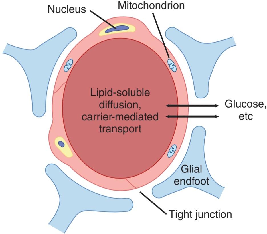
FIGURE 33-2 Transport across cerebral capillaries. Only free lipid-soluble substances can move passively across the endothelial cells. Water-soluble solutes, such as glucose, require active transport mechanisms. Proteins and protein-bound lipids are excluded.
شکل 33-2 انتقال از طریق مویرگ های مغزی. فقط مواد محلول در چربی آزاد می توانند به طور غیر فعال در سراسر سلول های اندوتلیال حرکت کنند. املاح محلول در آب، مانند گلوکز، به مکانیسم های انتقال فعال نیاز دارند. پروتئین ها و لیپیدهای متصل به پروتئین مستثنی هستند.
INNERVATION
Three systems of nerves innervate the cerebral blood vessels. Postganglionic sympathetic neurons have their cell bodies in the superior cervical ganglia, and their endings contain norepinephrine. Many also contain neuropeptide Y. Cholinergic neurons that probably originate in the sphenopalatine ganglia also innervate the cerebral vessels, and the postganglionic cholinergic neurons acting on the blood vessels contain acetylcholine. Many also contain vasoactive intestinal peptide (VIP) (see Chapter 7) and peptide histidyl methionine (PHM- 27). These nerves end primarily on large arteries. Sensory nerves are found on more distal arteries. They have their cell bodies in the trigeminal ganglia and contain substance P, neurokinin A, and calcitonin gene-related peptide (CGRP). Substance P, CGRP, VIP, and PHM-27 cause vasodilation, whereas neuropeptide Y is a vasoconstrictor. Touching or pulling on the cerebral vessels causes pain.
عصب
سه سیستم عصبی رگ های خونی مغز را عصب دهی می کنند. سلول های عصبی سمپاتیک پس گانگلیونی در گانگلیون فوقانی دهانه رحم قرار دارند و انتهای آنها حاوی نوراپی نفرین است. بسیاری نیز حاوی نوروپپتید Y هستند. نورون های کولینرژیک که احتمالاً از عقده های اسفنوپالاتین منشأ می گیرند، عروق مغزی را نیز عصب دهی می کنند، و نورون های کولینرژیک پس گانگلیونی که بر روی رگ های خونی عمل می کنند حاوی استیل کولین هستند. بسیاری از آنها همچنین حاوی پپتید روده ای فعال عروقی (VIP) (به فصل 7 مراجعه کنید) و پپتید هیستیدیل متیونین (PHM-27) دارند. این اعصاب در درجه اول به شریان های بزرگ ختم می شوند. اعصاب حسی در بیشتر شریان های دیستال یافت می شوند. آنها بدن سلولی خود را در گانگلیون سه قلو دارند و حاوی ماده P، نوروکینین A و پپتید مرتبط با ژن کلسی تونین (CGRP) هستند. ماده P، CGRP، VIP و PHM-27 باعث اتساع عروق می شوند، در حالی که نوروپپتید Y یک منقبض کننده عروق است. لمس یا کشیدن رگ های مغز باعث درد می شود.
CEREBROSPINAL FLUID
FORMATION & ABSORPTION
CSF fills the ventricles and subarachnoid space. In humans, the volume of CSF is about 150 mL, and the rate of CSF production is about 550 mL/d. Thus, the CSF turns over about 3.7 times a day. It has been estimated that 50-70% of the CSF is formed in the choroid plexuses and the remainder is formed around blood vessels and along the ventricular walls. The CSF in the ventricles flows through the foramens of Magendie and Luschka to the subarachnoid space and is absorbed through the arachnoid villi into veins, primarily the cerebral venous sinuses. The villi consist of projections of the fused arachnoid membrane and endothelium of the sinuses into low pressure venous sinuses. Similar, smaller villi project into veins around spinal nerve routes. These projections may contribute to the outflow of CSF into venous blood by a process known as bulk flow, which is unidirectional. An additional and perhaps more important route for CSF reabsorption into the bloodstream in health is via the cribriform plate above the nose and thence into the cervical lymphatics. However, reabsorption via one-way valves (of uncertain structural basis) in the arachnoid villi may assume a greater role if CSF pressure is elevated. Likewise, when CSF builds up abnormally, aquaporin water channels may be expressed in the choroid plexus and brain microvessels as a compensatory adaptation.
مایع مغزی نخاعی
تشکیل و جذب
CSF بطن ها و فضای زیر عنکبوتیه را پر می کند. در انسان، حجم CSF حدود 150 میلی لیتر و سرعت تولید CSF حدود 550 میلی لیتر در روز است. بنابراین، CSF حدود 3.7 بار در روز برمی گردد. تخمین زده شده است که 50-70 درصد CSF در شبکه های مشیمیه و بقیه در اطراف رگ های خونی و در امتداد دیواره های بطن تشکیل می شود. CSF در بطن ها از طریق سوراخ های Magendie و Luschka به فضای زیر عنکبوتیه جریان می یابد و از طریق پرزهای عنکبوتیه به وریدها جذب می شود، در درجه اول سینوس های ورید مغزی. پرزها از برجستگی های غشای عنکبوتیه ذوب شده و اندوتلیوم سینوس ها به سینوس های وریدی کم فشار تشکیل شده اند. پرزهای مشابه، کوچکتر به داخل سیاهرگهای اطراف مسیرهای عصبی نخاعی فرو می روند. این برجستگی ها ممکن است به خروج CSF به خون وریدی توسط فرآیندی به نام جریان توده ای که یک جهته است کمک کند. یک مسیر اضافی و شاید مهمتر برای بازجذب CSF به جریان خون در سلامتی، از طریق صفحه کریبریفورم بالای بینی و از آنجا به غدد لنفاوی گردنی است. با این حال، جذب مجدد از طریق دریچه های یک طرفه (با پایه ساختاری نامشخص) در پرزهای عنکبوتیه ممکن است نقش بیشتری را در صورت افزایش فشار CSF ایفا کند. به همین ترتیب، هنگامی که CSF به طور غیر طبیعی ایجاد می شود، کانال های آب آکواپورین ممکن است در شبکه مشیمیه و ریزرگ های مغز به عنوان یک سازگاری جبرانی بیان شوند.
CSF is formed continuously by the choroid plexus in two stages. First, plasma is passively filtered across the choroidal capillary endothelium. Next, secretion of water and ions across the choroidal epithelium provides for active control of CSF composition and quantity. Bicarbonate, chloride, and potassium ions enter the CSF via channels in the epithelial cell apical membranes. Aquaporins provide for water movement to balance osmotic gradients. The composition of CSF (Table 33-2) is essentially the same as that of brain extracellular fluid (ECF), which in living humans makes up 15% of the brain volume. In adults, free communication appears to take place between the brain interstitial fluid and CSF, although the diffusion distances from some parts of the brain to the CSF are appreciable. Consequently, equilibration may take some time to occur, and local areas of the brain may have extracellular microenvironments that are transiently different from CSF.
CSF به طور مداوم توسط شبکه مشیمیه در دو مرحله تشکیل می شود. اول، پلاسما به طور غیر فعال در سراسر اندوتلیوم مویرگی مشیمیه فیلتر می شود. سپس، ترشح آب و یونها در سراسر اپیتلیوم مشیمیه، کنترل فعال ترکیب و کمیت CSF را فراهم میکند. یون های بی کربنات، کلرید و پتاسیم از طریق کانال هایی در غشای آپیکال سلول های اپیتلیال وارد CSF می شوند. آکواپورین ها حرکت آب را برای متعادل کردن شیب اسمزی فراهم می کنند. ترکیب CSF (جدول 33-2) اساساً مشابه مایع خارج سلولی مغز (ECF) است که در انسان زنده 15 درصد حجم مغز را تشکیل می دهد. در بزرگسالان، به نظر می رسد که ارتباط آزاد بین مایع بینابینی مغز و CSF صورت می گیرد، اگرچه فواصل انتشار از برخی از قسمت های مغز تا CSF قابل ملاحظه است. در نتیجه، تعادل ممکن است مدتی طول بکشد تا اتفاق بیفتد و نواحی محلی مغز ممکن است دارای ریزمحیط های خارج سلولی باشند که به طور گذرا با CSF متفاوت هستند.
TABLE 33-2 Concentration of various substances in human cerebrospinal fluid (CSF) and plasma.
جدول 33-2 غلظت مواد مختلف در مایع مغزی نخاعی انسان (CSF) و پلاسما.

Lumbar CSF pressure is normally 70-180 mm H2O. Up to pressures well above this range, the rate of CSF formation is independent of intraventricular pressure. However, absorption is proportional to the pressure (Figure 33-3). At a pressure of 112 mm H2O, which is the average normal CSF pressure, formation and absorption are equal. Below a pressure of approximately 68 mm H2O, absorption stops. Large amounts of fluid accumulate when the capacity for CSF reabsorption is decreased (external hydrocephalus, communicating hydrocephalus). Fluid also accumulates proximal to the block and distends the ventricles when the foramens of Luschka and Magendie are blocked or there is obstruction within the ventricular system (internal hydrocephalus, noncommunicating hydrocephalus).
فشار CSF کمر به طور معمول 70-180 میلی متر H2O است. تا فشارهای بسیار بالاتر از این محدوده، سرعت تشکیل CSF مستقل از فشار داخل بطنی است. با این حال، جذب متناسب با فشار است (شکل 33-3). در فشار 112 میلی متر H2O، که میانگین فشار نرمال CSF است، تشکیل و جذب برابر است. زیر فشار تقریباً 68 میلی متر H2O، جذب متوقف می شود. هنگامی که ظرفیت بازجذب CSF کاهش می یابد (هیدروسفالی خارجی، هیدروسفالی ارتباطی) مقدار زیادی مایع جمع می شود. هنگامی که سوراخ های لوشکا و مگندی مسدود می شوند یا انسدادی در سیستم بطنی وجود دارد (هیدروسفالی داخلی، هیدروسفالی غیر ارتباطی)، مایع در نزدیکی بلوک تجمع می یابد و بطن ها را متسع می کند.
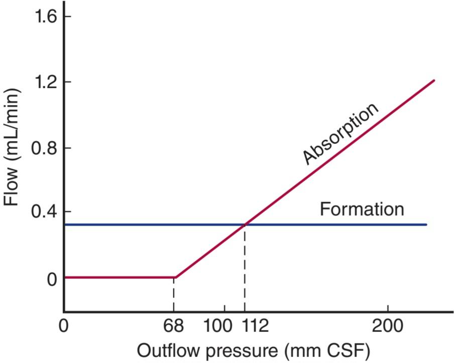
FIGURE 33-3 CSF formation and absorption in humans at various CSF pressures. Note that at 112 mm CSF, formation and absorption are equal, and at 68 mm CSF, absorption is zero. CSF, cerebrospinal fluid. (Modified with permission from Cutler RWP, et al: Formation and absorption of cerebrospinal fluid in man. Brain 1968;91(4):707-720.)
شکل 33-3 تشکیل و جذب CSF در انسان در فشارهای مختلف CSF. توجه داشته باشید که در CSF 112 میلی متر تشکیل و جذب برابر است و در CSF 68 میلی متر جذب صفر است. CSF، مایع مغزی نخاعی. (اصلاح شده با اجازه کاتلر RWP، و همکاران: تشکیل و جذب مایع مغزی نخاعی در انسان. Brain 1968;91(4):707-720.)
PROTECTIVE FUNCTION
The most critical role for CSF (and the meninges) is to protect the brain. The dura is attached firmly to bone. Normally, there is no “subdural space,” with the arachnoid being held to the dura by the surface tension of the thin layer of fluid between the two membranes. As shown in Figure 33-4, the brain itself is supported within the arachnoid by the blood vessels and nerve roots and by the multiple fine fibrous arachnoid trabeculae. The brain weighs about 1400 g in air, but in its “water bath” of CSF it has a net weight of only 50 g. The buoyancy of the brain in the CSF permits its relatively flimsy attachments to suspend it very effectively. When the head receives a blow, the arachnoid slides on the dura and the brain moves, but its motion is gently checked by the CSF cushion and by the arachnoid trabeculae.
عملکرد حفاظتی
حیاتی ترین نقش CSF (و مننژها) محافظت از مغز است. دورا محکم به استخوان چسبیده است. به طور معمول، “فضای سابدورال” وجود ندارد و عنکبوتیه توسط کشش سطحی لایه نازک مایع بین دو غشاء به سختگیری چسبیده میشود. همانطور که در شکل 33-4 نشان داده شده است، خود مغز در داخل عنکبوتیه توسط رگ های خونی و ریشه های عصبی و توسط ترابکول های عنکبوتیه فیبری ریز متعدد حمایت می شود. وزن مغز در هوا حدود 1400 گرم است، اما در “حمام آب” CSF خود تنها 50 گرم وزن دارد. شناوری مغز در CSF به اتصالات نسبتاً ضعیف آن اجازه می دهد تا آن را به طور بسیار مؤثری معلق کنند. هنگامی که سر ضربه ای دریافت می کند، عنکبوتیه بر روی سخت شامه می لغزد و مغز حرکت می کند، اما حرکت آن به آرامی توسط بالشتک CSF و ترابکول های عنکبوتیه بررسی می شود.

FIGURE 33-4 Investing membranes of the brain, showing their relation to the skull and to brain tissue. (Reproduced with permission from Young B, Heath JW: Wheater’s Functional Histology, 4th ed. Churchill Livingstone;2000.)
شکل 33-4 غشای سرمایه گذاری مغز، رابطه آنها را با جمجمه و بافت مغز نشان می دهد. (تکثیر شده با اجازه یانگ بی، هیث جی دبلیو: بافت شناسی عملکردی ویتر، ویرایش چهارم چرچیل لیوینگستون؛ 2000.)
The pain produced by spinal fluid deficiency illustrates the importance of CSF in supporting the brain. Removal of CSF during lumbar puncture can cause a severe headache after the fluid is removed, because the brain hangs on the vessels and nerve roots, and traction on them stimulates pain fibers. The pain can be relieved by intrathecal injection of sterile isotonic saline.
درد ناشی از کمبود مایع نخاعی اهمیت CSF را در حمایت از مغز نشان می دهد. برداشتن CSF در حین پونکسیون کمری می تواند باعث سردرد شدید پس از برداشتن مایع شود، زیرا مغز روی عروق و ریشه های عصبی آویزان است و کشش روی آنها باعث تحریک رشته های درد می شود. درد را می توان با تزریق داخل نخاعی سالین استریل ایزوتونیک تسکین داد.
HEAD INJURIES
Without the protection of the spinal fluid and the meninges, the brain would probably be unable to withstand even the minor traumas of everyday living; but with the protection afforded, it takes a fairly severe blow to produce cerebral damage. The brain is damaged most commonly when the skull is fractured and bone is driven into neural tissue (depressed skull fracture), when the brain moves far enough to tear the delicate bridging veins from the cortex to the bone, or when the brain is accelerated by a blow on the head and is driven against the skull or the tentorium at a point opposite where the blow was struck (contrecoup injury).
آسیب های سر
بدون محافظت از مایع نخاعی و مننژها، مغز احتمالاً قادر به تحمل آسیب های جزئی زندگی روزمره نخواهد بود. اما با حفاظت ارائه شده، ضربه نسبتاً شدیدی لازم است تا آسیب مغزی ایجاد شود. زمانی که جمجمه شکسته می شود و استخوان به داخل بافت عصبی هدایت می شود (شکستگی افسرده جمجمه)، زمانی که مغز به اندازه کافی حرکت می کند تا وریدهای پل زدن ظریف از قشر به استخوان را پاره کند، یا زمانی که مغز با ضربه ای به سر شتاب می گیرد و در مقابل آسیب جمجمه یا دهنه استخوانی در مقابل آسیب جمجمه قرار گرفته است، معمولا آسیب می بیند.
THE BLOOD-BRAIN BARRIER
The tight junctions between capillary endothelial cells in the brain and between the epithelial cells in the choroid plexus effectively prevent proteins from entering the brain in adults and slow the penetration of some smaller molecules as well. An example is the slow penetration of urea (Figure 33-5). This uniquely limited exchange of substances into the brain is referred to as the blood-brain barrier, a term most commonly used to encompass this barrier overall and more specifically the barrier in the choroid epithelium between blood and CSF.
سد خونی- مغزی
اتصالات محکم بین سلول های اندوتلیال مویرگی در مغز و بین سلول های اپیتلیال در شبکه مشیمیه به طور موثری از ورود پروتئین ها به مغز در بزرگسالان جلوگیری می کند و نفوذ برخی مولکول های کوچکتر را نیز کند می کند. به عنوان مثال، نفوذ آهسته اوره است (شکل 33-5). این تبادل منحصر به فرد مواد در مغز به عنوان سد خونی-مغزی نامیده می شود، اصطلاحی که بیشتر برای دربرگرفتن این سد به طور کلی و به طور خاص سد در اپیتلیوم مشیمیه بین خون و CSF استفاده می شود.

FIGURE 33-5 Penetration of urea into muscle, brain, and cerebrospinal fluid (CSF). Urea was administered by constant infusion.
شکل 33-5 نفوذ اوره به ماهیچه، مغز و مایع مغزی نخاعی (CSF). اوره با انفوزیون مداوم تجویز شد.
Passive diffusion across the tight cerebral capillaries is very limited, and little vesicular transport takes place. However, there are numerous carrier-mediated and active transport systems in the cerebral capillaries.
انتشار غیرفعال در سراسر مویرگ های سفت مغزی بسیار محدود است و انتقال تاولی کمی انجام می شود. با این حال، سیستم های حمل و نقل فعال و فعال متعددی در مویرگ های مغزی وجود دارد.
PENETRATION OF SUBSTANCES INTO THE BRAIN
Water, CO2, and O2 penetrate the brain with ease, as do the lipid-soluble free forms of steroid hormones, whereas their protein-bound forms and, in general, all proteins and polypeptides do not. The rapid passive penetration of CO2 contrasts with the regulated transcellular penetration of H+ and HCO3 and has physiologic significance in the regulation of respiration (see Chapter 35).
نفوذ مواد به مغز
آب، CO2 و O2 به آسانی به مغز نفوذ می کنند، همانطور که اشکال آزاد محلول در چربی هورمون های استروئیدی، در حالی که اشکال متصل به پروتئین آنها و به طور کلی، همه پروتئین ها و پلی پپتیدها اینطور نیستند. نفوذ غیرفعال سریع CO2 با نفوذ بین سلولی تنظیم شده +H و HCO3 در تضاد است و اهمیت فیزیولوژیکی در تنظیم تنفس دارد (به فصل 35 مراجعه کنید).
Glucose is the major ultimate source of energy for nerve cells. Its diffusion across the blood-brain barrier would be very slow, but the rate of transport into the CSF is markedly enhanced by the presence of specific transporters, including the glucose transporter 1 (GLUT1). The brain contains two forms of GLUT1: GLUT1 55K and GLUT1 45K. Both are encoded by the same gene, but they differ in the extent to which they are glycosylated. GLUT1 55K is present in high concentration in brain capillaries (Figure 33-6). Infants with congenital GLUT1 deficiency have low CSF glucose concentrations in the presence of normal plasma glucose, and they have seizures and delayed development. In addition, transporters for thyroid hormones; several organic acids; choline; nucleic acid precursors; and neutral, basic, and acidic amino acids are present at the blood-brain barrier.
گلوکز منبع اصلی انرژی برای سلول های عصبی است. انتشار آن در سد خونی مغزی بسیار آهسته خواهد بود، اما سرعت انتقال به CSF به طور قابل توجهی با حضور ناقلان خاص، از جمله ناقل گلوکز 1 (GLUT1) افزایش می یابد. مغز شامل دو شکل GLUT1 است: GLUT1 55K و GLUT1 45K. هر دو توسط یک ژن رمزگذاری می شوند، اما در میزان گلیکوزیله شدن با هم تفاوت دارند. GLUT1 55K در غلظت بالا در مویرگ های مغز وجود دارد (شکل 33-6). نوزادان مبتلا به کمبود مادرزادی GLUT1 در حضور گلوکز طبیعی پلاسما، غلظت گلوکز CSF پایینی دارند و دچار تشنج و تاخیر در رشد می شوند. علاوه بر این، انتقال دهنده های هورمون های تیروئید. چندین اسید آلی؛ کولین؛ پیش سازهای اسید نوکلئیک؛ و اسیدهای آمینه خنثی، بازی و اسیدی در سد خونی مغزی وجود دارند.

FIGURE 33-6 Localization of the various GLUT transporters in the brain. (Adapted with permission from Maher F, Vannucci SJ, Simpson IA: Glucose transporter proteins in brain. FASEB J 1994 Oct; 8(13):1003-1011.)
شکل 33-6 محلی سازی ناقل های مختلف GLUT در مغز. (اقتباس شده با اجازه Maher F, Vannucci SJ, Simpson IA: Glucose transporter proteins in brain. FASEB J اکتبر 1994؛ 8(13):1003-1011.)
A variety of drugs and peptides actually permeate into the endothelial cells of the cerebral capillaries but are promptly transported back into the blood by a nonspecific multidrug transporter in the apical membrane. This P-glycoprotein is a member of the family of adenosine triphosphate (ATP) binding cassette proteins that transport various proteins and lipids across cell membranes (see Chapter 2). In the absence of this transporter in mice, much larger proportions of systemically administered doses of various chemotherapeutic drugs, analgesics, and opioid peptides are found in the brain than in controls. If pharmacologic agents that inhibit this transporter can be developed, they could be of value in the treatment of brain tumors and other central nervous system (CNS) diseases in which it is difficult to introduce adequate amounts of therapeutic agents into the brain.
انواعی از داروها و پپتیدها در واقع به داخل سلولهای اندوتلیال مویرگهای مغزی نفوذ میکنند، اما به سرعت توسط یک ناقل چند دارویی غیر اختصاصی در غشای آپیکال به داخل خون منتقل میشوند. این P-گلیکوپروتئین عضوی از خانواده پروتئینهای کاست اتصال دهنده آدنوزین تری فسفات (ATP) است که پروتئینها و لیپیدهای مختلف را در غشای سلولی منتقل میکند (به فصل 2 مراجعه کنید). در غیاب این ناقل در موشها، نسبتهای بسیار بیشتری از دوزهای سیستمیک تجویز شده از داروهای مختلف شیمیدرمانی، مسکنها و پپتیدهای اپیوئیدی در مغز نسبت به گروه کنترل یافت میشود. اگر بتوان عوامل دارویی را که این ناقل را مهار میکنند توسعه داد، میتوانند در درمان تومورهای مغزی و سایر بیماریهای سیستم عصبی مرکزی (CNS) که در آنها وارد کردن مقادیر کافی از عوامل درمانی به مغز دشوار است، مفید باشند.
CIRCUMVENTRICULAR ORGANS
When dyes that bind to proteins in the plasma are injected, they stain many tissues but spare most of the brain. However, four small areas in or near the brainstem do take up the stain. These areas are: (1) the posterior pituitary (neurohypophysis) and the adjacent ventral part of the median eminence of the hypothalamus, (2) the area postrema, (3) the organum vasculosum of the lamina terminalis (OVLT, supraoptic crest), and (4) the subfornical organ (SFO).
اندام های دور بطنی
هنگامی که رنگ هایی که به پروتئین های پلاسما متصل می شوند تزریق می شوند، بسیاری از بافت ها را لکه دار می کنند اما بیشتر مغز را از بین می برند. با این حال، چهار ناحیه کوچک در یا نزدیک ساقه مغز، لکه را می گیرند. این نواحی عبارتند از: (1) هیپوفیز خلفی (هیپوفیز عصبی) و قسمت شکمی مجاور برجستگی میانی هیپوتالاموس، (2) ناحیه پسرما، (3) اندام عروقی لایه انتهایی (OVLT، تاج فوق اپتیک)، و (4) اندام زیرفرونی (SFO).
These areas are referred to collectively as the circumventricular organs (Figure 33-7). All have fenestrated capillaries, and because of their permeability they are said to be “outside the blood-brain barrier.” Some of them function as neurohemal organs; that is, areas in which polypeptides secreted by neurons enter the circulation. Others contain receptors for many different peptides and other substances, and function as chemoreceptor zones in which substances in the circulating blood can act to trigger changes in brain function without penetrating the blood-brain barrier. For example, the area postrema is a chemoreceptor trigger zone that initiates vomiting in response to chemical changes in the plasma (see Chapter 27). It is also concerned with cardiovascular control, where circulating angiotensin II acts to produce a neurally mediated increase in blood pressure. Angiotensin II also acts on the SFO and possibly on the OVLT to increase water intake. In addition, it appears that the OVLT is the site of the osmoreceptor controlling vasopressin secretion (see Chapter 38), and evidence suggests that circulating interleukin-1 (IL-1) produces fever by acting here too.
این نواحی در مجموع به عنوان اندام های دور بطنی نامیده می شوند (شکل 33-7). همه آنها مویرگهای دمدار دارند و به دلیل نفوذپذیری آنها گفته می شود که “خارج از سد خونی مغزی” هستند. برخی از آنها به عنوان اندام های عصبی عمل می کنند. یعنی مناطقی که پلی پپتیدهای ترشح شده توسط نورون ها وارد گردش خون می شوند. برخی دیگر حاوی گیرندههایی برای پپتیدهای مختلف و سایر مواد هستند و به عنوان مناطق گیرنده شیمیایی عمل میکنند که در آن مواد موجود در خون در گردش میتوانند باعث ایجاد تغییراتی در عملکرد مغز بدون نفوذ به سد خونی مغزی شوند. به عنوان مثال، ناحیه postrema ناحیه ماشه گیرنده شیمیایی است که در پاسخ به تغییرات شیمیایی در پلاسما، استفراغ را آغاز می کند (به فصل 27 مراجعه کنید). همچنین به کنترل قلبی عروقی مربوط می شود، جایی که آنژیوتانسین II در گردش باعث افزایش فشار خون با واسطه عصبی می شود. آنژیوتانسین II همچنین بر روی SFO و احتمالاً روی OVLT برای افزایش مصرف آب تأثیر می گذارد. علاوه بر این، به نظر می رسد که OVLT محل گیرنده اسموری است که ترشح وازوپرسین را کنترل می کند (به فصل 38 مراجعه کنید)، و شواهد نشان می دهد که اینترلوکین-1 در گردش (IL-1) با عمل در اینجا نیز باعث ایجاد تب می شود.
The subcommissural organ (Figure 33-7) is closely associated with the pineal gland and histologically resembles the circumventricular organs. However, it does not have fenestrated capillaries, and is not highly permeable. It may, instead, be involved in guiding axons during brain development. Conversely, the pineal and the anterior pituitary do have fenestrated capillaries and are outside the blood-brain barrier, but both are endocrine glands and are not part of the brain.
اندام زیرکمیسیورال (شکل 33-7) ارتباط نزدیکی با غده صنوبری دارد و از نظر بافت شناسی شبیه اندام های دور بطنی است. با این حال، مویرگ های فنس دار ندارد و نفوذپذیری بالایی ندارد. در عوض ممکن است در هدایت آکسون ها در طول رشد مغز نقش داشته باشد. برعکس، پینهآل و هیپوفیز قدامی دارای مویرگهای بالدار هستند و خارج از سد خونی مغزی هستند، اما هر دو غدد درونریز هستند و بخشی از مغز نیستند.

FIGURE 33-7 Circumventricular organs. The neurohypophysis (NH), organum vasculosum of the lamina terminalis (OVLT, organum vasculosum of the lamina terminalis), subfornical organ (SFO), and area postrema (AP) are shown projected on a sagittal section of the human brain. PI, pineal; SCO, subcommissural organ.
شکل 33-7 اندام های دور بطنی. نوروهیپوفیز (NH)، اندام واسکولوزوم لایه انتهایی (OVLT، اندام عروقی لایه انتهایی)، اندام ساب فورنیکال (SFO)، و ناحیه پسترما (AP) بر روی یک بخش ساژیتال مغز انسان نشان داده شده است. PI، پینه آل؛ سازمان SCO، ارگان فرعی.
FUNCTION OF THE BLOOD-BRAIN BARRIER
The blood-brain barrier strives to maintain the constancy of the environment of the neurons in the CNS (Clinical Box 33-1). Even minor variations in the concentrations of K+, Ca2+, Mg2+, H+, and other ions can have far-reaching consequences. The constancy of the composition of the ECF in all parts of the body is maintained by multiple homeostatic mechanisms (see Chapters 1 and 38), but because of the sensitivity of the cortical neurons to ionic change, it is not surprising that an additional defense has evolved to protect them. Other functions of the blood-brain barrier include protection of the brain from endogenous and exogenous toxins in the blood and prevention of the escape of neurotransmitters into the general circulation.
عملکرد سد خونی-مغزی
سد خونی مغزی برای حفظ ثبات محیط نورون ها در CNS تلاش می کند (جعبه بالینی 33-1). حتی تغییرات جزئی در غلظتهای +K+، Ca2+، Mg2+، H و سایر یونها میتواند پیامدهای گستردهای داشته باشد. ثبات ترکیب ECF در تمام قسمتهای بدن توسط مکانیسمهای هموستاتیک متعدد حفظ میشود (به فصلهای 1 و 38 مراجعه کنید)، اما به دلیل حساسیت نورونهای قشر مغز به تغییرات یونی، تعجب آور نیست که دفاع اضافی برای محافظت از آنها تکامل یافته است. سایر عملکردهای سد خونی مغزی شامل محافظت از مغز در برابر سموم درون زا و برون زا در خون و جلوگیری از فرار انتقال دهنده های عصبی به گردش خون عمومی است.
DEVELOPMENT OF THE BLOOD-BRAIN BARRIER
In experimental animals, many small molecules penetrate the brain more readily during the fetal and neonatal period than they do in the adult. On this basis, it is often stated that the blood-brain barrier is immature at birth. Humans are more mature at birth, and not surprisingly, detailed data on passive permeability of the normal human neonatal blood-brain barrier are not available. However, in severely jaundiced infants with high plasma levels of free bilirubin and an immature hepatic bilirubin-conjugating system, free bilirubin enters the brain and, in the presence of asphyxia, damages the basal ganglia (kernicterus). The counterpart of this situation in later life is the Crigler-Najjar syndrome in which there is a congenital deficiency of glucuronyl transferase. These individuals can have very high free bilirubin levels in the blood and develop encephalopathy. In other conditions, however, free bilirubin levels are generally not high enough to produce brain damage.
توسعه سد خونی مغزی
در حیوانات آزمایشی، بسیاری از مولکولهای کوچک در دوران جنینی و نوزادی راحتتر از بزرگسالان به مغز نفوذ میکنند. بر این اساس، اغلب گفته می شود که سد خونی مغزی در بدو تولد نابالغ است. انسانها در بدو تولد بالغتر هستند و جای تعجب نیست که دادههای دقیق در مورد نفوذپذیری غیرفعال سد خونی مغزی طبیعی نوزادان انسان در دسترس نیست. با این حال، در نوزادان مبتلا به زردی شدید با سطوح بالای بیلی روبین آزاد پلاسمایی و سیستم کونژوگه کننده بیلی روبین کبدی نابالغ، بیلی روبین آزاد وارد مغز شده و در صورت خفگی، به گانگلیون های پایه (کرنیکتروس) آسیب می رساند. همتای این وضعیت در زندگی بعدی، سندرم کریگلر-نجار است که در آن کمبود مادرزادی گلوکورونیل ترانسفراز وجود دارد. این افراد می توانند سطح بیلی روبین آزاد بسیار بالایی در خون داشته باشند و به آنسفالوپاتی مبتلا شوند. با این حال، در شرایط دیگر، سطح بیلی روبین آزاد به طور کلی به اندازه کافی بالا نیست که آسیب مغزی ایجاد کند.
CLINICAL BOX 33- 1
Clinical Implications of the Blood-Brain Barrier
Clinicians must know the degree to which drugs penetrate the brain in order to treat diseases of the nervous system intelligently. For example, it is clinically relevant that the amines dopamine and serotonin penetrate brain tissue to a very limited degree but their corresponding acid precursors, L- dopa and 5-hydroxytryptophan, respectively, enter with relative ease (see Chapters 7 and 12). Another important clinical consideration is the fact that the blood-brain barrier tends to break down in areas of infection or injury. Tumors develop new blood vessels, and the capillaries that are formed lack contact with normal astrocytes. Therefore, there are no tight junctions, and the vessels may even be fenestrated. The lack of a barrier helps in identifying the location of tumors; substances such as radioactive iodine-labeled albumin penetrate normal brain tissue very slowly, but they enter tumor tissue, making the tumor stand out as an island of radioactivity in the surrounding normal brain. The blood-brain barrier can also temporarily be disrupted by sudden marked increases in blood pressure or by intravenous injection of hypertonic fluids.
جعبه بالینی 33- 1
پیامدهای بالینی سد خونی مغزی
پزشکان برای درمان هوشمندانه بیماری های سیستم عصبی باید میزان نفوذ داروها به مغز را بدانند. برای مثال، از نظر بالینی مهم است که آمینهای دوپامین و سروتونین به میزان بسیار محدودی به بافت مغز نفوذ میکنند، اما پیشسازهای اسیدی مربوط به آنها، به ترتیب L-dopa و 5-hydroxytryptophan، با سهولت نسبی وارد میشوند (فصل 7 و 12 را ببینید). یکی دیگر از ملاحظات بالینی مهم این واقعیت است که سد خونی مغزی تمایل دارد در مناطق عفونت یا آسیب شکسته شود. تومورها رگ های خونی جدیدی ایجاد می کنند و مویرگ هایی که تشکیل می شوند با آستروسیت های طبیعی تماس ندارند. بنابراین، اتصالات محکمی وجود ندارد و حتی ممکن است رگ ها دارای فنس باشند. فقدان مانع به شناسایی محل تومورها کمک می کند. موادی مانند آلبومین نشاندار شده با ید رادیواکتیو بسیار آهسته به بافت طبیعی مغز نفوذ می کنند، اما وارد بافت تومور می شوند و تومور را به عنوان جزیره ای از رادیواکتیویته در مغز طبیعی اطراف برجسته می کند. سد خونی مغزی همچنین می تواند به طور موقت با افزایش ناگهانی فشار خون یا تزریق داخل وریدی مایعات هیپرتونیک مختل شود.
CEREBRAL BLOOD FLOW & ITS REGULATION
KETY METHOD
According to the Fick’s principle (see Chapter 30), the blood flow of any organ can be measured by determining the amount of a given substance (Q1) removed from the bloodstream by the organ per unit of time and dividing that value by the difference between the concentration of the substance in arterial blood and the concentration in the venous blood from the organ ([A、]-[V1]). Thus:
جریان خون مغزی و تنظیم آن
روش KETY
طبق اصل فیک (به فصل 30 مراجعه کنید)، جریان خون هر اندامی را می توان با تعیین مقدار ماده معین (Q1) که از جریان خون حذف می شود توسط اندام در واحد زمان و تقسیم آن مقدار بر اختلاف بین غلظت ماده در خون شریانی و غلظت خون وریدی اندام ([A]-[V) اندازه گیری کرد. بدین ترتیب:

This can be applied clinically using inhaled nitrous oxide (NO) (Kety method). The average cerebral blood flow in young adults is 54 mL/100 g/min. The average adult brain weighs about 1400 g, so the flow for the whole brain is about 756 mL/min. Note that the Kety method provides an average value for perfused areas of brain and gives no information about regional differences in blood flow. It also can only measure flow to perfused parts of the brain. If the blood flow to a portion of the brain is occluded, the measured flow does not change because the nonperfused area does not take up any N2O.
این را می توان به صورت بالینی با استفاده از اکسید نیتروژن استنشاقی (NO) (روش Kety) اعمال کرد. متوسط جریان خون مغزی در بزرگسالان جوان 54 میلی لیتر در 100 گرم در دقیقه است. میانگین وزن مغز بالغ حدود 1400 گرم است، بنابراین جریان برای کل مغز حدود 756 میلی لیتر در دقیقه است. توجه داشته باشید که روش Kety یک مقدار متوسط را برای مناطق پرفیوژن مغز ارائه می دهد و هیچ اطلاعاتی در مورد تفاوت های منطقه ای در جریان خون ارائه نمی دهد. همچنین فقط می تواند جریان را در قسمت های پرفیوژن مغز اندازه گیری کند. اگر جریان خون به بخشی از مغز مسدود شود، جریان اندازهگیری شده تغییر نمیکند زیرا ناحیه بدون پرفیوژن هیچ N2O را جذب نمیکند.
In spite of the marked local fluctuations in brain blood flow with neural activity, the cerebral circulation is regulated in such a way that total blood flow remains relatively constant. The factors involved in regulating the flow are summarized in Figure 33-8.
علیرغم نوسانات موضعی مشخص در جریان خون مغز با فعالیت عصبی، گردش خون مغزی به گونه ای تنظیم می شود که جریان خون کل نسبتاً ثابت می ماند. عوامل دخیل در تنظیم جریان در شکل 33-8 خلاصه شده است.
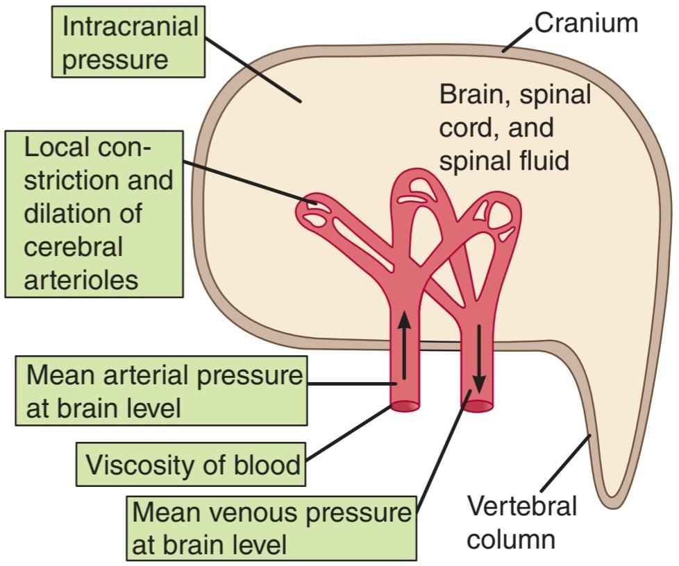
FIGURE 33-8 Diagrammatic summary of the factors affecting overall cerebral blood flow.
شکل 33-8 خلاصه نموداری عوامل مؤثر بر جریان کلی خون مغزی.
ROLE OF INTRACRANIAL PRESSURE
In adults, the brain, spinal cord, and spinal fluid are encased, along with the cerebral vessels, in a rigid bony enclosure. The cranial cavity normally contains a brain weighing approximately 1400 g, 75 mL of blood, and 75 mL of spinal fluid. Because brain tissue and spinal fluid are essentially incompressible, the volume of blood, spinal fluid, and brain in the cranium at any time must be relatively constant (Monro-Kellie doctrine). More importantly, the cerebral vessels are compressed whenever the intracranial pressure rises. Any change in venous pressure promptly causes a similar change in intracranial pressure. Thus, a rise in venous pressure decreases cerebral blood flow both by decreasing the effective perfusion pressure and by compressing the cerebral vessels. This relationship helps compensate for changes in arterial blood pressure at the level of the head. For example, if the body is accelerated upward (positive g), blood moves toward the feet and arterial pressure at the level of the head decreases. However, venous pressure also falls and intracranial pressure falls, so that the pressure on the vessels decreases and blood flow is much less severely compromised than it would otherwise be. Conversely, during acceleration downward, force acting toward the head (negative g) increases arterial pressure at head level, but intracranial pressure also rises, so that the vessels are supported and do not rupture. The cerebral vessels are protected during the straining associated with defecation or delivery in the same way.
نقش فشار داخل جمجمه ای
در بزرگسالان، مغز، نخاع و مایع نخاعی همراه با عروق مغزی در یک محفظه استخوانی سفت و محکم قرار دارند. حفره جمجمه معمولاً حاوی مغزی با وزن تقریبی 1400 گرم، 75 میلی لیتر خون و 75 میلی لیتر مایع نخاعی است. از آنجایی که بافت مغز و مایع نخاعی اساساً تراکم ناپذیر هستند، حجم خون، مایع نخاعی و مغز در جمجمه در هر زمان باید نسبتاً ثابت باشد (دکترین مونرو-کلی). مهمتر از آن، هر زمان که فشار داخل جمجمه افزایش یابد، رگ های مغز فشرده می شوند. هر گونه تغییر در فشار وریدی به سرعت باعث تغییر مشابه در فشار داخل جمجمه می شود. بنابراین، افزایش فشار وریدی، جریان خون مغزی را هم با کاهش فشار پرفیوژن موثر و هم با فشرده کردن عروق مغزی کاهش میدهد. این رابطه به جبران تغییرات فشار خون شریانی در سطح سر کمک می کند. به عنوان مثال، اگر بدن به سمت بالا شتاب بگیرد (g مثبت)، خون به سمت پاها حرکت می کند و فشار شریانی در سطح سر کاهش می یابد. با این حال، فشار وریدی نیز کاهش مییابد و فشار داخل جمجمه کاهش مییابد، به طوری که فشار روی رگها کاهش مییابد و جریان خون به شدت کمتر از آنچه در غیر این صورت بود به خطر میافتد. برعکس، در حین شتاب به سمت پایین، نیرویی که به سمت سر وارد می شود (g منفی) فشار شریانی را در سطح سر افزایش می دهد، اما فشار داخل جمجمه نیز افزایش می یابد، به طوری که رگ ها حمایت می شوند و پاره نمی شوند. رگ های مغزی در طول فشار ناشی از اجابت مزاج یا زایمان به همان روش محافظت می شوند.
AUTOREGULATION
As seen in other vascular beds, autoregulation is prominent in the brain (Figure 33-9). This process, by which the flow to many tissues is maintained at relatively constant levels despite variations in perfusion pressure, is discussed in Chapter 32. In the brain, autoregulation maintains a constant cerebral blood flow at arterial pressures of 65-140 mm Hg.
AUTOREGULATION
همانطور که در سایر بسترهای عروقی دیده می شود، خودتنظیمی در مغز برجسته است (شکل 33-9). این فرآیند، که توسط آن جریان به بسیاری از بافتها علیرغم تغییرات فشار پرفیوژن در سطوح نسبتاً ثابتی حفظ میشود، در فصل 32 مورد بحث قرار میگیرد. در مغز، تنظیم خودکار جریان خون مغزی را در فشارهای شریانی 65-140 میلیمتر جیوه حفظ میکند.

FIGURE 33-9 Autoregulation of cerebral blood flow (CBF) during steady- state conditions. The blue line shows the alteration produced by sympathetic stimulation during autoregulation.
شکل 33-9 تنظیم خودکار جریان خون مغزی (CBF) در شرایط حالت پایدار. خط آبی تغییر ایجاد شده توسط تحریک سمپاتیک در طول خودتنظیمی را نشان می دهد.
ROLE OF VASOMOTOR & SENSORY NERVES
The innervation of large cerebral blood vessels by postganglionic sympathetic and parasympathetic nerves and the additional distal innervation by sensory nerves have been described above. The nerves may also modulate tone indirectly, via the release of paracrine substances from astrocytes. The precise role of these nerves, however, remains a matter of debate. It has been argued that noradrenergic discharge occurs when the blood pressure is markedly elevated. This reduces the resultant passive increase in blood flow and helps protect the blood-brain barrier from the disruption that could otherwise occur (see above). Thus, vasomotor discharges affect autoregulation. With sympathetic stimulation, the constant-flow, or plateau, part of the pressure-flow curve is extended to the right (Figure 33-9); that is, greater increases in pressure can occur without an increase in flow. On the other hand, the vasodilator hydralazine and the angiotensin-converting enzyme (ACE) inhibitor captopril reduce the length of the plateau. Finally, neurovascular coupling may adjust local perfusion in response to changes in brain activity (see below).
نقش اعصاب وازوموتور و حسی
عصب دهی عروق خونی بزرگ مغز توسط اعصاب سمپاتیک و پاراسمپاتیک پس از عقده ای و عصب دیستال اضافی توسط اعصاب حسی در بالا توضیح داده شده است. اعصاب همچنین ممکن است به طور غیرمستقیم تن را از طریق آزادسازی مواد پاراکرین از آستروسیت ها تعدیل کنند. نقش دقیق این اعصاب، با این حال، موضوع بحث است. استدلال شده است که ترشح نورآدرنرژیک زمانی رخ می دهد که فشار خون به طور قابل توجهی افزایش یابد. این افزایش غیرفعال ناشی از جریان خون را کاهش می دهد و به محافظت از سد خونی مغزی در برابر اختلالاتی که در غیر این صورت ممکن است رخ دهد کمک می کند (به بالا مراجعه کنید). بنابراین، ترشحات وازوموتور بر تنظیم خودکار تأثیر می گذارد. با تحریک سمپاتیک، جریان ثابت، یا فلات، بخشی از منحنی فشار-جریان به سمت راست کشیده می شود (شکل 33-9). یعنی افزایش بیشتر فشار می تواند بدون افزایش جریان رخ دهد. از سوی دیگر، هیدرالازین گشادکننده عروق و کاپتوپریل مهارکننده آنزیم مبدل آنژیوتانسین (ACE) طول فلات را کاهش میدهند. در نهایت، جفت عصبی عروقی ممکن است پرفیوژن موضعی را در پاسخ به تغییرات در فعالیت مغز تنظیم کند (به زیر مراجعه کنید).
BLOOD FLOW IN VARIOUS PARTS OF THE BRAIN
A major advance in recent decades has been the development of techniques for monitoring regional blood flow in living, conscious humans. Among the most valuable methods are positron emission tomography (PET) and related techniques in which a short-lived radioisotope is used to label a compound and the compound is injected. The arrival and clearance of the tracer are monitored by scintillation detectors placed over the head. Because blood flow is tightly coupled to brain metabolism, local uptake of 2-deoxyglucose is also a good index of blood flow (see below and Chapter 1). If the 2-deoxyglucose is labeled with a short half-life positron emitter such as 18F, 110, or 150, its concentration in any part of the brain can be monitored.
جریان خون در قسمت های مختلف مغز
پیشرفت عمده در دهه های اخیر توسعه تکنیک هایی برای نظارت بر جریان خون منطقه ای در انسان های زنده و آگاه بوده است. از جمله با ارزش ترین روش ها می توان به توموگرافی انتشار پوزیترون (PET) و تکنیک های مرتبط با آن اشاره کرد که در آن از یک رادیوایزوتوپ کوتاه مدت برای برچسب زدن یک ترکیب و تزریق آن ترکیب استفاده می شود. ورود و پاکسازی ردیاب توسط آشکارسازهای سوسوزن که روی سر قرار داده شده است، کنترل می شود. از آنجایی که جریان خون به شدت با متابولیسم مغز مرتبط است، جذب موضعی 2-دئوکسی گلوکز نیز شاخص خوبی برای جریان خون است (به زیر و فصل 1 مراجعه کنید). اگر 2-دئوکسی گلوکز با یک ساطع کننده پوزیترون نیمه عمر کوتاه مانند 18F، 110 یا 150 برچسب زده شود، غلظت آن در هر قسمت از مغز قابل بررسی است.
Another valuable technique involves magnetic resonance imaging (MRI). MRI is based on detecting resonant signals from different tissues in a magnetic field. Functional magnetic resonance imaging (fMRI) measures the amount of blood in a tissue area. When neurons become active, their increased discharge alters the local field potential. A still unsettled mechanism triggers an increase in local blood flow and oxygen. The increase in oxygenated blood is detected by fMRI. PET scanning can be used to measure not only blood flow but also the concentration of molecules, such as dopamine, in various regions of the living brain. On the other hand, fMRI does not involve the use of radioactivity. Consequently, it can be used at frequent intervals to measure changes in regional blood flow in a single individual.
یکی دیگر از تکنیک های ارزشمند، تصویربرداری رزونانس مغناطیسی (MRI) است. MRI بر اساس تشخیص سیگنال های تشدید از بافت های مختلف در یک میدان مغناطیسی است. تصویربرداری رزونانس مغناطیسی عملکردی (fMRI) میزان خون را در یک ناحیه بافت اندازه گیری می کند. هنگامی که نورونها فعال میشوند، افزایش تخلیه آنها پتانسیل میدان محلی را تغییر میدهد. مکانیسمی که هنوز ثابت نشده است باعث افزایش جریان خون موضعی و اکسیژن می شود. افزایش خون اکسیژن دار توسط fMRI تشخیص داده می شود. اسکن PET می تواند نه تنها برای اندازه گیری جریان خون، بلکه غلظت مولکول هایی مانند دوپامین در نواحی مختلف مغز زنده مورد استفاده قرار گیرد. از طرف دیگر، fMRI شامل استفاده از رادیواکتیویته نمی شود. در نتیجه، می توان از آن در فواصل زمانی مکرر برای اندازه گیری تغییرات در جریان خون منطقه ای در یک فرد استفاده کرد.
In resting humans, the average blood flow in gray matter is 69 mL/100 g/min compared with 28 mL/100 g/min in white matter. A striking feature of cerebral function is the marked variation in local blood flow with changes in brain activity. In persons who are awake but at rest, blood flow is greatest in the premotor and frontal regions. This is the part of the brain that is believed to be concerned with decoding and analyzing afferent input and with intellectual activity. During voluntary clenching of the right hand, flow is increased in the hand area of the left motor cortex and the corresponding sensory areas in the postcentral gyrus. Especially when the movements being performed are sequential, the flow is also increased in the supplementary motor area. When persons talk, there is a bilateral increase in blood flow in the face, tongue, and mouth-sensory and motor areas and the upper premotor cortex in the categorical (usually the left) hemisphere. When the speech is stereotyped, Broca and Wernicke areas do not show increased flow, but when the speech is creative that is, when it involves ideas-flow increases in both these areas. Reading produces widespread increases in blood flow. Problem solving, reasoning, and motor ideation without movement produce increases in selected areas of the premotor and frontal cortex. In anticipation of a cognitive task, many of the brain areas that will be activated during the task are activated beforehand, as if the brain produces an internal model of the expected task. In right-handed individuals, blood flow to the left hemisphere is greater when a verbal task is being performed and blood flow to the right hemisphere is greater when a spatial task is being performed (Clinical Box 33-2). An example of how blood flow changes are regionalized during a mental task is shown in Figure 33-10.
در انسان های در حال استراحت، متوسط جریان خون در ماده خاکستری 69 میلی لیتر در 100 گرم در دقیقه در مقایسه با 28 میلی لیتر در 100 گرم در دقیقه در ماده سفید است. یکی از ویژگیهای بارز عملکرد مغز، تغییر مشخص در جریان خون موضعی همراه با تغییرات در فعالیت مغز است. در افرادی که بیدار هستند اما در حالت استراحت هستند، جریان خون در نواحی پیش حرکتی و جلویی بیشتر است. این بخشی از مغز است که اعتقاد بر این است که با رمزگشایی و تجزیه و تحلیل ورودی آوران و فعالیت فکری سروکار دارد. در حین فشردن اختیاری دست راست، جریان در ناحیه دست قشر حرکتی چپ و نواحی حسی مربوطه در شکنج پست مرکزی افزایش مییابد. به خصوص زمانی که حرکاتی که انجام می شوند به صورت متوالی باشند، جریان در ناحیه موتور تکمیلی نیز افزایش می یابد. هنگامی که افراد صحبت می کنند، افزایش دو طرفه در جریان خون در صورت، زبان و نواحی حسی و حرکتی دهان و قشر پیش حرکتی فوقانی در نیمکره طبقه ای (معمولاً چپ) وجود دارد. هنگامی که گفتار کلیشه ای است، نواحی بروکا و ورنیکه جریان بیشتری را نشان نمی دهند، اما زمانی که گفتار خلاقانه است، یعنی زمانی که شامل ایده باشد، جریان در هر دو حوزه افزایش می یابد. خواندن باعث افزایش گسترده جریان خون می شود. حل مسئله، استدلال، و ایده پردازی حرکتی بدون حرکت باعث افزایش در نواحی انتخابی پیش حرکتی و قشر پیشانی می شود. در انتظار انجام یک کار شناختی، بسیاری از نواحی مغزی که در حین کار فعال می شوند، از قبل فعال می شوند، گویی که مغز یک مدل درونی از کار مورد انتظار تولید می کند. در افراد راست دست، جریان خون به نیمکره چپ زمانی که یک تکلیف کلامی انجام می شود بیشتر است و جریان خون به نیمکره راست زمانی که یک کار فضایی انجام می شود بیشتر است (باکس بالینی 33-2). نمونه ای از نحوه منطقه ای شدن تغییرات جریان خون در طول یک کار ذهنی در شکل 33-10 نشان داده شده است.
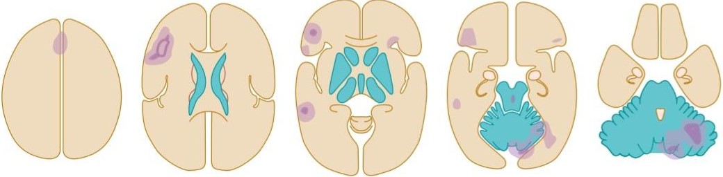
FIGURE 33-10 Activity in the human brain at five different horizontal levels while a subject generates a verb that is appropriate for each noun presented by an examiner. This mental task activates the frontal cortex (slices 1-4), anterior cingulate gyrus (slice 1), and posterior temporal lobe (slice 3) on the left side and the cerebellum (slices 4 and 5) on the right side. Light purple, moderate activation; dark purple, marked activation. (Based on PET scans in Posner MI, Raichle ME: Images of Mind. Scientific American Library, 1994.)
شکل 33-10 فعالیت در مغز انسان در پنج سطح افقی مختلف در حالی که یک آزمودنی یک فعل را تولید می کند که برای هر اسم ارائه شده توسط یک آزمونگر مناسب است. این کار ذهنی قشر پیشانی (برش های 1-4)، شکنج سینگولیت قدامی (برش 1) و لوب تمپورال خلفی (برش 3) در سمت چپ و مخچه (برش های 4 و 5) در سمت راست را فعال می کند. بنفش روشن، فعال سازی متوسط؛ بنفش تیره، فعال شدن مشخص. (براساس اسکن های PET در Posner MI، Raichle ME: Images of Mind. Scientific American Library، 1994.)
CLINICAL BOX 33-2
Changes in Cerebral Blood Flow in Disease
Several disease states are known to be associated with localized or general changes in cerebral blood flow, as revealed by PET scanning and fMRI. For example, epileptic foci are hyperemic during seizures, whereas flow is reduced in other parts of the brain. Between seizures, flow is sometimes reduced in the foci that generate the seizures. Parietooccipital flow is decreased in patients with symptoms of agnosia (see Chapter 15). In Alzheimer disease, the earliest change is decreased metabolism and blood flow in the superior parietal cortex, with later spread to the temporal and finally the frontal cortex. The precentral and postcentral gyri, basal ganglia, thalamus, brainstem, and cerebellum are relatively spared. In Huntington disease, blood flow is reduced bilaterally in the caudate nucleus, and this alteration in flow occurs early in the disease. In manic depressives (but interestingly, not in patients with unipolar depression), there is a general decrease in cortical blood flow when the patients are depressed. In schizophrenia, some evidence suggests decreased blood flow in the frontal lobes, temporal lobes, and basal ganglia. Finally, during the aura in patients with migraine, a bilateral decrease in blood flow starts in the occipital cortex and spreads anteriorly to the temporal and parietal lobes.
جعبه بالینی 33-2
تغییرات در جریان خون مغزی در بیماری
چندین حالت بیماری با تغییرات موضعی یا کلی در جریان خون مغزی مرتبط هستند، همانطور که توسط اسکن PET و fMRI نشان داده شده است. به عنوان مثال، کانون های صرع در هنگام تشنج پرخون هستند، در حالی که جریان در سایر قسمت های مغز کاهش می یابد. بین تشنج، گاهی اوقات جریان در کانون هایی که تشنج ایجاد می کند کاهش می یابد. جریان پاریتواکسیپیتال در بیماران مبتلا به علائم آگنوزیا کاهش می یابد (به فصل 15 مراجعه کنید). در بیماری آلزایمر، اولین تغییر کاهش متابولیسم و جریان خون در قشر جداری فوقانی است که بعداً به قشر تمپورال و در نهایت به قشر پیشانی گسترش می یابد. شکنج پیش مرکزی و پس مرکزی، عقده های قاعده ای، تالاموس، ساقه مغز و مخچه نسبتاً در امان هستند. در بیماری هانتینگتون، جریان خون به صورت دوطرفه در هسته دمی کاهش می یابد و این تغییر در جریان در اوایل بیماری رخ می دهد. در مبتلایان به افسردگی شیدایی (اما جالب است که نه در بیماران مبتلا به افسردگی تک قطبی)، هنگامی که بیماران افسرده هستند، جریان خون قشر مغز کاهش می یابد. در اسکیزوفرنی، برخی شواهد حاکی از کاهش جریان خون در لوب های فرونتال، لوب های تمپورال و گانگلیون های بازال است. در نهایت، در طول هاله در بیماران مبتلا به میگرن، کاهش دو طرفه در جریان خون از قشر اکسیپیتال شروع می شود و از جلو به لوب تمپورال و جداری گسترش می یابد.
BRAIN METABOLISM & OXYGEN REQUIREMENTS
UPTAKE & RELEASE OF SUBSTANCES BY THE BRAIN
If the cerebral blood flow is known, it is possible to calculate the consumption or production by the brain of O2, CO2, glucose, or any other substance present in the bloodstream by multiplying the cerebral blood flow by the difference between the concentration of the substance in arterial blood and cerebral venous blood (Table 33-3). When calculated in this manner, a negative value indicates that the brain is producing the substance.
متابولیسم مغز و نیازهای اکسیژن
جذب و آزادسازی مواد توسط مغز
اگر جریان خون مغزی مشخص باشد، می توان میزان مصرف یا تولید O2، CO2، گلوکز یا هر ماده دیگری که در جریان خون وجود دارد توسط مغز با ضرب جریان خون مغزی در اختلاف بین غلظت ماده در خون شریانی و خون وریدی مغز محاسبه شد (جدول 3-33). هنگامی که به این روش محاسبه می شود، یک مقدار منفی نشان می دهد که مغز در حال تولید ماده است.
TABLE 33-3 Utilization and production of substances by the adult human brain in vivo.
جدول 33-3 استفاده و تولید مواد توسط مغز انسان بالغ در داخل بدن.
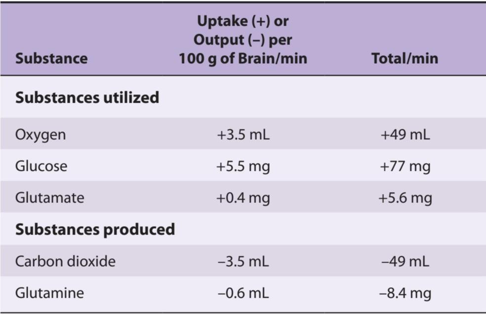
Substances not used or produced in the fed state: lactate, pyruvate, total ketones, and a-ketoglutarate.
موادی که در حالت تغذیه استفاده یا تولید نمی شوند: لاکتات، پیروات، کل کتون ها و a-ketoglutarate.
OXYGEN CONSUMPTION
O2 consumption by the human brain (cerebral metabolic rate for O2, CMRO2) averages approximately 20% of the total body resting O2 consumption (Table 33-1). The brain is extremely sensitive to hypoxia, and occlusion of its blood supply produces unconsciousness in a period as short as 10 s. The vegetative structures in the brainstem are more resistant to hypoxia than the cerebral cortex, and patients may recover from accidents such as cardiac arrest and other conditions causing fairly prolonged hypoxia with normal vegetative functions but severe, permanent intellectual deficiencies. The basal ganglia use O2 at a very high rate, and symptoms of Parkinson disease as well as intellectual deficits can be produced by chronic hypoxia. The thalamus and the inferior colliculus are also very susceptible to hypoxic damage (Clinical Box 33-3).
مصرف اکسیژن
مصرف O2 توسط مغز انسان (نرخ متابولیسم مغزی برای O2، CMRO2) به طور متوسط تقریباً 20٪ از کل مصرف O2 در حال استراحت بدن است (جدول 33-1). مغز به شدت به هیپوکسی حساس است و انسداد منبع خون آن باعث بیهوشی در یک دوره کوتاه 10 ثانیه می شود. ساختارهای رویشی در ساقه مغز نسبت به قشر مغز در برابر هیپوکسی مقاومتر هستند و بیماران ممکن است پس از حوادثی مانند ایست قلبی و سایر شرایطی که باعث هیپوکسی نسبتاً طولانیمدت با عملکردهای رویشی طبیعی، اما کمبودهای شدید و دائمی فکری میشوند، بهبود پیدا کنند. عقدههای قاعدهای از O2 با سرعت بسیار بالایی استفاده میکنند و علائم بیماری پارکینسون و همچنین نقصهای ذهنی میتوانند توسط هیپوکسی مزمن ایجاد شوند. تالاموس و کولیکولوس تحتانی نیز بسیار مستعد آسیب هیپوکسیک هستند (باکس بالینی 33-3).
CLINICAL BOX 33-3
Stroke
When the blood supply to a part of the brain is interrupted, ischemia damages or kills the cells in the area, producing the signs and symptoms of a stroke. There are two general types of strokes: hemorrhagic and ischemic. Hemorrhagic stroke occurs when a cerebral artery or arteriole ruptures, sometimes but not always at the site of a small aneurysm. Ischemic stroke occurs when flow in a vessel is compromised by atherosclerotic plaques on which thrombi form. Thrombi may also be produced elsewhere (eg, in the atria in patients with atrial fibrillation) and pass to the brain as emboli where they then lodge and interrupt flow. In the past, little could be done to modify the course of a stroke and its consequences. However, it has become clear that in the penumbra (the area surrounding the most severe brain damage) ischemia reduces glutamate uptake by astrocytes, and the increase in local glutamate causes excitotoxic damage and death to neurons (see Chapter 7).
جعبه بالینی 33-3
سکته
هنگامی که خون رسانی به بخشی از مغز قطع می شود، ایسکمی به سلول های آن ناحیه آسیب می زند یا می کشد و علائم و نشانه های سکته مغزی را ایجاد می کند. دو نوع کلی سکته مغزی وجود دارد: هموراژیک و ایسکمیک. سکته هموراژیک زمانی رخ می دهد که یک شریان مغزی یا شریان پاره شود، گاهی اوقات اما نه همیشه در محل یک آنوریسم کوچک. سکته مغزی ایسکمیک زمانی اتفاق میافتد که جریان در یک رگ توسط پلاکهای آترواسکلروتیک که بر روی آنها ترومبها تشکیل میشوند، به خطر بیفتد. ترومبوزها همچنین ممکن است در جاهای دیگر تولید شوند (مثلاً در دهلیزها در بیماران مبتلا به فیبریلاسیون دهلیزی) و به عنوان آمبولی به مغز منتقل می شوند و در آنجا مستقر می شوند و جریان را قطع می کنند. در گذشته، برای اصلاح روند سکته مغزی و عواقب آن کار چندانی نمی شد انجام داد. با این حال، مشخص شده است که در نیم سایه (ناحیه احاطه کننده شدیدترین آسیب مغزی) ایسکمی جذب گلوتامات توسط آستروسیت ها را کاهش می دهد و افزایش گلوتامات موضعی باعث آسیب اکسیتوتوکسیک و مرگ نورون ها می شود (به فصل 7 مراجعه کنید).
THERAPEUTIC HIGHLIGHTS
The clot-lysing drug, tissue-type plasminogen activator (t-PA) (see Chapter 31) is of great benefit in ischemic strokes. Drugs that prevent excitotoxic damage have had equivocal effects in human clinical trials, but growing insights into the mechanism of injury are yielding more targeted agents that may be of benefit via this pathway in the future. However, t-PA (and presumably any effective antiexcitotoxic treatment) must be given early in the course of a stroke to be of maximum benefit. This is why stroke has become a condition in which rapid diagnosis and treatment are extremely important. In addition, of course, it is important to determine if a stroke is thrombotic or hemorrhagic, since clot lysis is contraindicated in the latter. Trials are ongoing of therapies that enhance hemostasis in hemorrhagic stroke.
نکات برجسته درمانی
داروی لیز کننده لخته، فعال کننده پلاسمینوژن نوع بافتی (t-PA) (به فصل 31 مراجعه کنید) در سکته های ایسکمیک سود زیادی دارد. داروهایی که از آسیب اکسیتوتوکسیک جلوگیری میکنند، در آزمایشهای بالینی انسانی اثرات مبهم داشتهاند، اما بینشهای رو به رشد در مورد مکانیسم آسیب، عوامل هدفمندتری را به دست میدهند که ممکن است از طریق این مسیر در آینده سودمند باشند. با این حال، t-PA (و احتمالاً هر درمان مؤثر ضد تحریککنندهای) باید در اوایل دوره سکته مغزی داده شود تا حداکثر سود را داشته باشد. به همین دلیل است که سکته مغزی به شرایطی تبدیل شده است که در آن تشخیص و درمان سریع بسیار مهم است. علاوه بر این، البته، تعیین اینکه آیا سکته مغزی ترومبوتیک یا هموراژیک است، مهم است، زیرا لیز لخته در مورد دوم منع مصرف دارد. کارآزماییهایی درباره درمانهایی که هموستاز را در سکته هموراژیک افزایش میدهند، ادامه دارد.
ENERGY SOURCES
Glucose is the major ultimate source of energy for the brain; under normal conditions, 90% of the energy needed to maintain ion gradients across cell membranes and transmit electrical impulses comes from this source. Glucose enters the brain via GLUT1 in cerebral capillaries (see above). Other transporters then distribute it to neurons and glial cells.
منابع انرژی
گلوکز منبع اصلی انرژی برای مغز است. در شرایط عادی، 90 درصد انرژی مورد نیاز برای حفظ شیب یونی در غشای سلولی و انتقال تکانه های الکتریکی از این منبع تامین می شود. گلوکز از طریق GLUT1 در مویرگ های مغزی وارد مغز می شود (به بالا مراجعه کنید). سپس سایر ناقلان آن را به سلول های عصبی و گلیال توزیع می کنند.
Glucose is taken up from the blood in large amounts, and the RQ (respiratory quotient; see Chapter 26) of cerebral tissue is 0.95-0.99 in normal individuals. Importantly, insulin is not required for most cerebral cells to utilize glucose. In general, glucose utilization at rest parallels blood flow and O2 consumption. This does not mean that the total source of energy is always glucose. During prolonged starvation, appreciable utilization of other substances occurs. Indeed, evidence indicates that as much as 30% of the glucose taken up under normal conditions is converted to amino acids, lipids, and proteins, and those substances other than glucose are metabolized for energy during convulsions. Some utilization of amino acids from the circulation may also take place even though the amino acid arteriovenous difference across the brain is normally minute.
گلوکز به مقدار زیاد از خون گرفته می شود و RQ (نسبت تنفسی؛ به فصل 26 مراجعه کنید) بافت مغز در افراد عادی 0.95-0.99 است. نکته مهم این است که انسولین برای اکثر سلول های مغزی برای استفاده از گلوکز مورد نیاز نیست. به طور کلی، استفاده از گلوکز در حالت استراحت به موازات جریان خون و مصرف O2 است. این بدان معنا نیست که منبع کل انرژی همیشه گلوکز است. در طول گرسنگی طولانی مدت، استفاده قابل توجهی از مواد دیگر رخ می دهد. در واقع، شواهد نشان می دهد که 30 درصد از گلوکز جذب شده در شرایط عادی به اسیدهای آمینه، لیپیدها و پروتئین ها تبدیل می شود و این مواد به جز گلوکز برای انرژی در طی تشنج متابولیزه می شوند. برخی از اسیدهای آمینه از گردش خون نیز ممکن است انجام شود، حتی اگر تفاوت شریانی وریدی اسید آمینه در سراسر مغز به طور معمول بسیار اندک باشد.
The consequences of hypoglycemia in terms of neural function are discussed in Chapter 24.
پیامدهای هیپوگلیسمی از نظر عملکرد عصبی در فصل 24 مورد بحث قرار گرفته است.
GLUTAMATE & AMMONIA REMOVAL
The brain’s uptake of glutamate is approximately balanced by its output of glutamine. Glutamate entering the brain associates with ammonia and leaves as glutamine. The glutamate-glutamine conversion in the brain-the opposite of the reaction in the kidney that produces some of the ammonia entering the tubules serves as a detoxifying mechanism to keep the brain free of ammonia. Ammonia is very toxic to nerve cells, and ammonia intoxication is believed to be at least among the causes of neurologic symptoms that may emerge in advanced liver disease (hepatic encephalopathy, see Chapter 28).
حذف گلوتامات و آمونیاک
جذب گلوتامات توسط مغز تقریباً با خروجی گلوتامین متعادل می شود. ورود گلوتامات به مغز با آمونیاک همراه است و به صورت گلوتامین خارج می شود. تبدیل گلوتامات به گلوتامین در مغز – برعکس واکنش کلیه که مقداری از آمونیاک وارد لولهها را تولید میکند، به عنوان یک مکانیسم سمزدایی برای حفظ مغز از آمونیاک عمل میکند. آمونیاک برای سلول های عصبی بسیار سمی است و اعتقاد بر این است که مسمومیت با آمونیاک حداقل یکی از علل علائم عصبی است که ممکن است در بیماری پیشرفته کبد ظاهر شود (آنسفالوپاتی کبدی، به فصل 28 مراجعه کنید).
CORONARY CIRCULATION
ANATOMIC CONSIDERATIONS
The two coronary arteries that supply the myocardium arise from sinuses behind two of the cusps of the aortic valve at the root of the aorta (Figure 33-11). Most of the venous blood returns to the heart through the coronary sinus and anterior cardiac veins (Figure 33-12), which drain into the right atrium. In addition, there are other vessels that empty directly into the heart chambers. These include arteriosinusoidal vessels, sinusoidal capillary-like vessels that connect arterioles to the chambers; Thebesian veins that connect capillaries to the chambers; and a few arterioluminal vessels that are small arteries draining directly into the chambers. A few anastomoses occur between the coronary arterioles and extracardiac arterioles, especially around the mouths of the great veins.
گردش خون کرونر
ملاحظات آناتومیک
دو شریان کرونری که میوکارد را تامین می کنند از سینوس های پشت دو کاسپ دریچه آئورت در ریشه آئورت ایجاد می شوند (شکل 33-11). بیشتر خون وریدی از طریق سینوس کرونر و وریدهای قلبی قدامی به قلب باز می گردد (شکل 33-12)، که به دهلیز راست تخلیه می شود. علاوه بر این، عروق دیگری نیز وجود دارند که مستقیماً وارد حفره های قلب می شوند. اینها شامل عروق شریانی سینوسی، عروق مویرگی مانند سینوسی است که شریان ها را به اتاقک ها متصل می کند. وریدهای تبس که مویرگ ها را به اتاقک ها متصل می کنند. و چند رگ شریانی که شریان های کوچکی هستند که مستقیماً به داخل اتاقک تخلیه می شوند. چند آناستوموز بین شریانهای کرونر و شریانهای خارج قلب، بهویژه در اطراف دهان وریدهای بزرگ رخ میدهد.

FIGURE 33-11 Coronary arteries and their principal branches in humans. (Reproduced with permission from Ross G: Essentials of Human Physiology. Year Book Medical Publishers; 1978.)
شکل 33-11 عروق کرونر و شاخه های اصلی آنها در انسان. (تکثیر شده با اجازه Ross G: Essentials of Human Physiology. Year Book Medical Publishers; 1978.)

FIGURE 33–12 Diagram of the coronary circulation.
شکل 33-12 نمودار گردش خون کرونر.
PRESSURE GRADIENTS & FLOW IN THE CORONARY VESSELS
The heart is a muscle that, like skeletal muscle, compresses its blood vessels when it contracts. The pressure inside the left ventricle is slightly higher than in the aorta during systole (Table 33-4). Consequently, in the arteries supplying the subendocardial portion of the left ventricle, flow occurs only during diastole, although the force is sufficiently dissipated in the more superficial portions of the left ventricular myocardium to permit some flow in this region throughout the cardiac cycle. Because the relative length of diastole is shorter when the heart rate is high, left ventricular coronary flow is reduced during tachycardia. On the other hand, the pressure differential between the aorta and the right ventricle, and between the aorta and the atria, is somewhat greater during systole than during diastole. Consequently, coronary flow in those parts of the heart is not appreciably reduced during systole. Flow in the right and left coronary arteries is shown in Figure 33-13. Because no blood flow occurs during systole in the subendocardial portion of the left ventricle, this region is prone to ischemic damage and is the most common site of myocardial infarction. Blood flow to the left ventricle is decreased in patients with stenotic aortic valves because the pressure in the left ventricle must be much higher than that in the aorta to eject the blood. Consequently, the coronary vessels are more severely compressed during systole. Patients with aortic stenosis are particularly prone to develop symptoms of myocardial ischemia, in part because of this compression and in part because the myocardium requires more O2 to expel blood through the stenotic aortic valve. Coronary flow is also decreased when the aortic diastolic pressure is low. The rise in venous pressure in conditions such as heart failure reduces coronary flow because it decreases effective coronary perfusion pressure (Clinical Box 33-4).
شیب فشار و جریان در عروق کرونر
قلب ماهیچه ای است که مانند ماهیچه های اسکلتی، رگ های خونی خود را در هنگام انقباض فشرده می کند. فشار داخل بطن چپ در طی سیستول کمی بیشتر از آئورت است (جدول 33-4). در نتیجه، در شریانهایی که بخش ساب اندوکاردیال بطن چپ را تامین میکنند، جریان فقط در طول دیاستول رخ میدهد، اگرچه نیرو به اندازه کافی در قسمتهای سطحیتر میوکارد بطن چپ پخش میشود تا جریانی در این ناحیه در طول چرخه قلبی ایجاد شود. از آنجا که طول نسبی دیاستول زمانی که ضربان قلب زیاد است کوتاهتر است، جریان کرونری بطن چپ در طول تاکی کاردی کاهش می یابد. از سوی دیگر، اختلاف فشار بین آئورت و بطن راست، و بین آئورت و دهلیز، در طول سیستول تا حدودی بیشتر از دیاستول است. در نتیجه، جریان کرونری در آن قسمتهای قلب در طی سیستول به میزان قابلتوجهی کاهش نمییابد. جریان در عروق کرونر راست و چپ در شکل 33-13 نشان داده شده است. از آنجایی که هیچ جریان خونی در حین سیستول در بخش ساب اندوکاردیال بطن چپ رخ نمی دهد، این ناحیه مستعد آسیب ایسکمیک است و شایع ترین محل سکته قلبی است. جریان خون به بطن چپ در بیمارانی که دریچه های آئورت تنگی دارند کاهش می یابد زیرا فشار در بطن چپ باید بسیار بیشتر از فشار آئورت باشد تا خون خارج شود. در نتیجه، عروق کرونر در طول سیستول به شدت فشرده می شوند. بیماران مبتلا به تنگی آئورت به ویژه مستعد ابتلا به علائم ایسکمی میوکارد هستند، تا حدی به دلیل این فشرده سازی و تا حدی به این دلیل که میوکارد به O2 بیشتری برای دفع خون از طریق دریچه تنگی آئورت نیاز دارد. هنگامی که فشار دیاستولیک آئورت پایین است، جریان کرونر نیز کاهش می یابد. افزایش فشار وریدی در شرایطی مانند نارسایی قلبی جریان کرونر را کاهش می دهد زیرا فشار خونرسانی موثر کرونر را کاهش می دهد (باکس بالینی 33-4).
TABLE 33-4 Pressure in aorta and left and right ventricles (vent) in systole and diastole.
جدول 33-4 فشار در آئورت و بطن چپ و راست (دریچه) در سیستول و دیاستول.


FIGURE 33-13 Blood flow in the left and right coronary arteries during various phases of the cardiac cycle. Systole occurs between the two vertical dashed lines. (Reproduced with permission from Berne RM, Levy MN: Physiology, 2nd ed. Mosby; 1988.)
شکل 33-13 جریان خون در عروق کرونر چپ و راست در طی مراحل مختلف چرخه قلبی. سیستول بین دو خط چین عمودی رخ می دهد. (تکثیر شده با اجازه از Berne RM, Levy MN: Physiology, 2nd ed. Mosby, 1988.)
CLINICAL BOX 33-4
Coronary Artery Diseas
When flow through a coronary artery is reduced to the point that the myocardium it supplies becomes hypoxic, angina pectoris develops (see Chapter 30). If the myocardial ischemia is severe and prolonged, irreversible changes occur in the muscle, and the result is myocardial infarction. Many individuals have angina only on exertion, and coronary blood flow is normal at rest. Others have more severe restriction of blood flow and have anginal pain at rest as well. Partially occluded coronary arteries can be constricted further by vasospasm, producing myocardial infarction. However, it is now clear that the most common cause of myocardial infarction is rupture of an atherosclerotic plaque, or hemorrhage into it, which triggers the formation of a coronary-occluding clot at the site of the plaque. The electrocardiographic changes in myocardial infarction are discussed in Chapter 29. When myocardial cells die, they leak enzymes into the circulation, and measuring the rise in serum enzymes and isoenzymes produced by infarcted myocardial cells also plays an important role in the diagnosis of myocardial infarction. The enzymes most commonly measured are the MB isomer of creatine kinase (CK-MB), troponin T, and troponin I. Myocardial infarction is a very common cause of death in developed countries because of the widespread occurrence of atherosclerosis. In addition, there is a relation between atherosclerosis and circulating levels of lipoprotein(a) (Lp[a]). Lp(a) has an outer coat consisting of apo(a). It interferes with fibrinolysis by down-regulating plasmin generation (see Chapter 31). It now appears that atherosclerosis has an important inflammatory component as well. The lesions of the disease contain inflammatory cells, and there is a positive correlation between increased levels of C-reactive protein and other inflammatory markers in the circulation with subsequent myocardial infarction.
جعبه بالینی 33-4
بیماری های عروق کرونر
هنگامی که جریان از طریق یک شریان کرونر به حدی کاهش می یابد که میوکاردی که آن را تامین می کند هیپوکسیک می شود، آنژین صدری ایجاد می شود (به فصل 30 مراجعه کنید). اگر ایسکمی میوکارد شدید و طولانی باشد، تغییرات برگشت ناپذیری در عضله ایجاد می شود و نتیجه آن انفارکتوس میوکارد است. بسیاری از افراد تنها در هنگام فعالیت آنژین دارند و جریان خون کرونر در حالت استراحت طبیعی است. برخی دیگر محدودیت شدیدتری در جریان خون دارند و در هنگام استراحت نیز درد آنژین دارند. شریانهای کرونری که تا حدی مسدود شدهاند، میتوانند توسط وازواسپاسم بیشتر منقبض شوند و باعث انفارکتوس میوکارد شوند. با این حال، اکنون مشخص شده است که شایع ترین علت انفارکتوس میوکارد، پارگی پلاک آترواسکلروتیک یا خونریزی در آن است که باعث تشکیل لخته انسداد عروق کرونر در محل پلاک می شود. تغییرات الکتروکاردیوگرافی در انفارکتوس میوکارد در فصل 29 مورد بحث قرار میگیرد. هنگامی که سلولهای میوکارد میمیرند، آنزیمها را به گردش خون نشت میکنند و اندازهگیری افزایش آنزیمها و ایزوآنزیمهای سرم تولید شده توسط سلولهای انفارکتوس میوکارد نیز نقش مهمی در تشخیص انفارکتوس میوکارد دارد. آنزیمهایی که معمولاً اندازهگیری میشوند ایزومر MB کراتین کیناز (CK-MB)، تروپونین T و تروپونین I هستند. انفارکتوس میوکارد به دلیل وقوع گسترده آترواسکلروز، علت بسیار شایع مرگ در کشورهای توسعهیافته است. علاوه بر این، بین آترواسکلروز و سطوح در گردش لیپوپروتئین (a) (Lp[a]) رابطه وجود دارد. Lp(a) دارای پوشش بیرونی متشکل از apo(a) است. با کاهش تولید پلاسمین در فیبرینولیز تداخل می کند (به فصل 31 مراجعه کنید). اکنون به نظر می رسد که آترواسکلروز یک جزء التهابی مهم نیز دارد. ضایعات این بیماری حاوی سلول های التهابی هستند و یک ارتباط مثبت بین افزایش سطح پروتئین واکنشی C و سایر نشانگرهای التهابی در گردش خون با انفارکتوس میوکارد بعدی وجود دارد.
THERAPEUTIQ HIGHLIGHTS
Treatment of myocardial infarction aims to restore flow to the affected area as rapidly as possible while minimizing reperfusion injury. Needless to say, it should be started as promptly as possible to avoid irreversible changes in heart function. In acute disease, antithrombotic agents are often given, but these can be problematic, leading to increased mortality due to bleeding if cardiac surgery is subsequently needed. Mechanical/surgical approaches to coronary artery disease include balloon angioplasty and/or implantation of stents to hold the vessels open, or coronary artery bypass grafting (CABG).
نکات برجسته درمانی
هدف از درمان انفارکتوس میوکارد، بازگرداندن جریان به ناحیه آسیبدیده در سریعترین زمان ممکن و به حداقل رساندن آسیب خونرسانی مجدد است. نیازی به گفتن نیست که برای جلوگیری از تغییرات غیرقابل برگشت در عملکرد قلب، باید هرچه سریعتر شروع شود. در بیماری حاد، داروهای ضد ترومبوتیک اغلب تجویز میشود، اما این داروها میتوانند مشکلساز باشند و در صورت نیاز به جراحی قلب، منجر به افزایش مرگ و میر ناشی از خونریزی شود. رویکردهای مکانیکی/جراحی برای بیماری عروق کرونر شامل آنژیوپلاستی با بالون و/یا کاشت استنت برای باز نگه داشتن عروق یا پیوند بای پس عروق کرونر (CABG) است.
Coronary blood flow has been measured by inserting a catheter into the coronary sinus and applying the Kety method to the heart on the assumption that the N2O content of coronary venous blood is typical of the entire myocardial effluent. Coronary flow at rest in humans is about 250 mL/min (5% of the cardiac output). A number of techniques utilizing radionuclides detected with radiation detectors over the chest have been used to study regional blood flow in the heart and to detect areas of ischemia and infarct as well as to evaluate ventricular function. Radionuclides such as thallium-201 (201Tl) are pumped into cardiac muscle cells by Na+, K+ ATPase and equilibrate with the intracellular K+ pool. For the first 10–15 min after intravenous injection, 201Tl distribution is directly proportional to myocardial blood flow, and areas of ischemia can be detected by their low uptake. The uptake of this isotope is often determined soon after exercise and again several hours later to bring out areas in which exertion leads to compromised flow. Conversely, radiopharmaceuticals such as technetium-99m stannous pyrophosphate (99mTc-PYP) are selectively taken up by infarcted tissue and make infarcts stand out as “hot spots” on scintigrams of the chest. Coronary angiography can be combined with measurement of 133Xe washout (see above) to provide detailed analysis of coronary blood flow. Radiopaque contrast medium is first injected into the coronary arteries, and radiographs are used to outline their distribution. The angiographic camera is then replaced with a scintillation camera, and 133Xe washout is measured.
جریان خون کرونری با قرار دادن یک کاتتر در سینوس کرونر و اعمال روش Kety در قلب با این فرض که محتوای N2O خون وریدی کرونری نمونهای از کل پساب میوکارد است اندازهگیری شده است. جریان کرونری در حالت استراحت در انسان حدود 250 میلی لیتر در دقیقه (5 درصد برون ده قلبی) است. تعدادی از تکنیکها با استفاده از رادیونوکلئیدهای شناساییشده با آشکارسازهای تشعشع روی قفسه سینه برای مطالعه جریان خون منطقهای در قلب و تشخیص نواحی ایسکمی و انفارکتوس و همچنین برای ارزیابی عملکرد بطن استفاده شدهاند. رادیونوکلئیدهایی مانند تالیم-201 (201Tl) توسط Na+، K+ ATPase به سلولهای ماهیچههای قلب پمپ میشوند و با مخزن +K درون سلولی متعادل میشوند. برای 10-15 دقیقه اول پس از تزریق داخل وریدی، توزیع 201Tl مستقیماً با جریان خون میوکارد متناسب است و مناطق ایسکمی را می توان با جذب کم آنها تشخیص داد. جذب این ایزوتوپ اغلب بلافاصله پس از تمرین و دوباره چندین ساعت بعد تعیین می شود تا مناطقی را که اعمال فشار منجر به جریان مختل می شود، مشخص شود. برعکس، رادیوداروهایی مانند پیروفسفات قلع تکنسیوم-99m (99mTc-PYP) به طور انتخابی توسط بافت انفارکت شده جذب می شوند و انفارکتوس ها را به عنوان “نقاط داغ” روی سینتیگرام قفسه سینه برجسته می کنند. آنژیوگرافی عروق کرونر را می توان با اندازه گیری 133Xe Washout ترکیب کرد (به بالا مراجعه کنید) تا تجزیه و تحلیل دقیقی از جریان خون کرونر ارائه دهد. ماده حاجب رادیواپک ابتدا به داخل عروق کرونر تزریق می شود و از رادیوگرافی برای تشریح توزیع آنها استفاده می شود. سپس دوربین آنژیوگرافی با یک دوربین سوسوزن جایگزین می شود و شستشوی 133Xe اندازه گیری می شود.
VARIATIONS IN CORONARY FLOW
At rest, the heart extracts 70-80% of the O2 from each unit of blood delivered to it (Table 33-1). O2 delivery can only be increased significantly by increasing blood flow. Therefore, it is not surprising that blood flow increases when the metabolism of the myocardium is increased. The caliber of the coronary vessels, and consequently the rate of coronary blood flow, is influenced not only by pressure changes in the aorta but also by chemical and neural factors. The coronary circulation also shows considerable autoregulation.
تغییرات در جریان کرونر
در حالت استراحت، قلب 70-80% O2 را از هر واحد خونی که به آن تحویل داده می شود استخراج می کند (جدول 33-1). تحویل O2 تنها با افزایش جریان خون قابل افزایش است. بنابراین، تعجب آور نیست که با افزایش متابولیسم میوکارد، جریان خون افزایش می یابد. کالیبر عروق کرونر و در نتیجه سرعت جریان خون کرونر نه تنها تحت تأثیر تغییرات فشار در آئورت بلکه تحت تأثیر عوامل شیمیایی و عصبی قرار می گیرد. گردش خون کرونر نیز خودتنظیمی قابل توجهی را نشان می دهد.
CHEMICAL FACTORS
The close relationship between coronary blood flow and myocardial O2 consumption implies that one or more metabolic products causes coronary vasodilation. Factors suspected of playing this role include a lack of O2 and increased local concentrations of CO2, H*, K*, lactate, prostaglandins, adenine nucleotides, and adenosine. Likely several or all of these vasodilator metabolites act in an integrated manner, redundant manner, or both. Asphyxia, hypoxia, and intracoronary injections of cyanide all increase coronary blood flow 200-300% in denervated as well as intact hearts, and the feature common to these three stimuli is hypoxia of the myocardial fibers. A similar increase in flow is produced in the area supplied by a coronary artery if the artery is occluded and then released. This reactive hyperemia is similar to that seen in the skin (see below) and appears to be due to the release of adenosine.
عوامل شیمیایی
رابطه نزدیک بین جریان خون کرونر و مصرف O2 میوکارد نشان می دهد که یک یا چند محصول متابولیک باعث اتساع عروق کرونر می شود. عواملی که مشکوک به ایفای این نقش هستند عبارتند از کمبود O2 و افزایش غلظت موضعی CO2، H+، K، لاکتات، پروستاگلاندین ها، آدنین نوکلئوتیدها و آدنوزین. احتمالاً چندین یا همه این متابولیتهای گشادکننده عروق به صورت یکپارچه، اضافی یا هر دو عمل میکنند. خفگی، هیپوکسی و تزریق داخل کرونری سیانید همگی باعث افزایش 200 تا 300 درصدی جریان خون کرونر در قلب های عصب کشی شده و همچنین دست نخورده می شوند و ویژگی مشترک این سه محرک هیپوکسی رشته های میوکارد است. اگر شریان مسدود شده و سپس آزاد شود، افزایش مشابهی در جریان در ناحیه ای ایجاد می شود که توسط یک شریان کرونر تامین می شود. این پرخونی واکنشی مشابه آن چیزی است که در پوست مشاهده می شود (به زیر مراجعه کنید) و به نظر می رسد به دلیل آزاد شدن آدنوزین باشد.
NEURAL FACTORS
The coronary arterioles contain a-adrenergic receptors, which mediate vasoconstriction, and ẞ-adrenergic receptors, which mediate vasodilation. Activity in the noradrenergic nerves to the heart and injections of norepinephrine cause coronary vasodilation. Mechanistically, norepinephrine increases the heart rate and the force of cardiac contraction, and the vasodilation is due to production of vasodilator metabolites by the myocardium secondary to the increase in its activity. When the inotropic and chronotropic effects of noradrenergic discharge are blocked by a ẞ-adrenergic blocking drug, stimulation of the noradrenergic nerves or injection of norepinephrine in unanesthetized animals elicits coronary vasoconstriction. Thus, the direct effect of noradrenergic stimulation is constriction rather than dilation of the coronary vessels. On the other hand, stimulation of vagal fibers to the heart dilates the coronaries.
فاکتورهای عصبی
شریان های کرونری حاوی گیرنده های a-آدرنرژیک هستند که واسطه انقباض عروق هستند و گیرنده های آدرنرژیک که واسطه اتساع عروق هستند. فعالیت در اعصاب نورآدرنرژیک به قلب و تزریق نوراپی نفرین باعث اتساع عروق کرونر می شود. از نظر مکانیکی، نوراپی نفرین ضربان قلب و نیروی انقباض قلب را افزایش می دهد و اتساع عروق به دلیل تولید متابولیت های گشادکننده عروق توسط میوکارد ثانویه به افزایش فعالیت آن است. هنگامی که اثرات اینوتروپیک و کرونوتروپیک ترشحات نورآدرنرژیک توسط یک داروی مسدودکننده آدرنرژیک مسدود می شود، تحریک اعصاب نورآدرنرژیک یا تزریق نوراپی نفرین در حیوانات بیهوش باعث انقباض عروق کرونر می شود. بنابراین، اثر مستقیم تحریک نورآدرنرژیک، انقباض و نه گشاد شدن عروق کرونر است. از طرفی تحریک فیبرهای واگ به قلب باعث گشاد شدن عروق کرونر می شود.
When the systemic blood pressure falls, the overall effect of the reflex increase in noradrenergic discharge is increased coronary blood flow secondary to the metabolic changes in the myocardium at a time when the cutaneous, renal, and splanchnic vessels are constricted. In this way the circulation of the heart, like that of the brain, is preserved when flow to other organs is compromised.
هنگامی که فشار خون سیستمیک کاهش می یابد، اثر کلی افزایش رفلکس در ترشحات نورآدرنرژیک افزایش جریان خون کرونر ثانویه به تغییرات متابولیک در میوکارد در زمانی است که عروق پوستی، کلیوی و اسپلانکنیک منقبض می شوند. به این ترتیب گردش قلب، مانند گردش خون مغز، هنگامی که جریان به سایر اندام ها به خطر می افتد، حفظ می شود.
CUTANEOUS CIRCULATION
The amount of heat lost from the body is regulated to a large extent by varying the amount of blood flowing through the skin. The fingers, toes, palms, and earlobes contain well-innervated anastomotic connections between arterioles and venules (arteriovenous anastomoses; see Chapter 31). Blood flow in response to thermoregulatory stimuli can vary from 1 to as much as 150 mL/100 g of skin/min, and it has been postulated that these wide variations are possible because blood can be shunted through the anastomoses. The subdermal capillary and venous plexus is a blood reservoir of some importance, and the skin is one of the few places where the reactions of blood vessels can be observed visually.
گردش پوستی
مقدار گرمای از دست رفته از بدن تا حد زیادی با تغییر میزان جریان خون در پوست تنظیم می شود. انگشتان دست، انگشتان پا، کف دست و لاله گوش حاوی اتصالات آناستوموز خوب عصب دهی شده بین شریان ها و وریدها هستند (آناستوموزهای شریانی وریدی؛ به فصل 31 مراجعه کنید). جریان خون در پاسخ به محرک های تنظیم کننده حرارت می تواند از 1 تا 150 میلی لیتر در 100 گرم پوست در دقیقه متغیر باشد، و فرض شده است که این تغییرات گسترده امکان پذیر است زیرا خون می تواند از طریق آناستوموزها عبور کند. مویرگ زیر پوستی و شبکه وریدی یک مخزن خون با اهمیت است و پوست یکی از معدود مکان هایی است که می توان واکنش های رگ های خونی را به صورت بصری مشاهده کرد.
WHITE REACTION
When a pointed object is drawn lightly over the skin, the stroke lines become pale (white reaction). The mechanical stimulus apparently initiates contraction of the precapillary sphincters, and blood drains out of the capillaries and small veins. The response appears in about 15 s.
واکنش سفید
هنگامی که یک جسم نوک تیز به آرامی روی پوست کشیده می شود، خطوط ضربه رنگ پریده می شوند (واکنش سفید). محرک مکانیکی ظاهراً شروع کننده انقباض اسفنکترهای پیش مویرگی است و خون از مویرگ ها و وریدهای کوچک خارج می شود. پاسخ در حدود 15 ثانیه ظاهر می شود.
TRIPLE RESPONSE
When the skin is stroked more firmly with a pointed instrument, instead of the white reaction there is reddening at the site that appears in about 10 s (red reaction). This is followed in a few minutes by local swelling and diffuse, mottled reddening around the site. The initial redness is due to capillary dilation, a direct response of the capillaries to pressure. The swelling (wheal) is local edema due to increased permeability of the capillaries and postcapillary venules, with consequent extravasation of fluid. The redness spreading out from the site (flare) is due to arteriolar dilation. This three-part response—the red reaction, wheal, and flare—is called the triple response and is part of the normal reaction to injury (see Chapter 3). It persists after total sympathectomy. On the other hand, the flare is absent in locally anesthetized skin and in denervated skin after the sensory nerves have degenerated, but it is present immediately after nerve block or section above the site of the injury. This, plus other evidence, indicates that it is due to an axon reflex, a response in which impulses initiated in sensory nerves by the injury are relayed antidromically down other branches of the sensory nerve fibers (Figure 33-14). This is the one situation in the body in which there is substantial evidence for a physiologic effect due to antidromic conduction. The transmitter released at the central termination of the sensory C fiber neurons is substance P (see Chapter 7), and substance P and CGRP are present in all parts of the neurons. Both dilate arterioles and, in addition, substance P cause extravasation of fluid. Effective nonpeptide antagonists to substance P have been developed, and they reduce the extravasation. Thus, it appears that substance P produces the wheal.
پاسخ سه گانه
هنگامی که پوست با یک ابزار نوک تیز محکم تر نوازش می شود، به جای واکنش سفید، قرمزی در محل ایجاد می شود که در حدود 10 ثانیه ظاهر می شود (واکنش قرمز). پس از چند دقیقه تورم موضعی و قرمزی منتشر و خالدار در اطراف محل ایجاد می شود. قرمزی اولیه به دلیل گشاد شدن مویرگ ها است، که پاسخ مستقیم مویرگ ها به فشار است. تورم (ویل) ادم موضعی ناشی از افزایش نفوذپذیری مویرگ ها و وریدهای پس مویرگی و در نتیجه خارج شدن مایع است. قرمزی منتشر شده از محل (شعله ور شدن) به دلیل گشاد شدن شریان است. این پاسخ سه بخشی – واکنش قرمز، چرخش و شعله ور شدن – پاسخ سه گانه نامیده می شود و بخشی از واکنش طبیعی به آسیب است (به فصل 3 مراجعه کنید). پس از سمپاتکتومی کامل باقی می ماند. از سوی دیگر، شعله ور شدن در پوست بیهوش موضعی و در پوست عصبکشی شده پس از تحلیل رفتن اعصاب حسی وجود ندارد، اما بلافاصله پس از بلوک عصبی یا بخش بالای محل آسیب دیده میشود. این، به علاوه شواهد دیگر، نشان میدهد که به دلیل یک رفلکس آکسون است، پاسخی که در آن تکانههای آغاز شده در اعصاب حسی توسط آسیب به صورت ضددرومیک به سمت شاخههای دیگر رشتههای عصبی حسی منتقل میشوند (شکل 33-14). این تنها موقعیتی در بدن است که در آن شواهد قابل توجهی برای اثر فیزیولوژیکی ناشی از هدایت آنتی درومیک وجود دارد. فرستنده ای که در انتهای مرکزی نورون های فیبر حسی C منتشر می شود، ماده P است (به فصل 7 مراجعه کنید)، و ماده P و CGRP در تمام قسمت های نورون ها وجود دارد. هم شریانها را گشاد میکنند و هم ماده P باعث خارج شدن مایع میشود. آنتاگونیستهای غیرپپتیدی مؤثری برای ماده P ایجاد شدهاند و باعث کاهش تخلخل میشوند. بنابراین، به نظر می رسد که ماده P باعث تولید چرخ می شود.

FIGURE 33-14 Axon reflex.
شکل 33-14 رفلکس آکسون.
REACTIVE HYPEREMIA
A response of the blood vessels that occurs in many organs but is visible in the skin is reactive hyperemia, an increase in the amount of blood in a region when its circulation is reestablished after a period of occlusion. When the blood supply to a limb is occluded, the cutaneous arterioles below the occlusion dilate. When the circulation is reestablished, blood flowing into the dilated vessels makes the skin fiery red. O2 in the atmosphere can diffuse a short distance through the skin, and reactive hyperemia is prevented if the circulation of the limb is occluded in an atmosphere of 100% O2. Therefore, the arteriolar dilation is apparently due to a local effect of hypoxia.
هایپرمی واکنشی
پاسخ رگهای خونی که در بسیاری از اندامها رخ میدهد اما در پوست قابل مشاهده است، پرخونی واکنشی است، افزایش مقدار خون در ناحیهای که گردش خون پس از یک دوره انسداد دوباره برقرار میشود. هنگامی که خون رسانی به اندام مسدود می شود، شریان های پوستی زیر انسداد منبسط می شوند. هنگامی که گردش خون دوباره برقرار می شود، جریان خون به داخل عروق گشاد شده پوست را قرمز آتشین می کند. O2 موجود در اتمسفر می تواند در فاصله کوتاهی از پوست پخش شود و اگر گردش خون اندام در اتمسفر 100٪ O2 مسدود شود از پرخونی واکنشی جلوگیری می شود. بنابراین، اتساع شریانی ظاهراً به دلیل اثر موضعی هیپوکسی است.
GENERALIZED RESPONSES
Noradrenergic nerve stimulation and circulating epinephrine and norepinephrine constrict cutaneous blood vessels. No known vasodilator nerve fibers extend to the cutaneous vessels, and thus vasodilation is brought about by a decrease in constrictor tone as well as the local production of vasodilator metabolites. Skin color and temperature also depend on the state of the capillaries and venules. A cold blue or gray skin is one in which the arterioles are constricted and the capillaries dilated; a warm red skin is the one in which both are dilated.
پاسخ های کلی
تحریک عصبی نورآدرنرژیک و اپی نفرین و نوراپی نفرین در گردش، عروق خونی پوست را منقبض می کند. هیچ رشته عصبی گشادکننده عروقی شناخته شده ای به رگ های پوستی گسترش نمی یابد و بنابراین گشاد شدن عروق با کاهش تون منقبض کننده و همچنین تولید موضعی متابولیت های گشادکننده عروق ایجاد می شود. رنگ و دمای پوست نیز به وضعیت مویرگ ها و ونول ها بستگی دارد. پوست آبی یا خاکستری سرد پوستی است که در آن شریان ها منقبض شده و مویرگ ها گشاد می شوند. پوست قرمز گرم پوستی است که در آن هر دو گشاد می شوند.
Because painful stimuli cause diffuse noradrenergic discharge, a painful injury causes generalized cutaneous vasoconstriction in addition to the local triple response. When the body temperature rises during exercise, the cutaneous blood vessels dilate in spite of continuing noradrenergic discharge in other parts of the body. Dilation of cutaneous vessels in response to a rise in hypothalamic temperature overcomes other reflex activity. Shock is more profound in patients with elevated temperatures because of cutaneous vasodilation, and patients in shock should not be warmed to the point that their body temperature rises. Conversely, cold causes cutaneous vasoconstriction; however, with severe cold, superficial vasodilation may supervene. This latter vasodilation is the cause of the ruddy complexion seen on a cold day.
از آنجایی که محرک های دردناک باعث ترشحات نورآدرنرژیک منتشر می شوند، آسیب دردناک علاوه بر پاسخ سه گانه موضعی باعث انقباض عروق پوستی عمومی می شود. هنگامی که دمای بدن در حین ورزش افزایش مییابد، رگهای خونی پوستی با وجود ترشحات نورآدرنرژیک در سایر قسمتهای بدن منبسط میشوند. اتساع عروق پوستی در پاسخ به افزایش دمای هیپوتالاموس بر سایر فعالیت های رفلکس غلبه می کند. شوک در بیماران با درجه حرارت بالا به دلیل اتساع عروق پوستی شدیدتر است و بیماران شوک نباید تا حدی گرم شوند که دمای بدنشان افزایش یابد. برعکس، سرما باعث انقباض عروق پوستی می شود. با این حال، با سرمای شدید، اتساع عروق سطحی ممکن است تحت تأثیر قرار گیرد. این گشاد شدن عروق اخیر علت قرمزی رنگی است که در روزهای سرد مشاهده می شود.
PLACENTAL & FETAL CIRCULATION
UTERINE CIRCULATION
The blood flow of the uterus parallels the metabolic activity of the myometrium and endometrium and undergoes cyclic fluctuations that correlate with the menstrual cycle in nonpregnant women. The function of the spiral and basilar arteries of the endometrium in menstruation is discussed in Chapter 22. During pregnancy, blood flow increases rapidly as the uterus increases in size (Figure 33-15). Vasodilator metabolites are undoubtedly produced in the uterus, as they are in other active tissues. In early pregnancy, the arteriovenous O2 difference across the uterus is small, and it has been suggested that estrogens act on the blood vessels to increase uterine blood flow in excess of tissue O2 needs. Corticotrophin-releasing hormone also appears to play an important role in up- regulating uterine blood flow, as well as in the eventual timing of birth. However, even though uterine blood flow increases 20-fold during pregnancy, the size of the conceptus increases much more, changing from a single cell to a fetus plus a placenta that can weigh 4-5 kg at term in humans. Consequently, more O2 is extracted from the uterine blood during the latter part of pregnancy, and the O2 saturation of uterine blood falls.
گردش جفت و جنین
گردش خون رحم
جریان خون رحم به موازات فعالیت متابولیک میومتر و آندومتر است و دچار نوسانات چرخه ای می شود که با چرخه قاعدگی در زنان غیرباردار مرتبط است. عملکرد شریان های مارپیچی و بازیلار آندومتر در قاعدگی در فصل 22 مورد بحث قرار گرفته است. در دوران بارداری، با افزایش اندازه رحم، جریان خون به سرعت افزایش می یابد (شکل 33-15). متابولیت های وازودیلاتور بدون شک در رحم نیز مانند سایر بافت های فعال تولید می شوند. در اوایل بارداری، اختلاف O2 شریانی وریدی در سراسر رحم کم است و پیشنهاد شده است که استروژن ها بر روی رگ های خونی عمل می کنند تا جریان خون رحم را بیش از نیاز بافت O2 افزایش دهند. به نظر می رسد هورمون آزاد کننده کورتیکوتروفین نیز نقش مهمی در تنظیم جریان خون رحمی و همچنین در زمان نهایی تولد دارد. با این حال، حتی اگر جریان خون رحم در دوران بارداری 20 برابر افزایش می یابد، اندازه کانسپتوس بسیار بیشتر می شود و از یک سلول به یک جنین به اضافه جفتی که می تواند وزن آن 4-5 کیلوگرم در ترم در انسان باشد، تغییر می کند. در نتیجه، O2 بیشتری از خون رحم در اواخر بارداری استخراج می شود و اشباع O2 خون رحم کاهش می یابد.

FIGURE 33-15 Changes in uterine blood flow and the amount of O2 in uterine venous blood during pregnancy. (Modified with permission from Keele CA, Neil E: Samson Wright’s Applied Physiology, 12th ed. Oxford University Press; 1971.)
شکل 33-15 تغییرات در جریان خون رحم و مقدار O2 در خون وریدی رحم در دوران بارداری. (با اجازه Keele CA، Neil E: Samson Wright’s Applied Physiology، ویرایش دوازدهم انتشارات دانشگاه آکسفورد، 1971 اصلاح شده است.)
PLACENTA
The placenta is the “fetal lung” (Figures 33-16 and 33-17). Its maternal portion is in effect a large blood sinus. Into this “lake” project the villi of the fetal portion containing the small branches of the fetal umbilical arteries and vein (Figure 33–16). O2 is taken up by the fetal blood and CO2 is discharged into the maternal circulation across the walls of the villi in a manner analogous to O2 and CO2 exchange in the lungs (see Chapter 35). However, the cellular layers covering the villi are thicker and less permeable than the alveolar membranes in the lungs, and exchange is much less efficient. The placenta is also the route by which all nutritive materials enter the fetus and by which fetal wastes are discharged to the maternal blood.
جفت
جفت “ریه جنین” است (شکل های 16-33 و 17-33). بخش مادری آن در واقع یک سینوس خون بزرگ است. در این “دریاچه” پرزهای قسمت جنینی شامل شاخه های کوچک شریان ها و ورید نافی جنین است (شکل 33-16). O2 توسط خون جنین جذب می شود و CO2 به روشی مشابه با تبادل O2 و CO2 در ریه ها به گردش خون مادر در سراسر دیواره های پرزها تخلیه می شود (به فصل 35 مراجعه کنید). با این حال، لایه های سلولی پوشاننده پرزها ضخیم تر و کمتر از غشاهای آلوئولی در ریه ها نفوذ می کنند و تبادل بسیار کمتر کارآمد است. جفت همچنین مسیری است که از طریق آن تمام مواد مغذی وارد جنین می شود و از طریق آن فضولات جنین به خون مادر تخلیه می شود.

FIGURE 33-16 Diagram of a section through the human placenta, showing the way the fetal villi project into the maternal sinuses. (©Elsevier, Inc., Netterimages.com.)
شکل 33-16 نمودار مقطعی از جفت انسان، که راه پرزهای جنینی را به داخل سینوس های مادر نشان می دهد. (©Elsevier, Inc., Netterimages.com.)
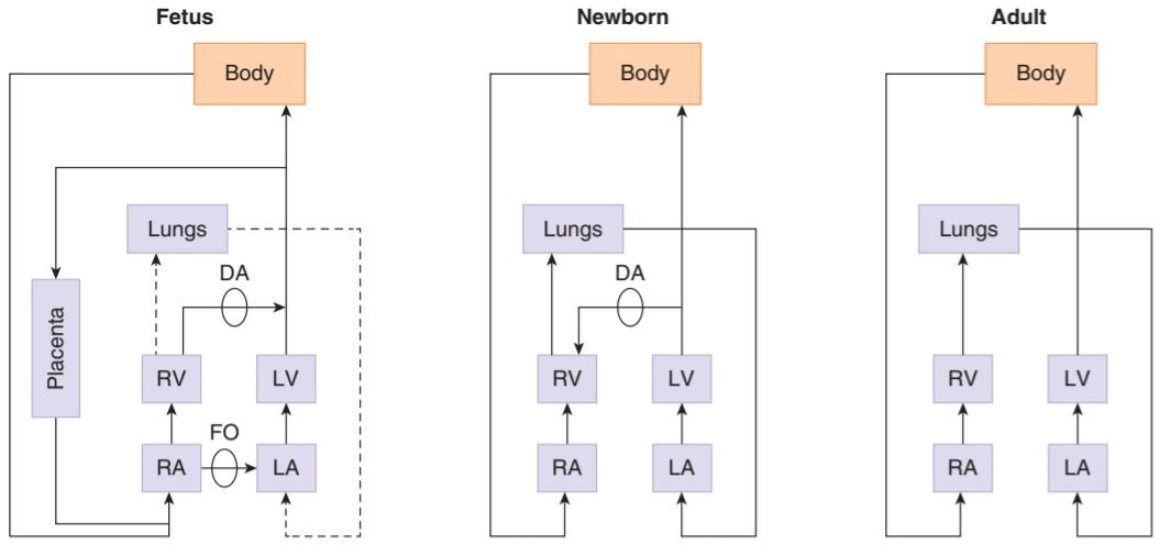
FIGURE 33-17 Diagram of the circulation in the fetus, the newborn infant, and the adult. DA, ductus arteriosus; FO, foramen ovale; LA, left atrium; LV, left ventricle; RA, right atrium; RV, right ventricle. Note that circulation to the fetal liver is not shown for simplicity (see Figure 33-18)
شکل 33-17 نمودار گردش خون در جنین، نوزاد تازه متولد شده و بزرگسالان. DA، مجرای شریانی؛ FO، فورامن بیضی؛ LA، دهلیز چپ؛ LV، بطن چپ؛ RA، دهلیز راست؛ RV، بطن راست. توجه داشته باشید که گردش خون به کبد جنین برای سادگی نشان داده نشده است (شکل 33-18 را ببینید).
FETAL CIRCULATION
The arrangement of the circulation in the fetus is shown diagrammatically in Figure 33-17. The blood in the umbilical vein in humans is believed to be about 80% saturated with O2, compared with 98% saturation in the arterial circulation of the adult. The ductus venosus (Figure 33-18) diverts some of this blood directly to the inferior vena cava, and the remainder mixes with the portal blood of the fetus. The portal and systemic venous blood of the fetus is only 26% saturated, and the saturation of the mixed blood in the inferior vena cava is approximately 67%. Most of the blood entering the heart through the inferior vena cava is diverted directly to the left atrium via the patent foramen ovale. Most of the blood from the superior vena cava enters the right ventricle and is expelled into the pulmonary artery. The resistance of the collapsed lungs is high, and the pressure in the pulmonary artery is several mm Hg higher than it is in the aorta, so that most of the blood in the pulmonary artery passes through the ductus arteriosus to the aorta. In this manner, the relatively unsaturated blood from the right ventricle is diverted to the trunk and lower body of the fetus, while the head of the fetus receives the better-oxygenated blood from the left ventricle. From the aorta, some of the blood is pumped into the umbilical arteries and back to the placenta. The O2 saturation of the blood in the lower aorta and umbilical arteries of the fetus is approximately 60%.
گردش خون جنین
ترتیب گردش خون در جنین به صورت نموداری در شکل 33-17 نشان داده شده است. اعتقاد بر این است که خون موجود در ورید ناف در انسان حدود 80٪ با O2 اشباع شده است، در حالی که 98٪ اشباع در گردش خون شریانی بزرگسالان است. مجرای وریدی (شکل 33-18) مقداری از این خون را مستقیماً به ورید اجوف تحتانی منحرف میکند و باقیمانده با خون پورتال جنین مخلوط میشود. خون ورید پورتال و سیستمیک جنین تنها 26 درصد اشباع است و اشباع خون مخلوط در ورید اجوف تحتانی تقریباً 67 درصد است. بیشتر خونی که از طریق ورید اجوف تحتانی وارد قلب می شود، مستقیماً از طریق سوراخ بیضی باز به دهلیز چپ منحرف می شود. بیشتر خون از ورید اجوف فوقانی وارد بطن راست می شود و به داخل شریان ریوی دفع می شود. مقاومت ریه های فروپاشیده زیاد است و فشار در شریان ریوی چندین میلی متر جیوه بیشتر از آئورت است، به طوری که بیشتر خون در شریان ریوی از مجرای شریانی به آئورت می گذرد. به این ترتیب، خون نسبتا غیر اشباع از بطن راست به سمت تنه و پایین بدن جنین منحرف می شود، در حالی که سر جنین خون با اکسیژن بهتر را از بطن چپ دریافت می کند. از آئورت، مقداری از خون به داخل شریانهای نافی پمپ میشود و به جفت باز میگردد. اشباع O2 خون در آئورت تحتانی و شریان های نافی جنین تقریباً 60٪ است.
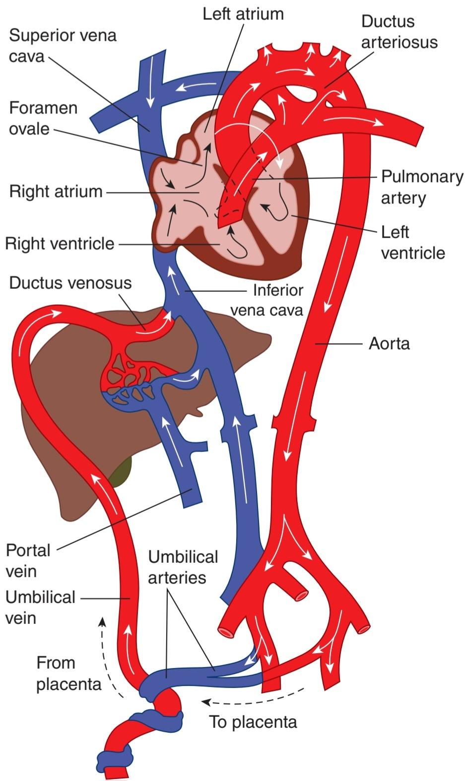
FIGURE 33-18 Circulation in the fetus. Most of the oxygenated blood reaching the heart via the umbilical vein and inferior vena cava is diverted through the foramen ovale and pumped out the aorta to the head, while the deoxygenated blood returned via the superior vena cava is mostly pumped through the pulmonary artery and ductus arteriosus to the feet and the umbilical arteries.
شکل 33-18 گردش خون در جنین. بیشتر خون اکسیژن دار که از طریق ورید ناف و ورید اجوف تحتانی به قلب می رسد از طریق سوراخ بیضی منحرف می شود و آئورت را به سمت سر پمپ می کند، در حالی که خون بدون اکسیژن که از طریق ورید اجوف فوقانی بازگردانده می شود، بیشتر از طریق شریان ریوی و مجرای شریانی و مجرای شریانی جنینی پمپ می شود.
FETAL RESPIRATION
The tissues of fetal and newborn mammals have a remarkable but poorly understood resistance to hypoxia. However, the O2 saturation of the maternal blood in the placenta is so low that the fetus might suffer hypoxic damage if fetal red cells did not have a greater O2 affinity than adult red cells (Figure 33-19). The fetal red cells contain fetal hemoglobin (hemoglobin F), whereas the adult cells contain adult hemoglobin (hemoglobin A). The cause of the difference in O2 affinity between the two is that hemoglobin F binds 2, 3-DPG less effectively than hemoglobin A does. The decrease in O2 affinity due to the binding of 2, 3- DPG is discussed in Chapter 31.
تنفس جنین
بافتهای پستانداران جنینی و تازه متولد شده مقاومت قابلتوجهی دارند، اما درک ضعیفی نسبت به هیپوکسی دارند. با این حال، اشباع O2 خون مادر در جفت آنقدر کم است که اگر گلبول های قرمز جنین تمایل بیشتری به O2 نسبت به گلبول های قرمز بالغ نداشته باشند، جنین ممکن است دچار آسیب هیپوکسیک شود (شکل 33-19). گلبول های قرمز جنین حاوی هموگلوبین جنینی (هموگلوبین F) هستند، در حالی که سلول های بالغ حاوی هموگلوبین بالغ (هموگلوبین A) هستند. علت تفاوت در میل ترکیبی O2 بین این دو این است که هموگلوبین F به 2، 3-DPG کمتر از هموگلوبین A متصل می شود. کاهش میل ترکیبی O2 به دلیل اتصال 2، 3- DPG در فصل 31 مورد بحث قرار گرفته است.
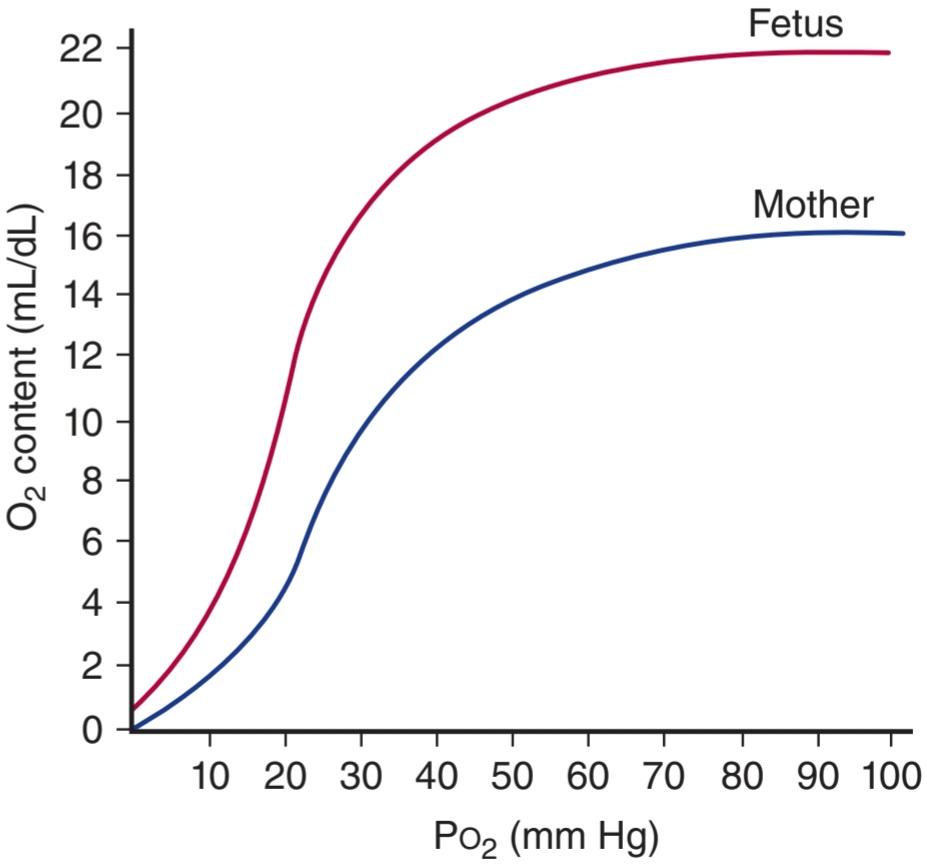
FIGURE 33-19 Dissociation curves of hemoglobin in human maternal and fetal blood.
شکل 33-19 منحنی های تفکیک هموگلوبین در خون مادر و جنین انسان.
CHANGES IN FETAL CIRCULATION & RESPIRATION AT BIRTH
Because of the patent ductus arteriosus and foramen ovale (Figure 33-18), the left heart and right heart pump in parallel in the fetus rather than in series as they do in the adult. At birth the circulation has to change from a parallel to a serial system and from placental to pulmonary gas exchange. This has to be largely completed within minutes of birth, although the complete changes may not be achieved for many hours. The essential element initiating the changes is the fall in the pulmonary vascular resistance when the lungs are first filled with a gas. The pulmonary vascular resistance falls to less than 20% of the value in utero after the lungs are expanded by the first few breaths.
تغییرات در گردش خون و تنفس جنین در بدو تولد
به دلیل وجود مجرای شریانی باز و فورامن اوال (شکل 33-18)، قلب چپ و قلب راست به طور موازی در جنین پمپ می کنند و نه به صورت سری که در بزرگسالان انجام می شود. در بدو تولد، گردش خون باید از یک سیستم موازی به یک سیستم سریالی و از جفت به تبادل گاز ریوی تغییر کند. این باید تا حد زیادی در عرض چند دقیقه پس از تولد تکمیل شود، اگرچه تغییرات کامل ممکن است برای ساعات زیادی به دست نیاید. عنصر اساسی آغاز کننده تغییرات، کاهش مقاومت عروق ریوی در زمانی است که ریه ها برای اولین بار با گاز پر می شوند. مقاومت عروق ریوی پس از انبساط ریه ها در چند نفس اول به کمتر از 20 درصد مقدار در رحم کاهش می یابد.
The stimulus for the baby to initiate breathing after birth is not fully understood. In a normal labor, there is no evidence that hypoxia plays a significant role and it is likely that the sudden exposure to light, sound, cold air, and other tactile stimuli are the most important triggers. Oxygen is a strong vasodilator of the pulmonary vascular tree, operating through the production of nitric oxide (NO). The high arterial oxygen tension and bradykinin, which is released from the lungs during their initial inflation, are vasoconstrictors for the umbilical arteries and ductus arteriosus.
محرک نوزاد برای شروع تنفس پس از تولد به طور کامل شناخته نشده است. در زایمان طبیعی، شواهدی وجود ندارد که هیپوکسی نقش مهمی ایفا کند و احتمالا قرار گرفتن ناگهانی در معرض نور، صدا، هوای سرد و سایر محرک های لمسی مهم ترین محرک ها هستند. اکسیژن یک گشادکننده عروق قوی درخت عروق ریوی است که از طریق تولید اکسید نیتریک (NO) عمل می کند. کشش بالای اکسیژن شریانی و برادی کینین که در طی بادکردن اولیه از ریه ها آزاد می شود، منقبض کننده عروق برای شریان های نافی و مجرای شریانی است.
Blood returning from the lungs raises the pressure in the left atrium, closing the foramen ovale by pushing the valve that guards it against the interatrial septum. The ductus arteriosus constricts and this, together with the fall in pulmonary vascular resistance, directs most of the output of the right ventricle through the lungs. The ductus arteriosus is functionally closed within a few hours of birth and permanent anatomic closure follows in the next 24-48 h due to extensive intimal thickening. In addition, relatively high concentrations of vasodilators are present in the ductus in utero-especially prostaglandin F2α- and synthesis of this prostaglandin is blocked by inhibition of cyclooxygenase at birth. In many premature infants the ductus fails to close spontaneously but closure can be produced by drugs that inhibit cyclooxygenase.
بازگشت خون از ریه ها فشار را در دهلیز چپ افزایش می دهد و با فشار دادن دریچه ای که از آن در برابر سپتوم بین دهلیزی محافظت می کند، فورامن اوال را می بندد. مجرای شریانی منقبض می شود و این، همراه با کاهش مقاومت عروق ریوی، بیشتر خروجی بطن راست را از طریق ریه ها هدایت می کند. مجرای شریانی در عرض چند ساعت پس از تولد از نظر عملکردی بسته می شود و بسته شدن آناتومیک دائمی در 24-48 ساعت آینده به دلیل ضخیم شدن گسترده انتیما دنبال می شود. علاوه بر این، غلظت نسبتاً بالایی از گشادکنندههای عروق در مجرای رحم وجود دارد، بهویژه پروستاگلاندین F2α- و سنتز این پروستاگلاندین با مهار سیکلواکسیژناز در بدو تولد مسدود میشود. در بسیاری از نوزادان نارس مجرای خود به خود بسته نمی شود، اما بسته شدن می تواند توسط داروهایی که سیکلواکسیژناز را مهار می کنند ایجاد شود.
After pulmonary respiration is established, highly oxygenated blood containing pulmonary bradykinin reaches the umbilical arteries, initiating constriction. Further constriction is also stimulated by cold and handling of the cord and the arteries are completely constricted within 3-5 minutes after birth. Venous flow continues as a result of the increased placental pressure within the contracted uterus and the negative intrathoracic pressure of the baby’s respiratory efforts (initially -30 to -50 mm Hg). This redistribution of blood within the fetoplacental circulation after birth is called the “placental transfusion.” Within 5 minutes, however, flow ceases within the umbilical cord and there is little residual blood remaining within the placenta.
پس از برقراری تنفس ریوی، خون بسیار اکسیژن دار حاوی برادی کینین ریوی به شریان های نافی می رسد و شروع به انقباض می کند. انقباض بیشتر نیز با سردی و دست زدن به بند ناف تحریک می شود و شریان ها در عرض 5-3 دقیقه پس از تولد کاملاً منقبض می شوند. جریان وریدی در نتیجه افزایش فشار جفت در رحم منقبض و فشار منفی داخل قفسه سینه ناشی از تلاشهای تنفسی نوزاد (در ابتدا 30- تا 50- میلیمتر جیوه) ادامه مییابد. این توزیع مجدد خون در گردش خون جنینی جفتی پس از تولد، “تزریق جفت” نامیده می شود. با این حال، در عرض 5 دقیقه، جریان در بند ناف متوقف می شود و خون باقیمانده کمی در جفت باقی می ماند.
Finally, in the fetal circulation the right ventricular output is slightly greater than that of the left. This is possible because of the parallel pump system and the shunts. After birth the serial system requires both ventricular outputs to be equal before the shunts can close.
در نهایت، در گردش خون جنین، خروجی بطن راست کمی بیشتر از بطن چپ است. این به دلیل سیستم پمپ موازی و شنت ها امکان پذیر است. پس از تولد، سیستم سریال نیاز دارد که هر دو خروجی بطنی قبل از بسته شدن شانت ها برابر باشند.
CHAPTER SUMMARY
خلاصه فصل
• Blood flow to the brain is via four arteries that ultimately unite to form the circle of Willis; from which six arteries supply the cerebral cortex. There is little cross-over of substances from one side of the arterial system to the other side of the brain. Venous drainage is via the deep veins and dural sinuses into the internal jugular veins.
• جریان خون به مغز از طریق چهار شریان است که در نهایت با هم متحد می شوند و دایره ویلیس را تشکیل می دهند. که از آن شش شریان قشر مغز را تامین می کنند. مواد متقاطع کمی از یک طرف سیستم شریانی به طرف دیگر مغز وجود دارد. تخلیه وریدی از طریق سیاهرگ های عمیق و سینوس های دورال به داخل سیاهرگ های ژوگولار داخلی انجام می شود.
• Cerebrospinal fluid is produced predominantly in the choroid plexus of the brain, driven mainly by active transport mechanisms in the choroid epithelial cells. Fluid is reabsorbed into the bloodstream to maintain appropriate pressure in the setting of continuous production.
• مایع مغزی نخاعی عمدتاً در شبکه مشیمیه مغز تولید می شود که عمدتاً توسط مکانیسم های انتقال فعال در سلول های اپیتلیال مشیمیه هدایت می شود. مایع دوباره جذب جریان خون می شود تا فشار مناسب را در شرایط تولید مداوم حفظ کند.
• The permeation of circulating substances into the brain is tightly controlled. Water, CO2, and O2 permeate freely. Other substances (such as glucose) require specific transport mechanisms, whereas entry of macromolecules is negligible. The effectiveness of the blood-brain barrier in preventing entry of xenobiotics is bolstered by active efflux mediated by P-glycoprotein.
• نفوذ مواد در گردش به مغز به شدت کنترل می شود. آب، CO2 و O2 آزادانه نفوذ می کنند. سایر مواد (مانند گلوکز) به مکانیسم های انتقال خاصی نیاز دارند، در حالی که ورود ماکرومولکول ها ناچیز است. اثربخشی سد خونی مغزی در جلوگیری از ورود بیگانهبیوتیکها توسط جریان فعال با واسطهگری P-گلیکوپروتئین تقویت میشود.
• The brain consumes approximately 20% of the total body resting O2 consumption, and occlusion of its blood supply can produce unconsciousness in seconds. Glucose is also critical to supply energy for brain function. When blood flow to a part of the brain is interrupted, the local injury results in a stroke.
• مغز تقریباً 20٪ از کل مصرف O2 در حال استراحت را مصرف می کند و انسداد خون آن می تواند در عرض چند ثانیه باعث بیهوشی شود. گلوکز همچنین برای تامین انرژی برای عملکرد مغز حیاتی است. هنگامی که جریان خون به بخشی از مغز قطع می شود، آسیب موضعی منجر به سکته می شود.
• The coronary circulation supplies oxygen to the contracting myocardium. Metabolic products and neural input induce vasodilation as needed for oxygen demand.
• گردش خون کرونر اکسیژن را به میوکارد منقبض می کند. محصولات متابولیک و ورودی عصبی در صورت نیاز به اکسیژن باعث اتساع عروق می شوند.
• Blockage of coronary arteries may lead to irreversible injury to heart tissue. Blood flows to the subendocardial portion of the left ventricle only during diastole, making this region most susceptible to myocardial infarction in the setting of coronary artery disease.
• انسداد عروق کرونر ممکن است منجر به آسیب غیر قابل برگشت به بافت قلب شود. خون به قسمت ساب اندوکاردیال بطن چپ فقط در طول دیاستول جریان می یابد و این ناحیه را در معرض ابتلا به انفارکتوس میوکارد در شرایط بیماری عروق کرونر قرار می دهد.
• Control of cutaneous blood flow is a key facet of temperature regulation, and is underpinned by varying levels of shunting through arteriovenous anastomoses. Hypoxia, axon reflexes, and sympathetic input are all important determinants of flow through the cutaneous vasculature.
• کنترل جریان خون پوستی یک جنبه کلیدی تنظیم دما است و با سطوح مختلف شانت از طریق آناستوموزهای شریانی وریدی پشتیبانی می شود. هیپوکسی، رفلکسهای آکسون و ورودی سمپاتیک همگی عوامل مهمی برای جریان از طریق عروق پوستی هستند.
• The fetal circulation cooperates with that of the placenta and uterus to deliver oxygen and nutrients to the growing fetus, as well as carrying away waste products. Unique anatomic features of the fetal circulation as well as biochemical properties of fetal hemoglobin serve to ensure adequate O2 supply, particularly to the head.
• گردش خون جنین با گردش خون جفت و رحم برای رساندن اکسیژن و مواد مغذی به جنین در حال رشد و همچنین حمل مواد زائد همکاری می کند. ویژگی های آناتومیک منحصر به فرد گردش خون جنین و همچنین خواص بیوشیمیایی هموگلوبین جنین برای اطمینان از تامین اکسیژن کافی به ویژه در سر است.
• At birth, the circulation must shift rapidly to a serial system where the right and left heart no longer pump in parallel. The foramen ovale and the ductus arteriosus close such that the neonatal lungs take over as the site for oxygen exchange. Following the establishment of pulmonary respiration, flow in the umbilical system rapidly ceases due to vasoconstrictive signals.
• در بدو تولد، گردش خون باید به سرعت به یک سیستم سریالی تغییر کند که در آن قلب راست و چپ دیگر به طور موازی پمپاژ نمی کنند. سوراخهای بیضی و مجرای شریانی بهگونهای بسته میشوند که ریههای نوزاد به عنوان محل تبادل اکسیژن قرار میگیرند. پس از برقراری تنفس ریوی، جریان در سیستم ناف به دلیل سیگنالهای انقباض عروقی به سرعت متوقف میشود.
MULTIPLE-CHOICE QUESTIONS
For all questions, select the single best answer unless otherwise directed.
سوالات چند گزینه ای
برای همه سؤالات، بهترین پاسخ را انتخاب کنید، مگر اینکه دستور دیگری داده شود.
1. Which of the following organs has the greatest blood flow per 100 g of tissue?
A. Brain
B. Heart muscle
C. Skin
D. Liver
E. Kidneys
1. در هر 100 گرم بافت کدام یک از اندام های زیر بیشترین جریان خون را دارد؟
الف. مغز
ب. عضله قلب
ج. پوست
د. کبد
ی. کلیه ها
2. A baby boy is brought to the hospital because of convulsions. In the course of a workup, his body temperature and plasma glucose are found to be normal, but his cerebrospinal fluid glucose is 12 mg/dL (normal, 65 mg/dL). A possible explanation of his condition is
A. constitutive activation of GLUT3 in neurons.
B. SGLT-1 deficiency in astrocytes.
C. GLUT5 deficiency in cerebral capillaries.
D. GLUT1 55K deficiency in cerebral capillaries.
E. GLUT1 45K deficiency in microglia.
2. نوزاد پسر به دلیل تشنج به بیمارستان آورده می شود. در طول یک کار، دمای بدن و گلوکز پلاسما طبیعی است، اما گلوکز مایع مغزی نخاعی او 12 میلی گرم در دسی لیتر (طبیعی، 65 میلی گرم در دسی لیتر) است. توضیح احتمالی وضعیت او این است
الف. فعال سازی سازنده GLUT3 در نورون ها.
ب. کمبود SGLT-1 در آستروسیت ها.
ج. کمبود GLUT5 در مویرگ های مغزی.
د. کمبود GLUT1 55K در مویرگ های مغزی.
ی. کمبود GLUT1 45K در میکروگلیا.
3. A scientist injects a rat intravenously with Evans blue, a dye that binds to serum albumin, then euthanizes the animal one hour later. When she examines sections of the brain, where would blue staining be expected?
A. Medulla
B. Area postrema
C. Motor area
D. Parietal lobe
E. Thalamus
3. دانشمندی به موشهای صحرایی رنگ Evans blue را به صورت داخل وریدی تزریق میکند، رنگی که به آلبومین سرم متصل میشود، سپس یک ساعت بعد حیوان را معدوم میکند. هنگامی که او بخشهایی از مغز را بررسی میکند، رنگ آبی در کجا انتظار میرود؟
الف. مدولا
ب. پوسته ناحیه ای
ج. ناحیه موتور
د. لوب جداری
ی. تالاموس
4. The pressure differential between the heart and the aorta is least in the
A. left ventricle during systole.
B. left ventricle during diastole.
C. right ventricle during systole.
D. right ventricle during diastole.
E. left atrium during systole.
4. اختلاف فشار بین قلب و آئورت در کمترین میزان است
الف. بطن چپ در طول سیستول.
ب. بطن چپ در طول دیاستول.
ج. بطن راست در طول سیستول.
د. بطن راست در طول دیاستول.
ی. دهلیز چپ در طول سیستول.
5. Injection of tissue plasminogen activator (t-PA) would probably be most beneficial
A. after at least 1 year of uncomplicated recovery following occlusion of a coronary artery.
B. after at least 2 months of rest and recuperation following occlusion of a coronary artery.
C. during the second week after occlusion of a coronary artery.
D. during the second day after occlusion of a coronary artery.
E. during the second hour after occlusion of a coronary artery.
5. تزریق فعال کننده پلاسمینوژن بافتی (t-PA) احتمالاً بیشترین سود را خواهد داشت.
الف. پس از حداقل 1 سال بهبودی بدون عارضه به دنبال انسداد شریان کرونر.
ب. پس از حداقل 2 ماه استراحت و بهبودی به دنبال انسداد عروق کرونر.
ج. در هفته دوم پس از انسداد شریان کرونر.
د. در روز دوم پس از انسداد شریان کرونر.
ی. در ساعت دوم پس از انسداد شریان کرونر.
6. Which of the following does not dilate arterioles in the skin?
A. Increased body temperature
B. Acetylcholine
C. Bradykinin
D. Substance P
E. Epinephrine
6. کدام یک از موارد زیر باعث گشاد شدن شریان های پوست نمی شود؟
الف. افزایش دمای بدن
ب. استیل کولین
ج. Bradykinin
د. ماده P
ی. اپی نفرین
7. Blood in which of the following vessels normally has the lowest Po2?
A. Maternal artery
B. Maternal uterine vein
C. Maternal femoral vein
D. Umbilical artery
E. Umbilical vein
7. خون در کدام یک از رگ های زیر به طور معمول کمترین Po2 را دارد؟
الف. شریان مادری
ب. ورید رحم مادر
ج. ورید فمورال مادر
د. شریان نافی
ی. ورید نافی
8. A female infant is delivered at a gestational age of 32 weeks and weighing 1 kg. Forty-eight hours after birth, she is noted to have a heart murmur and a bounding pulse, and is feeding poorly. Her symptoms resolve with administration of indomethacin. What is the most likely diagnosis?
A. Necrotizing enterocolitis
B. Bronchopulmonary dysplasia
C. Apnea
D. Patent ductus arteriosus
E. Respiratory distress syndrome
8. نوزاد دختر در سن حاملگی 32 هفته و وزن 1 کیلوگرم به دنیا می آید. چهل و هشت ساعت پس از تولد، سوفل قلب و نبض محدود کننده دارد و تغذیه ضعیفی دارد. علائم او با تجویز ایندومتاسین برطرف می شود. محتمل ترین تشخیص چیست؟
الف. انتروکولیت نکروزان
ب. دیسپلازی برونکوپولمونری
ج. آپنه
د. مجرای شریانی باز شده
ی. سندرم دیسترس تنفسی
کلیک کنید «منابع»
🚀 با ما همراه شوید!
تازهترین مطالب و آموزشهای مغز و اعصاب را از دست ندهید. با فالو کردن کانال تلگرام آیندهنگاران مغز، از ما حمایت کنید!



