
فیزیولوژی پزشکی گانونگ؛ مقدمه ای بر ساختار و مکانیک ریوی


دعای مطالعه [ نمایش ]
بِسْمِ الله الرَّحْمنِ الرَّحیمِ
اَللّهُمَّ اَخْرِجْنى مِنْ ظُلُماتِ الْوَهْمِ
خدايا مرا بيرون آور از تاريكىهاى وهم،
وَ اَكْرِمْنى بِنُورِ الْفَهْمِ
و به نور فهم گرامى ام بدار،
اَللّهُمَّ افْتَحْ عَلَيْنا اَبْوابَ رَحْمَتِكَ
خدايا درهاى رحمتت را به روى ما بگشا،
وَانْشُرْ عَلَيْنا خَزائِنَ عُلُومِكَ بِرَحْمَتِكَ يا اَرْحَمَ الرّاحِمينَ
و خزانههاى علومت را بر ما باز كن به امید رحمتت اى مهربانترين مهربانان.
کتاب «فیزیولوژی پزشکی گانونگ» بهعنوان یکی از جامعترین و معتبرترین منابع در حوزه علوم پزشکی، همچنان مرجع کلیدی برای درک عملکرد پیچیده بدن انسان است. این اثر با تکیه بر تازهترین پژوهشها و توضیحات دقیق از سازوکارهای فیزیولوژیک، پلی میان علوم پایه پزشکی و کاربردهای بالینی ایجاد میکند و نقشی بیبدیل در آموزش، پژوهش و ارتقای دانش سلامت ایفا مینماید.
ترجمه دقیق و علمی این شاهکار توسط برند علمی آیندهنگاران مغز به مدیریت داریوش طاهری، دسترسی فارسیزبانان به مرزهای نوین دانش فیزیولوژی را ممکن ساخته و رسالتی علمی برای ارتقای آموزش پزشکی، فهم عمیقتر سازوکارهای بدن و توسعه روشهای نوین در حوزه سلامت فراهم آورده است.
» کتاب فیزیولوژی پزشکی گانونگ
» » فصل ۳۴: مقدمه ای بر ساختار و مکانیک ریوی
در حال ویرایش
» Ganong’s Review of Medical Physiology
»» CHAPTER 34: Introduction to Pulmonary Structure & Mechanics

Respiration, or the uptake of O2 and removal of CO2 from the body as a whole, is the primary goal of the lungs. At rest, a normal human breathes 12-15 times a minute. With each breath containing ~500 mL of air, this translates to 6-8 L of air that is inspired and expired every minute. Once the air reaches the depths of the lungs in the alveoli, simple diffusion allows O2 to enter the blood in the pulmonary capillaries and CO2 to enter the alveoli, from where it can be expired. Using some basic math, on average, 250 mL of O2 enters the body per minute and 200 mL of CO2 is excreted. In addition to the O2 that enters the respiratory system, inspired air also contains a variety of particulates that must be properly filtered and/or removed to maintain lung health. Finally, although humans have a certain amount of control over breathing, most of the minute to minute function, including the fine adjustments necessary for proper lung function, are accomplished independent of voluntary control. The goal of this section is to review basic concepts that underlie important aspects of the control and outcome of breathing as well as to highlight other important functions in respiratory physiology.
تنفس، یا جذب O2 و حذف CO2 از بدن به عنوان یک کل، هدف اصلی ریه ها است. در حالت استراحت، یک انسان عادی 12-15 بار در دقیقه نفس می کشد. با هر تنفس حاوی حدود 500 میلی لیتر هوا، این به 6 تا 8 لیتر هوایی تبدیل می شود که در هر دقیقه دم کرده و منقضی می شود. هنگامی که هوا به اعماق ریه ها در آلوئول ها می رسد، انتشار ساده به O2 اجازه می دهد تا وارد خون در مویرگ های ریوی و CO2 وارد آلوئول ها شود و از آنجا می تواند منقضی شود. با استفاده از برخی ریاضیات پایه، به طور متوسط 250 میلی لیتر O2 در دقیقه وارد بدن می شود و 200 میلی لیتر CO2 دفع می شود. علاوه بر O2 که وارد سیستم تنفسی می شود، هوای دمیده شده نیز حاوی ذرات مختلفی است که برای حفظ سلامت ریه باید به درستی فیلتر و/یا حذف شوند. در نهایت، اگرچه انسان کنترل خاصی بر تنفس دارد، بیشتر عملکرد دقیقه به دقیقه، از جمله تنظیمات دقیق لازم برای عملکرد مناسب ریه، مستقل از کنترل ارادی انجام می شود. هدف این بخش مرور مفاهیم اساسی است که زیربنای جنبه های مهم کنترل و نتیجه تنفس است و همچنین برجسته کردن سایر عملکردهای مهم در فیزیولوژی تنفسی است.
The respiratory system is connected to the outside world by the upper airway that leads down a set of conduits before reaching the gas-exchanging areas (the alveoli). The function of the lungs is supported by a variety of anatomic features that serve to inflate/deflate the lungs, thereby allowing the movement of gases to and from the rest of the body. Supporting features include the chest wall, the respiratory muscles (which increase and decrease the size of the thoracic cavity), the areas in the brain that control the muscles, and the tracts and nerves that connect the brain to the muscles. Finally, the lungs support the pulmonary circulation, which allows for movement of gases to other organs and tissues of the body. In the first chapter of this section, the unique anatomic, histological, and cellular makeup of the respiratory system and how the intricate structure of the lungs contributes to respiratory physiology will be explored. This examination will lead into basic lung measurements that both define and allow for lung inflation/deflation as well as some of the nonrespiratory functions essential for lung health.
سیستم تنفسی توسط راه هوایی فوقانی به دنیای خارج متصل می شود که قبل از رسیدن به مناطق تبادل گاز (آلوئول ها) مجموعه ای از مجراها را به سمت پایین هدایت می کند. عملکرد ریهها توسط انواع ویژگیهای آناتومیک پشتیبانی میشود که برای باد کردن یا باد کردن ریهها عمل میکنند و در نتیجه امکان حرکت گازها به و از بقیه بدن را فراهم میکنند. ویژگی های حمایتی شامل دیواره قفسه سینه، ماهیچه های تنفسی (که اندازه حفره سینه را افزایش و کاهش می دهند)، مناطقی در مغز که ماهیچه ها را کنترل می کنند، و مجاری و اعصابی که مغز را به ماهیچه ها متصل می کنند، می باشد. در نهایت، ریهها از گردش خون ریوی پشتیبانی میکنند، که اجازه میدهد گازها به سایر اندامها و بافتهای بدن حرکت کنند. در فصل اول این بخش، ساختار منحصر به فرد آناتومیک، بافت شناسی و سلولی سیستم تنفسی و چگونگی کمک ساختار پیچیده ریه ها به فیزیولوژی تنفسی مورد بررسی قرار خواهد گرفت. این معاینه به اندازهگیریهای اساسی ریه منجر میشود که هم تورم / کاهش تورم ریه و هم برخی از عملکردهای غیر تنفسی ضروری برای سلامت ریه را تعریف میکنند و اجازه میدهند.
The discussion will continue with an overview of the primary function of the respiratory system-the capture of O2 from the outside environment and its delivery to tissues, as well as the simultaneous removal of CO2 from the tissues to the outside environment. During this discussion, the critical role of pH in gas exchange as well as the ability of the lungs to contribute to pH regulation of the blood is examined. A discussion of respiratory responses to altered O2 or CO2 concentrations, caused by environmental and/or pathophysiological changes, is used to better understand the overall control of coordinated uptake of O2 and excretion of CO2.
بحث با مروری بر عملکرد اولیه سیستم تنفسی – جذب O2 از محیط بیرون و تحویل آن به بافت ها، و همچنین حذف همزمان CO2 از بافت ها به محیط بیرون ادامه خواهد یافت. در طول این بحث، نقش حیاتی pH در تبادل گاز و همچنین توانایی ریه ها برای کمک به تنظیم pH خون مورد بررسی قرار می گیرد. بحث در مورد پاسخ های تنفسی به غلظت های O2 یا CO2 تغییر یافته، ناشی از تغییرات محیطی و/یا پاتوفیزیولوژیک، برای درک بهتر کنترل کلی جذب هماهنگ O2 و دفع CO2 استفاده می شود.
Control of breathing is quite complex and includes not only the repetitive neuronal firing that controls muscle movements that inflate/deflate the lungs but also a series of feedback loops that increase/decrease deflation depending on the gas content of the blood. The final chapter in this section begins with an overview of some of the key factors that aid in this regulation of respiration. Specific examples of common respiratory abnormalities and how they relate to altered regulation of breathing are also discussed to better understand the intricate feedback loops that help regulate breathing.
کنترل تنفس کاملاً پیچیده است و نه تنها شامل شلیک مکرر عصبی است که حرکات ماهیچهای را که ریهها را باد میکند، کنترل میکند، بلکه شامل یک سری حلقههای بازخوردی نیز میشود که بسته به محتوای گاز خون، کاهش باد را افزایش یا کاهش میدهد. فصل آخر در این بخش با مروری بر برخی از عوامل کلیدی که به تنظیم تنفس کمک می کنند، آغاز می شود. نمونههای خاصی از ناهنجاریهای تنفسی رایج و نحوه ارتباط آنها با تنظیم تغییر یافته تنفس نیز برای درک بهتر حلقههای بازخورد پیچیده که به تنظیم تنفس کمک میکنند، مورد بحث قرار میگیرند.
Due to the complexity of the lungs and thus the variety of working parts that can be compromised, there is a wide-ranging list of diseases that impact its function. Such diseases include common (and uncommon) respiratory infections, asthma, chronic obstructive pulmonary disease (COPD), acute respiratory distress syndrome, pulmonary hypertension, lung cancer, and many more. The health burden from such a diverse collection of disorders cannot be overstated. Using COPD as an example, current conservative estimates hold that over 12 million adults in the United States suffer from the condition. In fact, COPD is the fourth leading cause of death (and rising) in the world and a contributory factor in an equal number of non-COPD deaths. Although treatment strategies for COPD, largely based on continuing research efforts and understanding, have contributed to an improved lifestyle, the underlying causes are, as of yet, untreatable. The continued and improved understanding of respiratory physiology and lung function (and its dysfunction) will provide opportunities to develop new strategies for treatment of COPD, as well as the myriad of other lung diseases.
با توجه به پیچیدگی ریه ها و در نتیجه تنوع بخش های کاری که ممکن است در معرض خطر قرار گیرند، لیست گسترده ای از بیماری ها وجود دارد که بر عملکرد آن تأثیر می گذارد. چنین بیماری هایی عبارتند از عفونت های تنفسی رایج (و غیر معمول)، آسم، بیماری انسداد مزمن ریه (COPD)، سندرم دیسترس تنفسی حاد، فشار خون ریوی، سرطان ریه و بسیاری موارد دیگر. بار سلامتی ناشی از چنین مجموعه متنوعی از اختلالات را نمی توان اغراق کرد. با استفاده از COPD به عنوان مثال، برآوردهای محافظه کارانه فعلی نشان می دهد که بیش از 12 میلیون بزرگسال در ایالات متحده از این بیماری رنج می برند. در واقع، COPD چهارمین علت اصلی مرگ و میر (و افزایش) در جهان و یک عامل مؤثر در تعداد مساوی از مرگهای غیر COPD است. اگرچه راهبردهای درمانی COPD، عمدتاً بر اساس تلاشهای تحقیقاتی و درک مداوم، به بهبود سبک زندگی کمک کردهاند، علل زمینهای هنوز قابل درمان نیستند. درک مداوم و بهبود یافته از فیزیولوژی تنفسی و عملکرد ریه (و اختلال عملکرد آن) فرصت هایی را برای توسعه استراتژی های جدید برای درمان COPD و همچنین تعداد بی شماری از بیماری های ریوی دیگر فراهم می کند.
OBJECTIVES
After studying his chapter you should be able to:
• List the passages through which air passes from the exterior to the alveoli, and describe the cells that line each of them.
• List the major muscles involved in respiration, and state the role of each.
• Define the basic measures of lung volume and give approximate values for each in a normal adult.
• Define lung compliance and airway resistance.
• Compare the pulmonary and systemic circulations and list some major differences between them.
• Describe basic lung defense and metabolic functions.
• Define partial pressure and calculate the partial pressure of each of the important gases in the atmosphere at sea level.
اهداف
پس از مطالعه این فصل، باید بتوانید:
• مسیرهایی را که هوا از قسمت بیرونی به آلوئول ها عبور می کند را فهرست کنید و سلول هایی را که هر یک از آنها را پوشانده اند توصیف کنید.
• ماهیچه های اصلی درگیر در تنفس را فهرست کنید و نقش هر کدام را بیان کنید.
• معیارهای اساسی حجم ریه را تعریف کنید و مقادیر تقریبی را برای هر یک در یک بزرگسال عادی ارائه دهید.
• سازگاری ریه و مقاومت راه هوایی را تعریف کنید.
• گردش خون ریوی و سیستمیک را مقایسه کنید و چند تفاوت عمده بین آنها را فهرست کنید.
• دفاع اساسی ریه و عملکردهای متابولیک را شرح دهید.
• تعریف فشار جزئی و محاسبه فشار جزئی هر یک از گازهای مهم جو در سطح دریا.

INTRODUCTION
The structure of the respiratory system is uniquely suited to its primary function, the transport of gases in (O2) and out (CO2) of the body. In addition, the respiratory system provides a large volume of tissue that is constantly exposed to the outside environment, and thus, to potential infection and injury. Finally, the pulmonary system includes a unique circulation that must handle the blood flow; the lungs are the only organ that receives the whole cardiac output in the body. This chapter begins with the basic anatomy and cellular physiology that contribute to the respiratory system and their unique features. The chapter also includes discussion of how the anatomic features contribute to the basic mechanics of breathing, as well as highlights of nonrespiratory functions in the pulmonary system.
مقدمه
ساختار سیستم تنفسی به طور منحصر به فردی برای عملکرد اصلی آن یعنی انتقال گازها به داخل (O2) و خارج (CO2) از بدن مناسب است. علاوه بر این، سیستم تنفسی حجم زیادی از بافت را فراهم می کند که دائماً در معرض محیط بیرونی و در نتیجه عفونت و آسیب احتمالی است. در نهایت، سیستم ریوی شامل یک گردش خون منحصر به فرد است که باید جریان خون را کنترل کند. ریه ها تنها اندامی هستند که کل برون ده قلبی را در بدن دریافت می کنند. این فصل با آناتومی اولیه و فیزیولوژی سلولی که به سیستم تنفسی و ویژگی های منحصر به فرد آن کمک می کند، آغاز می شود. این فصل همچنین شامل بحث در مورد چگونگی کمک ویژگیهای آناتومیک به مکانیک اساسی تنفس، و همچنین برجستهترین عملکردهای غیر تنفسی در سیستم ریوی است.
ANATOMY OF THE LUNGS
REGIONS OF THE RESPIRATORY TRACT
Airflow through the respiratory system can be broken down into three interconnected regions: the upper airway, the conducting airway, and the alveolar airway (also known as the lung parenchyma or acinar tissue). The upper airway consists of the entry systems, the nose/nasal cavity and mouth that lead into the pharynx. The larynx extends from the lower part of the pharynx to complete the upper airway. The nose is the primary point of entry for inhaled air; therefore, the mucosal epithelium lining the nasopharyngeal airways is exposed to the highest concentration of inhaled allergens, toxicants, and particulate matter. With this in mind, it is easy to understand that in addition to olfaction, the nose and upper airway provides two additional crucial functions in airflow: (1) filtering out large particulates to prevent them from reaching the conducting and alveolar airways and (2) serving to warm and humidify air as it enters the body. Particulates larger than 30-50 μm in size (diameter) tend to not to be inhaled through the nose whereas particulates on the order of 5-10 μm impact on the nasopharynx and do enter the conducting airway. Most of these latter particles settle on mucous membranes in the nose and pharynx. Because of their momentum, they do not follow the airstream as it curves downward into the lungs, and they impact on or near the tonsils and adenoids, large collections of immunologically active lymphoid tissue in the back of the pharynx.
آناتومی ریه ها
مناطق دستگاه تنفسی
جریان هوا از طریق سیستم تنفسی را می توان به سه ناحیه به هم پیوسته تقسیم کرد: راه هوایی فوقانی، راه هوایی رسانا، و راه هوایی آلوئولی (همچنین به عنوان پارانشیم ریه یا بافت آسینار نیز شناخته می شود). راه هوایی فوقانی شامل سیستم های ورودی، حفره بینی / بینی و دهان است که به حلق منتهی می شود. حنجره از قسمت پایینی حلق امتداد می یابد تا راه هوایی فوقانی را کامل کند. بینی اولین نقطه ورود هوای استنشاقی است. بنابراین، اپیتلیوم مخاطی پوشاننده راه های هوایی نازوفارنکس در معرض بالاترین غلظت آلرژن های استنشاقی، سموم و ذرات معلق است. با در نظر گرفتن این موضوع، به راحتی می توان درک کرد که علاوه بر بویایی، بینی و راه هوایی فوقانی دو عملکرد حیاتی دیگر را در جریان هوا انجام می دهند: (1) فیلتر کردن ذرات بزرگ برای جلوگیری از رسیدن آنها به مجاری هوایی رسانا و آلوئولی و (2) خدمت به گرم کردن و مرطوب کردن هوا هنگام ورود به بدن. ذرات بزرگتر از 30 تا 50 میکرومتر در اندازه (قطر) تمایل دارند از طریق بینی استنشاق نشوند در حالی که ذرات 5 تا 10 میکرومتر بر روی نازوفارنکس تأثیر میگذارند و وارد راه هوایی رسانا میشوند. بیشتر این ذرات اخیر روی غشاهای مخاطی بینی و حلق می نشینند. آنها به دلیل حرکتشان جریان هوا را در حین انحنا به سمت پایین به سمت ریه ها دنبال نمی کنند و روی لوزه ها و آدنوئیدها یا نزدیک آنها، مجموعه های بزرگی از بافت لنفوئیدی فعال ایمونولوژیک در پشت حلق، ضربه می زنند.
CONDUCTING AIRWAY
The conducting airway begins at the trachea and branches dichotomously to greatly expand the surface area of the tissue in the lungs. The first 16 generations of passages form the conducting zone of the airways that transports gas from and to the upper airway described above (Figure 34-1). These branches are made up of bronchi, bronchioles, and terminal bronchioles. The conducting airway is made up of a variety of specialized cells that provide more than simply a conduit for air to reach the lungs (Figure 34-2). The mucosal epithelium is attached to a thin basement membrane, and beneath this, the lamina propria. Collectively these are referred to as the “airway mucosa.” Smooth muscle cells are found beneath the epithelium and an enveloping connective tissue is likewise interspersed with cartilage that is more predominant in the portions of the conducting airway of greater caliber. The epithelium is organized as a pseudostratified epithelium and contains several cell types, including ciliated and secretory cells (eg, goblet cells and glandular acini) that provide key components for airway innate immunity, and basal cells that can serve as progenitor cells for repair during injury. As the conducting airway transitions to terminal and transitional bronchioles, the histological appearance of the conducting tubes change. Secretory glands are absent from the epithelium of the bronchioles and terminal bronchioles, smooth muscle plays a more prominent role and cartilage is largely absent from the underlying tissue. Club cells (formerly termed “Clara cells”), nonciliated cuboidal epithelial cells that secrete important defense markers and serve as progenitor cells after injury, make up a large portion of the epithelial lining in the latter portions of the conducting airway.
هدایت راه هوایی
راه هوایی رسانا از نای شروع می شود و به صورت دوگانه منشعب می شود تا سطح بافت در ریه ها را تا حد زیادی گسترش دهد. 16 نسل اول گذرگاه ها ناحیه هدایت راه های هوایی را تشکیل می دهند که گاز را از و به راه هوایی فوقانی که در بالا توضیح داده شد منتقل می کند (شکل 34-1). این شاخه ها از نایژه ها، برونشیول ها و نایژه های انتهایی تشکیل شده اند. راه هوایی رسانا از انواع سلول های تخصصی تشکیل شده است که چیزی بیش از یک مجرای ساده برای رسیدن هوا به ریه ها فراهم می کند (شکل 34-2). اپیتلیوم مخاطی به یک غشای پایه نازک و در زیر آن، لایه پروپریا متصل است. در مجموع به آنها “مخاط راه هوایی” گفته می شود. سلولهای ماهیچهای صاف در زیر اپیتلیوم یافت میشوند و یک بافت همبند پوشاننده نیز با غضروفی که در بخشهایی از راه هوایی رسانا با کالیبر بیشتر غالب است، پراکنده است. اپیتلیوم به عنوان یک اپیتلیوم کاذب طبقه بندی شده سازماندهی شده است و شامل چندین نوع سلول، از جمله سلول های مژک دار و ترشحی (به عنوان مثال، سلول های جامی و آسین غده ای) است که اجزای کلیدی را برای ایمنی ذاتی راه هوایی فراهم می کند، و سلول های پایه که می توانند به عنوان سلول های پیش ساز برای ترمیم در هنگام آسیب عمل کنند. با انتقال راه هوایی رسانا به برونشیول های انتهایی و انتقالی، ظاهر بافتی لوله های هادی تغییر می کند. غدد ترشحی در اپیتلیوم برونشیول ها و برونشیول های انتهایی وجود ندارند، ماهیچه های صاف نقش برجسته تری دارند و غضروف تا حد زیادی در بافت زیرین وجود ندارد. سلولهای کلاب (که قبلاً «سلولهای کلارا» نامیده میشدند)، سلولهای اپیتلیال مکعبی غیرصلیبی که نشانگرهای دفاعی مهمی ترشح میکنند و به عنوان سلولهای پیشساز پس از آسیب عمل میکنند، بخش بزرگی از پوشش اپیتلیال را در قسمتهای آخر راه هوایی رسانا تشکیل میدهند.


FIGURE 34-1 Conducting and respiratory zones in the airway. A) Resin cast of the human airway tree shows dichotomous branching beginning at the trachea. Note the added pulmonary arteries (red) and veins (blue) displayed in the left lung. B) The branching pattern of the airway is sketched with individual regions of the conducting and respiratory airways identified. (Reproduced with permission from Fishman AP et al (eds): Fishman’s Pulmonary Diseases and Disorders. 4th ed. New York: McGraw-Hill; 2008.)
شکل 34-1 مناطق رسانا و تنفسی در راه هوایی. الف) رزین ریخته گری درخت راه هوایی انسان، انشعاب های دوتایی را نشان می دهد که از نای شروع می شود. به شریان های ریوی اضافه شده (قرمز) و سیاهرگ ها (آبی) که در ریه چپ نشان داده شده اند توجه کنید. ب) الگوی انشعاب راه هوایی با نواحی مجزا از راه های هوایی رسانا و تنفسی مشخص شده ترسیم می شود. (تکثیر شده با اجازه Fishman AP et al (eds): Fishman’s Pulmonary Diseases and Disorders. ویرایش چهارم نیویورک: McGraw-Hill؛ 2008.)

FIGURE 34-2 Cellular transition from conducting airway to the alveolus. The epithelial layer transitions from pseudostratified layer with submucosal glands to a cuboidal epithelium and then to a squamous epithelium. The underlying mesynchyme tissue and capillary structure also changes with the airway transition. (Adapted with permission from Fishman AP et al (eds): Fishman’s Pulmonary Diseases and Disorders. 4th ed. New York: McGraw-Hill; 2008.)
شکل 34-2 انتقال سلولی از راه هوایی رسانا به آلوئول. لایه اپیتلیال از لایه شبه چینه دار با غدد زیر مخاطی به اپیتلیوم مکعبی و سپس به اپیتلیوم سنگفرشی تبدیل می شود. بافت مزینشیم زیرین و ساختار مویرگی نیز با انتقال راه هوایی تغییر می کند. (اقتباس شده با اجازه از Fishman AP و همکاران (ویرایشگران): Fishman’s Pulmonary Diseases and Disorders. ویرایش چهارم نیویورک: McGraw-Hill؛ 2008.)
Epithelial cells in the conducting airway can secrete a variety of molecules that aid in lung defense. Secretory immunoglobulins (IgA), collectins (including surfactant protein (SP) A and SP-D), defensins and other peptides and proteases, reactive oxygen species (ROS), and reactive nitrogen species are all generated by airway epithelial cells. These secretions can act directly as antimicrobials to help keep the airway free of infection. Airway epithelial cells also secrete a variety of chemokines and cytokines that recruit traditional immune cells and other immune effector cells to site of infections. The smaller particles that make it through the upper airway, ~2-5 μm in diameter, generally fall on the walls of the bronchi as the airflow slows in the smaller passages. There they can initiate reflex bronchial constriction and coughing. Alternatively, they can be moved away from the lungs by the “mucociliary escalator.” The epithelium of the respiratory passages from the anterior third of the nose to the beginning of the respiratory bronchioles is ciliated (Figure 34-2). The cilia are bathed in a periciliary fluid where they typically beat at rates of 10-15 Hz. On top of the periciliary layer and the beating cilia rests a mucus layer, a complex mixture of proteins and polysaccharides secreted from specialized cells, glands, or both in the conducting airway. This combination allows for the trapping of foreign particles (in the mucus) and their transport out of the airway (powered by ciliary beat). The ciliary mechanism is capable of moving particles away from the lungs at a rate of at least 16 mm/min. When ciliary motility is defective, as can occur in smokers, or as a result of other environmental conditions or genetic deficiencies, mucus transport is virtually absent. This can lead to chronic sinusitis, recurrent lung infections, and bronchiectasis. Some of these symptoms are evident in cystic fibrosis (Clinical Box 34-1).
سلول های اپیتلیال در راه هوایی رسانا می توانند مولکول های مختلفی ترشح کنند که به دفاع ریه کمک می کند. ایمونوگلوبولین های ترشحی (IgA)، کلتین ها (شامل پروتئین سورفکتانت (SP) A و SP-D)، دفنسین ها و سایر پپتیدها و پروتئازها، گونه های فعال اکسیژن (ROS) و گونه های نیتروژن فعال همگی توسط سلول های اپیتلیال راه هوایی تولید می شوند. این ترشحات می توانند مستقیماً به عنوان ضد میکروبی عمل کنند و به عاری از عفونت راه هوایی کمک کنند. سلول های اپیتلیال راه هوایی نیز انواعی از کموکاین ها و سایتوکین ها ترشح می کنند که سلول های ایمنی سنتی و سایر سلول های عامل ایمنی را به محل عفونت جذب می کنند. ذرات کوچکتری که از راه هوایی فوقانی عبور می کنند، با قطر 2-5 میکرومتر، معمولاً روی دیواره های برونش می افتند، زیرا جریان هوا در مجاری کوچکتر کاهش می یابد. در آنجا آنها می توانند انقباض و سرفه رفلکس برونش را شروع کنند. از طرف دیگر، آنها را می توان با “پله برقی موکوسیلیاری” از ریه ها دور کرد. اپیتلیوم مجاری تنفسی از یک سوم قدامی بینی تا ابتدای برونشیول های تنفسی مژه دار است (شکل 34-2). مژک ها در یک مایع پیرامونی غوطه ور می شوند که در آنجا معمولاً با سرعت 10-15 هرتز می زنند. در بالای لایه اطراف و مژک کوبنده، یک لایه مخاط قرار دارد، مخلوط پیچیده ای از پروتئین ها و پلی ساکاریدها که از سلول ها، غدد یا هر دوی خاص در راه هوایی رسانا ترشح می شود. این ترکیب امکان به دام انداختن ذرات خارجی (در مخاط) و انتقال آنها به خارج از راه هوایی (با ضربان مژگانی) را می دهد. مکانیسم مژگانی قادر است ذرات را با سرعت حداقل 16 میلی متر در دقیقه از ریه ها دور کند. هنگامی که حرکت مژگانی معیوب باشد، همانطور که در افراد سیگاری رخ می دهد، یا در نتیجه سایر شرایط محیطی یا کمبودهای ژنتیکی، انتقال مخاط عملاً وجود ندارد. این می تواند منجر به سینوزیت مزمن، عفونت های مکرر ریه و برونشکتازی شود. برخی از این علائم در فیبروز کیستیک مشهود است (باکس بالینی 34-1).
The walls of the bronchi and bronchioles are innervated by the autonomic nervous system. Nerve cells in the airways sense mechanical stimuli or the presence of unwanted substances in the airways such as inhaled dusts, cold air, noxious gases, and cigarette smoke. These neurons can signal the respiratory centers to contract the respiratory muscles and initiate sneeze or cough reflexes. The receptors show rapid adaptation when they are continuously stimulated to limit sneeze and cough under normal conditions. The ẞ2-adrenergic receptors help mediate bronchodilation. They also increase bronchial secretions (eg, mucus), while a1-adrenergic receptors inhibit secretions.
دیواره برونش ها و برونشیول ها توسط سیستم عصبی خودمختار عصب دهی می شود. سلولهای عصبی در راههای هوایی، محرکهای مکانیکی یا وجود مواد ناخواسته در راههای هوایی مانند غبار استنشاقی، هوای سرد، گازهای مضر و دود سیگار را حس میکنند. این نورونها میتوانند به مراکز تنفسی سیگنال دهند که ماهیچههای تنفسی را منقبض کرده و رفلکسهای عطسه یا سرفه را آغاز کنند. گیرنده ها زمانی که به طور مداوم برای محدود کردن عطسه و سرفه در شرایط عادی تحریک می شوند، سازگاری سریع نشان می دهند. گیرنده های ۲ آدرنرژیک به واسطه اتساع برونش کمک می کنند. آنها همچنین ترشحات برونش را افزایش می دهند (مثلاً مخاط)، در حالی که گیرنده های a1-آدرنرژیک ترشحات را مهار می کنند.
CLINICAL BOX 34-1
Cystic Fibrosis
Among whites, cystic fibrosis is one of the most common genetic disorders; greater than 3% of the United States population are carriers for this autosomal recessive disease.
جعبه بالینی 34-1
فیبروز سیستیک
در میان سفیدپوستان، فیبروز کیستیک یکی از شایع ترین اختلالات ژنتیکی است. بیش از 3 درصد از جمعیت ایالات متحده ناقل این بیماری اتوزومال مغلوب هستند.
The gene that is abnormal in cystic fibrosis is located on the long arm of chromosome 7 and encodes the cystic fibrosis transmembrane conductance regulator (CFTR), a regulated Cl ̄ channel located on the apical membrane of various secretory and absorptive epithelia. The number of reported mutations in the CFTR gene that cause cystic fibrosis is large (> 1000) and the mutations are now grouped into five classes (I-V) based on their effects on cellular function. Class I mutations do not allow for synthesis of the protein. Class II mutations have protein processing defects. Class III mutations have a block in their channel regulation. Class IV mutations display altered conductance of the ion channel. Class V mutations display reduced synthesis of the protein. The severity of the defect varies with the class and the individual mutation. The most common mutation causing cystic fibrosis is loss of the phenylalanine residue at amino acid position 508 of the protein (AF508), a Class II mutation that limits the amount of CFTR protein that gets to the plasma membrane.
ژنی که در فیبروز کیستیک غیرطبیعی است در بازوی بلند کروموزوم 7 قرار دارد و تنظیم کننده هدایت غشایی گذرنده فیبروز کیستیک (CFTR) را کد می کند، یک کانال Cl ̄ تنظیم شده که بر روی غشای آپیکال اپیتلیوم های ترشحی و جذبی مختلف قرار دارد. تعداد جهشهای گزارششده در ژن CFTR که باعث فیبروز کیستیک میشوند، زیاد است (بیش از 1000) و جهشها اکنون بر اساس تأثیراتشان بر عملکرد سلولی به پنج کلاس (I-V) دستهبندی میشوند. جهش های کلاس I اجازه سنتز پروتئین را نمی دهند. جهش های کلاس II دارای نقص پردازش پروتئین هستند. جهش های کلاس III دارای یک بلوک در تنظیم کانال خود هستند. جهش های کلاس IV هدایت تغییر یافته کانال یونی را نشان می دهند. جهش های کلاس V باعث کاهش سنتز پروتئین می شوند. شدت نقص بسته به کلاس و جهش فردی متفاوت است. شایع ترین جهش که باعث فیبروز کیستیک می شود، از دست دادن باقیمانده فنیل آلانین در محل اسید آمینه 508 پروتئین (AF508) است، یک جهش کلاس II که مقدار پروتئین CFTR را که به غشای پلاسما می رسد محدود می کند.
One outcome of cystic fibrosis is repeated pulmonary infections, particularly with Pseudomonas aeruginosa, and progressive, eventually fatal destruction of the lungs. There is also suppressed chloride secretion across the wall of the airways. One would expect Na* reabsorption to be depressed as well, and indeed in sweat glands it is. However, in the lungs, it is enhanced, so that the Na* and water move out of airways, leaving their other secretions inspissated and sticky. This results in a reduced periciliary layer that inhibits function of the mucociliary and escalator and alters the local environment to reduce the effectiveness of antimicrobial secretions.
یکی از پیامدهای فیبروز کیستیک، عفونت های مکرر ریوی، به ویژه با سودوموناس آئروژینوزا، و تخریب پیشرونده و در نهایت کشنده ریه ها است. همچنین ترشح کلرید سرکوب شده در دیواره راه های هوایی وجود دارد. می توان انتظار داشت که بازجذب Na* نیز افسرده شود، و در واقع در غدد عرق این مشکل وجود دارد. با این حال، در ریهها افزایش مییابد، به طوری که سدیم* و آب از راههای هوایی خارج میشوند و سایر ترشحات آنها دمیده و چسبنده میشوند. این منجر به کاهش لایه پریسیلیاری می شود که عملکرد موکوسیلیاری و پله برقی را مهار می کند و محیط محلی را تغییر می دهد تا اثربخشی ترشحات ضد میکروبی را کاهش دهد.
THERAPEUTIQ HIGHLIGHTS
Traditional treatments of cystic fibrosis address the various symptoms. Chest physiotherapy and mucolytics are used to loosen thick mucus and aid lung clearance. Antibiotics are used to prevent new infections and keep chronic infections in check. Bronchodilators and anti-inflammatory medications are used to help expand and clear air passages. Pancreatic enzymes and nutritive supplements are used to increase nutrient absorption and promote weight gain. Because of the “single gene” mutation of this disease, gene therapy has been closely examined; however, results have not been successful. More recently, drugs that target the molecular defects have been advancing in clinical trials and are showing great promise for better treatments. The newly developed genome editing tool, CRISPR-Cas9 (clustered regularly interspaced short palindromic repeats (CRISPR) associated nuclease 9), will become a highly promising in vivo gene therapy for cystic fibrosis by correcting mutation(s) of CFTR gene.
نکات برجسته درمانی
درمان های سنتی فیبروز کیستیک علائم مختلف را برطرف می کند. فیزیوتراپی قفسه سینه و موکولیتیک ها برای شل کردن مخاط غلیظ و کمک به پاکسازی ریه استفاده می شود. آنتی بیوتیک ها برای جلوگیری از عفونت های جدید و کنترل عفونت های مزمن استفاده می شوند. برونکودیلاتورها و داروهای ضد التهابی برای کمک به گسترش و پاکسازی مجاری هوا استفاده می شود. آنزیم های پانکراس و مکمل های غذایی برای افزایش جذب مواد مغذی و افزایش وزن استفاده می شود. به دلیل جهش “تک ژن” این بیماری، ژن درمانی از نزدیک مورد بررسی قرار گرفته است. با این حال، نتایج موفقیت آمیز نبوده است. اخیراً، داروهایی که نقصهای مولکولی را هدف قرار میدهند، در آزمایشهای بالینی پیشرفت کردهاند و نویدبخش درمانهای بهتری هستند. ابزار ویرایش ژنوم جدید توسعهیافته، CRISPR-Cas9 (نوکلئاز 9 مرتبط با تکرارهای کوتاه پالیندرومیک خوشهای با فاصله منظم (CRISPR))، با اصلاح جهش(های) ژن CFTR به یک ژن درمانی در داخل بدن بسیار امیدوارکننده برای فیبروز کیستیک تبدیل خواهد شد.
ALVEOLAR AIRWAY
Between the trachea and the alveolar sacs, the airways divide 23 times. The last seven generations form the transitional and respiratory zones where gas exchange occurs are made up of transitional and respiratory bronchioles, alveolar ducts, and alveoli (Figure 34-1). These multiple divisions greatly increase the total cross-sectional area of the airways, from 2.5 cm2 in the trachea to 11,800 cm2 in the alveoli. Consequently, the velocity of airflow in the small airways declines to very low values. The transition from the conducting to the respiratory region that ends in the alveoli also includes a change in cellular arrangements (Figure 34-2 and Figure 34-3). There are 300 million alveoli in humans, and the total area of the alveolar walls in contact with capillaries in both lungs is about 70 m2.
راه هوایی آلوئولار
بین نای و کیسه های آلوئولی، راه های هوایی 23 برابر تقسیم می شوند. هفت نسل گذشته مناطق انتقالی و تنفسی را تشکیل می دهند که در آن تبادل گاز رخ می دهد که از برونشیول های انتقالی و تنفسی، مجاری آلوئولی و آلوئول ها تشکیل شده است (شکل 34-1). این تقسیمات چندگانه، سطح مقطع کل راه های هوایی را بسیار افزایش می دهد، از 2.5 سانتی متر مربع در نای به 11800 سانتی متر مربع در آلوئول ها. در نتیجه، سرعت جریان هوا در راه های هوایی کوچک به مقادیر بسیار پایین کاهش می یابد. انتقال از ناحیه رسانا به ناحیه تنفسی که به آلوئول ها ختم می شود نیز شامل تغییر آرایش سلولی است (شکل 34-2 و شکل 34-3). 300 میلیون آلوئول در انسان وجود دارد و مجموع مساحت دیواره آلوئول در تماس با مویرگ های هر دو ریه حدود 70 متر مربع است.

FIGURE 34-3 Prominent cells in the adult human alveolus. A) A cross- section of the respiratory zone shows the relationship between capillaries and the airway epithelium. Only 4 of the 18 alveoli are labeled. B) Enlargement of the boxed area from (A) displaying intimate relationship between capillaries, the interstitium, and the alveolar epithelium. C) Electron micrograph displaying a typical area depicted in (B). The pulmonary capillary (cap) in the septum contains plasma with red blood cells. Note the closely apposed endothelial and pulmonary epithelial cell membranes separated at places by additional connective tissue fibers (cf); en, nucleus of endothelial cell; epl, nucleus of type I alveolar epithelial cell; a, alveolar space; ma, alveolar macrophage. D) Type II cell formation and metabolism of surfactant. Lamellar bodies (LBs) are formed in type II alveolar epithelial cells and secreted by exocytosis into the fluid lining the alveoli. The released lamellar body material is converted to tubular myelin (TM), and the TM is the source of the phospholipid surface film (SF). Surfactant is taken up by endocytosis into alveolar macrophages and type II epithelial cells. CB, composite body; Golgi, Golgi apparatus; N, nucleus; RER, rough endoplasmic reticulum. (For (A) Reproduced with permission from Greep RO, Weiss L: Histology, 3rd ed. New York: New York, NY: McGraw-Hill; 1973; (B) Adapted with permission from Gong H, Drage CW: The Respiratory System: A Core Curriculum. Norwalk, CT: Appleton-Century-Crofts: 1982; (C) Reproduced with permission from Burri PA: Development and growth of the human lung. In: Handbook of Physiology, Section 3, The Respiratory System. Fishman AP, Fisher AB [editors]. American Physiological Society, 1985; and (D) Reproduced with permission from Wright JR: Metabolism and turnover of lung surfactant. Am Rev Respir Dis1987 Aug; 136(2):444, 1987.)
شکل 34-3 سلول های برجسته در آلوئول انسان بالغ. الف) مقطعی از ناحیه تنفسی رابطه بین مویرگها و اپیتلیوم راه هوایی را نشان می دهد. از 18 آلوئول فقط 4 مورد برچسب گذاری شده است. ب) بزرگ شدن ناحیه جعبه شده از (A) نمایش رابطه صمیمی بین مویرگ ها، بینابینی و اپیتلیوم آلوئولار. ج) میکروگراف الکترونی که یک ناحیه معمولی نشان داده شده در (B) را نمایش می دهد. مویرگ ریوی (کلاهک) در سپتوم حاوی پلاسما با گلبول های قرمز است. به غشاهای سلولی اپیتلیال اندوتلیال و ریوی که در مکانهایی با فیبرهای بافت همبند اضافی جدا شدهاند توجه کنید (cf). en، هسته سلول اندوتلیال. epl، هسته سلول اپیتلیال آلوئولی نوع I. الف، فضای آلوئولی؛ ماکروفاژ آلوئولی. د) تشکیل سلول های نوع II و متابولیسم سورفکتانت. اجسام لایه ای (LBs) در سلول های اپیتلیال آلوئولی نوع II تشکیل می شوند و توسط اگزوسیتوز به مایع پوشاننده آلوئول ها ترشح می شوند. مواد بدن لایه ای آزاد شده به میلین لوله ای (TM) تبدیل می شود و TM منبع لایه سطحی فسفولیپید (SF) است. سورفکتانت توسط اندوسیتوز به ماکروفاژهای آلوئولی و سلول های اپیتلیال نوع II جذب می شود. CB، بدنه کامپوزیت؛ گلژی، دستگاه گلژی; N، هسته؛ RER، شبکه آندوپلاسمی خشن. (برای (الف) تکثیر شده با اجازه از Greep RO، Weiss L: Histology، ویرایش 3. نیویورک: نیویورک، نیویورک: McGraw-Hill؛ 1973؛ (B) اقتباس شده با اجازه از Gong H، Drage CW: سیستم تنفسی: برنامه درسی اصلی. Norwalk, CTur: AppletonCro-8; تکثیر شده از Burri PA: توسعه و رشد انسان در: Handbook of Physiology, Section 3, The Respiratory AP, Fisher AB [ویراستاران] American Physiological Society, 1985 Dis1987 Aug 136(2):444, 1987.)
The alveoli are lined by two types of epithelial cells. Type I cells are flat cells with large cytoplasmic extensions and are the primary lining cells of the alveoli, covering approximately 95% of the alveolar epithelial surface area. Type II cells (granularpneumocytes) are thicker and contain numerous lamellar inclusion bodies. Although these cells make up only 5% of the surface area, they represent approximately 60% of the epithelial cells in the alveoli. Type II cells are important in alveolar repair as well as other lung cellular functions. One prime function of the type II cell is the production of surfactant (Figure 34-3D). Typical lamellar bodies, membrane-bound organelles containing whorls of phospholipid, are formed in these cells and secreted into the alveolar lumen by exocytosis. Tubes of lipid called tubular myelin form from the extruded bodies, and the tubular myelin in turn forms a phospholipid film. Following secretion, the phospholipids of surfactant line up in the alveoli with their hydrophobic fatty acid tails facing the alveolar lumen. This surfactant layer plays an important role in maintaining alveolar structure by reducing surface tension (see below). Surface tension is inversely proportional to the surfactant concentration per unit area. The surfactant molecules move further apart as the alveoli enlarge during inspiration, and surface tension increases, whereas it decreases when they move closer together during expiration. Some of the protein-lipid complexes in surfactant are taken up by endocytosis in type II alveolar cells and recycled.
آلوئول ها توسط دو نوع سلول اپیتلیال پوشیده شده اند. سلولهای نوع I سلولهای مسطحی با امتداد سیتوپلاسمی بزرگ هستند و سلولهای پوششی اولیه آلوئولها هستند و تقریباً 95 درصد از سطح اپیتلیال آلوئول را پوشش میدهند. سلولهای نوع II (پنوموسیتهای گرانولار) ضخیمتر هستند و حاوی لایههای متعددی هستند. اگرچه این سلول ها تنها 5 درصد از سطح را تشکیل می دهند، اما تقریباً 60 درصد از سلول های اپیتلیال در آلوئول ها را تشکیل می دهند. سلول های نوع II در ترمیم آلوئول و همچنین سایر عملکردهای سلولی ریه مهم هستند. یکی از عملکردهای اصلی سلول نوع II تولید سورفکتانت است (شکل 34-3D). اجسام لایهای معمولی، اندامکهای متصل به غشاء حاوی حلقههای فسفولیپید، در این سلولها تشکیل میشوند و با اگزوسیتوز به لومن آلوئولی ترشح میشوند. لوله های چربی به نام میلین لوله ای از اجسام اکسترود شده تشکیل می شوند و میلین لوله ای به نوبه خود یک فیلم فسفولیپیدی را تشکیل می دهد. پس از ترشح، فسفولیپیدهای سورفکتانت در آلوئول ها با دم های اسید چرب آبگریز خود رو به لومن آلوئولی قرار می گیرند. این لایه سورفکتانت با کاهش کشش سطحی نقش مهمی در حفظ ساختار آلوئولی ایفا می کند (به زیر مراجعه کنید). کشش سطحی با غلظت سورفکتانت در واحد سطح نسبت معکوس دارد. مولکولهای سورفکتانت با بزرگ شدن آلوئولها در طول دم از هم دورتر میشوند و کشش سطحی افزایش مییابد، در حالی که وقتی در طول بازدم به هم نزدیکتر میشوند، کاهش مییابد. برخی از کمپلکس های پروتئین-لیپیدی موجود در سورفکتانت توسط اندوسیتوز در سلول های آلوئولی نوع II جذب شده و بازیافت می شوند.
The alveoli are surrounded by pulmonary capillaries. In most areas, air and blood are separated only by the alveolar epithelium and the capillary endothelium, so they are about 0.5 μm apart (Figure 34–3). The alveoli also contain other specialized cells, including pulmonary alveolar macrophages (PAMs, or AMs), lymphocytes, plasma cells, neuroendocrine cells, and mast cells. PAMs are an important component of the pulmonary defense system. Like other macrophages, these cells come originally from the bone marrow. PAMs are actively phagocytic and ingest small particles that evade the mucociliary escalator and reach the alveoli. They also help process inhaled antigens for immunologic attack, and they secrete substances that attract granulocytes to the lungs as well as substances that stimulate granulocyte and monocyte formation in the bone marrow. PAM function can also be detrimental-when they ingest large amounts of the substances in cigarette smoke or other irritants, they may release lysosomal products into the extracellular space to cause inflammation.
آلوئول ها توسط مویرگ های ریوی احاطه شده اند. در بیشتر نواحی، هوا و خون فقط توسط اپیتلیوم آلوئولی و اندوتلیوم مویرگی از هم جدا می شوند، بنابراین حدود 0.5 میکرومتر از هم فاصله دارند (شکل 34-3). آلوئول ها همچنین حاوی سلول های تخصصی دیگری از جمله ماکروفاژهای آلوئولی ریوی (PAMs یا AMs)، لنفوسیت ها، سلول های پلاسما، سلول های عصبی غدد درون ریز و ماست سل ها هستند. PAM ها جزء مهمی از سیستم دفاعی ریوی هستند. مانند سایر ماکروفاژها، این سلول ها در اصل از مغز استخوان می آیند. PAM ها به طور فعال فاگوسیتز هستند و ذرات کوچکی را می بلعند که از پله برقی موکوسیلیاری فرار کرده و به آلوئول ها می رسند. آنها همچنین به پردازش آنتی ژن های استنشاقی برای حمله ایمونولوژیک کمک می کنند و موادی ترشح می کنند که گرانولوسیت ها را به سمت ریه ها جذب می کنند و همچنین موادی را ترشح می کنند که تشکیل گرانولوسیت و مونوسیت را در مغز استخوان تحریک می کنند. عملکرد PAM همچنین می تواند مضر باشد – زمانی که آنها مقادیر زیادی از مواد موجود در دود سیگار یا سایر محرک ها را مصرف می کنند، ممکن است محصولات لیزوزومی را در فضای خارج سلولی آزاد کنند تا باعث التهاب شوند.
RESPIRATORY MUSCLES
The lungs are positioned within the thoracic cavity, which is defined by the rib cage and the spinal column. The lungs are surrounded by a variety of muscles that contribute to breathing (Figure 34-4). Movement of the diaphragm accounts for 75% of the change in intrathoracic volume during quiet inspiration. Attached around the bottom of the thoracic cage, this muscle arches over the liver and moves downward like a piston when it contracts. The distance it moves ranges from 1.5 cm to as much as 7 cm with deep inspiration.
ماهیچه های تنفسی
ریه ها در داخل حفره سینه قرار دارند که توسط قفسه سینه و ستون فقرات مشخص می شود. ریه ها توسط ماهیچه های مختلفی احاطه شده اند که به تنفس کمک می کنند (شکل 34-4). حرکت دیافراگم 75 درصد از تغییر حجم داخل قفسه سینه را در حین دمیدن آرام تشکیل می دهد. این عضله که در اطراف انتهای قفس سینه چسبیده است، روی کبد قوس میدهد و هنگام انقباض مانند یک پیستون به سمت پایین حرکت میکند. فاصله حرکت آن با الهام عمیق از 1.5 سانتی متر تا 7 سانتی متر متغیر است.


FIGURE 34–4 Muscles and movement in respiration. A) An idealized diagram of respiratory muscles surrounding the rib cage. The diaphragm and intercostals play prominent roles in respiration. B) and C) Radiograph of chest in full expiration (B) and full inspiration (C). The dashed white line in C is an outline of the lungs in full expiration. Note the difference in intrathoracic volume. (For (A) Reproduced with permission from Fishman AP et al (eds): Fishman’s Pulmonary Diseases and Disorders. 4th ed. New York: McGraw-Hill; 2008; (B, C) Reproduced with permission from Comroe JH Jr: Physiology of Respiration, 2nd ed., Year Book; 1975.)
شکل 34-4 عضلات و حرکت در تنفس. الف) یک نمودار ایده آل از عضلات تنفسی اطراف قفسه سینه. دیافراگم و بین دنده ها نقش مهمی در تنفس دارند. ب) و ج) رادیوگرافی قفسه سینه در بازدم کامل (B) و دم کامل (C). خط سفید بریده بریده در C طرح کلی ریه ها در حال بازدم کامل است. به تفاوت حجم داخل قفسه سینه توجه کنید. (برای (الف) تکثیر شده با اجازه Fishman AP et al (eds): Fishman’s Pulmonary Diseases and Disorders. ویرایش چهارم نیویورک: McGraw-Hill؛ 2008؛ (B, C) تکثیر شده با اجازه Comroe JH Jr: Physiology of Respiration, 19,2nd Book.19,2nd).
The diaphragm has three parts: the portion, made up of muscle fibers that are attached to the ribs around the bottom of the thoracic cage; the crural portion, made up of fibers that are attached to the ligaments along the vertebrae; and the central tendon, into which the costal and the crural fibers insert. The central tendon is also the inferior part of the pericardium. The crural fibers pass on either side of the esophagus and can compress it when they contract. The costal and crural portions are innervated by different parts of the phrenic nerve and can contract separately. For example, during vomiting and eructation, intra- abdominal pressure is increased by contraction of the costal fibers but the crural fibers remain relaxed, allowing material to pass from the stomach into the esophagus.
دیافراگم دارای سه قسمت است: قسمتی که از فیبرهای عضلانی تشکیل شده است که به دنده های اطراف قسمت پایین قفس سینه متصل شده اند. قسمت متقاطع، متشکل از الیافی که به رباط ها در امتداد مهره ها متصل هستند. و تاندون مرکزی، که فیبرهای دنده ای و کروال وارد آن می شوند. تاندون مرکزی نیز قسمت تحتانی پریکارد است. فیبرهای کروال از دو طرف مری عبور می کنند و می توانند آن را در هنگام انقباض فشرده کنند. قسمت های دنده ای و کروال توسط قسمت های مختلف عصب فرنیک عصب دهی می شوند و می توانند به طور جداگانه منقبض شوند. به عنوان مثال، در هنگام استفراغ و نعوظ، فشار داخل شکمی با انقباض الیاف دنده ای افزایش می یابد، اما الیاف کروال آرام می مانند و اجازه می دهند مواد از معده به مری بروند.
The other important inspiratory muscles are the external intercostal muscles, which run obliquely downward and forward from rib to rib. The ribs pivot as if hinged at the back, so that when the external intercostals contract they elevate the lower ribs. This pushes the sternum outward and increases the anteroposterior diameter of the chest. The transverse diameter also increases but to a lesser degree. Either the diaphragm or the external intercostal muscles alone can maintain adequate ventilation at rest. Transection of the spinal cord above the third cervical segment is fatal without artificial respiration, but transection below the fifth cervical segment is not because it leaves the phrenic nerves that innervate the diaphragm intact; the phrenic nerves arise from cervical segments 3-5. Conversely, in patients with bilateral phrenic nerve palsy but intact innervation of their intercostal muscles, respiration is somewhat labored but adequate to maintain life. The scalene and sternocleidomastoid muscles in the neck are accessory inspiratory muscles that help elevate the thoracic cage during deep labored respiration.
دیگر ماهیچه های دمی مهم عضلات بین دنده ای خارجی هستند که به صورت مورب به سمت پایین و جلو از دنده به دنده حرکت می کنند. دندهها بهگونهای میچرخند که انگار در پشت لولا شدهاند، به طوری که وقتی بین دندههای خارجی منقبض میشوند، دندههای پایینی را بالا میبرند. این کار جناغ را به سمت بیرون می راند و قطر قدامی خلفی قفسه سینه را افزایش می دهد. قطر عرضی نیز افزایش می یابد اما به میزان کمتر. دیافراگم یا عضلات بین دنده ای خارجی به تنهایی می توانند تهویه کافی را در حالت استراحت حفظ کنند. قطع نخاع بالای بخش سوم گردن بدون تنفس مصنوعی کشنده است، اما برش زیر قسمت پنجم گردنی به این دلیل نیست که اعصاب فرنیک را که دیافراگم را عصب دهی می کنند دست نخورده باقی می گذارد. اعصاب فرنیک از بخش های گردن رحم 3-5 ایجاد می شود. برعکس، در بیماران مبتلا به فلج عصب فرنیک دوطرفه اما عصب دست نخورده عضلات بین دنده ای آنها، تنفس تا حدودی مشکل است اما برای حفظ زندگی کافی است. ماهیچههای اسکلن و استرنوکلیدوماستوئید در گردن، ماهیچههای دمی کمکی هستند که به بالا بردن قفس سینه در طی تنفس سخت عمیق کمک میکنند.
A decrease in intrathoracic volume and forced expiration result when the expiratory muscles contract. The internal intercostals have this action because they pass obliquely downward and posteriorly from rib to rib and therefore pull the rib cage downward when they contract. Contractions of the muscles of the anterior abdominal wall also aid expiration by pulling the rib cage downward and inward and by increasing the intra-abdominal pressure, which pushes the diaphragm upward.
کاهش حجم داخل قفسه سینه و بازدم اجباری هنگام انقباض ماهیچه های بازدمی ایجاد می شود. بین دنده های داخلی این عمل را انجام می دهند زیرا از دنده به دنده به صورت مورب به سمت پایین و خلف عبور می کنند و بنابراین هنگام انقباض قفسه سینه را به سمت پایین می کشند. انقباضات عضلات دیواره قدامی شکم نیز با کشیدن قفسه سینه به سمت پایین و داخل و با افزایش فشار داخل شکمی که دیافراگم را به سمت بالا می راند به بازدم کمک می کند.
In order for air to get into the conducting airway it must pass through the glottis, defined as the area including and between the vocal folds within the larynx. The abductor muscles in the larynx contract early in inspiration, pulling the vocal cords apart and opening the glottis. During swallowing or gagging, a reflex contraction of the adductor muscles closes the glottis and prevents aspiration of food, fluid, or vomitus into the lungs. In unconscious or anesthetized patients, glottic closure may be incomplete and vomitus may enter the trachea, causing an inflammatory reaction in the lungs (aspiration pneumonia).
برای اینکه هوا وارد راه هوایی رسانا شود، باید از گلوت عبور کند که به عنوان ناحیه ای شامل و بین چین های صوتی در حنجره تعریف می شود. ماهیچه های ابدکتور در حنجره در اوایل دم منقبض می شوند و تارهای صوتی را از هم جدا می کنند و گلوت را باز می کنند. در حین بلع یا نق زدن، انقباض رفلکس عضلات ادکتور گلوت را می بندد و از آسپیراسیون غذا، مایع یا استفراغ به داخل ریه ها جلوگیری می کند. در بیماران بیهوش یا بیهوش، بسته شدن گلوت ممکن است ناقص باشد و استفراغ ممکن است وارد نای شود و باعث واکنش التهابی در ریه ها (پنومونی آسپیراسیون) شود.
LUNG PLEURA
The pleural cavity and its infoldings serve as a lubricating fluid/area that allows for lung movement within the thoracic cavity (Figure 34-5A). There are two layers that contribute to the pleural cavity: the parietal pleura and the visceral pleura. The parietal pleura is a membrane that lines the chest cavity containing the lungs. The visceral pleura is a membrane that lines the lung surface. The pleural fluid (~15-20 mL) forms a thin layer between the pleural membranes and prevents friction between surfaces during inspiration and expiration.
پلورای ریه
حفره پلور و چین خوردگی های آن به عنوان یک مایع/ناحیه روان کننده عمل می کنند که امکان حرکت ریه در داخل حفره سینه را فراهم می کند (شکل 34-5A). دو لایه وجود دارد که به حفره پلور کمک می کنند: پلور جداری و پلور احشایی. پلور جداری غشایی است که حفره قفسه سینه حاوی ریه ها را می پوشاند. پلور احشایی غشایی است که سطح ریه را می پوشاند. مایع جنب (~15-20 میلی لیتر) یک لایه نازک بین غشاهای پلور تشکیل می دهد و از اصطکاک بین سطوح در طول دم و بازدم جلوگیری می کند.


FIGURE 34-5 Pleural space and connective fibers. A) Front sectional drawing of lungs within the rib cage. Note the parietal and visceral pleura and the infoldings around the lung lobes that include pleural space. B) Connective fiber tracts of the lung are highlighted. Note the axial fibers along the airways, the peripheral fibers in the pleura, and the septal fibers. (Adapted with permission from Fishman AP et al (eds): Fishman’s Pulmonary Diseases and Disorders. 4th ed. New York: McGraw-Hill; 2008.)
شکل 34-5 فضای جنب و فیبرهای همبند. الف) رسم مقطع جلویی ریه ها در داخل قفسه سینه. به پلور جداری و احشایی و چین خوردگی های اطراف لوب های ریه که فضای جنب را شامل می شود توجه کنید. ب) مسیرهای فیبر همبند ریه برجسته شده است. به رشته های محوری در امتداد راه های هوایی، رشته های محیطی در پلورا و فیبرهای سپتوم توجه کنید. (اقتباس شده با اجازه از Fishman AP و همکاران (ویرایشگران): Fishman’s Pulmonary Diseases and Disorders. ویرایش چهارم نیویورک: McGraw-Hill؛ 2008.)
The lungs themselves contain a vast amount of free space-it is ~80% air. Although this maximizes surface area for gas exchange, it also requires an extensive support network to maintain lung shape and function. The connective tissue within the visceral pleura contains three layers that help support the lungs. Elastic fibers that follow the mesothelium effectively wrap the three lobes of the right lung and the two lobes of the left lung (Figure 34-5B). A deep sheet of fine fibers that follow the outline of the alveoli provide support to individual air sacks. Between these two separate sheets lies connective tissue that is interspersed with individual cells for support and lung maintenance/function.
ریه ها خود حاوی مقدار زیادی فضای آزاد هستند – 80٪ آن هوا است. اگرچه این امر مساحت سطح را برای تبادل گاز به حداکثر میرساند، اما برای حفظ شکل و عملکرد ریه به یک شبکه پشتیبانی گسترده نیز نیاز دارد. بافت همبند در پلور احشایی شامل سه لایه است که به حمایت از ریه ها کمک می کند. الیاف الاستیک که از مزوتلیوم پیروی می کنند به طور موثر سه لوب ریه راست و دو لوب ریه چپ را می پوشانند (شکل 34-5B). یک ورقه عمیق از الیاف ریز که از طرح کلی آلوئول ها پیروی می کند، از کیسه های هوا پشتیبانی می کند. بین این دو ورقه مجزا، بافت همبند قرار دارد که برای حمایت و نگهداری/عملکرد ریه با سلولهای منفرد پراکنده شده است.
Blood & Lymph in the Lungs
Both the pulmonary circulation and the bronchial circulation contribute to blood flow in the lungs. In the pulmonary circulation (Figure 34-6), almost all the blood in the body passes via the pulmonary artery to the pulmonary capillary bed, where it is oxygenated and returned to the left atrium via the pulmonary veins. The pulmonary arteries strictly follow the branching of the bronchi down to the respiratory bronchioles. The pulmonary veins, however, are spaced between the bronchi on their return to the heart. The separate and much smaller bronchial circulation includes the bronchial arteries that come from systemic arteries. They form capillaries, which drain into bronchial veins or anastomose with pulmonary capillaries or veins. The bronchial veins drain into the azygos vein. The bronchial circulation nourishes the trachea down to the terminal bronchioles and also supplies the pleura and hilar lymph nodes. It should be noted that lymphatic channels are more abundant in the lungs than in any other organ. Lymph nodes are arranged along the bronchial tree and extend down until the bronchi are ~5 mm in diameter. Lymph node sizes can range from 1 mm at the bronchial periphery to 10 mm along the trachea. The nodes are connected by lymph vessels and allow for unidirectional flow of lymph to the subclavian veins.
خون و لنف در ریه ها
هم گردش خون ریوی و هم گردش خون برونش به جریان خون در ریه ها کمک می کنند. در گردش خون ریوی (شکل 34-6)، تقریباً تمام خون در بدن از طریق شریان ریوی به بستر مویرگ ریه میرود، جایی که اکسیژن میگیرد و از طریق سیاهرگهای ریوی به دهلیز چپ باز میگردد. شریان های ریوی به شدت از انشعاب برونش ها به سمت نایژه های تنفسی پیروی می کنند. اما وریدهای ریوی در بازگشت به قلب بین برونش ها فاصله دارند. گردش خون مجزا و بسیار کوچکتر برونش شامل شریان های برونش است که از شریان های سیستمیک می آیند. آنها مویرگ هایی را تشکیل می دهند که به داخل سیاهرگ های برونش تخلیه می شوند یا با مویرگ ها یا وریدهای ریوی آناستوموز می کنند. وریدهای برونش به داخل سیاهرگ آزیگوس تخلیه می شوند. گردش خون نای نای را به سمت نایژه های انتهایی تغذیه می کند و همچنین غدد لنفاوی پلورا و ناف را تامین می کند. لازم به ذکر است که کانال های لنفاوی در ریه ها بیش از هر اندام دیگری فراوان است. غدد لنفاوی در امتداد درخت برونش قرار گرفته و به سمت پایین امتداد می یابند تا قطر نایژه ها ~ 5 میلی متر باشد. اندازه غدد لنفاوی می تواند از 1 میلی متر در محیط برونش تا 10 میلی متر در امتداد نای متغیر باشد. گره ها توسط رگ های لنفاوی به هم متصل می شوند و امکان جریان یک طرفه لنف به وریدهای ساب ترقوه را فراهم می کنند.
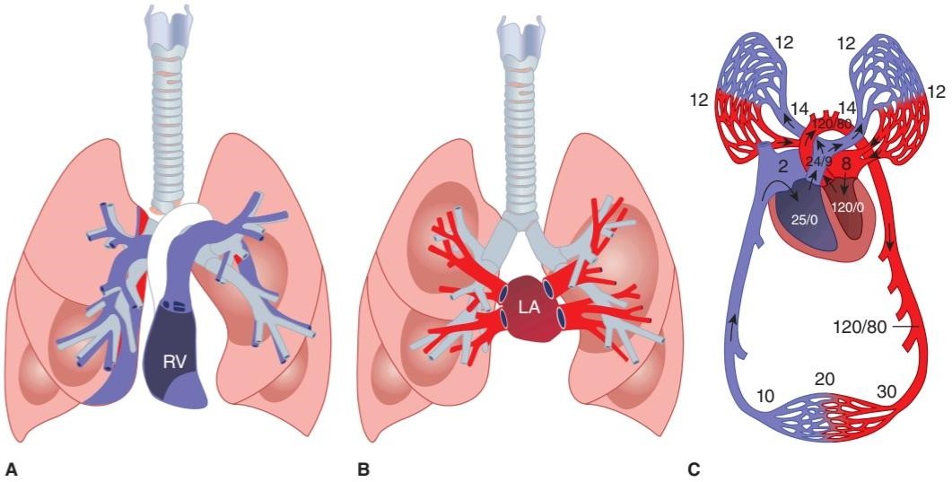
FIGURE 34-6 Pulmonary circulation. A) Schematic diagram of the relation of the main branches of the pulmonary arteries to the bronchial tree. B) Schematic of the pulmonary veins in relation to the bronchial tree. C) Representative areas of blood flow are labeled with corresponding blood pressure (mm Hg). In all diagrams, blue represents deoxygenated blood and red represents oxygenated blood. The deoxygenated/oxygenated shift in C occurs as blood traverses capillaries that wrap individual alveoli and is not as abrupt as shown. LA, left atrium; RV, right ventricle. (For (A,B) Reproduced with permission from Fishman AP et al (eds): Fishman’s Pulmonary Diseases and Disorders. 4th ed. New York: McGraw-Hill; 2008; (C) Modified with permission from Comroe JH Jr: Physiology of Respiration, 2nd ed. Year Book; 1974.)
شکل 34-6 گردش خون ریوی. الف) نمودار شماتیک ارتباط شاخه های اصلی شریان های ریوی با درخت برونش. ب) شماتیک وریدهای ریوی در رابطه با درخت برونش. ج) نواحی نماینده جریان خون با فشار خون متناظر (میلی متر جیوه) برچسب گذاری می شوند. در تمام نمودارها، رنگ آبی نشان دهنده خون بدون اکسیژن و قرمز نشان دهنده خون بدون اکسیژن است. تغییر اکسیژن زدایی شده/اکسیژنه در C زمانی اتفاق می افتد که خون از مویرگ هایی که آلوئول های منفرد را می پوشاند عبور می کند و آنطور که نشان داده شده ناگهانی نیست. LA، دهلیز چپ؛ RV، بطن راست. (برای (A, B) تکثیر شده با اجازه Fishman AP et al (eds): Fishman’s Pulmonary Diseases and Disorders. ویرایش 4. نیویورک: McGraw-Hill; 2008; (C) اصلاح شده با اجازه Comroe JH Jr: Physiology of Respiration، ویرایش دوم. کتاب سال؛ 1974.)
MECHANICS OF RESPIRATION
INSPIRATION & EXPIRATION
The lungs and the chest wall are elastic structures. Normally, no more than a thin layer of fluid is present between the lungs and the chest wall (intrapleural space). The lungs slide easily on the chest wall, but resist being pulled away from it in the same way that two moist pieces of glass slide on each other but resist separation. The pressure in the “space” between the lungs and chest wall (intrapleural pressure) is subatmospheric (Figure 34-7). The lungs are stretched when they expand at birth, and at the end of quiet expiration their tendency to recoil from the chest wall is just balanced by the tendency of the chest wall to recoil in the opposite direction. If the chest wall is opened, the lungs collapse; and if the lungs lose their elasticity, the chest expands and becomes barrel- shaped.
مکانیک تنفس
الهام و انقضا
ریه ها و دیواره قفسه سینه ساختارهای الاستیکی هستند. به طور معمول، بیش از یک لایه نازک مایع بین ریه ها و دیواره قفسه سینه (فضای داخل پلورال) وجود ندارد. ریه ها به راحتی روی دیواره قفسه سینه می لغزند، اما در برابر جدا شدن از آن مقاومت می کنند، به همان شکلی که دو تکه شیشه مرطوب روی هم می لغزند اما در برابر جدا شدن مقاومت می کنند. فشار در “فضای” بین ریه ها و دیواره قفسه سینه (فشار داخل پلورال) زیر اتمسفر است (شکل 34-7). ریه ها هنگام تولد کشیده می شوند و در پایان بازدم آرام، تمایل آنها به عقب نشینی از دیواره قفسه سینه با تمایل دیواره قفسه سینه به عقب نشینی در جهت مخالف متعادل می شود. اگر دیواره قفسه سینه باز شود، ریه ها فرو می ریزند. و اگر ریه ها خاصیت ارتجاعی خود را از دست بدهند، قفسه سینه منبسط شده و بشکه ای شکل می شود.
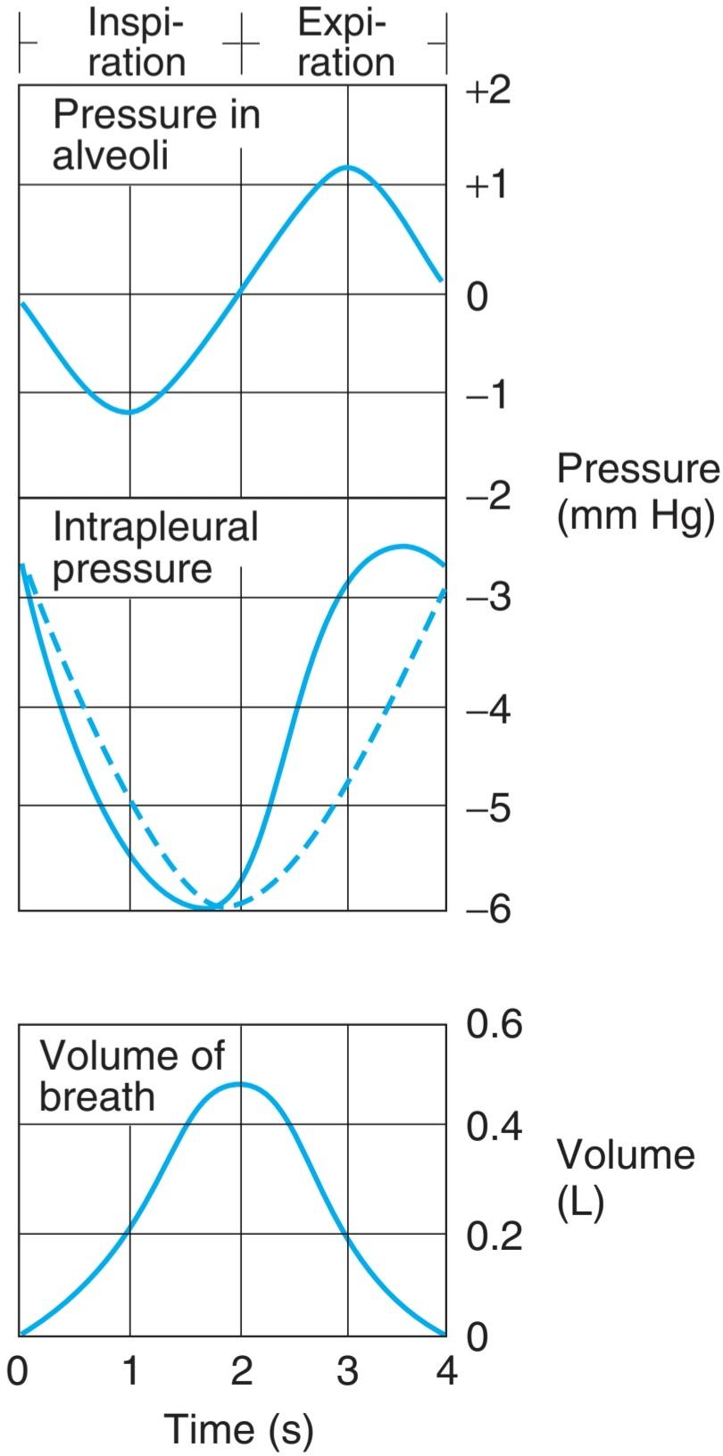
FIGURE 34-7 Pressure in the alveoli and the pleural space relative to atmospheric pressure during inspiration and expiration. The dashed line indicates what the intrapleural pressure would be in the absence of airway and tissue resistance; the actual curve (solid line) is skewed to the left by the resistance. Volume of breath during inspiration/expiration is graphed for comparison.
شکل 34-7 فشار در آلوئول ها و فضای پلور نسبت به فشار اتمسفر در طول دم و بازدم. خط چین نشان می دهد که فشار داخل پلور در غیاب مقاومت راه هوایی و بافت چقدر خواهد بود. منحنی واقعی (خط جامد) توسط مقاومت به سمت چپ منحرف می شود. حجم تنفس در طول دم/انقضا برای مقایسه نمودار شده است.
Inspiration is an active process. The contraction of the inspiratory muscles increases intrathoracic volume. The intrapleural pressure at the base of the lungs, which is normally about -2.5 mm Hg (relative to atmospheric) at the start of inspiration, decreases to about -6 mm Hg. The lungs are pulled into a more expanded position. The pressure in the airway becomes slightly negative, and air flows into the lungs. At the end of inspiration, the lung recoil begins to pull the chest back to the expiratory position, where the recoil pressures of the lungs and chest wall balance (see below). The pressure in the airway becomes slightly positive, and air flows out of the lungs. Expiration during quiet breathing is passive in the sense that no muscles that decrease intrathoracic volume contract. However, some contraction of the inspiratory muscles occurs in the early part of expiration. This contraction exerts a braking action on the recoil forces and slows expiration. Strong inspiratory efforts reduce intrapleural pressure to values as low as -30 mm Hg, producing correspondingly greater degrees of lung inflation. When ventilation is increased, the extent of lung deflation is also increased by active contraction of expiratory muscles that decrease intrathoracic volume.
الهام یک فرآیند فعال است. انقباض عضلات دمی باعث افزایش حجم داخل قفسه سینه می شود. فشار داخل پلور در قاعده ریه ها، که به طور معمول حدود 2.5- میلی متر جیوه (نسبت به اتمسفر) در شروع دم است، به حدود 6- میلی متر جیوه کاهش می یابد. ریهها به موقعیت منبسطتری کشیده میشوند. فشار در راه هوایی کمی منفی می شود و هوا به داخل ریه ها جریان می یابد. در پایان دم، پسکش ریه شروع به کشیدن قفسه سینه به سمت بازدم میکند، جایی که فشار پسکشی ریهها و دیواره قفسه سینه متعادل میشود (به زیر مراجعه کنید). فشار در راه هوایی کمی مثبت می شود و هوا از ریه ها خارج می شود. بازدم در طول تنفس آرام غیرفعال است به این معنا که هیچ ماهیچه ای که حجم داخل قفسه سینه را کاهش می دهد منقبض نمی شود. با این حال، برخی از انقباضات عضلات دمی در اوایل بازدم رخ می دهد. این انقباض یک عمل ترمز بر روی نیروهای پس زدگی اعمال می کند و انقباض را کند می کند. تلاشهای دمی قوی فشار داخل پلور را تا مقادیر کمتر از 30- میلیمتر جیوه کاهش میدهد و بهطور متناظر درجات بیشتری از تورم ریه ایجاد میکند. هنگامی که تهویه افزایش می یابد، میزان تخلیه ریه نیز با انقباض فعال عضلات بازدمی افزایش می یابد که حجم داخل قفسه سینه را کاهش می دهد.
QUANTITATING RESPIRATORY PHENOMENA
Modern spirometers permit direct measurement of gas intake and output. Since gas volumes vary with temperature and pressure and since the amount of water vapor in them varies, these devices have the ability to correct respiratory measurements involving volume to a stated set of standard conditions. It should be noted that correct measurements are highly dependent on the ability for the practitioner to properly encourage the patient to fully utilize the device. Modern techniques for gas analysis make possible rapid, reliable measurements of the composition of gas mixtures and the gas content of body fluids. For example, O2 and CO2 electrodes, small probes sensitive to O2 or CO2, can be inserted into the airway or into blood vessels or tissues and the PO2 and PCO2 recorded continuously. Chronic assessment of oxygenation is carried out noninvasively with a pulse oximeter, which can be easily placed on a fingertip.
کمیت کردن پدیده های تنفسی
اسپیرومترهای مدرن امکان اندازه گیری مستقیم گاز ورودی و خروجی را فراهم می کنند. از آنجایی که حجم گاز با دما و فشار متفاوت است و از آنجایی که مقدار بخار آب موجود در آنها متفاوت است، این دستگاه ها توانایی اصلاح اندازه گیری های تنفسی شامل حجم را به مجموعه ای از شرایط استاندارد دارند. لازم به ذکر است که اندازه گیری های صحیح به شدت به توانایی پزشک در تشویق صحیح بیمار برای استفاده کامل از دستگاه بستگی دارد. تکنیک های مدرن برای تجزیه و تحلیل گاز، اندازه گیری سریع و قابل اعتماد ترکیب مخلوط گازها و محتوای گاز مایعات بدن را ممکن می سازد. به عنوان مثال، الکترودهای O2 و CO2، پروبهای کوچک حساس به O2 یا CO2، میتوانند در راه هوایی یا رگهای خونی یا بافتها وارد شوند و PO2 و PCO2 به طور مداوم ثبت شوند. ارزیابی مزمن اکسیژن رسانی به صورت غیر تهاجمی با پالس اکسیمتر انجام می شود که به راحتی می توان آن را روی نوک انگشت قرار داد.
Lung Volumes & Capacities
Important quantitation of lung function can be gleaned from the displacement of air volume during inspiration and/or expiration. Lung capacities refer to subdivisions that contain two or more volumes. Volumes and capacities recorded on a spirometer from a healthy individual are shown in Figure 34-8. Diagnostic spirometry is used to assess a patient’s lung function for purposes of comparison with a normal population, or with previous measures from the same patient. The amount of air that moves into the lungs with each inspiration (or the amount that moves out with each expiration) during quiet breathing is called the tidal volume (TV). Typical values for TV are on the order of 500-750 mL. The air inspired with a maximal inspiratory effort in excess of the TV is the inspiratory reserve volume (IRV; ~2 L). The volume expelled by an active expiratory effort after passive expiration is the expiratory reserve volume (ERV; ~1 L), and the air left in the lungs after a maximal expiratory effort is the residual volume (RV; ~1.3 L). When all four of the above components are taken together, they make up the total lung capacity (TLC, ~5 L). The TLC can be broken down into alternative capacities that help define functioning lungs. The vital capacity (VC, ~3.5 L) refers to the maximum amount of air expired from the fully inflated lungs, or maximum inspiratory level (this represents TV + IRV + ERV). The inspiratory capacity (IC, ~2.5 L) is the maximum amount of air inspired from the end-expiratory level (IRV + TV). The functional residual capacity (FRC; ~2.5 L) represents the volume of the air remaining in the lungs after expiration of a normal breath (RV + ERV).
حجم و ظرفیت ریه
کمیت مهم عملکرد ریه را می توان از جابجایی حجم هوا در طول دم و/یا بازدم به دست آورد. ظرفیت ریه به زیربخش هایی اطلاق می شود که شامل دو یا چند جلد است. حجم ها و ظرفیت های ثبت شده روی یک اسپیرومتر از یک فرد سالم در شکل 34-8 نشان داده شده است. اسپیرومتری تشخیصی برای ارزیابی عملکرد ریه بیمار به منظور مقایسه با یک جمعیت طبیعی یا با اقدامات قبلی از همان بیمار استفاده می شود. مقدار هوایی که با هر دم به داخل ریه ها حرکت می کند (یا مقداری که با هر بازدم خارج می شود) در طول تنفس آرام، حجم جزر و مد (TV) نامیده می شود. مقادیر معمول برای تلویزیون در حدود 500-750 میلی لیتر است. هوای الهام گرفته شده با حداکثر تلاش دمی بیش از تلویزیون، حجم ذخیره دمی (IRV؛ ~ 2 L) است. حجم خارج شده توسط یک تلاش بازدمی فعال پس از بازدم غیرفعال، حجم ذخیره بازدمی (ERV؛ ~ 1 L) است و هوای باقی مانده در ریه ها پس از حداکثر تلاش بازدمی، حجم باقیمانده است (RV؛ ~1.3 L). وقتی هر چهار جزء فوق با هم گرفته شوند، ظرفیت کل ریه را تشکیل می دهند (TLC، ~ 5 لیتر). TLC را می توان به ظرفیت های جایگزینی تقسیم کرد که به تعریف عملکرد ریه ها کمک می کند. ظرفیت حیاتی (VC، ~ 3.5 L) به حداکثر مقدار هوای خارج شده از ریه های کاملاً باد شده یا حداکثر سطح دمی اشاره دارد (این نشان دهنده TV + IRV + ERV است). ظرفیت دمی (IC، ~ 2.5 L) حداکثر مقدار هوای دمیده شده از سطح انتهایی بازدم (IRV + TV) است. ظرفیت باقیمانده عملکردی (FRC؛ ~ 2.5 L) نشان دهنده حجم هوای باقی مانده در ریه ها پس از بازدم یک نفس طبیعی است (RV + ERV).
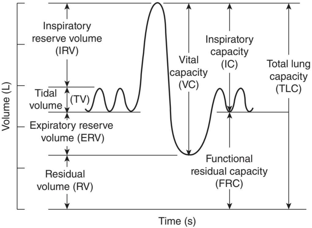
FIGURE 34-8 Lung volumes and capacity measurements. Lung volumes recorded by a spirometer. Lung capacities are determined from volume recordings. See text for definitions. (Reproduced with permission from Fishman AP et al (eds): Fishman’s Pulmonary Diseases and Disorders. 4th ed. New York: McGraw-Hill; 2008.)
شکل 34-8 اندازه گیری حجم و ظرفیت ریه. حجم ریه توسط اسپیرومتر ثبت می شود. ظرفیت ریه ها از روی حجم ضبط شده تعیین می شود. برای تعاریف به متن مراجعه کنید. (تکثیر شده با اجازه Fishman AP et al (eds): Fishman’s Pulmonary Diseases and Disorders. ویرایش چهارم نیویورک: McGraw-Hill؛ 2008.)
Dynamic measurements of lung volumes and capacities have been used to determine lung dysfunction. The forced vital capacity (FVC), the largest amount of air that can be expired after a maximal inspiratory effort, is frequently measured clinically as an index of pulmonary function. It gives useful information about the strength of the respiratory muscles and other aspects of pulmonary function. The fraction of the VC expired during the first second of a forced expiration is referred to as FEV1 (forced expiratory volume in the first second; Figure 34-9). The FEV1 to FVC ratio (FEV1/FVC) is a useful tool in the recognizing classes of airway disease (Clinical Box 34-2). Other dynamic measurements include the respiratory minute volume (RMV) and the maximal voluntary ventilation (MVV). RMV is normally ~6 L (500 mL/breath × 12 breaths/min). The MVV is the largest volume of gas that can be moved into and out of the lungs in 1 min by voluntary effort. Typically this is measured over a 15-s period and prorated to a minute; normal values range from 140 L/min to 180 L/min for healthy adult men. Changes in RMV and MVV in a patient can be indicative of lung dysfunction.
اندازه گیری های دینامیکی حجم و ظرفیت ریه برای تعیین اختلال عملکرد ریه استفاده شده است. ظرفیت حیاتی اجباری (FVC)، بزرگترین مقدار هوایی که می تواند پس از حداکثر تلاش دمی منقضی شود، اغلب از نظر بالینی به عنوان شاخصی از عملکرد ریوی اندازه گیری می شود. اطلاعات مفیدی در مورد قدرت ماهیچه های تنفسی و سایر جنبه های عملکرد ریوی می دهد. کسری از VC منقضی شده در اولین ثانیه از بازدم اجباری به عنوان FEV1 (حجم بازدمی اجباری در ثانیه اول؛ شکل 34-9) نامیده می شود. نسبت FEV1 به FVC (FEV1/FVC) ابزار مفیدی در تشخیص طبقات بیماری راه هوایی است (باکس بالینی 34-2). سایر اندازه گیری های دینامیکی شامل حجم دقیقه تنفسی (RMV) و حداکثر تهویه داوطلبانه (MVV) است. RMV معمولاً 6 لیتر (500 میلی لیتر در تنفس × 12 تنفس در دقیقه) است. MVV بزرگترین حجم گازی است که می تواند با تلاش داوطلبانه در 1 دقیقه به داخل و خارج ریه ها منتقل شود. به طور معمول این در یک دوره 15 ثانیه اندازه گیری می شود و به یک دقیقه تقسیم می شود. مقادیر طبیعی برای مردان بالغ سالم از 140 لیتر در دقیقه تا 180 لیتر در دقیقه متغیر است. تغییرات در RMV و MVV در یک بیمار می تواند نشان دهنده اختلال عملکرد ریه باشد.

FIGURE 34-9 Volume of gas expired by a normal adult man during a forced expiration, demonstrating the FEV1 and the FVC. From the graph, the forced expiratory volume in the first second (FEV1) to the forced vital capacity (FVC) ration (FEV1/FVC) can be calculated (4L/5L = 80%). (Reproduced with permission from Crapo RO: Pulmonary-function testing, N Engl J Med 1994; July 7; 331(1):25-30.)
شکل 34-9 حجم گاز منقضی شده توسط یک مرد بالغ طبیعی در طی یک بازدم اجباری، نشان دهنده FEV1 و FVC است. از نمودار، حجم بازدم اجباری در ثانیه اول (FEV1) تا جیره ظرفیت حیاتی اجباری (FVC) (FEV1/FVC) قابل محاسبه است (4L/5L = 80%). (تکثیر شده با مجوز Crapo RO: Pulmonary-function testing, N Engl J Med 1994؛ 7 ژوئیه؛ 331 (1): 25-30.)
CLINICAL BOX 34-2
Altered Airflow in Disease:
Representative lung volume expired over time in normal and diseased lungs during respiratory testing or pulmonary function test (PFT). A) Healthy normal subject. B) Patient with obstructive lung disease. C) Patient with restrictive lung disease. Note the differences in FEV1, FVC and FEV1/FVC at the bottom of the figure. FEV1, forced expiratory volume in the first second; FVC, forced vital capacity. (Reproduced with permission from Fishman AP et al (eds): Fishman’s Pulmonary Diseases and Disorders. 4th ed. New York: McGraw-Hill; 2008.)
جعبه بالینی 34-2
تغییر جریان هوا در بیماری:

حجم نماینده ریه به مرور زمان در ریه های نرمال و بیمار طی آزمایش تنفسی یا تست عملکرد ریوی (PFT) منقضی شد. الف) سوژه عادی سالم. ب) بیمار مبتلا به بیماری انسدادی ریه. ج) بیمار مبتلا به بیماری محدود کننده ریه. به تفاوت های FEV1، FVC و FEV1/FVC در پایین شکل توجه کنید. FEV1، حجم بازدم اجباری در ثانیه اول. FVC، ظرفیت حیاتی اجباری. (تکثیر شده با اجازه Fishman AP et al (eds): Fishman’s Pulmonary Diseases and Disorders. ویرایش چهارم نیویورک: McGraw-Hill؛ 2008.)
Airflow Measurements of Obstructive & Restrictive Disease
In the example above, a healthy or normal FVC is ~4.0 L and a healthy FEV1 is ~3.3 L. The calculated FEV1/FVC is ~80%. Patients with obstructive or restrictive lung diseases can display reduced FVC, on the order of 2.0 L in the example above. Measurement of FEV1, however, tends to vary significantly between the two diseases. In obstructive lung disorders, patients tend to show a slow, steady slope to the FVC, resulting in a small FEV1, on the order of 1.0 L in the example. However, in the restrictive lung disorder, patient’s airflow tends to be fast at first, and then quickly level out to approach FVC. The resultant FEV1 is much greater, on the order of 1.8 L in the example, even though FVC is equivalent (compare B, C above). A quick calculation of FEV1/FVC for patients with obstructive (50%) versus restrictive (90%) patterns defines the hallmark measurements in evaluating these two diseases. Obstructive disorders result in a marked decrease in both FVC and FEV1/FVC, whereas restrictive disorders result in a loss of FVC without loss in FEV1/FVC. It should be noted that these examples are idealized and several disorders can show mixed readings.
اندازه گیری جریان هوا بیماری انسدادی و محدود کننده
در مثال بالا، یک FVC سالم یا عادی ~4.0 لیتر و یک FEV1 سالم ~3.3 L است. FEV1/FVC محاسبه شده ~80٪ است. بیماران مبتلا به بیماریهای انسدادی یا محدودکننده ریه میتوانند FVC کاهش یافته را به ترتیب 2.0 لیتر در مثال بالا نشان دهند. با این حال، اندازه گیری FEV1 به طور قابل توجهی بین دو بیماری متفاوت است. در اختلالات انسدادی ریه، بیماران تمایل دارند شیب آهسته و ثابتی نسبت به FVC نشان دهند که منجر به یک FEV1 کوچک، در حد 1.0 لیتر در مثال میشود. با این حال، در اختلال ریه محدود کننده، جریان هوای بیمار ابتدا سریع است و سپس به سرعت به سمت FVC می رود. FEV1 حاصل بسیار بیشتر است، در مرتبه 1.8 لیتر در مثال، حتی اگر FVC معادل است (B، C را در بالا مقایسه کنید). محاسبه سریع FEV1/FVC برای بیماران با الگوهای انسدادی (50%) در مقابل الگوهای محدودکننده (90%)، معیارهای مشخصه را در ارزیابی این دو بیماری مشخص میکند. اختلالات انسدادی منجر به کاهش قابل توجه FVC و FEV1/FVC می شود، در حالی که اختلالات محدود کننده منجر به از دست دادن FVC بدون از دست دادن FEV1/FVC می شود. لازم به ذکر است که این نمونه ها ایده آل هستند و چندین اختلال می توانند قرائت های مختلط را نشان دهند.
Obstructive Disease-Asthma
Asthma is characterized by episodic or chronic wheezing, cough, and a feeling of tightness in the chest as a result of bronchoconstriction. Although the disease is not fully understood, three airway abnormalities are present: airway obstruction that is at least partially reversible, airway inflammation, and airway hyperresponsiveness to a variety of stimuli. A link to allergy has long been recognized, and plasma IgE levels are often elevated. Proteins released from eosinophils in the inflammatory reaction may damage the airway epithelium and contribute to the hyperresponsiveness. Leukotrienes are released from eosinophils and mast cells, and can enhance bronchoconstriction. Numerous other amines, neuropeptides, chemokines, and interleukins have effects on bronchial smooth muscle or produce inflammation, and they may be involved in asthma.
بیماری انسدادی – آسم
آسم با خس خس اپیزودیک یا مزمن، سرفه و احساس سفتی در قفسه سینه در نتیجه انقباض برونش مشخص می شود. اگرچه این بیماری به طور کامل شناخته نشده است، اما سه ناهنجاری راه هوایی وجود دارد: انسداد راه هوایی که حداقل تا حدی قابل برگشت است، التهاب راه هوایی، و واکنش بیش از حد راه هوایی به انواع محرک ها. مدتهاست که ارتباط با آلرژی شناخته شده است و سطح IgE پلاسما اغلب بالا می رود. پروتئین های آزاد شده از ائوزینوفیل ها در واکنش التهابی ممکن است به اپیتلیوم راه هوایی آسیب رسانده و به واکنش بیش از حد کمک کنند. لوکوترین ها از ائوزینوفیل ها و ماست سل ها آزاد می شوند و می توانند انقباض برونش را افزایش دهند. بسیاری دیگر از آمین ها، نوروپپتیدها، کموکاین ها و اینترلوکین ها بر روی ماهیچه صاف برونش تأثیر می گذارند یا التهاب ایجاد می کنند و ممکن است در آسم نقش داشته باشند.
Restrictive Disease-Idiopathic Pulmonary Fibrosis
Idiopathic pulmonary fibrosis (IPF), a type of pulmonary fibrosis, is a rare and irreversible interstitial lung disease that usually affects middle-aged and older adults (> 50 years old). The most common cause of death related to IPF is respiratory failure. Other causes of death include pulmonary hypertension (PH) and PH-associated right heart failure, pulmonary embolism and lung cancer. Up to 132,000 people in the United States are currently affected by IPF, and there are 30,000-40,000 new cases each year. Alveolar epithelial injury due to epithelial cell apoptosis and endoplasmic reticulum (ER) stress and epithelial-to-mesenchymal transition (EMT) have been implicated as cellular mechanisms involved in the development of IPF.
بیماری محدود کننده – فیبروز ریوی ایدیوپاتیک
فیبروز ریوی ایدیوپاتیک (IPF)، نوعی فیبروز ریوی، یک بیماری بینابینی ریه نادر و غیرقابل برگشت است که معمولاً افراد میانسال و مسن (بیش از 50 سال) را مبتلا می کند. شایع ترین علت مرگ ناشی از IPF نارسایی تنفسی است. سایر علل مرگ عبارتند از فشار خون ریوی (PH) و نارسایی قلب راست مرتبط با PH، آمبولی ریه و سرطان ریه. در حال حاضر بیش از 132000 نفر در ایالات متحده تحت تأثیر IPF قرار دارند و هر سال 30000 تا 40000 مورد جدید وجود دارد. آسیب اپیتلیال آلوئولی به دلیل آپوپتوز سلول های اپیتلیال و استرس شبکه آندوپلاسمی (ER) و انتقال اپیتلیال به مزانشیمی (EMT) به عنوان مکانیسم های سلولی درگیر در توسعه IPF نقش دارد.
THERAPEUTIC HIGHLIGHTS
Asthma
Because ẞ2-adrenergic receptors mediate bronchodilation, ẞ2-adrenergic agonists have long been the mainstay of “rescue” treatment for mild to moderate asthma attacks. Inhaled steroids are used even in mild to moderate cases to reduce inflammation; they are very effective, but their side effects can be a problem. Agents that block synthesis of leukotrienes or their CysLT1receptor inhibit acetylcholine-mediated activation of muscarinic (M) receptors and blockade Cl- channels and Ca2+ channels in mast cells have also proved useful in certain cases.
نکات برجسته درمانی
آسم
از آنجایی که گیرنده های 2-آدرنرژیک واسطه اتساع برونش هستند، آگونیست های 2-آدرنرژیک مدت هاست که اصلی ترین درمان “نجات” برای حملات خفیف تا متوسط آسم بوده است. استروئیدهای استنشاقی حتی در موارد خفیف تا متوسط برای کاهش التهاب استفاده می شود. آنها بسیار موثر هستند، اما عوارض جانبی آنها می تواند مشکل ساز باشد. عواملی که سنتز لکوترین ها یا گیرنده CysLT1 آنها را مسدود می کنند، فعال شدن گیرنده های موسکارینی (M) با واسطه استیل کولین را مهار می کنند و کانال های -Cl و کانال های +Ca2 را در ماست سل ها مسدود می کنند نیز در موارد خاص مفید هستند.
IPF
Clinical management and treatment have been focused on suppressing the inflammatory and fibrotic process of IPF. Agents with anti-fibrotic, anti- inflammation, and anti-oxidant effects (eg, nintedanib, pirfenidone, thalidomide) are often used for treatment of IPF. Lung transplantation (replacement of one or both lungs) is a surgical therapeutic intervention of proven benefits in IPF, which is often reserved for patients at the advanced stage of IPF; the 5-year survival rate is approximately 50% for lung transplant patients.
IPF
مدیریت و درمان بالینی بر سرکوب فرآیند التهابی و فیبروتیک IPF متمرکز شده است. داروهایی با اثرات ضد فیبروتیک، ضد التهابی و آنتی اکسیدانی (مانند نینتدانیب، پیرفنیدون، تالیدومید) اغلب برای درمان IPF استفاده می شوند. پیوند ریه (جایگزینی یک یا هر دو ریه) یک مداخله درمانی جراحی با مزایای ثابت شده در IPF است که اغلب برای بیماران در مرحله پیشرفته IPF محفوظ است. میزان بقای 5 ساله برای بیماران پیوند ریه تقریباً 50 درصد است.
COMPLIANCE OF THE LUNGS & CHEST WALL
Compliance is developed due to the tendency for tissue to resume its original position after an applied force has been removed. After an expiration during quiet breathing (eg, at the FRC), the lungs have a tendency to collapse and the chest wall has a tendency to expand. The interaction between the recoil of the lungs and recoil of the chest can be measured through a spirometer that has a valve just beyond the mouthpiece. The mouthpiece contains a pressure- measuring device. After the person inhales a given amount, the valve is shut, closing off the airway. The respiratory muscles are then relaxed while the pressure in the airway is recorded. The procedure is repeated after inhaling or actively exhaling various volumes. The curve of airway pressure obtained in this way, plotted against volume, is the pressure-volume curve of the total respiratory system (PTR in Figure 34–10). The pressure is zero at a lung volume that corresponds to the volume of gas in the lungs at FRC (ie, relaxation volume). As can be noted from Figure 34-10, this relaxation pressure is the sum of slightly negative pressure component from the chest wall (P) and a slightly positive pressure from the lungs (PL). PTR is positive at greater volumes and negative at smaller volumes. Compliance of the lungs and chest wall is measured as the slope of the PTR curve, or, as a change in lung volume per unit change in airway pressure (AV/AP). It is normally measured in the pressure range where the relaxation pressure curve is steepest, and normal values are ~0.2 L/cm H2O in a healthy adult man. However, compliance depends on lung volume and thus can vary. In an extreme example, an individual with only one lung has approximately half the AV for a given AP. Compliance is also slightly greater when measured during deflation than when measured during inflation. Consequently, it is more informative to examine the whole pressure-volume curve. The curve is shifted downward and to the right (compliance is decreased) by pulmonary edema and interstitial pulmonary fibrosis (Figure 34-11). Pulmonary fibrosis is a progressive restrictive airway disease in which there is stiffening and scarring of the lungs. The curve is shifted upward and to the left (compliance is increased) in emphysema.
انطباق ریه ها و دیواره قفسه سینه
انطباق به دلیل تمایل بافت برای از سرگیری موقعیت اولیه خود پس از برداشتن نیروی اعمال شده ایجاد می شود. پس از یک بازدم در طی تنفس آرام (مثلاً در FRC)، ریه ها تمایل به فروپاشی و دیواره قفسه سینه تمایل به انبساط دارند. برهمکنش بین پس زدن ریه ها و پس زدن قفسه سینه را می توان از طریق یک اسپیرومتر که دریچه ای درست در آن سوی قطعه دهانی دارد اندازه گیری کرد. قطعه دهانی حاوی یک دستگاه اندازه گیری فشار است. پس از اینکه فرد مقدار مشخصی را استنشاق کرد، دریچه بسته می شود و راه هوایی بسته می شود. سپس عضلات تنفسی شل می شوند و فشار در راه هوایی ثبت می شود. این روش پس از دم یا بازدم فعال حجم های مختلف تکرار می شود. منحنی فشار راه هوایی که از این طریق به دست می آید، در برابر حجم رسم می شود، منحنی فشار-حجم کل سیستم تنفسی است (PTR در شکل 34-10). فشار در حجم ریه برابر با حجم گاز در ریه ها در FRC صفر است (یعنی حجم آرامش). همانطور که از شکل 34-10 می توان اشاره کرد، این فشار آرامش حاصل مجموع مولفه فشار کمی منفی از دیواره قفسه سینه (P) و یک فشار کمی مثبت از ریه ها (PL) است. PTR در حجم های بیشتر مثبت و در حجم های کوچکتر منفی است. انطباق ریه ها و دیواره قفسه سینه به عنوان شیب منحنی PTR یا به عنوان تغییر در حجم ریه در واحد تغییر فشار راه هوایی (AV/AP) اندازه گیری می شود. معمولاً در محدوده فشاری اندازهگیری میشود که در آن منحنی فشار آرامش شدیدترین است، و مقادیر نرمال ~ 0.2 L/cm H2O در یک مرد بالغ سالم است. با این حال، انطباق به حجم ریه بستگی دارد و بنابراین می تواند متفاوت باشد. در یک مثال افراطی، فردی که تنها یک ریه دارد تقریباً نیمی از AV را برای یک AP معین دارد. انطباق نیز در هنگام کاهش تورم کمی بیشتر از زمانی است که در طول تورم اندازه گیری می شود. در نتیجه، بررسی کل منحنی فشار-حجم آموزنده تر است. منحنی به سمت پایین و به راست (انطباق کاهش می یابد) توسط ادم ریوی و فیبروز ریوی بینابینی منتقل می شود (شکل 34-11). فیبروز ریوی یک بیماری محدود کننده پیشرونده راه هوایی است که در آن ریه ها سفت و زخم می شوند. منحنی در آمفیزم به سمت بالا و به چپ جابه جا می شود (انطباق افزایش می یابد).

FIGURE 34-10 Pressure-volume curves in the lungs. The pressure-volume curves of the total respiratory system (PTR, black line), the lungs (P1, blue line), and the chest (Pw, green line) are plotted together with standard volumes for functional residual capacity and tidal volume. The transmural pressure is intrapulmonary pressure minus intrapleural pressure in the case of the lungs, intrapleural pressure minus outside (barometric) pressure in the case of the chest wall, and intrapulmonary pressure minus barometric pressure in the case of the total respiratory system. From these curves, the total and actual elastic work associated with breathing can be derived. (Modified with permission from Mines AH: Respiratory Physiology, 3rd ed. New York, NY: Raven Press; 1993.)
شکل 34-10 منحنی های فشار-حجم در ریه ها. منحنیهای فشار-حجم کل سیستم تنفسی (PTR، خط سیاه)، ریهها (P1، خط آبی)، و قفسه سینه (Pw، خط سبز) همراه با حجمهای استاندارد برای ظرفیت باقیمانده عملکردی و حجم جزر و مد رسم میشوند. فشار ترانس مورال فشار داخل ریوی منهای فشار داخل جنب در مورد ریه ها، فشار داخل پلور منهای فشار بیرونی (بارومتریک) در مورد دیواره قفسه سینه و فشار داخل ریوی منهای فشار فشار هوا در مورد کل سیستم تنفسی است. از این منحنی ها، کار الاستیک کلی و واقعی مرتبط با تنفس را می توان استخراج کرد. (اصلاح شده با اجازه Mines AH: Respiratory Physiology, 3rd ed. New York, NY: Raven Press; 1993.)

FIGURE 34-11 Static expiratory pressure-volume curves of lungs in healthy persons and persons with severe emphysema and pulmonary fibrosis. (Modified with permission from Pride NB, Macklem PT: Lung mechanics in disease. In: Handbook of Physiology. Section 3, The Respiratory System. Vol III, part 2. Fishman AP [editor]. American Physiological Society, 1986.)
شکل 34-11 منحنی های فشار بازدمی استاتیک-حجم ریه ها در افراد سالم و افراد مبتلا به آمفیزم شدید و فیبروز ریوی. (با مجوز Pride NB، Macklem PT: Lung mechanics in disease. In: Handbook of Physiology. بخش 3، سیستم تنفسی. جلد III، قسمت 2. Fishman AP [ویرایشگر]. انجمن فیزیولوژیک آمریکا، 1986، اصلاح شده است.)
Airway Resistance
Airway resistance is defined as the change of pressure (AP) from the alveoli to the mouth divided by the change in flow rate (V). Because of the structure of the bronchial tree, and thus the pathway for air that contributes to its resistance, it is difficult to apply mathematical estimates of the movement through the bronchial tree. However, measurements where alveolar and intrapleural pressure can be compared to actual pressure (eg, Figure 34–7 middle panel) show the contribution of airway resistance. Airway resistance is significantly increased as lung volume is reduced. Also, bronchi and bronchioles significantly contribute to airway resistance. Thus, contraction of the smooth muscle that lines the bronchial airways will increase airway resistance and make breathing more difficult.
مقاومت راه هوایی
مقاومت راه هوایی به عنوان تغییر فشار (AP) از آلوئول ها به دهان تقسیم بر تغییر سرعت جریان (V) تعریف می شود. به دلیل ساختار درخت برونش و در نتیجه مسیر هوا که به مقاومت آن کمک می کند، به کار بردن تخمین های ریاضی حرکت از طریق درخت نایژه دشوار است. با این حال، اندازه گیری هایی که در آن فشار آلوئولی و داخل پلورال را می توان با فشار واقعی مقایسه کرد (به عنوان مثال، شکل 34-7 پانل میانی) سهم مقاومت راه هوایی را نشان می دهد. با کاهش حجم ریه، مقاومت راه هوایی به طور قابل توجهی افزایش می یابد. همچنین برونش ها و برونشیول ها به میزان قابل توجهی در مقاومت راه هوایی کمک می کنند. بنابراین، انقباض ماهیچه صافی که راههای هوایی برونش را میپوشاند، مقاومت راه هوایی را افزایش داده و تنفس را دشوارتر میکند.
Role of Surfactant in Alveolar Surface Tension
An important factor affecting the compliance of the lungs is the surface tension of the film of fluid that lines the alveoli. The magnitude of this component at various lung volumes can be measured by removing the lungs from the body of an experimental animal and distending them alternately with saline and with air while measuring the intrapulmonary pressure. Because saline reduces the surface tension to nearly zero, the pressure-volume curve obtained with saline measures only the tissue elasticity (Figure 34-12), whereas the curve obtained with air measures both tissue elasticity and surface tension. The difference between the saline and air curves is much smaller when lung volumes are small. Differences are also obvious in the curves generated during inflation and deflation. This difference is termed hysteresis, and notably is not present in the saline generated curves (Figure 34-12). The alveolar environment, and specifically the secreted factors that help reduce surface tension and keep alveoli from collapsing, contribute to hysteresis.
نقش سورفکتانت در کشش سطح آلوئول
عامل مهمی که بر انطباق ریه ها تأثیر می گذارد، کشش سطحی لایه مایع است که آلوئول ها را می پوشاند. بزرگی این مولفه در حجم های مختلف ریه را می توان با برداشتن ریه ها از بدن حیوان آزمایشی و اتساع متناوب آنها با آب نمک و هوا در حین اندازه گیری فشار داخل ریوی اندازه گیری کرد. از آنجایی که سالین کشش سطحی را تقریباً به صفر می رساند، منحنی فشار-حجم به دست آمده با سالین فقط کشش بافت را اندازه می گیرد (شکل 34-12)، در حالی که منحنی به دست آمده با هوا هم کشش بافت و هم کشش سطحی را اندازه گیری می کند. تفاوت بین منحنی شور و هوا زمانی که حجم ریه کم است بسیار کمتر است. تفاوت ها در منحنی های ایجاد شده در طول تورم و کاهش تورم نیز آشکار است. این تفاوت پسماند نامیده می شود و به طور مشخص در منحنی های تولید شده با نمک وجود ندارد (شکل 34-12). محیط آلوئولی و به ویژه عوامل ترشحی که به کاهش کشش سطحی و جلوگیری از فروپاشی آلوئول ها کمک می کنند، به هیسترزیس کمک می کنند.

FIGURE 34-12 Pressure-volume curves in the lungs of a cat after removal from the body. Saline: lungs inflated and deflated with saline to reduce surface tension, resulting in a measurement of tissue elasticity. Air: lungs inflated (Inf) and deflated (Def) with air results in a measure of both tissue elasticity and surface tension. (Reproduced with permission from Morgan TE: Pulmonary surfactant. N Engl J Med 1971; May 27; 284(21):1185-1193.)
شکل 34-12 منحنی های فشار-حجم در ریه های گربه پس از خارج کردن از بدن. سالین: ریه ها با آب نمک باد شده و تخلیه می شوند تا کشش سطحی کاهش یابد و در نتیجه کشش بافت اندازه گیری شود. هوا: ریههای باد شده (Inf) و تخلیه (Def) با هوا منجر به اندازهگیری خاصیت ارتجاعی بافت و کشش سطحی میشود. (تکثیر شده با مجوز Morgan TE: Pulmonary surfactant. N Engl J Med 1971؛ May 27; 284(21):1185-1193.)
The low surface tension when the alveoli are small is due to the presence of surfactant in the fluid lining the alveoli. Surfactant is a mixture of dipalmitoylphosphatidylcholine (DPPC), other lipids, and proteins. If the surface tension is not kept low when the alveoli become smaller during expiration, they collapse in accordance with the law of Laplace. In spherical structures like an alveolus, the distending pressure (P) equals two times the tension (T) divided by the radius (r): P = 2T/r, if T is not reduced as r is reduced, the tension overcomes the distending pressure. Surfactant also helps prevent pulmonary edema. It has been calculated that if it were not present, the unopposed surface tension in the alveoli would produce a 20 mm Hg force; such a force would greatly favor transudation of fluid from the blood into the alveoli.
کشش سطحی کم هنگامی که آلوئول ها کوچک هستند به دلیل وجود سورفکتانت در مایع پوشاننده آلوئول ها است. سورفکتانت مخلوطی از دی پالمیتوئیل فسفاتیدیل کولین (DPPC)، سایر لیپیدها و پروتئین ها است. اگر کشش سطحی زمانی که آلوئول ها در حین بازدم کوچکتر می شوند پایین نگه داشته نشود، طبق قانون لاپلاس فرو می ریزند. در ساختارهای کروی مانند آلوئول، فشار اتساع (P) برابر است با دو برابر کشش (T) تقسیم بر شعاع (r): P = 2T/r، اگر T با کاهش r کاهش نیابد، کشش بر فشار انبساط غلبه می کند. سورفکتانت همچنین به جلوگیری از ادم ریوی کمک می کند. محاسبه شده است که اگر وجود نداشت، کشش سطحی بدون مخالف در آلوئول ها نیروی 20 میلی متر جیوه ایجاد می کرد. چنین نیرویی به شدت به انتقال مایع از خون به آلوئول کمک می کند.
Formation of the phospholipid film is greatly facilitated by the proteins in surfactant. This material contains four unique proteins: surfactant protein (SP)- A, SP-B, SP-C, and SP-D. SP-A is a large glycoprotein and has a collagen-like domain within its structure. It has multiple functions, including regulation of the feedback uptake of surfactant by the type II alveolar epithelial cells that secrete it. SP-B and SP-C are smaller proteins, which are the key protein members of the monomolecular film of surfactant. Like SP-A, SP-D is a glycoprotein. Its full function is uncertain; however, it plays an important role in the organization of SP-B and SP-C into the surfactant layer. Both SP-A and SP-D are members of the collectin family of proteins that are involved in innate immunity in the conducting airway as well as in the alveoli. Some clinical aspects of surfactant are discussed in Clinical Box 34-3.
تشکیل فیلم فسفولیپیدی توسط پروتئین های موجود در سورفکتانت بسیار تسهیل می شود. این ماده حاوی چهار پروتئین منحصر به فرد است: پروتئین سورفکتانت (SP)-A، SP-B، SP-C و SP-D. SP-A یک گلیکوپروتئین بزرگ است و یک دامنه کلاژن مانند در ساختار خود دارد. عملکردهای متعددی دارد، از جمله تنظیم بازخورد جذب سورفکتانت توسط سلول های اپیتلیال آلوئولی نوع II که آن را ترشح می کنند. SP-B و SP-C پروتئین های کوچکتری هستند که اعضای پروتئینی کلیدی فیلم تک مولکولی سورفکتانت هستند. مانند SP-A، SP-D یک گلیکوپروتئین است. عملکرد کامل آن نامشخص است. با این حال، نقش مهمی در سازماندهی SP-B و SP-C در لایه سورفکتانت ایفا می کند. هر دو SP-A و SP-D اعضای خانواده پروتئین های کلتین هستند که در ایمنی ذاتی در راه هوایی رسانا و همچنین در آلوئول ها نقش دارند. برخی از جنبه های بالینی سورفکتانت در جعبه بالینی 34-3 مورد بحث قرار گرفته است.
CLINICAL BOX 34-3
Surfactant
Surfactant is important at birth. The fetus makes respiratory movements in utero, but the lungs remain collapsed until birth. After birth, the infant makes several strong inspiratory movements and the lungs expand. Surfactant keeps them from collapsing again. Surfactant deficiency is an important cause of infant respiratory distress syndrome (IRDS, also known as hyaline membrane disease), the serious pulmonary disease that develops in infants born before their surfactant system is functional. Surface tension in the lungs of these infants is high, and the alveoli are collapsed in many areas (atelectasis). An additional factor in IRDS is retention of fluid in the lungs. During fetal life, Cl- is secreted with fluid by the pulmonary epithelial cells. At birth, there is a shift to Na* absorption by these cells via the epithelial Na* channels (ENaCs), and fluid is absorbed with the Na*. Prolonged immaturity of the ENaCs contributes to the pulmonary abnormalities in IRDS.
جعبه بالینی 34-3
سورفکتانت
سورفکتانت در بدو تولد مهم است. جنین در داخل رحم حرکات تنفسی انجام می دهد، اما ریه ها تا زمان تولد در هم می مانند. پس از تولد، نوزاد چندین حرکت دمی قوی انجام می دهد و ریه ها منبسط می شوند. سورفکتانت از فروپاشی مجدد آنها جلوگیری می کند. کمبود سورفکتانت یکی از علل مهم سندرم دیسترس تنفسی نوزادان (IRDS، که به عنوان بیماری غشای هیالین نیز شناخته می شود)، یک بیماری جدی ریوی است که در نوزادانی که قبل از عملکرد سیستم سورفکتانتشان متولد می شوند، ایجاد می شود. کشش سطحی در ریه های این نوزادان زیاد است و آلوئول ها در بسیاری از نواحی فرو می ریزند (آتلکتازی). یک عامل اضافی در IRDS احتباس مایع در ریه ها است. در طول زندگی جنینی، Cl- همراه با مایع توسط سلول های اپیتلیال ریوی ترشح می شود. در بدو تولد، جذب Na* توسط این سلولها از طریق کانالهای Na* اپیتلیال (ENaCs) تغییر میکند و مایع با Na* جذب میشود. نابالغی طولانی مدت ENaCs به ناهنجاری های ریوی در IRDS کمک می کند.
Overproduction/dysregulation of surfactant proteins can also lead to respiratory distress and is the cause of pulmonary alveolar proteinosis.
تولید بیش از حد/اختلال در تنظیم پروتئینهای سورفکتانت نیز میتواند منجر به دیسترس تنفسی شود و علت پروتئینوز آلوئولی ریوی است.
THERAPEUTIQ HIGHLIGHTS
Treatment of IRDS is commonly done with surfactant replacement therapy. Interestingly, such surfactant replacement therapy has not been as successful in clinical trials for adults experiencing respiratory distress due to surfactant dysfunction.
نکات برجسته درمانی
درمان IRDS معمولاً با درمان جایگزینی سورفکتانت انجام می شود. جالب توجه است که چنین درمان جایگزینی سورفکتانت در کارآزماییهای بالینی برای بزرگسالانی که به دلیل اختلال عملکرد سورفاکتانت دچار دیسترس تنفسی میشوند، موفقیتآمیز نبوده است.
WORK OF BREATHING
Work is performed by the respiratory muscles in stretching the elastic tissues of the chest wall and lungs (elastic work; approximately 65% of the total work), moving inelastic tissues (viscous resistance; 7% of total), and moving air through the respiratory passages (airway resistance; 28% of total). Because the dimensions of pressure × volume (g/cm2 × cm3 = g × cm) has the same dimensions as work (force × distance; g x cm), the work of breathing can be calculated from the previously presented pressure-volume curve (Figure 34–10). Note that the relaxation pressure curve of the total respiratory system (PTR) differs from that of the lungs alone (PL). The amount of elastic work required to inflate the whole respiratory system is less than the amount required to inflate the lungs alone because part of the work comes from elastic energy stored in the thorax.
کار از تنفس
کار توسط عضلات تنفسی در کشش بافت های الاستیک دیواره قفسه سینه و ریه ها (کار الاستیک؛ تقریباً 65٪ از کل کار)، حرکت بافت های غیر کشسان (مقاومت چسبناک؛ 7٪ از کل) و حرکت هوا از طریق مجاری تنفسی (مقاومت راه هوایی؛ 28٪ از کل) انجام می شود. از آنجایی که ابعاد فشار × حجم (g/cm2×cm3 = g×cm) دارای ابعاد یکسانی با کار است (نیرو × فاصله؛ g x cm)، کار تنفس را می توان از منحنی فشار-حجم قبلا ارائه شده محاسبه کرد (شکل 34-10). توجه داشته باشید که منحنی فشار آرامش کل سیستم تنفسی (PTR) با ریه ها به تنهایی (PL) متفاوت است. مقدار کار الاستیک مورد نیاز برای باد کردن کل سیستم تنفسی کمتر از مقدار مورد نیاز برای باد کردن ریه ها به تنهایی است زیرا بخشی از کار از انرژی الاستیک ذخیره شده در قفسه سینه ناشی می شود.
Estimates of the total work of quiet breathing range from 0.3 to 0.8 kg-m/min. The value rises markedly during exercise, but the energy cost of breathing in normal individuals represents less than 3% of the total energy expenditure during exercise. The work of breathing is greatly increased in diseases such as emphysema, asthma, and heart failure with dyspnea and orthopnea. The respiratory muscles have length-tension relations like those of other skeletal and cardiac muscles, and when they are severely stretched, they contract with less strength. They can also become fatigued and fail (pump failure), leading to inadequate ventilation.
برآورد کل کار تنفس آرام از 0.3 تا 0.8 کیلوگرم بر متر در دقیقه است. این مقدار در طول ورزش به طور قابل توجهی افزایش می یابد، اما هزینه انرژی تنفس در افراد عادی کمتر از 3٪ از کل انرژی مصرف شده در طول ورزش را نشان می دهد. کار تنفس در بیماری هایی مانند آمفیزم، آسم و نارسایی قلبی همراه با تنگی نفس و ارتوپنه بسیار افزایش می یابد. ماهیچه های تنفسی مانند سایر ماهیچه های اسکلتی و قلبی دارای روابط کششی طولی هستند و زمانی که به شدت کشیده می شوند با قدرت کمتری منقبض می شوند. آنها همچنین می توانند خسته و از کار بیفتند (شکست پمپ) که منجر به تهویه ناکافی می شود.
DIFFERENCES IN VENTILATION & BLOOD FLOW IN DIFFERENT PARTS OF THE LUNG
In the upright position, ventilation (V) per unit lung volume is greater at the base of the lung than at the apex. The reason for this is that at the start of inspiration, intrapleural pressure is less negative at the base than at the apex, and since the intrapulmonary intrapleural pressure difference is less than at the apex, the lung is less expanded. Conversely, at the apex, the lung is more expanded; that is, the percentage of maximum lung volume is greater. Because of the stiffness of the lung, the increase in lung volume per unit increase in pressure is smaller when the lung is initially more expanded, and ventilation is consequently greater at the base. Blood flow is also greater at the base than the apex. The relative change in blood flow from the apex to the base is greater than the relative change in ventilation, so the ventilation/perfusion (V/Q) ratio is low at the base and high at the apex.
تفاوت در تهویه و جریان خون در قسمت های مختلف ریه
در وضعیت عمودی، تهویه (V) به ازای واحد حجم ریه در پایه ریه بیشتر از راس است. دلیل این امر این است که در شروع دم، فشار داخل پلور در قاعده کمتر از راس منفی است و از آنجایی که اختلاف فشار داخل پلورال داخل ریوی کمتر از راس است، ریه کمتر منبسط میشود. برعکس، در راس، ریه بیشتر منبسط شده است. یعنی درصد حداکثر حجم ریه بیشتر است. به دلیل سفتی ریه، افزایش حجم ریه به ازای واحد افزایش فشار زمانی که ریه در ابتدا منبسط میشود کمتر است و در نتیجه تهویه در پایه بیشتر میشود. جریان خون نیز در قاعده بیشتر از راس است. تغییر نسبی در جریان خون از راس به قاعده بیشتر از تغییر نسبی تهویه است، بنابراین نسبت تهویه/پرفیوژن (V/Q) در قاعده کم و در راس زیاد است.
The ventilation and perfusion differences from the apex to the base of the lungs have usually been attributed to gravity: they tend to disappear in the supine position, and the weight of the lungs would be expected to create pressure at the base in the upright position. However, the inequalities of ventilation and blood flow in human lungs were found to persist to a remarkable degree in the weightlessness of space. Therefore, other factors also play a role in producing the inequalities.
تفاوتهای تهویه و پرفیوژن از راس تا پایه ریهها معمولاً به گرانش نسبت داده میشود: آنها در حالت خوابیده به پشت ناپدید میشوند و انتظار میرود وزن ریهها در حالت عمودی فشاری را در پایه ایجاد کند. با این حال، نابرابری های تهویه و جریان خون در ریه های انسان به میزان قابل توجهی در بی وزنی فضا ادامه دارد. بنابراین عوامل دیگری نیز در ایجاد نابرابری ها نقش دارند.
DEAD SPACE & UNEVEN VENTILATION
Because gaseous exchange in the respiratory system occurs only in the terminal portions of the airways, the gas that occupies the rest of the respiratory system is not available for gas exchange with pulmonary capillary blood. Normally, the volume (in mL) of this anatomic dead space is approximately equal to the body weight in pounds. As an example, in a man who weighs 150 lb (68 kg), only the first 350 mL of the 500 mL inspired with each breath at rest mixes with the air in the alveoli. Conversely, with each expiration, the first 150 mL expired is gas that occupied the dead space, and only the last 350 mL is gas from the alveoli. Consequently, the alveolar ventilation, that is, the amount of air reaching the alveoli per minute, is less than the RMV. Note that because of the dead space, rapid shallow breathing produces much less alveolar ventilation than slow deep breathing at the same RMV (Table 34-1).
فضای مرده و تهویه ناهموار
از آنجایی که تبادل گاز در سیستم تنفسی فقط در قسمت های انتهایی راه های هوایی اتفاق می افتد، گازی که بقیه سیستم تنفسی را اشغال می کند برای تبادل گاز با خون مویرگی ریوی در دسترس نیست. به طور معمول، حجم (بر حسب میلی لیتر) این فضای مرده آناتومیک تقریباً برابر با وزن بدن بر حسب پوند است. به عنوان مثال، در مردی که 150 پوند (68 کیلوگرم) وزن دارد، تنها 350 میلیلیتر اول از 500 میلیلیتر الهام گرفته شده با هر تنفس در حالت استراحت با هوای موجود در آلوئولها مخلوط میشود. برعکس، با هر انقضا، 150 میلی لیتر اول منقضی شده، گازی است که فضای مرده را اشغال کرده است، و تنها 350 میلی لیتر آخر گاز آلوئول ها است. در نتیجه، تهویه آلوئولی، یعنی مقدار هوایی که در دقیقه به آلوئول ها می رسد، کمتر از RMV است. توجه داشته باشید که به دلیل فضای مرده، تنفس کم عمق سریع تهویه آلوئولی بسیار کمتری نسبت به تنفس عمیق آهسته در همان RMV ایجاد می کند (جدول 34-1).
TABLE 34-1 Effect of variations in respiratory rate and depth on alveolar ventilation.
جدول 34-1 اثر تغییرات در تعداد و عمق تنفس بر تهویه آلوئولار.
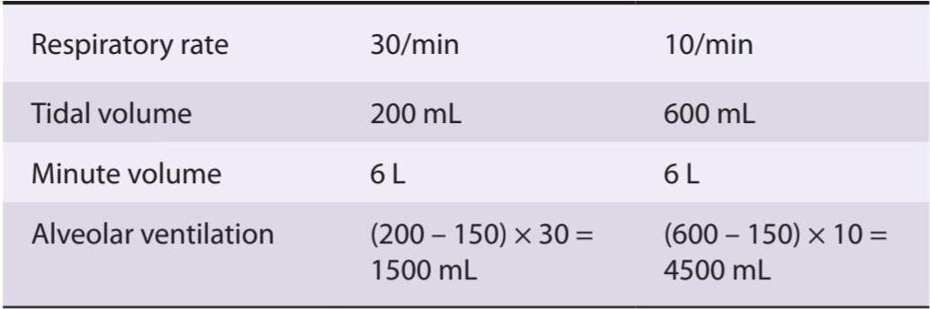
It is important to distinguish between the anatomic dead space (respiratory system volume exclusive of alveoli) and the total (physiological) dead space (volume of gas not equilibrating with blood; ie, wasted ventilation). In healthy individuals, the two dead spaces are identical and can be estimated by body weight. However, in disease states, no exchange may take place between the gas in some of the alveoli and the blood, and some of the alveoli may be overventilated. The volume of gas in nonperfused alveoli and any volume of air in the alveoli in excess of that necessary to oxygenate the blood in the alveolar capillaries is part of the dead space (nonequilibrating) gas volume. The anatomic dead space can be measured by analysis of the single-breath N2 curve (Figure 34-13). From mid-inspiration, the patient takes as deep a breath as possible of pure O2, then exhales steadily while the N2 content of the expired gas is continuously measured. The initial gas exhaled (phase I) is the gas that filled the dead space and that consequently contains no N2. This is followed by a mixture of dead space and alveolar gas (phase II) and then by alveolar gas (phase III). The volume of the dead space is the volume of the gas expired from peak inspiration to the midportion of phase II.
تمایز بین فضای مرده آناتومیک (حجم دستگاه تنفسی به استثنای آلوئول ها) و فضای مرده کل (فیزیولوژیکی) (حجم گازی که با خون متعادل نمی شود، یعنی تهویه هدر رفته) مهم است. در افراد سالم، دو فضای مرده یکسان هستند و با وزن بدن قابل تخمین هستند. با این حال، در حالات بیماری، ممکن است هیچ تبادلی بین گاز برخی از آلوئول ها و خون صورت نگیرد و برخی از آلوئول ها ممکن است بیش از حد تهویه شوند. حجم گاز در آلوئول های بدون پرفیوژن و هر حجمی از هوای موجود در آلوئول بیش از مقدار لازم برای اکسیژن رسانی به خون در مویرگ های آلوئولی بخشی از فضای مرده (غیر متعادل کننده) حجم گاز است. فضای مرده آناتومیک را می توان با تجزیه و تحلیل منحنی N2 تک تنفسی اندازه گیری کرد (شکل 34-13). از اواسط دم، بیمار تا حد امکان نفس عمیقی از O2 خالص می کشد، سپس به طور پیوسته بازدم می کند در حالی که محتوای N2 گاز منقضی شده به طور مداوم اندازه گیری می شود. گاز اولیه بازدم (فاز I) گازی است که فضای مرده را پر کرده و در نتیجه N2 ندارد. پس از آن مخلوطی از فضای مرده و گاز آلوئولی (فاز II) و سپس گاز آلوئولی (فاز III) انجام می شود. حجم فضای مرده، حجم گاز منقضی شده از اوج دم تا قسمت میانی فاز II است.

FIGURE 34-13 Single-breath N2 curve. From mid-inspiration, the patient takes a deep breath of pure O2 then exhales steadily. The changes in the N2 concentration of expired gas during expiration are shown, with the various phases of the curve indicated by roman numerals. Notably, region I is representative of the dead space (DS); from I-II is a mixture of DS and alveolar gas; the transition from III-IV is the closing volume (CV), and the end of IV is the residual volume (RV).
شکل 34-13 منحنی N2 تک تنفسی. از اواسط دم، بیمار یک نفس عمیق از O2 خالص می کشد و سپس به طور پیوسته بازدم می کند. تغییرات در غلظت N2 گاز منقضی شده در طول انقضا نشان داده شده است، با مراحل مختلف منحنی با اعداد رومی نشان داده شده است. قابل توجه، منطقه I نماینده فضای مرده (DS) است. از I-II مخلوطی از DS و گاز آلوئولی است. انتقال از III-IV حجم بسته شدن (CV) و انتهای IV حجم باقیمانده (RV) است.
Phase III of the single-breath N2 curve terminates at the closing volume (CV) and is followed by phase IV, during which the N2 content of the expired gas is increased. The CV is the lung volume above RV at which airways in the lower, dependent parts of the lungs begin to close off because of the lesser transmural pressure in these areas. The gas in the upper portions of the lungs is richer in N2 than the gas in the lower, dependent portions because the alveoli in the upper portions are more distended at the start of the inspiration of O2 and, consequently, the N2 in them is less diluted with O2. It is also worth noting that in most normal individuals, phase III has a slight positive slope even before phase IV is reached. This indicates that even during phase III there is a gradual increase in the proportion of the expired gas coming from the relatively N2-rich upper portions of the lungs.
فاز III منحنی N2 تک تنفسی به حجم بسته شدن (CV) ختم می شود و فاز IV به دنبال آن می آید که در طی آن مقدار N2 گاز منقضی شده افزایش می یابد. CV حجم ریه بالاتر از RV است که در آن راههای هوایی در قسمتهای تحتانی و وابسته ریهها به دلیل فشار بین دیواری کمتر در این نواحی شروع به بسته شدن میکنند. گاز در قسمتهای بالایی ریهها نسبت به گاز قسمتهای تحتانی و وابسته، غنیتر از N2 است، زیرا آلوئولهای قسمتهای بالایی در شروع دمکردن O2 بیشتر متسع میشوند و در نتیجه N2 در آنها کمتر با O2 رقیق میشوند. همچنین شایان ذکر است که در اکثر افراد عادی، فاز III حتی قبل از رسیدن به فاز IV دارای شیب مثبت خفیفی است. این نشان می دهد که حتی در طول فاز III افزایش تدریجی نسبت گاز منقضی شده از قسمت های نسبتاً غنی از N2 ریه ها وجود دارد.
The total dead space can be calculated from the PCO2 of expired air, the PCO2 of arterial blood, and the TV. The tidal volume (VT) times the PCO2 of the expired gas (PECO2) equals the arterial PCO2 (PaCO2) times the difference between the TV and the dead space (VD) plus the PCO2 of inspired air (PICO2) times VD (Bohr equation):
کل فضای مرده را می توان از PCO2 هوای منقضی شده، PCO2 خون شریانی و تلویزیون محاسبه کرد. حجم جزر و مد (VT) برابر PCO2 گاز منقضی شده (PECO2) برابر است با PCO2 شریانی (PaCO2) برابر اختلاف بین تلویزیون و فضای مرده (VD) به اضافه PCO2 هوای دمیده شده (PICO2) ضرب VD (معادله بور):

The term PICO2 × Vò is so small that it can be ignored and the equation solved for VD, where VD = VT – (PECO2 VT)/(PaCO2)
عبارت PICO2 × Vò آنقدر کوچک است که می توان آن را نادیده گرفت و معادله VD را حل کرد، که در آن VD = VT – (PECO2 VT)/(PaCO2)
If, for example: PECO2 = 28 mm Hg; Paco2 = 40 mm Hg; and V1 = 500 mL, then VD = 150 mL.
اگر، برای مثال: PECO2 = 28 میلی متر جیوه؛ Paco2 = 40 میلی متر جیوه؛ و V1 = 500 میلی لیتر، سپس VD = 150 میلی لیتر.
The equation can also be used to measure the anatomic dead space if one replaces Paco, with alveolar PCO2 (PACO2), which is the PCO2 of the last 10 mL of expired gas. Pco2 is an average of gas from different alveoli in proportion to their ventilation regardless of whether they are perfused. This is in contrast to Paco2, which is gas equilibrated only with perfused alveoli, and consequently, in individuals with underperfused alveoli, is greater than PACO2.
این معادله همچنین میتواند برای اندازهگیری فضای مرده آناتومیک استفاده شود، اگر کسی جایگزین Paco با PCO2 آلوئولی (PACO2) شود، که PCO2 آخرین 10 میلیلیتر گاز منقضی شده است. Pco2 میانگین گاز از آلوئول های مختلف به نسبت تهویه آنها بدون توجه به پرفیوژن آنها است. این برخلاف Paco2 است که گازی است که فقط با آلوئول های پرفیوژن متعادل می شود و در نتیجه در افراد با آلوئول های پرفیوژن کمتر از PACO2 بیشتر است.
GAS EXCHANGE IN THE LUNGS
PARTIAL PRESSURES
Unlike liquids, gases expand to fill the volume available to them, and the volume occupied by a given number of gas molecules at a given temperature and pressure is (ideally) the same regardless of the composition of the gas. Partial pressures are frequently used to describe gases in respiration. The pressure of a gas is proportional to its temperature and number of moles occupying a certain volume (Table 34-2). The pressure exerted by any one gas in a mixture of gases (its partial pressure) is equal to the total pressure times the fraction of the total amount of gas it represents.
تبادل گاز در ریه ها
فشارهای جزئی
بر خلاف مایعات، گازها منبسط می شوند تا حجم در دسترس آنها را پر کنند و حجم اشغال شده توسط تعداد معینی از مولکول های گاز در دما و فشار معین (در حالت ایده آل) بدون توجه به ترکیب گاز یکسان است. فشار جزئی اغلب برای توصیف گازهای موجود در تنفس استفاده می شود. فشار گاز متناسب با دما و تعداد مول های آن است که حجم معینی را اشغال می کنند (جدول 34-2). فشار وارد شده توسط هر یک از گازها در مخلوطی از گازها (فشار جزئی آن) برابر است با فشار کل ضربدر کسری از مقدار کل گازی که نشان می دهد.
TABLE 34-2 Properties of gases.
جدول 34-2 خواص گازها.

The pressure (P) of a gas is proportional to its temperature (T) and the number of moles (n) per volume (V); R, Gas constant.
فشار (P) گاز متناسب با دمای آن (T) و تعداد مول (n) در هر حجم (V) است. R، ثابت گاز.
The composition of dry air, or the fraction (F) of gas molecules in the dry air, is 20.98% O2 (FO2), 0.04% CO2 (FCO2), 78.06% N2 (FN2), and 0.92% other inert constituents such as argon and helium. The barometric pressure (PB) at sea level is 760 mm Hg (1 atmosphere). The partial pressure (indicated by the symbol P) of O2 (ie, Po2) in dry air is therefore 0.21 × 760, or 160 mm Hg at sea level. The PN2 and the other inert gases is 0.79 × 760, or 600 mm Hg; and the Pco2 is 0.0004 × 760, or 0.3 mm Hg. The water vapor in the air in most climates reduces these percentages, and therefore the partial pressures, to a slight degree. Air equilibrated with water is saturated with water vapor, and inspired air is saturated by the time it reaches the lungs. The PH2O at body temperature (37°C) is 47 mm Hg. Therefore, the partial pressures at sea level of the other gases in the air reaching the lungs are Po2, 150 mm Hg; Pco2, 0.3 mm Hg; and PN2 (including the other inert gases), 563 mm Hg.
ترکیب هوای خشک یا کسر (F) مولکول های گاز در هوای خشک، 20.98% O2 (FO2)، 0.04% CO2 (FCO2)، 78.06% N2 (FN2) و 0.92% سایر اجزای بی اثر مانند آرگون و هلیوم است. فشار هوا (PB) در سطح دریا 760 میلی متر جیوه (1 اتمسفر) است. فشار جزئی (که با نماد P نشان داده می شود) O2 (یعنی Po2) در هوای خشک بنابراین 0.21 × 760 یا 160 میلی متر جیوه در سطح دریا است. PN2 و سایر گازهای بی اثر 0.79 × 760 یا 600 میلی متر جیوه است. و Pco2 0.0004 × 760 یا 0.3 میلی متر جیوه است. بخار آب موجود در هوا در اکثر اقلیم ها این درصدها و در نتیجه فشارهای جزئی را تا حدودی کاهش می دهد. هوای متعادل شده با آب با بخار آب اشباع می شود و هوای دمیده شده تا زمانی که به ریه ها می رسد اشباع می شود. PH2O در دمای بدن (37 درجه سانتیگراد) 47 میلی متر جیوه است. بنابراین، فشار جزئی در سطح دریا از دیگر گازهای موجود در هوا که به ریه ها می رسد، Po2، 150 میلی متر جیوه است. Pco2، 0.3 میلی متر جیوه؛ و PN2 (از جمله سایر گازهای بی اثر)، 563 میلی متر جیوه.
Gas diffuses from areas of high partial pressure to areas of low partial pressure, with the rate of diffusion depending on the concentration gradient and the nature of the barrier between the two areas. When a mixture of gases is in contact with, and permitted to, equilibrate with a liquid, each gas in the mixture dissolves in the liquid to an extent determined by its partial pressure and its solubility in the fluid. The partial pressure of a gas in a liquid is the pressure that, in the gaseous phase in equilibrium with the liquid, would produce the concentration of gas molecules found in the liquid.
گاز از مناطق با فشار جزئی بالا به مناطق با فشار جزئی کم پخش می شود، با سرعت انتشار بستگی به گرادیان غلظت و ماهیت مانع بین دو منطقه دارد. هنگامی که مخلوطی از گازها با یک مایع در تماس است و اجازه مییابد با آن تعادل برقرار کند، هر گاز موجود در مخلوط تا حدی در مایع حل میشود که با فشار جزئی و حلالیت آن در سیال تعیین میشود. فشار جزئی یک گاز در مایع، فشاری است که در فاز گازی در حالت تعادل با مایع، غلظت مولکولهای گاز موجود در مایع را ایجاد میکند.
SAMPLING ALVEOLAR AIR
Theoretically, all but the first 150 mL expired from a healthy 150 lb man (ie, the dead space) with each expiration is the gas that was in the alveoli (alveolar air), but some mixing always occurs at the interface between the dead-space gas and the alveolar air (Figure 34-13). A later portion of expired air is therefore the portion taken for analysis. Using modern apparatus with a suitable automatic valve, it is possible to collect the last 10 mL expired gas during quiet breathing. The composition of alveolar gas is compared with that of inspired and expired air in Figure 34-14.
نمونه برداری از هوای آلوئولار
از نظر تئوری، تمام 150 میلی لیتر اول منقضی شده از یک مرد 150 پوندی سالم (یعنی فضای مرده) با هر بازدم گازی است که در آلوئول ها (هوای آلوئولی) بود، اما مقداری اختلاط همیشه در حد فاصل بین گاز فضای مرده و هوای آلوئولی رخ می دهد (شکل 34-13). بنابراین بخش بعدی هوای منقضی شده برای تجزیه و تحلیل گرفته می شود. با استفاده از دستگاه مدرن با دریچه اتوماتیک مناسب، می توان 10 میلی لیتر آخرین گاز تاریخ مصرف گذشته را در حین تنفس آرام جمع آوری کرد. ترکیب گاز آلوئولی با هوای دمیده شده و بازدمی شده در شکل 34-14 مقایسه شده است.
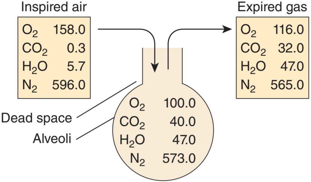
FIGURE 34-14 Partial pressures of gases (mm Hg) in various parts of the respiratory system. Typical partial pressures for inspired air, alveolar air, and expired air are given. See text for additional details.
شکل 34-14 فشار جزئی گازها (میلی متر جیوه) در قسمت های مختلف دستگاه تنفسی. فشارهای جزئی معمولی برای هوای دمیده شده، هوای آلوئولی و هوای منقضی شده داده شده است. برای جزئیات بیشتر به متن مراجعه کنید.
PAO2 can also be calculated from the alveolar gas equation:
PAO2 را می توان از معادله گاز آلوئولی نیز محاسبه کرد:

where FIO2 is the fraction of O2 molecules in the inspired (I) dry gas, PIO2 is the inspired Po2, and R is the respiratory exchange ratio; that is, the flow of CO2 molecules across the alveolar membrane per minute divided by the flow of O2 molecules across the membrane per minute.
که در آن FIO2 کسری از مولکول های O2 در گاز خشک الهام گرفته شده (I) است، PIO2 Po2 الهام گرفته شده است، و R نسبت تبادل تنفسی است. یعنی جریان مولکول های CO2 در طول غشای آلوئولی در دقیقه تقسیم بر جریان مولکول های O2 در سراسر غشاء در دقیقه.
COMPOSITION OF ALVEOLAR AIR
Oxygen continuously diffuses out of the gas in the alveoli into the bloodstream, and CO2 continuously diffuses into the alveoli from the blood. In the steady state, inspired air mixes with the alveolar gas, replacing the O2 that has entered the blood and diluting the CO2 that has entered the alveoli. Part of this mixture is expired. The O2 content of the alveolar gas then falls and its CO2 content rises until the next inspiration. Because the volume of gas in the alveoli is about 2 L at the end of expiration (FRC), each 350 mL increment of inspired and expired air has relatively little effect on Po2 and PCO2. Indeed, the composition of alveolar gas remains remarkably constant, not only at rest but also under a variety of other conditions.
ترکیب هوای آلوئولار
اکسیژن به طور مداوم از گاز موجود در آلوئول ها به جریان خون منتشر می شود و CO2 به طور مداوم از خون به آلوئول ها پخش می شود. در حالت ثابت، هوای دمیده شده با گاز آلوئولی مخلوط می شود و جایگزین O2 وارد شده به خون و رقیق شدن CO2 وارد شده به آلوئول می شود. بخشی از این مخلوط تاریخ مصرف گذشته است. سپس محتوای O2 گاز آلوئولی کاهش می یابد و محتوای CO2 آن تا دم بعدی افزایش می یابد. از آنجایی که حجم گاز موجود در آلوئول ها حدود 2 لیتر در پایان بازدم (FRC) است، هر 350 میلی لیتر افزایش هوای دمیده شده و منقضی شده تأثیر نسبتا کمی بر Po2 و PCO2 دارد. در واقع، ترکیب گاز آلوئولی نه تنها در حالت سکون بلکه در شرایط مختلف دیگر به طرز قابل ملاحظهای ثابت میماند.
DIFFUSION ACROSS THE ALVEOLOCAPILLARY MEMBRANE
Gases diffuse from the alveoli to the blood in the pulmonary capillaries or vice versa across the thin alveolocapillary membrane made up of the pulmonary epithelium, the capillary endothelium, and their fused basement membranes (Figure 34-3). Whether or not substances passing from the alveoli to the capillary blood reach equilibrium in the 0.75 s that blood takes to traverse the pulmonary capillaries at rest depends on their reaction with substances in the blood. Thus, for example, the anesthetic gas nitrous oxide (N2O) does not react and reaches equilibrium in about 0.1 s (Figure 34-15). In this situation, the amount of N2O taken up is not limited by diffusion but by the amount of blood flowing through the pulmonary capillaries; that is, it is flow-limited. On the other hand, carbon monoxide (CO) is taken up by hemoglobin in the red blood cells at such a high rate that the partial pressure of CO in the capillaries stays very low and equilibrium is not reached in the 0.75 s the blood is in the pulmonary capillaries. Therefore, the transfer of CO is not limited by perfusion at rest and instead is diffusion-limited. O2 is intermediate between N2O and CO;it is taken up by hemoglobin, but much less avidly than CO, and it reaches equilibrium with capillary blood in about 0.3 s. Thus, its uptake is perfusion- limited.
انتشار در سراسر غشاء آلوئولوکاپیلاری
گازها از آلوئول ها به خون در مویرگ های ریوی یا برعکس در غشای نازک آلوئولوکاپیلاری متشکل از اپیتلیوم ریوی، اندوتلیوم مویرگی و غشای پایه ذوب شده آنها منتشر می شوند (شکل 34-3). اینکه آیا موادی که از آلوئول ها به خون مویرگی عبور می کنند یا نه در 0.75 ثانیه ای که خون برای عبور از مویرگ های ریوی در حالت استراحت به تعادل می رسد یا نه بستگی به واکنش آنها با مواد موجود در خون دارد. بنابراین، به عنوان مثال، گاز بیهوش کننده اکسید نیتروژن (N2O) واکنش نشان نمی دهد و در حدود 0.1 ثانیه به تعادل می رسد (شکل 34-15). در این شرایط، مقدار N2O جذب شده توسط انتشار محدود نمی شود، بلکه با مقدار خونی که در مویرگ های ریوی جریان دارد، محدود می شود. یعنی محدود به جریان است. از طرف دیگر، مونوکسید کربن (CO) توسط هموگلوبین در گلبولهای قرمز خون با سرعت بالایی جذب میشود که فشار جزئی CO در مویرگها بسیار پایین میماند و در 0.75 ثانیه که خون در مویرگهای ریوی است، تعادل حاصل نمیشود. بنابراین، انتقال CO توسط پرفیوژن در حالت استراحت محدود نمی شود و در عوض انتشار محدود است. O2 حد واسط بین N2O و CO است؛ توسط هموگلوبین جذب می شود، اما بسیار کمتر از CO، و در حدود 0.3 ثانیه با خون مویرگی به تعادل می رسد. بنابراین، جذب آن از نظر پرفیوژن محدود است.

FIGURE 34-15 Uptake of various substances during the 0.75 s they are in transit through a pulmonary capillary. N2O is not bound in blood, so its partial pressure in blood rises rapidly to its partial pressure in the alveoli. Conversely, CO is avidly taken up by red blood cells, so its partial pressure reaches only a fraction of its partial pressure in the alveoli. O2 is intermediate between the two.
شکل 34-15 جذب مواد مختلف در طی 0.75 ثانیه که از طریق مویرگ ریوی در حال عبور هستند. N2O در خون محدود نمی شود، بنابراین فشار جزئی آن در خون به سرعت به فشار جزئی آن در آلوئول ها افزایش می یابد. برعکس، CO مشتاقانه توسط گلبول های قرمز جذب می شود، بنابراین فشار جزئی آن تنها به کسری از فشار جزئی آن در آلوئول ها می رسد. O2 حد واسط بین این دو است.
The diffusing capacity of the lungs for a given gas is directly proportional to the surface area of the alveolo-capillary membrane and inversely proportional to its thickness. The diffusing capacity for CO (DLCO) is measured as an index of diffusing capacity because its uptake is diffusion-limited. DLCO is proportional to the amount of CO entering the blood (ÿco) divided by the partial pressure of CO in the alveoli minus the partial pressure of CO in the blood entering the pulmonary capillaries. Except in habitual cigarette smokers, this latter term is close to zero, so it can be ignored and the equation becomes:
ظرفیت انتشار ریه ها برای یک گاز معین به طور مستقیم با سطح غشای آلوئولو-مویرگی و با ضخامت آن نسبت معکوس دارد. ظرفیت انتشار برای CO (DLCO) به عنوان شاخصی از ظرفیت انتشار اندازه گیری می شود زیرا جذب آن محدود به انتشار است. DLCO متناسب با مقدار CO وارد شده به خون (ÿco) تقسیم بر فشار جزئی CO در آلوئول ها منهای فشار جزئی CO در خون ورودی به مویرگ های ریوی است. به جز در افراد سیگاری معمولی، این جمله اخیر نزدیک به صفر است، بنابراین می توان آن را نادیده گرفت و معادله تبدیل می شود:

The normal value of DLCO at rest is about 25 mL/min/mm Hg. It increases up to threefold during exercise because of capillary dilation and an increase in the number of active capillaries. The Po2 of alveolar air is normally 100 mm Hg, and the Po2 of the blood entering the pulmonary capillaries is 40 mm Hg. The diffusing capacity for O2, like that for CO at rest, is about 25 mL/min/mm Hg, and the Po2 of blood is raised to 97 mm Hg, a value just under the alveolar Po2.
مقدار طبیعی DLCO در حالت استراحت حدود 25 میلی لیتر در دقیقه بر میلی متر جیوه است. به دلیل گشاد شدن مویرگ ها و افزایش تعداد مویرگ های فعال، در طول ورزش تا سه برابر افزایش می یابد. Po2 هوای آلوئولی معمولاً 100 میلی متر جیوه است و PO2 خون ورودی به مویرگ های ریوی 40 میلی متر جیوه است. ظرفیت انتشار O2، مانند CO در حالت استراحت، حدود 25 میلیلیتر بر دقیقه بر میلیمتر جیوه است، و Po2 خون به 97 میلیمتر جیوه افزایش مییابد، مقداری درست در زیر Po2 آلوئولی.
The PCO2 of venous blood is 46 mm Hg, whereas that of alveolar air is 40 mm Hg, and CO2 diffuses from the blood into the alveoli along this gradient. The PCO2 of blood leaving the lungs is 40 mm Hg. CO2 passes through all biological membranes with ease, and the diffusing capacity of the lungs for CO2 is much greater than the capacity for O2. It is for this reason that CO2 retention is rarely a problem in patients with alveolar fibrosis even when the reduction in diffusing capacity for O2 is severe.
PCO2 خون وریدی 46 میلیمتر جیوه است، در حالی که هوای آلوئولی 40 میلیمتر جیوه است و CO2 از خون به داخل آلوئولها در امتداد این گرادیان پخش میشود. PCO2 خون خروجی از ریه ها 40 میلی متر جیوه است. CO2 به راحتی از تمام غشاهای بیولوژیکی عبور می کند و ظرفیت انتشار ریه ها برای CO2 بسیار بیشتر از ظرفیت O2 است. به همین دلیل است که احتباس CO2 به ندرت در بیماران مبتلا به فیبروز آلوئولار مشکل ایجاد می کند، حتی زمانی که کاهش ظرفیت انتشار برای O2 شدید باشد.
PULMONARY CIRCULATION
PULMONARY BLOOD VESSELS
The pulmonary vascular bed resembles the systemic one, except that the walls of the pulmonary artery and its large branches are about 30% as thick as the wall of the aorta, and the small arterial vessels, unlike the systemic arterioles, contain relatively little smooth muscle in their walls. The walls of the pulmonary venous vessels also contain some smooth muscle. The pulmonary capillaries are large, and there are multiple anastomoses, so that each alveolus sits in a capillary basket.
گردش خون ریوی
رگ های خونی ریوی
بستر عروقی ریوی شبیه به سیستمیک است، با این تفاوت که ضخامت دیواره های شریان ریوی و شاخه های بزرگ آن حدود 30 درصد به اندازه دیواره آئورت است و عروق شریانی کوچک، بر خلاف شریان های سیستمیک، دارای عضلات صاف نسبتا کمی در دیواره خود هستند. دیواره رگ های وریدی ریوی نیز حاوی مقداری ماهیچه صاف است. مویرگ های ریوی بزرگ هستند و آناستوموزهای متعددی وجود دارد، به طوری که هر آلوئول در یک سبد مویرگی قرار می گیرد.
PRESSURE, VOLUME, & FLOW
With two quantitatively minor exceptions, the blood put out by the left ventricle returns to the right atrium and is ejected by the right ventricle, making the pulmonary vasculature unique in that it accommodates a blood flow that is almost equal to that of all the other organs in the body.
فشار، حجم و جریان
به جز دو استثنای کمّی جزئی، خون خارج شده توسط بطن چپ به دهلیز راست باز می گردد و توسط بطن راست خارج می شود و عروق ریوی را منحصر به فرد می کند زیرا جریان خون تقریباً برابر با سایر اندام های بدن را دارد.
One of the exceptions is part of the bronchial blood flow. There are anastomoses between the bronchial capillaries and the pulmonary capillaries and veins, and although some of the bronchial blood enters the bronchial veins, some enters the pulmonary capillaries and veins, bypassing the right ventricle. The other exception is blood that flows from the coronary arteries into the chambers of the left side of the heart. Because of the small physiological shunt created by those two exceptions, the blood in systemic arteries has a Po2 about 2 mm Hg lower than that of blood that has equilibrated with alveolar air, and the saturation of hemoglobin is 0.5% less.
یکی از استثناها بخشی از جریان خون برونش است. بین مویرگ های برونش و مویرگ ها و وریدهای ریوی آناستوموز وجود دارد و اگرچه مقداری از خون نایژه وارد سیاهرگ های برونش می شود، مقداری نیز وارد مویرگ ها و وریدهای ریوی می شود و بطن راست را دور می زند. استثنای دیگر خونی است که از شریان های کرونری به حفره های سمت چپ قلب می ریزد. به دلیل شنت فیزیولوژیکی کوچک ایجاد شده توسط این دو استثنا، خون در شریان های سیستمیک دارای Po2 حدود 2 میلی متر جیوه کمتر از خونی است که با هوای آلوئولی متعادل شده است و اشباع هموگلوبین 0.5٪ کمتر است.
The pressure in the various parts of the pulmonary portion of the pulmonary circulation is shown in Figure 34-6C. The pressure gradient in the pulmonary circulation system is about 7 mm Hg, compared with a gradient of about 90 mm Hg in the systemic circulation. Pulmonary capillary pressure is about 10 mm Hg, whereas the oncotic pressure is 25 mm Hg, so that an inward-directed pressure gradient of about 15 mm Hg keeps the alveoli free of all but a thin film of fluid. When the pulmonary capillary pressure is more than 25 mm Hg, pulmonary congestion and edema result.
فشار در قسمت های مختلف قسمت ریوی گردش خون ریوی در شکل 34-6C نشان داده شده است. گرادیان فشار در سیستم گردش خون ریوی حدود 7 میلی متر جیوه است، در مقایسه با شیب حدود 90 میلی متر جیوه در گردش خون سیستمیک. فشار مویرگی ریوی حدود 10 میلی متر جیوه است، در حالی که فشار انکوتیک 25 میلی متر جیوه است، به طوری که یک گرادیان فشار به سمت داخل حدود 15 میلی متر جیوه، آلوئول ها را عاری از هر چیزی به جز یک لایه نازک مایع نگه می دارد. هنگامی که فشار مویرگ ریوی بیش از 25 میلی متر جیوه باشد، احتقان ریوی و ادم ایجاد می شود.
The volume of blood in the pulmonary vessels at any one time is about 1 L, of which less than 100 mL is in the capillaries. The mean velocity of the blood in the root of the pulmonary artery is the same as that in the aorta (about 40 cm/s). It falls off rapidly, then rises slightly again in the larger pulmonary veins. It takes a red cell about 0.75 s to traverse the pulmonary capillaries at rest and 0.3 s or less during exercise.
حجم خون در رگ های ریوی در هر زمان حدود 1 لیتر است که کمتر از 100 میلی لیتر آن در مویرگ ها است. سرعت متوسط خون در ریشه شریان ریوی همان سرعت خون در آئورت است (حدود 40 سانتی متر بر ثانیه). به سرعت می افتد، سپس دوباره کمی در وریدهای ریوی بزرگتر بالا می رود. طی کردن مویرگ های ریوی در حالت استراحت حدود 0.75 ثانیه و در حین ورزش 0.3 ثانیه یا کمتر طول می کشد.
EFFECT OF GRAVITY
Gravity has a relatively marked effect on the pulmonary circulation. In the upright position, the upper portions of the lungs are well above the level of the heart, and the bases are at or below it. Consequently, in the upper part of the lungs, the blood flow is less, the alveoli are larger, and ventilation is less than at the base (Figure 34-16). The pressure in the capillaries at the top of the lungs is close to the atmospheric pressure in the alveoli. Pulmonary arterial pressure is normally just sufficient to maintain perfusion, but if it is reduced or if alveolar pressure is increased, some of the capillaries collapse. Under these circumstances, no gas exchange takes place in the affected alveoli and they become part of the physiological dead space.
اثر گرانش
جاذبه اثر نسبتاً مشخصی بر گردش خون ریوی دارد. در وضعیت عمودی، قسمتهای بالایی ریهها به خوبی بالاتر از سطح قلب هستند و پایهها در آن یا پایین آن قرار دارند. در نتیجه، در قسمت فوقانی ریه، جریان خون کمتر، آلوئول ها بزرگتر و تهویه کمتر از قاعده است (شکل 34-16). فشار مویرگ ها در بالای ریه ها نزدیک به فشار اتمسفر در آلوئول ها است. فشار شریانی ریوی معمولاً برای حفظ پرفیوژن کافی است، اما اگر کاهش یابد یا فشار آلوئولی افزایش یابد، برخی از مویرگ ها فرو می ریزند. در این شرایط، هیچ تبادل گازی در آلوئول های آسیب دیده انجام نمی شود و آنها بخشی از فضای مرده فیزیولوژیکی می شوند.
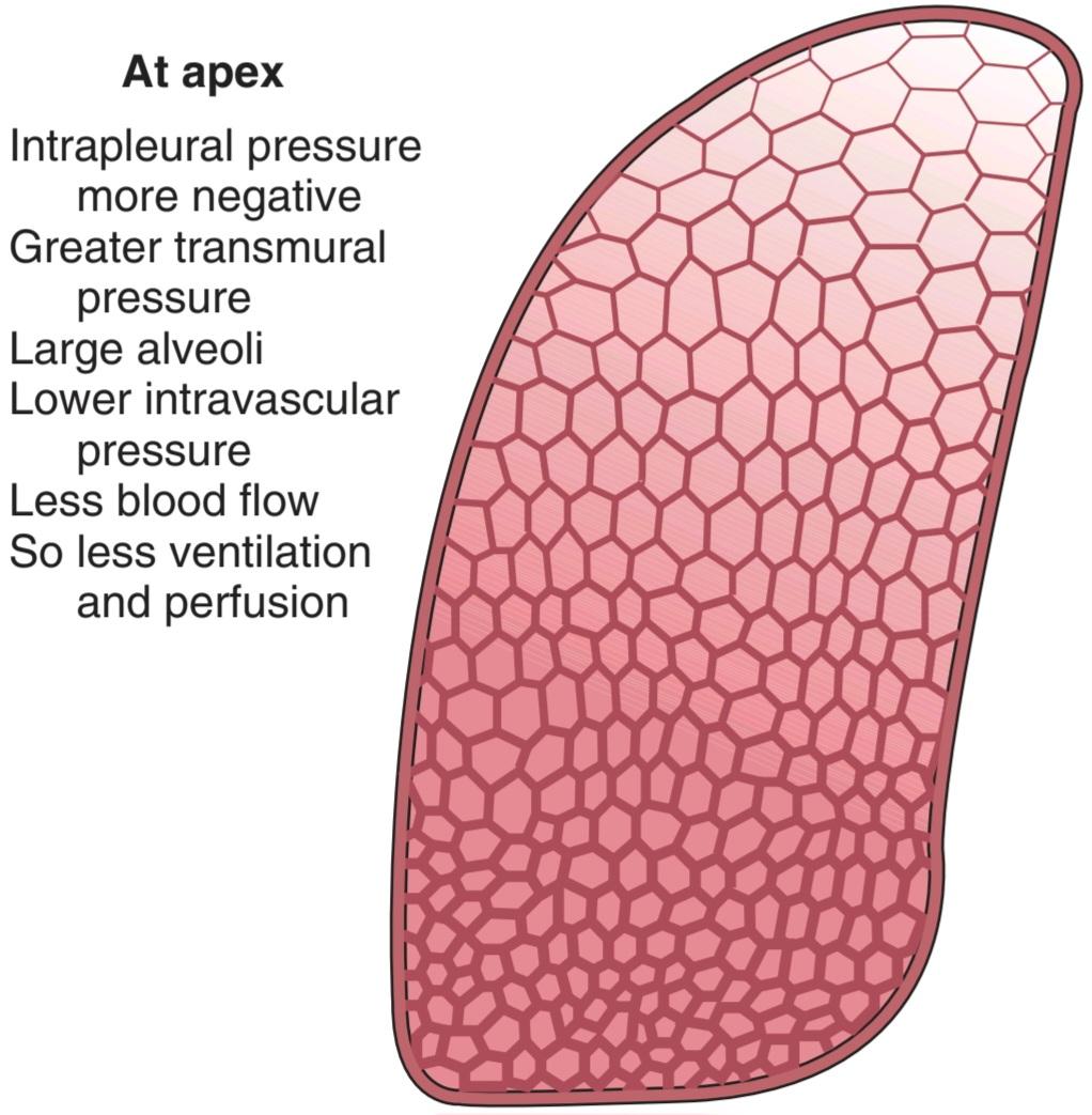
FIGURE 34-16 Diagram of normal differences in ventilation and perfusion of the lung in the upright position. Outlined areas are representative of changes in alveolar size (not actual size). Note the gradual change in alveolar size from top (apex) to bottom. Characteristic differences of alveoli at the apex of the lung are stated. (Modified with permission from Levitzky MG: Pulmonary Physiology, 6th ed. New York, NY: McGraw-Hill; 2003.)
شکل 34-16 نمودار تفاوت های طبیعی در تهویه و پرفیوژن ریه در وضعیت عمودی. نواحی مشخص شده نمایانگر تغییرات در اندازه آلوئول هستند (نه اندازه واقعی). به تغییر تدریجی اندازه آلوئول از بالا (راس) به پایین توجه کنید. تفاوت های مشخصه آلوئول ها در راس ریه بیان شده است. (با اجازه Levitzky MG: Pulmonary Physiology، ویرایش ششم نیویورک، نیویورک: McGraw-Hill؛ 2003 اصلاح شده است.)
In the middle portions of the lungs, the pulmonary arterial pressure and capillary pressure exceed alveolar pressure, but the pressure in the pulmonary venules may be lower than alveolar pressure during normal expiration, so they are collapsed. Under these circumstances, blood flow is determined by the difference between pulmonary arterial pressure and alveolar pressure rather than the difference between pulmonary arterial pressure and pulmonary venous pressure. Beyond the constriction, blood “falls” into the pulmonary veins, which are compliant and take whatever amount of blood the constriction lets flow into them. This has been called the waterfall effect. Obviously, the compression of vessels produced by alveolar pressure decreases and pulmonary blood flow increases as the arterial pressure increases toward the base of the lungs. In the lower portions of the lungs, alveolar pressure is lower than the pressure in all parts of the pulmonary circulation and blood flow is determined by the arterial-venous pressure difference. Examples of diseases affecting the pulmonary circulation are given in Clinical Box 34 4.
در قسمتهای میانی ریهها، فشار شریانی ریوی و فشار مویرگی از فشار آلوئولی بیشتر است، اما فشار در ونولهای ریوی ممکن است در طول بازدم طبیعی کمتر از فشار آلوئولی باشد، بنابراین فرو میروند. در این شرایط، جریان خون با تفاوت بین فشار شریانی ریوی و فشار آلوئولی به جای تفاوت بین فشار شریانی ریوی و فشار وریدی ریوی تعیین میشود. فراتر از انقباض، خون به داخل سیاهرگهای ریوی میریزد، این سیاهرگها سازگار هستند و هر مقدار خونی را که انقباض اجازه میدهد به آنها جاری شود، میگیرد. این اثر آبشار نامیده می شود. بدیهی است که با افزایش فشار شریانی به سمت قاعده ریه، فشردگی عروق تولید شده توسط فشار آلوئولی کاهش می یابد و جریان خون ریوی افزایش می یابد. در قسمتهای پایینی ریهها، فشار آلوئولی کمتر از فشار در تمام قسمتهای گردش خون ریوی است و جریان خون با اختلاف فشار شریانی و وریدی تعیین میشود. نمونههایی از بیماریهایی که بر گردش خون ریوی تأثیر میگذارند در کادر بالینی ۳۴-۴ آورده شده است.
CLINICAL BOX 34-4
Diseases Affecting the Pulmonary Circulation
Pulmonary Hypertension
Pulmonary hypertension (PH) is classified into five groups: pulmonary arterial hypertension (PAH; including persistent PH of the newborn), PH associated with left heart disease, PH due to lung diseases and/or hypoxia, chronic thromboembolic pulmonary hypertension (CTEPH), and PH with unclear multifactorial mechanisms. PH, as a syndrome with multiple causes, can occur at any age, but idiopathic PAH (a fatal and progressive form of PAH) predominantly affects young women. The causes for PH or PAH are different from those causing systemic arterial hypertension. For example, acute hypoxia causes pulmonary vasoconstriction and chronic hypoxia causes pulmonary hypertension, whereas hypoxia causes systemic vasodilation.Other unique causes for PH include inhalation of cocaine, treatment with dexfenfluramine and related appetite-suppressing drugs, HIV/HPV infection, systemic lupus erythematosus and scleroderma, and mutations of the genes encoding the bone morphogenetic protein receptor II (BMPRII), the pH- sensitive two-pore-domain K+ channel (KCNK3), the serotonin transporter (SERT), the activin receptor kinase-like 1 (ALK1), the transient receptor potential canonical channel 6 (TRPC6), the glycoprotein endoglin (ENG), and the scaffolding protein caveolin 1 (CAV1). Sustained pulmonary vasoconstriction, concentric pulmonary vascular remodeling, in situ pulmonary thrombosis, and increased pulmonary vascular wall stiffness all contribute to the increased pulmonary vascular resistance and pulmonary arterial pressure in patients with PH or PAH. If appropriate therapy is not initiated, the increased right ventricular afterload can lead eventually to right heart failure and death.
جعبه بالینی 34-4
بیماری های موثر بر گردش خون ریوی
فشار خون ریوی
فشار خون ریوی (PH) به پنج گروه طبقه بندی می شود: فشار خون شریانی ریوی (PAH؛ از جمله PH مداوم نوزاد)، PH مرتبط با بیماری قلب چپ، PH ناشی از بیماری های ریوی و/یا هیپوکسی، فشار خون مزمن ترومبوآمبولیک ریوی (CTEPH) و PH با مکانیسم های چند عاملی نامشخص. PH، به عنوان یک سندرم با علل متعدد، می تواند در هر سنی رخ دهد، اما PAH ایدیوپاتیک (شکل کشنده و پیشرونده PAH) عمدتا زنان جوان را تحت تاثیر قرار می دهد. علل PH یا PAH با علل ایجاد فشار خون شریانی سیستمیک متفاوت است. به عنوان مثال، هیپوکسی حاد باعث انقباض عروق ریوی و هیپوکسی مزمن باعث فشار خون ریوی می شود، در حالی که هیپوکسی باعث اتساع سیستمیک عروق می شود. سایر علل منحصر به فرد برای PH عبارتند از استنشاق کوکائین، درمان با دکسفن فلورامین و داروهای سرکوب کننده اشتها، عفونت HIV/HPV، عفونت HIV/HPV، عفونت سیستمیک لوپوس اریکوه و لوپوس اریکوتیسیون. گیرنده پروتئین مورفوژنتیک استخوان II (BMPRII)، کانال +K دو حفره ای حساس به pH (KCNK3)، ناقل سروتونین (SERT)، گیرنده اکتیوین کیناز مانند 1 (ALK1)، کانال متعارف بالقوه گیرنده گذرا 6 (TRPC6)، انتهای گلیکوپروتئین فولدینگ (گلیکوپروتئین فولدینگ1) (CAV1). انقباض عروق ریوی پایدار، بازسازی عروق ریوی متحدالمرکز، ترومبوز ریوی درجا، و افزایش سفتی دیواره عروق ریوی، همگی به افزایش مقاومت عروق ریوی و فشار شریان ریوی در بیماران مبتلا به PH یا PAH کمک می کنند. اگر درمان مناسب شروع نشود، افزایش بار پس از بطن راست می تواند در نهایت منجر به نارسایی قلب راست و مرگ شود.
THERAPEUTIQ HIGHLIGHTS
Treatment with vasodilators and antiproliferative agents, such as continuous intravenous infusion of prostacyclin (Flolan) and oral intake or inhalation of prostacyclin analogs (eg, beroprost, ilorost, treprostinil), is effective for patients with severe PH (eg, idiopathic, heritable and associated PAH). Conventional treatment includes diuretics, oxygen therapy, anticoagulation, digoxin, and exercise. Inhaled nitric oxide (NO) is effective for many pediatric patients. Advanced treatment includes voltage-gated Ca2+ channel blockers (eg, nifedipine, diltiazem, verapamil, amlodipine), prostanoids (eg, epoprostenol/PGI2, treprostinil, iloprost), endothelin receptor antagonists (eg, bosentan, ambrisentan), phosphodiesterase (PDE) inhibitors (eg, sildenafil/Viagra, tadalafil/Cialis), and combination therapy. Surgical treatment includes atrial septostomy, whole lung transplantation, living-donor lobar lung transplantation, pulmonary endarterectomy, and pulmonary arterioplasty.
نکات برجسته درمانی
درمان با گشادکننده عروق و عوامل ضد تکثیر، مانند انفوزیون مداوم وریدی پروستاسیکلین (Flolan) و مصرف خوراکی یا استنشاق آنالوگ های پروستاسیکلین (به عنوان مثال، beroprost، ilorost، treprostinil)، برای بیماران با PH شدید (مانند PAH ایدیوپاتیک، ارثی و مرتبط) موثر است. درمان مرسوم شامل دیورتیک ها، اکسیژن درمانی، ضد انعقاد، دیگوکسین و ورزش است. اکسید نیتریک استنشاقی (NO) برای بسیاری از بیماران اطفال موثر است. درمان پیشرفته شامل مسدود کننده های کانال های +Ca2 با ولتاژ (به عنوان مثال، نیفدیپین، دیلتیازم، وراپامیل، آملودیپین)، پروستانوئیدها (به عنوان مثال، اپوپروستنول/PGI2، ترپروستینیل، ایلوپروست)، آنتاگونیست های گیرنده اندوتلین (به عنوان مثال، بوسنتان، آمبریسنتان، اینترازبیت، phodies) سیلدنافیل/ویاگرا، تادالافیل/سیالیس) و درمان ترکیبی. درمان جراحی شامل سپتوستومی دهلیزی، پیوند ریه کامل، پیوند ریه لوبار از اهداکننده زنده، اندارترکتومی ریوی و آرتریوپلاستی ریوی است.
VENTILATION/PERFUSION RATIOS
The ratio of pulmonary ventilation (V) to pulmonary blood flow or perfusion (Q) for the whole lungs at rest is about 0.8 (4.2 L/min ventilation divided by 5.5 L/min blood flow). However, relatively marked differences occur in this ventilation/perfusion Q(V/Q) ratio in various parts of the normal lungs as a result of the effect of gravity, and local changes in the ventilation/perfusion ratio are common in disease. If the ventilation to an alveolus is reduced relative to its perfusion, the Po2 in the alveolus falls because less O2 is delivered to it and the PCO2 rises because less CO2 is expired. Conversely, if perfusion is reduced relative to ventilation, the Pco2 falls because less CO2 is delivered and the Po2 rises because less O2 enters the blood.
نسبت تهویه/پرفیوژن
نسبت تهویه ریوی (V) به جریان خون ریوی یا پرفیوژن (Q) برای کل ریه ها در حالت استراحت حدود 0.8 است (4.2 لیتر در دقیقه تهویه تقسیم بر 5.5 لیتر در دقیقه جریان خون). با این حال، تفاوتهای نسبتاً مشخصی در این نسبت Q(V/Q) تهویه/پرفیوژن در قسمتهای مختلف ریههای طبیعی در نتیجه اثر گرانش رخ میدهد و تغییرات موضعی در نسبت تهویه/پرفیوژن در بیماری شایع است. اگر تهویه آلوئول نسبت به پرفیوژن آن کاهش یابد، PO2 در آلوئول سقوط می کند زیرا O2 کمتری به آن می رسد و PCO2 افزایش می یابد زیرا CO2 کمتری منقضی می شود. برعکس، اگر پرفیوژن نسبت به تهویه کاهش یابد، Pco2 کاهش می یابد زیرا CO2 کمتری تحویل داده می شود و Po2 افزایش می یابد زیرا O2 کمتری وارد خون می شود.
As noted above, ventilation, as well as perfusion in the upright position, declines in a linear fashion from the bases to the apices of the lungs. However, the ventilation/perfusion ratios are high in the upper portions of the lungs. When widespread, nonuniformity of ventilation and perfusion in the lungs can cause CO2 retention and lowers systemic arterial Po2.
همانطور که در بالا ذکر شد، تهویه، و همچنین پرفیوژن در وضعیت عمودی، به صورت خطی از قاعده تا نوک ریه کاهش می یابد. با این حال، نسبت تهویه/پرفیوژن در قسمت های بالایی ریه ها بالا است. هنگامی که گسترده باشد، عدم یکنواختی تهویه و پرفیوژن در ریه ها می تواند باعث احتباس CO2 و کاهش Po2 شریان سیستمیک شود.
A unique mechanism that ensures the optimal ventilation/perfusion ratio in the lungs is hypoxic pulmonary vasoconstriction (HPV). HPV is a homeostatic mechanism that, in the adult men, diverts desaturated, mixed venous blood in the pulmonary artery from poorly ventilated (hypoxic) areas of the lungs to other better ventilated (normoxic) areas of the lungs. The alveolar hypoxia-induced vasoconstriction helps maintain an optimal ventilation/perfusion ratio and maximize the oxygenation of the mixed venous blood in the lungs.
مکانیسم منحصر به فردی که نسبت تهویه/پرفیوژن بهینه را در ریه ها تضمین می کند، انقباض عروق ریوی هیپوکسیک (HPV) است. HPV یک مکانیسم هموستاتیک است که در مردان بالغ، خون وریدی غیراشباع و مخلوط در شریان ریوی را از نواحی با تهویه ضعیف (هیپوکسیک) ریه ها به سایر مناطق با تهویه بهتر (نرموکسیک) ریه هدایت می کند. انقباض عروق ناشی از هیپوکسی آلوئولی به حفظ نسبت تهویه/پرفیوژن بهینه و به حداکثر رساندن اکسیژن رسانی خون وریدی مخلوط در ریه ها کمک می کند.
REGULATION OF PULMONARY BLOOD FLOW
It is unsettled whether pulmonary veins and pulmonary arteries are regulated separately, although constriction of the veins increases pulmonary capillary pressure and constriction of pulmonary arteries increases the load on the right side of the heart.
تنظیم جریان خون ریوی
مشخص نیست که آیا وریدهای ریوی و شریانهای ریوی به طور جداگانه تنظیم میشوند، اگرچه انقباض وریدها فشار مویرگ ریوی را افزایش میدهد و انقباض شریانهای ریوی باعث افزایش بار در سمت راست قلب میشود.
Pulmonary blood flow is affected by both active and passive factors. There is an extensive autonomic innervation of the pulmonary vessels, and stimulation of the cervical sympathetic ganglia reduces pulmonary blood flow by as much as 30%. The vessels also respond to circulating humoral agents. A diversity of the receptors involved in the regulation of pulmonary vasomotor tone and their effect on pulmonary smooth muscle are summarized in Table 34-3. Many of the dilator responses are endothelium-dependent and presumably operate via release of nitric oxide (NO).
جریان خون ریوی تحت تأثیر عوامل فعال و غیرفعال قرار می گیرد. یک عصب اتونوم گسترده عروق ریوی وجود دارد و تحریک عقده های سمپاتیک گردنی باعث کاهش جریان خون ریوی تا 30٪ می شود. عروق همچنین به عوامل هومورال در گردش پاسخ می دهند. تنوعی از گیرنده های دخیل در تنظیم تون وازوموتور ریوی و تأثیر آنها بر عضله صاف ریوی در جدول 34-3 خلاصه شده است. بسیاری از پاسخ های گشادکننده وابسته به اندوتلیوم هستند و احتمالاً از طریق آزادسازی اکسید نیتریک (NO) عمل می کنند.
TABLE 34-3 Receptors affecting smooth muscle in pulmonary arteries and veins.
جدول 34-3 گیرنده های موثر بر ماهیچه صاف در شریان ها و وریدهای ریوی.

Modified and reproduced with permission from Barnes PJ, Lin SF: Regulation of pulmonary vascular tone. Pharmacol Rev 1995;47:88.
اصلاح و تکثیر شده با مجوز Barnes PJ، Lin SF: تنظیم تون عروق ریوی. Pharmacol Rev 1995; 47:88.
Passive factors such as cardiac output and gravitational forces also have significant effects on pulmonary blood flow. Local adjustments of perfusion to ventilation occur with local changes in O2. With exercise, cardiac output increases and pulmonary arterial pressure rises. More red cells move through the lungs without any reduction in the O2 saturation of the hemoglobin in them, and consequently, the total amount of O2 delivered to the systemic circulation is increased. Capillaries dilate (also referred as capillary extension), and previously underperfused capillaries are “recruited” (also referred to as capillary recruitment) to carry blood. The net effect is a marked increase in pulmonary blood flow with few, if any, alterations in autonomic outflow to the pulmonary vessels.
عوامل غیرفعال مانند برون ده قلبی و نیروهای گرانشی نیز اثرات قابل توجهی بر جریان خون ریوی دارند. تنظیمات موضعی پرفیوژن برای تهویه با تغییرات موضعی در O2 رخ می دهد. با ورزش، برون ده قلبی افزایش می یابد و فشار شریان ریوی افزایش می یابد. گلبولهای قرمز بیشتری بدون هیچ کاهشی در اشباع O2 هموگلوبین در آنها در ریهها حرکت میکنند و در نتیجه، مقدار کل O2 تحویلی به گردش خون سیستمیک افزایش مییابد. مویرگها گشاد میشوند (به عنوان گسترش مویرگی نیز شناخته میشوند)، و مویرگهایی که قبلاً تحت پرفیوژن قرار داشتند برای حمل خون به کار گرفته میشوند (که به عنوان جذب مویرگی نیز نامیده میشود). اثر خالص افزایش قابل توجه در جریان خون ریوی با تغییرات اندک، در صورت وجود، در خروجی اتونومیک به عروق ریوی است.
When a bronchus or a bronchiole is obstructed, hypoxia develops in the underventilated alveoli beyond the obstruction. The O2 reduction apparently acts directly on vascular smooth muscle in the area to produce constriction (ie, HPV), shunting blood away from the hypoxic area. Accumulation of CO2 leads to a drop in pH in the area, and a decline in pH also produces vasoconstriction in the lungs, as opposed to the vasodilation it produces in other tissues. Conversely, reduction of the blood flow to a portion of the lung lowers the alveolar Pco2 in that area, and this leads to constriction of the bronchi supplying it, shifting ventilation away from the poorly perfused area. Systemic hypoxia also causes the pulmonary arterioles to constrict, with a resultant increase in pulmonary arterial pressure.
هنگامی که یک برونش یا یک برونشیول مسدود می شود، هیپوکسی در آلوئول های بدون تهویه فراتر از انسداد ایجاد می شود. کاهش O2 ظاهراً مستقیماً بر روی ماهیچه صاف عروق در ناحیه تأثیر می گذارد و باعث ایجاد انقباض (یعنی HPV) می شود و خون را از ناحیه هیپوکسیک دور می کند. تجمع CO2 منجر به افت pH در ناحیه می شود و کاهش pH همچنین باعث انقباض عروق در ریه ها می شود، برخلاف اتساع عروقی که در بافت های دیگر ایجاد می کند. برعکس، کاهش جریان خون به بخشی از ریه، Pco2 آلوئولی را در آن ناحیه کاهش میدهد و این منجر به انقباض برونشهای تامینکننده آن میشود و تهویه را از ناحیه با پرفیوژن ضعیف دور میکند. هیپوکسی سیستمیک همچنین باعث انقباض شریان های ریوی و در نتیجه افزایش فشار شریان ریوی می شود.
METABOLIC & ENDOCRINE FUNCTIONS OF THE LUNGS
In addition to their functions in gas exchange, the lungs have a number of metabolic functions. They manufacture surfactant for local use, as noted above. They also contain a fibrinolytic system that lyses clots in the pulmonary vessels. They release a variety of substances that enter the systemic arterial blood (Table 34-4), and they remove other substances from the systemic venous blood that reach them via the pulmonary artery. Prostaglandins are removed from the circulation, but they are also synthesized in the lungs and released into the blood when lung tissue is stretched.
عملکردهای متابولیک و غدد درون ریز ریه ها
ریه ها علاوه بر وظایف خود در تبادل گاز، تعدادی عملکرد متابولیک نیز دارند. همانطور که در بالا ذکر شد، آنها سورفکتانت را برای استفاده محلی تولید می کنند. آنها همچنین حاوی یک سیستم فیبرینولیتیک هستند که لخته ها را در رگ های ریوی لیز می کند. آنها مواد مختلفی را آزاد می کنند که وارد خون شریانی سیستمیک می شوند (جدول 34-4) و سایر مواد را از خون وریدی سیستمیک که از طریق شریان ریوی به آنها می رسد خارج می کنند. پروستاگلاندین ها از گردش خون خارج می شوند، اما در ریه ها نیز سنتز می شوند و با کشیده شدن بافت ریه در خون آزاد می شوند.
TABLE 34-4 Biologically active substances metabolized by the lungs.
جدول 34-4 مواد فعال بیولوژیکی که توسط ریه ها متابولیزه می شوند.

The lungs play an important role in activating angiotensin. The physiologically inactive decapeptide angiotensin I is converted to the pressor, aldosterone-stimulating octapeptide angiotensin II in the pulmonary circulation. The reaction occurs in other tissues as well, but it is particularly prominent in the lungs. Large amounts of the angiotensin-converting enzyme (ACE) responsible for this activation are located on the surface of the endothelial cells of the pulmonary capillaries. The converting enzyme also inactivates bradykinin, an endothelium-dependent vasodilator. Circulation time through the pulmonary capillaries is less than 1 s, yet 70% of the angiotensin I reaching the lungs is converted to angiotensin II in a single trip through the capillaries. Four other peptidases have been identified on the surface of the pulmonary endothelial cells, but their full physiological role is unsettled.
ریه ها نقش مهمی در فعال سازی آنژیوتانسین دارند. دکاپپتید غیرفعال فیزیولوژیکی آنژیوتانسین I در گردش خون ریوی به اکتاپپتید آنژیوتانسین II پرسور، تحریک کننده آلدوسترون تبدیل می شود. این واکنش در سایر بافت ها نیز رخ می دهد، اما به ویژه در ریه ها برجسته است. مقادیر زیادی از آنزیم مبدل آنژیوتانسین (ACE) مسئول این فعالسازی در سطح سلولهای اندوتلیال مویرگهای ریوی قرار دارد. آنزیم مبدل همچنین برادی کینین را غیرفعال می کند که یک گشادکننده عروق وابسته به اندوتلیوم است. زمان گردش خون در مویرگهای ریوی کمتر از 1 ثانیه است، با این حال 70 درصد آنژیوتانسین I که به ریهها میرسد در یک سفر از طریق مویرگها به آنژیوتانسین II تبدیل میشود. چهار پپتیداز دیگر بر روی سطح سلولهای اندوتلیال ریوی شناسایی شدهاند، اما نقش فیزیولوژیکی کامل آنها مشخص نیست.
Removal of serotonin and norepinephrine reduces the amounts of these vasoactive substances reaching the systemic circulation. However, many other vasoactive hormones pass through the lungs without being metabolized. These include epinephrine, dopamine, oxytocin, vasopressin, and angiotensin II. In addition, various amines and polypeptides are secreted by neuroendocrine cells in the lungs.
حذف سروتونین و نوراپی نفرین میزان رسیدن این مواد وازواکتیو به گردش خون سیستمیک را کاهش می دهد. با این حال، بسیاری از هورمون های وازواکتیو دیگر از ریه ها عبور می کنند بدون اینکه متابولیزه شوند. اینها عبارتند از اپی نفرین، دوپامین، اکسی توسین، وازوپرسین و آنژیوتانسین II. علاوه بر این، آمین ها و پلی پپتیدهای مختلفی توسط سلول های عصبی غدد درون ریز در ریه ها ترشح می شوند.
CHAPTER SUMMARY
خلاصه فصل
• Air enters the respiratory system in the upper airway, proceeds to the conducting airway and then on to the respiratory airway that ends in the alveoli. The cross-sectional area of the airway gradually increases through the conducting zone, and then rapidly increases during the transition from conducting to respiratory zones.
• هوا در راه هوایی فوقانی وارد سیستم تنفسی می شود، به سمت راه هوایی هدایت می شود و سپس به سمت راه هوایی تنفسی که به آلوئول ها ختم می شود، می رود. سطح مقطع راه هوایی به تدریج از طریق ناحیه رسانا افزایش می یابد و سپس در طول انتقال از ناحیه هدایت به ناحیه تنفسی به سرعت افزایش می یابد.
• The mucociliary escalator in the conducting airway helps keep particulates out of the respiratory zone.
• پله برقی موکوسیلیاری در راه هوایی رسانا به دور نگه داشتن ذرات از ناحیه تنفسی کمک می کند.
• There are several important measures of lung volume, including tidal volume (TV), inspiratory reserve volume (IRV), expiratory reserve volume (ERV), forced vital capacity (FVC), the forced expiratory volume in first second (FEV1), respiratory minute volume (RMV), and maximal voluntary ventilation.
• چندین معیار مهم برای حجم ریه وجود دارد، از جمله حجم جزر و مد (TV)، حجم ذخیره دمی (IRV)، حجم ذخیره بازدمی (ERV)، ظرفیت حیاتی اجباری (FVC)، حجم بازدم اجباری در ثانیه اول (FEV1)، حجم دقیقه تنفسی (RMV)، و حداکثر تهویه داوطلبانه.
• Net “driving pressure” for air movement into the lungs includes the force of muscle contraction, lung compliance (AP/AV), and airway resistance (AP/A .V). Surfactant decreases surface tension in the alveoli and helps keep them from deflating.
• خالص “فشار محرکه” برای حرکت هوا به داخل ریه ها شامل نیروی انقباض عضلانی، انطباق ریه (AP/AV) و مقاومت راه هوایی (AP/A .V) است. سورفکتانت کشش سطحی آلوئول ها را کاهش می دهد و به جلوگیری از باد کردن آنها کمک می کند.
• Not all air that enters the airway is available for gas exchange. The regions where gas is not exchanged in the airway are termed “dead space.” The conducting airway represents anatomic dead space. Increased dead space can occur in response to disease that affects air exchange in the respiratory zone (physiological dead space).
• تمام هوایی که وارد راه هوایی می شود برای تبادل گاز در دسترس نیست. مناطقی که در آن گاز در راه هوایی مبادله نمی شود “فضای مرده” نامیده می شود. راه هوایی هدایت کننده فضای مرده آناتومیک را نشان می دهد. افزایش فضای مرده می تواند در پاسخ به بیماری رخ دهد که بر تبادل هوا در ناحیه تنفسی (فضای مرده فیزیولوژیکی) تأثیر می گذارد.
• The pressure gradient in the pulmonary circulation system is much less than that in the systemic circulation.
• گرادیان فشار در سیستم گردش خون ریوی بسیار کمتر از گردش خون سیستمیک است.
• Hypoxic pulmonary vasoconstriction is a unique mechanism that directs the mixed venous blood from poorly ventilated (hypoxic) areas of the lungs to well (normoxic) ventilated areas to assure maximal oxygenation of the venous blood.
• انقباض عروق ریوی هیپوکسیک مکانیسم منحصر به فردی است که خون وریدی مخلوط را از نواحی با تهویه ضعیف (هیپوکسیک) ریه ها به مناطق دارای تهویه خوب (نورموکسیک) هدایت می کند تا حداکثر اکسیژن رسانی به خون وریدی را تضمین کند.
• There are a variety of biologically activated substances that are metabolized in the lungs. These include substances that are made and function in the lungs (eg, surfactant), substances that are released or removed from the blood (eg, prostaglandins), and substances that are activated as they pass through the lungs (eg, angiotensin II).
• انواع مختلفی از مواد فعال بیولوژیکی وجود دارد که در ریه ها متابولیزه می شوند. اینها شامل موادی هستند که در ریه ها ساخته شده و عمل می کنند (مثلاً سورفکتانت)، موادی که از خون آزاد یا خارج می شوند (مثلاً پروستاگلاندین ها) و موادی که هنگام عبور از ریه ها فعال می شوند (مثلاً آنژیوتانسین II).
MULTIPLE-CHOICE QUESTIONS
For all questions, select the single best answer unless otherwise directed.
سوالات چند گزینه ای
برای همه سؤالات، بهترین پاسخ را انتخاب کنید، مگر اینکه دستور دیگری داده شود.
1. On the summit of Mt. Everest, where the barometric pressure is about 250 mm Hg, the partial pressure of O2 in mm Hg is about
A. 0.1.
B. 0.5.
C. 5.
D. 50.
E. 100.
1. در قله اورست که فشار هوا حدود 250 میلی متر جیوه است، فشار جزئی O2 بر حسب میلی متر جیوه حدودا
الف. 0.1.
ب. 0.5.
ج. 5.
د. 50.
ی. 100.
2. The forced vital capacity is
A. the amount of air that normally moves into (or out of) the lungs with each respiration.
B. the amount of air that enters the lungs but does not participate in gas exchange.
C. the amount of air expired after maximal expiratory effort.
D. the largest amount of gas that can be moved into and out of the lungs in 1 min.
2. ظرفیت حیاتی اجباری است
الف. مقدار هوایی که به طور معمول با هر تنفس به داخل (یا خارج) ریه ها حرکت می کند.
ب. مقدار هوایی که وارد ریه ها می شود اما در تبادل گاز شرکت نمی کند.
ج. مقدار هوای منقضی شده پس از حداکثر تلاش بازدمی.
د. بیشترین مقدار گازی که می تواند در 1 دقیقه به داخل و خارج ریه ها منتقل شود.
3. The tidal volume is
A. the amount of air that normally moves into (or out of) the lungs with each respiration.
B. the amount of air that enters the lungs but does not participate in gas exchange.
C. the amount of air expired after maximal expiratory effort.
D. the amount of gas that can be moved into and out of the lungs in 1 min.
3. حجم جزر و مد است
الف. مقدار هوایی که به طور معمول با هر تنفس به داخل (یا خارج) ریه ها حرکت می کند.
ب. مقدار هوایی که وارد ریه ها می شود اما در تبادل گاز شرکت نمی کند.
ج. مقدار هوای منقضی شده پس از حداکثر تلاش بازدمی.
د. مقدار گازی که می تواند در 1 دقیقه به داخل و خارج ریه ها منتقل شود.
4. Which of the following is responsible for the movement of O2 from the alveoli into the blood in the pulmonary capillaries?
A. Active transport
B. Filtration
C. Secondary active transport
D. Facilitated diffusion
E. Passive diffusion
4. کدام یک از موارد زیر مسئول حرکت O2 از آلوئول ها است به خون در مویرگ های ریوی؟
الف. حمل و نقل فعال
ب. فیلتراسیون
ج. حمل و نقل فعال ثانویه
د. انتشار تسهیل شده
ی. انتشار غیرفعال
5. Airway resistance
A. is increased if the lungs are removed and inflated with saline. B. does not affect the work of breathing.
C. is increased in paraplegic patients.
D. is increased following bronchial smooth muscle contraction.
E. makes up 80% of the work of breathing.
5. مقاومت راه هوایی
الف. اگر ریه ها برداشته شده و با آب نمک باد شوند، افزایش می یابد.
ب. بر کار تنفس تأثیر نمی گذارد.
ج. در بیماران پاراپلژیک افزایش می یابد.
د. به دنبال انقباض عضلات صاف برونش افزایش می یابد.
ی. 80 درصد از کار تنفس را تشکیل می دهد.
6. Surfactant lining the alveoli
A. helps prevent alveolar collapse.
B. is produced in alveolar type I cells and secreted into the alveolus.
C. is increased in the lungs of heavy smokers.
D. is a glycolipid complex.
6. سورفکتانت پوشش آلوئول
الف. به جلوگیری از کلاپس آلوئول کمک می کند.
ب. در سلول های آلوئولی نوع I تولید شده و به داخل آلوئول ترشح می شود.
ج. در ریه افراد سیگاری شدید افزایش می یابد.
د. یک کمپلکس گلیکولیپیدی است.
کلیک کنید «منابع»
🚀 با ما همراه شوید!
تازهترین مطالب و آموزشهای مغز و اعصاب را از دست ندهید. با فالو کردن کانال تلگرام آیندهنگاران مغز، از ما حمایت کنید!



