
فیزیولوژی پزشکی گانونگ؛ انتقال گاز و pH


دعای مطالعه [ نمایش ]
بِسْمِ الله الرَّحْمنِ الرَّحیمِ
اَللّهُمَّ اَخْرِجْنى مِنْ ظُلُماتِ الْوَهْمِ
خدايا مرا بيرون آور از تاريكىهاى وهم،
وَ اَكْرِمْنى بِنُورِ الْفَهْمِ
و به نور فهم گرامى ام بدار،
اَللّهُمَّ افْتَحْ عَلَيْنا اَبْوابَ رَحْمَتِكَ
خدايا درهاى رحمتت را به روى ما بگشا،
وَانْشُرْ عَلَيْنا خَزائِنَ عُلُومِكَ بِرَحْمَتِكَ يا اَرْحَمَ الرّاحِمينَ
و خزانههاى علومت را بر ما باز كن به امید رحمتت اى مهربانترين مهربانان.
کتاب «فیزیولوژی پزشکی گانونگ» بهعنوان یکی از جامعترین و معتبرترین منابع در حوزه علوم پزشکی، همچنان مرجع کلیدی برای درک عملکرد پیچیده بدن انسان است. این اثر با تکیه بر تازهترین پژوهشها و توضیحات دقیق از سازوکارهای فیزیولوژیک، پلی میان علوم پایه پزشکی و کاربردهای بالینی ایجاد میکند و نقشی بیبدیل در آموزش، پژوهش و ارتقای دانش سلامت ایفا مینماید.
ترجمه دقیق و علمی این شاهکار توسط برند علمی آیندهنگاران مغز به مدیریت داریوش طاهری، دسترسی فارسیزبانان به مرزهای نوین دانش فیزیولوژی را ممکن ساخته و رسالتی علمی برای ارتقای آموزش پزشکی، فهم عمیقتر سازوکارهای بدن و توسعه روشهای نوین در حوزه سلامت فراهم آورده است.
» کتاب فیزیولوژی پزشکی گانونگ
» » فصل ۳۵: انتقال گاز و pH
در حال ویرایش
» Ganong’s Review of Medical Physiology
»» CHAPTER 35: Gas Transport & pH
OBJECTIVES
After studying his chapter, you should be able to:
• Describe the manner in which O2 flows “downhill” from the lungs to the tissues and CO2 flows “downhill” from the tissues to the lungs.
• List the important factors affecting the affinity of hemoglobin for O2 and the physiological significance of each.
• List the reactions that increase the amount of CO2 in the blood and draw the CO2 dissociation curve for arterial and venous blood.
• Define alkalosis and acidosis and list typical causes and compensatory responses to each.
• Define hypoxia and describe differences in subtypes of hypoxia.
• Describe the effects of hypercapnia and hypocapnia and give examples of conditions that can cause them.
اهداف
پس از مطالعه این فصل، باید بتوانید:
• روشی را توضیح دهید که در آن O2 از ریه ها به سمت بافت ها به سمت پایین جریان می یابد و CO2 از بافت ها به سمت ریه ها به سمت پایین جریان می یابد.
• عوامل مهم مؤثر بر تمایل هموگلوبین به O2 و اهمیت فیزیولوژیکی هر یک را فهرست کنید.
• واکنش هایی را که باعث افزایش میزان CO2 در خون می شوند فهرست کنید و منحنی تفکیک CO2 را برای خون شریانی و وریدی ترسیم کنید.
• آلکالوز و اسیدوز را تعریف کنید و علل معمولی و پاسخ های جبرانی به هر یک را فهرست کنید.
• هیپوکسی را تعریف کنید و تفاوت در انواع فرعی هیپوکسی را توضیح دهید.
• اثرات هایپرکاپنیا و هیپوکاپنیا را شرح دهید و مثال هایی از شرایطی که می توانند باعث ایجاد آنها شوند را بیان کنید.
INTRODUCTION
The concentrations for O2 and CO2 (measured as partial pressures, or Po2 and PcO2) change within each region of the lung, allowing for these gases to flow “downhill” or from higher partial pressures to lower partial pressures. For example, Po, is highest in the alveoli upon inspiration and lowest in the deoxygenated blood, whereas, PCO2 is exactly opposite. This allows for O2 to cross the alveoli and reoxygenate the blood in the pulmonary vasculature, while CO2 leaves the bloodstream and enters the alveoli where it is expired. However, the amount of both of these gases transported to and from the tissues would be grossly inadequate if it were not for the fact that about 99% of the O2 that dissolves in the blood combines with the O2-carrying protein hemoglobin and that about 94.5% of the CO2 that dissolves enters into a series of reversible chemical reactions that convert it into other compounds. Thus, the presence of hemoglobin increases the O2-carrying capacity of the blood 70-fold, and the reactions of CO2 increase the blood CO2 content 17-fold. In this chapter, physiological details that underlie O2 and CO2 movement under various conditions are discussed.
مقدمه
غلظت O2 و CO2 (اندازه گیری شده به عنوان فشارهای جزئی، یا Po2 و PcO2) در هر ناحیه از ریه تغییر می کند و به این گازها اجازه می دهد تا “سرازیری” یا از فشارهای جزئی بالاتر به فشارهای جزئی کمتر جریان پیدا کنند. به عنوان مثال، پو در آلوئول ها در هنگام دم بالاترین و در خون فاقد اکسیژن کمترین مقدار است، در حالی که PCO2 دقیقاً مخالف است. این اجازه می دهد تا O2 از آلوئول ها عبور کند و خون را در عروق ریوی دوباره اکسیژن کند، در حالی که CO2 جریان خون را ترک می کند و وارد آلوئول می شود که در آن منقضی شده است. با این حال، مقدار هر دوی این گازها که به بافتها و از بافتها منتقل میشوند، به شدت ناکافی میبود، اگر به خاطر این واقعیت نبود که حدود 99٪ از O2 که در خون حل میشود با هموگلوبین پروتئین حامل O2 ترکیب میشود و حدود 94.5٪ از CO2 که حل میشود وارد یک سری واکنشهای شیمیایی برگشتپذیر میشود. بنابراین، وجود هموگلوبین ظرفیت حمل O2 خون را 70 برابر افزایش می دهد و واکنش های CO2 باعث افزایش 17 برابری CO2 خون می شود. در این فصل، جزئیات فیزیولوژیکی که زمینه ساز حرکت O2 و CO2 در شرایط مختلف است، مورد بحث قرار می گیرد.
OXYGEN TRANSPORT
OXYGEN DELIVERY TO THE TISSUES
Oxygen delivery, or by definition, the volume of oxygen delivered to the systemic vascular bed per minute, is the product of the cardiac output and the arterial oxygen concentration. The ability to deliver O2 in the body depends on both the respiratory and the cardiovascular systems. O2 delivery to a particular tissue depends on the amount of O2 entering the lungs, the adequacy of pulmonary gas exchange, the blood flow to the tissue, and the capacity of the blood to carry O2. Blood flow to an individual tissue depends on cardiac output and the degree of constriction of the vascular bed in the tissue. The amount of O2 in the blood is determined by the amount of dissolved O2, the amount of hemoglobin in the blood, and the affinity of the hemoglobin for O2.
حمل و نقل اکسیژن
تحویل اکسیژن به بافت ها
تحویل اکسیژن یا طبق تعریف، حجم اکسیژن تحویلی به بستر عروقی سیستمیک در دقیقه، محصول برون ده قلبی و غلظت اکسیژن شریانی است. توانایی انتقال O2 در بدن به هر دو سیستم تنفسی و قلبی عروقی بستگی دارد. تحویل O2 به یک بافت خاص به مقدار O2 ورودی به ریه ها، کفایت تبادل گاز ریوی، جریان خون به بافت و ظرفیت خون برای حمل O2 بستگی دارد. جریان خون به یک بافت به برون ده قلبی و میزان انقباض بستر عروقی در بافت بستگی دارد. مقدار O2 در خون با مقدار O2 محلول، مقدار هموگلوبین خون و تمایل هموگلوبین به O2 تعیین می شود.
REACTION OF HEMOGLOBIN & OXYGEN
The dynamics of the reaction of hemoglobin with O2 make it a particularly suitable O2 carrier. Hemoglobin is a protein made up of four subunits, each of which contains a heme moiety attached to a polypeptide chain. In normal adults, most of the hemoglobin molecules contain two a and two ẞ chains. Heme (see Figure 31-7) is a porphyrin ring complex that includes one atom of ferrous iron. Each of the four iron atoms in hemoglobin can reversibly bind one O2 molecule. The iron stays in the ferrous state, so that the reaction is oxygenation (not oxidation). It has been customary to write the reaction of hemoglobin with O2 as Hb + O2 HbO2. Because it contains four deoxyhemoglobin (Hb) units, the hemoglobin molecule can also be represented as Hb4, and it actually reacts with four molecules of O2 to form Hb408.
واکنش هموگلوبین و اکسیژن
دینامیک واکنش هموگلوبین با O2 آن را به یک حامل O2 به خصوص مناسب می سازد. هموگلوبین پروتئینی است که از چهار زیرواحد تشکیل شده است که هر یک از آن ها دارای یک بخش هِم متصل به زنجیره پلی پپتیدی است. در بزرگسالان عادی، بیشتر مولکول های هموگلوبین حاوی دو زنجیره a و دو ẞ هستند. هم (شکل 31-7 را ببینید) یک کمپلکس حلقه پورفیرین است که شامل یک اتم آهن آهن است. هر یک از چهار اتم آهن موجود در هموگلوبین می تواند به طور برگشت پذیر یک مولکول O2 را متصل کند. آهن در حالت آهن باقی می ماند، به طوری که واکنش اکسیژن (و نه اکسیداسیون) است. مرسوم بوده است که واکنش هموگلوبین با O2 را به صورت Hb + O2 HbO2 بنویسند. از آنجایی که حاوی چهار واحد دئوکسی هموگلوبین (Hb) است، مولکول هموگلوبین را می توان به صورت Hb4 نیز نشان داد و در واقع با چهار مولکول O2 واکنش می دهد تا Hb408 را تشکیل دهد.
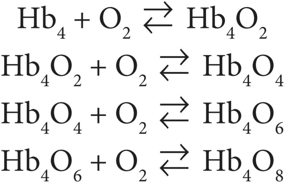
The reaction is rapid, requiring less than 0.01 s. The deoxygenation of Hb408 is also very rapid.
واکنش سریع است و کمتر از 0.01 ثانیه نیاز دارد. اکسیژن زدایی Hb408 نیز بسیار سریع است.
The quaternary structure of hemoglobin determines its affinity for O2. In deoxyhemoglobin, the globin units are tightly bound in a tense (T) configuration, which reduces the affinity of the molecule for O2. When O2 is first bound, the bonds holding the globin units are released, producing a relaxed (R) configuration, which exposes more O2-binding sites. The net result is a 500-fold increase in O2 affinity. In tissues, these reactions are reversed, resulting in O2 release. The transition from one state to another has been calculated to occur about 108 times in the life of a red blood cell.
ساختار چهارتایی هموگلوبین میل آن را برای O2 تعیین می کند. در دئوکسی هموگلوبین، واحدهای گلوبین در زمان (T) محکم بسته شده اند. پیکربندی، که میل ترکیبی مولکول برای O2 را کاهش می دهد. هنگامی که O2 برای اولین بار متصل می شود، پیوندهای نگهدارنده واحدهای گلوبین آزاد می شوند و یک پیکربندی آرام (R) ایجاد می کنند که مکان های اتصال O2 بیشتری را نشان می دهد. نتیجه خالص افزایش 500 برابری میل O2 است. در بافتها، این واکنشها معکوس میشوند و در نتیجه O2 آزاد میشود. انتقال از یک حالت به حالت دیگر حدود 108 بار در طول عمر یک گلبول قرمز محاسبه شده است.
The oxygen-hemoglobin dissociation curve relates percentage saturation of the O2-carrying power of hemoglobin (abbreviated as SaO2) to the Po2 (Figure 35-1). This curve has a characteristic sigmoid shape due to the T-R configuration interconversion. Combination of the first heme in the Hb molecule with O2 increases the affinity of the second heme for O2, and oxygenation of the second increases the affinity of the third, and so on, so that the affinity of Hb for the fourth O2 molecule is many times that for the first. Especially note that small changes at low Po2 lead to large changes in SaO2.
منحنی تفکیک اکسیژن-هموگلوبین درصد اشباع قدرت حامل O2 هموگلوبین (به اختصار SaO2) را به Po2 مرتبط می کند (شکل 35-1). این منحنی دارای شکل سیگموئید مشخصه به دلیل تبدیل درونی پیکربندی T-R است. ترکیب هم اول در مولکول Hb با O2 میل ترکیبی هم دوم را برای O2 افزایش می دهد و اکسیژن رسانی به دومی باعث افزایش میل ترکیبی سوم و غیره می شود، به طوری که میل ترکیبی Hb برای مولکول O2 چهارم چندین برابر مولکول اولی است. به خصوص توجه داشته باشید که تغییرات کوچک در Po2 کم منجر به تغییرات بزرگ در SaO2 می شود.
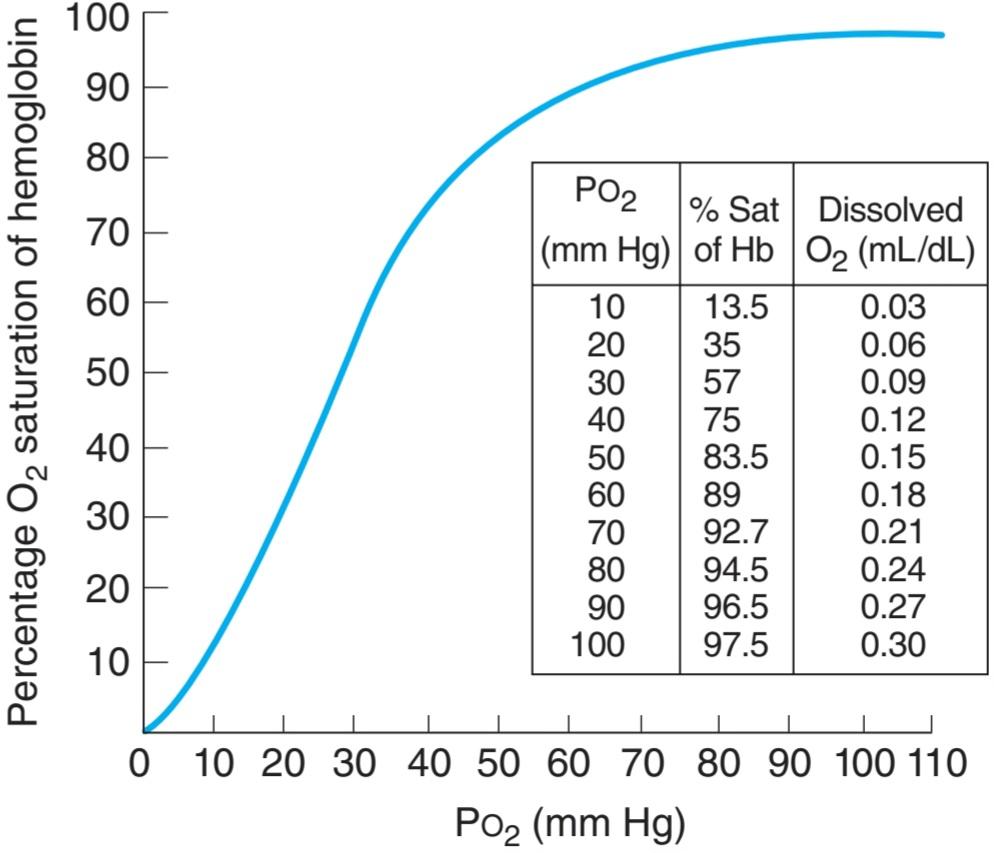
FIGURE 35-1 Oxygen-hemoglobin dissociation curve. pH 7.40, temperature 38°C. Inset table relates the percentage of saturated hemoglobin (SaO2) to Po2 and dissolved O2. (Modified with permission from Comroe JH Jr, et al: The Lung: Clinical Physiology and Pulmonary Function Tests, 2nd ed. Year Book; 1962.)
شکل 35-1 منحنی تفکیک اکسیژن-هموگلوبین. pH 7.40، دما 38 درجه سانتیگراد. جدول داخلی درصد هموگلوبین اشباع (SaO2) را به Po2 و O2 محلول مرتبط می کند. (با اجازه Comroe JH Jr و همکاران: The Lung: Clinical Physiology and Pulmonary Function Tests، ویرایش دوم سال کتاب، 1962، اصلاح شده است.)
When blood is equilibrated with 100% O2, the normal hemoglobin becomes 100% saturated. When fully saturated, each gram (g) of normal hemoglobin contains 1.39 mL of O2. However, blood normally contains small quantities of inactive hemoglobin derivatives, and the measured value in vivo is thus slightly lower. Using the traditional estimate of saturated hemoglobin in vivo, 1.34 mL of O2, the hemoglobin concentration in normal blood is about 15 g/dL (14 g/dL in women and 16 g/dL in men). Therefore, 1 dL of blood contains 20.1 mL (1.34 mL × 15) of O2 bound to hemoglobin when the hemoglobin is 100% saturated. The amount of dissolved O2 is a linear function of the Po2 (0.003 mL/dL blood/mm Hg Po2).
هنگامی که خون با 100٪ O2 متعادل می شود، هموگلوبین طبیعی 100٪ اشباع می شود. هنگامی که به طور کامل اشباع شود، هر گرم (گرم) هموگلوبین نرمال حاوی 1.39 میلی لیتر O2 است. با این حال، خون معمولاً حاوی مقادیر کمی از مشتقات هموگلوبین غیرفعال است و بنابراین مقدار اندازهگیری شده در داخل بدن کمی کمتر است. با استفاده از تخمین سنتی هموگلوبین اشباع در داخل بدن، 1.34 میلی لیتر O2، غلظت هموگلوبین در خون طبیعی حدود 15 گرم در دسی لیتر است (14 گرم در دسی لیتر در زنان و 16 گرم در دسی لیتر در مردان). بنابراین، 1 دسی لیتر خون حاوی 20.1 میلی لیتر (1.34 میلی لیتر × 15) O2 متصل به هموگلوبین زمانی که هموگلوبین 100٪ اشباع است. مقدار O2 محلول تابع خطی Po2 است (0.003 mL/dL خون/mm Hg Po2).
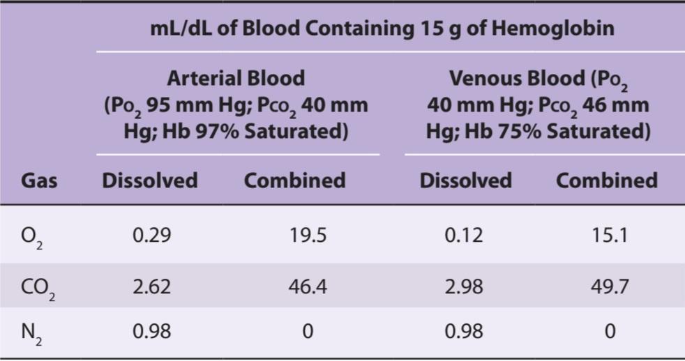
In vivo, the hemoglobin in the blood at the ends of the pulmonary capillaries is about 97.5% saturated with O2 (Po2 = 100 mm Hg). Because of a slight admixture with venous blood that bypasses the pulmonary capillaries (ie, physiological shunt), the hemoglobin in systemic arterial blood is only 97% saturated. The arterial blood therefore contains a total of about 19.8 mL of O2 per dL: 0.29 mL in solution and 19.5 mL bound to hemoglobin. In venous blood at rest, the hemoglobin is 75% saturated and the total O2 content is about 15.2 mL/dL: 0.12 mL in solution and 15.1 mL bound to hemoglobin. Thus, at rest the tissues remove about 4.6 mL of O2 from each deciliter (dL) of blood passing through them (Table 35-1); 0.17 mL of this total represents O2 that was in solution in the blood, and the remainder represents O2 that was liberated from hemoglobin. In this way, 250 mL of O2 per minute is transported from the blood to the tissues at rest.
در داخل بدن، هموگلوبین خون در انتهای مویرگ های ریوی حدود 97.5٪ با O2 اشباع شده است (Po2 = 100 میلی متر جیوه). به دلیل ترکیب جزئی با خون وریدی که مویرگ های ریوی را دور می زند (یعنی شانت فیزیولوژیکی)، هموگلوبین در خون شریانی سیستمیک تنها 97 درصد اشباع شده است. بنابراین خون شریانی در مجموع حاوی حدود 19.8 میلی لیتر O2 در هر دسی لیتر است: 0.29 میلی لیتر در محلول و 19.5 میلی لیتر متصل به هموگلوبین. در خون وریدی در حالت استراحت، هموگلوبین 75 درصد اشباع است و محتوای O2 کل حدود 15.2 میلی لیتر در دسی لیتر است: 0.12 میلی لیتر در محلول و 15.1 میلی لیتر به هموگلوبین متصل می شود. بنابراین، در حالت استراحت، بافت ها حدود 4.6 میلی لیتر O2 را از هر دسی لیتر (دسی لیتر) خون عبوری از خود حذف می کنند (جدول 35-1). 0.17 میلی لیتر از این مجموع نشان دهنده O2 است که در محلول در خون بود و باقی مانده نشان دهنده O2 است که از هموگلوبین آزاد شده است. به این ترتیب 250 میلی لیتر O2 در دقیقه در حالت استراحت از خون به بافت ها منتقل می شود.
TABLE 35-1 Gas content of blood.
جدول 35-1 محتوای گاز خون.
FACTORS AFFECTING THE AFFINITY OF HEMOGLOBIN FOR OXYGEN
Three important conditions affect the oxygen-hemoglobin dissociation curve: the pH, the temperature, and the concentration of 2,3-diphosphoglycerate (DPG; 2,3-DPG). A rise in temperature or a fall in pH shifts the curve to the right (Figure 35-2). When the curve is shifted in this direction, a higher Po2 is required for hemoglobin to bind a given amount of O2. Conversely, a fall in temperature or a rise in pH shifts the curve to the left, and a lower Po2 is required to bind a given amount of O2. A convenient index for comparison of such shifts is the P50, the Po2 at which hemoglobin is half saturated with O2. The higher the P50, the lower the affinity of hemoglobin for O2.
عوامل مؤثر بر تمایل هموگلوبین به اکسیژن
سه شرایط مهم بر منحنی تفکیک اکسیژن-هموگلوبین تأثیر میگذارند: pH، دما، و غلظت 2،3-دی فسفوگلیسرات (DPG؛ 2،3-DPG). افزایش دما یا کاهش pH منحنی را به سمت راست تغییر می دهد (شکل 35-2). هنگامی که منحنی در این جهت جابه جا می شود، برای هموگلوبین برای اتصال مقدار معینی از O2، به Po2 بالاتری نیاز است. برعکس، کاهش دما یا افزایش pH منحنی را به سمت چپ تغییر می دهد و برای اتصال مقدار معینی از O2 به Po2 کمتری نیاز است. یک شاخص مناسب برای مقایسه چنین تغییراتی P50 است، Po2 که در آن هموگلوبین نیمی از O2 اشباع شده است. هر چه P50 بالاتر باشد، تمایل هموگلوبین به O2 کمتر است.
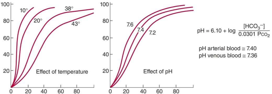
FIGURE 35-2 Effects of temperature and pH on the oxygen-hemoglobin dissociation curve. Both changes in temperature (left) and pH (right) can alter the affinity of hemoglobin for O2. Plasma pH can be estimated using the modified Henderson-Hasselbalch equation, as shown. (Modified with permission from Comroe JH Jr, et al: The Lung: Clinical Physiology and Pulmonary Function Tests, 2nd ed. Year Book; 1962.)
شکل 35-2 اثرات دما و pH بر منحنی تفکیک اکسیژن-هموگلوبین. هر دو تغییر در دما (چپ) و pH (راست) می توانند میل هموگلوبین را برای O2 تغییر دهند. همانطور که نشان داده شده است، می توان pH پلاسما را با استفاده از معادله اصلاح شده هندرسون- هاسلبالخ تخمین زد. (با اجازه Comroe JH Jr و همکاران: The Lung: Clinical Physiology and Pulmonary Function Tests، ویرایش دوم سال کتاب، 1962، اصلاح شده است.)
The decrease in O2 affinity of hemoglobin when the pH of blood falls is called the Bohr effect and is closely related to the fact that deoxygenated hemoglobin (deoxyhemoglobin) binds H* more actively than does oxygenated hemoglobin (oxyhemoglobin). The pH of blood falls as its CO2 content increases, so that when the PCO2 rises, the curve shifts to the right and the P50 rises. Most of the unsaturation of hemoglobin that occurs in the tissues is secondary to the decline in the Po2, but an extra 1-2% unsaturation is due to the rise in PCO2 and consequent shift of the dissociation curve to the right.
کاهش میل ترکیبی O2 هموگلوبین هنگام کاهش pH خون، اثر بور نامیده می شود و ارتباط نزدیکی با این واقعیت دارد که هموگلوبین بدون اکسیژن (دئوکسی هموگلوبین) +H را فعالتر از هموگلوبین اکسیژن دار (اکسی هموگلوبین) متصل می کند. pH خون با افزایش مقدار CO2 کاهش می یابد، به طوری که وقتی PCO2 افزایش می یابد، منحنی به سمت راست تغییر می کند و P50 افزایش می یابد. بیشتر اشباع نشدن هموگلوبین که در بافتها اتفاق میافتد، ثانویه به کاهش Po2 است، اما 1-2٪ غیراشباع اضافی به دلیل افزایش PCO2 و در نتیجه تغییر منحنی تفکیک به راست است.
2,3-DPG is very plentiful in red cells. It is formed from 3- phosphoglyceraldehyde, which is a product of glycolysis via the Embden- Meyerhof pathway. It is a highly charged anion that binds to the ẞ chains of deoxyhemoglobin. One mole of deoxyhemoglobin binds 1 mol of 2,3-DPG. In effect,
2،3-DPG در گلبول های قرمز بسیار فراوان است. از 3- فسفوگلیسرآلدئید که محصول گلیکولیز از طریق مسیر Embden-Meyerhof است تشکیل می شود. این یک آنیون بسیار باردار است که به زنجیره های ẞ دئوکسی هموگلوبین متصل می شود. یک مول دئوکسی هموگلوبین به 1 مول 2،3-DPG متصل می شود. در واقع،

In this equilibrium, an increase in the concentration of 2,3-DPG shifts the reaction to the right, causing more O2 to be liberated.
در این تعادل، افزایش غلظت 2،3-DPG واکنش را به سمت راست تغییر می دهد و باعث آزاد شدن O2 بیشتری می شود.
Because acidosis inhibits red cell glycolysis, the 2,3-DPG concentration falls when the pH is low. Conversely, thyroid hormones, growth hormones, and androgens can all increase the concentration of 2,3-DPG and the P50.
از آنجایی که اسیدوز گلیکولیز گلبول های قرمز را مهار می کند، غلظت 2،3-DPG زمانی که pH پایین است کاهش می یابد. برعکس، هورمون های تیروئید، هورمون های رشد و آندروژن ها همگی می توانند غلظت 2،3-DPG و P50 را افزایش دهند.
Exercise has been reported to produce an increase in 2,3-DPG within 60 min (although the rise may not occur in trained athletes). The P50 is also increased during exercise, because the temperature rises in active tissues and CO2 and metabolites accumulate, lowering the pH. In addition, much more O2 is removed from each unit of blood flowing through active tissues because the tissue Po2 declines. Finally, at low Po2 values, the oxygen-hemoglobin dissociation curve is steep, and large amounts of O2 are liberated per unit drop in Po2. Some clinical features of hemoglobin are discussed in Clinical Box 35-1.
گزارش شده است که ورزش باعث افزایش 2،3-DPG در 60 دقیقه می شود (اگرچه ممکن است این افزایش در ورزشکاران تمرین دیده رخ ندهد). P50 همچنین در طول ورزش افزایش می یابد، زیرا دما در بافت های فعال افزایش می یابد و CO2 و متابولیت ها انباشته می شوند و pH را کاهش می دهند. علاوه بر این، O2 بسیار بیشتری از هر واحد خونی که در بافتهای فعال جریان دارد، حذف میشود، زیرا Po2 بافت کاهش مییابد. در نهایت، در مقادیر پایین Po2، منحنی تفکیک اکسیژن-هموگلوبین شیب دار است و مقادیر زیادی O2 در هر واحد افت در Po2 آزاد می شود. برخی از ویژگی های بالینی هموگلوبین در جعبه بالینی 35-1 مورد بحث قرار گرفته است.
CLINICAL BOX 35-1
Hemoglobin & O2 Binding In Vivo
Cyanosis
Reduced hemoglobin has a dark color, and a dusky bluish discoloration of the tissues, called cyanosis, appears when the reduced hemoglobin concentration of the blood in the capillaries is more than 5 g/dL. Its occurrence depends on the total amount of hemoglobin in the blood, the degree of hemoglobin unsaturation, and the state of the capillary circulation. Cyanosis is most easily seen in the nail beds and mucous membranes and in the earlobes, lips, and fingers, where the skin is thin and there are plenty capillaries. Although visible observation is indicative of cyanosis, it is not fully reliable. Further tests of arterial oxygen tension and saturation, blood and hemoglobin counts can provide more reliable diagnoses.
جعبه بالینی 35-1
هموگلوبین و O2 اتصال In Vivo
سیانوز
هموگلوبین کاهش یافته رنگ تیره ای دارد و زمانی که غلظت هموگلوبین کاهش یافته خون در مویرگ ها بیش از 5 گرم در دسی لیتر باشد، تغییر رنگ مایل به آبی تیره بافت ها به نام سیانوز ظاهر می شود. وقوع آن به مقدار کل هموگلوبین خون، درجه اشباع نشدن هموگلوبین و وضعیت گردش خون مویرگی بستگی دارد. سیانوز به راحتی در بستر ناخن و غشاهای مخاطی و در لاله گوش، لب ها و انگشتان، جایی که پوست نازک است و مویرگ های زیادی وجود دارد، دیده می شود. اگرچه مشاهده مرئی نشان دهنده سیانوز است، اما کاملا قابل اعتماد نیست. آزمایشهای بیشتر تنش و اشباع اکسیژن شریانی، شمارش خون و هموگلوبین میتواند تشخیصهای قابل اعتمادتری را ارائه دهد.
Effects of 2,3-DPG on Fetal & Stored Blood
The affinity of fetal hemoglobin (hemoglobin F) for O2, which is greater than that for adult hemoglobin (hemoglobin A), facilitates the movement of O2 from the mother to the fetus. The cause of this greater affinity is the poor binding of 2,3-DPG by the y polypeptide chains that replace ẞ chains in fetal hemoglobin. Some abnormal hemoglobins in adults have low P50 values, and the resulting high O2 affinity of the hemoglobin causes enough tissue hypoxia to stimulate increased red cell formation, with resulting polycythemia. It is interesting to speculate that these hemoglobins may not bind 2,3-DPG.
اثرات 2،3-DPG بر خون جنین و ذخیره شده
تمایل هموگلوبین جنین (هموگلوبین F) به O2، که بیشتر از هموگلوبین بزرگسالان (هموگلوبین A) است، حرکت O2 را از مادر به جنین تسهیل می کند. علت این تمایل بیشتر، اتصال ضعیف 2،3-DPG توسط زنجیره های پلی پپتیدی y است که جایگزین زنجیره های ẞ در هموگلوبین جنین می شود. برخی از هموگلوبین های غیرطبیعی در بزرگسالان دارای مقادیر P50 پایینی هستند و در نتیجه میل ترکیبی O2 بالای هموگلوبین باعث هیپوکسی بافتی کافی برای تحریک افزایش تشکیل گلبول های قرمز و در نتیجه پلی سیتمی می شود. جالب است که حدس بزنیم که این هموگلوبین ها ممکن است به 2،3-DPG متصل نشوند.
Red cell 2,3-DPG concentration is increased in anemia and in a variety of diseases in which there is chronic hypoxia. This facilitates the delivery of O2 to the tissues by raising the PO2 at which O2 is released in peripheral capillaries. In banked blood that is stored, the 2,3-DPG level falls and the ability of this blood to release O2 to the tissues is reduced. This decrease, which obviously limits the benefit of the blood if it is transfused into a hypoxic patient, is less if the blood is stored in citrate-phosphate-dextrose solution rather than the usual acid-citrate-dextrose solution
غلظت گلبول قرمز 2،3-DPG در کم خونی و در انواع بیماری هایی که هیپوکسی مزمن در آنها وجود دارد افزایش می یابد. این امر با افزایش PO2 که در آن O2 در مویرگ های محیطی آزاد می شود، تحویل O2 به بافت ها را تسهیل می کند. در خون بانکی که ذخیره می شود، سطح 2،3-DPG کاهش می یابد و توانایی این خون برای آزادسازی O2 به بافت ها کاهش می یابد. این کاهش، که بدیهی است در صورت تزریق خون به یک بیمار هیپوکسیک، فایده آن را محدود می کند، در صورتی که خون در محلول سیترات-فسفات-دکستروز ذخیره شود کمتر است تا محلول معمول اسید سیترات-دکستروز.
THERAPEUTIC HIGHLIGHTS
Cyanosis is an indication of poorly oxygenated hemoglobin rather than a disease, and thus can have many causes, from cold exposure to drug overdose to chronic lung disease. As such, proper treatment depends on the underlying cause. For cyanosis caused by exposure to cold, maintaining a warm environment can be effective, whereas supplemental oxygen administration may be required under conditions of chronic and hypoxic disease.
نکات برجسته درمانی
سیانوز به جای یک بیماری، نشانه ای از هموگلوبین کم اکسیژن است و بنابراین می تواند دلایل زیادی داشته باشد، از قرار گرفتن در معرض سرما تا مصرف بیش از حد دارو تا بیماری مزمن ریوی. به این ترتیب، درمان مناسب به علت زمینه ای بستگی دارد. برای سیانوز ناشی از قرار گرفتن در معرض سرما، حفظ یک محیط گرم می تواند موثر باشد، در حالی که تجویز اکسیژن مکمل ممکن است در شرایط بیماری مزمن و هیپوکسیک مورد نیاز باشد.
An interesting contrast to hemoglobin is myoglobin, an iron-containing pigment found in skeletal muscle. Myoglobin resembles hemoglobin but binds 1 rather than 4 mol of O2 per mole protein. The lack of cooperative binding is reflected in the myoglobin dissociation curve, a rectangular hyperbola rather than the sigmoid curve observed for hemoglobin (Figure 35-3). Additionally, the leftward shift of the myoglobin O2 binding curve when compared with hemoglobin demonstrates a higher affinity for O2, and thus promotes a favorable transfer of O2 from hemoglobin in the blood. The steepness of the myoglobin curve also shows that O2 is released only at low Po2 values (eg, during exercise). The myoglobin content is greatest in muscles specialized for sustained contraction. The muscle blood supply is compressed during such contractions, and myoglobin can continue to provide O2 under reduced blood flow and/or reduced Po2 in the blood.
یک تضاد جالب با هموگلوبین، میوگلوبین است، یک رنگدانه حاوی آهن که در ماهیچه های اسکلتی یافت می شود. میوگلوبین شبیه هموگلوبین است اما 1 به جای 4 مول O2 در هر مول پروتئین متصل می کند. فقدان اتصال مشارکتی در منحنی تفکیک میوگلوبین، یک هذلول مستطیل شکل به جای منحنی سیگموئید مشاهده شده برای هموگلوبین منعکس می شود (شکل 35-3). علاوه بر این، تغییر منحنی اتصال به میوگلوبین O2 در مقایسه با هموگلوبین به سمت چپ، تمایل بیشتری به O2 نشان میدهد و بنابراین باعث انتقال مطلوب O2 از هموگلوبین در خون میشود. شیب منحنی میوگلوبین همچنین نشان می دهد که O2 فقط در مقادیر کم Po2 (مثلاً در حین ورزش) آزاد می شود. محتوای میوگلوبین در ماهیچه هایی که برای انقباض پایدار تخصص دارند بیشترین مقدار است. جریان خون ماهیچه ای در طول چنین انقباضاتی فشرده می شود و میوگلوبین می تواند به ارائه O2 تحت کاهش جریان خون و/یا کاهش Po2 در خون ادامه دهد.
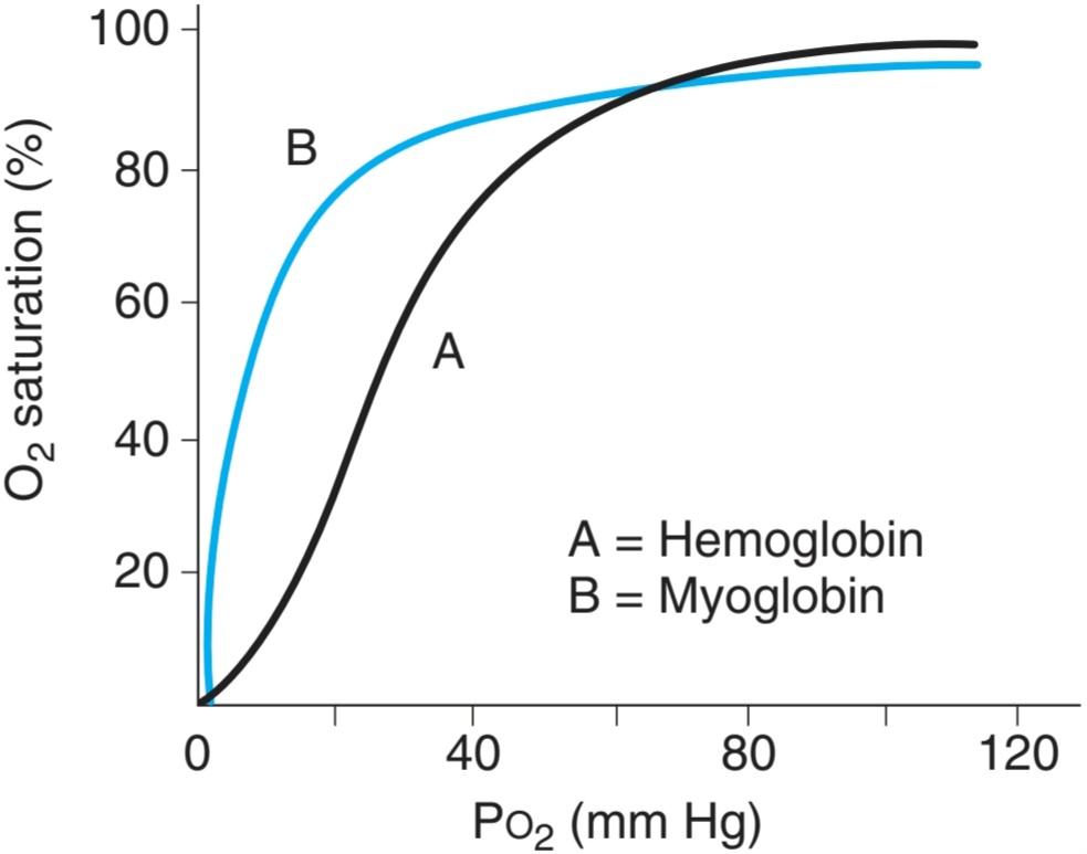
FIGURE 35-3 Comparison of dissociation curves for hemoglobin and myoglobin. The myoglobin binding curve (B) lacks the sigmoidal shape of the hemoglobin binding curve (A) because of the single O2-binding site in each molecule. Myoglobin also has greater affinity for O2 than hemoglobin (curve shifted left) and thus can release O2 in muscle when Po2 in blood is low (eg, during exercise).
شکل 35-3 مقایسه منحنی های تفکیک هموگلوبین و میوگلوبین. منحنی اتصال میوگلوبین (B) فاقد شکل سیگموئیدی منحنی اتصال هموگلوبین (A) است زیرا محل اتصال O2 در هر مولکول است. همچنین میوگلوبین تمایل بیشتری به O2 نسبت به هموگلوبین دارد (منحنی به چپ منتقل می شود) و بنابراین می تواند O2 در ماهیچه آزاد کند زمانی که Po2 در خون کم است (مثلاً در حین ورزش).
CARBON DIOXIDE TRANSPORT
MOLECULAR FATE OF CARBON DIOXIDE IN BLOOD
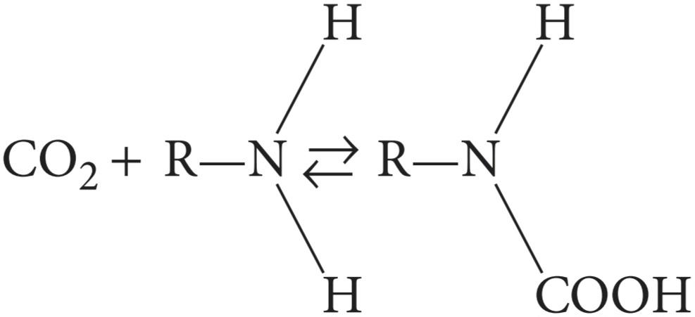
The solubility of CO2 in blood is about 20 times that of O2; therefore, considerably more CO2 than O2 is present in simple solution at equal partial pressures. The CO2 that diffuses into red blood cells is rapidly hydrated to H2CO3 because of the presence of carbonic anhydrase (Figure 35-4). The H2CO3 dissociates to H* and HCO3, and the H+ is buffered, primarily by hemoglobin, while the HCO3 enters the plasma. Some of the CO2 in the red cells reacts with the amino groups of hemoglobin and other proteins (R), forming carbamino compounds.
انتقال دی اکسید کربن
سرنوشت مولکولی دی اکسید کربن در خون
حلالیت CO2 در خون حدود 20 برابر O2 است. بنابراین، CO2 به طور قابل توجهی بیشتر از O2 در محلول ساده در فشارهای جزئی برابر وجود دارد. CO2 که در گلبول های قرمز منتشر می شود به دلیل وجود کربنیک انیدراز به سرعت به H2CO3 هیدراته می شود (شکل 35-4). H2CO3 به +H و HCO3 تجزیه می شود و +H عمدتاً توسط هموگلوبین بافر می شود در حالی که HCO3 وارد پلاسما می شود. مقداری از CO2 موجود در گلبولهای قرمز با گروههای آمینه هموگلوبین و سایر پروتئینها (R) واکنش داده و ترکیبات کربامینو را تشکیل میدهند.
Because deoxyhemoglobin binds more H* than oxyhemoglobin and forms carbamino compounds more readily, binding of O2 to hemoglobin reduces its affinity for CO2. The Haldane effect refers to the increased capacity of deoxygenated hemoglobin to bind and carry CO2. Consequently, venous blood carries more CO2 than arterial blood, CO2 uptake is facilitated in the tissues, and CO2 release is facilitated in the lungs. About 11% of the CO2 added to the blood in the systemic capillaries is carried to the lungs as carbamino-CO2.
از آنجایی که دئوکسی هموگلوبین به +H بیشتری نسبت به اکسی هموگلوبین متصل می شود و ترکیبات کربامینو را با سهولت بیشتری تشکیل می دهد، اتصال O2 به هموگلوبین میل آن را برای CO2 کاهش می دهد. اثر Haldane به افزایش ظرفیت هموگلوبین بدون اکسیژن برای اتصال و حمل CO2 اشاره دارد. در نتیجه، خون وریدی CO2 بیشتری را نسبت به خون شریانی حمل می کند، جذب CO2 در بافت ها تسهیل می شود و انتشار CO2 در ریه ها تسهیل می شود. حدود 11 درصد از CO2 اضافه شده به خون در مویرگ های سیستمیک به عنوان carbamino-CO2 به ریه ها منتقل می شود.
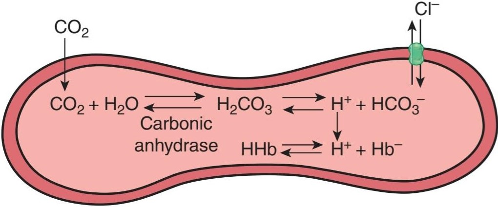
FIGURE 35-4 Fate of CO2 in the red blood cell. Upon entering the red blood cell, CO2 is rapidly hydrated to H2CO3 by carbonic anhydrase. H2CO3 is in equilibrium with H* and its conjugate base, HCO3. H* can interact with deoxyhemoglobin, whereas HCO3 can be transported outside of the cell via anion exchanger 1 (AE1 or Band 3). In effect, for each CO2 molecule that enters the red cell, there is an additional HCO3 or Cl ̄ in the cell.
شکل 35-4 سرنوشت CO2 در گلبول قرمز. پس از ورود به گلبول قرمز، CO2 به سرعت توسط کربنیک انیدراز به H2CO3 هیدراته می شود. H2CO3 با +H و پایه مزدوج آن HCO3 در تعادل است. +H می تواند با دئوکسی هموگلوبین تعامل کند، در حالی که HCO3 می تواند از طریق مبدل آنیونی 1 (AE1 یا Band 3) به خارج از سلول منتقل شود. در واقع، برای هر مولکول CO2 که وارد گلبول قرمز می شود، یک HCO3 یا Cl ̄ اضافی در سلول وجود دارد.
CHLORIDE SHIFT
Because the rise in the HCO3 content of red cells is much greater than that in plasma as the blood passes through the capillaries, about 70% of the HCO3 formed in the red cells enters the plasma. The excess HCO3 ̄ leaves the red cells in exchange for Cl (Figure 35-4). This process is mediated by anion exchanger 1 (AE1; also called Band 3), a major membrane protein in the red blood cell. Because of this chloride shift, the Cl content of the red cells in venous blood is significantly greater than that in arterial blood. The chloride shift occurs rapidly and is essentially complete within 1 s.
تغییر کلرید
از آنجایی که افزایش محتوای HCO3 در گلبول های قرمز بسیار بیشتر از پلاسما است که خون از مویرگ ها عبور می کند، حدود 70 درصد از HCO3 تشکیل شده در گلبول های قرمز وارد پلاسما می شود. ̄HCO3 اضافی از گلبول های قرمز در ازای Cl خارج می شود (شکل 35-4). این فرآیند توسط مبدل آنیونی 1 (AE1 که باند 3 نیز نامیده می شود)، یک پروتئین غشایی اصلی در گلبول قرمز انجام می شود. به دلیل این تغییر کلرید، محتوای کلر گلبول های قرمز در خون وریدی به طور قابل توجهی بیشتر از خون شریانی است. تغییر کلرید به سرعت اتفاق می افتد و اساساً در عرض 1 ثانیه کامل می شود.
Note that for each CO2 molecule added to a red cell, there is an increase of one osmotically active particle in the cell—either an HCO3 or a Cl ̄ (Figure 35-4). Consequently, the red cells take up water and increase in size. For this reason, plus the fact that a small amount of fluid in the arterial blood returns via the lymphatics rather than the veins, the hematocrit of venous blood is normally 3% greater than that of the arterial blood. In the lungs, the Cl- moves back out of the cells and they shrink.
توجه داشته باشید که به ازای هر مولکول CO2 که به یک گلبول قرمز اضافه می شود، یک ذره فعال اسمزی در سلول افزایش می یابد – اعم از HCO3 یا ̄Cl (شکل 35-4). در نتیجه، گلبول های قرمز آب را می گیرند و اندازه آنها افزایش می یابد. به همین دلیل، به علاوه این واقعیت که مقدار کمی مایع در خون شریانی از طریق لنفاوی به جای وریدها باز می گردد، هماتوکریت خون وریدی به طور معمول 3 درصد بیشتر از خون شریانی است. در ریه ها، -Cl از سلول ها عقب می رود و آنها کوچک می شوند.
SPATIAL DISTRIBUTION OF CARBON DIOXIDE IN BLOOD
For convenience, the various fates of CO2 in the plasma and red cells are summarized in Table 35-2. The extent to which they increase the capacity of the blood to carry CO2 is indicated by the difference between the lines indicating the dissolved CO2 and the total CO2 in the dissociation curves for CO2 shown in Figure 35-5.
توزیع فضایی دی اکسید کربن در خون
برای سهولت، سرنوشت های مختلف CO2 در پلاسما و گلبول های قرمز در جدول 35-2 خلاصه شده است. میزان افزایش ظرفیت خون برای حمل CO2 با تفاوت بین خطوط نشان دهنده CO2 محلول و CO2 کل در منحنی های تفکیک CO2 نشان داده شده در شکل 35-5 نشان داده شده است.
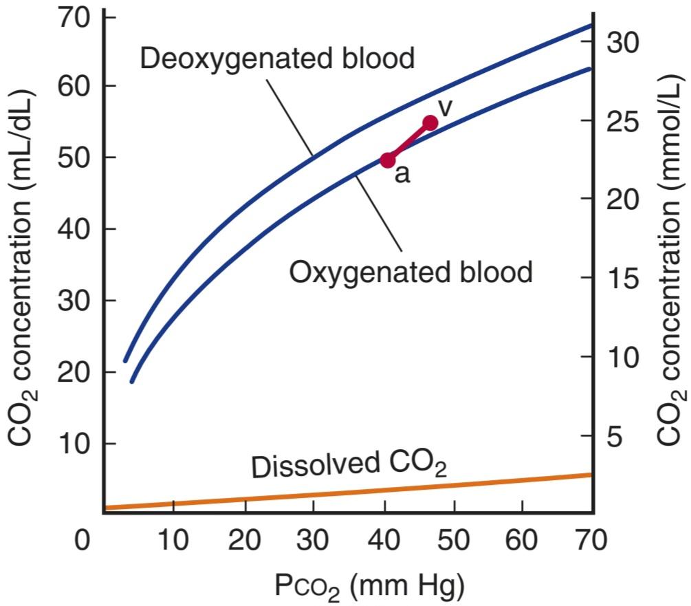
FIGURE 35-5 CO2 dissociation curves. The arterial point (a) and the venous point (v) indicate the total CO2 content found in arterial blood and venous blood of normal resting humans. Note the low amount of CO2 that is dissolved (orange trace) compared to that which can be carried by other means (Table 35-2). (Modified with permission from Schmidt RF, Thews G [editors]: Human Physiology. Springer; 1983.)
شکل 35-5 منحنی های تفکیک CO2. نقطه شریانی (a) و نقطه وریدی (v) میزان کل CO2 موجود در خون شریانی و خون وریدی انسانهای عادی در حال استراحت را نشان میدهد. به مقدار کم CO2 حل شده (ردپای نارنجی) در مقایسه با آنچه که می توان با وسایل دیگر حمل کرد (جدول 35-2) توجه کنید. (اصلاح شده با اجازه اشمیت RF، Thews G [ویراستاران]: فیزیولوژی انسانی. Springer؛ 1983.)
TABLE 35-2 Fate of CO2 in blood.
جدول 35-2 سرنوشت CO2 در خون.
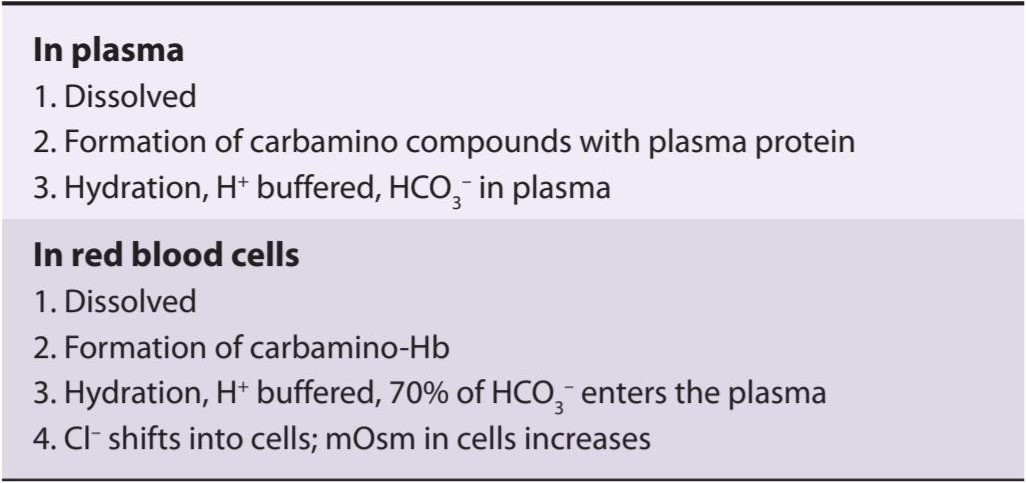
Of the approximately 49 mL of CO2 in each deciliter of arterial blood (Table 35-1), 2.6 mL is dissolved, 2.6 mL is in carbamino compounds, and 43.8 mL is in HCO3. In the tissues, 3.7 mL of CO2 per deciliter of blood is added, 0.4 mL stays in solution, 0.8 mL forms carbamino compounds, and 2.5 mL forms HCO3. The pH of the blood drops from 7.40 to 7.36. In the lungs, the processes are reversed, and the 3.7 mL of CO2 is discharged into the alveoli. In this manner, 200 mL of CO2 per minute at rest and much larger amounts during exercise are transported from the tissues to the lungs and excreted. It is worth noting that this amount of CO2 is equivalent in 24 h to over 12,500 mEq of H*.
از تقریباً 49 میلی لیتر CO2 در هر دسی لیتر خون شریانی (جدول 35-1)، 2.6 میلی لیتر در ترکیبات کربامینو و 43.8 میلی لیتر در HCO3 است. در بافت ها، 3.7 میلی لیتر CO2 در هر دسی لیتر خون اضافه می شود، 0.4 میلی لیتر در محلول باقی می ماند، 0.8 میلی لیتر ترکیبات کربامینو و 2.5 میلی لیتر HCO3 تشکیل می دهد. pH خون از 7.40 به 7.36 کاهش می یابد. در ریه ها، فرآیندها معکوس می شوند و 3.7 میلی لیتر CO2 به آلوئول ها تخلیه می شود. به این ترتیب 200 میلی لیتر CO2 در دقیقه در حالت استراحت و مقادیر بسیار بیشتر در حین ورزش از بافت ها به ریه ها منتقل شده و دفع می شود. شایان ذکر است که این مقدار CO2 در 24 ساعت معادل بیش از 12500 +mEq H است.
ACID-BASE BALANCE & GAS TRANSPORT
The major source of acids in the blood under normal conditions is through cellular metabolism. The CO2 formed by metabolism in the tissues is in large part hydrated to H2CO3, resulting in the large total H+ load noted above (> 12,500 mEq/day). However, most of the CO2 is excreted in the lungs, and the small quantities of the remaining H* are excreted by the kidneys.
تعادل اسید و باز و حمل و نقل گاز
منبع اصلی اسیدهای خون در شرایط عادی از طریق متابولیسم سلولی است. CO2 تشکیل شده توسط متابولیسم در بافت ها تا حد زیادی به H2CO3 هیدراته می شود و در نتیجه بار کل +H که در بالا ذکر شد (> 12500 mEq/day) ایجاد می شود. با این حال، بیشتر CO2 در ریه ها دفع می شود و مقدار کمی از +H باقی مانده توسط کلیه ها دفع می شود.
BUFFERING IN THE BLOOD
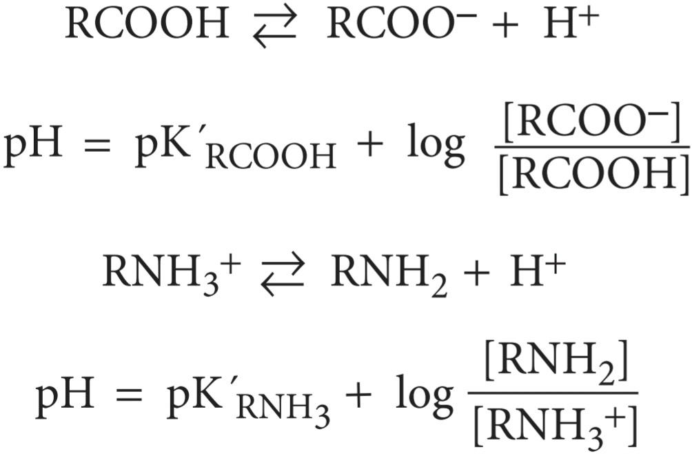
Acid and base shifts in the blood are largely controlled by three main buffers in blood: (1) proteins, (2) hemoglobin, and (3) the carbonic acid-bicarbonate system. Plasma proteins are effective buffers because both their free carboxyl and their free amino groups dissociate:
بافر در خون
تغییرات اسید و باز در خون تا حد زیادی توسط سه بافر اصلی در خون کنترل می شود: (1) پروتئین ها، (2) هموگلوبین، و (3) سیستم کربنیک اسید-بی کربنات. پروتئین های پلاسما بافرهای موثری هستند زیرا هم کربوکسیل آزاد و هم گروه های آمینه آزاد آنها از هم جدا می شوند:
The second buffer system is provided by the dissociation of the imidazole groups of the histidine residues in hemoglobin.
سیستم بافر دوم با تفکیک گروه های ایمیدازول از باقی مانده های هیستیدین در هموگلوبین ایجاد می شود.
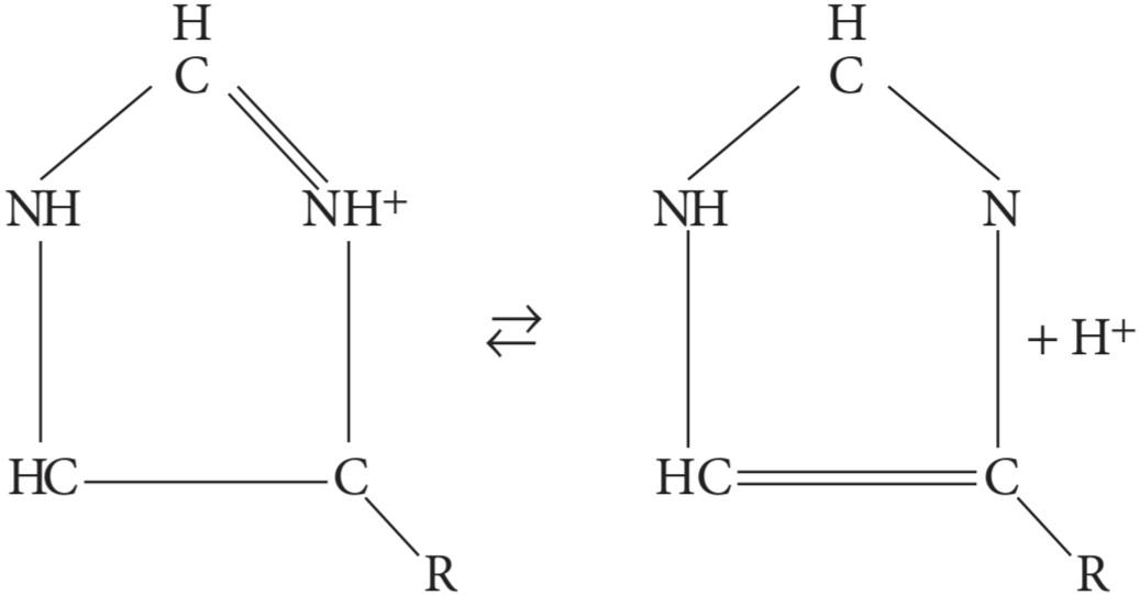
In the pH 7.0-7.7 range, the free carboxyl and amino groups of hemoglobin contribute relatively little to its buffering capacity. However, the hemoglobin molecule contains 38 histidine residues, and on this basis-plus the fact that hemoglobin is present in large amounts-the hemoglobin in blood has six times the buffering capacity of the plasma proteins. In addition, the action of hemoglobin is unique because the imidazole groups of deoxyhemoglobin (Hb) dissociate less than those of oxyhemoglobin (HbO2), making Hb a weaker acid and therefore a better buffer than HbO2. The titration curves for Hb and HbO2 (Figure 35-6) illustrate the differences in H+ buffering capacity.
در محدوده pH 7.0-7.7، گروه های کربوکسیل و آمینو آزاد هموگلوبین سهم نسبتا کمی در ظرفیت بافر آن دارند. با این حال، مولکول هموگلوبین حاوی 38 باقیمانده هیستیدین است و بر این اساس – به علاوه این واقعیت که هموگلوبین در مقادیر زیادی وجود دارد – هموگلوبین خون شش برابر ظرفیت بافری پروتئین های پلاسما دارد. علاوه بر این، عملکرد هموگلوبین منحصر به فرد است، زیرا گروه های ایمیدازول دئوکسی هموگلوبین (Hb) کمتر از اکسی هموگلوبین (HbO2) تفکیک می کنند و Hb را اسید ضعیف تری می کند و بنابراین بافر بهتری نسبت به HbO2 می کند. منحنی های تیتراسیون برای Hb و HbO2 (شکل 35-6) تفاوت در ظرفیت بافر +H را نشان می دهد.
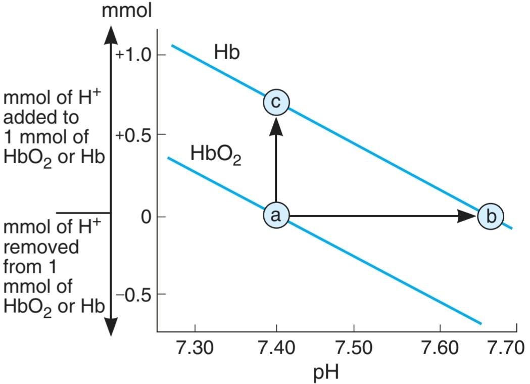
FIGURE 35-6 Comparative titration curves for oxygenated hemoglobin (HbO2) and deoxyhemoglobin (Hb). The arrow from a to c indicates the number of additional millimoles of H+ that Hb can buffer compared with a similar concentration of HbO2 (ie, no shift in pH). The arrow from a to b indicates the pH shift that would occur on deoxygenation of HbO2 without additional H+.
شکل 35-6 منحنی های تیتراسیون مقایسه ای برای هموگلوبین اکسیژن دار (HbO2) و دی اکسی هموگلوبین (Hb). پیکان از a به c نشاندهنده تعداد میلیمولهای اضافی +H است که Hb میتواند در مقایسه با غلظت مشابه HbO2 (یعنی بدون تغییر در pH) بافر کند. پیکان از a به b نشاندهنده تغییر pH است که در اکسیژنزدایی از HbO2 بدون +H اضافی رخ میدهد.
The third and major buffer system in blood is the carbonic acid-bicarbonate system:
سومین و اصلی ترین سیستم بافر خون، سیستم کربنیک اسید-بی کربنات است:

The Henderson-Hasselbalch equation for this system is
معادله هندرسون- هاسلبالخ برای این سیستم است

The pK for this system in an ideal solution is low (about 3), and the amount of H2CO3 is small and hard to measure accurately. However, in the body, H2CO3 is in equilibrium with CO2:
pK برای این سیستم در یک محلول ایده آل کم است (حدود 3) و مقدار H2CO3 کم است و اندازه گیری دقیق آن سخت است. با این حال، در بدن، H2CO3 با CO2 در تعادل است:

If the pK is changed to pK’ (apparent ionization constant; distinguished from the true pK due to less than ideal conditions for the solution) and [CO2] is substituted for [H2CO3], the pK’ is 6.1:
اگر pK به pK’ (ثابت یونیزاسیون ظاهری؛ متمایز از pK واقعی به دلیل شرایط کمتر از ایده آل برای محلول) تغییر یابد و [CO2] جایگزین [H2CO3] شود، pK’ 6.1 است:
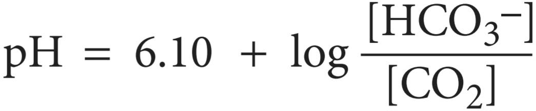
The clinically relevant form of this equation is:
شکل مرتبط بالینی این معادله به صورت زیر است:

since the amount of dissolved CO2 is proportional to the partial pressure of CO2 and the solubility coefficient of CO2 in mmol/L/mm Hg is 0.0301. [HCO3 ̄] cannot be measured directly, but pH and PCO2 can be measured with suitable accuracy with pH and PCO2 glass electrodes, and [HCO3] can then be calculated.
از آنجایی که مقدار CO2 محلول با فشار جزئی CO2 متناسب است و ضریب حلالیت CO2 بر حسب mmol/L/mm Hg 0.0301 است. [HCO3 ̄] را نمی توان مستقیماً اندازه گیری کرد، اما pH و PCO2 را می توان با دقت مناسب با pH و الکترودهای شیشه ای PCO2 اندازه گیری کرد و سپس می توان [HCO3] را محاسبه کرد.
The pK’ of this system is still low relative to the pH of the blood, but the system is one of the most effective buffer systems in the body because the amount of dissolved CO2 is controlled by respiration (ie, it is an “open” system). Additional control of the plasma concentration of HCO3 is provided by the kidneys. When H+ is added to the blood, HCO3 declines as more H2CO3 is formed. If the extra H2CO3 were not converted to CO2 and H2O and the CO2 excreted in the lungs, the H2CO3 concentration would rise. Without CO2 removal to reduce H2CO3, sufficient H* addition that would halve the plasma HCO3 would alter the pH 7.4 to 6.0. However, such a H* concentration increase is tolerated because: (1) extra H2CO3 that is formed is removed and (2) the H* rise stimulates respiration and therefore produces a drop in PCO2, so that some additional H2CO3 is removed. The net pH after such an increase in H* concentration is actually 7.2 or 7.3.
pK’ این سیستم هنوز نسبت به pH خون پایین است، اما این سیستم یکی از موثرترین سیستم های بافر در بدن است زیرا مقدار CO2 محلول توسط تنفس کنترل می شود (یعنی یک سیستم “باز” است). کنترل اضافی غلظت HCO3 پلاسما توسط کلیه ها انجام می شود. هنگامی که +H به خون اضافه می شود، HCO3 با تشکیل H2CO3 بیشتر کاهش می یابد. اگر H2CO3 اضافی به CO2 و H2O تبدیل نمی شد و CO2 در ریه ها دفع می شد، غلظت H2CO3 افزایش می یافت. بدون حذف CO2 برای کاهش H2CO3، افزودن +H کافی که HCO3 پلاسما را نصف میکند، pH 7.4 تا 6.0 را تغییر میدهد. با این حال، چنین افزایش غلظت +H قابل تحمل است زیرا: (1) H2CO3 اضافی که تشکیل می شود حذف می شود و (2) افزایش +H باعث تحریک تنفس می شود و بنابراین باعث کاهش PCO2 می شود، به طوری که مقداری H2CO3 اضافی حذف می شود. pH خالص پس از چنین افزایشی در غلظت +H در واقع 7.2 یا 7.3 است.
There are two additional factors that make the carbonic acid-bicarbonate system such a good biological buffer. First, the reaction CO2 + H2O H2CO3 proceeds slowly in either direction unless the enzyme carbonic anhydrase is present. There is no carbonic anhydrase in plasma, but there is an abundant supply in red blood cells, spatially confining and controlling the reaction. Second, the presence of hemoglobin in the blood increases the buffering of the system by binding free H+ produced by the hydration of CO2 and allowing for movement of the HCO3 into the plasma.
دو عامل اضافی وجود دارد که سیستم کربنیک اسید-بی کربنات را به یک بافر بیولوژیکی خوب تبدیل می کند. اول، واکنش CO2 + H2O H2CO3 به آرامی در هر جهت پیش می رود مگر اینکه آنزیم کربنیک انیدراز وجود داشته باشد. هیچ کربنیک انیدراز در پلاسما وجود ندارد، اما مقدار زیادی در گلبول های قرمز خون وجود دارد که واکنش را از نظر مکانی محدود و کنترل می کند. دوم، حضور هموگلوبین در خون، بافر سیستم را با اتصال +H آزاد تولید شده توسط هیدراتاسیون CO2 افزایش می دهد و اجازه می دهد HCO3 به داخل پلاسما حرکت کند.
ACIDOSIS & ALKALOSIS
The pH of the arterial plasma is normally 7.40 and that of venous plasma slightly lower. A decrease in pH below the norm (acidosis) is technically present whenever the arterial pH is below 7.40 and an increase in pH (alkalosis) is technically present whenever pH is above 7.40. In practice, variations of up to 0.05 pH unit occur without untoward effects. Acid-base disorders are split into four categories: respiratory acidosis, respiratory alkalosis, metabolic acidosis, and metabolic alkalosis. In addition, these disorders can occur in combination. Some causes of acid-base disturbances are shown in Table 35-3.
اسیدوز و قلیایی
pH پلاسمای شریانی معمولاً 7.40 و پلاسمای وریدی کمی کمتر است. کاهش pH کمتر از حد نرمال (اسیدوز) از نظر فنی زمانی که pH شریانی زیر 7.40 باشد و افزایش pH (آلکالوز) از نظر فنی هر زمان که pH بالاتر از 7.40 باشد وجود دارد. در عمل، تغییرات تا 0.05 واحد pH بدون اثرات نامطلوب رخ می دهد. اختلالات اسید-باز به چهار دسته تقسیم می شوند: اسیدوز تنفسی، آلکالوز تنفسی، اسیدوز متابولیک و آلکالوز متابولیک. علاوه بر این، این اختلالات می توانند به صورت ترکیبی ایجاد شوند. برخی از علل اختلالات اسید-باز در جدول 35-3 نشان داده شده است.
TABLE 35-3 Plasma pH, HCO3, and PCO2 values in various typical disturbances of acid-base balance.a
جدول 35-3 مقادیر pH پلاسما، HCO3 و PCO2 در اختلالات معمولی مختلف تعادل اسید-باز.a
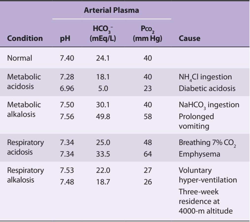
aIn the diabetic acidosis and prolonged vomiting examples, respiratory compensation for primary metabolic acidosis and alkalosis has occurred, and the Pco, has shifted from 40 mm Hg. In the emphysema and high-altitude examples, renal compensation for primary respiratory acidosis and alkalosis has occurred and has made the deviations from normal of the plasma HCO3– larger than they would otherwise be.
aدر نمونه های اسیدوز دیابتی و استفراغ طولانی مدت، جبران تنفسی برای اسیدوز متابولیک اولیه و آلکالوز رخ داده است و PCO از 40 میلی متر جیوه تغییر کرده است. در نمونه های آمفیزم و ارتفاع بالا، جبران کلیوی برای اسیدوز تنفسی اولیه و آلکالوز رخ داده است و انحراف از طبیعی -HCO3 پلاسما را بزرگتر از آنچه در غیر این صورت بود، کرده است.
RESPIRATORY ACIDOSIS
Any short-term rise in arterial Pco2 (ie, above 40 mm Hg, due to hypoventilation) results in respiratory acidosis. Recall that CO2 that is retained is in equilibrium with H2CO3, which in turn is in equilibrium with HCO3. The effective rise in plasma HCO3 means that a new equilibrium is reached at a lower pH. This can be indicated graphically on a plot of plasma HCO3- concentration versus pH (Figure 35-7). The pH change observed at any increase in Pco2 during respiratory acidosis depends on the buffering capacity of the blood. The initial changes shown in Figure 35-7 are those that occur independently of any compensatory mechanism; that is, they are those of uncompensated respiratory acidosis.
اسیدوز تنفسی
هر گونه افزایش کوتاه مدت در Pco2 شریانی (یعنی بالای 40 میلی متر جیوه، به دلیل هیپوونتیلاسیون) منجر به اسیدوز تنفسی می شود. به یاد بیاورید که CO2 که حفظ می شود با H2CO3 در تعادل است که به نوبه خود با HCO3 در تعادل است. افزایش موثر HCO3 پلاسما به این معنی است که در pH پایین تر به تعادل جدیدی رسیده است. این را می توان به صورت گرافیکی بر روی نمودار غلظت -HCO3 پلاسما در مقابل pH نشان داد (شکل 35-7). تغییر pH مشاهده شده در هر افزایش Pco2 در طی اسیدوز تنفسی به ظرفیت بافر خون بستگی دارد. تغییرات اولیه نشان داده شده در شکل 35-7 آنهایی هستند که مستقل از هر مکانیزم جبرانی رخ می دهند. یعنی آنها اسیدوز تنفسی جبران نشده هستند.
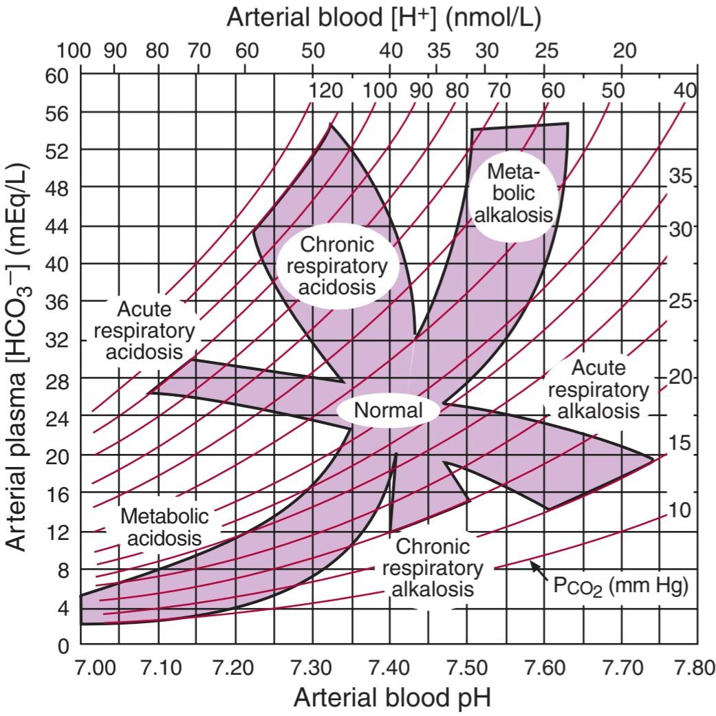
FIGURE 35–7 Acid-base nomogram. Changes in the PCO2 (curved lines), plasma HCO3, and pH (or [H*]) of arterial blood in respiratory and metabolic acidosis are shown. Note the shifts in HCO3 and pH as acute respiratory acidosis and alkalosis are compensated, producing their chronic counterparts. (Reproduced with permission from Brenner BM, Rector FC Jr. [editors]: Brenner & Rector’s The Kidney, 7th ed. St. Louis, MO: Saunders; 2004.)
شکل 35-7 نوموگرام اسید-باز. تغییرات در PCO2 (خطوط منحنی)، HCO3 پلاسما، و pH (یا [+H]) خون شریانی در اسیدوز تنفسی و متابولیک نشان داده شده است. توجه داشته باشید که تغییرات در HCO3 و pH زمانی که اسیدوز و آلکالوز حاد تنفسی جبران میشوند و همتایان مزمن خود را ایجاد میکنند. (تکثیر شده با اجازه Brenner BM, Rector FC Jr. [ویراستاران]: Brenner & Rector’s The Kidney, ویرایش هفتم سنت لوئیس, MO: ساندرز؛ 2004.)
RESPIRATORY ALKALOSIS
Any short-term lowering of PCO2 below what is needed for proper CO2 exchange (ie, below 35 mm Hg, as can occur during hyperventilation) results in respiratory alkalosis. The decreased CO2 shifts the equilibrium of the carbonic acid-bicarbonate system to effectively lower [H+] and increase pH. As in respiratory acidosis, initial pH changes corresponding to respiratory alkalosis (Figure 35-7) are those that occur independently of any compensatory mechanism and are thus uncompensated respiratory alkalosis.
آلکالوز تنفسی
هر گونه کاهش کوتاه مدت PCO2 کمتر از مقدار مورد نیاز برای تبادل CO2 مناسب (یعنی زیر 35 میلی متر جیوه، همانطور که در طول هیپرونتیلاسیون رخ می دهد) منجر به آلکالوز تنفسی می شود. کاهش CO2، تعادل سیستم کربنیک اسید-بی کربنات را به طور موثری کاهش می دهد و pH را افزایش می دهد. مانند اسیدوز تنفسی، تغییرات pH اولیه مربوط به آلکالوز تنفسی (شکل 35-7) آنهایی هستند که مستقل از هر مکانیسم جبرانی رخ می دهند و بنابراین آلکالوز تنفسی جبران نشده هستند.
METABOLIC ACIDOSIS & ALKALOSIS
Blood pH changes can also arise by nonrespiratory mechanism. Metabolic acidosis (or nonrespiratory acidosis) occurs when strong acids are added to blood. If, for example, a large amount of acid is ingested (eg, aspirin overdose), acids in the blood are quickly increased. The H2CO3 that is formed is converted to H2O and CO2, and the CO2 is rapidly excreted via the lungs. This is the situation in uncompensated metabolic acidosis (Figure 35-7). Note that in contrast to respiratory acidosis, metabolic acidosis does not include a change in PcO2; the shift toward metabolic acidosis occurs along an isobar line (Figure 35-8). When the free [H+] level falls as a result of addition of alkali, or more commonly, the removal of large amounts of acid (eg, following vomiting), metabolic alkalosis results. In uncompensated metabolic alkalosis the pH rises along the isobar line (Figures 35-7 and 35-8).
اسیدوز متابولیک و قلیایی
تغییرات pH خون نیز می تواند با مکانیسم غیر تنفسی ایجاد شود. اسیدوز متابولیک (یا اسیدوز غیر تنفسی) زمانی رخ می دهد که اسیدهای قوی به خون اضافه شود. به عنوان مثال، اگر مقدار زیادی اسید مصرف شود (مثلاً مصرف بیش از حد آسپرین)، اسیدهای خون به سرعت افزایش می یابد. H2CO3 تشکیل شده به H2O و CO2 تبدیل می شود و CO2 به سرعت از طریق ریه ها دفع می شود. این وضعیت در اسیدوز متابولیک جبران نشده است (شکل 35-7). توجه داشته باشید که برخلاف اسیدوز تنفسی، اسیدوز متابولیک شامل تغییر در PcO2 نمی شود. تغییر به سمت اسیدوز متابولیک در امتداد یک خط ایزوبار رخ می دهد (شکل 35-8). هنگامی که سطح [+H] آزاد در نتیجه افزودن قلیایی، یا معمولاً حذف مقادیر زیادی اسید (مثلاً به دنبال استفراغ) کاهش می یابد، آلکالوز متابولیک ایجاد می شود. در آلکالوز متابولیک جبران نشده pH در امتداد خط ایزوبار افزایش می یابد (شکل های 35-7 و 35-8).
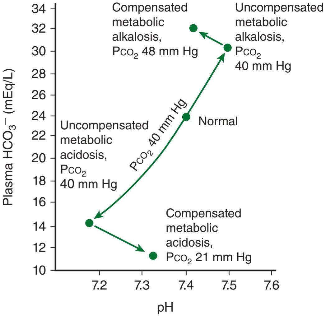
FIGURE 35-8 Acid-base paths during metabolic acidosis. Changes in true plasma pH, HCO3, and PCO2 at rest, during metabolic acidosis and alkalosis, and following respiratory compensation are plotted. Metabolic acidosis or alkalosis causes changes in pH along the Pco, isobar line (middle line). Respiratory compensation moves pH toward normal by altering PCO2 (top and bottom arrows). (Modified with permission from Davenport HW: The ABC of Acid-Base Chemistry, 6th ed. University of Chicago Press; 1974.)
شکل 35-8 مسیرهای اسید-باز در طی اسیدوز متابولیک. تغییرات در pH واقعی پلاسما، HCO3 و PCO2 در حالت استراحت، در طی اسیدوز متابولیک و آلکالوز، و به دنبال جبران تنفسی رسم شده است. اسیدوز متابولیک یا آلکالوز باعث تغییر در pH در امتداد خط Pco، ایزوبار (خط وسط) می شود. جبران تنفسی با تغییر PCO2 (فلش های بالا و پایین) pH را به سمت نرمال می برد. (با اجازه Davenport HW: The ABC of Acid-Base Chemistry، ویرایش ششم انتشارات دانشگاه شیکاگو، 1974 اصلاح شده است.)
RESPIRATORY & RENAL COMPENSATION
Uncompensated acidosis and alkalosis as described above are seldom seen because of compensation systems. The two main compensatory systems are respiratory compensation and renal compensation.
جبران تنفسی و کلیوی
اسیدوز و آلکالوز جبران نشده همانطور که در بالا توضیح داده شد به ندرت به دلیل سیستم های جبران دیده می شوند. دو سیستم اصلی جبرانی عبارتند از جبران تنفسی و جبران کلیه.
The respiratory system compensates for metabolic acidosis or alkalosis by altering ventilation, and consequently, the PCO2, which can directly change blood pH. Respiratory mechanisms are fast. In response to metabolic acidosis, ventilation is increased, resulting in a decrease of Pco2 (eg, from 40 mm Hg to 20 mm Hg) and a subsequent increase in pH toward normal (Figure 35-8). In response to metabolic alkalosis, ventilation is decreased, Pco2 is increased, and a subsequent decrease in pH occurs. Because respiratory compensation is a quick response, the graphical representation in Figure 35-8 overstates the two-step adjustment in blood pH. In actuality, as soon as metabolic acidosis begins, respiratory compensation is invoked and the large shifts in pH depicted do not occur.
سیستم تنفسی اسیدوز متابولیک یا آلکالوز را با تغییر تهویه و در نتیجه PCO2 که می تواند مستقیماً pH خون را تغییر دهد، جبران می کند. مکانیسم های تنفسی سریع هستند. در پاسخ به اسیدوز متابولیک، تهویه افزایش می یابد و در نتیجه Pco2 کاهش می یابد (مثلاً از 40 میلی متر جیوه به 20 میلی متر جیوه) و متعاقباً pH به سمت نرمال افزایش می یابد (شکل 35-8). در پاسخ به آلکالوز متابولیک، تهویه کاهش مییابد، Pco2 افزایش مییابد و متعاقباً pH کاهش مییابد. از آنجایی که جبران تنفسی یک پاسخ سریع است، نمایش گرافیکی در شکل 35-8 تنظیم دو مرحله ای pH خون را بیش از حد نشان می دهد. در واقع، به محض شروع اسیدوز متابولیک، جبران تنفسی فراخوانی می شود و تغییرات زیادی در pH نشان داده شده رخ نمی دهد.
For complete compensation from respiratory or metabolic acidosis/alkalosis, renal compensatory mechanisms are invoked. The kidney responds to acidosis by actively secreting fixed acids while retaining filtered HCO3. In contrast, the kidney responds to alkalosis by decreasing H* secretion and by decreasing the retention of filtered HCO3.
برای جبران کامل اسیدوز/آلکالوز تنفسی یا متابولیک، مکانیسم های جبرانی کلیوی مورد استفاده قرار می گیرد. کلیه با ترشح فعال اسیدهای ثابت در حالی که HCO3 فیلتر شده را حفظ می کند، به اسیدوز پاسخ می دهد. در مقابل، کلیه با کاهش ترشح +H و با کاهش احتباس HCO3 فیلتر شده به آلکالوز پاسخ می دهد.
Renal tubule cells in the kidney have active carbonic anhydrase and thus can produce H+ and HCO3 ̄ from CO2. In response to acidosis, these cells secrete H+ into the tubular fluid in exchange for Na* while the HCO3 is actively reabsorbed into the peritubular capillary; for each H* secreted, one Na* and one HCO3 are added to the blood. The result of this renal compensation for respiratory acidosis is shown graphically in the shift from acute to chronic respiratory acidosis in Figure 35–7. Conversely, in response to alkalosis, the kidney decreases H* secretion and depresses HCO3 reabsorption. The result of this renal compensation for respiratory alkalosis is shown graphically in the shift from acute to chronic respiratory alkalosis in Figure 35-7. Clinical evaluations of acid-base status are discussed in Clinical Box 35-2, and the role of the kidneys in acid-base homeostasis is discussed in more detail in Chapter 38.
سلول های توبول کلیه در کلیه دارای کربنیک انیدراز فعال هستند و بنابراین می توانند +H و ̄ HCO3 از CO2 تولید کنند. در پاسخ به اسیدوز، این سلولها +H را در مایع لولهای در ازای +Na ترشح میکنند در حالی که HCO3 به طور فعال در مویرگ اطراف لولهای بازجذب میشود. به ازای هر +H ترشح شده، یک +Na و یک HCO3 به خون اضافه می شود. نتیجه این جبران کلیوی برای اسیدوز تنفسی به صورت گرافیکی در تغییر اسیدوز تنفسی حاد به مزمن در شکل 35-7 نشان داده شده است. برعکس، در پاسخ به آلکالوز، کلیه ترشح +H را کاهش داده و بازجذب HCO3 را کاهش می دهد. نتیجه این جبران کلیوی برای آلکالوز تنفسی به صورت گرافیکی در تغییر آلکالوز تنفسی حاد به مزمن در شکل 35-7 نشان داده شده است. ارزیابیهای بالینی وضعیت اسید-باز در جعبه بالینی 35-2 مورد بحث قرار گرفته است و نقش کلیهها در هموستاز اسید-باز با جزئیات بیشتر در فصل 38 مورد بحث قرار گرفته است.
CLINICAL BOX 35-2
Clinical Evaluation of Acid-Base Status
In evaluating disturbances of acid-base balance, it is important to know the pH and HCO3 content of arterial plasma. Reliable pH determinations can be made with a pH meter and a glass pH electrode. Using pH and a direct measurement of the PCO2 with a CO2 electrode, HCO3 concentration can be calculated. The Pco2 is ~8 mm Hg higher and the pH 0.03-0.04 unit lower in venous than arterial plasma because venous blood contains the CO2 being carried from the tissues to the lungs. Therefore, the calculated HCO3 concentration is about 2 mmol/L higher. However, if this is kept in mind, free-flowing venous blood can be substituted for arterial blood in most clinical situations.
جعبه بالینی 35-2
ارزیابی بالینی وضعیت اسید و باز
در ارزیابی اختلالات تعادل اسید و باز، دانستن pH و محتوای HCO3 پلاسمای شریانی مهم است. تعیین pH قابل اعتماد را می توان با یک pH متر و یک الکترود pH شیشه ای انجام داد. با استفاده از pH و اندازه گیری مستقیم PCO2 با الکترود CO2، غلظت HCO3 را می توان محاسبه کرد. Pco2 8 میلی متر جیوه بیشتر و pH 0.03-0.04 واحد وریدی نسبت به پلاسمای شریانی کمتر است زیرا خون وریدی حاوی CO2 است که از بافت ها به ریه ها منتقل می شود. بنابراین، غلظت HCO3 محاسبه شده حدود 2 میلی مول در لیتر بیشتر است. با این حال، اگر این را در نظر داشته باشید، خون وریدی با جریان آزاد می تواند جایگزین خون شریانی در اکثر موقعیت های بالینی شود.
A measurement that is of some value in the differential diagnosis of metabolic acidosis is the anion gap. This gap, which is something of a misnomer, refers to the difference between the concentration of cations other than Na* and the concentration of anions other than Cl ̄ and HCO3 in the plasma. It consists for the most part of proteins in anionic form, HPO2, SO42, and organic acids; a normal value is about 12 mEq/L. It is increased when the plasma concentration of K+, Ca2+, or Mg+ is decreased; when the concentration of (or the charge on) plasma proteins is increased; or when organic anions such as lactate or foreign anions accumulate in blood. It is decreased when cations are increased or when plasma albumin is decreased. The anion gap is increased in metabolic acidosis due to ketoacidosis, lactic acidosis, and other forms of acidosis in which organic anions are increased.
اندازه گیری که در تشخیص افتراقی اسیدوز متابولیک ارزش دارد، شکاف آنیونی است. این شکاف، که یک نام اشتباه است، به تفاوت بین غلظت کاتیون های غیر از +Na و غلظت آنیون های غیر از Cl ̄ و HCO3 در پلاسما اشاره دارد. بیشتر از پروتئین های آنیونی، HPO2، SO42 و اسیدهای آلی تشکیل شده است. مقدار نرمال حدود 12 میلی اکیوالان در لیتر است. هنگامی که غلظت پلاسمایی +K+، Ca2 یا +Mg کاهش می یابد، افزایش می یابد. هنگامی که غلظت (یا شارژ روی) پروتئین های پلاسما افزایش می یابد. یا زمانی که آنیون های آلی مانند لاکتات یا آنیون های خارجی در خون جمع می شوند. با افزایش کاتیون ها یا کاهش آلبومین پلاسما کاهش می یابد. شکاف آنیون در اسیدوز متابولیک به دلیل کتواسیدوز، اسیدوز لاکتیک و سایر اشکال اسیدوز افزایش می یابد که در آن آنیون های آلی افزایش می یابد.
HYPOXIA
Hypoxia is O2 deficiency at the tissue level. It is a more correct term than anoxia (lack of O2), since there is rarely no O2 at all left in the tissues.
هیپوکسی
هیپوکسی کمبود O2 در سطح بافت است. این اصطلاح صحیح تر از آنوکسی (کمبود O2) است، زیرا به ندرت هیچ O2 در بافت ها باقی نمی ماند.
Numerous classifications for hypoxia have been used, but the more traditional four-type system still has considerable utility if the definitions of the terms are kept clearly in mind. The four categories are (1) hypoxemia (sometimes termed hypoxic hypoxia), in which the PO2 of the arterial blood is reduced; (2) anemic hypoxia, in which the arterial Po2 is normal but the amount of hemoglobin available to carry O2 is reduced; (3) ischemic or stagnant hypoxia, in which the blood flow to a tissue is so low that adequate O2 is not delivered to it despite a normal Po2 and hemoglobin concentration; and (4) histotoxic hypoxia, in which the amount of O2 delivered to a tissue is adequate but, because of the action of a toxic agent, the tissue cells cannot make use of the O2 supplied to them. Some specific effects of hypoxia on cells and tissues are discussed in Clinical Box 35- 3.
طبقهبندیهای متعددی برای هیپوکسی استفاده شده است، اما اگر تعاریف اصطلاحات به وضوح در نظر گرفته شود، سیستم چهار نوع سنتیتر هنوز کاربرد قابلتوجهی دارد. این چهار دسته عبارتند از (1) هیپوکسمی (گاهی اوقات هیپوکسی هیپوکسیک نامیده می شود)، که در آن PO2 خون شریانی کاهش می یابد. (2) هیپوکسی کم خون، که در آن Po2 شریانی طبیعی است اما مقدار هموگلوبین موجود برای حمل O2 کاهش می یابد. (3) هیپوکسی ایسکمیک یا راکد، که در آن جریان خون به بافت آنقدر کم است که علیرغم غلظت طبیعی Po2 و هموگلوبین، O2 کافی به آن منتقل نمی شود. و (4) هیپوکسی هیستوتوکسیک، که در آن مقدار O2 تحویلی به بافت کافی است، اما به دلیل عملکرد یک عامل سمی، سلول های بافت نمی توانند از O2 عرضه شده به آنها استفاده کنند. برخی از اثرات خاص هیپوکسی بر سلول ها و بافت ها در جعبه بالینی 35-3 مورد بحث قرار گرفته است.
CLINICAL BOX 35-3
Effects of Hypoxia on Cells and Selected Tissues
Effects on Cells
Hypoxia causes the production of transcription factors (hypoxia-inducible factors; HIFs). These are made up of a and ẞ subunits. In normally oxygenated tissues, the a subunits are rapidly ubiquitinated and destroyed. However, in hypoxic cells, the a subunits dimerize with ẞ subunits, and the dimers activate genes that produce several proteins including angiogenic factors and erythropoietin, among others.
جعبه بالینی 35-3
اثرات هیپوکسی بر سلول ها و بافت های منتخب
اثرات بر سلول ها
هیپوکسی باعث تولید فاکتورهای رونویسی می شود (عوامل القاء کننده هیپوکسی؛ HIFs). اینها از زیر واحدهای a و ẞ ساخته شده اند. در بافتهای دارای اکسیژن طبیعی، زیرواحدهای a به سرعت یوبیکوئیتین شده و از بین میروند. با این حال، در سلولهای هیپوکسیک، زیرواحدهای a با زیر واحدهای ẞ دیمر میشوند و دایمرها ژنهایی را فعال میکنند که چندین پروتئین از جمله فاکتورهای رگزایی و اریتروپویتین را تولید میکنند.
Effects on the Brain
In hypoxymia and the other generalized forms of hypoxia, the brain is affected first. A sudden drop in the inspired Po2 to less than 20 mm Hg, which occurs, for example, when cabin pressure is suddenly lost in a plane flying above 16,000 m, causes loss of consciousness in 10-20 s and death in 4-5 min. Less severe hypoxia causes a variety of mental aberrations not unlike those produced by alcohol: impaired judgment, drowsiness, dulled pain sensibility, excitement, disorientation, loss of time sense, and headache. Other symptoms include anorexia, nausea, vomiting, tachycardia, and, when the hypoxia is severe, hypertension. The rate of ventilation is increased in proportion to the severity of the hypoxia of the carotid chemoreceptor cells.
اثرات بر روی مغز
در هیپوکسی و سایر اشکال عمومی هیپوکسی، ابتدا مغز تحت تأثیر قرار می گیرد. افت ناگهانی Po2 الهامشده به کمتر از 20 میلیمتر جیوه، که برای مثال، زمانی که فشار کابین به طور ناگهانی در هواپیمای بالای 16000 متر از بین میرود، رخ میدهد، باعث از دست دادن هوشیاری در 10-20 ثانیه و مرگ در 4-5 دقیقه میشود. هیپوکسی کمتر شدید باعث انواع انحرافات ذهنی می شود که بی شباهت به آنهایی است که توسط الکل ایجاد می شود: اختلال در قضاوت، خواب آلودگی، حساسیت درد مبهم، هیجان، بی نظمی، از دست دادن حس زمان و سردرد. علائم دیگر عبارتند از بی اشتهایی، حالت تهوع، استفراغ، تاکی کاردی و در صورت شدید بودن هیپوکسی، فشار خون بالا. سرعت تهویه متناسب با شدت هیپوکسی سلول های گیرنده شیمیایی کاروتید افزایش می یابد.
Respiratory Stimulation
Dyspnea is by definition difficult or labored breathing in which the person is conscious of shortness of breath; hyperpnea is the general term for an increase in the rate or depth of breathing regardless of the patient’s subjective sensations. Tachypnea is rapid, shallow breathing. In general, a normal individual is not conscious of respiration until ventilation is doubled, and breathing is not uncomfortable until ventilation is tripled or quadrupled. Whether or not a given level of ventilation is uncomfortable also appears to depend on a variety of other factors. Hypercapnia and, to a lesser extent, hypoxia cause dyspnea. An additional factor is the effort involved in moving the air in and out of the lungs (the work of breathing).
تحریک تنفسی
تنگی نفس طبق تعریف، تنفس دشوار یا سخت است که در آن فرد از تنگی نفس آگاه است. هیپرپنه اصطلاح کلی برای افزایش سرعت یا عمق تنفس بدون توجه به احساسات ذهنی بیمار است. تاکی پنه تنفس سریع و کم عمق است. به طور کلی، یک فرد عادی تا زمانی که تهویه دو برابر نشود، از تنفس آگاه نیست و تا زمانی که تهویه سه یا چهار برابر نشود، تنفس ناراحت کننده نیست. به نظر می رسد که آیا سطح مشخصی از تهویه ناخوشایند است یا نه، به عوامل مختلفی بستگی دارد. هایپرکاپنی و به میزان کمتر هیپوکسی باعث تنگی نفس می شود. یک عامل اضافی، تلاشی است که در حرکت هوا به داخل و خارج از ریه ها انجام می شود (کار تنفس).
HYPOXEMIA
By definition, hypoxemia is a condition of reduced arterial Po2. Hypoxemia is a problem in normal individuals at high altitudes and is a complication of pneumonia and a variety of other diseases of the respiratory system.
هیپوکسمی
طبق تعریف، هیپوکسمی یک وضعیت کاهش Po2 شریانی است. هیپوکسمی مشکلی در افراد عادی در ارتفاعات است و از عوارض ذات الریه و انواع بیماری های دیگر دستگاه تنفسی است.
EFFECTS OF DECREASED BAROMETRIC PRESSURE
The composition of air stays the same, but the total barometric pressure falls with increasing altitude (Figure 35-9), and thus, the Po2 also falls. At 3000 m (~10,000 ft) above sea level, the alveolar Po2 is about 60 mm Hg and there is enough hypoxic stimulation of the chemoreceptors under normal breathing to cause increased ventilation. As one ascends higher, the alveolar Po2 falls less rapidly and the alveolar Pco2 declines because of the hyperventilation. The resulting fall in arterial PCO2 produces respiratory alkalosis. A number of compensatory mechanisms operate over a period of time to increase altitude tolerance (acclimatization), but in unacclimatized persons, mental symptoms such as irritability appear at about 3700 m. At 5500 m, the hypoxic symptoms are severe; and at altitudes above 6100 m (20,000 ft), consciousness is usually lost.
اثرات کاهش فشار بارومتریک
ترکیب هوا ثابت میماند، اما فشار بارومتری کل با افزایش ارتفاع کاهش مییابد (شکل 35-9)، و بنابراین، Po2 نیز سقوط میکند. در ارتفاع 3000 متری (~ 10000 فوت) بالاتر از سطح دریا، Po2 آلوئولی حدود 60 میلی متر جیوه است و به اندازه کافی تحریک هیپوکسیک گیرنده های شیمیایی در تنفس طبیعی وجود دارد که باعث افزایش تهویه می شود. با بالا رفتن بالاتر، Po2 آلوئولی با سرعت کمتری سقوط می کند و Pco2 آلوئولی به دلیل هیپرونتیلاسیون کاهش می یابد. کاهش PCO2 شریانی باعث ایجاد آلکالوز تنفسی می شود. تعدادی از مکانیسم های جبرانی در یک دوره زمانی برای افزایش تحمل ارتفاع (آقلیم سازی) عمل می کنند، اما در افراد غیر سازگار، علائم ذهنی مانند تحریک پذیری در حدود 3700 متر ظاهر می شود. در ارتفاع 5500 متری، علائم هیپوکسی شدید است. و در ارتفاعات بالاتر از 6100 متر (20000 فوت) معمولاً هوشیاری از دست می رود.
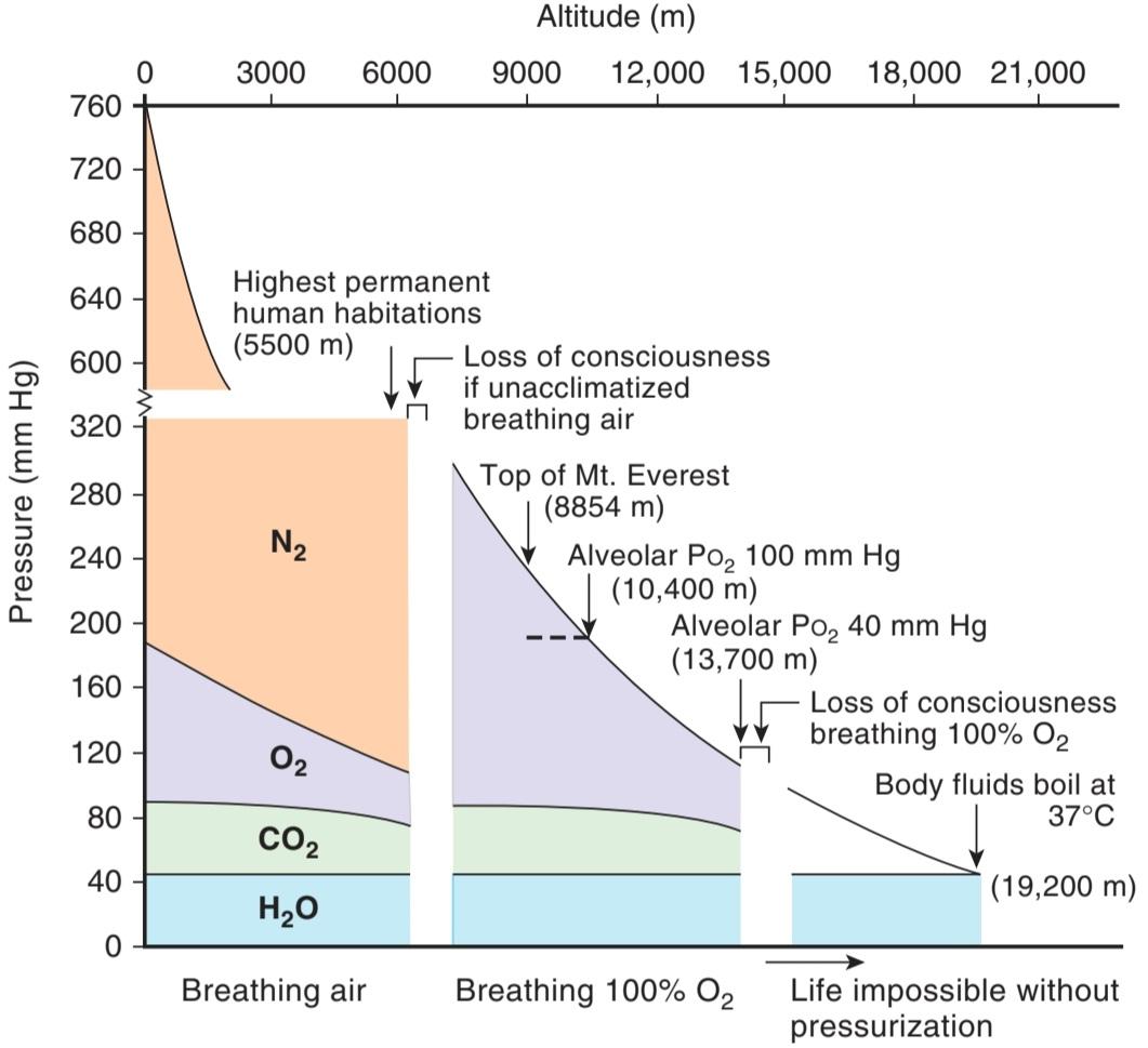
FIGURE 35-9 Composition of alveolar air in individuals breathing air (0- 6100 m) and 100% O1⁄2 (6100-13,700 m). The minimal alveolar Po2 that an unacclimatized person can tolerate without loss of consciousness is about 35-40 mm Hg. Note that with increasing altitude, the alveolar PCO2 drops because of the hyperventilation due to hypoxic stimulation of the carotid and aortic chemoreceptors. The fall in barometric pressure with increasing altitude is not linear because air is compressible.
شکل 35-9 ترکیب هوای آلوئولی در افراد تنفس کننده هوا (0-6100 متر) و 100% O1⁄2 (6100-13700 متر). حداقل PO2 آلوئولی که یک فرد سازگار نشده می تواند بدون از دست دادن هوشیاری تحمل کند حدود 35-40 میلی متر جیوه است. توجه داشته باشید که با افزایش ارتفاع، PCO2 آلوئولی به دلیل تهویه بیش از حد به دلیل تحریک هیپوکسیک گیرنده های شیمیایی کاروتید و آئورت کاهش می یابد. کاهش فشار هوا با افزایش ارتفاع خطی نیست زیرا هوا قابل تراکم است.
HYPOXIC SYMPTOMS & BREATHING OXYGEN
Some of the effects of high altitude can be offset by breathing 100% O2. Under these conditions, the total atmospheric pressure becomes the limiting factor in altitude tolerance.
علائم هیپوکسیک و اکسیژن تنفسی
برخی از اثرات ارتفاع بالا را می توان با تنفس 100% O2 جبران کرد. تحت این شرایط، فشار کل اتمسفر عامل محدود کننده در تحمل ارتفاع می شود.
The partial pressure of water vapor in the alveolar air is constant at 47 mm Hg, and that of CO2 is normally 40 mm Hg, so that the lowest barometric pressure at which a normal alveolar Po2 of 100 mm Hg is possible is 187 mm Hg, the pressure at about 10,400 m (34,000 ft). At greater altitudes, the increased ventilation due to the decline in alveolar Po2 lowers the alveolar PCO2 somewhat, but the maximum alveolar Po2 that can be attained when breathing 100% O2 at the ambient barometric pressure of 100 mm Hg at 13,700 m is ~40 mm Hg. At ~14,000 m, consciousness is lost in spite of the administration of 100% O2. At 19,200 m, the barometric pressure is 47 mm Hg, and at or below this pressure the body fluids boil at body temperature. The point is largely academic, however, because any individual exposed to such a low pressure would be dead of hypoxia before the bubbles of body fluids could cause death.
فشار جزئی بخار آب در هوای آلوئولی در 47 میلیمتر جیوه ثابت است و فشار CO2 معمولاً 40 میلیمتر جیوه است، به طوری که کمترین فشار هوا که در آن یک Po2 آلوئولی طبیعی 100 میلیمتر جیوه امکانپذیر است، 187 میلیمتر جیوه و فشار در حدود 10400 متر (34،00) است. در ارتفاعات بیشتر، افزایش تهویه به دلیل کاهش Po2 آلوئولی، PCO2 آلوئولی را تا حدودی کاهش میدهد، اما حداکثر Po2 آلوئولی که میتوان با تنفس 100% O2 در فشار هوای محیط 100 میلیمتر جیوه در 13700 متر به دست آورد، 40 میلیمتر جیوه است. در ارتفاع 14000 متری، علیرغم تجویز 100% O2، هوشیاری از دست میرود. در 19200 متر، فشار هوا 47 میلی متر جیوه است و در این فشار یا کمتر از آن، مایعات بدن در دمای بدن می جوشند. با این حال، موضوع تا حد زیادی آکادمیک است، زیرا هر فردی که در معرض چنین فشار پایینی قرار میگیرد، قبل از اینکه حبابهای مایعات بدن باعث مرگ شوند، به دلیل هیپوکسی مرده خواهند بود.
Of course, an artificial atmosphere can be created around an individual; in a pressurized suit or cabin supplied with O2 and a system to remove CO2, it is possible to ascend to any altitude and to live in the vacuum of interplanetary space. Some delayed effects of high altitude are discussed in Clinical Box 35-4.
البته می توان فضایی مصنوعی در اطراف یک فرد ایجاد کرد. در یک لباس یا کابین تحت فشار مجهز به O2 و سیستم حذف CO2، می توان به هر ارتفاعی صعود کرد و در خلاء فضای بین سیاره ای زندگی کرد. برخی از اثرات تاخیری ارتفاع بالا در جعبه بالینی 35-4 مورد بحث قرار گرفته است.
CLINICAL BOX 35-4
Delayed Effects of High Altitude
When they first arrive at a high altitude, many individuals develop acute “mountain sickness.” This syndrome develops 8-24 h after arrival at altitude and lasts 4-8 days. It is characterized by headache, irritability, insomnia, breathlessness, and nausea and vomiting. Its cause is unsettled, but it appears to be associated with cerebral edema. The low Po2 at high altitude causes systemic arteriolar dilation, and if cerebral autoregulation does not compensate, there is an increase in capillary pressure that favors increased transudation of fluid into brain tissue.
جعبه بالینی 35-4
اثرات تاخیری ارتفاع بالا
هنگامی که آنها برای اولین بار به ارتفاعات می رسند، بسیاری از افراد به “بیماری کوهستانی” حاد مبتلا می شوند. این سندرم 8-24 ساعت پس از رسیدن به ارتفاع ایجاد می شود و 4-8 روز طول می کشد. با سردرد، تحریک پذیری، بی خوابی، تنگی نفس و حالت تهوع و استفراغ مشخص می شود. علت آن مشخص نیست، اما به نظر می رسد که با ادم مغزی مرتبط باشد. PO2 کم در ارتفاع بالا باعث اتساع سیستمیک شریانی می شود و اگر خودتنظیمی مغزی جبران نشود، فشار مویرگی افزایش می یابد که به افزایش انتقال مایع به بافت مغز کمک می کند.
Two more serious syndromes that are associated with high-altitude illness: high-altitude cerebral edema and high-altitude pulmonary edema. In high-altitude cerebral edema, the capillary leakage in mountain sickness progresses to frank brain swelling, with ataxia, disorientation, and in some cases coma and death due to herniation of the brain through the tentorium. High-altitude pulmonary edema is a patchy edema of the lungs that is related to the marked pulmonary hypertension that develops at high altitude. It has been argued that it occurs because not all pulmonary arteries have enough smooth muscle to constrict in response to hypoxia, and in the capillaries supplied by those arteries, the general rise in pulmonary arterial pressure causes a capillary pressure increase that disrupts their walls (stress failure).
دو سندرم جدی تر که با بیماری در ارتفاعات مرتبط هستند: ادم مغزی در ارتفاع بالا و ادم ریوی در ارتفاع بالا. در ادم مغزی در ارتفاعات بالا، نشت مویرگی در بیماری کوهستان به تورم آشکار مغز، همراه با آتاکسی، بیحسی و در برخی موارد کما و مرگ به دلیل فتق مغز از طریق تنتوریوم پیشرفت میکند. ادم ریوی در ارتفاع بالا یک ادم تکه تکه ریه است که مربوط به فشار خون ریوی مشخص است که در ارتفاع بالا ایجاد می شود. استدلال شده است که به این دلیل رخ می دهد که همه شریان های ریوی دارای عضلات صاف کافی نیستند تا در پاسخ به هیپوکسی منقبض شوند، و در مویرگ های تامین شده توسط آن شریان ها، افزایش عمومی فشار شریان ریوی باعث افزایش فشار مویرگی می شود که دیواره آنها را مختل می کند (نارسایی استرس).
THERAPEUTIC HIGHLIGHTS
All forms of high-altitude illness are benefited by descent to lower altitude and by treatment with the diuretic acetazolamide. This drug inhibits carbonic anhydrase and results in stimulated respiration, increased PaCO2, and reduced formation of CSF. When cerebral edema is marked, large doses of glucocorticoids are often administered as well. Their mechanism of action is unsettled. In high-altitude pulmonary edema, prompt treatment with O2 is essential—and, if available, use of a hyperbaric chamber. Portable hyperbaric chambers are now available in a number of mountain areas. Nifedipine, a Ca2+ channel blocker that lowers pulmonary artery pressure, can also be useful.
نکات برجسته درمانی
همه اشکال بیماری در ارتفاعات با فرود به ارتفاعات پایین تر و درمان با استازولامید دیورتیک سود می برند. این دارو کربنیک انیدراز را مهار کرده و باعث تحریک تنفس، افزایش PaCO2 و کاهش تشکیل CSF می شود. هنگامی که ادم مغزی مشخص می شود، اغلب دوزهای زیادی از گلوکوکورتیکوئیدها نیز تجویز می شود. مکانیسم عمل آنها نابسامان است. در ادم ریوی در ارتفاعات، درمان سریع با O2 ضروری است – و در صورت وجود، استفاده از یک اتاقک هایپرباریک. اتاقک های پرتابل هایپرباریک در حال حاضر در تعدادی از مناطق کوهستانی موجود است. نیفدیپین، یک مسدود کننده کانال +Ca2 که فشار شریان ریوی را کاهش می دهد، نیز می تواند مفید باشد.
ACCLIMATIZATION
Acclimatization to altitude is due to the operation of a variety of compensatory mechanisms. The respiratory alkalosis produced by the hyperventilation shifts the oxygen-hemoglobin dissociation curve to the left, but a concomitant increase in red blood cell 2,3-DPG tends to decrease the O2 affinity of hemoglobin. The net effect is a small increase in P50. The decrease in O2 affinity makes more O2 available to the tissues. However, the value of the increase in P50 is limited because when the arterial Po2 is markedly reduced, the decreased O2 affinity also interferes with O2 uptake by hemoglobin in the lungs.
سازگاری
سازگاری با ارتفاع به دلیل عملکرد انواع مکانیسم های جبرانی است. آلکالوز تنفسی ایجاد شده توسط هیپرونتیلاسیون منحنی تفکیک اکسیژن-هموگلوبین را به سمت چپ تغییر می دهد، اما افزایش همزمان گلبول قرمز 2،3-DPG تمایل به کاهش میل ترکیبی O2 هموگلوبین دارد. اثر خالص افزایش اندکی در P50 است. کاهش میل ترکیبی O2 باعث می شود O2 بیشتری در دسترس بافت ها قرار گیرد. با این حال، مقدار افزایش P50 محدود است زیرا زمانی که Po2 شریانی به طور قابل توجهی کاهش می یابد، کاهش میل O2 نیز با جذب O2 توسط هموگلوبین در ریه ها تداخل می کند.
The initial ventilatory response to increased altitude is relatively small, because the alkalosis tends to counteract the stimulating effect of hypoxia. However, ventilation steadily increases over the next 4 days (Figure 35-10) because the active transport of H+ into cerebrospinal fluid (CSF), or possibly a developing lactic acidosis in the brain, causes a fall in CSF pH that increases the response to hypoxia. After 4 days, the ventilatory response begins to decline slowly, but it takes years of residence at higher altitudes for it to decline to the initial level, if it is reached at all.
پاسخ تهویه اولیه به افزایش ارتفاع نسبتاً کوچک است، زیرا آلکالوز تمایل دارد با اثر تحریک کننده هیپوکسی مقابله کند. با این حال، تهویه به طور پیوسته در طی 4 روز آینده افزایش می یابد (شکل 35-10) زیرا انتقال فعال +H به مایع مغزی نخاعی (CSF)، یا احتمالاً یک اسیدوز لاکتیک در حال توسعه در مغز، باعث کاهش pH CSF می شود که پاسخ به هیپوکسی را افزایش می دهد. پس از 4 روز، پاسخ تهویه به آرامی شروع به کاهش می کند، اما سالها اقامت در ارتفاعات بالاتر طول می کشد تا به سطح اولیه کاهش یابد، اگر اصلاً به آن برسد.
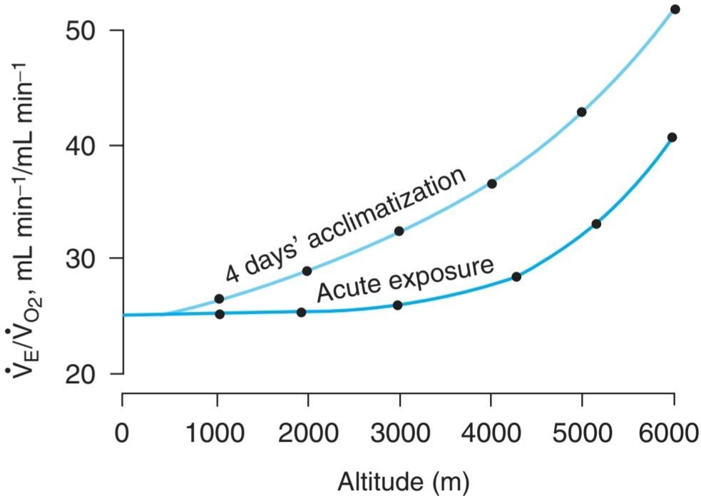
FIGURE 35-10 Effect of acclimatization on the ventilatory response at various altitudes. VE/VO, is the ventilatory equivalent, the ratio of expired minute volume (VE) to the O2 consumption (Vo). (Reproduced with permission from Lenfant C, Sullivan K: Adaptation to high altitude. N Engl J Med 1971; June 10; 284(23):1298-1309.)
شکل 35-10 اثر سازگاری بر پاسخ تهویه در ارتفاعات مختلف. VE/VO، معادل تهویه، نسبت حجم دقیقه منقضی شده (VE) به مصرف O2 (Vo) است. (تکثیر شده با اجازه از Lenfant C، Sullivan K: Adaptation to high altitude. N Engl J Med 1971؛ 10 ژوئن؛ 284(23):1298-1309.)
Erythropoietin secretion increases promptly on ascent to high altitude and then falls somewhat over the following 4 days as the ventilatory response increases and the arterial Po2 rises. The increase in circulating red blood cells triggered by the erythropoietin begins in 2-3 days and is sustained as long as the individual remains at high altitude. Compensatory changes also occur in the tissues. The mitochondria, which are the site of oxidative reactions, increase in number, and myoglobin increases, which facilitates the movement of O2 into the tissues. The tissue content of cytochrome oxidase also increases.
ترشح اریتروپویتین در صعود به ارتفاع بالا به سرعت افزایش می یابد و سپس در طی 4 روز بعد با افزایش پاسخ تهویه و افزایش Po2 شریانی تا حدودی کاهش می یابد. افزایش گلبول های قرمز در گردش که توسط اریتروپویتین ایجاد می شود در 2-3 روز شروع می شود و تا زمانی که فرد در ارتفاع بالا باقی می ماند ادامه دارد. تغییرات جبرانی نیز در بافت ها رخ می دهد. میتوکندری ها که محل واکنش های اکسیداتیو هستند، تعدادشان افزایش می یابد و میوگلوبین افزایش می یابد که باعث تسهیل حرکت O2 به داخل بافت ها می شود. محتوای بافت سیتوکروم اکسیداز نیز افزایش می یابد.
The effectiveness of the acclimatization process is indicated by the fact that permanent human habitations exist in the Andes and Himalayas at elevations above 5500 m (18,000 ft). The natives who live in these villages are barrel- chested and markedly polycythemic. They have low alveolar Po2 values, but in most other ways they are remarkably normal.
اثربخشی فرآیند سازگاری با این واقعیت نشان می دهد که سکونتگاه های دائمی انسان در آند و هیمالیا در ارتفاعات بالای 5500 متر (18000 فوت) وجود دارد. بومیهایی که در این روستاها زندگی میکنند، سینهشان بشکهای هستند و بهطور مشخصی چند سلولی هستند. آنها مقادیر Po2 آلوئولی پایینی دارند، اما از بسیاری جهات دیگر به طور قابل ملاحظه ای طبیعی هستند.
DISEASES CAUSING HYPOXEMIA
Hypoxemia is the most common form of hypoxia seen clinically. The diseases that cause it can be roughly divided into those in which the gas exchange apparatus fails, those such as congenital heart disease in which large amounts of blood are shunted from the venous to the arterial side of the circulation, and those in which the respiratory pump fails. Lung failure occurs when conditions such as pulmonary fibrosis produce alveolar-capillary block, or there is ventilation-perfusion imbalance. Pump failure can be due to fatigue of the respiratory muscles in conditions in which the work of breathing is increased or to a variety of mechanical defects such as pneumothorax or bronchial obstruction that limit ventilation. It can also be caused by abnormalities of the neural mechanisms that control ventilation, such as depression of the respiratory neurons in the medulla by morphine and other drugs. Some specific causes of hypoxemia are discussed in the following text.
بیماری هایی که باعث هیپوکسمی می شوند
هیپوکسمی شایع ترین شکل هیپوکسی است که به صورت بالینی دیده می شود. بیماریهایی که باعث آن میشوند را میتوان به طور کلی به بیماریهایی که در آن دستگاه تبادل گاز از کار میافتد، بیماریهایی مانند بیماری مادرزادی قلبی که در آن مقادیر زیادی خون از وریدی به سمت شریانی گردش خون منتقل میشود، و بیماریهایی که در آن پمپ تنفسی از کار میافتد، تقسیم میشوند. نارسایی ریه زمانی اتفاق می افتد که شرایطی مانند فیبروز ریوی بلوک آلوئولی-مویرگی ایجاد کند یا عدم تعادل تهویه-پرفیوژن وجود داشته باشد. خرابی پمپ می تواند به دلیل خستگی ماهیچه های تنفسی در شرایطی باشد که کار تنفس افزایش می یابد یا به دلیل انواع نقص های مکانیکی مانند پنوموتوراکس یا انسداد برونش که تهویه را محدود می کند. همچنین میتواند ناشی از ناهنجاریهای مکانیسمهای عصبی کنترل کننده تهویه باشد، مانند فرورفتگی نورونهای تنفسی در مدولا توسط مورفین و سایر داروها. برخی از علل خاص هیپوکسمی در متن زیر مورد بحث قرار گرفته است.
VENOUS-TO-ARTERIAL SHUNTS
When a cardiovascular abnormality such as an interatrial septal defect permits large amounts of unoxygenated venous blood to bypass the pulmonary capillaries and dilute the oxygenated blood in the systemic arteries (“right-to-left shunt”), chronic hypoxemia and cyanosis (cyanotic congenital heart disease) result. Administration of 100% O2 raises the O2 content of alveolar air but has little effect on hypoxia due to venous-to-arterial shunts. This is because the deoxygenated venous blood does not have the opportunity to get to the lung to be oxygenated.
شانت های وریدی به شریانی
هنگامی که یک ناهنجاری قلبی عروقی مانند نقص سپتوم بین دهلیزی به مقادیر زیادی خون وریدی بدون اکسیژن اجازه می دهد که مویرگ های ریوی را دور بزند و خون اکسیژن دار در شریان های سیستمیک را رقیق کند (“شانت راست به چپ”)، هیپوکسمی مزمن و سیانوز (بیماری مادرزادی سیانوتیک) نتیجه می شود. تجویز 100% O2 باعث افزایش محتوای O2 هوای آلوئولی می شود اما تأثیر کمی بر هیپوکسی ناشی از شانت وریدی به شریانی دارد. این به این دلیل است که خون وریدی بدون اکسیژن فرصتی برای رسیدن به ریه برای اکسیژن رسانی ندارد.
VENTILATION-PERFUSION IMBALANCE
Patchy ventilation-perfusion imbalance is by far the most common cause of hypoxemia in clinical situations. In disease processes that prevent ventilation of some of the alveoli, the ventilation-blood flow ratios in different parts of the lung determine the extent to which systemic arterial Po2 declines. If nonventilated alveoli are perfused, the nonventilated but perfused portion of the lung is in effect a right-to-left shunt, dumping unoxygenated blood into the left side of the heart. Lesser degrees of ventilation-perfusion imbalance are more common. In the example illustrated in Figure 35-11, the balanced ventilation- perfusion example on the left illustrates a uniform distribution throughout gas exchange. However, when ventilation is not in balance with perfusion, O2 exchange is compromised. Note that the underventilated alveoli (B) have a low alveolar Po2, whereas the overventilated alveoli (A) have a high alveolar Po2 while both have the same blood flow. The unsaturation of the hemoglobin of the blood coming from B is not completely compensated by the slightly greater saturation of the blood coming from A, because hemoglobin is normally nearly saturated in the lungs and the higher alveolar Po2 adds only a little more O2 to the hemoglobin than it normally carries. Consequently, the arterial blood is unsaturated. The CO2 content of the arterial blood is generally normal in such situations, since extra loss of CO2 in overventilated regions can balance diminished loss in underventilated areas.
عدم تعادل تهویه-پرفیوژن
عدم تعادل تهویه-پرفیوژن تکه تکه شایع ترین علت هیپوکسمی در موقعیت های بالینی است. در فرآیندهای بیماری که از تهویه برخی از آلوئول ها جلوگیری می کند، نسبت تهویه به جریان خون در قسمت های مختلف ریه میزان کاهش Po2 شریان سیستمیک را تعیین می کند. اگر آلوئول های بدون تهویه پرفیوژن شوند، قسمت بدون تهویه اما پرفیوژن ریه در واقع یک شنت راست به چپ است که خون بدون اکسیژن را به سمت چپ قلب می ریزد. درجات کمتری از عدم تعادل تهویه-پرفیوژن شایع تر است. در مثال نشان داده شده در شکل 35-11، مثال متعادل تهویه- پرفیوژن در سمت چپ، توزیع یکنواخت در سراسر تبادل گاز را نشان می دهد. با این حال، زمانی که تهویه با پرفیوژن در تعادل نباشد، تبادل O2 به خطر می افتد. توجه داشته باشید که آلوئول های بدون تهویه (B) دارای Po2 آلوئولی کم هستند، در حالی که آلوئول های دارای تهویه بیش از حد (A) دارای Po2 آلوئولی بالا هستند در حالی که هر دو دارای جریان خون یکسان هستند. اشباع نشدن هموگلوبین خون از B به طور کامل با اشباع کمی بیشتر خون حاصل از A جبران نمی شود، زیرا هموگلوبین به طور معمول در ریه ها تقریباً اشباع شده است و Po2 آلوئولی بالاتر فقط کمی بیشتر از مقدار معمولی O2 به هموگلوبین اضافه می کند. در نتیجه، خون شریانی اشباع نشده است. محتوای CO2 خون شریانی به طور کلی در چنین شرایطی طبیعی است، زیرا از دست دادن اضافی CO2 در نواحی دارای تهویه بیش از حد می تواند کاهش کاهش را در نواحی تهویه نشده متعادل کند.
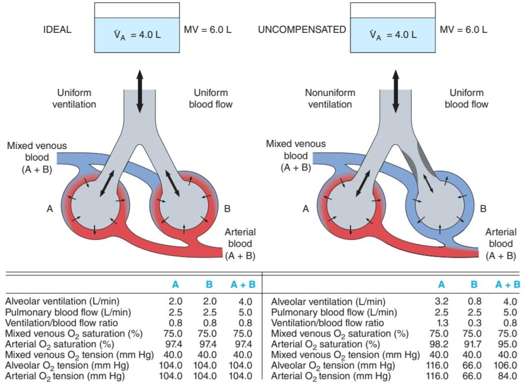
FIGURE 35-11 Comparison of ventilation/blood flow relationships in health and disease. Left: “Ideal” ventilation/blood flow relationship. Right: Nonuniform ventilation and uniform blood flow, uncompensated. VA, alveolar ventilation; MV, respiratory minute volume. See text for details. (Reproduced with permission from Comroe JH Jr, et al: The Lung: Clinical Physiology and Pulmonary Function Tests, 2nd ed. Year Book; 1962.)
شکل 35-11 مقایسه روابط تهویه/جریان خون در سلامت و بیماری. سمت چپ: رابطه تهویه / جریان خون “ایده آل”. سمت راست: تهویه غیریکنواخت و جریان خون یکنواخت، بدون جبران. VA، تهویه آلوئولار؛ MV، حجم دقیقه تنفسی. برای جزئیات به متن مراجعه کنید (تکثیر شده با اجازه Comroe JH Jr و همکاران: The Lung: Clinical Physiology and Pulmonary Function Tests, 2nd ed. Year Book, 1962.)
OTHER FORMS OF HYPOXIA
ANEMIC HYPOXIA
Hypoxia due to anemia is not severe at rest unless the hemoglobin deficiency is marked, because 2,3-DPG increases in the red blood cells. However, anemic patients may have considerable difficulty during exercise because of a limited ability to increase O2 delivery to the active tissues (Figure 35-12).
سایر اشکال هیپوکسی
هیپوکسی کم خون
هیپوکسی ناشی از کم خونی در حالت استراحت شدید نیست مگر اینکه کمبود هموگلوبین مشخص باشد، زیرا 2،3-DPG در گلبول های قرمز افزایش می یابد. با این حال، بیماران کم خون ممکن است در طول ورزش به دلیل توانایی محدود در افزایش تحویل O2 به بافت های فعال، مشکلات قابل توجهی داشته باشند (شکل 35-12).
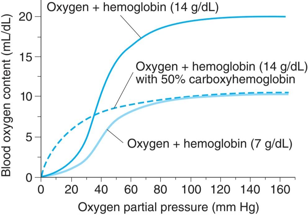
FIGURE 35-12 Effects of anemia and CO on hemoglobin binding of O2. Normal oxyhemoglobin (14 g/dL hemoglobin) dissociation curve compared with anemia (7 g/dL hemoglobin) and with oxyhemoglobin dissociation curves in CO poisoning (50% arboxy-hemoglobin). Note that the CO-poisoning curve is shifted to the left of the anemia curve. (Reproduced with permission from Leff AR, Schumacker PT: Respiratory Physiology: Basics and Applications. Saunders; 1993.)
شکل 35-12 اثرات کم خونی و CO بر اتصال هموگلوبین O2. منحنی تفکیک اکسی هموگلوبین طبیعی (14 گرم در دسی لیتر هموگلوبین) در مقایسه با کم خونی (7 گرم در دسی لیتر هموگلوبین) و با منحنی های تفکیک اکسی هموگلوبین در مسمومیت با CO (50٪ آربوکسی-هموگلوبین). توجه داشته باشید که منحنی مسمومیت با CO به سمت چپ منحنی کم خونی منتقل می شود. (تکثیر شده با اجازه Leff AR، Schumacker PT: Respiratory Physiology: Basics and Applications. Saunders; 1993.)
CARBON MONOXIDE POISONING
Small amounts of carbon monoxide (CO) are formed in the body, and this gas may function as a chemical messenger in the brain and elsewhere. In larger amounts, it is poisonous. Outside the body, it is formed by incomplete combustion of carbon. It was used by the Greeks and Romans to execute criminals, and today it causes more deaths than any other gas. CO poisoning has become less common in the United States, since natural gas replaced other gases such as coal gas, which contain large amounts of CO. CO is, however, still readily available, as the exhaust of gasoline engines is 6% or more CO.
مسمومیت با مونوکسید کربن
مقادیر کمی مونوکسید کربن (CO) در بدن تشکیل می شود و این گاز ممکن است به عنوان یک پیام رسان شیمیایی در مغز و جاهای دیگر عمل کند. در مقادیر بیشتر سمی است. در خارج از بدنه، با احتراق ناقص کربن تشکیل می شود. یونانی ها و رومی ها از آن برای اعدام مجرمان استفاده می کردند و امروزه بیش از هر گاز دیگری باعث مرگ می شود. مسمومیت با CO در ایالات متحده کمتر رایج شده است، زیرا گاز طبیعی جایگزین گازهای دیگر مانند گاز زغال سنگ شده است که حاوی مقادیر زیادی CO است.
CO is toxic because it reacts with hemoglobin to form carbon monoxyhemoglobin (carboxyhemoglobin, COHb), and COHb does not take up O2 (Figure 35-12). CO poisoning is often listed as a form of anemic hypoxia because the amount of hemoglobin that can carry O2 is reduced, but the total hemoglobin content of the blood is unaffected by CO. The affinity of hemoglobin for CO is 210 times its affinity for O2, and COHb liberates CO very slowly. An additional difficulty is that when COHb is present, the dissociation curve of the remaining HbO2 shifts to the left, decreasing the amount of O2 released. This is why an anemic individual who has 50% of the normal amount of HbO2 may be able to perform moderate work, whereas an individual with HbO2 reduced to the same level because of the formation of COHb is seriously incapacitated.
CO سمی است زیرا با هموگلوبین واکنش می دهد و کربن مونوکسی هموگلوبین (کربوکسی هموگلوبین، COHb) را تشکیل می دهد و COHb O2 را جذب نمی کند (شکل 35-12). مسمومیت با CO اغلب به عنوان نوعی هیپوکسی کم خون ذکر می شود زیرا مقدار هموگلوبینی که می تواند O2 را حمل کند کاهش می یابد، اما محتوای هموگلوبین کل خون تحت تأثیر CO قرار نمی گیرد. تمایل هموگلوبین به CO 210 برابر میل ترکیبی آن برای O2 است و COHb COH را بسیار آهسته آزاد می کند. یک مشکل دیگر این است که وقتی COHb وجود دارد، منحنی تفکیک HbO2 باقیمانده به سمت چپ تغییر میکند و مقدار O2 آزاد شده را کاهش میدهد. به همین دلیل است که یک فرد کم خونی که 50 درصد از مقدار طبیعی HbO2 را دارد، ممکن است بتواند کار متوسطی انجام دهد، در حالی که فردی که HbO2 به دلیل تشکیل COHb به همان میزان کاهش یافته است، به طور جدی ناتوان است.
Because of the affinity of CO for hemoglobin, progressive COHb formation occurs when the alveolar Pco is greater than 0.4 mm Hg. However, the amount of COHb formed depends on the duration of exposure to CO as well as the concentration of CO in the inspired air and the alveolar ventilation.
به دلیل تمایل COH به هموگلوبین، تشکیل تدریجی COHb زمانی اتفاق میافتد که Pco آلوئولی بیشتر از 0.4 میلیمتر جیوه باشد. با این حال، مقدار COHb تشکیل شده به مدت زمان قرار گرفتن در معرض CO و همچنین غلظت CO در هوای دمیده شده و تهویه آلوئولی بستگی دارد.
CO is also toxic to the cytochromes in the tissues, but the amount of CO required to poison the cytochromes is 1000 times the lethal dose; tissue toxicity thus plays no role in clinical CO poisoning.
CO همچنین برای سیتوکرومهای بافتها سمی است، اما مقدار CO مورد نیاز برای مسموم کردن سیتوکرومها 1000 برابر دوز کشنده است. بنابراین سمیت بافتی هیچ نقشی در مسمومیت بالینی CO ایفا نمی کند.
The symptoms of CO poisoning are those of any type of hypoxia, especially headache and nausea, but there is little stimulation of respiration, since in the arterial blood, Po2 remains normal and the carotid and aortic chemoreceptors are not stimulated. The cherry-red color of COHb is visible in the skin, nail beds, and mucous membranes. Death results when about 70-80% of the circulating hemoglobin is converted to COHb. The symptoms produced by chronic exposure to sublethal concentrations of CO are those of progressive brain damage, including mental changes and, sometimes, a parkinsonism-like state.
علائم مسمومیت با CO علائم هر نوع هیپوکسی، به ویژه سردرد و حالت تهوع است، اما تحریک کمی برای تنفس وجود دارد، زیرا در خون شریانی، Po2 طبیعی باقی می ماند و گیرنده های شیمیایی کاروتید و آئورت تحریک نمی شوند. رنگ قرمز گیلاسی COHb در پوست، بستر ناخن و غشاهای مخاطی قابل مشاهده است. زمانی که حدود 70 تا 80 درصد از هموگلوبین در گردش به COHb تبدیل شود، مرگ رخ می دهد. علائم ناشی از قرار گرفتن مزمن در معرض غلظتهای کشنده دیاکسید کربن، علائم آسیب تدریجی مغز، از جمله تغییرات ذهنی و گاهی اوقات، حالتی شبیه پارکینسون است.
Treatment of CO poisoning consists of immediate termination of the exposure and adequate ventilation, by artificial respiration if necessary. Ventilation with O2 is preferable to ventilation with fresh air, since O2 hastens the dissociation of COHb. Hyperbaric oxygenation (see below) is useful in this condition.
درمان مسمومیت با CO شامل قطع فوری تماس و تهویه مناسب، در صورت لزوم با تنفس مصنوعی است. تهویه با O2 به تهویه با هوای تازه ترجیح داده می شود، زیرا O2 تجزیه COHb را تسریع می کند. اکسیژن رسانی هیپرباریک (به زیر مراجعه کنید) در این شرایط مفید است.
ISCHEMIC HYPOXIA
Ischemic hypoxia, or stagnant hypoxia, is due to slow circulation and is a problem in organs such as the kidneys and heart during shock. The liver and possibly the brain are damaged by ischemic hypoxia in heart failure. The blood flow to the lung is normally very large, and it takes prolonged hypotension to produce significant damage. However, acute respiratory distress syndrome (ARDS) can develop when there is prolonged circulatory collapse.
هیپوکسی ایسکمیک
هیپوکسی ایسکمیک یا هیپوکسی راکد به دلیل گردش خون آهسته است و مشکلی در اندام هایی مانند کلیه ها و قلب در هنگام شوک است. کبد و احتمالا مغز در اثر هیپوکسی ایسکمیک در نارسایی قلبی آسیب می بینند. جریان خون به ریه به طور معمول بسیار زیاد است و برای ایجاد آسیب قابل توجه به افت فشار طولانی مدت نیاز دارد. با این حال، سندرم دیسترس تنفسی حاد (ARDS) می تواند در صورت فروپاشی طولانی مدت گردش خون ایجاد شود.
HISTOTOXIC HYPOXIA
Hypoxia due to inhibition of tissue oxidative processes is most commonly the result of cyanide poisoning. Cyanide inhibits cytochrome oxidase and possibly other enzymes. Methylene blue or nitrites are used to treat cyanide poisoning. They act by forming methemoglobin, which then reacts with cyanide to form cyanmethemoglobin, a nontoxic compound. The extent of treatment with these compounds is, of course, limited by the amount of methemoglobin that can be safely formed. Hyperbaric oxygenation may also be useful.
هیپوکسی هیستوتوکسیک
هیپوکسی ناشی از مهار فرآیندهای اکسیداتیو بافتی بیشتر در نتیجه مسمومیت با سیانید است. سیانید سیتوکروم اکسیداز و احتمالا سایر آنزیم ها را مهار می کند. متیلن بلو یا نیتریت برای درمان مسمومیت با سیانید استفاده می شود. آنها با تشکیل متهموگلوبین عمل می کنند، که سپس با سیانید واکنش می دهد و سیانمتهموگلوبین، یک ترکیب غیر سمی را تشکیل می دهد. گستره درمان با این ترکیبات البته با مقدار متهموگلوبینی که می تواند به طور ایمن تشکیل شود محدود می شود. اکسیژن رسانی هیپرباریک نیز ممکن است مفید باشد.
OXYGEN TREATMENT OF HYPOXIA
Administration of oxygen-rich gas mixtures is of very limited value in hypoperfusion, anemic, and histotoxic hypoxia because all that can be accomplished in this way is an increase in the amount of dissolved O2 in the arterial blood. This is also true in hypoxemia when it is due to shunting of unoxygenated venous blood past the lungs. In other forms of hypoxemia, O2 is great benefit. Treatment regimens that deliver less than 100% O2 are of value both acutely and chronically, and administration of O2 24 h/day for 2 years in this manner has been shown to significantly decrease the mortality of chronic obstructive pulmonary disease. O2 toxicity and therapy are discussed in Clinical Box 35-5.
درمان با اکسیژن هیپوکسی
تجویز مخلوط های گاز غنی از اکسیژن در هیپوپرفیوژن، کم خونی و هیپوکسی هیستوتوکسیک ارزش بسیار محدودی دارد، زیرا تنها کاری که می توان از این طریق انجام داد افزایش مقدار O2 محلول در خون شریانی است. این موضوع در هیپوکسمی نیز صدق می کند که به دلیل شنت خون وریدی بدون اکسیژن از ریه ها باشد. در سایر اشکال هیپوکسمی، O2 فواید زیادی دارد. رژیمهای درمانی که کمتر از 100% O2 را ارائه میکنند، هم بهصورت حاد و هم بهصورت مزمن دارای ارزش هستند، و نشان داده شده است که تجویز 24 ساعت در روز O2 به مدت 2 سال به این شیوه، مرگومیر بیماری انسدادی مزمن ریه را بهطور چشمگیری کاهش میدهد. سمیت O2 و درمان در جعبه بالینی 35-5 مورد بحث قرار گرفته است.
CLINICAL BOX 35-5
Administration of Oxygen & Its Potential Toxicity
It is interesting that while O2 is necessary for life in aerobic organisms, it is also toxic. Indeed, 100% O2 has been demonstrated to exert toxic effects not only in animals but also in bacteria, fungi, cultured animal cells, and plants. The toxicity seems to be due to the production of reactive oxygen species including superoxide anion (O2) and H2O2. When 80-100% O2 is administered to humans for periods of 8 h or more, the respiratory passages become irritated, causing substernal distress, nasal congestion, sore throat, and coughing.
جعبه بالینی 35-5
مدیریت اکسیژن و سمیت بالقوه آن
جالب است که در حالی که O2 برای زندگی در موجودات هوازی ضروری است، سمی نیز هست. در واقع، ثابت شده است که 100٪ O2 اثرات سمی نه تنها در حیوانات بلکه در باکتری ها، قارچ ها، سلول های حیوانی کشت شده و گیاهان نیز دارد. به نظر می رسد سمیت به دلیل تولید گونه های فعال اکسیژن از جمله آنیون سوپراکسید (O2) و H2O2 باشد. هنگامی که 80-100% O2 برای دوره های 8 ساعت یا بیشتر به انسان داده شود، مجاری تنفسی تحریک می شود و باعث ناراحتی زیر سینه، احتقان بینی، گلودرد و سرفه می شود.
A chronic condition characterized by lung cysts and densities (bronchopulmonary dysplasia) develops in some infants treated with O2 for respiratory distress syndrome. This syndrome may be a manifestation of O2 toxicity. Another complication in these infants is retinopathy of prematurity (retrolental fibroplasia), the formation of opaque vascular tissue in the eyes, which can lead to serious visual defects. The retinal receptors mature from the center to the periphery of the retina, and they use considerable O2. This causes the retina to become vascularized in an orderly manner. Oxygen treatment before maturation is complete provides the needed O2 to the photoreceptors, and consequently the normal vascular pattern fails to develop. Evidence indicates that this condition can be prevented or ameliorated by treatment with vitamin E, which exerts an anti-oxidant effect and, in animals, by growth hormone inhibitors.
یک بیماری مزمن که با کیست و تراکم ریه مشخص می شود (دیسپلازی برونکوپولمونری) در برخی از نوزادان تحت درمان با O2 برای سندرم دیسترس تنفسی ایجاد می شود. این سندرم ممکن است تظاهر سمیت O2 باشد. یکی دیگر از عوارض این نوزادان، رتینوپاتی نارس (فیبروپلازی رترولنتال)، تشکیل بافت عروقی مات در چشم است که می تواند منجر به نقص های بینایی جدی شود. گیرنده های شبکیه از مرکز به سمت حاشیه شبکیه بالغ می شوند و از O2 قابل توجهی استفاده می کنند. این باعث می شود که شبکیه به روشی منظم عروقی شود. درمان با اکسیژن قبل از کامل شدن بلوغ اکسیژن مورد نیاز را برای گیرنده های نوری فراهم می کند و در نتیجه الگوی عروقی طبیعی ایجاد نمی شود. شواهد نشان می دهد که می توان با درمان با ویتامین E که اثر آنتی اکسیدانی دارد و در حیوانات با مهارکننده های هورمون رشد از این وضعیت پیشگیری کرد یا آن را بهبود بخشید.
Administration of 100% O2 at increased pressure accelerates the onset of O2 toxicity, with the production not only of tracheobronchial irritation but also of muscle twitching, ringing in the ears, dizziness, convulsions, and coma. The speed with which these symptoms develop is proportional to the pressure at which the O2 is administered; for example, at 4 atm, symptoms develop in 30 min in half the patients, whereas at 6 atm, convulsions develop in a few minutes.
تجویز 100% O2 با فشار افزایش یافته، شروع سمیت O2 را تسریع میکند و نه تنها باعث تحریک نای برونشیال میشود، بلکه باعث انقباض عضلانی، صدای زنگ در گوش، سرگیجه، تشنج و کما میشود. سرعت ایجاد این علائم متناسب با فشاری است که O2 در آن تجویز می شود. به عنوان مثال، در 4 اتمسفر، علائم در 30 دقیقه در نیمی از بیماران ایجاد می شود، در حالی که در 6 atm، تشنج در چند دقیقه ایجاد می شود.
On the other hand, exposure to 100% O2 at 2-3 atm can increase dissolved O2 in arterial blood to the point that arterial O2 tension is greater than 2000 mm Hg and tissue O2 tension is 400 mm Hg. If exposure is limited to 5 h or less at these pressures, O2 toxicity is not a problem. Therefore, hyperbaric O2 therapy in closed tanks is used to treat diseases in which improved oxygenation of tissues cannot be achieved in other ways. It is of demonstrated value in carbon monoxide poisoning, radiation-induced tissue injury, gas gangrene, very severe blood loss anemia, diabetic leg ulcers, and other wounds that are slow to heal, and rescue of skin flaps and grafts in which the circulation is marginal. It is also the primary treatment for decompression sickness and air embolism.
از طرف دیگر، قرار گرفتن در معرض 100٪ O2 در 2-3 اتمسفر می تواند O2 محلول در خون شریانی را تا حدی افزایش دهد که کشش O2 شریانی بیشتر از 2000 میلی متر جیوه و کشش O2 بافتی 400 میلی متر جیوه باشد. اگر قرار گرفتن در معرض در این فشارها به 5 ساعت یا کمتر محدود شود، سمیت O2 مشکلی ایجاد نمی کند. بنابراین، O2 درمانی هیپرباریک در مخازن بسته برای درمان بیماری هایی استفاده می شود که در آنها اکسیژن رسانی بهتر بافت ها از راه های دیگر امکان پذیر نیست. در مسمومیت با مونوکسید کربن، آسیب بافتی ناشی از تشعشع، گانگرن گازی، کم خونی از دست دادن خون بسیار شدید، زخم های پای دیابتی، و سایر زخم هایی که دیر التیام می یابند، و نجات فلپ ها و گرافت های پوستی که در آنها گردش خون حاشیه ای است، ارزش اثبات شده ای دارد. همچنین درمان اولیه بیماری رفع فشار و آمبولی هوا است.
In hypercapnic patients in severe pulmonary failure, the CO2 level may be so high that it depresses rather than stimulates respiration. Some of these patients keep breathing only because the carotid and aortic chemoreceptors drive the respiratory center. If the hypoxic drive is withdrawn by administering O2, breathing may stop. During the resultant apnea, the arterial Po2 drops but breathing may not start again, as PCO2 further depresses the respiratory center. Therefore, O2 therapy in this situation must be started with care.
در بیماران هیپرکاپنیک در نارسایی شدید ریوی، سطح CO2 ممکن است آنقدر بالا باشد که به جای تحریک تنفس، باعث کاهش تنفس شود. برخی از این بیماران فقط به این دلیل نفس می کشند که گیرنده های شیمیایی کاروتید و آئورت مرکز تنفسی را هدایت می کنند. اگر با تجویز O2، محرک هیپوکسیک قطع شود، ممکن است تنفس متوقف شود. در طول آپنه ناشی از آن، Po2 شریانی کاهش می یابد، اما ممکن است تنفس دوباره شروع نشود، زیرا PCO2 مرکز تنفسی را بیشتر تحت فشار قرار می دهد. بنابراین، درمان O2 در این شرایط باید با احتیاط آغاز شود.
HYPERCAPNIA & HYPOCAPNIA
HYPERCAPNIA
Retention of CO2 in the body (hypercapnia) initially stimulates respiration. Retention of larger amounts produces such symptoms as confusion, diminished sensory acuity and, eventually, coma with respiratory depression and death due to depression of the central nervous system. In patients with these symptoms, the PcO2 is markedly elevated and severe respiratory acidosis is present. Large amounts of HCO3 are excreted, but more HCO3 is reabsorbed, raising the plasma HCO3 and partially compensating for the acidosis.
هایپرکاپنیا و هیپوکاپنیا
هایپرکاپنیا
حفظ CO2 در بدن (هیپرکاپنیا) در ابتدا باعث تحریک تنفس می شود. احتباس مقادیر بیشتر علائمی مانند گیجی، کاهش حدت حسی و در نهایت کما همراه با افسردگی تنفسی و مرگ ناشی از افسردگی سیستم عصبی مرکزی را ایجاد می کند. در بیماران با این علائم، PcO2 به طور قابل توجهی افزایش یافته و اسیدوز تنفسی شدید وجود دارد. مقادیر زیادی HCO3 دفع می شود، اما HCO3 بیشتری دوباره جذب می شود، HCO3 پلاسما را بالا می برد و تا حدی اسیدوز را جبران می کند.
CO2 is so much more soluble than O2 that hypercapnia is rarely a problem in patients with pulmonary fibrosis. However, it does occur in ventilation- perfusion inequality and when for any reason alveolar ventilation is inadequate in the various forms of pump failure. It is exacerbated when CO2 production is increased. For example, in febrile patients there is a 13% increase in CO2 production for each 1°C rise in temperature, and a high carbohydrate intake increases CO2 production because of the increase in the respiratory quotient. Normally, alveolar ventilation increases and the extra CO2 is expired, but it accumulates when ventilation is compromised.
CO2 بسیار بیشتر از O2 محلول است که هایپرکاپنی به ندرت در بیماران مبتلا به فیبروز ریوی مشکل ایجاد می کند. با این حال، در نابرابری تهویه- پرفیوژن و زمانی که به هر دلیلی تهویه آلوئولی در اشکال مختلف خرابی پمپ ناکافی است، رخ می دهد. هنگامی که تولید CO2 افزایش یابد تشدید می شود. به عنوان مثال، در بیماران تب دار به ازای هر یک درجه سانتی گراد افزایش دما، 13 درصد افزایش تولید CO2 وجود دارد و مصرف زیاد کربوهیدرات به دلیل افزایش ضریب تنفسی، تولید CO2 را افزایش می دهد. به طور معمول، تهویه آلوئولار افزایش مییابد و CO2 اضافی منقضی میشود، اما زمانی که تهویه به خطر میافتد، انباشته میشود.
HYPOCAPNIA
Hypocapnia is the result of hyperventilation. During voluntary hyperventilation, the arterial Pco, falls from 40 mm Hg to as low as 15 mm Hg while the alveolar Po2 rises to 120-140 mm Hg.
هیپوکاپنیا
هیپوکاپنی نتیجه هیپرونتیلاسیون است. در طول هیپرونتیلاسیون ارادی، Pco شریانی از 40 میلیمتر جیوه به 15 میلیمتر جیوه کاهش مییابد در حالی که Po2 آلوئولی به 120-140 میلیمتر جیوه افزایش مییابد.
The more chronic effects of hypocapnia are seen in neurotic patients who chronically hyperventilate. Cerebral blood flow may be reduced 30% or more because of the direct constrictor effect of hypocapnia on the cerebral vessels. The cerebral ischemia causes light-headedness, dizziness, and paresthesias. Hypocapnia also increases cardiac output. It has a direct constrictor effect on many peripheral vessels, but it depresses the vasomotor center, so that the blood pressure is usually unchanged or only slightly elevated.
اثرات مزمن هیپوکاپنیا در بیماران عصبی دیده می شود که به طور مزمن هیپرونتیله می کنند. جریان خون مغزی ممکن است 30 درصد یا بیشتر کاهش یابد، زیرا اثر منقبض کننده مستقیم هیپوکاپنی بر عروق مغزی دارد. ایسکمی مغزی باعث سبکی سر، سرگیجه و پارستزی می شود. هیپوکاپنی همچنین برون ده قلبی را افزایش می دهد. این یک اثر منقبض کننده مستقیم روی بسیاری از رگ های محیطی دارد، اما مرکز وازوموتور را تحت فشار قرار می دهد، به طوری که فشار خون معمولاً تغییر نمی کند یا فقط کمی افزایش می یابد.
Other consequences of hypocapnia are due to the associated respiratory alkalosis, the blood pH being increased to 7.5 or 7.6. The plasma HCO3 level is low, but HCO3 reabsorption is decreased because of the inhibition of renal acid secretion by the low Pco2. The plasma total calcium level does not change, but the plasma Ca2+ level falls and hypocapnic individuals develop carpopedal spasm, a positive Chvostek sign, and other signs of tetany.
سایر عواقب هیپوکاپنی به دلیل آلکالوز تنفسی مرتبط است، pH خون به 7.5 یا 7.6 افزایش می یابد. سطح HCO3 پلاسما پایین است، اما بازجذب HCO3 به دلیل مهار ترشح اسید کلیوی توسط Pco2 پایین کاهش می یابد. سطح کلسیم کل پلاسما تغییر نمی کند، اما سطح کلسیم پلاسما کاهش می یابد و افراد هیپوکاپنیک دچار اسپاسم کارپوپدال، علامت Chvostek مثبت و سایر علائم تتانی می شوند.
CHAPTER SUMMARY
خلاصه فصل
• Partial pressure differences between air and blood for O2 and CO2 dictate a net flow of O2 into the blood and CO2 out of the blood in the pulmonary system.
• اختلاف فشار جزئی بین هوا و خون برای O2 و CO2 باعث جریان خالص O2 به داخل خون و خروج CO2 از خون در سیستم ریوی می شود.
• The amount of O2 in the blood is determined by the amount dissolved (minor) and the amount bound (major) to hemoglobin. Each hemoglobin molecule contains four subunits that each can bind O2. Hemoglobin O2 binding is cooperative and also affected by pH, temperature, and the concentration of 2,3-diphosphoglycerate (2,3-DPG).
• مقدار O2 در خون با مقدار حل شده (مینور) و مقدار متصل (ماژور) به هموگلوبین تعیین می شود. هر مولکول هموگلوبین شامل چهار زیر واحد است که هر کدام می توانند O2 را متصل کنند. اتصال هموگلوبین O2 همکاری می کند و همچنین تحت تأثیر pH، دما و غلظت 2،3-دی فسفوگلیسرات (2،3-DPG) قرار می گیرد.
• CO2 in blood is rapidly converted into H2CO3 due to the activity of carbonic anhydrase. CO2 also readily forms carbamino compounds with blood proteins (including hemoglobin). The rapid net loss of CO2 allows more CO2 to dissolve in blood.
• CO2 در خون به دلیل فعالیت کربنیک انیدراز به سرعت به H2CO3 تبدیل می شود. CO2 همچنین به آسانی ترکیبات کربامینو را با پروتئین های خون (از جمله هموگلوبین) تشکیل می دهد. از دست دادن خالص سریع CO2 اجازه می دهد تا CO2 بیشتری در خون حل شود.
• The pH of plasma is 7.4. A decrease in plasma pH is termed acidosis and an increase of plasma pH is termed alkalosis. A short-term change in arterial Pco, due to decreased ventilation results in respiratory acidosis. A short-term change in arterial Pco2 due to increased ventilation results in respiratory alkalosis. Metabolic acidosis occurs when strong acids are added to the blood, and metabolic alkalosis occurs when strong bases are added to (or strong acids are removed from) the blood.
• pH پلاسما 7.4 است. کاهش pH پلاسما اسیدوز و افزایش pH پلاسما آلکالوز نامیده می شود. تغییر کوتاه مدت در Pco شریانی به دلیل کاهش تهویه منجر به اسیدوز تنفسی می شود. تغییر کوتاه مدت در Pco2 شریانی به دلیل افزایش تهویه منجر به آلکالوز تنفسی می شود. اسیدوز متابولیک زمانی اتفاق میافتد که اسیدهای قوی به خون اضافه شود و آلکالوز متابولیک زمانی رخ میدهد که بازهای قوی به خون اضافه شوند (یا اسیدهای قوی از آن خارج شوند).
• Respiratory compensation to acidosis or alkalosis involves quick changes in ventilation. Such changes effectively change the PCO2 in the blood plasma. Renal compensation mechanisms are much slower and involve H+ secretion or HCO3 reabsorption.
• جبران تنفسی اسیدوز یا آلکالوز شامل تغییرات سریع در تهویه است. چنین تغییراتی به طور موثری PCO2 را در پلاسمای خون تغییر می دهد. مکانیسم های جبران کلیوی بسیار کندتر است و شامل ترشح +H یا بازجذب HCO3 می شود.
• Hypoxia is a deficiency of O2 at the tissue level. Hypoxia has powerful consequences at the cellular, tissue, and organ level: It can alter cellular transcription factors and thus protein expression; it can quickly alter brain function and produce symptoms similar to alcohol (eg, dizziness, impaired mental function, drowsiness, headache); and it can affect ventilation. Long- term hypoxia results in cell and tissue death.
• هیپوکسی کمبود O2 در سطح بافت است. هیپوکسی پیامدهای قدرتمندی در سطح سلولی، بافتی و اندامی دارد: می تواند فاکتورهای رونویسی سلولی و در نتیجه بیان پروتئین را تغییر دهد. می تواند به سرعت عملکرد مغز را تغییر دهد و علائمی شبیه الکل ایجاد کند (مانند سرگیجه، اختلال در عملکرد ذهنی، خواب آلودگی، سردرد). و می تواند بر تهویه تاثیر بگذارد. هیپوکسی طولانی مدت منجر به مرگ سلولی و بافتی می شود.
MULTIPLE-CHOICE QUESTIONS
For all questions, select the single best answer unless otherwise directed.
سوالات چند گزینه ای
برای همه سؤالات، بهترین پاسخ را انتخاب کنید، مگر اینکه دستور دیگری داده شود.
1. Most of the CO2 transported in the blood is
A. dissolved in plasma.
B. in carbamino compounds formed from plasma proteins.
C. in carbamino compounds formed from hemoglobin.
D. bound to Cl.
E. in HCO3.
1. بیشتر CO2 منتقل شده در خون است
الف. محلول در پلاسما.
ب. در ترکیبات کربامینوی تشکیل شده از پروتئین های پلاسما.
ج. در ترکیبات کربامینوی تشکیل شده از هموگلوبین.
د. محدود به Cl.
ی. در HCO3.
2. Which of the following has the greatest effect on the ability of blood to transport oxygen?
A. Capacity of the blood to dissolve oxygen
B. Amount of hemoglobin in the blood
C. pH of plasma
D. CO2 content of red blood cells
E. Temperature of the blood
2- کدام یک از موارد زیر بیشترین تأثیر را بر توانایی خون در انتقال اکسیژن دارد؟
الف. ظرفیت خون برای حل کردن اکسیژن
ب. مقدار هموگلوبین در خون
ج. pH پلاسما
د. محتوای CO2 گلبول های قرمز
ی. دمای خون
3. Which of the following is true of the system?
A. Reaction 2 is catalyzed by carbonic anhydrase.
B. Because of reaction 2, the pH of blood declines during hyperventilation.
C. Reaction 1 occurs in the red blood cell.
D. Reaction 1 occurs primarily in plasma.
E. The reactions move to the right when there is excess H+ in the tissues.
3. کدام یک از موارد زیر در مورد سیستم صادق است؟
 الف. واکنش 2 توسط کربنیک انیدراز کاتالیز می شود.
ب. به دلیل واکنش 2، pH خون در طول هیپرونتیلاسیون کاهش می یابد.
ج. واکنش 1 در گلبول قرمز رخ می دهد.
د. واکنش 1 عمدتاً در پلاسما رخ می دهد.
ی. هنگامی که +H اضافی در بافت ها وجود دارد، واکنش ها به سمت راست حرکت می کنند.
4. In comparing uncompensated respiratory acidosis and uncompensated metabolic acidosis which one of the following is true?
A. Plasma pH change is always greater in uncompensated respiratory acidosis compared to uncompensated metabolic acidosis.
B. There are no compensation mechanisms for respiratory acidosis, whereas there is respiratory compensation for metabolic acidosis.
C. Uncompensated respiratory acidosis involves changes in plasma [HCO3 ̄], whereas plasma [HCO3] is unchanged in uncompensated metabolic acidosis.
D. Uncompensated respiratory acidosis is associated with a change in Pco2, whereas in uncompensated metabolic acidosis PCO2 is constant.
4. در مقایسه اسیدوز تنفسی جبران نشده و اسیدوز متابولیک جبران نشده کدام یک از موارد زیر صحیح است؟
الف. تغییر pH پلاسما در اسیدوز تنفسی جبران نشده در مقایسه با اسیدوز متابولیک جبران نشده همیشه بیشتر است.
ب. هیچ مکانیسم جبرانی برای اسیدوز تنفسی وجود ندارد، در حالی که برای اسیدوز متابولیک جبران تنفسی وجود دارد.
ج. اسیدوز تنفسی جبران نشده شامل تغییرات در پلاسما [HCO3 ̄] است، در حالی که پلاسما [HCO3] در اسیدوز متابولیک جبران نشده بدون تغییر است.
د. اسیدوز تنفسی جبران نشده با تغییر در Pco2 همراه است، در حالی که در اسیدوز متابولیک جبران نشده PCO2 ثابت است.
کلیک کنید «منابع»
🚀 با ما همراه شوید!
تازهترین مطالب و آموزشهای مغز و اعصاب را از دست ندهید. با فالو کردن کانال تلگرام آیندهنگاران مغز، از ما حمایت کنید!



