
فیزیولوژی پزشکی گانونگ؛ مکانیسم های تنظیم کننده قلب و عروق


دعای مطالعه [ نمایش ]
بِسْمِ الله الرَّحْمنِ الرَّحیمِ
اَللّهُمَّ اَخْرِجْنى مِنْ ظُلُماتِ الْوَهْمِ
خدايا مرا بيرون آور از تاريكىهاى وهم،
وَ اَكْرِمْنى بِنُورِ الْفَهْمِ
و به نور فهم گرامى ام بدار،
اَللّهُمَّ افْتَحْ عَلَيْنا اَبْوابَ رَحْمَتِكَ
خدايا درهاى رحمتت را به روى ما بگشا،
وَانْشُرْ عَلَيْنا خَزائِنَ عُلُومِكَ بِرَحْمَتِكَ يا اَرْحَمَ الرّاحِمينَ
و خزانههاى علومت را بر ما باز كن به امید رحمتت اى مهربانترين مهربانان.
کتاب «فیزیولوژی پزشکی گانونگ» بهعنوان یکی از جامعترین و معتبرترین منابع در حوزه علوم پزشکی، همچنان مرجع کلیدی برای درک عملکرد پیچیده بدن انسان است. این اثر با تکیه بر تازهترین پژوهشها و توضیحات دقیق از سازوکارهای فیزیولوژیک، پلی میان علوم پایه پزشکی و کاربردهای بالینی ایجاد میکند و نقشی بیبدیل در آموزش، پژوهش و ارتقای دانش سلامت ایفا مینماید.
ترجمه دقیق و علمی این شاهکار توسط برند علمی آیندهنگاران مغز به مدیریت داریوش طاهری، دسترسی فارسیزبانان به مرزهای نوین دانش فیزیولوژی را ممکن ساخته و رسالتی علمی برای ارتقای آموزش پزشکی، فهم عمیقتر سازوکارهای بدن و توسعه روشهای نوین در حوزه سلامت فراهم آورده است.
» کتاب فیزیولوژی پزشکی گانونگ
» » فصل ۳۲: مکانیسم های تنظیم کننده قلب و عروق
در حال ویرایش
» Ganong’s Review of Medical Physiology
»» CHAPTER 32: Cardiovascular Regulatory Mechanisms
OBJECTIVES
After studying his chapter, you should be able to:
• Contrast the sympathetic and parasympathetic control of the heart and vasculature.
• Identify the components and neurotransmitters in the central neural pathways that control arterial blood pressure and heart rate and list factors that determine the activity within the pathways.
• Identify the locations of the receptors and afferent and efferent pathways, the effector responses, and the functions of the baroreceptor reflex pathway.
• Describe the role of atrial and cardiopulmonary receptors in the control of the cardiovascular system in health and disease.
• Contrast the locations, properties, and functions of the peripheral and central chemoreceptor reflexes.
• Define how the process of autoregulation contributes to control of vascular caliber.
• Identify the paracrine factors, particularly those secreted by the endothelium, that regulate vascular tone and their mechanisms of action.
• List circulating vasodilators and vasoconstrictors.
اهداف
پس از مطالعه این فصل، باید بتوانید:
• کنترل سمپاتیک و پاراسمپاتیک قلب و عروق را مقایسه کنید.
• اجزاء و انتقال دهنده های عصبی را در مسیرهای عصبی مرکزی که فشار خون شریانی و ضربان قلب را کنترل می کنند، شناسایی کنید و عواملی را فهرست کنید که فعالیت درون مسیرها را تعیین می کنند.
• مکان گیرنده ها و مسیرهای آوران و وابران، پاسخ های موثر و عملکرد مسیر رفلکس بارورسپتور را شناسایی کنید.
• نقش گیرنده های دهلیزی و قلبی ریوی را در کنترل سیستم قلبی عروقی در سلامت و بیماری توضیح دهید.
• مکان ها، خواص و عملکرد رفلکس های گیرنده شیمیایی محیطی و مرکزی را با هم مقایسه کنید.
• تعریف کنید که چگونه فرآیند خودتنظیمی به کنترل کالیبر عروقی کمک می کند.
• فاکتورهای پاراکرین، به ویژه آنهایی که از اندوتلیوم ترشح می شوند، که تون عروق و مکانیسم های عمل آنها را تنظیم می کنند، شناسایی کنید.
• داروهای گشادکننده عروق و منقبض کننده عروق در گردش را فهرست کنید.
INTRODUCTION
Multiple cardiovascular regulatory mechanisms exist that help increase the blood supply to active tissues and increase or decrease heat loss from the body by redistributing the blood. In the face of challenges such as hemorrhage, they maintain blood flow to the heart and brain. When the challenge faced is severe, flow to these vital organs is maintained at the expense of the circulation to the rest of the body.
مقدمه
مکانیسم های تنظیم کننده قلبی عروقی متعددی وجود دارد که به افزایش خون رسانی به بافت های فعال و افزایش یا کاهش اتلاف حرارت از بدن با توزیع مجدد خون کمک می کند. در مواجهه با چالش هایی مانند خونریزی، جریان خون را به قلب و مغز حفظ می کنند. وقتی چالشی که با آن مواجه میشوید شدید است، جریان به این اندامهای حیاتی به هزینه گردش خون به بقیه بدن حفظ میشود.
Circulatory adjustments are effected by altering the output of the heart), changing the diameter of the resistance vessels (primarily the arterioles), or altering the amount of blood pooled in the capacitance vessels (the veins). Regulation of cardiac output is discussed in Chapter 30. The caliber of the arterioles is adjusted in part by autoregulation (Table 32-1). It is also increased in active tissues by locally produced vasodilator metabolites and substances secreted by the endothelium. The diameter of the arterioles is also regulated by circulating vasoactive substances and the sympathetic nerves that innervate them. Circulating vasoactive substances and sympathetic nerves also determine the caliber of the capacitance vessels. The systemic regulatory mechanisms synergize with the local mechanisms and adjust vascular responses throughout the body.
تنظیمات گردش خون با تغییر برونده قلب، تغییر قطر رگهای مقاومتی (عمدتاً شریانچهها) یا تغییر مقدار خون جمعشده در رگهای ظرفیتی (سیاهرگها) انجام میشود. تنظیم برونده قلبی در فصل 30 مورد بحث قرار گرفته است. کالیبر شریانچهها تا حدی توسط خودتنظیمی تنظیم میشود (جدول 32-1). همچنین در بافتهای فعال توسط متابولیتهای گشادکننده عروق تولید شده موضعی و مواد ترشح شده توسط اندوتلیوم افزایش مییابد. قطر شریانچهها نیز توسط مواد وازواکتیو در گردش و اعصاب سمپاتیکی که آنها را عصبدهی میکنند، تنظیم میشود. مواد وازواکتیو در گردش و اعصاب سمپاتیک نیز کالیبر رگهای ظرفیتی را تعیین میکنند. مکانیسمهای تنظیمی سیستمیک با مکانیسمهای موضعی همافزایی میکنند و پاسخهای عروقی را در سراسر بدن تنظیم میکنند.
TABLE 32-1 Summary of factors affecting the caliber of the arterioles.
جدول 32-1 خلاصه عوامل موثر بر کالیبر سرخرگ ها.

The terms vasoconstriction and vasodilation are generally used to refer to constriction and dilation of the resistance vessels. Changes in the caliber of the veins are referred to as venoconstriction or venodilation.
اصطلاحات انقباض عروق و اتساع عروق معمولاً برای اشاره به انقباض و اتساع عروق مقاومت استفاده می شود. تغییرات در کالیبر وریدها را انقباض رگها یا گشاد شدن رگها می نامند.
NEURAL CONTROL OF THE CARDIOVASCULAR SYSTEM
INNERVATION OF THE BLOOD VESSELS
Most of the vasculature is an example of an autonomic effector organ that receives innervation from the sympathetic but not the parasympathetic division of the autonomic nervous system (see Chapter 13). Sympathetic postganglionic fibers terminate on vascular smooth muscle in all parts of the body and release norepinephrine to act on a1-adrenoceptors to mediate vasoconstriction. In the vasculature of exercising muscles, activation of the sympathetic nervous system and release of epinephrine from the adrenal medulla can also mediate vasodilation by the activation of ẞ2-adrenoceptors on skeletal muscle blood vessels.
کنترل عصبی سیستم قلبی عروقی
عصب کشی رگ های خونی
قسمت اعظم عروق نمونهای از یک اندام مؤثر خودکار است که عصبگیری را از بخش سمپاتیک، اما نه از بخش پاراسمپاتیک سیستم عصبی خودمختار دریافت میکند (به فصل 13 مراجعه کنید). فیبرهای پس گانگلیونی سمپاتیک به ماهیچه صاف عروق در تمام قسمتهای بدن ختم میشوند و نوراپی نفرین را آزاد میکنند تا بر روی گیرندههای a1 آدرنرژیک تأثیر بگذارد و باعث انقباض عروق شود. در عروق ماهیچههای تمرینکننده، فعال شدن سیستم عصبی سمپاتیک و آزاد شدن اپی نفرین از بصل الکلیوی آدرنال نیز میتواند با فعال شدن گیرندههای ۲ آدرنرژیک در عروق خونی عضله اسکلتی باعث اتساع عروق شود.
Although the arterioles and the other resistance vessels are most densely innervated, all blood vessels except capillaries and venules contain smooth muscle and receive motor nerve fibers from the sympathetic division of the autonomic nervous system. The fibers to the resistance vessels regulate tissue blood flow and arterial pressure. The fibers to the venous capacitance vessels vary the volume of blood “stored” in the veins. The innervation of most veins is sparse, but the splanchnic veins are well innervated. Venoconstriction is produced by stimuli that also activate the vasoconstrictor nerves to the arterioles. The resultant decrease in venous capacity increases venous return, shifting blood to the arterial side of the circulation.
اگرچه شریان ها و سایر رگ های مقاومتی به شدت عصب دهی می شوند، همه رگ های خونی به جز مویرگ ها و ونول ها حاوی ماهیچه های صاف هستند و رشته های عصبی حرکتی را از بخش سمپاتیک سیستم عصبی خودمختار دریافت می کنند. فیبرهای موجود در رگ های مقاومتی جریان خون بافت و فشار شریانی را تنظیم می کنند. الیاف به رگ های ظرفیت وریدی، حجم خون “ذخیره” در سیاهرگ ها را تغییر می دهد. عصب دهی بیشتر وریدها کم است، اما وریدهای splanchnic به خوبی عصب دهی می شوند. انقباض سیاهرگی توسط محرک هایی ایجاد می شود که اعصاب منقبض کننده عروق را به شریان ها نیز فعال می کند. کاهش ظرفیت وریدی منجر به افزایش بازگشت وریدی می شود و خون را به سمت شریانی گردش خون منتقل می کند.
When the sympathetic nerves are damaged (sympathectomy or neuropathy), the blood vessels dilate. A change in the level of activity (increase or decrease) in sympathetic nerves is just one of the many factors that mediate vasoconstriction or vasodilation (Table 32-1).
هنگامی که اعصاب سمپاتیک آسیب می بینند (سمپاتکتومی یا نوروپاتی)، رگ های خونی منبسط می شوند. تغییر در سطح فعالیت (افزایش یا کاهش) در اعصاب سمپاتیک تنها یکی از عوامل متعددی است که باعث انقباض عروق یا اتساع عروق می شود (جدول 32-1).
INNERVATION OF THE HEART
The heart is one example of an effector organ that receives opposing influences from the sympathetic and parasympathetic divisions of the autonomic nervous system (see Chapter 13). Release of norepinephrine from postganglionic sympathetic nerves activates ẞ1-adrenoceptors in the heart, notably on the sinoatrial (SA) node, atrioventricular (AV) node, His-Purkinje conductive tissue, and atrial and ventricular contractile tissue. Stimulation of sympathetic nerves leads to an increase in heart rate (chronotropy), rate of transmission in the cardiac conductive tissue (dromotropy), and the force of ventricular contraction (inotropy). On the other hand, release of acetylcholine from postganglionic parasympathetic (vagus) nerves activates muscarinic receptors in the heart, notably on the SA and AV nodes and atrial muscle. Stimulation of the vagus nerve reduces the heart rate, the rate of transmission through the AV node, and atrial contractility.
عصب کشی قلب
قلب نمونهای از اندام مؤثر است که تأثیرات متضادی را از بخشهای سمپاتیک و پاراسمپاتیک سیستم عصبی خودمختار دریافت میکند (به فصل 13 مراجعه کنید). آزاد شدن نوراپی نفرین از اعصاب سمپاتیک پس گانگلیونی، گیرنده های آدرنرژیک ۱ را در قلب فعال می کند، به ویژه در گره سینوسی دهلیزی (SA)، گره دهلیزی (AV)، بافت رسانای His-Purkinje، و بافت انقباضی دهلیزی و بطنی. تحریک اعصاب سمپاتیک منجر به افزایش ضربان قلب (کرنوتروپی)، سرعت انتقال در بافت رسانای قلب (دروموتروپی) و نیروی انقباض بطنی (اینوتروپی) می شود. از سوی دیگر، آزاد شدن استیل کولین از اعصاب پاراسمپاتیک (واگ) پس گانگلیونی، گیرنده های موسکارینی در قلب، به ویژه در گره های SA و AV و عضله دهلیزی را فعال می کند. تحریک عصب واگ باعث کاهش ضربان قلب، سرعت انتقال از طریق گره AV و انقباض دهلیزی می شود.
The above description presents an oversimplified explanation of autonomic control of cardiac function. There are adrenergic and cholinergic receptors on autonomic nerve terminals that modulate transmitter release from nerve endings. For example, release of acetylcholine from vagal nerve terminals inhibits the release of norepinephrine from sympathetic nerve terminals, so this can enhance the effects of vagal nerve activation on the heart.
شرح فوق توضیحی بیش از حد ساده از کنترل خودکار عملکرد قلب ارائه می دهد. گیرنده های آدرنرژیک و کولینرژیک در پایانه های عصبی خودمختار وجود دارند که آزادسازی فرستنده از انتهای عصبی را تعدیل می کنند. به عنوان مثال، آزاد شدن استیل کولین از پایانه های عصب واگ، آزاد شدن نوراپی نفرین را از پایانه های عصبی سمپاتیک مهار می کند، بنابراین می تواند اثرات فعال شدن عصب واگ را بر قلب افزایش دهد.
There is a moderate amount of tonic discharge in the cardiac sympathetic nerves at rest, but there is considerable tonic vagal discharge (vagal tone) in humans and other large animals. After the administration of muscarinic receptor antagonists such as atropine, the heart rate in humans increases from 70 beats/min, its normal resting value, to 150-180 beats/min because the sympathetic tone is unopposed. In humans in whom both noradrenergic and cholinergic systems are blocked, the heart rate is approximately 100 beats/min.
مقدار متوسطی از ترشحات مقوی در اعصاب سمپاتیک قلبی در حالت استراحت وجود دارد، اما ترشحات مقوی واگ (تن واگ) در انسان و سایر حیوانات بزرگ وجود دارد. پس از تجویز آنتاگونیستهای گیرنده موسکارینی مانند آتروپین، ضربان قلب در انسان از 70 ضربه در دقیقه که مقدار طبیعی آن در حالت استراحت است، به 150-180 ضربه در دقیقه افزایش مییابد زیرا تون سمپاتیک بدون مخالفت است. در انسان هایی که سیستم نورآدرنرژیک و کولینرژیک در آنها مسدود شده است، ضربان قلب تقریباً 100 ضربه در دقیقه است.
CENTRAL NEURAL CONTROL OF THE CARDIOVASCULAR SYSTEM
The cardiovascular system is under neural influences coming from several parts of the brainstem, forebrain, and insular cortex. The brainstem receives feedback from sensory receptors in the vasculature (eg, baroreceptors and chemoreceptors). A simplified model of the feedback control circuit is shown in Figure 32-1. An increase in neural output from the brainstem to sympathetic nerves leads to a decrease in blood vessel diameter (arteriolar vasoconstriction) and increases in stroke volume and heart rate, which contribute to a rise in blood pressure. This in turn causes an increase in baroreceptor activity, which signals the brainstem to reduce the neural output to sympathetic nerves. The baroreceptor reflex is described in detail below.
کنترل عصبی مرکزی سیستم قلبی عروقی
سیستم قلبی عروقی تحت تأثیرات عصبی است که از چندین قسمت ساقه مغز، پیش مغز و قشر منزوی می آید. ساقه مغز از گیرنده های حسی موجود در عروق (مانند گیرنده های فشاری و شیمیایی) بازخورد دریافت می کند. یک مدل ساده شده از مدار کنترل فیدبک در شکل 32-1 نشان داده شده است. افزایش برون ده عصبی از ساقه مغز به اعصاب سمپاتیک منجر به کاهش قطر رگ خونی (انقباض عروق شریانی) و افزایش حجم ضربه و ضربان قلب می شود که به افزایش فشار خون کمک می کند. این به نوبه خود باعث افزایش فعالیت بارورسپتور می شود که به ساقه مغز سیگنال می دهد تا خروجی عصبی را به اعصاب سمپاتیک کاهش دهد. رفلکس بارورسپتور در زیر به تفصیل توضیح داده شده است.

FIGURE 32-1 Feedback control of blood pressure. Brainstem excitatory input to sympathetic nerves to the heart and vasculature increases heart rate and stroke volume and reduces vessel diameter. Together these increase blood pressure, which activates the baroreceptor reflex to reduce the activity in the brainstem.
شکل 32-1 کنترل بازخورد فشار خون. ورودی تحریکی ساقه مغز به اعصاب سمپاتیک به قلب و عروق، ضربان قلب و حجم ضربه را افزایش می دهد و قطر رگ را کاهش می دهد. اینها با هم فشار خون را افزایش می دهند، که رفلکس بارورسپتور را برای کاهش فعالیت در ساقه مغز فعال می کند.
Venoconstriction and a decrease in the stores of blood in the venous reservoirs usually accompany increases in arteriolar constriction, although changes in the capacitance vessels do not always parallel changes in the resistance vessels. In the presence of an increase in sympathetic nerve activity to the heart and vasculature, there is usually an associated decrease in the activity of vagal fibers to the heart. Conversely, a decrease in sympathetic activity causes vasodilation, a fall in blood pressure, and an increase in the storage of blood in the venous reservoirs. There is usually a concomitant decrease in heart rate, but this is mostly due to stimulation of the vagal innervation of the heart.
انقباض وریدی و کاهش ذخایر خون در مخازن وریدی معمولاً با افزایش انقباض شریانی همراه است، اگرچه تغییرات در رگهای ظرفیت همیشه به موازات تغییرات عروق مقاومت نیست. در حضور افزایش فعالیت عصب سمپاتیک به قلب و عروق، معمولاً کاهش مرتبطی در فعالیت فیبرهای واگ به قلب وجود دارد. برعکس، کاهش فعالیت سمپاتیک باعث اتساع عروق، کاهش فشار خون و افزایش ذخیره خون در مخازن وریدی می شود. معمولاً کاهش همزمان ضربان قلب وجود دارد، اما این بیشتر به دلیل تحریک عصب واگ قلب است.
One of the major sources of excitatory input to sympathetic nerves controlling the vasculature is a group of neurons located near the pial surface of the medulla in the rostral ventrolateral medulla (RVLM; Figure 32-2). The axons of RVLM neurons course dorsally and medially and then descend in the lateral column of the spinal cord to the thoracolumbar intermediolateral cell column (IML). They contain phenylethanolamine-N-methyltransferase (PNMT; see Chapter 7), but glutamate is the excitatory transmitter they secrete to activate preganglionic sympathetic neurons. Neurovascular compression of the RVLM has been linked to some cases of essential hypertension in humans (Clinical Box 32-1).
یکی از منابع اصلی ورودی تحریکی به اعصاب سمپاتیک که عروق را کنترل می کنند، گروهی از نورون ها هستند که در نزدیکی سطح پیال مدولا در بصل النخاع شکمی جانبی منقاری قرار دارند (RVLM؛ شکل 32-2). آکسونهای نورونهای RVLM بهصورت پشتی و میانی حرکت میکنند و سپس در ستون جانبی نخاع به سمت ستون سلولی میانی جانبی قفسه سینه (IML) فرود میآیند. آنها حاوی فنیل اتانول آمین-N-متیل ترانسفراز هستند (PNMT؛ به فصل 7 مراجعه کنید)، اما گلوتامات فرستنده تحریکی است که آنها برای فعال کردن نورون های سمپاتیک پیش گانگلیونی ترشح می کنند. فشرده سازی عصبی عروقی RVLM با برخی موارد فشار خون ضروری در انسان مرتبط است (باکس بالینی 32-1).

FIGURE 32-2 Basic neural pathways involved in the control of blood pressure. The drawing shows a parasagittal section of the brainstem and its connections to the thoracic spinal cord. The rostral ventrolateral medulla (RVLM) is one of the major sources of excitatory input to sympathetic nerves controlling the vasculature. These neurons receive inhibitory input from the baroreceptors via an inhibitory (GABAergic) neuron in the caudal ventrolateral medulla (CVLM). The nucleus of the tractus solitarius (NTS) is the site of termination of baroreceptor afferent fibers that release glutamate. ACh, acetylcholine; GABA, y-aminobutyric acid; Glu, glutamate; IML, intermediolateral cell column; NE, norepinephrine.
شکل 32-2 مسیرهای عصبی اساسی درگیر در کنترل فشار خون. این نقاشی بخش پاراساژیتال ساقه مغز و اتصالات آن به نخاع قفسه سینه را نشان می دهد. مدولای شکمی جانبی منقاری (RVLM) یکی از منابع اصلی ورودی تحریکی به اعصاب سمپاتیک است که عروق را کنترل می کنند. این نورونها ورودی مهاری را از گیرندههای بارورژیک از طریق یک نورون بازدارنده (GABAergic) در بصل النخاع شکمی جانبی دمی (CVLM) دریافت میکنند. هسته tractus solitarius (NTS) محل خاتمه الیاف آوران بارورسپتور است که گلوتامات را آزاد می کنند. ACH، استیل کولین؛ GABA، y-aminobutyric اسید؛ گلو، گلوتامات؛ IML، ستون سلولی میانی. NE، نوراپی نفرین.
Clinical Box 32- 1
Essential Hypertension & Neurovascular Compression of the RVLM
In about 88% of patients with elevated blood pressure, the cause of the hypertension is unknown, and they are said to have essential hypertension (see Chapter 31). Neurovascular compression of the RVLM is associated with essential hypertension in some persons. For example, patients with a schwannoma (acoustic neuroma) or meningioma lying close to the RVLM also have hypertension. Magnetic resonance angiography (MRA) has been used to compare the incidence of neurovascular compression in hypertensive and normotensive individuals and to correlate indices of increased sympathetic nerve activity with the presence or absence of compression. Some of these studies showed a higher incidence of coexistence of neurovascular compression with essential hypertension than in other forms of hypertension or normotension, but others showed the presence of a compression in normotensive subjects. On the other hand, there was a strong positive relationship between the presence of neurovascular compression and increased sympathetic nerve activity.
جعبه بالینی 32-1
فشار خون ضروری و فشرده سازی عصبی عروقی RVLM
در حدود 88٪ از بیماران مبتلا به فشار خون بالا، علت فشار خون بالا ناشناخته است و گفته می شود که آنها فشار خون اساسی دارند (به فصل 31 مراجعه کنید). فشرده سازی عصبی عروقی RVLM در برخی افراد با فشار خون ضروری همراه است. به عنوان مثال، بیماران مبتلا به شوانوما (نوروم آکوستیک) یا مننژیوم در نزدیکی RVLM نیز دارای فشار خون بالا هستند. آنژیوگرافی رزونانس مغناطیسی (MRA) برای مقایسه میزان فشرده سازی عصبی عروقی در افراد دارای فشار خون بالا و فشار خون طبیعی و برای ارتباط شاخص های افزایش فعالیت عصب سمپاتیک با وجود یا عدم وجود فشرده سازی استفاده شده است. برخی از این مطالعات بروز همزیستی فشرده سازی عصبی عروقی با فشار خون ضروری را نسبت به سایر اشکال فشار خون یا فشار خون طبیعی نشان دادند، اما برخی دیگر وجود فشرده سازی را در افراد دارای فشار خون طبیعی نشان دادند. از سوی دیگر، یک رابطه مثبت قوی بین وجود فشرده سازی عصبی عروقی و افزایش فعالیت عصب سمپاتیک وجود داشت.
THERAPEUTIQ HIGHLIGHTS
In the 1970s, Dr. Peter Jannetta, a neurosurgeon in Pittsburgh, PA, developed a technique for “microvascular decompression” of the medulla to treat trigeminal neuralgia and hemifacial spasm, which he attributed to pulsatile compression of the vertebral and posterior inferior cerebellar arteries impinging on the fifth and seventh cranial nerves. Moving the arteries away from the nerves led to reversal of the neurologic symptoms in many cases. Some of these patients were also hypertensive, and they showed reductions in blood pressure postoperatively. Later, a few human studies showed that surgical decompression of the RVLM could sometimes relieve hypertension. There are also reports that hypertension is relieved after surgical decompression in patients with a schwannoma or meningioma in the vicinity of the RVLM.
نکات برجسته درمانی
در دهه 1970، دکتر پیتر جانتا، جراح مغز و اعصاب در پیتسبورگ، PA، تکنیکی را برای “کم فشار ریز عروقی” بصل النخاع برای درمان نورالژی سه قلو و اسپاسم همی صورت ابداع کرد که او آن را به فشرده سازی ضربانی مهره ها و ماهیچه های پشتی شکم بر روی ماهیچه های ران تحتانی گردن می دهد. اعصاب دور کردن شریان ها از اعصاب منجر به معکوس شدن علائم عصبی در بسیاری از موارد شد. برخی از این بیماران فشار خون نیز داشتند و پس از عمل کاهش فشار خون را نشان دادند. بعداً، چند مطالعه انسانی نشان داد که برداشتن فشار از طریق جراحی RVLM گاهی اوقات میتواند فشار خون را کاهش دهد. همچنین گزارش هایی وجود دارد که فشار خون بالا پس از رفع فشار جراحی در بیماران مبتلا به شوانوما یا مننژیوم در مجاورت RVLM کاهش می یابد.
The activity of RVLM neurons is determined by many factors (see Table 32- 2). They include input from a pathway that originates in the cerebral cortex (particularly the limbic cortex) and has a relay in the hypothalamus. This pathway is responsible for the blood pressure rise and tachycardia produced by emotions such as stress, sexual excitement, and anger. The RVLM also receives input from other brain regions and from arterial baroreceptors and the carotid and aortic chemoreceptors. In addition, some stimuli act directly on RVLM neurons to increase their firing rate.
فعالیت نورون های RVLM توسط عوامل زیادی تعیین می شود (جدول 32-2 را ببینید). آنها شامل ورودی از مسیری هستند که از قشر مغز (به ویژه قشر لیمبیک) منشا می گیرد و دارای یک رله در هیپوتالاموس است. این مسیر مسئول افزایش فشار خون و تاکی کاردی ناشی از احساساتی مانند استرس، هیجانات جنسی و خشم است. RVLM همچنین ورودی را از سایر مناطق مغز و از بارورسپتورهای شریانی و گیرنده های شیمیایی کاروتید و آئورت دریافت می کند. علاوه بر این، برخی از محرک ها مستقیماً روی نورون های RVLM عمل می کنند تا سرعت شلیک آنها را افزایش دهند.
TABLE 32-2 Factors affecting the activity of the RVLM.
جدول 32-2 عوامل مؤثر بر فعالیت RVLM.

Inflation of the lungs causes vasodilation and a decrease in blood pressure. This response is mediated via vagal afferents from the lungs that inhibit RVLM and sympathetic nerve activities. Pain usually causes a rise in blood pressure via afferent impulses converging in the RVLM. However, prolonged severe pain may cause vasodilation and fainting. The activity in afferents from exercising muscles probably exerts a similar pressor effect via a pathway to the RVLM. The pressor response to stimulation of somatic afferent nerves is called the somatosympathetic reflex.
تورم ریه ها باعث اتساع عروق و کاهش فشار خون می شود. این پاسخ از طریق آوران های واگ از ریه ها انجام می شود که RVLM و فعالیت های عصبی سمپاتیک را مهار می کنند. درد معمولاً از طریق تکانه های آوران که در RVLM همگرا می شوند باعث افزایش فشار خون می شود. با این حال، درد شدید طولانی مدت ممکن است باعث اتساع عروق و غش شود. فعالیت در آوران ها از تمرین عضلات احتمالاً اثر فشاری مشابهی را از طریق مسیری به RVLM اعمال می کند. پاسخ فشار دهنده به تحریک اعصاب آوران سوماتیک را رفلکس سوماتوسمپاتیک می نامند.
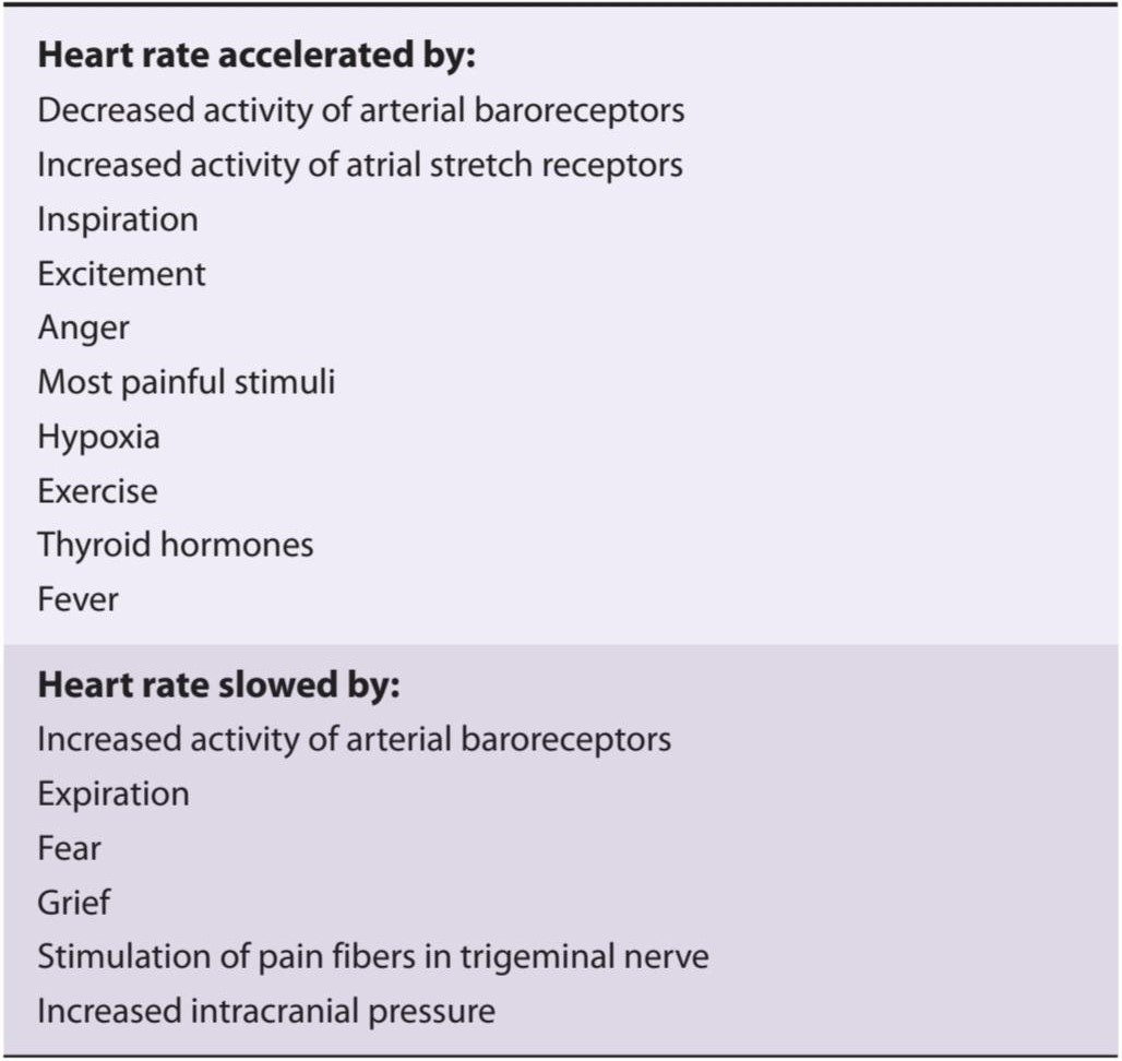
The medulla is also a major site of origin of excitatory input to cardiac vagal motor neurons in the nucleus ambiguus (Figure 32-3). Table 32-3 is a summary of factors that affect heart rate. In general, stimuli that increase heart rate also increase blood pressure, whereas those that decrease heart rate lower blood pressure. However, there are exceptions, such as combined hypotension and tachycardia with stimulation of atrial stretch receptors and hypertension and bradycardia during increased intracranial pressure.
مدولا همچنین محل اصلی منشاء ورودی تحریکی به نورونهای حرکتی واگ قلب در هسته مبهم است (شکل 32-3). جدول 3-32 خلاصه ای از عوامل موثر بر ضربان قلب است. به طور کلی، محرک هایی که ضربان قلب را افزایش می دهند، فشار خون را نیز افزایش می دهند، در حالی که آنهایی که ضربان قلب را کاهش می دهند، فشار خون را کاهش می دهند. با این حال، استثنائاتی مانند افت فشار خون و تاکی کاردی ترکیبی با تحریک گیرنده های کشش دهلیزی و فشار خون بالا و برادی کاردی در هنگام افزایش فشار داخل جمجمه وجود دارد.
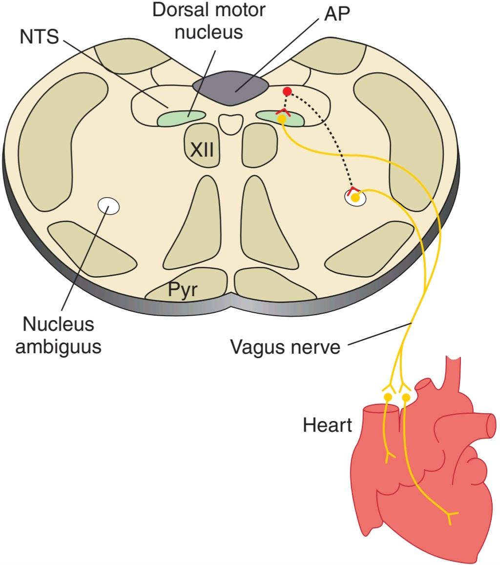
FIGURE 32-3 Basic pathways involved in the control of heart rate by the vagus nerves. Neurons in the nucleus of the tractus solitarius (NTS) project to and excite cardiac preganglionic parasympathetic neurons primarily in the nucleus ambiguus. Some are also located in the dorsal motor nucleus of the vagus; however, this nucleus primarily contains vagal motor neurons that project to the gastrointestinal tract. AP, area postrema; Pyr, pyramid; XII, hypoglossal nucleus.
شکل 32-3 مسیرهای اساسی درگیر در کنترل ضربان قلب توسط اعصاب واگ. نورونهای موجود در هسته tractus solitarius (NTS) به نورونهای پاراسمپاتیک پیشگانگلیونی قلبی عمدتاً در هسته مبهم میپردازند و آنها را تحریک میکنند. برخی نیز در هسته حرکتی پشتی واگ قرار دارند. با این حال، این هسته در درجه اول حاوی نورون های حرکتی واگ است که به سمت دستگاه گوارش می رود. AP، postrema ناحیه; پیر، هرم؛ XII، هسته هیپوگلوسال.
TABLE 32-3 Factors affecting heart rate.
جدول 32-3 عوامل موثر بر ضربان قلب.
BARORECEPTORS
The baroreceptors are stretch receptors in the walls of the heart and blood vessels. The carotid sinus and aortic arch receptors monitor the arterial circulation. There are also stretch receptors in the low-pressure part of the circulation (cardiopulmonary receptors). These include receptors in the walls of the right and left atria at the entrance of the superior and inferior vena cava, in the pulmonary veins, and in the pulmonary circulation.
بارورسپتورها
بارورسپتورها گیرنده های کششی در دیواره های قلب و رگ های خونی هستند. گیرنده های سینوس کاروتید و قوس آئورت بر گردش خون شریانی نظارت دارند. همچنین گیرنده های کششی در قسمت کم فشار گردش خون (گیرنده های قلبی ریوی) وجود دارد. اینها شامل گیرنده هایی در دیواره دهلیز راست و چپ در ورودی ورید اجوف فوقانی و تحتانی، در وریدهای ریوی و در گردش خون ریوی است.
The carotid sinus is a small dilation of the internal carotid artery just above the bifurcation of the common carotid into external and internal carotid branches (Figure 32-4). Baroreceptors are in the adventitia of the vessel within this dilation as well as in the wall of the arch of the aorta. The afferent nerve fibers from the carotid sinus form a distinct branch of the glossopharyngeal nerve, the carotid sinus nerve. The fibers from the aortic arch form a branch of the vagus nerve, the aortic depressor nerve.
سینوس کاروتید گشاد شدن کوچک شریان کاروتید داخلی درست بالای دوشاخه کاروتید مشترک به شاخه های کاروتید خارجی و داخلی است (شکل 32-4). بارورسپتورها در مجاورت رگ درون این اتساع و همچنین در دیواره قوس آئورت قرار دارند. رشتههای عصبی آوران از سینوس کاروتید شاخهای متمایز از عصب گلوسوفارنژیال، عصب سینوس کاروتید را تشکیل میدهند. فیبرهای قوس آئورت شاخه ای از عصب واگ را تشکیل می دهند که عصب دپرسور آئورت است.

FIGURE 32-4 Baroreceptor areas in the carotid sinus and aortic arch. One set of baroreceptors (stretch receptors) is located in the carotid sinus, a small dilation of the internal carotid artery just above the bifurcation of the common carotid into external and internal carotid branches. These receptors are innervated by a branch of the glossopharyngeal nerve, the carotid sinus nerve. A second set of baroreceptors is located in the wall of the arch of the aorta. These receptors are innervated by a branch of the vagus nerve, the aortic depressor nerve.
شکل 32-4 مناطق بارورسپتور در سینوس کاروتید و قوس آئورت. یک مجموعه از بارورسپتورها (گیرنده های کششی) در سینوس کاروتید، گشاد شدن کوچک شریان کاروتید داخلی درست بالای دو شاخه شدن کاروتید مشترک به شاخه های کاروتید خارجی و داخلی قرار دارد. این گیرنده ها توسط شاخه ای از عصب گلوفارنکس، عصب سینوس کاروتید عصب دهی می شوند. مجموعه دوم بارورسپتورها در دیواره قوس آئورت قرار دارند. این گیرنده ها توسط شاخه ای از عصب واگ، عصب دپرسور آئورت، عصب دهی می شوند.
The baroreceptors are stimulated by distension of the structures in which they are located; they discharge at an increased rate when the pressure in the carotid sinus and aortic arch rises. Their afferent fibers pass via the glossopharyngeal and vagus nerves to the medulla. These fibers release glutamate to excite neurons in the nucleus of the tractus solitarius (NTS) as the first synapse in the baroreceptor reflex (Figure 32-2). These NTS neurons then excite neurons in the caudal ventrolateral medulla (CVLM). These CVLM neurons release the inhibitory neurotransmitter y-aminobutyric acid (GABA) into the RVLM to reduce the firing rate of these. Excitatory projections also extend from the NTS to the vagal motor neurons in the nucleus ambiguus and dorsal motor nucleus (Figure 32-3). Thus, increased baroreceptor discharge inhibits the tonic discharge of sympathetic nerves and excites the vagal innervation of the heart. These neural changes produce vasodilation, venodilation, hypotension, bradycardia, and a decrease in cardiac output.
بارورسپتورها با اتساع ساختارهایی که در آن قرار دارند تحریک می شوند. هنگامی که فشار در سینوس کاروتید و قوس آئورت بالا می رود، با سرعت بیشتری ترشح می شوند. فیبرهای آوران آنها از طریق اعصاب گلوفارنکس و واگ به مدولا می گذرد. این فیبرها گلوتامات را آزاد می کنند تا نورون های موجود در هسته tractus solitarius (NTS) را به عنوان اولین سیناپس در رفلکس بارورسپتور تحریک کنند (شکل 32-2). سپس این نورونهای NTS نورونها را در بصل النخاع شکمی جانبی دمی (CVLM) تحریک میکنند. این نورونهای CVLM، انتقالدهنده عصبی مهاری y-aminobutyric acid (GABA) را در RVLM آزاد میکنند تا سرعت شلیک آنها را کاهش دهند. برجستگی های تحریکی نیز از NTS به نورون های حرکتی واگ در هسته مبهم و هسته حرکتی پشتی گسترش می یابد (شکل 32-3). بنابراین، افزایش ترشحات بارورسپتور ترشح تونیک اعصاب سمپاتیک را مهار می کند و عصب واگ قلب را تحریک می کند. این تغییرات عصبی باعث اتساع عروق، گشاد شدن رگ، افت فشار خون، برادی کاردی و کاهش برون ده قلبی می شود.
BARORECEPTOR NERVE ACTIVITY
Baroreceptors are more sensitive to pulsatile pressure than to constant pressure. A decline in pulse pressure without any change in mean pressure decreases the rate of baroreceptor discharge and provokes a rise in systemic blood pressure and tachycardia. At normal blood pressure levels (about 100 mm Hg mean pressure), a burst of action potentials appears in a single baroreceptor fiber during systole, but there are few action potentials in early diastole (Figure 32- 5). At lower mean pressures, this phasic change in firing is even more dramatic with activity only occurring during systole. At these lower pressures, the overall firing rate is considerably reduced. The threshold for eliciting activity in the carotid sinus nerve is approximately 50 mm Hg; maximal activity occurs at approximately 200 mm Hg, with activity throughout the cardiac cycle.
فعالیت عصبی گیرنده بار
بارورسپتورها به فشار ضربانی حساس تر از فشار ثابت هستند. کاهش فشار نبض بدون تغییر در فشار متوسط، سرعت تخلیه بارورسپتور را کاهش میدهد و باعث افزایش فشار خون سیستمیک و تاکی کاردی میشود. در سطوح نرمال فشار خون (حدود 100 میلی متر جیوه فشار متوسط)، یک انفجار پتانسیل عمل در یک فیبر بارورسپتور منفرد در طول سیستول ظاهر می شود، اما پتانسیل عمل کمی در دیاستول اولیه وجود دارد (شکل 32-5). در فشارهای متوسط کمتر، این تغییر فازی در شلیک حتی با فعالیتی که فقط در طول سیستول رخ می دهد، چشمگیرتر است. در این فشارهای پایین تر، میزان شلیک کلی به میزان قابل توجهی کاهش می یابد. آستانه برای برانگیختن فعالیت در عصب سینوس کاروتید تقریباً 50 میلی متر جیوه است. حداکثر فعالیت در حدود 200 میلی متر جیوه، با فعالیت در طول چرخه قلبی رخ می دهد.

FIGURE 32-5 Discharges (vertical lines) in a single afferent nerve fiber from the carotid sinus at various levels of mean arterial pressures, plotted against changes in aortic pressure with time. Baroreceptors are very sensitive to changes in pulse pressure as shown by the record of phasic aortic pressure. (Reproduced with permission from Levy MN, Pappano AJ: Cardiovascular Physiology, 9th ed. Mosby; 2007.)
شکل 32-5 ترشحات (خطوط عمودی) در یک فیبر عصبی آوران منفرد از سینوس کاروتید در سطوح مختلف فشار متوسط شریانی، در برابر تغییرات فشار آئورت با گذشت زمان رسم شده است. گیرنده های باروری به تغییرات فشار نبض بسیار حساس هستند، همانطور که در رکورد فشار آئورت فازیک نشان داده شده است. (تکثیر شده با اجازه Levy MN، Pappano AJ: Cardiovascular Physiology، ویرایش نهم Mosby؛ 2007.)
From the foregoing discussion, it is apparent that the baroreceptors on the arterial side of the circulation, their afferent connections to the medullary cardiovascular areas, and the efferent pathways from these areas constitute a reflex feedback mechanism that operates to stabilize blood pressure and heart rate. Any drop in systemic arterial pressure decreases the discharge in the buffer nerves, and there is a compensatory rise in blood pressure and cardiac output. Any rise in pressure produces dilation of the arterioles and decreases cardiac output until the blood pressure returns to its previous baseline level.
با توجه به بحث فوق، آشکار است که بارورسپتورهای سمت شریانی گردش خون، اتصالات آوران آنها به نواحی قلبی عروقی مدولاری، و مسیرهای وابران از این نواحی یک مکانیسم بازخورد بازتابی را تشکیل می دهند که برای تثبیت فشار خون و ضربان قلب عمل می کند. هر افت فشار شریانی سیستمیک باعث کاهش ترشحات در اعصاب بافر می شود و افزایش جبرانی در فشار خون و برون ده قلبی وجود دارد. هر افزایش فشار باعث اتساع شریان ها و کاهش برون ده قلبی می شود تا زمانی که فشار خون به سطح اولیه قبلی خود بازگردد.
ROLE OF BARORECEPTORS IN SHORT-TERM CONTROL OF BLOOD PRESSURE
The changes in heart rate and blood pressure that occur on standing up or lying down are due for the most part to baroreceptor reflexes. Baroreceptors are very important in short-term control of arterial pressure. Activation of the reflex allows for rapid adjustments in blood pressure in response to abrupt changes in posture, blood volume, cardiac output, or peripheral resistance during exercise. Removal of the baroreceptor reflex prevents an individual from adjusting their blood pressure in response to stimuli that cause abrupt changes in blood volume, cardiac output, or peripheral resistance, including exercise and postural changes.
نقش گیرنده های باروری در کنترل کوتاه مدت فشار خون
تغییرات در ضربان قلب و فشار خون که هنگام ایستادن یا دراز کشیدن رخ می دهد بیشتر به دلیل رفلکس های بارورسپتور است. بارورسپتورها در کنترل کوتاه مدت فشار شریانی بسیار مهم هستند. فعال سازی رفلکس امکان تنظیم سریع فشار خون را در پاسخ به تغییرات ناگهانی در وضعیت بدنی، حجم خون، برون ده قلبی یا مقاومت محیطی در حین ورزش فراهم می کند. حذف رفلکس بارورسپتور از تنظیم فشار خون فرد در پاسخ به محرک هایی که باعث تغییرات ناگهانی در حجم خون، برون ده قلبی یا مقاومت محیطی می شوند، از جمله ورزش و تغییرات وضعیتی، جلوگیری می کند.
In chronic hypertension, the baroreceptor reflex mechanism is “reset” to maintain an elevated rather than a normal blood pressure. In perfusion studies on hypertensive experimental animals, raising the pressure in the isolated carotid sinus lowers the elevated systemic pressure; and decreasing the perfusion pressure raises the elevated pressure. A long-term change in blood pressure resulting from loss of baroreceptor reflex control is called neurogenic hypertension.
در پرفشاری خون مزمن، مکانیسم رفلکس بارورسپتور برای حفظ فشار خون بالا به جای طبیعی “بازنشانی” می شود. در مطالعات پرفیوژن روی حیوانات آزمایشگاهی دارای فشار خون بالا، افزایش فشار در سینوس کاروتید جدا شده، فشار سیستمیک بالا را کاهش می دهد. و کاهش فشار پرفیوژن باعث افزایش فشار می شود. تغییر طولانی مدت در فشار خون ناشی از از دست دادن کنترل رفلکس بارورسپتور فشار خون عصبی نامیده می شود.
ATRIAL STRETCH & CARDIOPULMONARY RECEPTORS
The stretch receptors in the atria are of two types: those that discharge primarily during atrial systole (type A), and those that discharge primarily in late diastole, at the time of peak atrial filling (type B). The discharge of type B baroreceptors is increased when venous return is increased and decreased by positive-pressure breathing, indicating that these baroreceptors respond primarily to distension of the atrial walls. The reflex circulatory adjustments initiated by activation of these receptors include vasodilation and a fall in blood pressure. However, the heart rate is increased rather than decreased.
کشش دهلیزی و گیرنده های قلبی ریوی
گیرنده های کششی در دهلیزها دو نوع هستند: آنهایی که عمدتاً در طول سیستول دهلیزی (نوع A) تخلیه می شوند، و گیرنده هایی که عمدتاً در دیاستول دیررس، در زمان اوج پر شدن دهلیز تخلیه می شوند (نوع B). تخلیه بارورسپتورهای نوع B هنگامی که بازگشت وریدی با تنفس با فشار مثبت افزایش و کاهش می یابد افزایش می یابد، که نشان می دهد این بارورسپتورها عمدتاً به اتساع دیواره های دهلیزی پاسخ می دهند. تنظیم رفلکس گردش خون که با فعال شدن این گیرنده ها آغاز می شود شامل اتساع عروق و کاهش فشار خون است. با این حال، ضربان قلب به جای کاهش، افزایش می یابد.
Receptors in the endocardial surfaces of the ventricles are activated during ventricular distension. The response is a vagal bradycardia and hypotension, comparable to a baroreceptor reflex. Left ventricular stretch receptors may play a role in the maintenance of vagal tone that keeps the heart rate low at rest. Various chemicals are known to elicit reflexes due to activation of cardiopulmonary chemoreceptors and may play a role in various cardiovascular disorders (Clinical Box 32-2).
گیرنده ها در سطوح اندوکاردی بطن ها در طول اتساع بطن فعال می شوند. پاسخ یک برادی کاردی واگ و افت فشار خون است که با رفلکس بارورسپتور قابل مقایسه است. گیرنده های کشش بطن چپ ممکن است در حفظ تون واگ نقش داشته باشند که ضربان قلب را در حالت استراحت پایین نگه می دارد. مواد شیمیایی مختلف به دلیل فعال شدن گیرنده های شیمیایی قلبی ریوی باعث ایجاد رفلکس می شوند و ممکن است در اختلالات قلبی عروقی مختلف نقش داشته باشند (باکس بالینی 32-2).
Clinical Box 32-2
Cardiopulmonary Chemosensitive Receptors
Activation of chemosensitive vagal C fibers in the cardiopulmonary region (eg, juxtacapillary region of alveoli, ventricles, atria, great veins, and pulmonary artery) causes profound bradycardia, hypotension, and a brief period of apnea followed by rapid shallow breathing. This response pattern is called the Bezold-Jarisch reflex and can be elicited by a variety of substances including capsaicin, serotonin, phenylbiguanide, and veratridine.
جعبه بالینی 32-2
گیرنده های شیمیایی حساس قلبی ریوی
فعال شدن فیبرهای شیمیایی C واگ حساس در ناحیه قلبی ریوی (به عنوان مثال، ناحیه کنار مویرگ آلوئول ها، بطن ها، دهلیزها، سیاهرگ های بزرگ و شریان ریوی) باعث برادی کاردی عمیق، افت فشار خون و یک دوره کوتاه آپنه و به دنبال آن تنفس سریع کم عمق می شود. این الگوی پاسخ، رفلکس بزولد-جاریش نامیده می شود و می تواند توسط مواد مختلفی از جمله کپسایسین، سروتونین، فنیل بیگوانید و وراتریدین ایجاد شود.
The Bezold-Jarisch reflex is activated during certain pathophysiologic conditions. For example, this reflex may be activated during myocardial ischemia and reperfusion as a result of increased production of oxygen radicals and by agents used as radiocontrast for coronary angiography. This can contribute to the hypotension that is frequently a stubborn complication of heart disease. Activation of cardiopulmonary chemosensitive receptors may also be part of a defense mechanism protecting individuals from toxic chemical hazards. Activation of cardiopulmonary reflexes may help reduce the amount of inspired pollutants that gets absorbed into the blood, protecting vital organs from potential toxicity of these pollutants, and facilitating their elimination. Finally, the syndrome of cardiac slowing with hypotension (vasovagal syncope) has also been attributed to activation of the Bezold- Jarisch reflex. Vasovagal syncope can occur after prolonged upright posture that results in pooling of blood in the lower extremities and diminished intracardiac blood volume (also called postural syncope). This phenomenon is exaggerated if combined with dehydration. The resultant arterial hypotension is sensed in the carotid sinus baroreceptors, and afferent fibers from these receptors trigger autonomic signals that increase cardiac rate and contractility. However, pressure receptors in the wall of the left ventricle respond by sending signals that trigger paradoxical bradycardia and decreased contractility, resulting in sudden marked hypotension. The individual also feels lightheaded and may experience a brief episode of loss of consciousness.
رفلکس بزولد-جاریش در شرایط خاص پاتوفیزیولوژیک فعال می شود. به عنوان مثال، این رفلکس ممکن است در طول ایسکمی میوکارد و خونرسانی مجدد در نتیجه افزایش تولید رادیکال های اکسیژن و توسط عواملی که به عنوان کنتراست رادیویی برای آنژیوگرافی عروق کرونر استفاده می شود، فعال شود. این می تواند به افت فشار خون کمک کند که اغلب یک عارضه سرسخت بیماری قلبی است. فعالسازی گیرندههای شیمیایی حساس قلبی-ریوی نیز ممکن است بخشی از یک مکانیسم دفاعی باشد که از افراد در برابر خطرات شیمیایی سمی محافظت میکند. فعالسازی رفلکسهای قلبی ریوی ممکن است به کاهش میزان جذب آلایندههای الهامشده در خون کمک کند، از اندامهای حیاتی در برابر سمیت احتمالی این آلایندهها محافظت کند و حذف آنها را تسهیل کند. در نهایت، سندرم کند شدن قلب همراه با افت فشار خون (سنکوپ وازوواگال) نیز به فعال شدن رفلکس بزولد-جاریش نسبت داده شده است. سنکوپ وازوواگال می تواند پس از وضعیت عمودی طولانی مدت رخ دهد که منجر به تجمع خون در اندام تحتانی و کاهش حجم خون داخل قلب می شود (که سنکوپ وضعیتی نیز نامیده می شود). این پدیده اگر با کم آبی همراه باشد اغراق آمیز است. هیپوتانسیون شریانی حاصل در بارورسپتورهای سینوس کاروتید حس میشود و فیبرهای آوران از این گیرندهها سیگنالهای اتونومی را تحریک میکنند که ضربان قلب و انقباض را افزایش میدهد. با این حال، گیرندههای فشار در دیواره بطن چپ با ارسال سیگنالهایی پاسخ میدهند که باعث برادی کاردی متناقض و کاهش انقباض میشود و در نتیجه افت فشار خون ناگهانی مشخص میشود. فرد همچنین احساس سبکی سر می کند و ممکن است یک دوره کوتاه از دست دادن هوشیاری را تجربه کند.
THERAPEUTIQ HIGHLIGHTS
The most critical intervention for individuals who experience episodes of neurogenic syncope is to avoid dehydration and to avoid situations that trigger the adverse event. Episodes of syncope may be reduced or prevented by an increased dietary salt intake or administration of mineralocorticoids. Vasovagal syncope has been treated with the use of ẞ-adrenoceptor antagonists and disopyramide, an antiarrhythmic agent that blocks Na+ channels. Cardiac pacemakers have also been used to stabilize the heart rate during episodes that normally trigger bradycardia.
نکات برجسته درمانی
مهمترین مداخله برای افرادی که دورههای سنکوپ عصبی را تجربه میکنند، اجتناب از کمآبی بدن و اجتناب از موقعیتهایی است که باعث بروز عارضه نامطلوب میشوند. اپیزودهای سنکوپ ممکن است با افزایش مصرف نمک در رژیم غذایی یا تجویز مینرالوکورتیکوئیدها کاهش یا پیشگیری شود. سنکوپ وازوواگال با استفاده از آنتاگونیست های گیرنده آدرنرژیک و دیسوپیرامید، یک عامل ضد آریتمی که کانال های Na+ را مسدود می کند، درمان شده است. ضربان سازهای قلبی نیز برای تثبیت ضربان قلب در طول دوره هایی که به طور معمول باعث برادی کاردی می شوند، استفاده شده است.
VALSALVA MANEUVER
The function of the receptors can also be tested by monitoring the changes in pulse pressure and blood pressure that occur in response to brief periods of straining (forced expiration against a closed glottis: the Valsalva maneuver). Valsalva maneuvers occur regularly during coughing, defecation, and heavy lifting. The blood pressure rises at the onset of straining (Figure 32-6) because the increase in intrathoracic pressure is added to the pressure of the blood in the aorta. It then falls because the high intrathoracic pressure compresses the veins, decreasing venous return and cardiac output. The decreases in arterial pressure and pulse pressure inhibit the baroreceptors, causing tachycardia and a rise in peripheral resistance. When the glottis is opened and the intrathoracic pressure returns to normal, cardiac output is restored but the peripheral vessels are constricted. The blood pressure therefore rises above normal, and this stimulates the baroreceptors, causing bradycardia and a drop in pressure to normal levels.
مانور والسالوا
عملکرد گیرنده ها را نیز می توان با نظارت بر تغییرات فشار نبض و فشار خون که در پاسخ به دوره های کوتاه فشار روی می دهد (بازدم اجباری در برابر گلوت بسته: مانور والسالوا) آزمایش کرد. مانورهای والسالوا به طور منظم در هنگام سرفه، مدفوع و بلند کردن اجسام سنگین انجام می شود. فشار خون در شروع زور زدن افزایش می یابد (شکل 32-6) زیرا افزایش فشار داخل قفسه سینه به فشار خون در آئورت اضافه می شود. سپس سقوط می کند زیرا فشار داخل قفسه سینه بالا وریدها را فشرده می کند و بازگشت وریدی و برون ده قلبی را کاهش می دهد. کاهش فشار شریانی و فشار نبض، بارورسپتورها را مهار می کند و باعث تاکی کاردی و افزایش مقاومت محیطی می شود. هنگامی که گلوت باز می شود و فشار داخل قفسه سینه به حالت عادی باز می گردد، برون ده قلبی بازیابی می شود اما رگ های محیطی منقبض می شوند. بنابراین فشار خون از حد طبیعی بالاتر می رود و این باعث تحریک بارورسپتورها می شود و باعث برادی کاردی و کاهش فشار به سطح طبیعی می شود.

FIGURE 32–6 Diagram of the response to straining (the Valsalva maneuver) in a healthy man, recorded with a needle in the brachial artery. Blood pressure rises at the onset of straining because increased intrathoracic pressure is added to the pressure of the blood in the aorta. It then falls because the high intrathoracic pressure compresses veins, decreasing venous return and cardiac output. (Used with permission of M Mcllroy.)
شکل 32-6 نمودار پاسخ به زور زدن (مانور والسالوا) در یک مرد سالم، که با یک سوزن در شریان بازویی ثبت شده است. فشار خون در شروع زور زدن افزایش می یابد زیرا افزایش فشار داخل قفسه سینه به فشار خون در آئورت اضافه می شود. سپس سقوط می کند زیرا فشار داخل قفسه سینه بالا وریدها را فشرده می کند و بازگشت وریدی و برون ده قلبی را کاهش می دهد. (با اجازه M Mcllroy استفاده می شود.)
In patients whose sympathetic nervous system is not functional, heart rate changes still occur because the baroreceptors and the vagi are intact. However, in patients with autonomic insufficiency, a syndrome in which autonomic function is widely disrupted, the heart rate changes are absent. For unknown reasons, patients with primary hyperaldosteronism also fail to show the heart rate changes and the blood pressure rise when the intrathoracic pressure returns to normal. Their response to the Valsalva maneuver returns to normal after removal of the aldosterone-secreting tumor.
در بیمارانی که سیستم عصبی سمپاتیک آنها کار نمی کند، تغییرات ضربان قلب همچنان رخ می دهد زیرا گیرنده های فشار و واگی دست نخورده هستند. با این حال، در بیماران مبتلا به نارسایی اتونومیک، سندرمی که در آن عملکرد اتونوم به طور گسترده ای مختل می شود، تغییرات ضربان قلب وجود ندارد. به دلایل ناشناخته، بیماران مبتلا به هیپرآلدوسترونیسم اولیه نیز قادر به نشان دادن تغییرات ضربان قلب و افزایش فشار خون با بازگشت فشار داخل قفسه سینه به حالت طبیعی نیستند. پاسخ آنها به مانور والسالوا پس از برداشتن تومور ترشح کننده آلدوسترون به حالت عادی باز می گردد.
PERIPHERAL CHEMORECEPTOR REFLEX
Peripheral arterial chemoreceptors in the carotid and aortic bodies are exposed to very high rates of blood flow. These receptors are primarily activated by a reduction in arterial partial pressure of oxygen (PaO2), but they also respond to an increase in the arterial partial pressure of carbon dioxide (PaCO2) and pH. Chemoreceptors exert their main effects on respiration; however, their activation also leads to vasoconstriction. Heart rate changes are variable and depend on various factors, including changes in respiration. A direct effect of chemoreceptor activation is to increase vagal nerve activity. However, hypoxia also produces hyperpnea and increased catecholamine release from the adrenal medulla, both of which produce tachycardia and an increase in cardiac output. Hemorrhage that produces hypotension leads to chemoreceptor stimulation due to decreased blood flow to the chemoreceptors and consequent stagnant anoxia of these organs. Chemoreceptor activity may also contribute to the production of Mayer waves, which are slow, regular oscillations in arterial pressure that occur at the rate of about one every 20-40 s during hypotension. Under these conditions, hypoxia stimulates the chemoreceptors to increase blood pressure; this improves the blood flow in the receptor organs and eliminates the stimulus to the chemoreceptors; blood pressure then falls and a new cycle is initiated. Mayer waves should not be confused with Traube-Hering waves, which are fluctuations in blood pressure synchronized with respiration.
رفلکس شیمی گیرنده محیطی
گیرنده های شیمیایی شریانی محیطی در بدن کاروتید و آئورت در معرض نرخ بسیار بالایی از جریان خون قرار دارند. این گیرنده ها در درجه اول با کاهش فشار جزئی اکسیژن شریانی (PaO2) فعال می شوند، اما به افزایش فشار جزئی شریانی دی اکسید کربن (PaCO2) و pH نیز پاسخ می دهند. گیرنده های شیمیایی اثرات اصلی خود را بر روی تنفس اعمال می کنند. با این حال، فعال شدن آنها نیز منجر به انقباض عروق می شود. تغییرات ضربان قلب متغیر است و به عوامل مختلفی از جمله تغییر در تنفس بستگی دارد. اثر مستقیم فعال شدن گیرنده های شیمیایی افزایش فعالیت عصب واگ است. با این حال، هیپوکسی همچنین باعث هیپرپنه و افزایش ترشح کاتکول آمین از بصل الکلیوی آدرنال می شود که هر دو باعث تاکی کاردی و افزایش برون ده قلبی می شوند. خونریزی که باعث کاهش فشار خون می شود به دلیل کاهش جریان خون به گیرنده های شیمیایی و در نتیجه آنکسی راکد این اندام ها منجر به تحریک شیمی درمانی می شود. فعالیت گیرنده شیمیایی ممکن است به تولید امواج مایر نیز کمک کند، که نوسانات آهسته و منظمی در فشار شریانی است که در طول افت فشار خون هر 20 تا 40 ثانیه یک بار رخ می دهد. تحت این شرایط، هیپوکسی گیرنده های شیمیایی را برای افزایش فشار خون تحریک می کند. این جریان خون را در اندام های گیرنده بهبود می بخشد و محرک گیرنده های شیمیایی را از بین می برد. سپس فشار خون کاهش می یابد و چرخه جدیدی آغاز می شود. امواج مایر را نباید با امواج Traube-Hering که نوسانات فشار خون همزمان با تنفس هستند، اشتباه گرفت.
CENTRAL CHEMORECEPTORS
When intracranial pressure is increased, the blood supply to the brain is compromised, and the local hypercapnia activates central chemoreceptors located along the ventrolateral medullary surface. Their activation induces a rise in systemic arterial pressure (Cushing reflex) that tends to restore the blood flow to the brain. Over a considerable range, the blood pressure rise is proportional to the increase in intracranial pressure. The rise in blood pressure causes a reflex decrease in heart rate via the arterial baroreceptors. This is why bradycardia rather than tachycardia is characteristically seen in patients with increased intracranial pressure.
گیرنده های شیمیایی مرکزی
هنگامی که فشار داخل جمجمه افزایش می یابد، خون رسانی به مغز به خطر می افتد و هیپرکاپنی موضعی گیرنده های شیمیایی مرکزی واقع در امتداد سطح مدولاری بطنی جانبی را فعال می کند. فعال شدن آنها باعث افزایش فشار شریانی سیستمیک (رفلکس کوشینگ) می شود که تمایل دارد جریان خون را به مغز بازگرداند. در محدوده قابل توجهی، افزایش فشار خون متناسب با افزایش فشار داخل جمجمه است. افزایش فشار خون باعث کاهش رفلکس ضربان قلب از طریق بارورسپتورهای شریانی می شود. به همین دلیل است که برادی کاردی به جای تاکی کاردی مشخصاً در بیماران با افزایش فشار داخل جمجمه دیده می شود.
Hypercapnia directly stimulates the RVLM (Table 32-3), but the direct peripheral effect of hypercapnia is vasodilation. Therefore, the peripheral and central actions tend to cancel each other out. Moderate hyperventilation that significantly lowers the CO2 tension of the blood causes cutaneous and cerebral vasoconstriction, but there is little change in blood pressure. Exposure to high concentrations of CO2 is associated with marked cutaneous and cerebral vasodilation, but vasoconstriction occurs elsewhere and usually there is a slow rise in blood pressure.
هایپرکاپنی به طور مستقیم RVLM را تحریک می کند (جدول 32-3)، اما اثر محیطی مستقیم هیپرکاپنی اتساع عروق است. بنابراین، کنش های محیطی و مرکزی تمایل به خنثی کردن یکدیگر دارند. هیپرونتیلاسیون متوسط که به طور قابل توجهی تنش CO2 خون را کاهش می دهد، باعث انقباض عروق پوستی و مغزی می شود، اما تغییر کمی در فشار خون وجود دارد. قرار گرفتن در معرض غلظت های بالای CO2 با گشاد شدن عروق پوستی و مغزی همراه است، اما انقباض عروق در جاهای دیگر رخ می دهد و معمولاً افزایش آهسته فشار خون وجود دارد.
LOCAL REGULATION
AUTOREGULATION
The capacity of tissues to regulate their own blood flow is referred to as autoregulation. Most vascular beds have an intrinsic capacity to compensate for moderate changes in perfusion pressure by changes in vascular resistance, so that blood flow remains relatively constant. This capacity is well developed in the kidneys (see Chapter 37), but it has also been observed in the mesentery, skeletal muscle, brain, liver, and myocardium. It is due in part to the intrinsic contractile response of smooth muscle to stretch (myogenic theory of autoregulation). As the pressure rises, the blood vessels are distended and the vascular smooth muscle fibers that surround the vessels contract. If it is postulated that the muscle responds to the tension in the vessel wall, this theory explains the greater degree of contraction at higher pressures; the wall tension is proportional to the distending pressure times the radius of the vessel (law of Laplace; see Chapter 31), and the maintenance of a stable wall tension as the pressure rises would require a decrease in radius. Vasodilator substances tend to accumulate in active tissues, and these “metabolites” also contribute to autoregulation (metabolic theory of autoregulation). When blood flow decreases, they accumulate and the vessels dilate; when blood flow increases, they tend to be washed away.
مقررات محلی
AUTOREGULATION
ظرفیت بافت ها برای تنظیم جریان خون خود به عنوان خود تنظیم نامیده می شود. بیشتر بسترهای عروقی دارای ظرفیت ذاتی برای جبران تغییرات متوسط در فشار خونرسانی با تغییر در مقاومت عروقی هستند، به طوری که جریان خون نسبتا ثابت باقی می ماند. این ظرفیت در کلیه ها به خوبی توسعه یافته است (به فصل 37 مراجعه کنید)، اما در مزانتر، ماهیچه های اسکلتی، مغز، کبد و میوکارد نیز مشاهده شده است. این تا حدی به دلیل پاسخ انقباضی ذاتی عضله صاف به کشش است (نظریه میوژنیک خودتنظیمی). با افزایش فشار، رگ های خونی منبسط شده و رشته های عضلانی صاف عروقی که رگ ها را احاطه کرده اند منقبض می شوند. اگر فرض شود که عضله به کشش دیواره رگ پاسخ می دهد، این نظریه درجه انقباض بیشتر در فشارهای بالاتر را توضیح می دهد. کشش دیواره متناسب با فشار انبساطی ضربدر شعاع ظرف است (قانون لاپلاس؛ فصل 31 را ببینید)، و حفظ کشش دیواره پایدار با افزایش فشار مستلزم کاهش شعاع است. مواد گشادکننده عروق تمایل به تجمع در بافتهای فعال دارند و این متابولیتها نیز به خودتنظیمی کمک میکنند (تئوری متابولیک خودتنظیمی). هنگامی که جریان خون کاهش می یابد، انباشته می شوند و عروق گشاد می شوند. هنگامی که جریان خون افزایش می یابد، آنها تمایل دارند که شسته شوند.
VASODILATOR METABOLITES
The metabolic changes that produce vasodilation include, in most tissues, decreases in O2 tension and pH. These changes cause relaxation of the arterioles and precapillary sphincters. A local fall in O2 tension, in particular, can initiate a program of vasodilatory gene expression secondary to production of hypoxia- inducible factor-1a (HIF-1a), a transcription factor with multiple targets. Increases in CO2 tension and osmolality also dilate the vessels. The direct dilator action of CO2 is most pronounced in the skin and brain. The neutrally mediated vasoconstrictor effects of systemic, as opposed to local, hypoxia and hypercapnia have been discussed above. A rise in temperature exerts a direct vasodilator effect, and the temperature rise in active tissues (due to the heat of metabolism) may contribute to vasodilation. K✶ is another substance that accumulates locally, and has demonstrated dilator activity secondary to the hyperpolarization of vascular smooth muscle cells. Lactate may also contribute to the dilation. In injured tissues, histamine released from damaged cells increases capillary permeability. Thus, it is probably responsible for some of the swelling in areas of inflammation. Adenosine plays a vasodilator role in cardiac muscle but not in skeletal muscle. It also inhibits the release of norepinephrine.
متابولیت های گشاد کننده عروق
تغییرات متابولیکی که باعث اتساع عروق می شود شامل کاهش تنش O2 و pH در بیشتر بافت ها می شود. این تغییرات باعث شل شدن شریان ها و اسفنکترهای پیش مویرگی می شود. کاهش موضعی در تنش O2، به ویژه، میتواند برنامهای برای بیان ژن گشادکننده عروق ثانویه به تولید فاکتور 1a القایی هیپوکسی (HIF-1a)، یک فاکتور رونویسی با اهداف متعدد، آغاز کند. افزایش کشش CO2 و اسمولالیته نیز باعث گشاد شدن عروق می شود. اثر گشادکننده مستقیم CO2 در پوست و مغز بارزتر است. اثرات انقباض عروقی با واسطه خنثی سیستمیک، برخلاف موضعی، هیپوکسی و هیپرکاپنی در بالا مورد بحث قرار گرفته است. افزایش دما یک اثر گشادکننده مستقیم عروق دارد و افزایش دما در بافتهای فعال (به دلیل گرمای متابولیسم) ممکن است به اتساع عروق کمک کند. +K ماده دیگری است که به صورت موضعی تجمع می یابد و فعالیت گشادکننده ثانویه به هیپرپلاریزه شدن سلول های ماهیچه صاف عروقی را نشان داده است. لاکتات همچنین ممکن است به اتساع کمک کند. در بافت های آسیب دیده، هیستامین آزاد شده از سلول های آسیب دیده، نفوذپذیری مویرگی را افزایش می دهد. بنابراین، احتمالاً مسئول برخی از تورم در مناطق التهابی است. آدنوزین نقش گشادکننده عروق در عضله قلب دارد اما در عضله اسکلتی نقشی ندارد. همچنین ترشح نوراپی نفرین را مهار می کند.
LOCALIZED VASOCONSTRICTION
Injured arteries and arterioles constrict strongly. The constriction appears to be due in part to the local liberation of serotonin from platelets that stick to the vessel wall in the injured area. Injured veins also constrict.
انقباض عروق موضعی
شریان ها و شریان های آسیب دیده به شدت منقبض می شوند. به نظر می رسد این انقباض تا حدی به دلیل آزادسازی موضعی سروتونین از پلاکت هایی است که به دیواره عروق در ناحیه آسیب دیده می چسبند. وریدهای آسیب دیده نیز منقبض می شوند.
A drop in tissue temperature causes vasoconstriction, and this local response to cold plays a part in temperature regulation (see Chapter 17).
کاهش دمای بافت باعث انقباض عروق می شود و این پاسخ موضعی به سرما در تنظیم دما نقش دارد (به فصل 17 مراجعه کنید).
SUBSTANCES SECRETED BY THE ENDOTHELIUM
ENDOTHELIAL CELLS
As noted in Chapter 31, the endothelial cells constitute a large and important tissue. They secrete many growth factors and vasoactive substances. The vasoactive substances include prostaglandins and thromboxanes, nitric oxide (NO), and endothelins.
موادی که توسط اندوتلیوم ترشح می شود
سلول های اندوتلیال
همانطور که در فصل 31 اشاره شد، سلول های اندوتلیال بافت بزرگ و مهمی را تشکیل می دهند. آنها بسیاری از عوامل رشد و مواد وازواکتیو ترشح می کنند. مواد وازواکتیو شامل پروستاگلاندین ها و ترومبوکسان ها، اکسید نیتریک (NO) و اندوتلین ها هستند.
PROSTACYCLIN & THROMBOXANE A2
Prostacyclin is produced by endothelial cells and thromboxane A2 by platelets from their common precursor, arachidonic acid, via the cyclooxygenase pathway. Thromboxane A2 promotes platelet aggregation and vasoconstriction, whereas prostacyclin inhibits platelet aggregation and promotes vasodilation. The balance between thromboxane A2 and prostacyclin fosters localized platelet aggregation and consequent clot formation (see Chapter 31) while preventing excessive extension of the clot and maintaining blood flow around it.
پروستاسیکلین و ترومبوکسان A2
پروستاسیکلین توسط سلول های اندوتلیال و ترومبوکسان A2 توسط پلاکت ها از پیش ساز رایج آنها، اسید آراشیدونیک، از طریق مسیر سیکلواکسیژناز تولید می شود. ترومبوکسان A2 تجمع پلاکتی و انقباض عروق را افزایش می دهد، در حالی که پروستاسیکلین تجمع پلاکتی را مهار می کند و باعث اتساع عروق می شود. تعادل بین ترومبوکسان A2 و پروستاسیکلین باعث افزایش تجمع پلاکتی موضعی و در نتیجه تشکیل لخته می شود (به فصل 31 مراجعه کنید) در حالی که از گسترش بیش از حد لخته و حفظ جریان خون در اطراف آن جلوگیری می کند.
The thromboxane A2-prostacyclin balance can be shifted toward prostacyclin by administration of low doses of aspirin. Aspirin produces irreversible inhibition of cyclooxygenase by acetylating a serine residue in its active site. Obviously, this reduces production of both thromboxane A2 and prostacyclin. However, endothelial cells produce new cyclooxygenase in a matter of hours, whereas platelets cannot manufacture the enzyme, and the level rises only as new platelets enter the circulation. This is a slow process because platelets have a half-life of about 4 days. Therefore, administration of small amounts of aspirin for prolonged periods reduces clot formation and has been shown to be of value in preventing myocardial infarctions, unstable angina, transient ischemic attacks, and stroke.
تعادل ترومبوکسان A2-پروستاسیکلین را می توان با تجویز دوزهای پایین آسپرین به سمت پروستاسیکلین تغییر داد. آسپرین با استیل کردن یک باقیمانده سرین در محل فعال آن، مهار غیر قابل برگشت سیکلواکسیژناز را ایجاد می کند. بدیهی است که این امر باعث کاهش تولید ترومبوکسان A2 و پروستاسیکلین می شود. با این حال، سلولهای اندوتلیال در عرض چند ساعت سیکلواکسیژناز جدید تولید میکنند، در حالی که پلاکتها نمیتوانند آنزیم را بسازند و سطح آن تنها با ورود پلاکتهای جدید به گردش خون افزایش مییابد. این یک روند کند است زیرا پلاکت ها نیمه عمری در حدود 4 روز دارند. بنابراین، تجویز مقادیر کم آسپرین برای دوره های طولانی، تشکیل لخته را کاهش می دهد و نشان داده شده است که در پیشگیری از انفارکتوس میوکارد، آنژین ناپایدار، حملات ایسکمیک گذرا و سکته مغزی مفید است.
NITRIC OXIDE
Many different stimuli act on endothelial cells to produce endothelium-derived relaxing factor (EDRF), a substance that is now known to be nitric oxide. NO is synthesized from arginine (Figure 32-7) in a reaction catalyzed by nitric oxide synthase (NO synthase, NOS). Three isoforms of NOS have been identified: NOS 1, found in the nervous system; NOS 2, found in macrophages and other immune cells; and NOS 3, found in endothelial cells. NOS 1 and NOS 3 are activated by agents that increase intracellular Ca2+ concentrations, including the vasodilators acetylcholine and bradykinin. The NOS in immune cells is not activated by Ca2+ but is induced by cytokines. NO formed in the endothelium diffuses to smooth muscle cells, where it activates soluble guanylyl cyclase, producing cyclic 3,5-guanosine monophosphate (cGMP; see Figure 32- 7), which in turn mediates the relaxation of vascular smooth muscle. NO is short-lived and is inactivated by hemoglobin.
اکسید نیتریک
بسیاری از محرکهای مختلف بر روی سلولهای اندوتلیال برای تولید فاکتور آرامشبخش مشتق از اندوتلیوم (EDRF)، مادهای که اکنون به عنوان اکسید نیتریک شناخته میشود، عمل میکنند. NO از آرژنین سنتز می شود (شکل 32-7) در واکنشی که توسط اکسید نیتریک سنتاز (NO synthase, NOS) کاتالیز می شود. سه ایزوفرم NOS شناسایی شده است: NOS 1 که در سیستم عصبی یافت می شود. NOS 2 که در ماکروفاژها و سایر سلول های ایمنی یافت می شود. و NOS 3 در سلول های اندوتلیال یافت می شود. NOS 1 و NOS 3 توسط عواملی فعال می شوند که غلظت +Ca2 داخل سلولی را افزایش می دهند، از جمله گشادکننده عروق استیل کولین و برادی کینین. NOS در سلول های ایمنی توسط +Ca2 فعال نمی شود بلکه توسط سیتوکین ها القا می شود. NO تشکیل شده در اندوتلیوم به سلول های ماهیچه صاف منتشر می شود، جایی که گوانیلیل سیکلاز محلول را فعال می کند و 3،5-گوانوزین مونوفسفات حلقوی تولید می کند (cGMP؛ به شکل 32-7 مراجعه کنید)، که به نوبه خود واسطه شل شدن عضلات صاف عروق است. NO کوتاه مدت است و توسط هموگلوبین غیر فعال می شود.
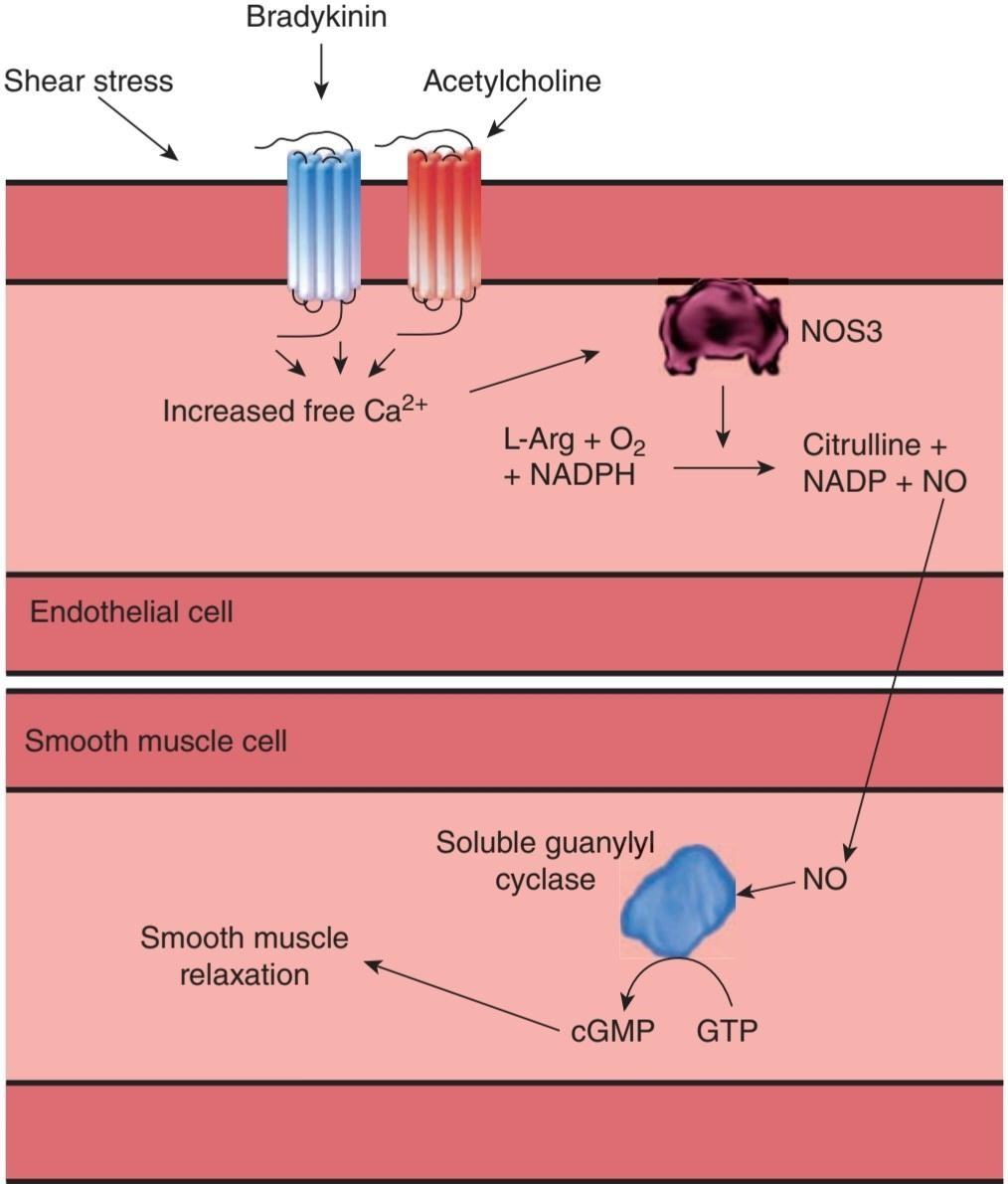
FIGURE 32-7 Endothelial-derived NO leads to smooth muscle relaxation. The endothelial form of nitric oxide synthase (NOS3) is activated by increased free Ca2+ and can be induced by a variety of extracellular signals. NOS3 acts on L-arginine, NADPH, and O2 to yield citrulline, NADP, and NO. A variety of cofactors are required for this reaction (not shown). NO can diffuse to adjacent smooth muscle cells where it activates soluble guanylyl cyclase. This leads to the production of cGMP and subsequent smooth muscle relaxation.
شکل 32-7 NO مشتق از اندوتلیال منجر به شل شدن عضلات صاف می شود. شکل اندوتلیال نیتریک اکساید سنتاز (NOS3) با افزایش +Ca2 آزاد فعال می شود و می تواند توسط انواع سیگنال های خارج سلولی القا شود. NOS3 روی ال-آرژنین، NADPH و O2 برای تولید سیترولین، NADP و NO عمل می کند. کوفاکتورهای مختلفی برای این واکنش مورد نیاز است (نشان داده نشده است). NO میتواند به سلولهای عضله صاف مجاور نفوذ کند، جایی که گوانیلیل سیکلاز محلول را فعال میکند. این منجر به تولید cGMP و متعاقب آن شل شدن عضلات صاف می شود.
Adenosine, atrial natriuretic peptide (ANP), and histamine via H2 receptors produce relaxation of vascular smooth muscle that is independent of the endothelium. However, acetylcholine, histamine via H1 receptors, bradykinin, vasoactive intestinal peptide (VIP), substance P, and some other polypeptides act via the endothelium, and various vasoconstrictors that act directly on vascular smooth muscle would produce much greater constriction if their effects were not limited by their ability simultaneously to cause release of NO. When flow to a tissue is suddenly increased by arteriolar dilation, the large arteries to the tissue also dilate. This flow-induced dilation is due to local release of NO. Products of platelet aggregation also cause release of NO, and the resulting vasodilation helps keep blood vessels with an intact endothelium patent. This is in contrast to injured blood vessels, where the endothelium is damaged at the site of injury and platelets therefore aggregate and produce vasoconstriction (see Chapter 31).
آدنوزین، پپتید ناتریورتیک دهلیزی (ANP) و هیستامین از طریق گیرنده های H2 باعث شل شدن عضلات صاف عروق می شود که مستقل از اندوتلیوم است. با این حال، استیل کولین، هیستامین از طریق گیرنده های H1، برادی کینین، پپتید وازواکتیو روده ای (VIP)، ماده P و برخی پلی پپتیدهای دیگر از طریق اندوتلیوم عمل می کنند، و منقبض کننده های عروق مختلف که مستقیماً بر روی ماهیچه صاف عروق تأثیر می گذارند، اگر اثرات آنها به دلیل توانایی آزادسازی NO به طور محدود محدود نشود، انقباض بیشتری ایجاد می کنند. هنگامی که جریان به یک بافت به طور ناگهانی توسط اتساع شریانی افزایش می یابد، شریان های بزرگ به بافت نیز منبسط می شوند. این اتساع ناشی از جریان به دلیل انتشار موضعی NO است. محصولات تجمع پلاکتی همچنین باعث آزاد شدن NO می شوند و اتساع عروقی حاصل از آن به حفظ رگ های خونی با اندوتلیوم دست نخورده کمک می کند. این برخلاف رگ های خونی آسیب دیده است، جایی که اندوتلیوم در محل آسیب آسیب می بیند و پلاکت ها جمع می شوند و باعث انقباض عروق می شوند (به فصل 31 مراجعه کنید).
Further evidence for a physiologic role of NO is the observation that mice lacking NOS 3 are hypertensive. This suggests that tonic release of NO is necessary to maintain normal blood pressure.
شواهد بیشتر برای نقش فیزیولوژیکی NO مشاهده این است که موش های فاقد NOS 3 دارای فشار خون بالا هستند. این نشان می دهد که آزادسازی تونیک NO برای حفظ فشار خون طبیعی ضروری است.
NO is also involved in vascular remodeling and angiogenesis, and NO may be involved in the pathogenesis of atherosclerosis. It is interesting in this regard that in some patients with heart transplants, an accelerated form of atherosclerosis develops in the vessels of the transplant, and there is reason to believe that this is triggered by endothelial damage. Nitroglycerin and other nitrovasodilators that are of great value in the treatment of angina act by stimulating guanylyl cyclase in the same manner as NO.
NO همچنین در بازسازی عروق و رگزایی نقش دارد و NO ممکن است در پاتوژنز آترواسکلروز نقش داشته باشد. از این نظر جالب است که در برخی از بیماران پیوند قلب، شکل تسریع شده آترواسکلروز در عروق پیوند ایجاد می شود و دلایلی وجود دارد که معتقد باشیم این امر در اثر آسیب اندوتلیال ایجاد می شود. نیتروگلیسیرین و دیگر گشادکنندههای نیترووازی که در درمان آنژین از ارزش بالایی برخوردار هستند، با تحریک گوانیلیل سیکلاز به روشی مشابه NO عمل میکنند.
Penile erection is also produced by release of NO, with consequent vasodilation and engorgement of the corpora cavernosa (see Chapter 23). This accounts for the efficacy of drugs such as Viagra that slow the breakdown of cGMP.
نعوظ آلت تناسلی نیز با آزاد شدن NO ایجاد می شود و در نتیجه اتساع عروق و انباشتگی اجسام غارنوردی ایجاد می شود (به فصل 23 مراجعه کنید). این به دلیل اثربخشی داروهایی مانند ویاگرا است که تجزیه cGMP را کند می کند.
OTHER FUNCTIONS OF NO
NO is present in the brain and, acting via cGMP, it is important in brain function (see Chapter 7). NO is also necessary for the antimicrobial and cytotoxic activity of various inflammatory cells, although the net effect of NO in inflammation and tissue injury depends on the amount and kinetics of release, which in turn may depend on the specific NOS isoform involved. In the gastrointestinal tract, NO is important in the relaxation of smooth muscle. Other functions of NO are mentioned in other parts of this book.
سایر عملکردهای NO
NO در مغز وجود دارد و با عمل از طریق cGMP، در عملکرد مغز مهم است (به فصل 7 مراجعه کنید). NO همچنین برای فعالیت ضد میکروبی و سیتوتوکسیک سلول های التهابی مختلف ضروری است، اگرچه اثر خالص NO در التهاب و آسیب بافت به مقدار و سینتیک آزادسازی بستگی دارد که به نوبه خود ممکن است به ایزوفرم NOS خاص درگیر بستگی داشته باشد. در دستگاه گوارش، NO در شل شدن عضلات صاف مهم است. سایر عملکردهای NO در قسمت های دیگر این کتاب ذکر شده است.
CARBON MONOXIDE
The production of carbon monoxide (CO) from heme is shown in Figure 28-4. Heme oxygenase-2 (HO-2), the enzyme that catalyzes the reaction, is also present in cardiovascular tissues, and there is growing evidence that CO as well as NO produces local dilation in blood vessels. Interestingly, hydrogen sulfide is likewise emerging as a third gas transmitter that regulates vascular tone, although the relative roles of NO, CO, and H2S have yet to be established.
مونوکسید کربن
تولید مونوکسید کربن (CO) از هم در شکل 28-4 نشان داده شده است. هم اکسیژناز 2 (HO-2)، آنزیمی که واکنش را کاتالیز میکند، در بافتهای قلبی عروقی نیز وجود دارد و شواهد فزایندهای وجود دارد که CO و همچنین NO باعث اتساع موضعی در رگهای خونی میشوند. جالب اینجاست که سولفید هیدروژن نیز به عنوان سومین فرستنده گازی ظاهر می شود که تون عروق را تنظیم می کند، اگرچه نقش نسبی NO، CO و H2S هنوز مشخص نشده است.
ENDOTHELINS
Endothelial cells also produce endothelin-1, one of the most potent vasoconstrictor agents yet isolated. Endothelin-1 (ET-1), endothelin-2 (ET-2), and endothelin-3 (ET-3) are the members of a family of three similar 21-amino- acid polypeptides. Each is encoded by a different gene.
اندوتلین ها
سلول های اندوتلیال همچنین اندوتلین-1 را تولید می کنند که یکی از قوی ترین عوامل منقبض کننده عروقی است که هنوز جدا شده است. اندوتلین-1 (ET-1)، اندوتلین-2 (ET-2) و اندوتلین-3 (ET-3) اعضای خانواده ای از سه پلی پپتید 21 آمینو اسیدی مشابه هستند. هر کدام توسط یک ژن متفاوت کدگذاری می شوند.
ENDOTHELIN-1
In endothelial cells, the product of the endothelin-1 gene is processed to a 39- amino-acid prohormone, big endothelin-1, which has about 1% of the activity of endothelin-1. The prohormone is cleaved by endothelin-converting enzyme at a tryptophan-valine (Trp-Val) bond to form endothelin-1. Small amounts of big endothelin-1 and endothelin-1 are secreted into the blood, but for the most part, they are secreted locally and act in a paracrine fashion.
اندوتلین-1
در سلول های اندوتلیال، محصول ژن اندوتلین-1 به یک پروهورمون 39 آمینو اسیدی به نام اندوتلین-1 بزرگ پردازش می شود که حدود 1 درصد از فعالیت اندوتلین-1 را دارد. پروهورمون توسط آنزیم تبدیل کننده اندوتلین در پیوند تریپتوفان-والین (Trp-Val) شکسته می شود و اندوتلین-1 را تشکیل می دهد. مقادیر کمی از اندوتلین-1 و اندوتلین-1 بزرگ در خون ترشح می شوند، اما در بیشتر موارد، به صورت موضعی ترشح می شوند و به روش پاراکرین عمل می کنند.
Two different endothelin receptors have been cloned, both of which are coupled via G-proteins to phospholipase C (see Chapter 2). The ET ̧ receptor, which is specific for endothelin-1, is found in many tissues and mediates the vasoconstriction produced by endothelin-1. The ETB receptor responds to all three endothelins and is coupled to G1. It may mediate vasodilation, and it appears to mediate the developmental effects of the endothelins (see below).
دو گیرنده اندوتلین مختلف شبیه سازی شده اند، که هر دو از طریق پروتئین های G به فسفولیپاز C جفت می شوند (به فصل 2 مراجعه کنید). گیرنده ET ̧ که مخصوص اندوتلین-1 است، در بسیاری از بافت ها یافت می شود و واسطه انقباض عروق تولید شده توسط اندوتلین-1 است. گیرنده ETB به هر سه اندوتلین پاسخ می دهد و با G1 جفت می شود. ممکن است واسطه اتساع عروق باشد و به نظر می رسد که واسطه اثرات رشدی اندوتلین ها باشد (به زیر مراجعه کنید).
REGULATION OF SECRETION
Endothelin-1 is not stored in secretory granules, and most regulatory factors alter the transcription of its gene, with changes in secretion occurring promptly thereafter. Factors activating and inhibiting the gene are summarized in Table 32-4.
مقررات ترشح
اندوتلین-1 در گرانول های ترشحی ذخیره نمی شود و بیشتر عوامل تنظیمی رونویسی ژن آن را تغییر می دهند و تغییراتی در ترشح بلافاصله پس از آن رخ می دهد. عوامل فعال و مهار کننده ژن در جدول 32-4 خلاصه شده است.
TABLE 32-4 Regulation of endothelin-1 secretion via transcription of its gene.
جدول 32-4 تنظیم ترشح اندوتلین-1 از طریق رونویسی ژن آن.

ANP, atrial natriuretic peptide; HDL, high-density lipoprotein; LDL, low-density lipoprotein; NO, nitric oxide; PGE2, prostaglandin E2; VIP, vasoactive intestinal polypeptide.
ANP، پپتید ناتریورتیک دهلیزی؛ HDL، لیپوپروتئین با چگالی بالا؛ LDL، لیپوپروتئین با چگالی کم؛ NO، اکسید نیتریک؛ PGE2، پروستاگلاندین E2؛ VIP، پلی پپتید وازواکتیو روده ای.
CARDIOVASCULAR FUNCTIONS
As noted above, endothelin-1 appears primarily to be a paracrine regulator of vascular tone. However, endothelin-1 is not increased in hypertension, and in mice in which one allele of the endothelin-1 gene is knocked out, blood pressure is actually elevated rather than reduced. The concentration of circulating endothelin-1 is, however, elevated in heart failure and after myocardial infarction, so it may play a role in the pathophysiology of these diseases.
عملکردهای قلبی عروقی
همانطور که در بالا ذکر شد، اندوتلین-1 در درجه اول یک تنظیم کننده پاراکرین تون عروقی به نظر می رسد. با این حال، اندوتلین-1 در فشار خون بالا افزایش نمی یابد، و در موش هایی که در آنها یکی از آلل های ژن اندوتلین-1 حذف شده است، فشار خون در واقع به جای کاهش، افزایش می یابد. غلظت اندوتلین 1 در گردش در نارسایی قلبی و پس از انفارکتوس میوکارد افزایش می یابد، بنابراین ممکن است در پاتوفیزیولوژی این بیماری ها نقش داشته باشد.
OTHER FUNCTIONS OF ENDOTHELINS
Endothelin-1 is found in the brain and kidneys as well as the endothelial cells. Endothelin-2 is produced primarily in the kidneys and intestine. Endothelin-3 is present in the blood and is found in high concentrations in the brain. It is also found in the kidneys and gastrointestinal tract. In the brain, endothelins are abundant and, in early life, are produced by both astrocytes and neurons. They are found in the dorsal root ganglia, ventral horn cells, the cortex, the hypothalamus, and cerebellar Purkinje cells. They also play a role in regulating transport across the blood-brain barrier. There are endothelin receptors on mesangial cells (see Chapter 37), and the polypeptide participates in tubuloglomerular feedback.
سایر عملکردهای اندوتلین ها
اندوتلین-1 در مغز و کلیه ها و همچنین سلول های اندوتلیال یافت می شود. اندوتلین-2 عمدتاً در کلیه ها و روده تولید می شود. اندوتلین-3 در خون وجود دارد و در غلظت های بالایی در مغز یافت می شود. همچنین در کلیه ها و دستگاه گوارش یافت می شود. در مغز، اندوتلین ها فراوان هستند و در اوایل زندگی، هم توسط آستروسیت ها و هم توسط نورون ها تولید می شوند. آنها در گانگلیون های ریشه پشتی، سلول های شاخ شکمی، قشر، هیپوتالاموس و سلول های پورکنژ مخچه یافت می شوند. آنها همچنین در تنظیم حمل و نقل از طریق سد خونی مغزی نقش دارند. گیرنده های اندوتلین روی سلول های مزانژیال وجود دارد (به فصل 37 مراجعه کنید)، و پلی پپتید در بازخورد توبولوگلومرولی شرکت می کند.
SYSTEMIC REGULATION BY NEUROHUMORAL AGENTS
Many circulating substances affect the vascular system. The vasodilator regulators include kinins, VIP, and ANP. Circulating vasoconstrictor hormones include vasopressin, norepinephrine, epinephrine, and angiotensin II.
تنظیم سیستمی توسط عوامل عصبی
بسیاری از مواد در گردش بر سیستم عروقی تأثیر می گذارند. تنظیم کننده های گشاد کننده عروق شامل کینین ها، VIP و ANP هستند. هورمون های منقبض کننده عروق در گردش عبارتند از وازوپرسین، نوراپی نفرین، اپی نفرین و آنژیوتانسین II.
KININS
Two related vasodilator peptides called kinins are found in the body. One is the nonapeptide bradykinin, and the other is the decapeptide kallidin, also known as lysylbradykinin (Figure 32-8). Kallidin can be converted to bradykinin by aminopeptidase. Both peptides are metabolized to forms that are active at the type 1 bradykinin receptor by kininase I, a carboxypeptidase that removes the carboxyl terminal arginine (Arg). In addition, the dipeptidylcarboxypeptidase kininase II inactivates bradykinin and kallidin by removing phenylalanine- arginine (Phe-Arg) from the carboxyl terminal as well as additional cleavages. Kininase II is the same enzyme as angiotensin-converting enzyme (ACE), which removes histidine-leucine (His-Leu) from the carboxyl terminal end of angiotensin I.
KININS
دو پپتید گشاد کننده عروق مرتبط به نام کینین در بدن یافت می شود. یکی غیراپپتید برادی کینین و دیگری دکاپپتید کالیدین است که به نام لیزیل برادیکینین نیز شناخته می شود (شکل 32-8). کالیدین می تواند توسط آمینوپپتیداز به برادی کینین تبدیل شود. هر دو پپتید به اشکالی متابولیزه می شوند که در گیرنده برادی کینین نوع 1 توسط کینیناز I، کربوکسی پپتیدازی که آرژنین انتهایی کربوکسیل (Arg) را حذف می کند، فعال هستند. علاوه بر این، دی پپتیدیل کربوکسی پپتیداز کینیناز II با حذف فنیل آلانین- آرژنین (Phe-Arg) از انتهای کربوکسیل و همچنین شکاف های اضافی، برادی کینین و کالیدین را غیرفعال می کند. کینیناز II همان آنزیم تبدیل کننده آنژیوتانسین (ACE) است که هیستیدین-لوسین (His-Leu) را از انتهای انتهایی کربوکسیل آنژیوتانسین I حذف می کند.

FIGURE 32-8 Kinins. Kallidin (lysylbradykinin) can be converted to bradykinin by aminopeptidase. Both kallidin and bradykinin act at type 2 bradykinin receptors (B2R). The peptides are converted by kininase I to their des-Arg derivatives that are ligands for type 1 bradykinin receptors (B1R). They can also be cleaved by angiotensin-converting enzyme (ACE, also known as kininase II) to shorter fragments that lack biologic activity.
شکل 32-8 کینین. کالیدین (لیزیل برادیکینین) می تواند توسط آمینوپپتیداز به برادی کینین تبدیل شود. هر دو کالیدین و برادی کینین در گیرنده های برادی کینین نوع 2 (B2R) عمل می کنند. پپتیدها توسط کینیناز I به مشتقات des-Arg تبدیل می شوند که لیگاندهای گیرنده های برادی کینین نوع 1 (B1R) هستند. آنها همچنین می توانند توسط آنزیم مبدل آنژیوتانسین (ACE، که به عنوان کینیناز II نیز شناخته می شود) به قطعات کوتاه تری که فاقد فعالیت بیولوژیکی هستند، تقسیم شوند.
Bradykinin and kallidin are formed from two precursor proteins: high- molecular-weight kininogen and low-molecular-weight kininogen (Figure 32-9). They are formed by alternative splicing of a single gene located on chromosome 3. Proteases called kallikreins release the peptides from their precursors. There are two types of kallikreins: plasma kallikrein, which circulates in an inactive form, and tissue kallikrein, which appears to be located primarily on the apical membranes of cells concerned with transcellular electrolyte transport. Tissue kallikrein is found in many tissues, including sweat and salivary glands, the pancreas, the prostate, the intestine, and the kidneys. Tissue kallikrein acts on high-molecular-weight kininogen to form bradykinin and low-molecular-weight kininogen to form kallidin. When activated, plasma kallikrein acts on high-molecular-weight kininogen to form bradykinin.
برادی کینین و کالیدین از دو پروتئین پیش ساز تشکیل می شوند: کینینوژن با وزن مولکولی بالا و کینینوژن با وزن مولکولی کم (شکل 32-9). آنها از پیوند جایگزین یک ژن منفرد واقع در کروموزوم 3 تشکیل می شوند. پروتئازهایی به نام کالیکرئین پپتیدها را از پیش سازهای خود آزاد می کنند. دو نوع کالیکرئین وجود دارد: کالیکرئین پلاسما، که به شکل غیر فعال در گردش است، و کالیکرئین بافت، که به نظر می رسد در درجه اول بر روی غشای آپیکال سلول های مرتبط با انتقال الکترولیت بین سلولی قرار دارد. کالیکرئین بافتی در بسیاری از بافت ها از جمله غدد عرق و بزاقی، پانکراس، پروستات، روده و کلیه ها یافت می شود. کالیکرئین بافت بر روی کینینوژن با وزن مولکولی بالا برای تشکیل برادی کینین و کینینوژن با وزن مولکولی پایین برای تشکیل کالیدین عمل می کند. وقتی کالیکرئین پلاسما فعال می شود، روی کینینوژن با وزن مولکولی بالا عمل می کند و برادی کینین را تشکیل می دهد.
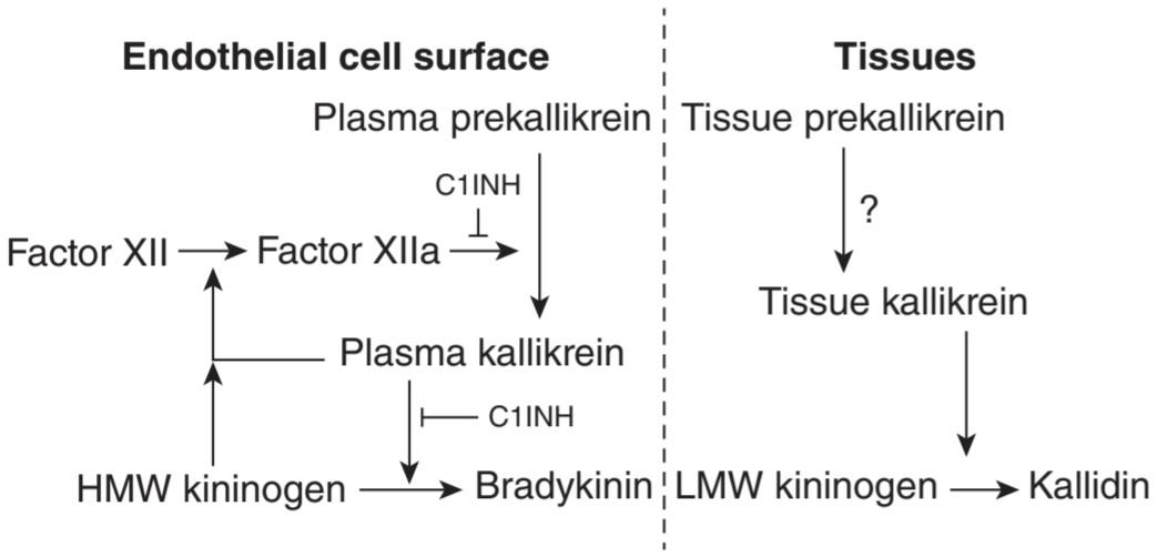
FIGURE 32-9 Formation of kinins from high-molecular-weight (HMW) and low-molecular-weight (LMW) kininogens. Note that the formation of bradykinin is subject to a positive feedback loop that can be interrupted in two places by C1-esterase inhibitor (C1INH).
شکل 32-9 تشکیل کینین ها از کینینوژن های با وزن مولکولی بالا (HMW) و وزن مولکولی پایین (LMW). توجه داشته باشید که تشکیل برادی کینین در معرض یک حلقه بازخورد مثبت است که می تواند در دو مکان توسط مهارکننده C1-esterase (C1INH) قطع شود.
Inactive plasma kallikrein (prekallikrein) is converted to the active form, kallikrein, by active factor XII, the factor that initiates the intrinsic blood clotting cascade. Kallikrein also activates factor XII in a positive feedback loop, and high-molecular-weight kininogen has a factor XII-activating action (see Figure 31-12). This system is normally kept in check by the C1-esterase inhibitor (C1INH), which is a member of the serpin protein family that inhibits several enzymes in the clotting cascade by binding irreversibly to their active sites.
کالیکرئین پلاسمای غیر فعال (پرکالیکرئین) توسط فاکتور XII فعال به شکل فعال کالیکرئین تبدیل می شود، عاملی که آبشار لخته شدن خون را آغاز می کند. Kallikrein همچنین فاکتور XII را در یک حلقه بازخورد مثبت فعال می کند و کینینوژن با وزن مولکولی بالا دارای یک عمل فعال کننده فاکتور XII است (شکل 31-12 را ببینید). این سیستم معمولاً توسط بازدارنده C1-esterase (C1INH) کنترل می شود، که عضوی از خانواده پروتئین سرپین است که چندین آنزیم را در آبشار لخته شدن با اتصال غیرقابل برگشت به مکان های فعال آنها مهار می کند.
The actions of both kinins resemble those of histamine. They are primarily paracrines, although small amounts are also found in the circulating blood. They cause contraction of visceral smooth muscle, but they relax vascular smooth muscle via NO, lowering blood pressure. They also significantly increase vascular permeability resulting in edema, attract leukocytes, and cause pain upon injection under the skin. They are formed during active secretion in sweat glands, salivary glands, and the exocrine portion of the pancreas, and they are probably responsible for the increase in blood flow when these tissues are actively secreting their products. In keeping with the scheme for bradykinin production outlined above, mutations in C1INH lead to the rare bradykinin- mediated swelling disorder known as hereditary angioedema.
عملکرد هر دو کینین شبیه هیستامین است. آنها در درجه اول پاراکرین هستند، اگرچه مقادیر کمی نیز در خون در گردش یافت می شود. آنها باعث انقباض ماهیچه صاف احشایی می شوند، اما عضلات صاف عروق را از طریق NO شل می کنند و فشار خون را کاهش می دهند. آنها همچنین به طور قابل توجهی نفوذپذیری عروقی را افزایش می دهند که منجر به ادم، جذب لکوسیت ها و ایجاد درد در هنگام تزریق در زیر پوست می شود. آنها در طول ترشح فعال در غدد عرق، غدد بزاقی و بخش برون ریز پانکراس تشکیل می شوند و احتمالاً مسئول افزایش جریان خون زمانی هستند که این بافت ها به طور فعال محصولات خود را ترشح می کنند. مطابق با طرح تولید برادی کینین که در بالا ذکر شد، جهش در C1INH منجر به اختلال نادر تورم ناشی از برادی کینین می شود که به عنوان آنژیوادم ارثی شناخته می شود.
Two bradykinin receptors, B1R and B2R, have been identified. The B1R may mediate the pain-producing effects of the kinins, and antagonists have entered clinical trials for the treatment of pain and inflammation. The B2 receptor has strong homology to the histamine H2-receptor and is found in many different tissues.
دو گیرنده برادی کینین، B1R و B2R، شناسایی شده است. B1R ممکن است واسطه اثرات تولید درد کینین ها باشد و آنتاگونیست ها برای درمان درد و التهاب وارد آزمایشات بالینی شده اند. گیرنده B2 همسانی قوی با گیرنده H2 هیستامین دارد و در بسیاری از بافت های مختلف یافت می شود.
NATRIURETIC HORMONES
A family of natriuretic peptides is also involved in vascular regulation, including ANP secreted by the heart, B-type natriuretic peptide (BNP), and C-type natriuretic peptide (CNP). They are released in response to hypervolemia. ANP and BNP circulate, whereas CNP acts predominantly in a paracrine fashion. In general, these peptides antagonize the action of various vasoconstrictor agents and lower blood pressure. ANP and BNP also serve to coordinate the control of vascular tone with fluid and electrolyte homeostasis via actions on the kidney.
هورمون های ناتریورتیک
خانواده ای از پپتیدهای ناتریورتیک نیز در تنظیم عروق نقش دارند، از جمله ANP ترشح شده از قلب، پپتید ناتریورتیک نوع B (BNP) و پپتید ناتریورتیک نوع C (CNP). آنها در پاسخ به هیپرولمی آزاد می شوند. ANP و BNP در گردش هستند، در حالی که CNP عمدتاً به صورت پاراکرین عمل می کند. به طور کلی، این پپتیدها با عملکرد عوامل مختلف منقبض کننده عروق مخالفت می کنند و فشار خون را کاهش می دهند. ANP و BNP همچنین برای هماهنگ کردن کنترل تون عروقی با هموستاز مایعات و الکترولیت ها از طریق اعمال روی کلیه عمل می کنند.
CIRCULATING VASOCONSTRICTORS
Vasopressin is a potent vasoconstrictor, but when it is injected in healthy individuals, there is a compensating decrease in cardiac output, so that there is little change in blood pressure. Its role in blood pressure regulation is discussed in Chapter 17.
منقبض کننده های عروقی در گردش
وازوپرسین یک منقبض کننده عروق قوی است، اما هنگامی که در افراد سالم تزریق می شود، کاهش جبرانی در برون ده قلبی وجود دارد، به طوری که تغییر کمی در فشار خون ایجاد می شود. نقش آن در تنظیم فشار خون در فصل 17 مورد بحث قرار گرفته است.
Norepinephrine has a generalized vasoconstrictor action, whereas epinephrine dilates the vessels in skeletal muscle and the liver. The relative unimportance of circulating norepinephrine, as opposed to norepinephrine released from vasomotor nerves, is pointed out in Chapter 19, where the cardiovascular actions of catecholamines are discussed in detail.
نوراپی نفرین دارای یک اثر منقبض کننده عروق عمومی است، در حالی که اپی نفرین عروق ماهیچه اسکلتی و کبد را گشاد می کند. بی اهمیت بودن نسبی نوراپی نفرین در گردش، در مقابل نوراپی نفرین آزاد شده از اعصاب وازوموتور، در فصل 19 اشاره شده است، جایی که اعمال قلبی عروقی کاتکول آمین ها به تفصیل مورد بحث قرار می گیرد.
Angiotensin II has a generalized vasoconstrictor action. It is formed by the action of ACE on angiotensin I, which itself is liberated by the action of renin from the kidney on circulating angiotensinogen (see Chapter 38). Renin secretion, in turn, is increased when the blood pressure falls or extracellular fluid (ECF) volume is reduced, and angiotensin II therefore helps maintain blood pressure. Angiotensin II also increases water intake and stimulates aldosterone secretion, and increased formation of angiotensin II is part of a homeostatic mechanism that operates to maintain ECF volume (see Chapter 19). In addition, there are renin-angiotensin systems in many different organs, including the walls of blood vessels. Angiotensin II produced in blood vessel walls could be important in some forms of clinical hypertension. The role of angiotensin II in cardiovascular regulation is also amply demonstrated in the widespread use of ACE inhibitors as antihypertensive medications.
آنژیوتانسین II یک اثر منقبض کننده عروق عمومی دارد. این توسط اثر ACE بر روی آنژیوتانسین I ایجاد می شود که خود با اثر رنین از کلیه بر روی آنژیوتانسینوژن در گردش آزاد می شود (به فصل 38 مراجعه کنید). ترشح رنین به نوبه خود با کاهش فشار خون یا کاهش حجم مایع خارج سلولی (ECF) افزایش می یابد و بنابراین آنژیوتانسین II به حفظ فشار خون کمک می کند. آنژیوتانسین II همچنین مصرف آب را افزایش میدهد و ترشح آلدوسترون را تحریک میکند، و افزایش تشکیل آنژیوتانسین II بخشی از یک مکانیسم هموستاتیک است که برای حفظ حجم ECF عمل میکند (به فصل 19 مراجعه کنید). علاوه بر این، سیستم های رنین-آنژیوتانسین در بسیاری از اندام های مختلف از جمله دیواره رگ های خونی وجود دارد. آنژیوتانسین II تولید شده در دیواره رگ های خونی می تواند در برخی از اشکال فشار خون بالینی مهم باشد. نقش آنژیوتانسین II در تنظیم قلبی عروقی نیز در استفاده گسترده از مهارکننده های ACE به عنوان داروهای ضد فشار خون به طور گسترده نشان داده شده است.
Urotensin-II, a polypeptide first isolated from the spinal cord of fish, is present in human cardiac and vascular tissue. It is one of the most potent mammalian vasoconstrictors known, and is being explored for its role in a large range of different human disease states. For example, levels of both urotensin-II and its receptor have been shown to be elevated in hypertension and heart failure, and may be markers of disease in these and other conditions.
Urotensin-II، پلی پپتیدی که ابتدا از نخاع ماهی جدا شد، در بافت قلب و عروق انسان وجود دارد. این یکی از قوی ترین منقبض کننده های عروق پستانداران شناخته شده است و به دلیل نقش آن در طیف وسیعی از بیماری های مختلف انسانی مورد بررسی قرار گرفته است. به عنوان مثال، نشان داده شده است که هم سطح urotensin-II و هم گیرنده آن در فشار خون بالا و نارسایی قلبی بالا است و ممکن است نشانگر بیماری در این شرایط و سایر شرایط باشد.
CHAPTER SUMMARY
خلاصه فصل
• The vasculature is innervated by sympathetic nerves that mediate vasoconstriction; the heart is innervated both by sympathetic nerves that mediate an increase in heart rate, AV conduction, and ventricular contractility and by parasympathetic (vagal) nerves that produce a reduction in heart rate and conduction through the AV node.
• عروق توسط اعصاب سمپاتیک که واسطه انقباض عروق هستند عصب دهی می شود. قلب هم توسط اعصاب سمپاتیک که واسطه افزایش ضربان قلب، هدایت AV و انقباض بطنی هستند و هم توسط اعصاب پاراسمپاتیک (واگ) که باعث کاهش ضربان قلب و هدایت از طریق گره AV می شوند، عصب دهی می شود.
• RVLM neurons project to the thoracolumbar IML and release glutamate on preganglionic sympathetic neurons that innervate the heart and vasculature. The NTS is the major excitatory input to cardiac vagal motor neurons in the nucleus ambiguus. RVLM and NTS neurons receive input from other brain regions, baroreceptors, chemoreceptors, and other sources.
• نورون های RVLM به سمت IML قفسه سینه ی کمری حرکت می کنند و گلوتامات را روی نورون های سمپاتیک پیش گانگلیونی آزاد می کنند که قلب و عروق را عصب دهی می کنند. NTS ورودی تحریک کننده اصلی به نورون های حرکتی واگ قلب در ابهام هسته است. نورون های RVLM و NTS ورودی را از سایر مناطق مغز، بارورسپتورها، گیرنده های شیمیایی و سایر منابع دریافت می کنند.
• Carotid sinus and aortic depressor baroreceptors are innervated by branches of the ninth and tenth cranial nerves, respectively (glossopharyngeal and aortic depressor nerves). These receptors are most sensitive to changes in pulse pressure but also respond to changes in mean arterial pressure. Baroreceptor nerves terminate in the NTS and release glutamate. NTS neurons project to the CVLM and nucleus ambiguus and release glutamate. CVLM neurons project to RVLM and release GABA. This leads to a reduction in sympathetic activity and an increase in vagal activity (ie, the baroreceptor reflex).
• بارورسپتورهای فشاردهنده سینوس کاروتید و آئورت به ترتیب توسط شاخههای اعصاب نهم و دهم جمجمه (اعصاب کاهشدهنده گلوسوفارنکس و آئورت) عصببندی میشوند. این گیرندهها نسبت به تغییرات فشار پالس حساسترین هستند، اما به تغییرات فشار متوسط شریانی نیز پاسخ میدهند. اعصاب بارورسپتور در NTS خاتمه یافته و گلوتامات آزاد می کنند. نورون های NTS به CVLM و ابهام هسته پرتاب می شوند و گلوتامات آزاد می کنند. نورون های CVLM به RVLM پروژه می دهند و GABA را آزاد می کنند. این منجر به کاهش فعالیت سمپاتیک و افزایش فعالیت واگ می شود (یعنی رفلکس بارورسپتور).
• The reflex circulatory adjustments initiated by activation of atrial stretch receptors include a reduction in blood pressure and an increase in heart rate. Activation of ventricular stretch receptors induces hypotension and bradycardia. Activation of cardiopulmonary chemoreceptors induces the Bezold-Jarisch reflex that plays a role in various cardiovascular disorders (eg, myocardial ischemia and reperfusion and vasovagal syncope).
• تنظیمات رفلکس گردش خون که با فعال شدن گیرنده های کشش دهلیزی آغاز می شود شامل کاهش فشار خون و افزایش ضربان قلب است. فعال شدن گیرنده های کشش بطنی باعث کاهش فشار خون و برادی کاردی می شود. فعالسازی گیرندههای شیمیایی قلبی ریوی، رفلکس بزولد-جاریش را القا میکند که در اختلالات قلبی عروقی مختلف (مانند ایسکمی میوکارد و خونرسانی مجدد و سنکوپ وازوواگال) نقش دارد.
• Activation of peripheral chemoreceptors in the carotid and aortic bodies, primarily by a reduction in PaO2, leads to an increase in vasoconstriction. Heart rate changes are variable and depend on various factors including changes in respiration. Central chemoreceptors are activated primarily by hypercapnia; activation leads to an increase in blood pressure and a baroreceptor reflex-induced bradycardia.
• فعال شدن گیرنده های شیمیایی محیطی در بدن کاروتید و آئورت، عمدتاً با کاهش PaO2، منجر به افزایش انقباض عروق می شود. تغییرات ضربان قلب متغیر است و به عوامل مختلفی از جمله تغییر در تنفس بستگی دارد. گیرنده های شیمیایی مرکزی در درجه اول توسط هیپرکاپنی فعال می شوند. فعال شدن منجر به افزایش فشار خون و برادی کاردی ناشی از رفلکس بارورسپتور می شود.
• Most vascular beds have a capacity to respond to changes in blood pressure within a certain range by altering vascular resistance to maintain stable blood flow. This property is known as autoregulation and is related to intrinsic properties of the vascular smooth muscle as well as the actions of local vasoactive metabolites.
• بیشتر بسترهای عروقی این ظرفیت را دارند که به تغییرات فشار خون در محدوده معینی با تغییر مقاومت عروقی برای حفظ جریان خون پایدار پاسخ دهند. این خاصیت به عنوان خودتنظیمی شناخته می شود و به ویژگی های ذاتی ماهیچه صاف عروق و همچنین عملکرد متابولیت های وازواکتیو موضعی مربوط می شود.
• Local factors such as oxygen tension, pH, temperature, and metabolic products contribute to vascular regulation; many produce vasodilation to restore blood flow.
• عوامل محلی مانند تنش اکسیژن، pH، دما و محصولات متابولیک به تنظیم عروق کمک می کنند. بسیاری از آنها اتساع عروقی را برای بازگرداندن جریان خون ایجاد می کنند.
• The endothelium is an important source of vasoactive mediators that act to either contract or relax vascular smooth muscle. In particular, three gaseous mediators—NO, CO, and H2S—are important regulators of vasodilation.
• اندوتلیوم منبع مهمی از واسطه های وازواکتیو است که برای انقباض یا شل کردن عضلات صاف عروق عمل می کنند. به طور خاص، سه واسطه گازی – NO، CO، و H2S – تنظیم کننده مهم اتساع عروق هستند.
• Endothelins and angiotensin II induce vasoconstriction and may be involved in the pathogenesis of some forms of hypertension.
• اندوتلین ها و آنژیوتانسین II باعث انقباض عروق می شوند و ممکن است در پاتوژنز برخی از اشکال فشار خون بالا نقش داشته باشند.
MULTIPLE-CHOICE QUESTIONS
For all questions, select the single best answer unless otherwise directed.
سوالات چند گزینه ای
برای همه سؤالات، بهترین پاسخ را انتخاب کنید، مگر اینکه دستور دیگری داده شود.
1. When a pheochromocytoma (tumor of the adrenal medulla) suddenly discharges a large amount of epinephrine into the circulation, the patient’s heart rate would be expected to
A. increase because the increase in blood pressure stimulates the carotid and aortic baroreceptors.
B. increase because epinephrine has a direct chronotropic effect on the heart.
C. increase because of increased tonic parasympathetic discharge to the heart.
D. decrease because the increase in blood pressure stimulates the carotid and aortic chemoreceptors.
E. decrease because of increased tonic parasympathetic discharge to the heart.
1. هنگامی که یک فئوکروموسیتوم (تومور مدولای آدرنال) به طور ناگهانی مقدار زیادی اپی نفرین را وارد گردش خون می کند، انتظار می رود ضربان قلب بیمار
الف. افزایش می یابد زیرا افزایش فشار خون باعث تحریک بارورسپتورهای کاروتید و آئورت می شود.
ب. افزایش می یابد زیرا اپی نفرین اثر کرونوتروپیک مستقیم بر قلب دارد.
ج. افزایش به دلیل افزایش ترشحات پاراسمپاتیک تونیک به قلب.
د. کاهش می یابد زیرا افزایش فشار خون باعث تحریک گیرنده های شیمیایی کاروتید و آئورت می شود.
ی. کاهش به دلیل افزایش ترشحات پاراسمپاتیک تونیک به قلب.
2. A 45-year-old woman had a blood pressure of 155/95 mm Hg when she was at her physician’s office for a physical. She was worried as this was her first physical in over 10 years. The clinician suggested that she begin monitoring her blood pressure at home. Stress-induced activation of which neurons might contribute to the higher BP while in her physician’s office?
A. Limbic cortex and NTS
B. Hypothalamus and CVLM
C. NTS and CVLM
D. Hypothalamus and RVLM
E. Limbic cortex and nucleus ambiguus
2. زنی 45 ساله زمانی که برای معاینه فیزیکی در مطب پزشکش بود فشار خون 155/95 میلی متر جیوه داشت. او نگران بود زیرا این اولین بار در 10 سال گذشته بود. پزشک به او پیشنهاد کرد که فشار خون خود را در خانه کنترل کند. فعالسازی ناشی از استرس کدام نورونها ممکن است در هنگام حضور در مطب پزشک به فشار خون بالاتر کمک کنند؟
الف. قشر لیمبیک و NTS
ب. هیپوتالاموس و CVLM
ج. NTS و CVLM
د. هیپوتالاموس و RVLM
ی. قشر لیمبیک و هسته مبهم
3. Which group of neurons within central autonomic pathways release glutamate on their postsynaptic targets?
A. CVLM neurons projecting to the IML and NTS neurons projecting to the nucleus ambiguus.
B. RVLM neurons projecting to the IML and NTS neurons projecting to the CVLM.
C. NTS neurons projecting to the nucleus ambiguus and CVLM neurons projecting to the RVLM.
D. NTS neurons projecting to the RVLM and CVLM neurons projecting to the RVLM.
E. CVLM neurons projecting to the NTS and RVLM neurons projecting to the IML.
3. کدام گروه از نورون ها در مسیرهای اتونوم مرکزی گلوتامات را روی اهداف پس سیناپسی خود آزاد می کنند؟
الف. نورونهای CVLM که به نورونهای IML و NTS پیشتاب میکنند به ابهام هسته.
ب. نورون های RVLM که به IML و نورون های NTS که به CVLM برمی خیزند.
ج. نورونهای NTS که به هسته مبهم و نورونهای CVLM برمیتابند به RVLM.
د. نورون های NTS که به RVLM و نورون های CVLM که به RVLM برمی خیزند.
ی. نورون های CVLM که به NTS و نورون های RVLM که به IML برآمده اند.
4. List the location of the arterial baroreceptors, their afferent innervation, and the central site of termination of the afferent fibers.
A. Atria and pulmonary veins, vagus nerve, and nucleus ambiguus
B. Carotid body and aortic body, glossopharyngeal nerve and vagus nerve, and NTS
C. Carotid sinus and aortic arch, carotid sinus nerve and aortic depressor nerve, and NTS
D. Carotid sinus and atria, glossopharyngeal and vagus nerves, and nucleus ambiguus
E. Carotid body and ventricle, dorsal root fibers, and RVLM
4. محل بارورسپتورهای شریانی، عصب آوران آنها و محل مرکزی خاتمه فیبرهای آوران را فهرست کنید.
الف. دهلیزها و وریدهای ریوی، عصب واگ و هسته مبهم
ب. بدن کاروتید و بدن آئورت، عصب گلوفارنکس و عصب واگ و NTS
ج. سینوس کاروتید و قوس آئورت، عصب سینوس کاروتید و عصب دپرسور آئورت و NTS
د. سینوس و دهلیز کاروتید، اعصاب گلوفارنکس و واگ، و هسته مبهم
ی. بدن و بطن کاروتید، الیاف ریشه پشتی و RVLM
5. A 30-year-old woman experienced profound bradycardia, hypotension, and a brief period of apnea followed by rapid shallow breathing. This description is consistent with the activation of which of the following reflexes?
A. Cushing reflex
B. Central chemoreceptor reflex
C. Peripheral chemoreceptor reflex
D. Atrial stretch receptor reflex
E. Bezold-Jarisch reflex
5. یک زن 30 ساله برادی کاردی عمیق، افت فشار خون و یک دوره کوتاه آپنه و به دنبال تنفس سریع کم عمق را تجربه کرد. این توصیف با فعال شدن کدام یک از رفلکس های زیر مطابقت دارد؟
الف. رفلکس کوشینگ
ب. رفلکس گیرنده شیمیایی مرکزی
ج. رفلکس گیرنده شیمیایی محیطی
د. رفلکس گیرنده کششی دهلیزی
ی. رفلکس بزولد-جاریش
6. A 53-year-old woman with chronic lung disease was experiencing difficulty breathing. Her PaO2 and PaCO2 were 50 mm Hg and 60 mm Hg, respectively. Peripheral chemoreceptors are very sensitive to and central chemoreceptors are very sensitive to Fill in the missing information
A. Small increases in PaCO2 and small decreases in local partial pressure of 02.
B. Small decreases in PaCO2 and small increases in local partial pressure of 02.
C. Small decreases in PaO2 and small increases in local partial pressure of CO2.
D. Small increases in PaO2 and small decreases in local partial pressure of CO2
E. Small decreases in arterial pH and small increases in local pH.
6. زن 53 ساله مبتلا به بیماری مزمن ریوی دچار مشکل تنفسی شده بود. PaO2 و PaCO2 او به ترتیب 50 میلی متر جیوه و 60 میلی متر جیوه بود. گیرنده های شیمیایی محیطی بسیار حساس و مرکزی هستند گیرنده های شیمیایی نسبت به پر کردن اطلاعات از دست رفته بسیار حساس هستند
الف. افزایش اندک در PaCO2 و کاهش اندک در فشار جزئی موضعی 02.
ب. کاهش اندک در PaCO2 و افزایش اندک در فشار جزئی موضعی 02.
ج. کاهش اندک در PaO2 و افزایش اندک در فشار جزئی موضعی CO2.
د. افزایش اندک در PaO2 و کاهش اندک در فشار جزئی موضعی CO2
ی. کاهش اندک در pH شریانی و افزایش اندک در pH موضعی.
7. Due to a partial vascular occlusion, a portion of the small bowel becomes hypoxic. Factors that would likely contribute to an adaptive increase in blood flow in the affected segment would include all of the following except
A. HIF-1α
B. Increased local tension of CO2
C. Relaxation of precapillary sphincters
D. Increased local concentration of K+
E. Changes in gene expression
7. به دلیل انسداد نسبی عروق، قسمتی از روده کوچک هیپوکسیک می شود. عواملی که احتمالاً به افزایش تطبیقی در جریان خون در بخش آسیبدیده کمک میکنند شامل همه موارد زیر میشود به جز
الف. HIF-1α
ب. افزایش تنش موضعی CO2
ج. شل شدن اسفنکترهای پیش مویرگی
د. افزایش غلظت موضعی +K
ی. تغییرات در بیان ژن
8. A 55-year-old man comes to his primary care clinician complaining of erectile dysfunction. He is given a prescription for Viagra, and on follow-up, reports that his ability to sustain an erection has been improved markedly by this treatment. The action of which of the following vasoactive mediators would primarily be increased in this patient?
A. Histamine
B. Endothelin-1
C. Prostacyclin
D. Nitric oxide
E. Atrial natriuretic peptide
8. مردی 55 ساله با شکایت از اختلال نعوظ به پزشک مراقبت های اولیه مراجعه می کند. به او نسخه ای برای ویاگرا داده می شود و در پیگیری، گزارش می دهد که توانایی او برای حفظ نعوظ به طور قابل توجهی با این درمان بهبود یافته است. عمل کدام یک از واسطه های وازواکتیو زیر در این بیمار در درجه اول افزایش می یابد؟
الف. هیستامین
ب. اندوتلین-1
ج. پروستاسیکلین
د. اکسید نیتریک
ی. پپتید ناتریورتیک دهلیزی
9. Why is the dilator response to injected acetylcholine changed to a constrictor response when the endothelium is damaged?
A. More Nat is generated.
B. More bradykinin is generated.
C. The damage lowers the pH of the remaining layers of the artery.
D. The damage augments the production of endothelin by the endothelium.
E. The damage interferes with the production of NO by the endothelium.
9. چرا وقتی اندوتلیوم آسیب می بیند، پاسخ گشادکننده به استیل کولین تزریقی به پاسخ منقبض کننده تغییر می کند
الف. +Na بیشتری تولید می شود.
ب. برادی کینین بیشتری تولید می شود.
ج. آسیب باعث کاهش PH لایه های باقیمانده شریان می شود.
د. آسیب باعث افزایش تولید اندوتلین توسط اندوتلیوم می شود.
ی. آسیب با تولید NO توسط اندوتلیوم تداخل می کند.
کلیک کنید «منابع»
🚀 با ما همراه شوید!
تازهترین مطالب و آموزشهای مغز و اعصاب را از دست ندهید. با فالو کردن کانال تلگرام آیندهنگاران مغز، از ما حمایت کنید!



