
فیزیولوژی پزشکی گانونگ؛ خون به عنوان یک مایع گردش خون و دینامیک جریان خون و لنف


دعای مطالعه [ نمایش ]
بِسْمِ الله الرَّحْمنِ الرَّحیمِ
اَللّهُمَّ اَخْرِجْنى مِنْ ظُلُماتِ الْوَهْمِ
خدايا مرا بيرون آور از تاريكىهاى وهم،
وَ اَكْرِمْنى بِنُورِ الْفَهْمِ
و به نور فهم گرامى ام بدار،
اَللّهُمَّ افْتَحْ عَلَيْنا اَبْوابَ رَحْمَتِكَ
خدايا درهاى رحمتت را به روى ما بگشا،
وَانْشُرْ عَلَيْنا خَزائِنَ عُلُومِكَ بِرَحْمَتِكَ يا اَرْحَمَ الرّاحِمينَ
و خزانههاى علومت را بر ما باز كن به امید رحمتت اى مهربانترين مهربانان.
کتاب «فیزیولوژی پزشکی گانونگ» بهعنوان یکی از جامعترین و معتبرترین منابع در حوزه علوم پزشکی، همچنان مرجع کلیدی برای درک عملکرد پیچیده بدن انسان است. این اثر با تکیه بر تازهترین پژوهشها و توضیحات دقیق از سازوکارهای فیزیولوژیک، پلی میان علوم پایه پزشکی و کاربردهای بالینی ایجاد میکند و نقشی بیبدیل در آموزش، پژوهش و ارتقای دانش سلامت ایفا مینماید.
ترجمه دقیق و علمی این شاهکار توسط برند علمی آیندهنگاران مغز به مدیریت داریوش طاهری، دسترسی فارسیزبانان به مرزهای نوین دانش فیزیولوژی را ممکن ساخته و رسالتی علمی برای ارتقای آموزش پزشکی، فهم عمیقتر سازوکارهای بدن و توسعه روشهای نوین در حوزه سلامت فراهم آورده است.
» کتاب فیزیولوژی پزشکی گانونگ
» » فصل ۳۱: خون به عنوان یک مایع گردش خون و دینامیک جریان خون و لنف
در حال ویرایش
» Ganong’s Review of Medical Physiology
»» CHAPTER 31: Blood as a Circulatory Fluid & the Dynamics of Blood & Lymph Flow
OBJECTIVES
After studying his chapter you should be able to:
• Describe the cellular components of blood, and their origins, how the fragility of red blood cells is controlled, the role of hemoglobin in transporting oxygen in red blood cells, and the basis of hemoglobinopathies.
• Define the composition of plasma and lymph and identify the sources of their constituents.
• Understand the molecular basis of blood groups and the reasons for transfusion reactions.
• Delineate the process of hemostasis that restricts blood loss when vessels are damaged, the adverse consequences of intravascular thrombosis, and the basis of clotting disorders.
• Identify the types of blood and lymphatic vessels that make up the circulatory system and the regulation and function of their primary constituent cell types.
• Describe how physical principles dictate the flow of blood and lymph around the body.
• Understand the basis of methods used to measure blood flow and blood pressure in various vascular segments.
• Understand the basis and consequences of a sustained elevation of systemic arterial pressure (hypertension).
اهداف
پس از مطالعه فصل، باید بتوانید:
• اجزای سلولی خون و منشا آنها، نحوه کنترل شکنندگی گلبول های قرمز، نقش هموگلوبین در انتقال اکسیژن در گلبول های قرمز و اساس هموگلوبینوپاتی ها را شرح دهید.
• ترکیب پلاسما و لنف را تعریف کنید و منابع تشکیل دهنده آنها را شناسایی کنید.
• اساس مولکولی گروه های خونی و دلایل واکنش های انتقال خون را درک کنید.
• روند هموستاز را که از دست دادن خون در هنگام آسیب دیدگی عروق، پیامدهای نامطلوب ترومبوز داخل عروقی و اساس اختلالات لخته شدن خون را محدود می کند، مشخص کنید.
• انواع رگ های خونی و لنفاوی که سیستم گردش خون را تشکیل می دهند و تنظیم و عملکرد انواع سلول های تشکیل دهنده اولیه آنها را شناسایی کنید.
• توضیح دهید که چگونه اصول فیزیکی جریان خون و لنف در بدن را تعیین می کند.
• اساس روش های مورد استفاده برای اندازه گیری جریان خون و فشار خون در بخش های مختلف عروقی را بدانید.
• اساس و پیامدهای افزایش مداوم فشار شریانی سیستمیک (فشار خون) را درک کنید.

INTRODUCTION
The circulatory system supplies inspired O2 as well as substances absorbed from the gastrointestinal tract to the tissues, returns CO2 to the lungs and other products of metabolism to the kidneys, functions in the regulation of body temperature, and distributes hormones and other agents that regulate cell function. The blood, the carrier of these substances, is pumped through a closed system of blood vessels by the heart. From the left ventricle, blood is pumped through the arteries and arterioles to the capillaries, where it equilibrates with the interstitial fluid. The capillaries drain through venules into the veins and back to the right atrium. Some tissue fluids enter another system of closed vessels, the lymphatics, which drain lymph via the thoracic duct and the right lymphatic duct into the venous system. The circulation is controlled by multiple regulatory systems that function in general to maintain adequate capillary blood flow when possible in all organs, but particularly in the heart and brain.
مقدمه
سیستم گردش خون O2 الهام گرفته شده و همچنین مواد جذب شده از دستگاه گوارش را به بافت ها می رساند، CO2 را به ریه ها و سایر محصولات متابولیسم را به کلیه ها برمی گرداند، در تنظیم دمای بدن عمل می کند و هورمون ها و سایر عواملی را که عملکرد سلول را تنظیم می کنند توزیع می کند. خون، حامل این مواد، از طریق یک سیستم بسته از رگ های خونی توسط قلب پمپ می شود. از بطن چپ، خون از طریق شریانها و شریانها به مویرگها پمپ میشود، جایی که با مایع بینابینی تعادل برقرار میکند. مویرگ ها از طریق ونول ها به داخل سیاهرگ ها تخلیه می شوند و به دهلیز راست باز می گردند. برخی از مایعات بافتی وارد سیستم دیگری از عروق بسته می شوند، لنفاوی، که لنف را از طریق مجرای قفسه سینه و مجرای لنفاوی سمت راست را به سیستم وریدی تخلیه می کند. گردش خون توسط سیستم های تنظیمی متعددی کنترل می شود که به طور کلی برای حفظ جریان خون مویرگی کافی در صورت امکان در همه اندام ها، به ویژه در قلب و مغز، عمل می کنند.
Blood flows through the circulation primarily because of the forward motion imparted to it by the pumping of the heart, although in the case of the systemic circulation, diastolic recoil of the walls of the arteries, compression of the veins by skeletal muscles during exercise, and the negative pressure in the thorax during inspiration also move the blood forward. The resistance to flow depends to a minor degree on the viscosity of the blood but mostly on the diameter of the vessels, and principally that of the arterioles. The blood flow to each tissue is regulated by local chemical and general neural and humoral mechanisms that dilate or constrict its vessels. All of the blood flows through the lungs, but the systemic circulation is made up of numerous different circuits in parallel (Figure 31-1). The arrangement permits wide variations in regional blood flow without changing total systemic flow.
خون از طریق گردش خون عمدتاً به دلیل حرکت رو به جلوی است که توسط پمپاژ قلب به آن منتقل می شود، اگرچه در مورد گردش خون سیستمیک، پس زدن دیاستولیک دیواره های شریان ها، فشرده شدن وریدها توسط ماهیچه های اسکلتی در حین ورزش و فشار منفی در قفسه سینه در طول دم نیز خون را به جلو می برد. مقاومت در برابر جریان تا حدودی به ویسکوزیته خون بستگی دارد اما بیشتر به قطر رگ ها و عمدتاً به قطر شریان ها بستگی دارد. جریان خون به هر بافت توسط مکانیسم های شیمیایی و عمومی عصبی و هومورال موضعی تنظیم می شود که عروق آن را گشاد یا منقبض می کند. تمام خون از طریق ریه ها جریان می یابد، اما گردش خون سیستمیک از چندین مدار مختلف به صورت موازی تشکیل شده است (شکل 31-1). این آرایش تغییرات گسترده ای را در جریان خون منطقه ای بدون تغییر در جریان کلی سیستمیک اجازه می دهد.
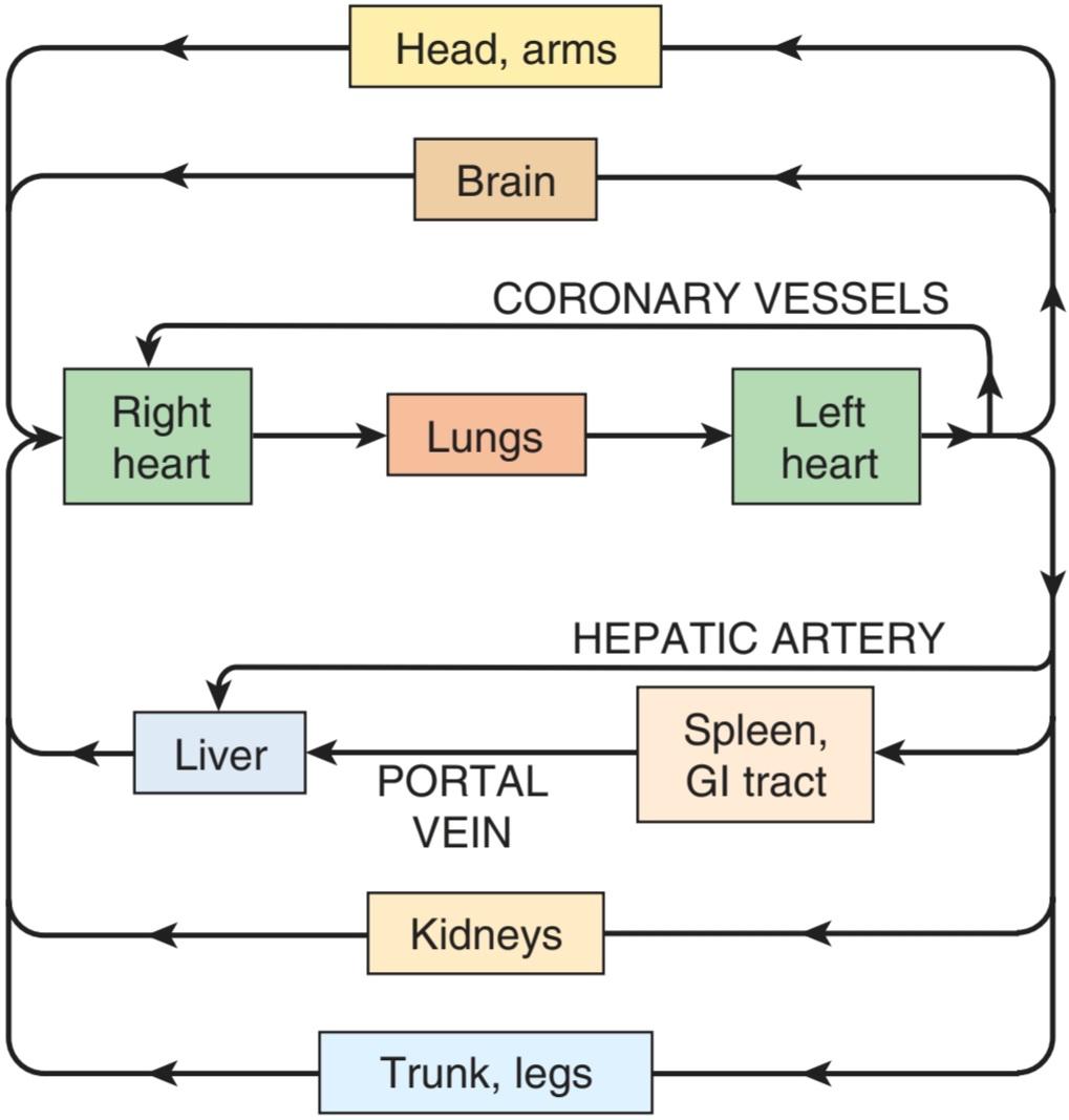
FIGURE 31-1 Diagram of the circulation in the adult.
شکل 31-1 نمودار گردش خون در بزرگسالان.
This chapter is concerned with blood and lymph and with the multiple functions of the cells they contain. It will also address general principles that apply to all parts of the circulation and pressure and flow in the systemic circulation. The homeostatic mechanisms operating to adjust flow are the subject of Chapter 32. The special characteristics of the pulmonary and renal circulations are discussed in Chapters 34 and 37. Likewise, the role of blood as the carrier of many immune effector cells will not be discussed here, but rather was covered in Chapter 3.
این فصل به خون و لنف و عملکردهای چندگانه سلول های موجود در آنها می پردازد. همچنین به اصول کلی مربوط به تمام بخشهای گردش خون و فشار و جریان در گردش خون سیستمیک میپردازد. مکانیسمهای هموستاتیکی که برای تنظیم جریان کار میکنند موضوع فصل 32 است. ویژگیهای خاص گردش خون ریوی و کلیوی در فصلهای 34 و 37 مورد بحث قرار میگیرد. به همین ترتیب، نقش خون به عنوان حامل بسیاری از سلولهای مؤثر ایمنی در اینجا مورد بحث قرار نخواهد گرفت، بلکه در فصل 3 مورد بررسی قرار گرفت.
BLOOD AS A CIRCULATORY FLUID
Blood consists of a protein-rich fluid known as plasma, in which are suspended cellular elements: white blood cells, red blood cells, and platelets. The normal total circulating blood volume is about 8% of the body weight (5600 mL in a 70- kg man). About 55% of this volume is plasma.
خون به عنوان یک مایع گردش خون
خون از مایع غنی از پروتئین به نام پلاسما تشکیل شده است که در آن عناصر سلولی معلق هستند: گلبول های سفید، گلبول های قرمز و پلاکت ها. حجم طبیعی کل خون در گردش حدود 8 درصد وزن بدن است (5600 میلی لیتر در یک مرد 70 کیلوگرمی). حدود 55 درصد از این حجم پلاسما است.
BONE MARROW
In the adult, red blood cells, many white blood cells, and platelets are formed in the bone marrow. In the fetus, blood cells are also formed in the liver and spleen, and in adults such extramedullary hematopoiesis may occur in diseases in which the bone marrow becomes destroyed or fibrosed. In children, blood cells are actively produced in the marrow cavities of all the bones. By age 20, the marrow in the cavities of the long bones, except for the upper humerus and femur, has become inactive (Figure 31-2).
مغز استخوان
در بزرگسالان، گلبول های قرمز، بسیاری از گلبول های سفید و پلاکت ها در مغز استخوان تشکیل می شوند. در جنین، سلولهای خونی نیز در کبد و طحال تشکیل میشوند و در بزرگسالان چنین خونسازی خارج مدولاری ممکن است در بیماریهایی رخ دهد که در آن مغز استخوان از بین میرود یا فیبروز میشود. در کودکان، سلول های خونی به طور فعال در حفره های مغزی تمام استخوان ها تولید می شوند. در سن 20 سالگی، مغز استخوان در حفره های استخوان های بلند، به جز استخوان بازو و استخوان ران، غیرفعال شده است (شکل 31-2).

FIGURE 31–2 Changes in red bone marrow cellularity in various bones with age. Hundred percent equals the degree of cellularity at birth. (Reproduced with permission from Whitby LEH, Britton CJC: Disorders of the Blood, 10th ed. Churchill Livingstone; 1969.)
شکل 31-2 تغییرات در سلولی بودن مغز استخوان قرمز در استخوان های مختلف با افزایش سن. صد در صد برابر با درجه سلولی در بدو تولد است. (تکثیر شده با مجوز Whitby LEH، Britton CJC: Disorders of the Blood، ویرایش دهم چرچیل لیوینگستون، 1969.)
The bone marrow is actually one of the largest organs in the body, approaching the size and weight of the liver. It is also one of the most active. Normally, 75% of the cells in the marrow belong to the white blood cell- producing myeloid series and only 25% are maturing red cells, even though there are over 500 times as many red cells in the circulation as there are white cells. This difference in the marrow reflects the fact that the average life span of white cells is short, whereas that of red cells is long.
مغز استخوان در واقع یکی از بزرگترین اندام های بدن است که به اندازه و وزن کبد نزدیک می شود. همچنین یکی از فعال ترین هاست. به طور معمول، 75 درصد از سلولهای مغز متعلق به سری میلوئید تولیدکننده گلبولهای سفید هستند و تنها 25 درصد سلولهای قرمز بالغ هستند، حتی اگر بیش از 500 برابر تعداد گلبولهای قرمز در گردش خون وجود داشته باشد. این تفاوت در مغز نشان دهنده این واقعیت است که طول عمر متوسط گلبول های سفید کوتاه است، در حالی که طول عمر گلبول های قرمز طولانی است.
Hematopoietic stem cells (HSCs) are bone marrow cells that are capable of producing all types of blood cells. They differentiate into one or another type of committed stem cells (progenitor cells). These in turn form the various differentiated types of blood cells. There are separate pools of progenitor cells for megakaryocytes, lymphocytes, erythrocytes, eosinophils, and basophils; neutrophils and monocytes arise from a common precursor. The bone marrow stem cells are also the source of osteoclasts (see Chapter 21), Kupffer cells (see Chapter 28), mast cells, dendritic cells, and Langerhans cells. The HSCs are few in number but are capable of completely replacing the bone marrow when injected into a patient whose own bone marrow has been entirely destroyed.
سلول های بنیادی خونساز (HSCs) سلول های مغز استخوان هستند که قادر به تولید انواع سلول های خونی هستند. آنها به یک یا نوع دیگری از سلول های بنیادی متعهد (سلول های پیش ساز) تمایز می یابند. اینها به نوبه خود انواع متفاوت سلول های خونی را تشکیل می دهند. مخازن جداگانه ای از سلول های پیش ساز برای مگاکاریوسیت ها، لنفوسیت ها، گلبول های قرمز، ائوزینوفیل ها و بازوفیل ها وجود دارد. نوتروفیل ها و مونوسیت ها از یک پیش ساز معمولی به وجود می آیند. سلول های بنیادی مغز استخوان همچنین منبع استئوکلاست ها (به فصل 21)، سلول های کوپفر (به فصل 28 مراجعه کنید)، ماست سل ها، سلول های دندریتیک و سلول های لانگرهانس هستند. تعداد HSCها کم است، اما وقتی به بیمار تزریق می شود که مغز استخوان او کاملاً از بین رفته است، می توانند به طور کامل جایگزین مغز استخوان شوند.
The HSCs are derived from uncommitted, totipotent stem cells that can be stimulated to form any cell in the body. Adults have a few of these, but they are more readily obtained from the blastocysts of embryos. There is not surprisingly immense interest in stem cell research due to its potential to regenerate diseased tissues, but ethical issues are involved, and debate on these issues will undoubtedly continue.
HSC ها از سلول های بنیادی غیرمتعهد و تمام توان مشتق شده اند که می توانند برای تشکیل هر سلولی در بدن تحریک شوند. بزرگسالان تعداد کمی از این موارد را دارند، اما آنها به راحتی از بلاستوسیست های جنین به دست می آیند. به دلیل پتانسیل آن برای بازسازی بافتهای بیمار، علاقه زیادی به تحقیقات سلولهای بنیادی وجود ندارد، اما مسائل اخلاقی در آن دخیل هستند و بحث در مورد این موضوعات بدون شک ادامه خواهد داشت.
WHITE BLOOD CELLS
Normally, human blood contains 4000-11,000 white blood cells per microliter (Table 31-1). Of these, the granulocytes (polymorphonuclear leukocytes, PMNs) are the most numerous. Young granulocytes have horseshoe-shaped nuclei that become multilobed as the cells grow older (Figure 31–3). Most of them contain neutrophilic granules (neutrophils), but a few contain granules that stain with acidic dyes (eosinophils), and some have basophilic granules (basophils). The other two cell types found normally in peripheral blood are lymphocytes, which have large round nuclei and scanty cytoplasm, and monocytes, which have abundant agranular cytoplasm and kidney-shaped nuclei (Figure 31-3). Acting together, these cells provide the body with the powerful defenses against tumors and viral, bacterial, and parasitic infections that were discussed in Chapter 3.
گلبول های سفید خون
به طور معمول، خون انسان حاوی 4000-11000 گلبول سفید در هر میکرولیتر است (جدول 31-1). از این میان، گرانولوسیتها (لکوسیتهای پلیمورفونوکلئر، PMN) از همه بیشتر هستند. گرانولوسیت های جوان دارای هسته های نعل اسبی هستند که با بزرگتر شدن سلول ها چند لوبی می شوند (شکل 31-3). بیشتر آنها حاوی گرانول های نوتروفیل (نوتروفیل) هستند، اما تعداد کمی حاوی گرانول هایی هستند که با رنگ های اسیدی (ائوزینوفیل ها) رنگ می شوند و برخی دارای گرانول های بازوفیل (بازوفیل) هستند. دو نوع سلول دیگر که به طور معمول در خون محیطی یافت می شوند، لنفوسیت ها هستند که دارای هسته های گرد بزرگ و سیتوپلاسم کمی هستند، و مونوسیت ها که دارای سیتوپلاسم دانه ای فراوان و هسته های کلیه شکل هستند (شکل 31-3). این سلول ها با هم عمل می کنند و بدن را با دفاع قدرتمند در برابر تومورها و عفونت های ویروسی، باکتریایی و انگلی که در فصل 3 مورد بحث قرار گرفت، فراهم می کنند.
TABLE 31-1 Normal values for the cellular elements in human blood.
جدول 31-1 مقادیر طبیعی عناصر سلولی در خون انسان.

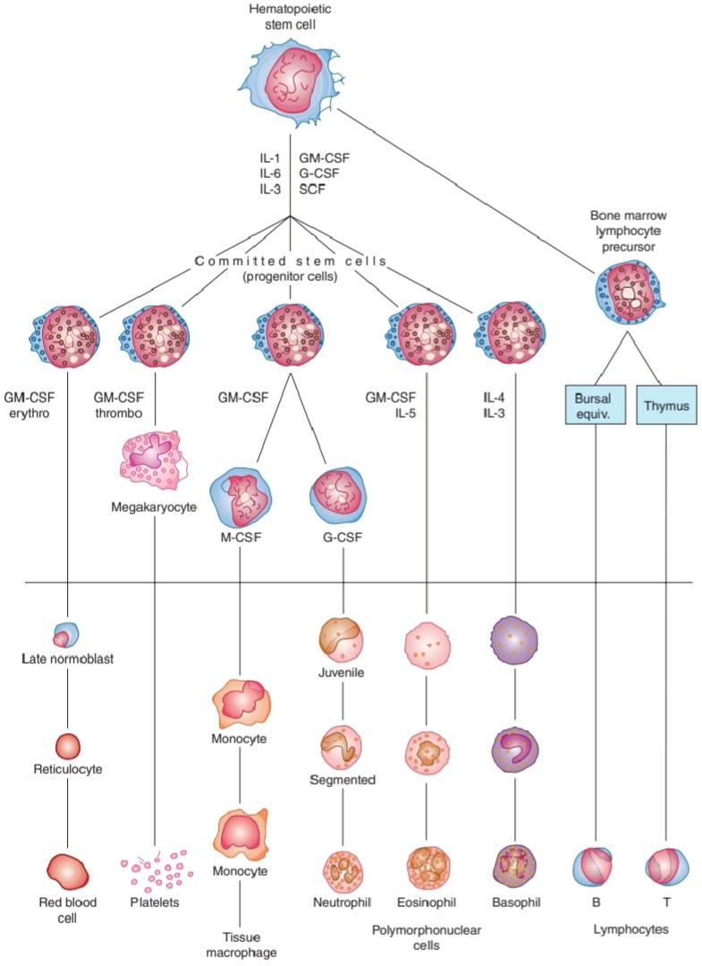
FIGURE 31-3 Development of various formed elements of the blood from bone marrow cells. Cells below the horizontal line are found in normal peripheral blood. The principal sites of action of erythropoietin (erythro) and the various colony-stimulating factors (CSFs) that stimulate the differentiation of the components are indicated. G, granulocyte; M, macrophage; IL, interleukin; thrombo, thrombopoietin; erythro, erythropoietin; SCF, stem cell factor.
شکل 31-3 توسعه عناصر تشکیلشده مختلف خون از سلولهای مغز استخوان. سلول های زیر خط افقی در خون محیطی طبیعی یافت می شوند. مکانهای اصلی عمل اریتروپویتین (اریترو) و عوامل مختلف محرک کلنی (CSFs) که تمایز اجزا را تحریک میکنند نشان داده شدهاند. G، گرانولوسیت؛ M، ماکروفاژ; IL، اینترلوکین؛ ترومبو، ترومبوپوئیتین؛ اریترو، اریتروپویتین؛ SCF، فاکتور سلول های بنیادی.
PLATELETS
Platelets are small, granulated bodies that aggregate at sites of vascular injury. They lack nuclei and are 2-4 μm in diameter (Figure 31-3). There are about 300,000/μL of circulating blood, and they normally have a half-life of about 4 days. The megakaryocytes, giant cells in the bone marrow, form platelets by pinching off bits of cytoplasm and extruding them into the circulation. Between 60% and 75% of the platelets that have been extruded from the bone marrow are in the circulating blood, and the remainder are mostly in the spleen. Splenectomy causes an increase in the platelet count (thrombocytosis).
پلاکت ها
پلاکت ها اجسام کوچک و دانه بندی شده ای هستند که در محل آسیب عروقی تجمع می یابند. آنها فاقد هسته هستند و 2-4 میکرومتر قطر دارند (شکل 31-3). حدود 300000/μL خون در گردش وجود دارد و نیمه عمر آنها معمولاً حدود 4 روز است. مگاکاریوسیتها، سلولهای غولپیکر در مغز استخوان، پلاکتها را با فشار دادن تکههایی از سیتوپلاسم و بیرون راندن آنها در گردش خون تشکیل میدهند. بین 60 تا 75 درصد پلاکتهایی که از مغز استخوان اکسترود شدهاند در خون در گردش هستند و بقیه بیشتر در طحال هستند. اسپلنکتومی باعث افزایش تعداد پلاکت ها (ترومبوسیتوز) می شود.
RED BLOOD CELLS
The red blood cells (erythrocytes) carry hemoglobin in the circulation. They are biconcave disks (Figure 31-4) that are manufactured in the bone marrow. In mammals, they lose their nuclei before entering the circulation. In humans, they survive in the circulation for an average of 120 days. The average normal red blood cell count is 5.4 million/μL in men and 4.8 million/μL in women. The number of red cells is also conveniently expressed as the hematocrit, or the percentage of the blood, by volume, that is occupied by erythrocytes. Each human red blood cell is about 7.5 μm in diameter and 2 μm thick, and each contains approximately 29 pg of hemoglobin (Table 31-2). There are thus about 3 × 10¹³ red blood cells and about 900 g of hemoglobin in the circulating blood of an adult man (Figure 31-5).
گلبول های قرمز
گلبول های قرمز خون (گلبول های قرمز) هموگلوبین را در گردش خون حمل می کنند. آنها دیسک های دوقعر هستند (شکل 31-4) که در مغز استخوان ساخته می شوند. در پستانداران، آنها قبل از ورود به گردش خون هسته خود را از دست می دهند. در انسان به طور متوسط 120 روز در گردش خون زنده می مانند. میانگین تعداد طبیعی گلبول های قرمز خون در مردان 5.4 میلیون در میکرولیتر و در زنان 4.8 میلیون در میکرولیتر است. تعداد گلبول های قرمز نیز به راحتی به صورت هماتوکریت یا درصد خونی که توسط گلبول های قرمز اشغال شده است بیان می شود. هر گلبول قرمز خون انسان حدود 7.5 میکرومتر قطر و 2 میکرومتر ضخامت دارد و هر گلبول تقریباً 29 pg هموگلوبین دارد (جدول 31-2). بنابراین حدود 3 × 10¹³ گلبول قرمز و حدود 900 گرم هموگلوبین در خون در گردش یک مرد بالغ وجود دارد (شکل 31-5).
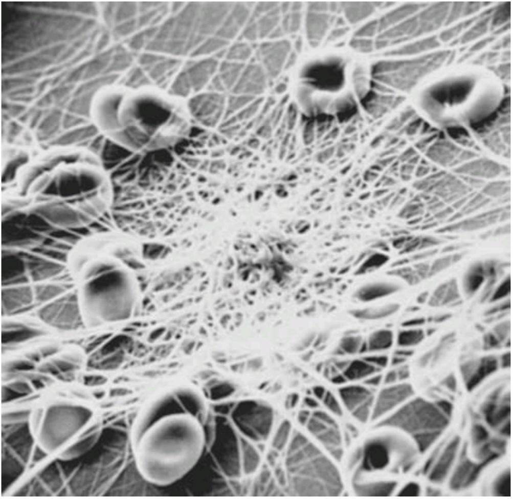
FIGURE 31-4 Human red blood cells and fibrin fibrils. Blood was placed on a polyvinyl chloride surface, fixed, and photographed with a scanning electron microscope. Reduced from ×2590. (Used with permission of NF Rodman.)
شکل 31-4 گلبول های قرمز خون انسان و فیبریل های فیبرین. خون روی سطح پلی وینیل کلرید قرار داده شد، ثابت شد و با میکروسکوپ الکترونی روبشی عکسبرداری شد. از 2590 × کاهش یافته است. (با اجازه NF Rodman استفاده می شود.)
TABLE 31-2 Characteristics of human red cells.a
جدول 31-2 ویژگی های گلبول های قرمز انسان.a
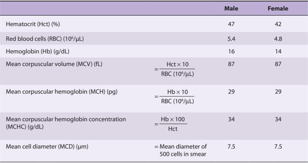
aCells with MCVs > 95 fL are called macrocytes; cells with MCVs < 80 fL are called microcytes; cells with MCHCs < 25 g/dL are called hypochromic.
aسلول های دارای MCV > 95 fL ماکروسیت نامیده می شوند. سلول های دارای MCV کمتر از 80 fL میکروسیت نامیده می شوند. سلول های دارای MCHC کمتر از 25 گرم در دسی لیتر هیپوکرومیک نامیده می شوند.
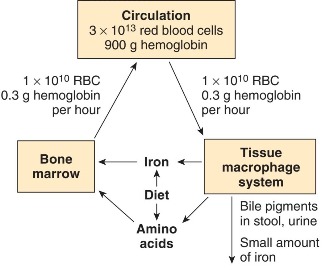
FIGURE 31-5 Red cell formation and destruction. RBC, red blood cells.
شکل 31-5 تشکیل و تخریب گلبول های قرمز. RBC، گلبول های قرمز.
The feedback control of erythropoiesis by erythropoietin is discussed in Chapter 38, and the role of IL-1, IL-3, IL-6 (interleukin), and GM-CSF (granulocyte-macrophage colony-stimulating factor) in development of the relevant erythroid stem cells is shown in Figure 31-3.
کنترل بازخورد اریتروپوئز توسط اریتروپویتین در فصل 38 مورد بحث قرار گرفته است و نقش IL-1، IL-3، IL-6 (اینترلوکین)، و GM-CSF (فاکتور تحریک کننده کلنی گرانولوسیت-ماکروفاژ) در توسعه سلول های بنیادی اریتروئید مربوطه در شکل 31-3 نشان داده شده است.
ROLE OF THE SPLEEN
The spleen is an important blood filter that removes aged or abnormal red cells. It also contains many platelets and plays a significant role in the immune system. Abnormal red cells are removed if they are not as flexible as normal red cells and consequently are unable to squeeze through the slits between the endothelial cells that line the splenic sinuses (Clinical Box 31-1).
نقش طحال
طحال یک فیلتر خون مهم است که گلبول های قرمز پیر یا غیر طبیعی را حذف می کند. همچنین حاوی تعداد زیادی پلاکت است و نقش مهمی در سیستم ایمنی دارد. اگر گلبولهای قرمز غیرطبیعی مانند گلبولهای قرمز نرمال انعطافپذیر نباشند و در نتیجه نتوانند از طریق شکافهای بین سلولهای اندوتلیال که سینوسهای طحال را پوشاندهاند فشار دهند، برداشته میشوند (باکس بالینی 31-1).
CLINICAL BOX 31-1
Red Cell Fragility
Red blood cells, like other cells, shrink in solutions with an osmotic pressure greater than that of normal plasma. In solutions with a lower osmotic pressure they swell, become spherical rather than disk-shaped, and eventually lose their hemoglobin (hemolysis). The hemoglobin of hemolyzed red cells dissolves in the plasma, coloring it red. A 0.9% sodium chloride solution is isotonic with plasma. When osmotic fragility is normal, red cells begin to hemolyze when suspended in 0.5% saline; 50% lysis occurs in 0.40-0.42% saline, and lysis is complete in 0.35% saline. In hereditary spherocytosis (congenital hemolytic icterus), the cells are spherocytic in normal plasma and hemolyze more readily than normal cells in hypotonic sodium chloride solutions. Abnormal spherocytes are also trapped and destroyed in the spleen, meaning that hereditary spherocytosis is one of the most common causes of hereditary hemolytic anemia. The spherocytosis is caused by mutations in proteins that make up the membrane skeleton of the erythrocyte, which normally maintain the shape and flexibility of the red cell membrane, including spectrin, the transmembrane protein band 3, and the linker protein, ankyrin. Red cells can also be lysed by drugs (especially penicillin and sulfa drugs) and infections. The susceptibility of red cells to hemolysis by these agents is increased by deficiency of the enzyme glucose-6-phosphate dehydrogenase (G6PD), which catalyzes the initial step in the oxidation of glucose via the hexose monophosphate pathway (see Chapter 1). This pathway generates dihydronicotinamide adenine dinucleotide phosphate (NADPH), which is needed for the maintenance of normal red cell fragility. Severe G6PD deficiency also inhibits the killing of bacteria by granulocytes and predisposes to severe infections.
جعبه بالینی 31-1
شکنندگی گلبول قرمز
گلبولهای قرمز مانند سایر سلولها در محلولهایی با فشار اسمزی بیشتر از پلاسمای طبیعی منقبض میشوند. در محلولهایی که فشار اسمزی کمتری دارند، متورم میشوند، به جای دیسکی شکل، کروی میشوند و در نهایت هموگلوبین خود را از دست میدهند (همولیز). هموگلوبین گلبول های قرمز همولیز شده در پلاسما حل می شود و آن را قرمز رنگ می کند. محلول 0.9% کلرید سدیم با پلاسما ایزوتونیک است. هنگامی که شکنندگی اسمزی طبیعی است، گلبول های قرمز زمانی که در نمک 0.5% معلق می شوند شروع به همولیز می کنند. 50% لیز در 0.40-0.42% سالین رخ می دهد و لیز در 0.35% سالین کامل است. در اسفروسیتوز ارثی (ایکتروس همولیتیک مادرزادی)، سلول ها در پلاسمای طبیعی کروی هستند و راحت تر از سلول های طبیعی در محلول های کلرید سدیم هیپوتونیک همولیز می شوند. اسفروسیت های غیر طبیعی نیز در طحال به دام افتاده و از بین می روند، به این معنی که اسفروسیتوز ارثی یکی از شایع ترین علل کم خونی همولیتیک ارثی است. اسفروسیتوز در اثر جهش در پروتئینهایی ایجاد میشود که اسکلت غشایی گلبول قرمز را تشکیل میدهند، که معمولاً شکل و انعطافپذیری غشای گلبول قرمز را حفظ میکنند، از جمله اسپکترین، باند پروتئین گذرنده 3 و پروتئین پیوندی، آنکیرین. گلبول های قرمز همچنین می توانند توسط داروها (به ویژه داروهای پنی سیلین و سولفا) و عفونت ها لیز شوند. حساسیت گلبولهای قرمز به همولیز توسط این عوامل با کمبود آنزیم گلوکز-6-فسفات دهیدروژناز (G6PD) افزایش مییابد که مرحله اولیه اکسیداسیون گلوکز را از طریق مسیر هگزوز مونوفسفات کاتالیز میکند (به فصل 1 مراجعه کنید). این مسیر دی هیدرونیکوتین آمید آدنین دی نوکلئوتید فسفات (NADPH) را تولید می کند که برای حفظ شکنندگی طبیعی گلبول های قرمز مورد نیاز است. کمبود شدید G6PD همچنین از کشتن باکتری ها توسط گرانولوسیت ها جلوگیری می کند و مستعد ابتلا به عفونت های شدید می شود.
THERAPEUTIC HIGHLIGHTS
Severe cases of hereditary spherocytosis can be treated by splenectomy, but this is not without other risks, such as sepsis. Milder cases can be treated with dietary folate supplementation and/or blood transfusions. Treatment of other forms of hemolytic anemia depends on the underlying cause. Some forms are autoimmune in nature, and have been shown to benefit from treatment with corticosteroids.
نکات برجسته درمانی
موارد شدید اسفروسیتوز ارثی را می توان با اسپلنکتومی درمان کرد، اما این بدون خطرات دیگری مانند سپسیس نیست. موارد خفیف تر را می توان با مکمل غذایی فولات و/یا تزریق خون درمان کرد. درمان سایر اشکال کم خونی همولیتیک به علت اصلی آن بستگی دارد. برخی از اشکال در طبیعت خود ایمنی هستند و نشان داده شده است که از درمان با کورتیکواستروئیدها سود می برند.
HEMOGLOBIN
The red, oxygen-carrying pigment in the red blood cells of vertebrates is the protein hemoglobin. Hemoglobin is a globular molecule made up of four subunits (Figure 31–6). Each subunit contains a heme moiety conjugated to a polypeptide. Heme is an iron-containing porphyrin derivative (Figure 31-7). The polypeptides are referred to collectively as the globin portion of the hemoglobin molecule. There are two pairs of polypeptides in each hemoglobin molecule. In normal adult human hemoglobin (hemoglobin A), the two polypeptides are called a chains and ẞ chains. Thus, hemoglobin A is designated a2ẞ2. Not all the hemoglobin in the blood of normal adults is hemoglobin A. About 2.5% of the hemoglobin is hemoglobin A2, in which ẞ chains are replaced by 8 chains (α282).
هموگلوبین
رنگدانه قرمز و حامل اکسیژن در گلبول های قرمز مهره داران پروتئین هموگلوبین است. هموگلوبین یک مولکول کروی است که از چهار زیر واحد تشکیل شده است (شکل 31-6). هر زیرواحد حاوی یک نیمه هم به یک پلی پپتید است. هِم یک مشتق پورفیرین حاوی آهن است (شکل 31-7). پلی پپتیدها در مجموع به عنوان بخش گلوبین مولکول هموگلوبین نامیده می شوند. در هر مولکول هموگلوبین دو جفت پلی پپتید وجود دارد. در هموگلوبین طبیعی انسان بالغ (هموگلوبین A)، این دو پلی پپتید یک زنجیره و زنجیره ẞ نامیده می شوند. بنابراین، هموگلوبین A a2ẞ2 نامیده می شود. تمام هموگلوبین موجود در خون بزرگسالان عادی هموگلوبین A نیست. حدود 2.5 درصد از هموگلوبین هموگلوبین A2 است که در آن زنجیره های ẞ با 8 زنجیره (α282) جایگزین می شوند.
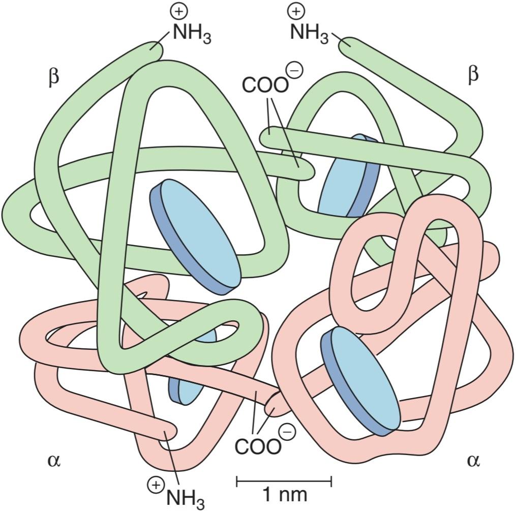
FIGURE 31-6 Diagrammatic representation of a molecule of hemoglobin A, showing the four subunits. There are two a and two ẞ polypeptide chains, each containing a heme moiety. These moieties are represented by the blue disks. (Reproduced with permission from Harper HA, et al: Physiologische Chemie. Springer; 1975.)
شکل 31-6 نمایش نموداری یک مولکول هموگلوبین A، که چهار زیرواحد را نشان می دهد. دو زنجیره پلی پپتیدی a و دو ẞ وجود دارد که هر کدام حاوی یک قسمت هم هستند. این قسمت ها با دیسک های آبی نشان داده می شوند. (تکثیر شده با اجازه هارپر HA، و همکاران: Physiologische Chemie. Springer؛ 1975.)
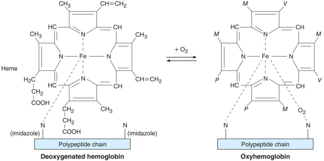
FIGURE 31–7 Reaction of heme with O2. The abbreviations M, V, and P stand for the groups shown on the molecule on the left.
شکل 31-7 واکنش هم با O2. اختصارات M، V و P مخفف گروه هایی هستند که روی مولکول سمت چپ نشان داده شده اند.
There are small amounts of hemoglobin A derivatives closely associated with hemoglobin A that represent glycated hemoglobins. One of these, hemoglobin Alc (HbA1c), has a glucose attached to the terminal valine in each ẞ chain and is of special interest because it increases in the blood of patients with poorly controlled diabetes mellitus (see Chapter 24), and is measured clinically as a marker of the progression of that disease and/or the effectiveness of treatment.
مقادیر کمی از مشتقات هموگلوبین A وجود دارد که نزدیک به هموگلوبین A مرتبط است که نشان دهنده هموگلوبین گلیکوزیله است. یکی از اینها، هموگلوبین Alc (HbA1c)، دارای گلوکز متصل به والین انتهایی در هر زنجیره ẞ است و مورد توجه ویژه است زیرا در خون بیماران مبتلا به دیابت قندی کنترل نشده افزایش می یابد (به فصل 24 مراجعه کنید)، و از نظر بالینی به عنوان نشانگر پیشرفت آن بیماری و/یا اثربخشی درمان اندازه گیری می شود.
REACTIONS OF HEMOGLOBIN
O2 binds to the Fe2+ in the heme moiety of hemoglobin to form oxyhemoglobin. The affinity of hemoglobin for O2 is affected by pH, temperature, and the concentration in the red cells of 2,3-bisphosphoglycerate (2,3-BPG). 2,3-BPG and H* compete with O2 for binding to deoxygenated hemoglobin, decreasing the affinity of hemoglobin for O2 by shifting the positions of the four peptide chains (quaternary structure). The details of the oxygenation and deoxygenation of hemoglobin and the physiologic role of these reactions in O2 transport are discussed in Chapter 35.
واکنش های هموگلوبین
O2 به +Fe2 در قسمت هموگلوبین متصل می شود و اکسی هموگلوبین را تشکیل می دهد. تمایل هموگلوبین به O2 تحت تأثیر pH، دما و غلظت 2،3-بیس فسفوگلیسرات (2،3-BPG) در گلبول های قرمز قرار می گیرد. 2،3-BPG و +H برای اتصال به هموگلوبین بدون اکسیژن با O2 رقابت می کنند و با تغییر موقعیت چهار زنجیره پپتیدی (ساختار چهارتایی) میل هموگلوبین برای O2 را کاهش می دهند. جزئیات اکسیژن رسانی و اکسیژن زدایی هموگلوبین و نقش فیزیولوژیکی این واکنش ها در انتقال O2 در فصل 35 مورد بحث قرار گرفته است.
When blood is exposed to various drugs and other oxidizing agents in vitro or in vivo, the ferrous iron (Fe2+) that is normally present in hemoglobin is converted to ferric iron (Fe3+), forming methemoglobin. Methemoglobin is dark-colored, and when it is present in large quantities in the circulation, it causes a dusky discoloration of the skin resembling cyanosis (see Chapter 35). Some oxidation of hemoglobin to methemoglobin occurs normally, but an enzyme system in the red cells, the dihydronicotinamide adenine dinucleotide (NADH)-methemoglobin reductase system, converts methemoglobin back to hemoglobin. Congenital absence of this system is one cause of hereditary methemoglobinemia.
هنگامی که خون در شرایط آزمایشگاهی یا درون تنی در معرض داروهای مختلف و سایر عوامل اکسید کننده قرار می گیرد، آهن آهنی (+Fe2) که به طور معمول در هموگلوبین وجود دارد، به آهن آهن (+Fe3) تبدیل می شود و متهموگلوبین را تشکیل می دهد. متهموگلوبین تیره رنگ است و هنگامی که به مقدار زیاد در گردش خون وجود داشته باشد، باعث تغییر رنگ تیره پوست شبیه سیانوز می شود (به فصل 35 مراجعه کنید). مقداری اکسیداسیون هموگلوبین به متهموگلوبین به طور معمول رخ می دهد، اما یک سیستم آنزیمی در گلبول های قرمز، سیستم دی هیدرونیکوتین آمید آدنین دی نوکلئوتید (NADH) – متهموگلوبین ردوکتاز، متهموگلوبین را دوباره به هموگلوبین تبدیل می کند. عدم وجود مادرزادی این سیستم یکی از علل متهموگلوبینمی ارثی است.
Carbon monoxide reacts with hemoglobin to form carbon monoxyhemoglobin (carboxyhemoglobin). The affinity of hemoglobin for O2 is much lower than its affinity for carbon monoxide, which consequently displaces O2 on hemoglobin, reducing the oxygen-carrying capacity of blood (see Chapter 35).
مونوکسید کربن با هموگلوبین واکنش داده و کربن مونوکسی هموگلوبین (کربوکسی هموگلوبین) تشکیل می دهد. میل ترکیبی هموگلوبین برای O2 بسیار کمتر از تمایل آن به مونوکسید کربن است، که در نتیجه O2 را روی هموگلوبین جابجا می کند و ظرفیت حمل اکسیژن خون را کاهش می دهد (به فصل 35 مراجعه کنید).
HEMOGLOBIN IN THE FETUS
The blood of the human fetus normally contains fetal hemoglobin (hemoglobin F). Its structure is similar to that of hemoglobin A except that the ẞ chains are replaced by y chains; that is, hemoglobin F is α¿Y1⁄2. Fetal hemoglobin is normally replaced by adult hemoglobin soon after birth (Figure 31-8). In certain individuals, it fails to disappear and persists throughout life. In the body, its O2 content at a given PO2 is greater than that of adult hemoglobin because it binds 2,3-BPG less avidly. Hemoglobin F is therefore critical to facilitate movement of O2 from the maternal to the fetal circulation, particularly at later stages of gestation where oxygen demand increases (see Chapter 33). In young embryos there are, in addition, and ε chains, forming Gower 1 hemoglobin (22) and Gower 2 hemoglobin (α22). Switching from one form of hemoglobin to another during development seems to be regulated largely by oxygen availability, with relative hypoxia favoring the production of hemoglobin F both via direct effects on globin gene expression, as well as upregulated production of erythropoietin.
هموگلوبین در جنین
خون جنین انسان به طور معمول حاوی هموگلوبین جنینی (هموگلوبین F) است. ساختار آن شبیه هموگلوبین A است با این تفاوت که زنجیره های ẞ با زنجیره های y جایگزین می شوند. یعنی هموگلوبین F α¿Y1⁄2 است. هموگلوبین جنینی به طور معمول بلافاصله پس از تولد با هموگلوبین بالغ جایگزین می شود (شکل 31-8). در افراد خاص، ناپدید نمی شود و در طول زندگی ادامه می یابد. در بدن، محتوای O2 آن در یک PO2 معین بیشتر از هموگلوبین بزرگسالان است زیرا 2،3-BPG را با اشتیاق کمتر متصل می کند. بنابراین هموگلوبین F برای تسهیل حرکت O2 از گردش خون مادر به جنین، به ویژه در مراحل بعدی بارداری که نیاز به اکسیژن افزایش می یابد، حیاتی است (به فصل 33 مراجعه کنید). در جنین های جوان، علاوه بر این، زنجیره های ε نیز وجود دارد که هموگلوبین Gower 1 (22) و Gower 2 (α22) را تشکیل می دهند. به نظر می رسد تغییر از یک شکل هموگلوبین به شکل دیگر در طول توسعه تا حد زیادی توسط در دسترس بودن اکسیژن تنظیم می شود، با هیپوکسی نسبی به تولید هموگلوبین F هم از طریق تأثیر مستقیم بر بیان ژن گلوبین و هم از طریق افزایش تولید اریتروپویتین.

FIGURE 31-8 Development of human hemoglobin chains. The graph shows the normal rate of synthesis of the various hemoglobin chains in utero, and how these change after birth.
شکل 31-8 توسعه زنجیره های هموگلوبین انسانی. نمودار سرعت طبیعی سنتز زنجیره های مختلف هموگلوبین در رحم و چگونگی تغییر آنها پس از تولد را نشان می دهد.
SYNTHESIS OF HEMOGLOBIN
The average normal hemoglobin content of blood is 16 g/dL in men and 14 g/dL in women, all of it in red cells. In the body of a 70-kg man, there are about 900 g of hemoglobin, and 0.3 g of hemoglobin is destroyed and 0.3 g synthesized every hour (Figure 31-5). The heme portion of the hemoglobin molecule is synthesized from glycine and succinyl-CoA (Clinical Box 31-2).
سنتز هموگلوبین
میانگین محتوای طبیعی هموگلوبین خون در مردان 16 گرم در دسیلیتر و در زنان 14 گرم در دسیلیتر است که تمام آن در گلبولهای قرمز وجود دارد. در بدن یک مرد 70 کیلوگرمی، حدود 900 گرم هموگلوبین وجود دارد و هر ساعت 0.3 گرم هموگلوبین از بین میرود و 0.3 گرم سنتز میشود (شکل 31-5). بخش هِم مولکول هموگلوبین از گلیسین و سوکسینیل-کوآ سنتز میشود (کادر بالینی 31-2).
CLINICAL BOX 31-2
Abnormalities of Hemoglobin Production
There are two major types of inherited disorders of hemoglobin in humans: the hemoglobinopathies, in which abnormal globin polypeptide chains are produced, and the thalassemias and related disorders, in which the chains are normal in structure but produced in decreased amounts or absent because of defects in the regulatory portion of the globin genes. Mutant genes that cause the production of abnormal hemoglobins are widespread, and over 1000 abnormal hemoglobins have been described in humans. In one of the most common examples, hemoglobin S, the a chains are normal but the ẞ chains have a single substitution of a valine residue for one glutamic acid, leading to sickle cell anemia (Table 31-3). When an abnormal gene inherited from one parent dictates formation of an abnormal hemoglobin (ie, when the individual is heterozygous), half the circulating hemoglobin is abnormal and half is normal. When identical abnormal genes are inherited from both parents, the individual is homozygous and all the hemoglobin is abnormal. It is theoretically possible to inherit two different abnormal hemoglobins, one from the father and one from the mother. Studies of the inheritance and geographic distribution of abnormal hemoglobins have made it possible in some cases to decide where the mutant gene originated and approximately how long ago the mutation occurred. In general, harmful mutations tend to die out, but mutant genes that confer traits with survival value persist and spread in the population. Many of the abnormal hemoglobins are harmless; however, some have abnormal O2 equilibriums, while others cause anemia. For example, hemoglobin S polymerizes at low O2 tensions, and this causes the red cells to become sickle-shaped, hemolyze, and form aggregates that block blood vessels. The sickle cell gene is an example of a gene that has persisted and spread in the population due to its beneficial effect when present in heterozygous form. It originated in Africa, and confers resistance to one type of malaria. In some parts of Africa, 40% of the population is heterozygous for hemoglobin S. There is a corresponding prevalence of 10% among African Americans in the United States.
جعبه بالینی 31-2
ناهنجاری های تولید هموگلوبین
دو نوع عمده اختلالات ارثی هموگلوبین در انسان وجود دارد: هموگلوبینوپاتی ها، که در آن زنجیره های پلی پپتیدی گلوبین غیرطبیعی تولید می شود، و تالاسمی ها و اختلالات مرتبط، که در آن زنجیره ها از نظر ساختار طبیعی هستند اما به دلیل نقص در بخش تنظیم کننده گلوبین، در مقادیر کاهش یافته یا وجود ندارند. ژنهای جهشیافته که باعث تولید هموگلوبینهای غیرطبیعی میشوند، گسترده هستند و بیش از 1000 هموگلوبین غیرطبیعی در انسان توصیف شده است. در یکی از رایجترین نمونهها، هموگلوبین S، زنجیرههای a طبیعی هستند، اما زنجیرههای ẞ یک جانشین یک باقیمانده والین به جای یک اسید گلوتامیک دارند که منجر به کم خونی سلول داسی میشود (جدول 31-3). هنگامی که یک ژن غیر طبیعی که از یکی از والدین به ارث رسیده است تشکیل یک هموگلوبین غیرطبیعی را دیکته می کند (یعنی زمانی که فرد هتروزیگوت است)، نیمی از هموگلوبین در گردش غیر طبیعی و نیمی طبیعی است. هنگامی که ژن های غیر طبیعی یکسان از هر دو والدین به ارث می رسد، فرد هموزیگوت است و تمام هموگلوبین غیر طبیعی است. از نظر تئوری امکان به ارث بردن دو هموگلوبین غیرطبیعی متفاوت، یکی از پدر و دیگری از مادر وجود دارد. مطالعات وراثت و توزیع جغرافیایی هموگلوبین های غیرطبیعی در برخی موارد این امکان را فراهم کرده است که تصمیم گیری شود که ژن جهش یافته از کجا منشأ گرفته و تقریباً چه مدت پیش جهش رخ داده است. به طور کلی، جهشهای مضر تمایل به از بین رفتن دارند، اما ژنهای جهشیافته که ویژگیهایی با ارزش بقا میدهند، باقی میمانند و در جمعیت پخش میشوند. بسیاری از هموگلوبین های غیر طبیعی بی ضرر هستند. با این حال، برخی دارای تعادل O2 غیر طبیعی هستند، در حالی که برخی دیگر باعث کم خونی می شوند. به عنوان مثال، هموگلوبین S در تنش های O2 کم پلیمریزه می شود و این باعث می شود گلبول های قرمز داسی شکل شوند، همولیز شوند و تجمعاتی تشکیل دهند که رگ های خونی را مسدود می کنند. ژن سلول داسی نمونهای از ژنهایی است که به دلیل تأثیر مفید آن در صورت وجود به شکل هتروزیگوت باقی مانده و در جمعیت گسترش یافته است. منشا آن در آفریقا است و در برابر یک نوع مالاریا مقاومت می کند. در برخی از بخشهای آفریقا، 40 درصد جمعیت از نظر هموگلوبین S هتروزیگوت هستند. شیوع مشابهی 10 درصدی در میان آمریکاییهای آفریقایی تبار در ایالات متحده وجود دارد.
THERAPEUTIQ HIGHLIGHTS
Hemoglobin F decreases the polymerization of deoxygenated hemoglobin S, and hydroxyurea stimulates production of hemoglobin F in children and adults. Hydroxyurea has therefore proven to be a very valuable agent for the treatment of sickle cell disease. In patients with severe sickle cell disease, hematopoietic stem cell transplantation has also been shown to have some benefit, and prophylactic treatment with antibiotics has also been shown to be helpful. The clinically important thalassemias result in severe anemia, often requiring repeated blood transfusions. However, these run the risk of iron overload, and often must be accompanied by treatment with drugs that chelate iron. Hematopoietic stem cell transplantation is also being explored for treatment of the thalassemias.
نکات برجسته درمانی
هموگلوبین F پلیمریزاسیون هموگلوبین S بدون اکسیژن را کاهش می دهد و هیدروکسی اوره تولید هموگلوبین F را در کودکان و بزرگسالان تحریک می کند. بنابراین ثابت شده است که هیدروکسی اوره یک عامل بسیار ارزشمند برای درمان بیماری سلول داسی شکل است. در بیماران مبتلا به بیماری سلول داسی شکل شدید، پیوند سلول های بنیادی خونساز نیز فوایدی دارد و درمان پیشگیرانه با آنتی بیوتیک ها نیز مفید بوده است. تالاسمی های مهم بالینی منجر به کم خونی شدید می شوند که اغلب نیاز به تزریق خون مکرر دارند. با این حال، اینها خطر اضافه بار آهن را به همراه دارند و اغلب باید با داروهایی که آهن را کلات می کنند همراه شوند. پیوند سلول های بنیادی خونساز نیز برای درمان تالاسمی ها در حال بررسی است.
CATABOLISM OF HEMOGLOBIN
When old red blood cells are destroyed by tissue macrophages, the globin portion of the hemoglobin molecule is split off, and the heme is converted to biliverdin. The enzyme involved is a subtype of heme oxygenase (see Figure 28-4), and CO is formed in the process. CO is an intercellular messenger, like NO (see Chapters 2 and 3). In humans, most of the biliverdin is converted to bilirubin and excreted in the bile (see Chapter 28). The iron from the heme is reused for hemoglobin synthesis.
کاتابولیسم هموگلوبین
هنگامی که گلبول های قرمز قدیمی توسط ماکروفاژهای بافتی از بین می روند، بخش گلوبین مولکول هموگلوبین جدا شده و هِم به بیلیوردین تبدیل می شود. آنزیم درگیر یک زیرگروه از هم اکسیژناز است (شکل 28-4 را ببینید)، و CO در این فرآیند تشکیل می شود. CO یک پیام رسان بین سلولی است، مانند NO (به فصل های 2 و 3 مراجعه کنید). در انسان، بیشتر بیلی وردین به بیلی روبین تبدیل شده و در صفرا دفع می شود (به فصل 28 مراجعه کنید). آهن حاصل از هم برای سنتز هموگلوبین مجددا استفاده می شود.
Exposure of the skin to white light converts bilirubin to lumirubin, which has a shorter half-life than bilirubin. Phototherapy (exposure to light) is of value in treating infants with jaundice due to hemolysis. Iron is essential for hemoglobin synthesis; if blood is lost from the body and the iron deficiency is not corrected, iron deficiency anemia results.
قرار گرفتن پوست در معرض نور سفید، بیلی روبین را به لومی روبین تبدیل می کند که نیمه عمر آن کوتاه تر از بیلی روبین است. فتوتراپی (قرار گرفتن در معرض نور) در درمان نوزادان مبتلا به زردی ناشی از همولیز ارزشمند است. آهن برای سنتز هموگلوبین ضروری است. اگر خون از بدن خارج شود و کمبود آهن اصلاح نشود، کم خونی ناشی از فقر آهن ایجاد می شود.
TABLE 31-3 Partial amino acid composition of normal human ẞ chain, and some hemoglobins with abnormal ẞ chains.a
جدول 31-3 ترکیب آمینو اسیدی جزئی از زنجیره طبیعی انسان و برخی هموگلوبینها با زنجیرههای غیرطبیعی.a
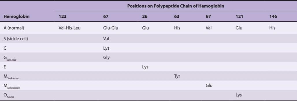
aOther hemoglobins have abnormal a chains. Abnormal hemoglobins that are very similar electrophoretically but differ slightly in composition are indicated by the same letter and a subscript indicating the geographic location where they were first discovered; hence, MSaskatoon and MMilwaukee.
aسایر هموگلوبین ها دارای یک زنجیره غیر طبیعی هستند. هموگلوبین های غیرعادی که از نظر الکتروفورزی بسیار شبیه به هم هستند اما از نظر ترکیب کمی متفاوت هستند، با یک حرف و یک زیرنویس نشان می دهد که موقعیت جغرافیایی را که برای اولین بار در آن کشف شده است نشان می دهد. از این رو، MSaskatoon و MMilwaukee.
PLASMA
The fluid portion of the blood, the plasma, is a remarkable solution containing an immense number of ions, inorganic molecules, and organic molecules that are in transit to various parts of the body or aid in the transport of other substances. Normal plasma volume is about 5% of body weight, or roughly 3500 mL in a 70-kg man. Plasma clots on standing, remaining fluid only if an anticoagulant is added. If whole blood is allowed to clot and the clot is removed, the remaining fluid is called serum. Serum has essentially the same composition as plasma, except that its fibrinogen and clotting factors II, V, and VIII (Table 31-4) have been removed and it has a higher serotonin content because of the breakdown of platelets during clotting.
پلاسما
بخش مایع خون، پلاسما، محلول قابل توجهی است که حاوی تعداد زیادی یون، مولکول های معدنی و مولکول های آلی است که در حال انتقال به قسمت های مختلف بدن هستند یا به انتقال مواد دیگر کمک می کنند. حجم طبیعی پلاسما حدود 5 درصد وزن بدن یا تقریباً 3500 میلی لیتر در یک مرد 70 کیلوگرمی است. پلاسما در حالت ایستاده لخته می شود و فقط در صورت اضافه شدن یک ضد انعقاد مایع باقی می ماند. اگر اجازه داده شود که خون کامل لخته شود و لخته برداشته شود، مایع باقی مانده سرم نامیده می شود. سرم اساساً ترکیبی مشابه پلاسما دارد، با این تفاوت که فیبرینوژن و فاکتورهای انعقادی II، V و VIII (جدول 31-4) حذف شده اند و به دلیل تجزیه پلاکت ها در طول لخته شدن، محتوای سروتونین بالاتری دارد.
TABLE 31-4 System for naming blood-clotting factors.
جدول 31-4 سیستم نامگذاری عوامل لخته کننده خون.

aFactor VI is not a separate entity and has been dropped.
aFactor VI یک موجودیت جداگانه نیست و حذف شده است.
PLASMA PROTEINS
The plasma proteins consist of albumin, globulin, and fibrinogen fractions. Most capillary walls are relatively impermeable to the proteins in plasma, and the proteins therefore exert an osmotic force of about 25 mm Hg across the capillary wall (oncotic pressure; see Chapter 1) that pulls water into the blood. The plasma proteins are also responsible for 15% of the buffering capacity of proteins in the blood (including hemoglobin; see Chapter 39) because of the weak ionization of their substituent COOH and NH2 groups. At the normal plasma pH of 7.40, the proteins are mostly in the anionic form (see Chapter 1). Plasma proteins may have specific functions (eg, antibodies and the proteins concerned with blood clotting), whereas others function as nonspecific carriers for various hormones, other solutes, and drugs.
پروتئین های پلاسما
پروتئین های پلاسما شامل آلبومین، گلوبولین و بخش های فیبرینوژن هستند. بیشتر دیواره های مویرگ ها نسبت به پروتئین های پلاسما نفوذناپذیر هستند و بنابراین پروتئین ها نیروی اسمزی حدود 25 میلی متر جیوه در سراسر دیواره مویرگ اعمال می کنند (فشار انکوتیک؛ فصل 1 را ببینید) که آب را به داخل خون می کشد. پروتئین های پلاسما همچنین مسئول 15 درصد ظرفیت بافری پروتئین ها در خون هستند (از جمله هموگلوبین؛ به فصل 39 مراجعه کنید) به دلیل یونیزاسیون ضعیف گروه های COOH و NH2 جایگزین آنها. در pH طبیعی پلاسما 7.40، پروتئین ها بیشتر به شکل آنیونی هستند (به فصل 1 مراجعه کنید). پروتئین های پلاسما ممکن است عملکردهای خاصی داشته باشند (مثلاً آنتی بادی ها و پروتئین های مربوط به لخته شدن خون)، در حالی که بقیه به عنوان حامل های غیر اختصاصی برای هورمون های مختلف، سایر املاح و داروها عمل می کنند.
ORIGIN OF PLASMA PROTEINS
Circulating antibodies are manufactured by lymphocytes. Most of the other plasma proteins are synthesized in the liver. These proteins and their principal functions are listed in Table 31-5.
منشا پروتئین های پلاسما
آنتی بادی های در گردش توسط لنفوسیت ها ساخته می شوند. بیشتر سایر پروتئین های پلاسما در کبد سنتز می شوند. این پروتئین ها و عملکردهای اصلی آنها در جدول 31-5 آمده است.
TABLE 31-5 Some of the proteins synthesized by the liver: Physiologic functions and properties.
جدول 31-5 برخی از پروتئین های سنتز شده توسط کبد: عملکردها و خواص فیزیولوژیکی.
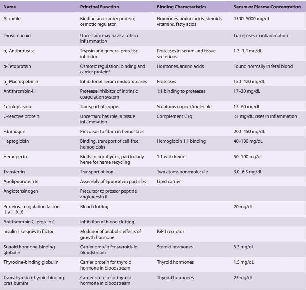
aThe function of a-fetoprotein is uncertain, but because of its structural homology to albumin it is often assigned these functions.
aعملکرد a-fetoprotein نامشخص است، اما به دلیل همسانی ساختاری آن با آلبومین، اغلب این وظایف به آن اختصاص داده می شود.
Data on the turnover of albumin show that synthesis plays an important role in the maintenance of normal levels. In normal adult humans, the plasma albumin level is 3.5-5.0 g/dL, and the total exchangeable albumin pool is 4.0- 5.0 g/kg body weight; 38-45% of this albumin is intravascular, and much of the rest of it is in the skin. Between 6% and 10% of the exchangeable pool is degraded per day, and the degraded albumin is replaced by hepatic synthesis of 200-400 mg/kg/day. The albumin is probably transported to the extravascular areas by vesicular transport across the walls of the capillaries (see Chapter 2). Albumin synthesis is carefully regulated. It is decreased during fasting and increased in conditions such as nephrosis in which there is excessive albumin loss.
داده های مربوط به گردش آلبومین نشان می دهد که سنتز نقش مهمی در حفظ سطوح طبیعی دارد. در انسان بالغ طبیعی، سطح آلبومین پلاسما 3.5-5.0 گرم در دسی لیتر است و کل آلبومین قابل تعویض 4.0-5.0 گرم بر کیلوگرم وزن بدن است. 38 تا 45 درصد این آلبومین داخل عروقی است و بقیه آن در پوست است. بین 6 تا 10 درصد از استخر قابل تعویض در روز تجزیه می شود و آلبومین تجزیه شده با سنتز کبدی 200-400 میلی گرم بر کیلوگرم در روز جایگزین می شود. آلبومین احتمالاً با انتقال تاولی از طریق دیواره های مویرگ ها به نواحی خارج عروقی منتقل می شود (به فصل 2 مراجعه کنید). سنتز آلبومین به دقت تنظیم می شود. در طول روزه داری کاهش می یابد و در شرایطی مانند نفروز که در آن از دست دادن بیش از حد آلبومین وجود دارد، افزایش می یابد.
HYPOPROTEINEMIA
Plasma protein levels are maintained during starvation until body protein stores are markedly depleted. However, in prolonged starvation and in malabsorption syndromes due to intestinal diseases, plasma protein levels are low (hypoproteinemia). They are also low in liver disease, because hepatic protein synthesis is depressed, and in nephrosis, because large amounts of albumin are lost in the urine. Because of the decrease in the plasma oncotic pressure, edema tends to develop. Rarely, there is congenital absence of one or another plasma protein. An example of congenital protein deficiency is the congenital form of afibrinogenemia, characterized by defective blood clotting.
هیپوپروتئینمی
سطح پروتئین پلاسما در طول گرسنگی تا زمانی که ذخایر پروتئین بدن به طور قابل توجهی تخلیه شود حفظ می شود. با این حال، در گرسنگی طولانی مدت و در سندرم های سوء جذب ناشی از بیماری های روده، سطح پروتئین پلاسما پایین است (هیپوپروتئینمی). آنها همچنین در بیماری کبدی کم هستند، زیرا سنتز پروتئین کبدی کاهش می یابد، و در نفروز، زیرا مقادیر زیادی آلبومین در ادرار از بین می رود. به دلیل کاهش فشار انکوتیک پلاسما، ادم تمایل دارد ایجاد شود. به ندرت، فقدان مادرزادی یک یا آن پروتئین پلاسما وجود دارد. نمونه ای از کمبود مادرزادی پروتئین، شکل مادرزادی آفیبرینوژنمی است که با لخته شدن خون معیوب مشخص می شود.
LYMPH
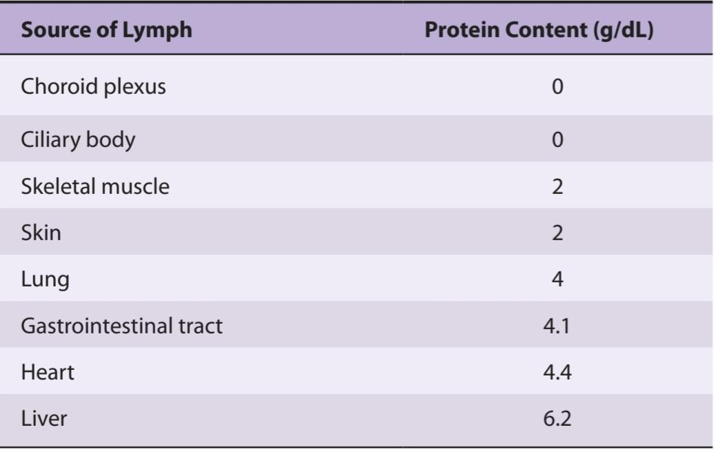
Lymph is tissue fluid that enters the lymphatic vessels. It drains into the venous blood via the thoracic and right lymphatic ducts. It contains clotting factors and clots on standing in vitro. In most locations, it also contains proteins that have traversed capillary walls and can then return to the blood via the lymph. Nevertheless, its protein content is generally lower than that of plasma, which contains about 7 g/dL, but lymph protein content varies with the region from which the lymph drains (Table 31-6). Water-insoluble fats are absorbed from the intestine into the lymphatics, and the lymph in the thoracic duct after a meal is milky because of its high fat content (see Chapter 26). Lymphocytes also enter the circulation principally through the lymphatics, and there are appreciable numbers of lymphocytes in thoracic duct lymph.
لنف
لنف مایع بافتی است که وارد عروق لنفاوی می شود. از طریق مجاری لنفاوی سینه ای و راست به خون وریدی تخلیه می شود. حاوی فاکتورهای انعقادی و لخته در حالت ایستاده در شرایط آزمایشگاهی است. در بیشتر مکانها، حاوی پروتئینهایی است که از دیوارههای مویرگی عبور میکنند و سپس میتوانند از طریق لنف به خون بازگردند. با این وجود، محتوای پروتئین آن به طور کلی کمتر از پلاسما است، که حاوی حدود 7 گرم در دسی لیتر است، اما محتوای پروتئین لنفاوی با منطقه ای که لنف از آن تخلیه می شود، متفاوت است (جدول 31-6). چربی های نامحلول در آب از روده به غدد لنفاوی جذب می شوند و لنف در مجرای قفسه سینه بعد از غذا به دلیل محتوای چربی زیاد آن شیری رنگ است (به فصل 26 مراجعه کنید). لنفوسیت ها نیز عمدتاً از طریق غدد لنفاوی وارد گردش خون می شوند و تعداد قابل توجهی لنفوسیت در لنف مجرای قفسه سینه وجود دارد.
TABLE 31-6 Approximate protein content of lymph in humans.
جدول 31-6 محتوای پروتئین تقریبی لنف در انسان.
BLOOD TYPES
The membranes of human red cells contain a variety of blood group antigens, which are also called agglutinogens. The most important and best known of these are the A and B antigens, but there are many more.
گروه های خونی
غشای گلبول های قرمز انسان حاوی انواع آنتی ژن های گروه خونی است که به آنها آگلوتینوژن نیز می گویند. مهم ترین و شناخته شده ترین آنها آنتی ژن های A و B هستند، اما تعداد بیشتری نیز وجود دارد.
THE ABO SYSTEM
The A and B antigens are inherited as mendelian dominants, and individuals are divided into four major blood types on this basis. Type A individuals have the A antigen, type B have the B, type AB have both, and type O have neither. The A and B antigens are complex oligosaccharides that differ in their terminal sugar. An H gene codes for a fucose transferase that adds a terminal fucose, forming the H antigen that is usually present in individuals of all blood types (Figure 31- 9). Individuals who are type A also express a second transferase that catalyzes placement of a terminal N-acetylgalactosamine on the H antigen, whereas individuals who are type B express a transferase that places a terminal galactose. Individuals who are type AB have both transferases. Individuals who are type O have neither, so the H antigen persists.
سیستم ABO
آنتی ژن های A و B به صورت غالب مندلی به ارث می رسند و افراد بر این اساس به چهار گروه خونی اصلی تقسیم می شوند. افراد نوع A دارای آنتی ژن A، نوع B دارای B، نوع AB هر دو و نوع O هیچ کدام را ندارند. آنتی ژن های A و B الیگوساکاریدهای پیچیده ای هستند که از نظر قند نهایی متفاوت هستند. یک ژن H یک فوکوز ترانسفراز را کد می کند که یک فوکوز انتهایی را اضافه می کند و آنتی ژن H را تشکیل می دهد که معمولاً در افراد با همه گروه های خونی وجود دارد (شکل 31-9). افرادی که نوع A هستند، ترانسفراز دومی را نیز بیان می کنند که قرارگیری یک N-استیل گالاکتوزامین پایانی را بر روی آنتی ژن H کاتالیز می کند، در حالی که افرادی که نوع B هستند، ترانسفرازی را بیان می کنند که یک گالاکتوز انتهایی را قرار می دهد. افرادی که نوع AB هستند هر دو ترانسفراز دارند. افرادی که نوع O هستند هیچ کدام را ندارند، بنابراین آنتی ژن H باقی می ماند.
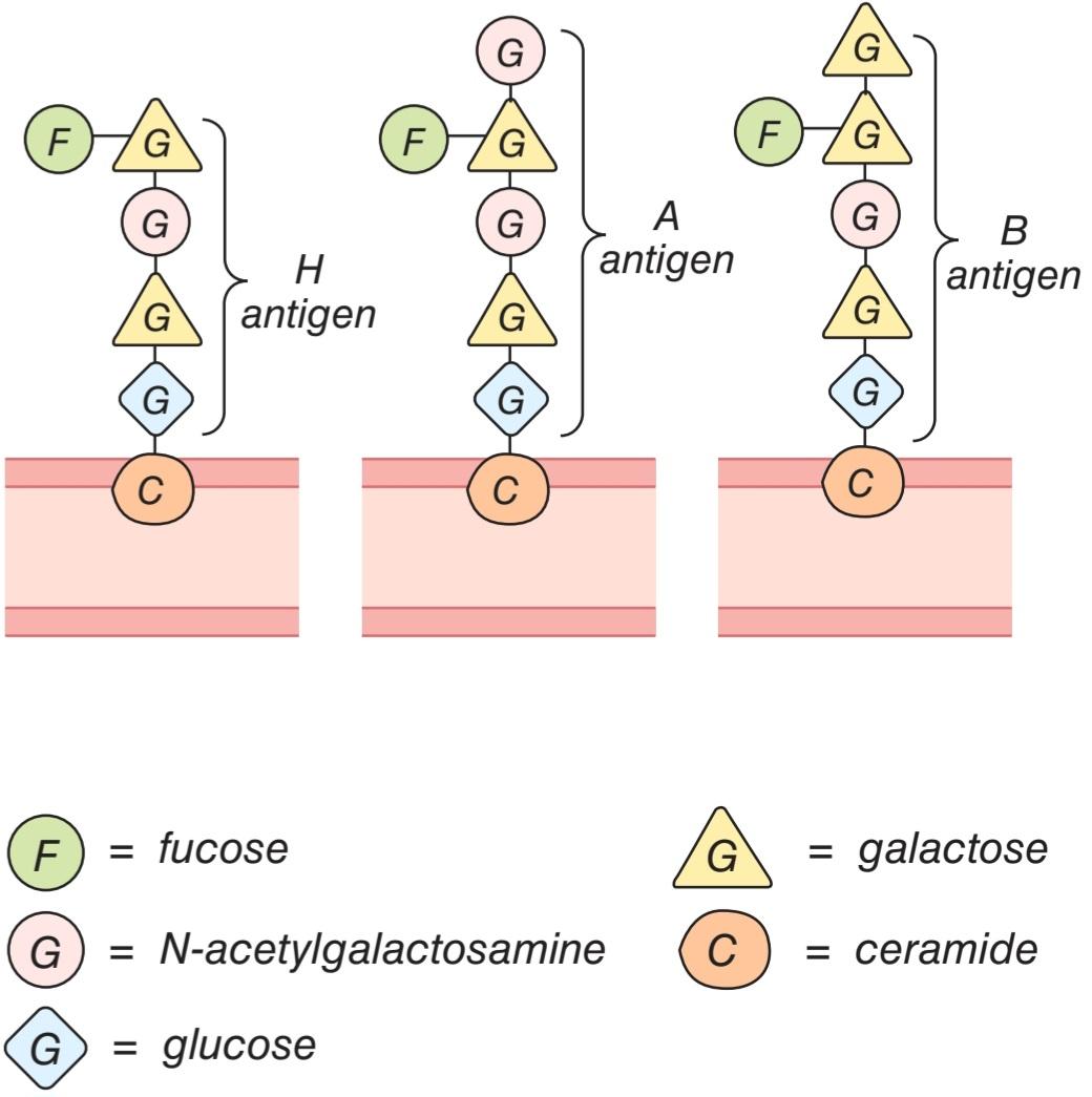
FIGURE 31-9 Antigens of the ABO system on the surface of red blood cells.
شکل 31-9 آنتی ژن های سیستم ABO روی سطح گلبول های قرمز خون.
Antibodies against red cell agglutinogens are called agglutinins. Antigens very similar to A and B are common in intestinal bacteria and possibly in foods to which newborn individuals are exposed. Therefore, infants rapidly develop antibodies against the antigens not present in their own cells. Thus, type A individuals develop anti-B antibodies, type B individuals develop anti-A antibodies, type O individuals develop both, and type AB individuals develop neither (Table 31-7). When the plasma of a type A individual is mixed with type B red cells, the anti-B antibodies cause the type B red cells to clump (agglutinate), as shown in Figure 31-10. The other agglutination reactions produced by mismatched plasma and red cells are summarized in Table 31-7. ABO blood typing is performed by mixing an individual’s red blood cells with antisera containing the various agglutinins on a slide and seeing whether agglutination occurs.
آنتی بادی ها علیه آگلوتینوژن های گلبول قرمز آگلوتینین نامیده می شوند. آنتی ژن های بسیار شبیه به A و B در باکتری های روده و احتمالاً در غذاهایی که افراد تازه متولد شده در معرض آنها قرار دارند، رایج هستند. بنابراین، نوزادان به سرعت آنتیبادیهایی را علیه آنتیژنهایی که در سلولهای خودشان وجود ندارند تولید میکنند. بنابراین، افراد نوع A آنتی بادی های ضد B، افراد نوع B آنتی بادی های ضد A، افراد نوع O هر دو را ایجاد می کنند و افراد نوع AB هیچ کدام را ایجاد نمی کنند (جدول 31-7). همانطور که در شکل 31-10 نشان داده شده است، هنگامی که پلاسمای یک فرد نوع A با گلبول های قرمز نوع B مخلوط می شود، آنتی بادی های ضد B باعث جمع شدن گلبول های قرمز نوع B می شوند (آگلوتینه). سایر واکنش های آگلوتیناسیون تولید شده توسط پلاسمای نامتناسب و گلبول های قرمز در جدول 31-7 خلاصه شده است. گروهبندی خونی ABO با مخلوط کردن گلبولهای قرمز خون فرد با آنتیسرمهای حاوی آگلوتینینهای مختلف روی یک اسلاید و دیدن اینکه آیا آگلوتیناسیون رخ میدهد یا خیر، انجام میشود.
TABLE 31-7 Summary of ABO system.
جدول 31-7 خلاصه سیستم ABO.


FIGURE 31-10 Red cell agglutination in incompatible plasma.
شکل 31-10 آگلوتیناسیون گلبول های قرمز در پلاسمای ناسازگار.
TRANSFUSION REACTIONS
Dangerous hemolytic transfusion reactions occur when blood is transfused into an individual with an incompatible blood type; that is, an individual who has agglutinins against the red cells in the transfusion. The plasma in the transfusion is usually so diluted in the recipient that it rarely causes agglutination even when the titer of agglutinins against the recipient’s cells is high. However, when the recipient’s plasma has agglutinins against the donor’s red cells, the cells agglutinate and hemolyze. Free hemoglobin is liberated into the plasma. The severity of the resulting transfusion reaction may vary from an asymptomatic minor rise in the plasma bilirubin level to severe jaundice and renal tubular damage leading to anuria and death.
واکنش های انتقال خون
واکنش های خطرناک همولیتیک انتقال خون زمانی رخ می دهد که خون به فردی با گروه خونی ناسازگار تزریق می شود. یعنی فردی که دارای آگلوتینین در برابر گلبول های قرمز در تزریق است. پلاسما در تزریق معمولاً آنقدر در گیرنده رقیق می شود که به ندرت باعث آگلوتیناسیون می شود حتی زمانی که تیتر آگلوتینین ها در برابر سلول های گیرنده بالا باشد. با این حال، هنگامی که پلاسمای گیرنده دارای آگلوتینین در برابر گلبول های قرمز اهداکننده باشد، سلول ها آگلوتینه شده و همولیز می شوند. هموگلوبین آزاد در پلاسما آزاد می شود. شدت واکنش حاصل از انتقال خون ممکن است از افزایش جزئی بدون علامت در سطح بیلی روبین پلاسما تا زردی شدید و آسیب لوله های کلیوی که منجر به آنوری و مرگ می شود متفاوت باشد.
Incompatibilities in the ABO blood group system are summarized in Table 31-7. Persons with type AB blood are “universal recipients” because they have no circulating agglutinins and can be given blood of any type without developing a transfusion reaction due to ABO incompatibility. Type O individuals are “universal donors” because they lack A and B antigens, and type O blood can be given to anyone without producing a transfusion reaction due to ABO incompatibility. This does not mean, however, that blood should ever be transfused without being cross-matched except in the most extreme emergencies, since the possibility of reactions or sensitization due to incompatibilities in systems other than ABO systems always exists. In cross-matching, donor red cells are mixed with recipient plasma on a slide and checked for agglutination.
ناسازگاری های سیستم گروه خونی ABO در جدول 31-7 خلاصه شده است. افراد دارای گروه خونی AB “دریافت کننده جهانی” هستند زیرا آگلوتینین در گردش ندارند و می توان خون از هر نوع را بدون ایجاد واکنش انتقال خون به دلیل ناسازگاری ABO به آنها داد. افراد نوع O “اهدای جهانی” هستند زیرا فاقد آنتی ژن های A و B هستند و خون نوع O را می توان بدون ایجاد واکنش انتقال خون به دلیل ناسازگاری ABO به هر کسی داد. با این حال، این بدان معنا نیست که خون باید بدون تلاقی متقابل تزریق شود، مگر در شدیدترین مواقع اضطراری، زیرا امکان واکنش یا حساسیت به دلیل ناسازگاری در سیستمهایی غیر از سیستمهای ABO همیشه وجود دارد. در تطبیق متقابل، گلبولهای قرمز اهداکننده با پلاسمای گیرنده بر روی یک اسلاید مخلوط میشوند و از نظر آگلوتیناسیون بررسی میشوند.
A procedure that has recently become popular is to withdraw the patient’s own blood in advance of elective surgery and then infuse this blood back (autologous transfusion) if a transfusion is needed during the surgery. With iron treatment, 1000-1500 mL can be withdrawn over a 3-week period. The popularity of banking one’s own blood is primarily due to fear of transmission of infectious diseases by heterologous transfusions, but of course another advantage is elimination of the risk of transfusion reactions.
روشی که اخیراً رایج شده است، برداشتن خون خود بیمار قبل از جراحی انتخابی و سپس تزریق مجدد خون (ترانسفوزیون اتولوگ) در صورت نیاز به تزریق در طول جراحی است. با درمان آهن، 1000-1500 میلی لیتر را می توان در یک دوره 3 هفته ای حذف کرد. محبوبیت بانک خون خود در درجه اول به دلیل ترس از انتقال بیماری های عفونی با تزریق هترولوگ است، اما البته مزیت دیگر حذف خطر واکنش های انتقال خون است.
OTHER AGGLUTINOGENS
In addition to the ABO system of antigens in human red cells, there are systems such as the Rh, MNSS, Lutheran, Kell, Kidd, and many others. There are over 500 billion possible known blood group phenotypes, and because undiscovered antigens undoubtedly exist, it has been calculated that the number of phenotypes is actually in the trillions.
سایر آگلوتینوژن ها
علاوه بر سیستم آنتی ژن ABO در گلبول های قرمز انسان، سیستم هایی مانند Rh، MNSS، Lutheran، Kell، Kidd و بسیاری دیگر وجود دارد. بیش از 500 میلیارد فنوتیپ گروه خونی شناخته شده وجود دارد، و از آنجا که آنتی ژن های کشف نشده بدون شک وجود دارند، محاسبه شده است که تعداد فنوتیپ ها در واقع به تریلیون ها می رسد.
The number of blood groups in animals is as large as it is in humans. An interesting question is why this degree of polymorphism developed and has persisted through evolution. Certain diseases are more common in individuals with one blood type or another, but the differences are not great. The significance of a recognition code of this complexity is therefore unknown.
تعداد گروه های خونی در حیوانات به اندازه انسان است. یک سوال جالب این است که چرا این درجه از چندشکلی توسعه یافته و در طول تکامل باقی مانده است. برخی از بیماری ها در افراد با یک گروه خونی یا دیگری شایع تر است، اما تفاوت ها زیاد نیست. بنابراین اهمیت کد شناسایی با این پیچیدگی ناشناخته است.
THE RH GROUP
Aside from the antigens of the ABO system, those of the Rh system are of the greatest clinical importance. The Rh factor, named for the rhesus monkey because it was first studied using the blood of this animal, is a system composed primarily of the C, D, and E antigens, although it actually contains many more. Unlike the ABO antigens, the system has not been detected in tissues other than red cells. D is by far the most antigenic component, and the term Rh-positive as it is generally used means that the individual has agglutinogen D. The Rh- negative individual has no D antigen and forms the anti-D agglutinin when injected with D-positive cells. The Rh typing serum used in routine blood typing is anti-D serum. Eighty-five percent of whites are D-positive and 15% are D- negative; over 99% of Asians are D-positive. Unlike the antibodies of the ABO system, anti-D antibodies do not develop without exposure of a D-negative individual to D-positive red cells by transfusion or entrance of fetal blood into the maternal circulation. However, D-negative individuals who have received a transfusion of D-positive blood (even years previously) can have appreciable anti-D titers and thus may develop transfusion reactions when transfused again with D-positive blood.
گروه RH
جدا از آنتی ژن های سیستم ABO، آنتی ژن های سیستم Rh بیشترین اهمیت بالینی را دارند. فاکتور Rh که برای میمون رزوس نامگذاری شده است زیرا برای اولین بار با استفاده از خون این حیوان مورد مطالعه قرار گرفت، سیستمی است که عمدتاً از آنتی ژن های C، D و E تشکیل شده است، اگرچه در واقع حاوی تعداد زیادی آنتی ژن است. برخلاف آنتی ژن های ABO، این سیستم در بافت های دیگری به جز گلبول های قرمز شناسایی نشده است. D تا حد زیادی آنتی ژنی ترین جزء است، و اصطلاح Rh مثبت همانطور که به طور کلی استفاده می شود به این معنی است که فرد دارای آگلوتینوژن D است. فرد Rh منفی آنتی ژن D ندارد و هنگامی که با سلول های D مثبت تزریق می شود، آگلوتینین ضد D را تشکیل می دهد. سرم Rh تایپ مورد استفاده در گروه بندی خون معمولی سرم ضد D است. 85 درصد سفیدپوستان D مثبت و 15 درصد D منفی هستند. بیش از 99 درصد از آسیایی ها D مثبت هستند. برخلاف آنتی بادیهای سیستم ABO، آنتیبادیهای ضد D بدون قرار گرفتن در معرض گلبولهای قرمز D مثبت با انتقال خون جنین یا ورود خون جنین به گردش خون مادر، ایجاد نمیشوند. با این حال، افراد D منفی که خون D مثبت دریافت کردهاند (حتی سالها قبل) میتوانند تیتر ضد D قابلتوجهی داشته باشند و بنابراین ممکن است هنگام تزریق مجدد خون D مثبت، واکنشهای انتقال خون ایجاد کنند.
HEMOLYTIC DISEASE OF THE NEWBORN
Another complication due to Rh incompatibility arises when an Rh-negative mother carries an Rh-positive fetus. Small amounts of fetal blood leak into the maternal circulation at the time of delivery, and some mothers develop significant titers of anti-Rh agglutinins during the postpartum period. During the next pregnancy, the mother’s agglutinins cross the placenta to the fetus. When anti-Rh agglutinins cross the placenta to an Rh-positive fetus, they can cause hemolysis and various forms of hemolytic disease of the newborn (erythroblastosis fetalis). If hemolysis in the fetus is severe, the infant may die in utero or may develop anemia, severe jaundice, and edema (hydrops fetalis). Kernicterus, a neurologic syndrome in which unconjugated bilirubin is deposited in the basal ganglia, may also develop, especially if birth is complicated by a period of hypoxia. Bilirubin rarely penetrates the brain in adults, but it does in infants with erythroblastosis, possibly in part because the blood-brain barrier is more permeable in infancy. However, the main reasons that the concentration of unconjugated bilirubin is very high in this condition are that production is increased and the bilirubin-conjugating system is not yet mature.
بیماری همولیتیک نوزاد
عارضه دیگر ناشی از ناسازگاری Rh زمانی ایجاد می شود که یک مادر Rh منفی جنین Rh مثبت را حمل کند. مقادیر کمی از خون جنین در زمان زایمان به گردش خون مادر نشت می کند و برخی از مادران در طول دوره پس از زایمان، تیترهای قابل توجهی از آگلوتینین ضد Rh ایجاد می کنند. در بارداری بعدی، آگلوتینین های مادر از جفت به جنین می گذرد. هنگامی که آگلوتینین های ضد Rh از جفت عبور می کنند و به جنین Rh مثبت می رسند، می توانند باعث همولیز و اشکال مختلف بیماری همولیتیک نوزاد (اریتروبلاستوز فتالیس) شوند. اگر همولیز در جنین شدید باشد، ممکن است نوزاد در رحم بمیرد یا ممکن است دچار کم خونی، زردی شدید و ادم (هیدروپس جنین) شود. کرنیکتروس، یک سندرم عصبی که در آن بیلی روبین غیر کونژوگه در گانگلیون های پایه رسوب می کند، نیز ممکن است ایجاد شود، به خصوص اگر تولد با یک دوره هیپوکسی پیچیده شود. بیلی روبین در بزرگسالان به ندرت به مغز نفوذ می کند، اما در نوزادان مبتلا به اریتروبلاستوز نفوذ می کند، احتمالاً تا حدی به این دلیل که سد خونی مغزی در دوران نوزادی نفوذپذیرتر است. با این حال، دلایل اصلی که غلظت بیلی روبین غیر کونژوگه در این شرایط بسیار زیاد است این است که تولید افزایش یافته و سیستم بیلی روبین کونژوگه هنوز بالغ نشده است.
About 50% of Rh-negative individuals are sensitized (develop an anti-Rh titer) by transfusion of Rh-positive blood. Because sensitization of Rh-negative mothers by carrying an Rh-positive fetus generally occurs at birth, the first child is usually normal. However, hemolytic disease occurs in about 17% of the Rh- positive fetuses born to Rh-negative mothers who have previously been pregnant one or more times with Rh-positive fetuses. Fortunately, it is usually possible to prevent sensitization from occurring the first time by administering a single dose of anti-Rh antibodies in the form of Rh immune globulin during the postpartum period. Such passive immunization does not harm the mother and has been demonstrated to prevent active antibody formation by the mother. In obstetric clinics, the institution of such treatment on a routine basis to unsensitized Rh- negative women who have delivered an Rh-positive baby has reduced the overall incidence of hemolytic disease by more than 90%. In addition, fetal Rh typing with material obtained by amniocentesis or chorionic villus sampling is now possible, and treatment with a small dose of Rh immune serum will prevent sensitization during pregnancy.
حدود 50 درصد از افراد Rh منفی با تزریق خون Rh مثبت حساس می شوند (تیتر ضد Rh ایجاد می کنند). از آنجایی که حساسیت مادران Rh منفی با حمل جنین Rh مثبت معمولا در بدو تولد اتفاق می افتد، اولین فرزند معمولا طبیعی است. با این حال، بیماری همولیتیک در حدود 17 درصد از جنین های Rh مثبت متولد شده از مادران Rh منفی که قبلا یک یا چند بار با جنین های Rh مثبت باردار شده اند، رخ می دهد. خوشبختانه، معمولاً می توان با تجویز یک دوز از آنتی بادی های ضد Rh به شکل گلوبولین ایمنی Rh در طول دوره پس از زایمان از ایجاد حساسیت در اولین بار جلوگیری کرد. چنین ایمن سازی غیرفعال به مادر آسیبی نمی رساند و نشان داده شده است که از تشکیل آنتی بادی فعال توسط مادر جلوگیری می کند. در کلینیک های زنان و زایمان، انجام چنین درمان هایی به صورت روتین برای زنان غیر حساس Rh منفی که نوزاد Rh مثبت به دنیا آورده اند، شیوع کلی بیماری همولیتیک را بیش از 90 درصد کاهش داده است. علاوه بر این، تایپ Rh جنین با مواد بدست آمده از آمنیوسنتز یا نمونه برداری از پرزهای کوریونی اکنون امکان پذیر است و درمان با دوز کمی از سرم ایمنی Rh از ایجاد حساسیت در دوران بارداری جلوگیری می کند.
HEMOSTASIS
Hemostasis is the process of forming clots in the walls of damaged blood vessels and preventing blood loss while maintaining blood in a fluid state within the vascular system. A collection of complex interrelated systemic mechanisms operates to maintain a balance between coagulation and anticoagulation.
هموستاز
هموستاز فرآیند تشکیل لخته در دیواره رگ های خونی آسیب دیده و جلوگیری از از دست دادن خون و در عین حال حفظ خون در حالت مایع در داخل سیستم عروقی است. مجموعه ای از مکانیسم های پیچیده سیستمیک مرتبط به هم برای حفظ تعادل بین انعقاد و ضد انعقاد عمل می کند.
RESPONSE TO INJURY
When a small blood vessel is transected or damaged, the injury initiates a series of events (Figure 31-11) that lead to the formation of a clot. This seals off the damaged region and prevents further blood loss. A temporary hemostatic plug of platelets is triggered when platelets bind to collagen and aggregate. There is also constriction of the vessel, with constriction of an injured arteriole or small artery occasionally so marked that its lumen is obliterated, at least temporarily. The vasoconstriction is due to serotonin (5-HT) and other vasoconstrictors liberated from the platelets adhered to the walls of the damaged vessel. This is then followed by conversion of the plug into the definitive clot.
پاسخ به آسیب
هنگامی که یک رگ خونی کوچک قطع می شود یا آسیب می بیند، آسیب شروع کننده مجموعه ای از رویدادها است (شکل 31-11) که منجر به تشکیل لخته می شود. این ناحیه آسیب دیده را میبندد و از از دست دادن خون بیشتر جلوگیری میکند. زمانی که پلاکت ها به کلاژن متصل می شوند و تجمع می یابند، پلاکت هموستاتیک موقتی از پلاکت ها ایجاد می شود. رگ نیز انقباض دارد، با انقباض شریان آسیب دیده یا شریان کوچک، گاهی چنان مشخص می شود که مجرای آن، حداقل به طور موقت، محو می شود. انقباض عروق ناشی از سروتونین (5-HT) و سایر منقبض کننده های عروق آزاد شده از پلاکت های چسبیده به دیواره رگ آسیب دیده است. سپس این کار با تبدیل پلاگین به لخته قطعی دنبال می شود.
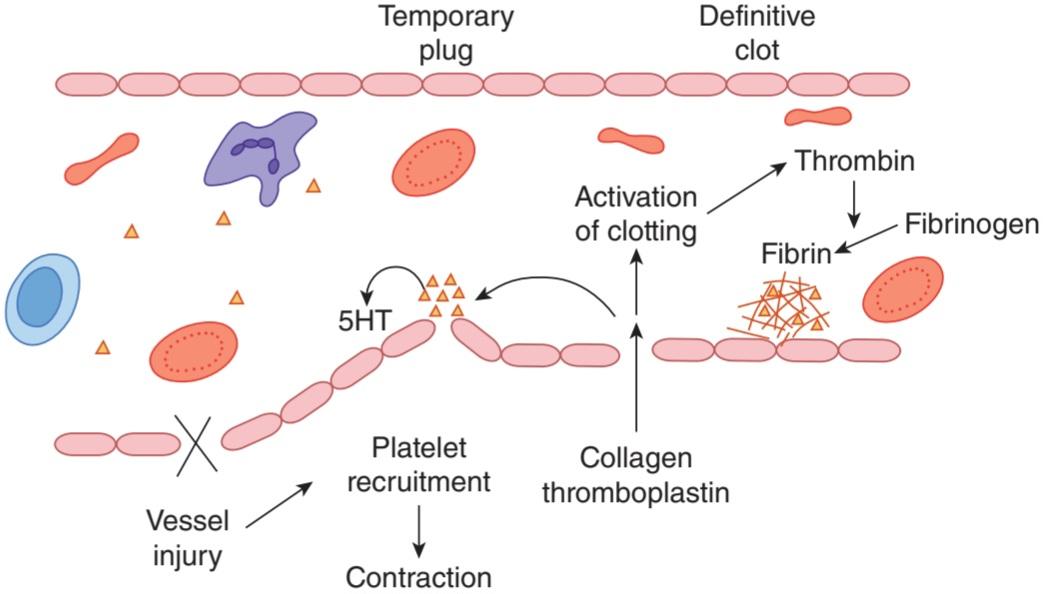
FIGURE 31-11 Summary of reactions involved in hemostasis. Injury to a blood vessel exposes collagen and thromboplastin, recruiting platelets to the site of injury to form a temporary plug. Platelets release 5-hydroxytryptamine, among other factors, resulting in smooth muscle contraction and vasoconstriction. Activation of the clotting cascade in response to collagen and thromboplastin activates thrombin, which converts circulating fibrinogen to fibrin monomers. Fibrin monomers polymerize and are cross-linked and accumulate with platelets at the site of injury to form the definitive clot.
شکل 31-11 خلاصه ای از واکنش های دخیل در هموستاز. آسیب به رگ خونی، کلاژن و ترومبوپلاستین را در معرض دید قرار می دهد و پلاکت ها را به محل آسیب جذب می کند تا یک پلاگ موقت تشکیل دهد. پلاکت ها 5-هیدروکسی تریپتامین را در کنار سایر عوامل آزاد می کنند که منجر به انقباض عضلات صاف و انقباض عروق می شود. فعال شدن آبشار لخته شدن در پاسخ به کلاژن و ترومبوپلاستین، ترومبین را فعال می کند که فیبرینوژن در گردش را به مونومرهای فیبرین تبدیل می کند. مونومرهای فیبرین پلیمریزه شده و به صورت متقاطع متصل می شوند و با پلاکت ها در محل آسیب تجمع می یابند تا لخته قطعی را تشکیل دهند.
THE CLOTTING MECHANISM
The loose aggregation of platelets in the temporary plug is bound together and converted into the definitive clot by fibrin. Fibrin formation involves a cascade of enzymatic reactions and a series of numbered clotting factors (Table 31-4). The fundamental reaction is conversion of the soluble plasma protein fibrinogen to insoluble fibrin (Figure 31-12). The process involves the release of two pairs of polypeptides from each fibrinogen molecule. The remaining portion, fibrin monomer, then polymerizes with other monomer molecules to form fibrin. The fibrin is initially a loose mesh of interlacing strands. It is converted by the formation of covalent cross-linkages to a dense, tight aggregate (stabilization). This latter reaction is catalyzed by activated factor XIII and requires Ca2+.
مکانیسم انعقاد
تجمع شل پلاکت ها در پلاگ موقت به هم متصل شده و توسط فیبرین به لخته قطعی تبدیل می شود. تشکیل فیبرین شامل یک آبشار از واکنش های آنزیمی و یک سری فاکتورهای انعقادی شماره گذاری شده است (جدول 31-4). واکنش اساسی تبدیل فیبرینوژن پروتئین محلول پلاسما به فیبرین نامحلول است (شکل 31-12). این فرآیند شامل آزادسازی دو جفت پلی پپتید از هر مولکول فیبرینوژن است. بخش باقیمانده، مونومر فیبرین، سپس با مولکول های مونومر دیگر پلیمریزه می شود و فیبرین را تشکیل می دهد. فیبرین در ابتدا یک شبکه شل از رشته های در هم تنیده است. با تشکیل پیوندهای عرضی کووالانسی به یک سنگدانه متراکم و محکم (تثبیت) تبدیل می شود. این واکنش دوم توسط فاکتور XIII فعال شده کاتالیز می شود و به +Ca2 نیاز دارد.

FIGURE 31-12 The clotting mechanism. a, active form of clotting factor. TFI, tissue factor pathway inhibitor; TPL, tissue thromboplastin. For other abbreviations, see Table 31-4.
شکل 31-12 مکانیسم لخته شدن. الف، شکل فعال فاکتور انعقادی. TFI، مهارکننده مسیر فاکتور بافتی. TPL، ترومبوپلاستین بافتی. برای سایر اختصارات، جدول 31-4 را ببینید.
The conversion of fibrinogen to fibrin is catalyzed by thrombin. Thrombin is a serine protease that is formed from its circulating precursor, prothrombin, by the action of activated factor X. It has additional actions, including activation of platelets, endothelial cells, and leukocytes via so-called proteinase activated receptors, which are G-protein-coupled.
تبدیل فیبرینوژن به فیبرین توسط ترومبین کاتالیز می شود. ترومبین یک پروتئاز سرین است که از پیش ساز در گردش آن، پروترومبین، با اثر فاکتور X فعال شده تشکیل می شود. این ماده دارای اقدامات اضافی از جمله فعال سازی پلاکت ها، سلول های اندوتلیال و لکوسیت ها از طریق گیرنده های به اصطلاح فعال پروتئیناز است که با پروتئین G همراه هستند.
Factor X can be activated by either of two systems, known as intrinsic and extrinsic (Figure 31-12). The initial reaction in the intrinsic system is conversion of inactive factor XII to active factor XII (XIIa). This activation, which is catalyzed by high-molecular-weight kininogen and kallikrein (see Chapter 32), can be brought about in vitro by exposing the blood to glass, or in vivo by collagen fibers underlying the endothelium. Active factor XII then activates factor XI, and active factor XI activates factor IX. Activated factor IX forms a complex with active factor VIII, which is activated when it is separated from von Willebrand factor. The complex of IXa and VIIIa activate factor X. Phospholipids from aggregated platelets (PL) and Ca2+ are necessary for full activation of factor X. The extrinsic system is triggered by the release of tissue thromboplastin (TPL), a protein-phospholipid mixture that activates factor VII. Tissue thromboplastin and factor VII activate factors IX and X. In the presence of PL, Ca2+, and factor V, activated factor X catalyzes the conversion of prothrombin to thrombin. The extrinsic pathway is inhibited by a tissue factor pathway inhibitor (TFI) that forms a quaternary structure with TPL, factor VIIa, and factor Xa.
فاکتور X می تواند توسط هر یک از دو سیستم که به نام های درونی و بیرونی شناخته می شوند فعال شود (شکل 31-12). واکنش اولیه در سیستم ذاتی تبدیل فاکتور XII غیر فعال به فاکتور XII فعال (XIIa) است. این فعالسازی، که توسط کینینوژن و کالیکرئین با وزن مولکولی بالا کاتالیز میشود (به فصل 32 مراجعه کنید)، میتوان در شرایط آزمایشگاهی با قرار دادن خون در معرض شیشه، یا در داخل بدن توسط رشتههای کلاژنی که در زیر اندوتلیوم قرار دارند، انجام شود. فاکتور XII فعال فاکتور XI را فعال می کند و عامل XI فعال عامل IX را فعال می کند. فاکتور IX فعال یک کمپلکس با فاکتور هشت فعال تشکیل می دهد که با جدا شدن از فاکتور فون ویلبراند فعال می شود. مجموعه IXa و VIIIa فاکتور X را فعال میکند. فسفولیپیدهای پلاکتهای تجمع یافته (PL) و +Ca2 برای فعالسازی کامل فاکتور X ضروری هستند. سیستم بیرونی با آزاد شدن ترومبوپلاستین بافتی (TPL)، یک مخلوط پروتئین-فسفولیپید که فاکتور VII را فعال میکند، تحریک میشود. ترومبوپلاستین بافتی و فاکتور VII فاکتورهای IX و X را فعال می کنند. در حضور +PL، Ca2 و فاکتور V، فاکتور X فعال تبدیل پروترومبین به ترومبین را کاتالیز می کند. مسیر بیرونی توسط یک بازدارنده مسیر فاکتور بافتی (TFI) مهار می شود که یک ساختار چهارتایی با TPL، فاکتور VIIa و فاکتور Xa را تشکیل می دهد.
ANTICLOTTING MECHANISMS
The tendency of blood to clot is balanced in vivo by reactions that prevent clotting inside the blood vessels, break down any clots that do form, or both. These reactions include the interaction between the platelet-aggregating effect of thromboxane A2 and the antiaggregating effect of prostacyclin, which causes clots to form at the site when a blood vessel is injured but keeps the vessel lumen free of clot (see Chapter 32 and Clinical Box 31-3).
مکانیسم های ضد انعقاد
تمایل خون به لخته شدن در داخل بدن با واکنش هایی که از لخته شدن در داخل رگ های خونی جلوگیری می کند، لخته هایی را که تشکیل می شود یا هر دو را می شکنند، متعادل می شود. این واکنشها شامل برهمکنش بین اثر تجمع پلاکتی ترومبوکسان A2 و اثر ضد تجمع پروستاسیکلین میشود، که باعث ایجاد لخته در محل در هنگام آسیب دیدگی رگ خونی میشود اما مجرای رگ را عاری از لخته نگه میدارد (به فصل 32 و جعبه بالینی 31-3 مراجعه کنید).
Antithrombin III is a circulating protease inhibitor that binds to serine proteases in the coagulation system, blocking their activity as clotting factors. This binding is facilitated by heparin, a naturally occurring anticoagulant that is a mixture of sulfated polysaccharides. The clotting factors that are inhibited are the active forms of factors IX, X, XI, and XII.
آنتی ترومبین III یک مهارکننده پروتئاز در گردش است که به پروتئازهای سرین در سیستم انعقادی متصل می شود و فعالیت آنها را به عنوان فاکتورهای انعقادی مسدود می کند. این اتصال توسط هپارین، یک ضد انعقاد طبیعی که مخلوطی از پلی ساکاریدهای سولفاته است، تسهیل می شود. عوامل انعقادی که مهار می شوند، اشکال فعال فاکتورهای IX، X، XI و XII هستند.
CLINICAL BOX 31-3
Abnormalities of Hemostasis
In addition to clotting abnormalities due to platelet disorders, hemorrhagic diseases can be produced by selective deficiencies of most of the clotting factors (Table 31–8). Hemophilia A, which is caused by factor VIII deficiency, is relatively common. von Willebrand factor deficiency likewise causes a bleeding disorder (von Willebrand disease) by reducing platelet adhesion and by lowering plasma factor VIII. The condition can be congenital or acquired. The large von Willebrand molecule is subject to cleavage and resulting inactivation by the plasma metalloprotease ADAM-13 in vascular areas where fluid shear stress is elevated. Finally, when absorption of vitamin K is depressed along with absorption of other fat-soluble vitamins (see Chapter 26), the resulting clotting factor deficiencies may cause the development of a significant bleeding tendency.
جعبه بالینی 31-3
ناهنجاری های هموستاز
علاوه بر ناهنجاری های لخته شدن ناشی از اختلالات پلاکتی، بیماری های هموراژیک را می توان در اثر کمبودهای انتخابی بیشتر عوامل لخته شدن ایجاد کرد (جدول 31-8). هموفیلی A که در اثر کمبود فاکتور 8 ایجاد می شود، نسبتاً شایع است. کمبود فاکتور فون ویلبراند نیز با کاهش چسبندگی پلاکت ها و کاهش فاکتور VIII پلاسما باعث اختلال خونریزی (بیماری فون ویلبراند) می شود. این بیماری می تواند مادرزادی یا اکتسابی باشد. مولکول بزرگ فون ویلبراند توسط متالوپروتئاز پلاسما ADAM-13 در نواحی عروقی که تنش برشی سیال افزایش مییابد در معرض شکست و غیرفعال شدن است. در نهایت، هنگامی که جذب ویتامین K همراه با جذب سایر ویتامینهای محلول در چربی کاهش مییابد (به فصل 26 مراجعه کنید)، کمبود فاکتور لختهکننده ممکن است باعث ایجاد تمایل به خونریزی قابل توجهی شود.
Formation of clots inside blood vessels is called thrombosis to distinguish it from the normal extravascular clotting of blood. Thromboses are a major medical problem. They are particularly prone to occur where blood flow is sluggish because the slow flow permits activated clotting factors to accumulate instead of being washed away. They also occur in vessels when the intima is damaged by atherosclerotic plaques, and over areas of damage to the endocardium. They frequently occlude the arterial supply to the organs in which they form, and bits of thrombus (emboli) sometimes break off and travel in the bloodstream to distant sites, damaging other organs. An example is obstruction of the pulmonary artery or its branches by thrombi from the leg veins (pulmonary embolism). Congenital absence of protein C leads to uncontrolled intravascular coagulation and, in general, death in infancy. If this condition is diagnosed and treatment is instituted, the coagulation defect disappears. Resistance to activated protein C is another cause of thrombosis, and this condition is common. It is due to a point mutation in the gene for factor V, which prevents activated protein C from inactivating the factor.
تشکیل لخته در داخل رگ های خونی ترومبوز نامیده می شود تا آن را از لخته شدن خارج عروقی طبیعی تشخیص دهد. ترومبوز یک مشکل عمده پزشکی است. آنها به ویژه در جاهایی که جریان خون کند است به وجود می آیند زیرا جریان آهسته اجازه می دهد تا عوامل انعقادی فعال به جای شسته شدن جمع شوند. آنها همچنین در عروق هنگامی که انتیما توسط پلاک های آترواسکلروتیک آسیب می بیند و در مناطق آسیب دیده به اندوکارد رخ می دهد. آنها اغلب منبع شریانی اندامهایی را که در آن تشکیل میشوند مسدود میکنند، و تکههای ترومبوز (آمبولی) گاهی اوقات شکسته میشوند و در جریان خون به نقاط دوردست میروند و به سایر اندامها آسیب میزنند. به عنوان مثال، انسداد شریان ریوی یا شاخههای آن توسط ترومبهای وریدهای پا (آمبولی ریه) است. عدم وجود مادرزادی پروتئین C منجر به انعقاد داخل عروقی کنترل نشده و به طور کلی مرگ در دوران نوزادی می شود. اگر این بیماری تشخیص داده شود و درمان شروع شود، نقص انعقادی از بین می رود. مقاومت در برابر پروتئین فعال C یکی دیگر از علل ترومبوز است و این وضعیت شایع است. این به دلیل جهش نقطه ای در ژن فاکتور V است که مانع از غیرفعال کردن پروتئین C فعال می شود.
Disseminated intravascular coagulation is another serious complication of septicemia, extensive tissue injury, and other diseases in which fibrin is deposited in the vascular system and many small- and medium-sized vessels are thrombosed. The increased consumption of platelets and coagulation factors causes bleeding to occur at the same time. The cause of the condition appears to be increased generation of thrombin due to increased TPL activity without adequate TFI activity.
انعقاد داخل عروقی منتشر یکی دیگر از عوارض جدی سپتی سمی، آسیب بافتی گسترده و سایر بیماری هاست که در آن فیبرین در سیستم عروقی رسوب می کند و بسیاری از عروق کوچک و متوسط ترومبوز می شوند. افزایش مصرف پلاکت ها و فاکتورهای انعقادی باعث خونریزی همزمان می شود. به نظر می رسد علت این وضعیت افزایش تولید ترومبین به دلیل افزایش فعالیت TPL بدون فعالیت TFI کافی باشد.
THERAPEUTIC HIGHLIGHTS
Hemophilia has been treated with factor VIII-rich preparations made from plasma, or, more recently, factor VIII produced by recombinant DNA techniques. Some patients with von Willebrand disease are treated with desmopressin, which stimulates production of factor VIII, particularly prior to dental procedures or surgery. Thrombotic disorders, on the other hand, are treated with anticoagulants such as heparin.
نکات برجسته درمانی
هموفیلی با داروهای غنی از فاکتور VIII ساخته شده از پلاسما یا اخیراً فاکتور هشت تولید شده توسط تکنیک های DNA نوترکیب درمان شده است. برخی از بیماران مبتلا به بیماری فون ویلبراند با دسموپرسین درمان می شوند که باعث تحریک تولید فاکتور VIII می شود، به ویژه قبل از عمل های دندانپزشکی یا جراحی. از طرف دیگر اختلالات ترومبوتیک با داروهای ضد انعقاد مانند هپارین درمان می شوند.
TABLE 31-8 Examples of diseases due to deficiency of clotting factors.
جدول 31-8 نمونه هایی از بیماری های ناشی از کمبود فاکتورهای انعقادی.

The endothelium of the blood vessels also plays an active role in preventing the extension of clots. All endothelial cells except those in the cerebral microcirculation produce thrombomodulin, a thrombin-binding protein, on their surfaces. In circulating blood, thrombin is a procoagulant that activates factors V and VIII, but when it binds to thrombomodulin, it becomes an anticoagulant in that the thrombomodulin-thrombin complex activates protein C (Figure 33-13). Activated protein C (APC), along with its cofactor protein S, inactivates factors V and VIII and inactivates an inhibitor of tissue plasminogen activator, increasing the formation of plasmin.
اندوتلیوم رگ های خونی نیز نقش فعالی در جلوگیری از گسترش لخته دارد. تمام سلول های اندوتلیال به جز آنهایی که در میکروسیرکولاسیون مغزی قرار دارند، ترومبومودولین، یک پروتئین متصل به ترومبین، را بر روی سطوح خود تولید می کنند. در گردش خون، ترومبین یک پیش انعقاد کننده است که فاکتورهای V و VIII را فعال می کند، اما هنگامی که به ترومبومودولین متصل می شود، به یک ضد انعقاد تبدیل می شود، زیرا کمپلکس ترومبومودولین-ترومبین پروتئین C را فعال می کند (شکل 33-13). پروتئین فعال C (APC)، همراه با پروتئین کوفاکتور S خود، فاکتورهای V و VIII را غیرفعال می کند و یک مهارکننده فعال کننده پلاسمینوژن بافتی را غیرفعال می کند و تشکیل پلاسمین را افزایش می دهد.

FIGURE 31–13 The fibrinolytic system and its regulation by protein C. u- PA, urokinase-type plasminogen activator.
شکل 31-13 سیستم فیبرینولیتیک و تنظیم آن توسط پروتئین C.u-PA، فعال کننده پلاسمینوژن نوع اوروکیناز.
Plasmin (fibrinolysin) is the active component of the plasminogen (fibrinolytic) system (Figure 31-13). This enzyme lyses fibrin and fibrinogen, with the production of fibrinogen degradation products (FDPs) that inhibit thrombin. Plasmin is formed from its inactive precursor, plasminogen, by the action of thrombin and tissue-type plasminogen activator (t-PA). Plasminogen is converted to active plasmin when t-PA hydrolyzes the bond between Arg 560 and Val 561. It is also activated by urokinase-type plasminogen activator (u- PA). If the t-PA gene or the u-PA gene is knocked out in mice, some fibrin deposition occurs and clot lysis is slowed. However, when both are knocked out, spontaneous fibrin deposition is extensive.
پلاسمین (فیبرینولیزین) جزء فعال سیستم پلاسمینوژن (فیبرینولیتیک) است (شکل 31-13). این آنزیم فیبرین و فیبرینوژن را با تولید محصولات تخریب فیبرینوژن (FDPs) لیز می کند که ترومبین را مهار می کند. پلاسمین از پیش ساز غیرفعال آن، پلاسمینوژن، با عمل ترومبین و فعال کننده پلاسمینوژن نوع بافتی (t-PA) تشکیل می شود. هنگامی که t-PA پیوند بین Arg 560 و Val 561 را هیدرولیز می کند، پلاسمینوژن به پلاسمین فعال تبدیل می شود. همچنین توسط فعال کننده پلاسمینوژن نوع urokinase (u-PA) فعال می شود. اگر ژن t-PA یا ژن u-PA در موش حذف شود، مقداری رسوب فیبرین رخ می دهد و لیز لخته کند می شود. با این حال، زمانی که هر دو حذف شوند، رسوب خود به خود فیبرین گسترده است.
Plasminogen receptors are located on the surfaces of many different types of cells and are plentiful on endothelial cells. When plasminogen binds to its receptors, it becomes activated, so intact blood vessel walls are provided with a mechanism that discourages clot formation.
گیرنده های پلاسمینوژن بر روی سطوح انواع مختلف سلول ها قرار دارند و در سلول های اندوتلیال به وفور یافت می شوند. هنگامی که پلاسمینوژن به گیرنده های خود متصل می شود، فعال می شود، بنابراین دیواره های رگ های خونی دست نخورده با مکانیسمی فراهم می شود که از تشکیل لخته جلوگیری می کند.
Human t-PA is produced by recombinant DNA techniques for clinical use in myocardial infarction and stroke. Streptokinase, a bacterial enzyme, is also fibrinolytic and is also used in the treatment of early myocardial infarction (see Chapter 33).
T-PA انسانی با تکنیک های DNA نوترکیب برای استفاده بالینی در انفارکتوس میوکارد و سکته مغزی تولید می شود. استرپتوکیناز، یک آنزیم باکتریایی، همچنین فیبرینولیتیک است و همچنین در درمان انفارکتوس اولیه میوکارد استفاده می شود (به فصل 33 مراجعه کنید).
ANTICOAGULANTS
As noted above, heparin is a naturally occurring anticoagulant that facilitates the action of antithrombin III. Low-molecular-weight fragments have been produced from unfractionated heparin, and these are seeing increased clinical use because they have a longer half-life and produce a more predictable anticoagulant response than unfractionated heparin. The highly basic protein protamine forms an irreversible complex with heparin and is used clinically to neutralize heparin.
ضد انعقادها
همانطور که در بالا ذکر شد، هپارین یک ضد انعقاد طبیعی است که عملکرد آنتی ترومبین III را تسهیل می کند. قطعات با وزن مولکولی پایین از هپارین شکسته نشده تولید شدهاند و استفاده بالینی آنها افزایش یافته است زیرا نیمه عمر طولانیتری دارند و پاسخ ضد انعقادی قابل پیشبینیتری نسبت به هپارین شکستهنشده ایجاد میکنند. پروتئین بسیار پایه پروتامین یک کمپلکس برگشت ناپذیر با هپارین تشکیل می دهد و از نظر بالینی برای خنثی کردن هپارین استفاده می شود.
In vivo, a plasma Ca2+ level low enough to interfere with blood clotting is incompatible with life, but clotting can be prevented in vitro if Ca2+ is removed from the blood by the addition of substances such as oxalates, which form insoluble salts with Ca2+, or chelating agents, which bind Ca2+. Coumarin derivatives such as dicumarol and warfarin are also effective anticoagulants. They inhibit the action of vitamin K, which is a necessary cofactor for the enzyme that catalyzes the conversion of glutamic acid residues to y- carboxyglutamic acid residues. Factors II (prothrombin), VII, IX, and X, protein C, and protein S (see above) require conversion of a number of glutamic acid residues to y-carboxyglutamic acid residues before being released into the circulation, and hence all six are vitamin K-dependent.
در داخل بدن، سطح Ca2 پلاسما به حدی پایین که با لخته شدن خون تداخل داشته باشد، با زندگی ناسازگار است، اما اگر +Ca2 از خون با افزودن موادی مانند اگزالاتها که نمکهای نامحلول را با +Ca2 تشکیل میدهند، یا عوامل کیلکننده که +Ca2 را متصل میکنند، در شرایط آزمایشگاهی حذف شود، میتوان از لخته شدن جلوگیری کرد. مشتقات کومارین مانند دیکومارول و وارفارین نیز ضد انعقاد موثر هستند. آنها عملکرد ویتامین K را که یک کوفاکتور ضروری برای آنزیمی است که تبدیل باقی مانده های اسید گلوتامیک به باقی مانده های اسید y-کربوکسی گلوتامیک را کاتالیز می کند، مهار می کنند. فاکتورهای II (پروترومبین)، VII، IX، و X، پروتئین C و پروتئین S (به بالا مراجعه کنید) نیاز به تبدیل تعدادی از باقیماندههای اسید گلوتامیک به باقیماندههای اسید y-کربوکسی گلوتامیک قبل از آزاد شدن در گردش خون دارند و از این رو هر شش مورد وابسته به ویتامین K هستند.
STRUCTURAL FEATURES OF THE CIRCULATION
Here, we will discuss the two major cell types that make up the blood vessels and how they are arranged into the various vessel types that subserve the needs of the circulation.
ویژگی های ساختاری گردش
در اینجا، ما در مورد دو نوع سلول اصلی که رگهای خونی را تشکیل میدهند و نحوه چیدمان آنها در انواع مختلف عروق که نیازهای گردش خون را برآورده میکنند، بحث خواهیم کرد.
ENDOTHELIUM
Located between the circulating blood and the media and adventitia of the blood vessels, the endothelial cells constitute a large and important organ. They respond to flow changes, stretch, a variety of circulating substances, and inflammatory mediators. They secrete growth regulators and vasoactive substances (see below and Chapter 32).
اندوتلیوم
سلولهای اندوتلیال که بین خون در گردش و محیط و مجاورت رگهای خونی قرار دارند، اندام بزرگ و مهمی را تشکیل میدهند. آنها به تغییرات جریان، کشش، انواع مواد در گردش و واسطه های التهابی پاسخ می دهند. آنها تنظیم کننده های رشد و مواد وازواکتیو ترشح می کنند (به زیر و فصل 32 مراجعه کنید).
VASCULAR SMOOTH MUSCLE
The smooth muscle in blood vessel walls has been one of the most-studied forms of visceral smooth muscle because of its importance in the regulation of blood pressure and hypertension. The membranes of the muscle cells contain various types of K+, Ca2+, and Cl channels. Contraction is produced primarily by the myosin light chain mechanism described in Chapter 5. However, vascular smooth muscle also undergoes prolonged contractions that determine vascular tone. These may be due in part to the latch-bridge mechanism (see Chapter 5), but other factors also play a role. Some of the molecular mechanisms that appear to be involved in contraction and relaxation are shown in Figure 31-14.
عضله صاف عروقی
عضله صاف در دیواره رگ های خونی به دلیل اهمیت آن در تنظیم فشار خون و فشار خون، یکی از انواع مورد مطالعه عضله صاف احشایی بوده است. غشاهای سلول های ماهیچه ای حاوی انواع مختلفی از کانال های +K+، Ca2 و Cl هستند. انقباض عمدتاً توسط مکانیسم زنجیره سبک میوزین که در فصل 5 توضیح داده شده است ایجاد می شود. با این حال، ماهیچه صاف عروقی نیز تحت انقباضات طولانی مدت قرار می گیرد که تون عروق را تعیین می کند. اینها ممکن است تا حدی به دلیل مکانیسم پل چفت باشد (به فصل 5 مراجعه کنید)، اما عوامل دیگری نیز نقش دارند. برخی از مکانیسم های مولکولی که به نظر می رسد در انقباض و شل شدن نقش دارند در شکل 31-14 نشان داده شده است.

FIGURE 31-14 Some of the established and postulated mechanisms involved in the contraction and relaxation of vascular smooth muscle. A, agonist; AA, arachidonic acid; BK, Ca*-activated K+ channel; G, heterotrimeric G-protein; MLC, myosin light chain; MLCK, myosin light chain kinase; PLD, phospholipase D; R, receptor; RR, ryanodine receptors; SR, sarcoplasmic reticulum; VGCC, voltage-gated Ca2+ channel. For other abbreviations, see Chapter 2.
شکل 31-14 برخی از مکانیسم های ایجاد شده و فرض شده درگیر در انقباض و شل شدن ماهیچه صاف عروقی. الف، آگونیست؛ AA، اسید آراشیدونیک؛ BK، کانال +K فعال شده با Ca+. G، هتروتریمر G-پروتئین؛ MLC، زنجیره سبک میوزین؛ MLCK، میوزین زنجیره سبک کیناز. PLD، فسفولیپاز D. R، گیرنده; RR، گیرنده های رایانودین؛ SR، شبکه سارکوپلاسمی. VGCC، کانال +Ca2 دارای ولتاژ. برای سایر اختصارات، به فصل 2 مراجعه کنید.
Vascular smooth muscle cells provide an interesting example of the way high and low cytosolic Ca2+ can have different and even opposite effects (see Chapter 2). In these cells, influx of Ca2+ via voltage-gated Ca2+ channels produces a diffuse increase in cytosolic Ca2+ that initiates contraction. However, the Ca2+ influx also initiates Ca2+ release from the sarcoplasmic reticulum via ryanodine receptors (see Chapter 5), and the high local Ca2+ concentration produced by these Ca2+ sparks increases the activity of Ca2+-activated K+ channels in the cell membrane. These are also known as big K or BK channels because K+ flows through them at a high rate. The resulting K* efflux increases the membrane potential, shutting off voltage-gated Ca2+ channels and producing relaxation. The site of action of the Ca2+ sparks is the ẞ1-subunit of the BK channel, and mice in which this subunit is knocked out develop increased vascular tone and blood pressure.
سلولهای ماهیچه صاف عروقی مثال جالبی از روشی ارائه میکنند که سیتوزولی +Ca2 بالا و پایین میتواند اثرات متفاوت و حتی مخالف داشته باشد (به فصل 2 مراجعه کنید). در این سلولها، هجوم +Ca2 از طریق کانالهای +Ca2 دارای ولتاژ باعث افزایش انتشاری +Ca2 سیتوزولی میشود که انقباض را آغاز میکند. با این حال، هجوم +Ca2 همچنین آزادسازی +Ca2 را از شبکه سارکوپلاسمی از طریق گیرندههای رایانودین آغاز میکند (به فصل 5 مراجعه کنید)، و غلظت بالای محلی +Ca2 تولید شده توسط این جرقههای +Ca2، فعالیت کانالهای +K فعالشده با +Ca2 را در غشای سلولی افزایش میدهد. این کانالها بهعنوان کانالهای K یا BK بزرگ نیز شناخته میشوند، زیرا +K با سرعت بالایی در آنها جریان دارد. جریان خروجی +K پتانسیل غشا را افزایش می دهد و کانال های +Ca2 دارای ولتاژ را می بندد و آرامش ایجاد می کند. محل عمل جرقه های +Ca2 زیرواحد ẞ1 کانال BK است و موش هایی که این زیر واحد در آنها ناک اوت می شود، تون عروق و فشار خون افزایش می یابد.
ARTERIES & ARTERIOLES
The characteristics of the various types of blood vessels are listed in Table 31–9. The walls of all arteries are made up of an outer layer of connective tissue, the adventitia; a middle layer of smooth muscle, the media; and an inner layer, the intima, made up of the endothelium and underlying connective tissue (Figure 31-15). The walls of the aorta and other large diameter arteries contain a relatively large amount of elastic tissue, primarily located in the inner and external elastic laminas. They are stretched during systole and recoil on the blood during diastole. The walls of the arterioles contain less elastic tissue but much more smooth muscle. The muscle is innervated by noradrenergic nerve fibers, which function as constrictors, and in some instances by cholinergic fibers, which dilate the vessels. The arterioles are the major site of the resistance to blood flow, and small changes in their caliber cause large changes in the total peripheral resistance.
شریان ها و شریان ها
ویژگی های انواع مختلف رگ های خونی در جدول 31-9 فهرست شده است. دیواره همه شریان ها از یک لایه بیرونی بافت همبند، adventitia تشکیل شده است. لایه میانی عضله صاف، رسانه؛ و یک لایه داخلی، انتیما، که از اندوتلیوم و بافت همبند زیرین تشکیل شده است (شکل 31-15). دیوارههای آئورت و سایر شریانهای با قطر بزرگ حاوی مقدار نسبتاً زیادی بافت الاستیک هستند که عمدتاً در لایههای الاستیک داخلی و خارجی قرار دارند. آنها در طول سیستول کشیده می شوند و در هنگام دیاستول روی خون پس می زنند. دیواره سرخرگ ها دارای بافت الاستیک کمتری است اما ماهیچه صاف بسیار بیشتری دارد. عضله توسط فیبرهای عصبی نورآدرنرژیک، که به عنوان منقبض کننده عمل می کنند، و در برخی موارد توسط رشته های کولینرژیک، که عروق را گشاد می کنند، عصب دهی می شود. سرخرگ ها محل اصلی مقاومت در برابر جریان خون هستند و تغییرات جزئی در کالیبر آنها باعث تغییرات زیادی در مقاومت کلی محیطی می شود.
TABLE 31-9 Characteristics of various types of blood vessels in humans.
جدول 31-9 ویژگی های انواع مختلف رگ های خونی در انسان.
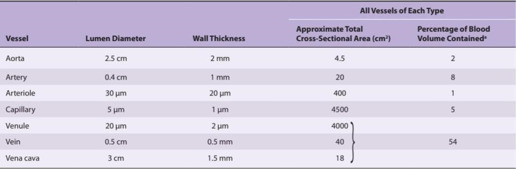
aIn systemic vessels; there is an additional 12% in the heart and 18% in the pulmonary circulation.
a در عروق سیستمیک؛ 12% اضافی در قلب و 18% در گردش خون ریوی وجود دارد.

FIGURE 31-15 Structure of a normal muscle artery. (Reproduced with permission from Ross R, Glomset JA: The pathogenesis of atherosclerosis. N Engl J Med 1976; August 12; 295(7):369-377.)
شکل 31-15 ساختار یک شریان عضلانی طبیعی. (تکثیر شده با اجازه Ross R، Glomset JA: The pathogenesis of atherosclerosis. N Engl J Med 1976؛ 12 اوت؛ 295 (7): 369-377.)
CAPILLARIES
The arterioles divide into smaller muscle-walled vessels called metarterioles, and these in turn feed into capillaries (Figure 31-16). The openings of the capillaries are surrounded on the upstream side by minute smooth muscle precapillary sphincters. It is unsettled whether the metarterioles are innervated, and it appears that the precapillary sphincters are not. However, they can of course respond to local or circulating vasoconstrictor substances. The capillaries are about 5 pm in diameter at the arterial end and 9 μm in diameter at the venous end. When the sphincters are dilated, the diameter of the capillaries is just sufficient to permit red blood cells to squeeze through in “single file.” As they pass through the capillaries, the red cells become thimble- or parachute-shaped, with the flow pushing the center ahead of the edges. This configuration appears to be simply due to the pressure in the center of the vessel, whether or not the edges of the red blood cell are in contact with the capillary walls.
مویرگ ها
شریان ها به عروق کوچکتر با دیواره عضلانی به نام متارتریول تقسیم می شوند و به نوبه خود به مویرگ ها تغذیه می کنند (شکل 31-16). دهانه های مویرگ ها در سمت بالادست توسط اسفنکترهای عضله صاف پیش مویرگ احاطه شده اند. مشخص نیست که آیا متارتریول ها عصب دهی شده اند یا خیر، و به نظر می رسد که اسفنکترهای پیش مویرگی عصب دهی نشده اند. با این حال، آنها البته می توانند به مواد منقبض کننده عروق موضعی یا در گردش پاسخ دهند. قطر مویرگ ها در انتهای شریانی حدود 5 بعد از ظهر و در انتهای وریدی 9 میکرومتر است. هنگامی که اسفنکترها گشاد می شوند، قطر مویرگ ها به اندازه ای است که به گلبول های قرمز خون اجازه می دهد تا در “یک فایل” فشرده شوند. با عبور از مویرگها، گلبولهای قرمز به شکل انگشتانه یا چتر در میآیند و جریان مرکز را جلوتر از لبهها میبرد. به نظر می رسد این پیکربندی صرفاً به دلیل فشار در مرکز رگ باشد، چه لبه های گلبول قرمز در تماس با دیواره های مویرگ باشد یا نه.
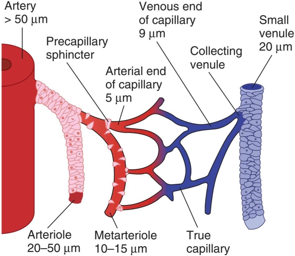
FIGURE 31-16 The microcirculation. Arterioles give rise to metarterioles, which give rise to capillaries. The capillaries drain via short collecting venules to the venules. The walls of the arteries, arterioles, and small venules contain relatively large amounts of smooth muscle. There are scattered smooth muscle cells in the walls of the metarterioles, and the openings of the capillaries are guarded by muscular precapillary sphincters. The diameters of the various vessels are also shown. (Used with permission of JN Diana.)
شکل 31-16 میکروسیرکولاسیون. شریان ها متارتریول هایی را ایجاد می کنند که مویرگ ها را به وجود می آورند. مویرگ ها از طریق ونول های جمع کننده کوتاه به ونول ها تخلیه می شوند. دیواره سرخرگ ها، شریان ها و ونول های کوچک حاوی مقادیر نسبتاً زیادی عضله صاف هستند. سلول های ماهیچه صاف پراکنده در دیواره متارتریول ها وجود دارد و دهانه مویرگ ها توسط اسفنکترهای عضلانی پیش مویرگی محافظت می شود. قطر رگ های مختلف نیز نشان داده شده است. (با اجازه JN Diana استفاده می شود.)
The total area of all the capillary walls in the body exceeds 6300 m2 in the adult. The walls, which are about 1 μm thick, are made up of a single layer of endothelial cells. The structure of the walls varies from organ to organ. In many beds, including those in skeletal, cardiac, and smooth muscle, the junctions between the endothelial cells (Figure 31-17) permit the passage of molecules up to 10 nm in diameter. It also appears that plasma and its dissolved proteins can be taken up by endocytosis, transported across the endothelial cells, and discharged by exocytosis (vesicular transport; see Chapter 2). However, this process can account for only a small portion of the transport across the endothelium. In the brain, the capillaries resemble the capillaries in muscle, but the junctions between endothelial cells are tighter, and transport across them is largely limited to small molecules (although pathological conditions may open these junctions). In most endocrine glands, the intestinal villi, and parts of the kidneys, on the other hand, the cytoplasm of the endothelial cells is attenuated to form gaps called fenestrations. These fenestrations are 20-100 nm in diameter and may be opened to permit the passage of larger molecules, although their permeability is likely significantly reduced under normal circumstances by a thick layer of endothelial glycocalyx. An exception to this, however, is found in the liver, where the sinusoidal capillaries are extremely porous, the endothelium is discontinuous, and gaps occur between endothelial cells that are not closed by membranes (see Figure 28-2). Some of the gaps are 600 nm in diameter, and others may be as large as 3000 nm. They therefore permit the passage of large molecules, including plasma proteins, which is important for hepatic function (see Chapter 28). The permeability of capillaries in various parts of the body, expressed in terms of their hydraulic conductivity, are summarized in Table 31- 10.
مساحت کل دیواره های مویرگی بدن در بزرگسالان بیش از 6300 متر مربع است. دیواره ها که حدود 1 میکرومتر ضخامت دارند از یک لایه سلول های اندوتلیال تشکیل شده اند. ساختار دیواره ها از اندامی به اندام دیگر متفاوت است. در بسیاری از بسترها، از جمله آنهایی که در ماهیچه های اسکلتی، قلبی و صاف هستند، اتصالات بین سلول های اندوتلیال (شکل 31-17) اجازه عبور مولکول هایی تا قطر 10 نانومتر را می دهد. همچنین به نظر میرسد که پلاسما و پروتئینهای محلول آن میتوانند توسط اندوسیتوز جذب شوند، در سلولهای اندوتلیال منتقل شوند و توسط اگزوسیتوز تخلیه شوند (انتقال تاولی؛ فصل 2 را ببینید). با این حال، این فرآیند می تواند تنها بخش کوچکی از حمل و نقل در سراسر اندوتلیوم را تشکیل دهد. در مغز، مویرگها شبیه مویرگهای عضلانی هستند، اما اتصالات بین سلولهای اندوتلیال محکمتر است و حمل و نقل از طریق آنها تا حد زیادی به مولکولهای کوچک محدود میشود (اگرچه شرایط پاتولوژیک ممکن است این اتصالات را باز کند). در اکثر غدد درون ریز، پرزهای روده و قسمت هایی از کلیه ها، از طرف دیگر، سیتوپلاسم سلول های اندوتلیال ضعیف می شود و شکاف هایی به نام فنستراسیون ایجاد می شود. این فنسترها 20 تا 100 نانومتر قطر دارند و ممکن است برای اجازه عبور مولکول های بزرگتر باز شوند، اگرچه نفوذپذیری آنها احتمالاً در شرایط عادی به طور قابل توجهی توسط یک لایه ضخیم گلیکوکالیکس اندوتلیال کاهش می یابد. با این حال، یک استثنا در این مورد، در کبد یافت میشود، جایی که مویرگهای سینوسی بسیار متخلخل هستند، اندوتلیوم ناپیوسته است، و شکافهایی بین سلولهای اندوتلیال که توسط غشاها بسته نمیشوند رخ میدهد (شکل 28-2 را ببینید). قطر برخی از شکاف ها 600 نانومتر و برخی دیگر ممکن است به 3000 نانومتر نیز برسد. بنابراین آنها اجازه عبور مولکول های بزرگ از جمله پروتئین های پلاسما را می دهند که برای عملکرد کبد مهم است (به فصل 28 مراجعه کنید). نفوذپذیری مویرگها در قسمتهای مختلف بدن که بر حسب هدایت هیدرولیکی آنها بیان شده است در جدول 31-10 خلاصه شده است.
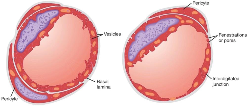
FIGURE 31-17 Cross-sections of capillaries. Left: Type of capillary found in muscle. Right: Fenestrated type of capillary. (Reproduced with permission from Orbison JL and D Smith, editors: Peripheral Blood Vessels. Baltimore: Williams & Wilkins, 1962.)
شکل 31-17 مقاطع عرضی مویرگها. سمت چپ: نوع مویرگ موجود در عضله. سمت راست: نوع فنس دار مویرگ. (تکثیر شده با اجازه Orbison JL و D Smith، ویراستاران: Peripheral Blood Vessels. Baltimore: Williams & Wilkins، 1962.)
TABLE 31-10 Hydraulic conductivity of capillaries in various parts of the body.
جدول 31-10 هدایت هیدرولیکی مویرگ ها در قسمت های مختلف بدن.
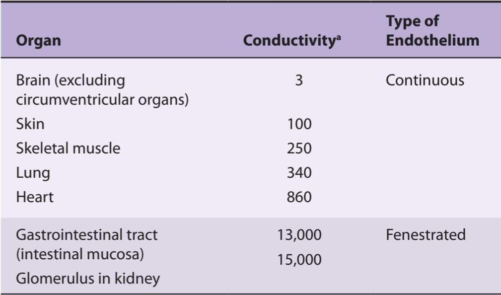
aUnits of conductivity are 10-13 cm3 s1 dyne-1.
Data from JN Diana.
aواحدهای رسانایی 10-13 سانتی متر مکعب s1 dyne-1 است.
داده ها از JN Diana.
Capillaries and postcapillary venules have pericytes around their endothelial cells (Figure 31-17). These cells have long processes that wrap around the vessels. They are contractile and release a wide variety of vasoactive agents. They also synthesize and release constituents of the basement membrane and extracellular matrix. One of their physiologic functions appears to be regulation of flow through the junctions between endothelial cells, particularly in the presence of inflammation. They are closely related to the mesangial cells in the renal glomeruli (see Chapter 37).
مویرگ ها و ونول های پس مویرگی دارای پری سیتی در اطراف سلول های اندوتلیال خود هستند (شکل 31-17). این سلول ها فرآیندهای طولانی دارند که دور رگ ها می پیچند. آنها انقباضی هستند و طیف گسترده ای از عوامل وازواکتیو را آزاد می کنند. آنها همچنین ترکیبات غشای پایه و ماتریکس خارج سلولی را سنتز و آزاد می کنند. به نظر می رسد یکی از عملکردهای فیزیولوژیکی آنها تنظیم جریان از طریق اتصالات بین سلول های اندوتلیال، به ویژه در حضور التهاب است. آنها ارتباط نزدیکی با سلول های مزانژیال در گلومرول های کلیوی دارند (به فصل 37 مراجعه کنید).
LYMPHATICS
The lymphatics serve to collect plasma and its constituents that have exuded from the capillaries into the interstitial space (ie, the lymph). They drain from the body tissues via a system of vessels that coalesce and eventually enter the right and left subclavian veins at their junctions with the respective internal jugular veins. The lymph vessels contain valves and regularly traverse lymph nodes along their course. The ultrastructure of the small lymph vessels differs from that of the capillaries in several details: No fenestrations are visible in the lymphatic endothelium; very little if any basal lamina is present under the endothelium; and the junctions between endothelial cells are open, with no tight intercellular connections.
لنفاوی
غدد لنفاوی برای جمع آوری پلاسما و اجزای تشکیل دهنده آن که از مویرگ ها به فضای بینابینی (یعنی لنف) تراوش کرده اند، خدمت می کنند. آنها از بافتهای بدن از طریق سیستمی از عروق تخلیه میشوند که به هم میپیوندند و در نهایت وارد سیاهرگهای ساب ترقوه راست و چپ در محل اتصالشان به سیاهرگهای ژوگولار داخلی میشوند. عروق لنفاوی حاوی دریچه هایی هستند و به طور منظم از گره های لنفاوی در طول مسیر خود عبور می کنند. فراساختار عروق لنفاوی کوچک در چندین جزئیات با مویرگ ها متفاوت است: هیچ گونه فنستراسیونی در اندوتلیوم لنفاوی قابل مشاهده نیست. در صورتی که لایه بازال زیر اندوتلیوم وجود داشته باشد بسیار کم است. و اتصالات بین سلول های اندوتلیال باز است، بدون اتصالات بین سلولی محکم.
ARTERIOVENOUS ANASTOMOSES
In the fingers, palms, and ear lobes, short channels connect arterioles to venules, bypassing the capillaries. These arteriovenous (A-V) anastomoses, or shunts, have thick, muscular walls and are abundantly innervated, presumably by vasoconstrictor nerve fibers.
آناستوموزهای شریانی وریدی
در انگشتان، کف دست ها و لبه های گوش، کانال های کوتاه، شریان ها را به ونول ها متصل می کنند و مویرگ ها را دور می زنند. این آناستوموزهای شریانی وریدی (A-V) یا شنتها، دیوارههای ضخیم و عضلانی دارند و احتمالاً توسط رشتههای عصبی منقبضکننده عروق، به وفور عصببندی میشوند.
VENULES & VEINS
The walls of the venules are only slightly thicker than those of the capillaries. The walls of the veins are also thin and easily distended. They contain relatively little smooth muscle, but considerable venoconstriction is produced by activity in the noradrenergic nerves to the veins and by circulating vasoconstrictors such as endothelins. Variations in venous tone are important in circulatory adjustments.
وریدها و وریدها
دیوارههای ونولها فقط کمی ضخیمتر از دیوارههای مویرگها هستند. دیواره رگ ها نیز نازک است و به راحتی متسع می شود. آنها حاوی عضله صاف نسبتا کمی هستند، اما انقباض سیاهرگی قابل توجهی با فعالیت در اعصاب نورآدرنرژیک به وریدها و با گردش خون منقبض کننده های عروقی مانند اندوتلین ایجاد می شود. تغییرات در تن وریدی در تنظیم گردش خون مهم است.
The intima of the limb veins is folded at intervals to form venous valves that prevent retrograde flow. However, no valves are present in very small veins, the great veins (superior and inferior vena cava plus the pulmonary veins), or the veins from the brain and viscera.
انتیما وریدهای اندام در فواصل زمانی تا می شود تا دریچه های وریدی تشکیل شود که از جریان رتروگراد جلوگیری می کند. با این حال، هیچ دریچه ای در وریدهای بسیار کوچک، وریدهای بزرگ (ورید اجوف فوقانی و تحتانی به اضافه وریدهای ریوی) یا وریدهای مغز و احشاء وجود ندارد.
ANGIOGENESIS
When tissues grow, blood vessels must proliferate if the tissue is to maintain a normal blood supply. Therefore, angiogenesis, the formation of new blood vessels, is important during fetal life and growth to adulthood. It is also important in adulthood for processes such as wound healing, formation of the corpus luteum after ovulation, and formation of new endometrium after menstruation. Abnormally, it is important in tumor growth since tumors that do not develop a blood supply cannot grow.
آنژیوژنز
هنگامی که بافت ها رشد می کنند، رگ های خونی باید تکثیر شوند تا بافت طبیعی خون را حفظ کند. بنابراین، رگ زایی، تشکیل رگ های خونی جدید، در طول زندگی جنین و رشد تا بزرگسالی مهم است. همچنین در بزرگسالی برای فرآیندهایی مانند بهبود زخم، تشکیل جسم زرد پس از تخمک گذاری و تشکیل آندومتر جدید پس از قاعدگی مهم است. به طور غیر طبیعی، در رشد تومور مهم است، زیرا تومورهایی که منبع خونی ایجاد نمی کنند، نمی توانند رشد کنند.
During embryonic development, a network of leaky capillaries is formed in tissues from angioblasts: this process is sometimes called vasculogenesis. Branches from nearby vessels then hook up with the capillaries and provide them with smooth muscle, which brings about their maturation. Angiogenesis in adults is presumably similar, but consists of new vessel formation by branching from preexisting vessels rather than from angioblasts.
در طول رشد جنینی، شبکهای از مویرگهای نشتی در بافتهای آنژیوبلاستها تشکیل میشود: این فرآیند گاهی اوقات واسکولوژنز نامیده میشود. سپس شاخههای رگهای مجاور به مویرگها متصل میشوند و ماهیچههای صاف را برای آنها فراهم میکنند که باعث بلوغ آنها میشود. رگ زایی در بزرگسالان احتمالاً مشابه است، اما شامل تشکیل عروق جدید با انشعاب از عروق از قبل موجود به جای آنژیوبلاست ها است.
Many factors are involved in angiogenesis. A key compound is vascular endothelial growth factor (VEGF). VEGF is primarily responsible for vasculogenesis, whereas the budding of vessels that connect to the immature capillary network is regulated by other as yet unidentified factors. Some VEGF isoforms and receptors may play a more prominent role in the formation of lymphatic vessels (lymphangiogenesis) than that of blood vessels.
عوامل زیادی در رگ زایی نقش دارند. یک ترکیب کلیدی فاکتور رشد اندوتلیال عروقی (VEGF) است. VEGF در درجه اول مسئول واسکولوژنز است، در حالی که جوانه زدن عروقی که به شبکه مویرگی نابالغ متصل می شوند توسط عوامل دیگری که هنوز شناسایی نشده اند تنظیم می شود. برخی از ایزوفرم ها و گیرنده های VEGF ممکن است نقش برجسته تری در تشکیل عروق لنفاوی (لنفانژیوژنز) نسبت به عروق خونی داشته باشند.
The actions of VEGF and related factors have received considerable attention in recent years because of the requirement for angiogenesis in the development of tumors. VEGF antagonists and other angiogenesis inhibitors have entered clinical practice as adjunctive therapies for malignancies and are being tested as first-line therapies as well.
اقدامات VEGF و عوامل مرتبط با آن در سال های اخیر به دلیل نیاز به رگزایی در ایجاد تومورها مورد توجه قابل توجهی قرار گرفته است. آنتاگونیست های VEGF و سایر مهارکننده های رگ زایی به عنوان درمان های کمکی برای بدخیمی ها وارد عمل بالینی شده اند و به عنوان درمان های خط اول نیز در حال آزمایش هستند.
BIOPHYSICAL CONSIDERATIONS FOR CIRCULATORY PHYSIOLOGY
FLOW, PRESSURE, & RESISTANCE
Blood always flows, of course, from areas of high pressure to areas of low pressure, except in certain situations when momentum transiently sustains flow (see Figure 30-3). The relationship between mean flow, mean pressure, and resistance in the blood vessels is analogous in a general way to the relationship between the current, electromotive force (voltage), and resistance in an electrical circuit as expressed in Ohm law:
ملاحظات بیوفیزیکی برای فیزیولوژی گردش خون
جریان، فشار، و مقاومت
البته خون همیشه از نواحی پرفشار به نواحی با فشار پایین جریان دارد، مگر در شرایط خاصی که تکانه به طور موقت جریان را حفظ می کند (شکل 30-3 را ببینید). رابطه بین جریان متوسط، فشار متوسط و مقاومت در رگهای خونی به طور کلی با رابطه بین جریان، نیروی الکتروموتور (ولتاژ) و مقاومت در یک مدار الکتریکی که در قانون اهم بیان شده است مشابه است:

Flow in any portion of the vascular system is therefore equal to the effective perfusion pressure in that portion divided by the resistance. The effective perfusion pressure is the mean intraluminal pressure at the arterial end minus the mean pressure at the venous end. The units of resistance (pressure divided by flow) are dyne.s/cm3. To avoid dealing with such complex units, resistance in the cardiovascular system is sometimes expressed in R units, which are obtained by dividing pressure in mm Hg by flow in mL/s (see also Table 33-1). Thus, for example, when the mean aortic pressure is 90 mm Hg and the left ventricular output is 90 mL/s, the total peripheral resistance is
بنابراین جریان در هر بخش از سیستم عروقی برابر است با فشار پرفیوژن موثر در آن قسمت تقسیم بر مقاومت. فشار پرفیوژن موثر میانگین فشار داخل مجرای انتهای شریانی منهای فشار متوسط در انتهای وریدی است. واحدهای مقاومت (فشار تقسیم بر جریان) dyne.s/cm3 هستند. برای جلوگیری از برخورد با چنین واحدهای پیچیده، مقاومت در سیستم قلبی عروقی گاهی اوقات با واحدهای R بیان می شود که از تقسیم فشار بر حسب میلی متر جیوه بر جریان بر حسب میلی لیتر بر ثانیه به دست می آید (جدول 33-1 را نیز ببینید). بنابراین، برای مثال، زمانی که فشار متوسط آئورت 90 میلیمتر جیوه و خروجی بطن چپ 90 میلیلیتر بر ثانیه باشد، مقاومت محیطی کل است.

METHODS FOR MEASURING BLOOD FLOW
Various noninvasive devices have been developed to measure blood flow. Most commonly, blood velocity can be measured with Doppler flow meters. Ultrasonic waves are sent into a vessel diagonally, and the waves reflected from the red and white blood cells are picked up by a downstream sensor. The frequency of the reflected waves is higher by an amount that is proportional to the rate of flow toward the sensor because of the Doppler effect.
روش های اندازه گیری جریان خون
دستگاه های غیرتهاجمی مختلفی برای اندازه گیری جریان خون ساخته شده اند. معمولاً سرعت خون را می توان با فلومتر داپلر اندازه گیری کرد. امواج اولتراسونیک به صورت مورب به داخل یک رگ فرستاده می شود و امواج منعکس شده از گلبول های قرمز و سفید خون توسط یک حسگر پایین دست دریافت می شود. فرکانس امواج منعکس شده به دلیل اثر داپلر با مقداری که متناسب با سرعت جریان به سمت حسگر است بیشتر است.
Indirect methods for measuring the blood flow in various organs in humans include adaptations of the Fick and indicator dilution techniques described in Chapter 30. One example is the use of the Kety N2O method for measuring cerebral blood flow (see Chapter 33). Another is determination of the renal blood flow by measuring the clearance of para-aminohippuric acid (see Chapter 37). A considerable amount of data on blood flow in the extremities have been obtained by plethysmography. The forearm, for example, is sealed in a watertight chamber (plethysmograph). Changes in the volume of the forearm, reflecting changes in the amount of blood and interstitial fluid it contains, displace the water, and this displacement is measured with a volume recorder. When the venous drainage of the forearm is occluded, the rate of increase in the volume of the forearm is a function of the arterial blood flow (venous occlusion plethysmography).
روشهای غیرمستقیم برای اندازهگیری جریان خون در اندامهای مختلف در انسان شامل سازگاری با تکنیکهای Fick و رقیقسازی نشانگر است که در فصل 30 توضیح داده شد. یک مثال استفاده از روش Kety N2O برای اندازهگیری جریان خون مغزی است (به فصل 33 مراجعه کنید). دیگری تعیین جریان خون کلیوی با اندازه گیری کلیرانس اسید پاراآمینو هیپوریک است (به فصل 37 مراجعه کنید). اطلاعات قابل توجهی در مورد جریان خون در اندام ها با پلتیسموگرافی به دست آمده است. به عنوان مثال، ساعد در یک محفظه ضد آب (پلتیسموگرافی) مهر و موم شده است. تغییرات در حجم ساعد، منعکس کننده تغییرات در مقدار خون و مایع بینابینی موجود در آن، آب را جابجا می کند و این جابجایی با یک ضبط کننده حجم اندازه گیری می شود. هنگامی که درناژ وریدی ساعد مسدود می شود، میزان افزایش حجم ساعد تابعی از جریان خون شریانی است (پلتیسموگرافی انسداد وریدی).
APPLICABILITY OF PHYSICAL PRINCIPLES TO FLOW IN BLOOD VESSELS
Physical principles and equations that describe the behavior of perfect fluids in rigid tubes have often been used indiscriminately to explain the behavior of blood in blood vessels, whereas blood vessels are not rigid tubes, and the blood is not a perfect fluid but a two-phase system of liquid and cells. Therefore, the behavior of the circulation deviates, sometimes markedly, from that predicted by physical principles. However, these principles are of value when used as an aid to understanding what goes on in the body.
کاربرد اصول فیزیکی برای جریان در رگ های خونی
اصول و معادلات فیزیکی که رفتار سیالات کامل را در لولههای صلب توصیف میکنند، اغلب برای توضیح رفتار خون در رگهای خونی بهطور بیتوجهی استفاده میشوند، در حالی که رگهای خونی لولههای صلب نیستند و خون یک مایع کامل نیست، بلکه یک سیستم دو فازی از مایع و سلولها است. بنابراین، رفتار گردش خون، گاهی اوقات به طور قابل توجهی، از رفتار پیش بینی شده توسط اصول فیزیکی منحرف می شود. با این حال، این اصول زمانی ارزشمند هستند که به عنوان کمکی برای درک آنچه در بدن می گذرد استفاده شوند.
LAMINAR FLOW
The flow of blood in straight blood vessels, like the flow of liquids in narrow rigid tubes, is normally laminar. Within the blood vessels, an infinitely thin layer of blood in contact with the wall of the vessel does not move. The next layer within the vessel has a low velocity, the next a higher velocity, and so forth, velocity being greatest in the center of the stream (Figure 31-18). Laminar flow occurs at velocities up to a certain critical velocity. At or above this velocity, flow is turbulent. The probability of turbulence is also related to the diameter of the vessel and the viscosity of the blood. This probability can be expressed by the ratio of inertial to viscous forces as follows:
جریان LAMINAR
جریان خون در رگ های خونی مستقیم، مانند جریان مایعات در لوله های سفت و باریک، معمولاً آرام است. در داخل رگ های خونی، یک لایه بی نهایت نازک از خون در تماس با دیواره رگ حرکت نمی کند. لایه بعدی درون کشتی دارای سرعت کم، لایه بعدی سرعت بالاتر و به همین ترتیب، سرعت در مرکز جریان بیشتر است (شکل 31-18). جریان آرام در سرعت هایی تا یک سرعت بحرانی خاص رخ می دهد. در این سرعت یا بالاتر از آن، جریان متلاطم است. احتمال تلاطم با قطر رگ و ویسکوزیته خون نیز مرتبط است. این احتمال را می توان با نسبت نیروهای اینرسی به ویسکوز به صورت زیر بیان کرد:


FIGURE 31-18 Diagram of the velocities of concentric laminas of a viscous fluid flowing in a tube, illustrating the parabolic distribution of velocities.
شکل 31-18 نمودار سرعت لایه های متحدالمرکز یک سیال چسبناک که در یک لوله جریان دارد، که توزیع سهموی سرعت ها را نشان می دهد.
where Re is the Reynolds number; p is the density of the fluid; D is the diameter of the tube; V. is the velocity of the flow; and n is the viscosity of the fluid. The higher the value of Re, the greater the probability of turbulence. When D is in cm, V. is in cm/s1, and n is in poise; flow is usually not turbulent if Re is less than 2000. When Re is more than 3000, turbulence is almost always present. Laminar flow can be disturbed at the branching points of arteries, and the resulting turbulence may increase the likelihood that atherosclerotic plaques will be deposited. Constriction of an artery likewise increases the velocity of blood flow through the constriction, producing turbulence and sound beyond the constriction (Figure 31-19). Examples are bruits heard over arteries constricted by atherosclerotic plaques and the sounds of Korotkoff heard when measuring blood pressure (see below). In healthy humans, the critical velocity is sometimes exceeded in the ascending aorta at the peak of systolic ejection, but it is usually exceeded only when an artery is constricted.
که در آن Re عدد رینولدز است. p چگالی سیال است. D قطر لوله است. V. سرعت جریان است. و n ویسکوزیته سیال است. هر چه مقدار Re بیشتر باشد، احتمال تلاطم بیشتر است. وقتی D بر حسب سانتیمتر است، V. بر حسب سانتیمتر/s1 است و n در پویز است. اگر Re کمتر از 2000 باشد، جریان معمولاً متلاطم نیست. وقتی Re بیش از 3000 باشد، تلاطم تقریباً همیشه وجود دارد. جریان آرام می تواند در نقاط انشعاب شریان ها مختل شود و تلاطم حاصل ممکن است احتمال رسوب پلاک های آترواسکلروتیک را افزایش دهد. انقباض شریان به همین ترتیب سرعت جریان خون را از طریق انقباض افزایش می دهد و تلاطم و صدایی فراتر از انقباض ایجاد می کند (شکل 31-19). به عنوان مثال می توان به ضربات شنیده شده روی شریان های منقبض شده توسط پلاک های آترواسکلروتیک و صداهای کوروتکف که هنگام اندازه گیری فشار خون شنیده می شود اشاره کرد (به زیر مراجعه کنید). در انسانهای سالم، سرعت بحرانی در آئورت صعودی در اوج جهش سیستولیک گاهی اوقات فراتر میرود، اما معمولاً تنها زمانی که یک شریان منقبض شده است، از آن بیشتر میشود.

FIGURE 31-19 Top: Effect of constriction (C) on the profile of velocities in a blood vessel. The arrows indicate direction of velocity components, and their length is proportional to their magnitude. Bottom: Range of velocities at each point along the vessel. In the area of turbulence, there are many different anterograde (A) and some retrograde (R) velocities. (Modified with permission from Richards KE: Doppler echocardiographic diagnosis and quantification of vascular heart disease. Curr Probl Cardiol 1985 February; 10(2):1-49.)
شکل 31-19 بالا: اثر انقباض (C) بر روی مشخصات سرعت در رگ خونی. فلش ها جهت مولفه های سرعت را نشان می دهند و طول آنها متناسب با بزرگی آنها است. پایین: محدوده سرعت در هر نقطه در طول کشتی. در ناحیه تلاطم، سرعت های متجانس (A) و برخی رتروگراد (R) بسیار متفاوت است. (اصلاح شده با مجوز ریچاردز KE: تشخیص اکوکاردیوگرافی داپلر و تعیین کمیت بیماری قلبی عروقی. Curr Probl Cardiol 1985 فوریه؛ 10(2):1-49.)
SHEAR STRESS & GENE ACTIVATION
Flowing blood creates a force on the endothelium that is parallel to the long axis of the vessel. This shear stress (y) is proportional to viscosity (n) times the shear rate (dy/dr), which is the rate at which the axial velocity increases from the vessel wall toward the lumen.
تنش برشی و فعال سازی ژن
جریان خون نیرویی بر روی اندوتلیوم ایجاد می کند که موازی با محور طولانی رگ است. این تنش برشی (y) متناسب با ویسکوزیته (n) برابر نرخ برش (dy/dr) است، که سرعتی است که در آن سرعت محوری از دیواره رگ به سمت لومن افزایش مییابد.

Change in shear stress and other physical variables, such as cyclic strain and stretch, produce marked changes in the expression of genes by endothelial cells. The genes that are activated include those that produce growth factors, integrins, and related molecules (Figure 31-20). Recent evidence has implicated endothelial surface structures known as primary cilia as the sensors of shear stress, which in turn activate signaling pathways to alter cell function. However, other components may also be involved, such as ion channels, adhesion molecules, and the cytoskeleton.
تغییر در تنش برشی و سایر متغیرهای فیزیکی، مانند کرنش و کشش حلقوی، تغییرات مشخصی در بیان ژنها توسط سلولهای اندوتلیال ایجاد میکند. ژن هایی که فعال می شوند شامل ژن هایی هستند که فاکتورهای رشد، اینتگرین ها و مولکول های مرتبط را تولید می کنند (شکل 31-20). شواهد اخیر ساختارهای سطح اندوتلیال معروف به مژک های اولیه را به عنوان حسگرهای تنش برشی دخیل می دانند که به نوبه خود مسیرهای سیگنالینگ را برای تغییر عملکرد سلول فعال می کنند. با این حال، اجزای دیگر نیز ممکن است درگیر باشند، مانند کانالهای یونی، مولکولهای چسبنده و اسکلت سلولی.

FIGURE 31-20 Endothelial transcription factors and target genes activated by shear stress. ACE, angiotensin-converting enzyme; AP-1, activator protein- 1; Egr-1, early growth response protein-1; ICAM-1; intercellular adhesion molecule-1; MCP-1, monocyte chemoattractant protein-1; NF-κB, nuclear factor-KB; NOS 3, nitric oxide synthase 3; PDGF, platelet-derived growth factor; SP1, specificity protein 1; SSRE, shear stress responsive element; TGF-ß, transforming growth factor-ẞ; VCAM-1, vascular cell adhesion molecule-1. (Data from Braddock M, et al: Fluid shear stress modulation of gene expression in endothelial cells. News Physiol Sci 1998;13:241.)
شکل 31-20 فاکتورهای رونویسی اندوتلیال و ژن های هدف فعال شده توسط تنش برشی. ACE، آنزیم مبدل آنژیوتانسین؛ AP-1، پروتئین فعال کننده- 1; Egr-1، پروتئین پاسخ رشد اولیه-1. ICAM-1; مولکول چسبندگی بین سلولی-1; MCP-1، پروتئین جذب کننده شیمیایی مونوسیتی-1. NF-kB، فاکتور هسته ای-KB. NOS 3، نیتریک اکسید سنتاز 3. PDGF، فاکتور رشد مشتق از پلاکت. SP1، پروتئین اختصاصی 1; SSRE، عنصر پاسخگو به تنش برشی. TGF-ß، تبدیل کننده فاکتور رشد-ẞ; VCAM-1، مولکول چسبندگی سلول عروقی-1. (داده های Braddock M و همکاران: تعدیل استرس برشی سیال بیان ژن در سلول های اندوتلیال. News Physiol Sci 1998; 13:241.)
AVERAGE VELOCITY
When considering flow in a system of tubes, it is important to distinguish between velocity, which is displacement per unit time (eg, cm/s), and flow, which is volume per unit time (eg, cm3/s). Velocity (v) is proportional to flow (Q) divided by the area of the conduit (A):
سرعت متوسط
هنگام در نظر گرفتن جریان در سیستم لولهها، مهم است که بین سرعت، که جابجایی در واحد زمان (مثلاً سانتیمتر بر ثانیه) است، و جریان، که حجم در واحد زمان (مثلاً سانتیمتر مکعب بر ثانیه) است، تمایز قائل شویم. سرعت (v) متناسب با جریان (Q) تقسیم بر مساحت مجرای (A) است:

Therefore, Q = AV, and if flow stays constant, velocity increases in direct proportion to any decrease in A (Figure 31–19).
بنابراین، Q = AV، و اگر جریان ثابت بماند، سرعت به نسبت مستقیم با کاهش A افزایش می یابد (شکل 31-19).
The average velocity of fluid movement at any point in a system of tubes in parallel is inversely proportional to the total cross-sectional area at that point. Therefore, the average velocity of the blood is high in the aorta, declines steadily in the smaller vessels, and is lowest in the capillaries, which have 1000 times the total cross-sectional area of the aorta (Table 31-9). The average velocity of blood flow increases again as the blood enters the veins and is relatively high in the vena cava, although not so high as in the aorta. Clinically, the velocity of the circulation can be measured by injecting a bile salt preparation into an arm vein and timing the first appearance of the bitter taste it produces (Figure 31-21). The average normal arm-to-tongue circulation time is 15 s.
سرعت متوسط حرکت سیال در هر نقطه از سیستم لوله های موازی با کل سطح مقطع در آن نقطه نسبت معکوس دارد. بنابراین، سرعت متوسط خون در آئورت زیاد است، در رگ های کوچکتر به طور پیوسته کاهش می یابد و در مویرگ ها که 1000 برابر کل سطح مقطع آئورت دارند، کمترین میزان را دارد (جدول 31-9). سرعت متوسط جریان خون با ورود خون به سیاهرگها دوباره افزایش مییابد و در ورید اجوف نسبتاً زیاد است، اگرچه به اندازه آئورت نیست. از نظر بالینی، سرعت گردش خون را می توان با تزریق یک نمک صفراوی به ورید بازو و تعیین زمان اولین ظاهر طعم تلخی که ایجاد می کند اندازه گیری کرد (شکل 31-21). متوسط زمان عادی گردش بازو به زبان 15 ثانیه است.
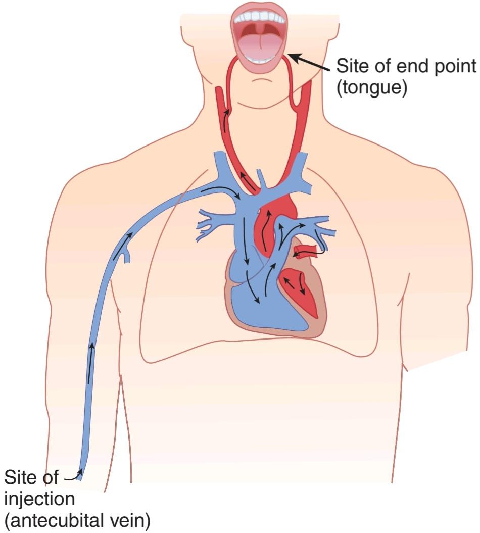
FIGURE 31-21 Pathway traversed by the injected material when the armto-tongue circulation time is measured.
شکل 31-21 مسیری که توسط مواد تزریق شده طی می شود، زمانی که زمان گردش بازو به زبان اندازه گیری می شود.
POISEUILLE-HAGEN FORMULA
The relationship between the flow in a long narrow tube, the viscosity of the fluid, and the radius of the tube is expressed mathematically in the Poiseuille- Hagen formula:
فرمول POISEUILLE-هاگن
رابطه بین جریان در یک لوله باریک بلند، ویسکوزیته سیال و شعاع لوله به صورت ریاضی در فرمول Poiseuille-Hagen بیان می شود:

where
کجا

Because flow is equal to pressure difference divided by resistance (R),
از آنجا که جریان برابر است با اختلاف فشار تقسیم بر مقاومت (R)،

Because flow varies directly and resistance inversely with the fourth power of the radius, blood flow and resistance in vivo are markedly affected by small changes in the caliber of the vessels. Thus, for example, flow through a vessel is doubled by an increase of only 19% in its radius; and when the radius is doubled, resistance is reduced to 6% of its previous value. This is why organ blood flow is so effectively regulated by small changes in the caliber of the arterioles and why variations in arteriolar diameter have such a pronounced effect on systemic arterial pressure.
از آنجایی که جریان به طور مستقیم و مقاومت به طور معکوس با توان چهارم شعاع تغییر می کند، جریان خون و مقاومت در داخل بدن به طور قابل توجهی تحت تأثیر تغییرات کوچک در کالیبر عروق قرار می گیرند. بنابراین، برای مثال، جریان از طریق یک کشتی با افزایش تنها 19٪ در شعاع آن دو برابر می شود. و هنگامی که شعاع دو برابر می شود، مقاومت به 6% مقدار قبلی کاهش می یابد. به همین دلیل است که جریان خون اندام به طور موثری با تغییرات کوچک در کالیبر سرخرگها تنظیم میشود و به همین دلیل است که تغییرات در قطر شریانی چنین تأثیر برجستهای بر فشار شریانی سیستمیک دارد.
VISCOSITY & RESISTANCE
The resistance to blood flow is determined not only by the radius of the blood vessels but also by the viscosity of the blood. Plasma is about 1.8 times as viscous as water, whereas whole blood is 3-4 times as viscous as water. Thus, viscosity depends for the most part on the hematocrit. The effect of viscosity in vivo deviates from that predicted by the Poiseuille-Hagen formula. In large vessels, increases in hematocrit cause appreciable increases in viscosity. However, in vessels smaller than 100 μm in diameter that is, in arterioles,capillaries, and venules the viscosity change per unit change in hematocrit is much less than it is in large-bore vessels. This is due to a difference in the nature of flow through the small vessels, known as the Fahraeus-Lindqvist effect. In small vessels, erythrocytes move to the center of the vessel, leaving cell-free plasma at the vessel wall. Therefore, the net change in viscosity per unit change in hematocrit is considerably smaller in the body than if it is measured in vitro (Figure 31-22). This is why hematocrit changes have relatively little effect on the peripheral resistance except when the changes are large. In severe polycythemia (excessive production of red blood cells), the increase in resistance does increase the work of the heart. Conversely, in marked anemia, peripheral resistance is decreased, in part because of the decline in viscosity. Of course, the decrease in hemoglobin decreases the O2-carrying ability of the blood, but the improved blood flow due to the decrease in viscosity partially compensates for this.
ویسکوزیته و مقاومت
مقاومت در برابر جریان خون نه تنها با شعاع رگ های خونی بلکه با ویسکوزیته خون نیز تعیین می شود. پلاسما 1.8 برابر آب چسبناک است، در حالی که خون کامل 3 تا 4 برابر آب چسبناک است. بنابراین، ویسکوزیته بیشتر به هماتوکریت بستگی دارد. اثر ویسکوزیته در داخل بدن از آنچه توسط فرمول Poiseuille-Hagen پیش بینی شده بود منحرف می شود. در عروق بزرگ، افزایش هماتوکریت باعث افزایش قابل ملاحظه ویسکوزیته می شود. با این حال، در عروق با قطر کمتر از 100 میکرومتر، یعنی در شریانها، مویرگها و ونولها، تغییر ویسکوزیته در واحد تغییر در هماتوکریت بسیار کمتر از رگهای با سوراخ بزرگ است. این به دلیل تفاوت در ماهیت جریان از طریق عروق کوچک است که به عنوان اثر Fahraeus-Lindqvist شناخته می شود. در عروق کوچک، گلبول های قرمز به سمت مرکز رگ حرکت می کنند و پلاسمای بدون سلول را در دیواره عروق باقی می گذارند. بنابراین، تغییر خالص ویسکوزیته در واحد تغییر در هماتوکریت در بدن به طور قابل توجهی کمتر از اندازه گیری در شرایط آزمایشگاهی است (شکل 31-22). به همین دلیل است که تغییرات هماتوکریت تأثیر نسبتا کمی بر مقاومت محیطی دارد، مگر زمانی که تغییرات زیاد باشد. در پلی سیتمی شدید (تولید بیش از حد گلبول های قرمز خون)، افزایش مقاومت باعث افزایش کار قلب می شود. برعکس، در کم خونی مشخص، مقاومت محیطی کاهش می یابد، تا حدی به دلیل کاهش ویسکوزیته. البته کاهش هموگلوبین توانایی حمل O2 خون را کاهش می دهد، اما بهبود جریان خون به دلیل کاهش ویسکوزیته تا حدودی این را جبران می کند.

FIGURE 31-22 Effect of changes in hematocrit on the relative viscosity of blood measured in vivo and in vitro. (Data simplified and replotted from Whittaker SRF, Winton FR: The apparent viscosity of blood flowing in the isolated hind limb of the dog, and its variation with corpuscular concentration. J Physiol [Lond] 1933; 78:338.)
شکل 31-22 اثر تغییرات هماتوکریت بر ویسکوزیته نسبی خون اندازه گیری شده در داخل بدن و در شرایط آزمایشگاهی. (دادهها سادهشده و بازنگریشده از Whittaker SRF، Winton FR: ویسکوزیته ظاهری خون جاری در اندام عقبی جدا شده سگ، و تغییر آن با غلظت جسمی. J Physiol [Lond] 1933؛ 78:338.)
Viscosity is also affected by the composition of the plasma and the resistance of the cells to deformation. Clinically significant increases in viscosity are seen in diseases in which plasma proteins such as immunoglobulins are markedly elevated as well as when red blood cells are abnormally rigid (hereditary spherocytosis).
ویسکوزیته نیز تحت تأثیر ترکیب پلاسما و مقاومت سلول ها در برابر تغییر شکل قرار می گیرد. افزایش قابل توجه ویسکوزیته از نظر بالینی در بیماری هایی که در آن پروتئین های پلاسما مانند ایمونوگلوبولین ها به طور قابل توجهی افزایش می یابد و همچنین هنگامی که گلبول های قرمز به طور غیر طبیعی سفت و سخت هستند (اسفروسیتوز ارثی) مشاهده می شود.
CRITICAL CLOSING PRESSURE
In rigid tubes, the relationship between pressure and flow of homogeneous fluids is linear, but in thin-walled blood vessels in vivo it is not. When the pressure in a small blood vessel is reduced, a point is reached at which no blood flows, even though the pressure is not zero (Figure 31-23). This is because the vessels are surrounded by tissues that exert a small but definite pressure on them, and when the intraluminal pressure falls below the tissue pressure, they collapse. In inactive tissues, for example, the pressure in many capillaries is low because the precapillary sphincters and metarterioles are constricted, and many of these capillaries are collapsed. The pressure at which flow ceases is called the critical closing pressure.
فشار بسته شدن بحرانی
در لوله های صلب، رابطه بین فشار و جریان مایعات همگن خطی است، اما در عروق خونی جدار نازک در داخل بدن این رابطه وجود ندارد. هنگامی که فشار در یک رگ خونی کوچک کاهش می یابد، به نقطه ای می رسد که در آن هیچ خونی جریان نمی یابد، حتی اگر فشار صفر نباشد (شکل 31-23). این به این دلیل است که رگ ها توسط بافت هایی احاطه شده اند که فشار کوچک اما مشخصی بر آنها وارد می کنند و زمانی که فشار داخل مجرا از فشار بافت پایین می آید، فرو می ریزند. به عنوان مثال، در بافتهای غیرفعال، فشار در بسیاری از مویرگها کم است، زیرا اسفنکترهای پیش مویرگ و متارتریولها منقبض شدهاند و بسیاری از این مویرگها فرو میروند. فشاری که در آن جریان متوقف می شود، فشار بسته شدن بحرانی نامیده می شود.

FIGURE 31–23 Relation of pressure to flow in thin-walled blood vessels.
شکل 31-23 رابطه فشار با جریان در عروق خونی با دیواره نازک.
LAW OF LAPLACE
It is perhaps surprising that structures as thin-walled and delicate as the capillaries are not more prone to rupture. The principal reason for their relative invulnerability is their small diameter. The protective effect of small size in this case is an example of the operation of the law of Laplace, an important physical principle with several other applications in physiology. This law states that tension in the wall of a cylinder (T) is equal to the product of the transmural pressure (P) and the radius (r) divided by the wall thickness (w):
قانون لاپلاس
شاید تعجب آور باشد که ساختارهایی به دیواره نازک و ظریف مانند مویرگ ها بیشتر مستعد پارگی نیستند. دلیل اصلی آسیب ناپذیری نسبی آنها قطر کوچک آنها است. اثر محافظتی اندازه کوچک در این مورد نمونه ای از عملکرد قانون لاپلاس است، یک اصل فیزیکی مهم با چندین کاربرد دیگر در فیزیولوژی. این قانون می گوید که کشش در دیواره یک سیلندر (T) برابر است با حاصل ضرب فشار بین دیواری (P) و شعاع (r) تقسیم بر ضخامت دیواره (w):

The transmural pressure is the pressure inside the cylinder minus the pressure outside the cylinder, but because tissue pressure in the body is low, it can generally be ignored and P equated to the pressure inside the blood vessel. In a thin-walled vessel, w is very small and it too can be ignored, but it becomes a significant factor in vessels such as arteries. Therefore, in a thin-walled vessel, P = T divided by the two principal radii of curvature of the structure:
فشار transmural فشار داخل سیلندر منهای فشار خارج از سیلندر است، اما از آنجایی که فشار بافت در بدن کم است، به طور کلی می توان آن را نادیده گرفت و P را معادل فشار داخل رگ خونی دانست. در یک رگ با دیواره نازک، w بسیار کوچک است و می توان آن را نیز نادیده گرفت، اما عامل مهمی در عروقی مانند شریان ها می شود. بنابراین، در یک ظرف دیوار نازک، P = T تقسیم بر دو شعاع اصلی انحنای سازه:
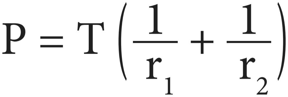
In a cylinder such as a blood vessel, one radius is infinite, so
در استوانه ای مانند رگ خونی، یک شعاع بی نهایت است، بنابراین

Consequently, the smaller the radius of a blood vessel, the lower the tension in the wall necessary to balance the distending pressure. In the human aorta, for example, the tension at normal pressures is about 170,000 dynes/cm, and in the vena cava it is about 21,000 dynes/cm; but in the capillaries, it is approximately 16 dynes/cm.
در نتیجه، هرچه شعاع یک رگ خونی کوچکتر باشد، کشش دیواره لازم برای متعادل کردن فشار انبساط کمتر است. به عنوان مثال، در آئورت انسان، کشش در فشارهای طبیعی حدود 170000 dynes/cm و در ورید اجوف حدود 21000 dynes/cm است. اما در مویرگها تقریباً 16 dynes/cm است.
The law of Laplace also makes clear a disadvantage faced by dilated hearts. When the radius of a cardiac chamber is increased, a greater tension must be developed in the myocardium to produce any given pressure; consequently, a dilated heart must do more work than a nondilated heart.
قانون لاپلاس همچنین نقطه ضعفی را که قلب های گشاد شده با آن روبرو هستند را روشن می کند. هنگامی که شعاع اتاق قلب افزایش می یابد، باید کشش بیشتری در میوکارد ایجاد شود تا فشار معینی ایجاد شود. در نتیجه، قلب گشاد شده باید بیشتر از قلب غیر متسع کار کند.
RESISTANCE & CAPACITANCE VESSELS
In vivo, the veins are an important blood reservoir. Normally, they are partially collapsed and oval in cross-section. A large amount of blood can be added to the venous system before the veins become distended to the point where further increments in volume produce a large rise in venous pressure. The veins are therefore called capacitance vessels. The small arteries and arterioles are referred to as resistance vessels because they are the principal site of the peripheral resistance (see below).
مخازن مقاومت و ظرفیت
در داخل بدن، وریدها یک مخزن خون مهم هستند. به طور معمول، آنها تا حدی جمع شده و در مقطع بیضی شکل هستند. مقدار زیادی خون می تواند به سیستم وریدی اضافه شود، قبل از اینکه وریدها به حدی متسع شوند که افزایش حجم بیشتر باعث افزایش شدید فشار وریدی شود. بنابراین رگه ها را مخازن ظرفیتی می نامند. شریان ها و شریان های کوچک به عنوان عروق مقاومت شناخته می شوند زیرا محل اصلی مقاومت محیطی هستند (به زیر مراجعه کنید).
At rest, at least 50% of the circulating blood volume is in the systemic veins, 12% is in the heart cavities, and 18% is in the low-pressure pulmonary circulation. Only 2% is in the aorta, 8% in the arteries, 1% in the arterioles, and 5% in the capillaries (Table 31-9). When extra blood is administered by transfusion, less than 1% of it is distributed in the arterial system (the “high- pressure system”), and all the rest is found in the systemic veins, pulmonary circulation, and heart chambers other than the left ventricle (the “low-pressure system”).
در حالت استراحت، حداقل 50 درصد از حجم خون در گردش در وریدهای سیستمیک، 12 درصد در حفره های قلب و 18 درصد در گردش خون ریوی کم فشار است. فقط 2 درصد در آئورت، 8 درصد در شریان ها، 1 درصد در شریان ها و 5 درصد در مویرگ ها قرار دارد (جدول 31-9). هنگامی که خون اضافی از طریق تزریق تزریق می شود، کمتر از 1٪ از آن در سیستم شریانی (“سیستم فشار بالا”) توزیع می شود و بقیه در وریدهای سیستمیک، گردش خون ریوی و حفره های قلب به غیر از بطن چپ (“سیستم فشار پایین”) یافت می شود.
ARTERIAL & ARTERIOLAR CIRCULATION
The pressure and velocities of the blood in the various parts of the systemic circulation are summarized in Figure 31-24. The general relationships in the pulmonary circulation are similar, but the pressure in the pulmonary artery is 25/10 mm Hg or less.
گردش خون شریانی و شریانی
فشار و سرعت خون در قسمت های مختلف گردش خون سیستمیک در شکل 31-24 خلاصه شده است. روابط عمومی در گردش خون ریوی مشابه است، اما فشار در شریان ریوی 25/10 میلی متر جیوه یا کمتر است.

FIGURE 31-24 Diagram of the changes in pressure and velocity as blood flows through the systemic circulation. TA, total cross-sectional area of the vessels, which increases from 4.5 cm2 in the aorta to 4500 cm2 in the capillaries (Table 31-9). RR, relative resistance, which is highest in the arterioles.
شکل 31-24 نمودار تغییرات فشار و سرعت در جریان جریان خون در گردش خون سیستمیک. TA، سطح مقطع کل عروق، که از 4.5 سانتی متر مربع در آئورت به 4500 سانتی متر مربع در مویرگ ها افزایش می یابد (جدول 31-9). RR، مقاومت نسبی، که بالاترین میزان در شریان ها است.
PULSATILE FLOW OF BLOOD
Although the mean velocity of the blood in the proximal portion of the aorta is 40 cm/s, the flow is phasic, and velocity ranges from 120 cm/s during systole to a negative value at the time of the transient backflow before the aortic valve closes in diastole. In the distal portions of the aorta and in the large arteries, velocity is also much greater in systole than it is in diastole. However, the vessels are elastic, and forward flow is continuous because of the recoil during diastole of the vessel walls that have been stretched during systole (Figure 31- 25). Pulsatile flow appears to maintain optimal function of the tissues, apparently via distinct effects on gene transcription.
جریان ضربانی خون
اگرچه میانگین سرعت خون در قسمت پروگزیمال آئورت 40 سانتیمتر بر ثانیه است، جریان فازی است و سرعت آن از 120 سانتیمتر بر ثانیه در طول سیستول تا مقدار منفی در زمان جریان برگشتی گذرا قبل از بسته شدن دریچه آئورت در دیاستول متغیر است. در بخشهای انتهایی آئورت و در شریانهای بزرگ، سرعت در سیستول بسیار بیشتر از دیاستول است. با این حال، عروق کشسان هستند، و جریان رو به جلو پیوسته است، زیرا در هنگام دیاستول دیواره های عروق کشیده شده است (شکل 31-25). به نظر می رسد جریان ضربانی عملکرد مطلوب بافت ها را حفظ می کند، ظاهراً از طریق تأثیرات متمایز بر رونویسی ژن.

FIGURE 31-25 Changes in blood flow during the cardiac cycle in the dog. Diastole is followed by systole starting at 0.1 and again at 0.5 s. Flow patterns in humans are similar. Ao, aorta; IVC, inferior vena cava; PA, pulmonary artery; PV, pulmonary vein; RA, renal artery. (Reproduced with permission from Milnor WR: Pulsatile blood flow. N Engl J Med 1972; July 6; 287(1):27-34.)
شکل 31-25 تغییرات در جریان خون در طول چرخه قلبی در سگ. پس از دیاستول، سیستول از 0.1 شروع می شود و دوباره در 0.5 ثانیه شروع می شود. الگوهای جریان در انسان مشابه است. آئورت، آئورت؛ IVC، ورید اجوف تحتانی؛ PA، شریان ریوی؛ PV، ورید ریوی؛ RA، شریان کلیوی. (تکثیر شده با اجازه Milnor WR: Pulsatile blood flow. N Engl J Med 1972؛ 6 ژوئیه؛ 287(1):27-34.)
ARTERIAL PRESSURE
The pressure in the aorta and in the brachial and other large arteries in a young adult human rises to a peak value (systolic pressure) of about 120 mm Hg during each heart cycle and falls to a minimum (diastolic pressure) of about 70 mm Hg. The arterial pressure is conventionally written as systolic pressure over diastolic pressure, for example, 120/70 mm Hg. The pulse pressure, the difference between the systolic and diastolic pressures, is normally about 50 mm Hg. The mean pressure is the average pressure throughout the cardiac cycle. Because systole is shorter than diastole, the mean pressure is slightly less than the value halfway between systolic and diastolic pressure. It can be determined only by integrating the area of the pressure curve (Figure 31-26); however, as an approximation, mean pressure equals the diastolic pressure plus one-third of the pulse pressure.
فشار شریانی
فشار در آئورت و در شریان های بازویی و دیگر شریان های بزرگ در یک انسان جوان بالغ به حداکثر مقدار (فشار سیستولیک) حدود 120 میلی متر جیوه در طول هر چرخه قلبی افزایش می یابد و به حداقل (فشار دیاستولیک) حدود 70 میلی متر جیوه کاهش می یابد. فشار شریانی معمولاً به عنوان فشار سیستولیک بر فشار دیاستولیک نوشته می شود، به عنوان مثال، 120/70 میلی متر جیوه. فشار نبض، تفاوت بین فشار سیستولیک و دیاستولیک، به طور معمول حدود 50 میلی متر جیوه است. فشار متوسط، فشار متوسط در طول چرخه قلبی است. از آنجایی که سیستول کوتاهتر از دیاستول است، فشار متوسط کمی کمتر از مقدار بین فشار سیستولیک و دیاستولیک است. می توان آن را تنها با ادغام مساحت منحنی فشار تعیین کرد (شکل 31-26). با این حال، به عنوان تقریبی، فشار متوسط برابر است با فشار دیاستولیک به اضافه یک سوم فشار نبض.
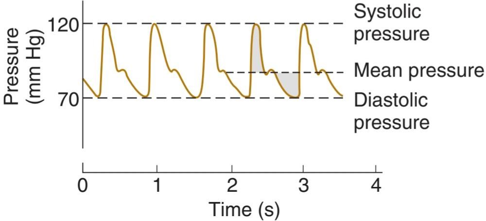
FIGURE 31-26 Brachial artery pressure curve of a normal young human, showing the relation of systolic and diastolic pressure to mean pressure. The shaded area above the mean pressure line is equal to the shaded area below it.
شکل 31-26 منحنی فشار شریان بازویی یک انسان جوان طبیعی، رابطه فشار سیستولیک و دیاستولیک را با فشار متوسط نشان می دهد. ناحیه سایه دار بالای خط فشار متوسط برابر با ناحیه سایه دار زیر آن است.
The pressure falls very slightly in the large- and medium-sized arteries because their resistance to flow is small, but it falls rapidly in the small arteries and arterioles, which are the main sites of the peripheral resistance against which the heart pumps. The mean pressure at the end of the arterioles is 30-38 mm Hg. Pulse pressure also declines rapidly to about 5 mm Hg at the ends of the arterioles. The magnitude of the pressure drop along the arterioles varies considerably depending on whether they are constricted or dilated.
فشار در شریان های بزرگ و متوسط بسیار اندک کاهش می یابد زیرا مقاومت آنها در برابر جریان کوچک است، اما در شریان ها و شریان های کوچک که محل های اصلی مقاومت محیطی هستند که قلب در برابر آن پمپ می کند، به سرعت کاهش می یابد. فشار متوسط در انتهای شریان ها 30-38 میلی متر جیوه است. فشار نبض نیز به سرعت به حدود 5 میلی متر جیوه در انتهای شریان ها کاهش می یابد. میزان افت فشار در امتداد شریان ها بسته به اینکه آنها منقبض یا متسع شده باشند به طور قابل توجهی متفاوت است.
EFFECT OF GRAVITY
The pressures in Figure 31-24 are those in blood vessels at heart level. The pressure in any vessel below heart level is increased and that in any vessel above heart level is decreased by the effect of gravity. The magnitude of the gravitational effect is 0.77 mm Hg/cm of vertical distance above or below the heart. Thus, in an adult human in the upright position, when the mean arterial pressure at heart level is 100 mm Hg, the mean pressure in a large artery in the head (50 cm above the heart) is 62 mm Hg (100 [0.77 x 50]) and the pressure in a large artery in the foot (105 cm below the heart) is 180 mm Hg (100+ [0.77 × 10⁵]). The effect of gravity on venous pressure is similar (Figure 31-27).
اثر گرانش
فشارهای شکل 31-24 فشارهای موجود در عروق خونی در سطح قلب است. فشار در هر رگ زیر سطح قلب افزایش می یابد و در هر رگ بالاتر از سطح قلب توسط اثر گرانش کاهش می یابد. بزرگی اثر گرانشی 0.77 میلی متر جیوه بر سانتی متر از فاصله عمودی بالا یا پایین قلب است. بنابراین، در یک انسان بالغ در وضعیت عمودی، زمانی که فشار متوسط شریانی در سطح قلب 100 میلیمتر جیوه است، فشار متوسط در یک سرخرگ بزرگ در سر (50 سانتیمتر بالای قلب) 62 میلیمتر جیوه (50×0.77 100) و فشار در یک شریان بزرگ در پا (105 سانتیمتر زیر 0×1 میلیمتر Hg 0.8) [0. 105]). اثر گرانش بر فشار وریدی مشابه است (شکل 31-27).
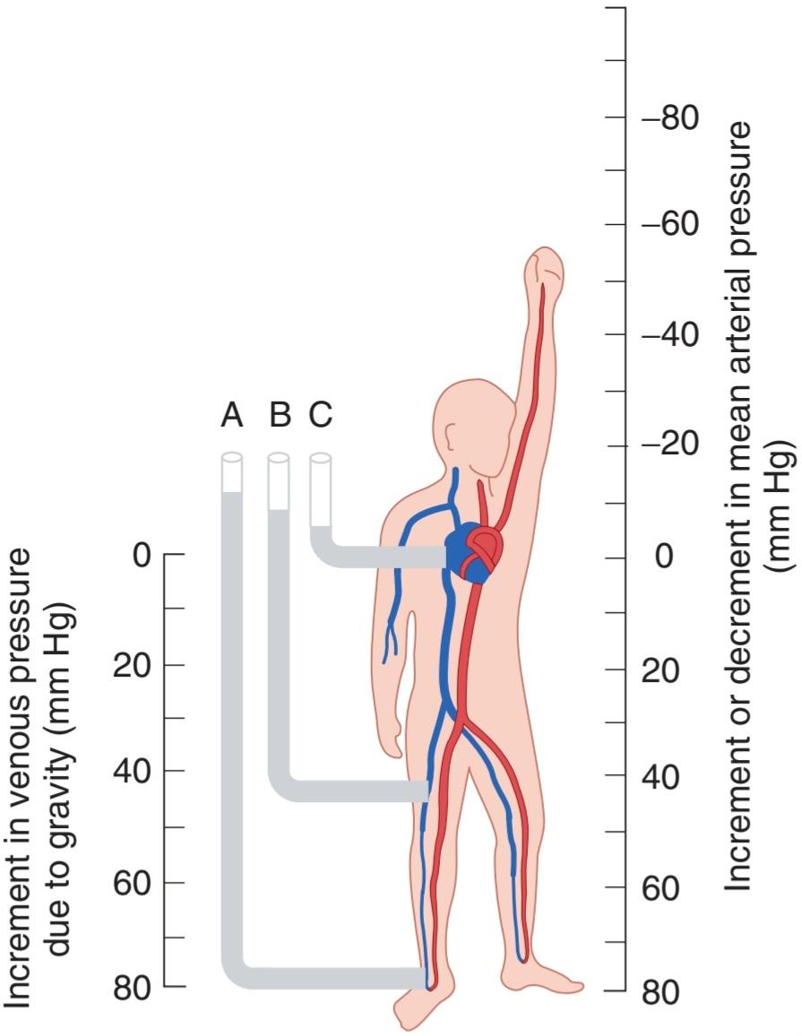
FIGURE 31-27 Effects of gravity on arterial and venous pressure. The scale on the right indicates the increment (or decrement) in mean pressure in a large artery at each level. The mean pressure in all large arteries is approximately 100 mm Hg when they are at the level of the left ventricle. The scale on the left indicates the increment in venous pressure at each level due to gravity. The manometers on the left of the figure indicate the height to which a column of blood in a tube would rise if connected to an ankle vein (A), the femoral vein (B), or the right atrium (C), with the subject in the standing position. The approximate pressures in these locations in the recumbent position; that is, when the ankle, thigh, and right atrium are at the same level, are A, 10 mm Hg; B, 7.5 mm Hg; and C, 4.6 mm Hg.
شکل 31-27 اثرات گرانش بر فشار شریانی و وریدی. مقیاس سمت راست نشان دهنده افزایش (یا کاهش) فشار متوسط در یک شریان بزرگ در هر سطح است. فشار متوسط در تمام شریان های بزرگ زمانی که در سطح بطن چپ قرار دارند تقریباً 100 میلی متر جیوه است. مقیاس سمت چپ نشان دهنده افزایش فشار وریدی در هر سطح به دلیل گرانش است. مانومترهای سمت چپ شکل نشان دهنده ارتفاعی است که یک ستون خون در یک لوله در صورتی که به ورید مچ پا (A)، ورید فمورال (B) یا دهلیز راست (C) وصل شود و سوژه در وضعیت ایستاده قرار گیرد، تا چه اندازه بالا می رود. فشارهای تقریبی در این مکان ها در حالت خوابیده. یعنی وقتی مچ پا، ران و دهلیز راست در یک سطح هستند، A، 10 میلی متر جیوه است. B، 7.5 میلی متر جیوه; و C، 4.6 میلی متر جیوه.
METHODS OF MEASURING BLOOD PRESSURE
If a cannula is inserted into an artery, the arterial pressure can be measured directly with a mercury manometer or a suitably calibrated strain gauge. When an artery is tied off beyond the point at which the cannula is inserted, an end pressure is recorded, flow in the artery is interrupted, and all the kinetic energy of flow is converted into pressure energy. If, alternatively, a T tube is inserted into a vessel and the pressure is measured in the side arm of the tube, the recorded side pressure, under conditions where pressure drop due to resistance is negligible, is lower than the end pressure by the kinetic energy of flow. This is because in a tube or a blood vessel the total energy-the sum of the kinetic energy of flow and the potential energy-is constant (Bernoulli principle).
روش های اندازه گیری فشار خون
اگر یک کانول در شریان قرار داده شود، فشار شریانی را می توان مستقیماً با یک مانومتر جیوه ای یا یک فشار سنج کالیبره شده مناسب اندازه گیری کرد. هنگامی که یک سرخرگ فراتر از نقطه ای که کانولا در آن قرار می گیرد بسته می شود، فشار انتهایی ثبت می شود، جریان در شریان قطع می شود و تمام انرژی جنبشی جریان به انرژی فشار تبدیل می شود. اگر یک لوله T در یک ظرف قرار داده شود و فشار در بازوی جانبی لوله اندازه گیری شود، فشار جانبی ثبت شده، در شرایطی که افت فشار به دلیل مقاومت ناچیز است، کمتر از فشار انتهایی توسط انرژی جنبشی جریان است. این به این دلیل است که در یک لوله یا یک رگ خونی انرژی کل – مجموع انرژی جنبشی جریان و انرژی پتانسیل – ثابت است (اصل برنولی).
It is worth noting that the pressure drop in any segment of the arterial system is due both to resistance and to conversion of potential into kinetic energy. The pressure drop due to energy lost in overcoming resistance is irreversible, since the energy is dissipated as heat; but the pressure drop due to conversion of potential to kinetic energy as a vessel narrows is reversed when the vessel widens out again (Figure 31-28).
شایان ذکر است که افت فشار در هر بخش از سیستم شریانی هم به دلیل مقاومت و هم به دلیل تبدیل پتانسیل به انرژی جنبشی است. افت فشار ناشی از انرژی از دست رفته در غلبه بر مقاومت غیر قابل برگشت است، زیرا انرژی به صورت گرما تلف می شود. اما افت فشار ناشی از تبدیل پتانسیل به انرژی جنبشی با باریک شدن ظرف زمانی که ظرف دوباره پهن می شود معکوس می شود (شکل 31-28).
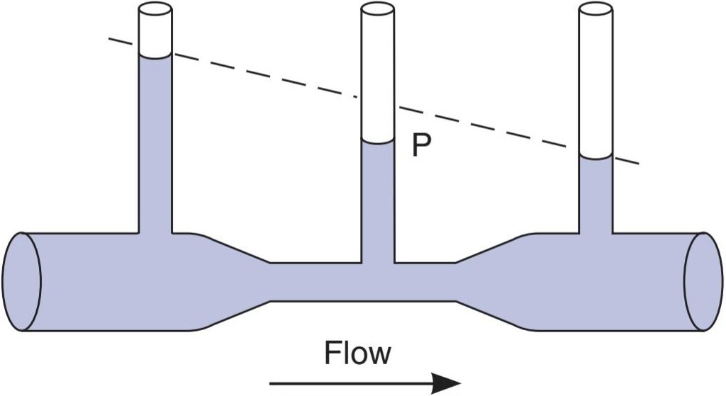
FIGURE 31-28 Bernoulli principle. When fluid flows through the narrow portion of the tube, the kinetic energy of flow is increased as the velocity increases, and the potential energy is reduced. Consequently, the measured pressure (P) is lower than it would have been at that point if the tube had not been narrowed. The dashed line indicates what the pressure drop due to frictional forces would have been if the tube had been of uniform diameter.
شکل 31-28 اصل برنولی. هنگامی که سیال از قسمت باریک لوله عبور می کند، انرژی جنبشی جریان با افزایش سرعت افزایش می یابد و انرژی پتانسیل کاهش می یابد. در نتیجه، فشار اندازهگیریشده (P) کمتر از آن چیزی است که اگر لوله باریک نمیشد، در آن نقطه بود. خط چین نشان می دهد که اگر قطر لوله یکنواخت بود، افت فشار ناشی از نیروهای اصطکاکی چقدر بود.
The Bernoulli principle also has a significant application in pathophysiology. According to the principle, the greater the velocity of flow in a vessel, the lower the lateral pressure distending its walls. When a vessel is narrowed, the velocity of flow in the narrowed portion increases and the distending pressure decreases. Therefore, when a vessel is narrowed by a pathologic process such as an atherosclerotic plaque, the lateral pressure at the constriction is decreased and the narrowing tends to maintain itself.
اصل برنولی در پاتوفیزیولوژی نیز کاربرد قابل توجهی دارد. بر اساس این اصل، هرچه سرعت جریان در یک ظرف بیشتر باشد، فشار جانبی که دیوارههای آن را متسع میکند کمتر میشود. هنگامی که یک ظرف باریک می شود، سرعت جریان در قسمت باریک افزایش می یابد و فشار انبساط کاهش می یابد. بنابراین، هنگامی که یک رگ توسط یک فرآیند پاتولوژیک مانند پلاک آترواسکلروتیک باریک می شود، فشار جانبی در انقباض کاهش می یابد و باریک شدن تمایل به حفظ خود دارد.
AUSCULTATORY METHOD
The arterial blood pressure in humans is routinely measured by the auscultatory method. An inflatable cuff attached to a mercury manometer (sphygmomanometer) is wrapped around the arm and a stethoscope is placed over the brachial artery at the elbow. The cuff is rapidly inflated until the pressure is well above the expected systolic pressure in the brachial artery. The artery is occluded by the cuff, and no sound is heard with the stethoscope. The pressure in the cuff is then lowered slowly. At the point at which systolic pressure in the artery just exceeds the cuff pressure, a spurt of blood passes through with each heartbeat and, synchronously with each beat, a tapping sound is heard below the cuff. The cuff pressure at which the sounds are first heard is the systolic pressure. As the cuff pressure is lowered further, the sounds become louder, then dull and muffled. These are the sounds of Korotkoff. Finally, in most individuals, they disappear. When direct and indirect blood pressure measurements are made simultaneously, the diastolic pressure in resting adults correlates best with the pressure at which the sound disappears. However, in adults after exercise and in children, the diastolic pressure correlates best with the pressure at which the sounds become muffled. This is also true in diseases such as hyperthyroidism and aortic regurgitation.
روش سمعي
فشار خون شریانی در انسان به طور معمول با روش سمعی اندازه گیری می شود. یک کاف بادی که به یک مانومتر جیوه ای (فشنگ سنج) متصل است به دور بازو پیچیده می شود و یک گوشی پزشکی روی شریان بازویی در آرنج قرار می گیرد. کاف به سرعت باد می شود تا زمانی که فشار از فشار سیستولیک مورد انتظار در شریان بازویی بیشتر شود. شریان توسط کاف مسدود می شود و صدایی با گوشی پزشکی شنیده نمی شود. سپس فشار در کاف به آرامی کاهش می یابد. در نقطه ای که فشار سیستولیک در شریان فقط از فشار کاف بیشتر می شود، با هر ضربان قلب یک فواره خون از آن عبور می کند و همزمان با هر ضربان، صدای ضربه زدن زیر کاف شنیده می شود. فشار کاف که در آن صداها برای اولین بار شنیده می شود فشار سیستولیک است. با کاهش بیشتر فشار کاف، صداها بلندتر، سپس کسل کننده و خفه می شوند. اینها صداهای کوروتکف هستند. در نهایت، در اکثر افراد، آنها ناپدید می شوند. هنگامی که اندازه گیری مستقیم و غیرمستقیم فشار خون به طور همزمان انجام می شود، فشار دیاستولیک در بزرگسالان در حال استراحت به بهترین وجه با فشاری که در آن صدا ناپدید می شود همبستگی دارد. با این حال، در بزرگسالان پس از ورزش و در کودکان، فشار دیاستولیک به بهترین وجه با فشاری که در آن صداها خفه می شوند همبستگی دارد. این موضوع در بیماری هایی مانند پرکاری تیروئید و نارسایی آئورت نیز صادق است.
The sounds of Korotkoff are produced by turbulent flow in the brachial artery. When the artery is narrowed by the cuff, the velocity of flow through the constriction exceeds the critical velocity and turbulent flow results (Figure 31- 19). At cuff pressures just below the systolic pressure, flow through the artery occurs only at the peak of systole, and the intermittent turbulence produces a tapping sound. As long as the pressure in the cuff is above the diastolic pressure in the artery, flow is interrupted at least during part of diastole, and the intermittent sounds have a staccato quality. When the cuff pressure is near the arterial diastolic pressure, the vessel is still constricted, but the turbulent flow is continuous. Continuous sounds have a muffled rather than a staccato quality.
صداهای کوروتکف توسط جریان متلاطم در شریان بازویی ایجاد می شود. هنگامی که شریان توسط کاف باریک می شود، سرعت جریان از طریق انقباض از سرعت بحرانی فراتر می رود و جریان متلاطم حاصل می شود (شکل 31-19). در فشارهای کاف درست زیر فشار سیستولیک، جریان از طریق شریان فقط در اوج سیستول رخ می دهد و تلاطم متناوب صدای ضربه زدن ایجاد می کند. تا زمانی که فشار در کاف بالاتر از فشار دیاستولیک در شریان باشد، جریان حداقل در بخشی از دیاستول قطع می شود و صداهای متناوب دارای کیفیت استاکاتو هستند. هنگامی که فشار کاف نزدیک به فشار دیاستولیک شریانی است، رگ همچنان منقبض است، اما جریان متلاطم پیوسته است. صداهای پیوسته به جای استاکاتو کیفیتی خفه دارند.
CHANGES IN ARTERIAL BLOOD PRESSURE
Because the arterial pressure is the product of the cardiac output and the peripheral resistance, it is affected by conditions that affect either or both of these factors. Emotion increases the cardiac output and peripheral resistance, and about 20% of hypertensive patients have blood pressures that are higher in the doctor’s office than at home (“white coat hypertension”). Blood pressure normally falls up to 20 mm Hg during sleep. This fall is reduced or absent in hypertension.
تغییرات در فشار خون شریانی
از آنجایی که فشار شریانی محصول برون ده قلبی و مقاومت محیطی است، تحت تأثیر شرایطی قرار می گیرد که بر یکی یا هر دوی این عوامل تأثیر می گذارد. احساسات برون ده قلبی و مقاومت محیطی را افزایش می دهد و حدود 20 درصد از بیماران مبتلا به فشار خون فشار خونی دارند که در مطب پزشک بیشتر از خانه است (“فشار خون کت سفید”). فشار خون به طور معمول در هنگام خواب تا 20 میلی متر جیوه کاهش می یابد. این سقوط در فشار خون بالا کاهش می یابد یا وجود ندارد.
There is general agreement that blood pressure rises with advancing age, but the magnitude of this rise is uncertain because hypertension is a common disease and its incidence also increases with advancing age (Clinical Box 31-4). Individuals who have systolic blood pressures < 120 mm Hg at age 50-60 and never develop clinical hypertension still have systolic pressures that rise throughout life (Figure 31–29). This rise may be the closest approximation to the rise in normal individuals. Diastolic pressure also rises, but then starts to fall in middle age as the stiffness of arteries increases. Consequently, pulse pressure rises with advancing age. It is interesting that systolic and diastolic blood pressures are lower in women than in men until age 55-65, after which they become comparable. Because there is a positive correlation between blood pressure and the incidence of heart attacks and strokes (see below), the lower blood pressure before menopause in women may be one reason that, on average, they live longer than men.
توافق کلی وجود دارد که فشار خون با افزایش سن افزایش می یابد، اما میزان این افزایش نامشخص است زیرا فشار خون یک بیماری شایع است و بروز آن نیز با افزایش سن افزایش می یابد (باکس بالینی 31-4). افرادی که فشار خون سیستولیک کمتر از 120 میلیمتر جیوه در سنین 60-50 سالگی دارند و هرگز به فشار خون بالینی مبتلا نمیشوند، همچنان فشار سیستولیک دارند که در طول زندگی افزایش مییابد (شکل 31-29). این افزایش ممکن است نزدیکترین تقریب به افزایش در افراد عادی باشد. فشار دیاستولیک نیز افزایش می یابد، اما در میانسالی با افزایش سفتی شریان ها شروع به کاهش می کند. در نتیجه، فشار نبض با افزایش سن افزایش می یابد. جالب است که فشار خون سیستولیک و دیاستولیک در زنان تا سن 55 تا 65 سالگی کمتر از مردان است و پس از آن قابل مقایسه است. از آنجایی که ارتباط مثبتی بین فشار خون و بروز حملات قلبی و سکته وجود دارد (به زیر مراجعه کنید)، کاهش فشار خون قبل از یائسگی در زنان ممکن است یکی از دلایلی باشد که به طور متوسط آنها نسبت به مردان عمر طولانیتری دارند.
CLINICAL BOX 31-4
Hypertension
Hypertension is a sustained elevation of the systemic arterial pressure. It is most commonly due to increased peripheral resistance and is a very common abnormality in humans. It can be produced by many diseases (Table 31-11) and causes a number of serious disorders. When the resistance against which the left ventricle must pump (afterload) is elevated for a long period, the cardiac muscle hypertrophies. The initial response is activation of immediate- early genes in the ventricular muscle, followed by activation of a series of genes involved in growth during fetal life. Left ventricular hypertrophy is associated with a poor prognosis. The total O2 consumption of the heart, already increased by the work of expelling blood against a raised pressure (see Chapter 30), is increased further because there is more muscle. Therefore, any decrease in coronary blood flow has more serious consequences in hypertensive patients than it does in normal individuals, and degrees of coronary vessel narrowing that do not produce symptoms when the size of the heart is normal may produce myocardial infarction when the heart is enlarged.
جعبه بالینی 31-4
فشار خون بالا
فشار خون بالا افزایش مداوم فشار شریانی سیستمیک است. این بیشتر به دلیل افزایش مقاومت محیطی است و یک ناهنجاری بسیار شایع در انسان است. این می تواند توسط بسیاری از بیماری ها تولید شود (جدول 31-11) و باعث تعدادی از اختلالات جدی می شود. هنگامی که مقاومتی که بطن چپ باید در برابر آن پمپاژ کند (پس از بارگیری) برای مدت طولانی افزایش می یابد، عضله قلب هیپرتروفی می شود. پاسخ اولیه، فعال شدن ژن های فوری و اولیه در عضله بطنی، و به دنبال آن فعال شدن یک سری از ژن های دخیل در رشد در طول زندگی جنینی است. هیپرتروفی بطن چپ با پیش آگهی ضعیف همراه است. کل مصرف O2 قلب، که قبلاً با کار دفع خون در برابر فشار افزایش یافته افزایش یافته است (به فصل 30 مراجعه کنید)، بیشتر افزایش می یابد زیرا ماهیچه های بیشتری وجود دارد. بنابراین، هر گونه کاهش در جریان خون کرونری در بیماران مبتلا به فشار خون عواقب جدی تری نسبت به افراد عادی دارد و درجاتی از باریک شدن عروق کرونر که زمانی که اندازه قلب طبیعی است علائمی ایجاد نمی کند، ممکن است در هنگام بزرگ شدن قلب باعث انفارکتوس میوکارد شود.
The incidence of atherosclerosis increases in hypertension, and myocardial infarcts are common even when the heart is not enlarged. Eventually, the ability to compensate for the high peripheral resistance is exceeded, and the heart fails. Hypertensive individuals are also predisposed to thromboses of cerebral vessels and cerebral hemorrhage. An additional complication is chronic kidney disease. However, the incidence of heart failure, strokes, and chronic kidney disease can be markedly reduced by active treatment of hypertension, even when the hypertension is relatively mild. In most patients with elevated blood pressure, the cause of the hypertension is unknown, and they are said to have essential hypertension (Table 31-11). At present, essential hypertension is treatable but not curable. In other, less common forms of hypertension, the cause is known. A review of these is helpful because it emphasizes ways disordered physiology can lead to disease. Pathology that compromises the renal blood supply leads to renal hypertension, as does narrowing (coarctation) of the thoracic aorta, which both increases renin secretion and increases peripheral resistance. Pheochromocytomas, adrenal medullary tumors that secrete norepinephrine and epinephrine, can cause sporadic or sustained hypertension (see Chapter 20). Estrogens increase angiotensinogen secretion, and contraceptive pills containing large amounts of estrogen cause hypertension (pill hypertension) on this basis (see Chapter 22). Increased secretion of aldosterone or other mineralocorticoids causes renal Na* retention, which leads to hypertension. A primary increase in plasma mineralocorticoids inhibits renin secretion. For unknown reasons, plasma renin is also low in 10-15% of patients with essential hypertension and normal circulating mineralocortical levels (low renin hypertension). Mutations in a number of single genes are also known to cause hypertension. These cases of monogenic hypertension are rare but informative. One of these is glucocorticoid-remediable aldosteronism (GRA), in which a hybrid gene encodes an adrenocorticotropic hormone (ACTH)- sensitive aldosterone synthase, with resulting hyperaldosteronism (see Chapter 20). 11-ẞ Hydroxylase deficiency also causes hypertension by increasing the secretion of deoxycorticosterone (see Chapter 20). Normal blood pressure is restored when ACTH secretion is inhibited by administering a glucocorticoid. Mutations that decrease 11-ẞ hydroxysteroid dehydrogenase cause loss of specificity of the mineralocorticoid receptors (see Chapter 20) with stimulation of them by cortisol and, in pregnancy, by the elevated circulating levels of progesterone. Finally, mutations of the genes for ENaCs that reduce degradation of the ẞ or y subunits increase ENaC activity and lead to excess renal Na* retention and hypertension (Liddle syndrome; see Chapter 37).
بروز آترواسکلروز در فشار خون بالا افزایش می یابد و انفارکتوس میوکارد حتی زمانی که قلب بزرگ نشده باشد شایع است. در نهایت، توانایی جبران مقاومت محیطی بالا از بین می رود و قلب از کار می افتد. افراد مبتلا به فشار خون نیز مستعد ترومبوز عروق مغزی و خونریزی مغزی هستند. یک عارضه اضافی بیماری مزمن کلیه است. با این حال، بروز نارسایی قلبی، سکته مغزی و بیماری مزمن کلیوی را می توان به طور قابل توجهی با درمان فعال فشار خون بالا کاهش داد، حتی زمانی که فشار خون بالا نسبتاً خفیف باشد. در اکثر بیماران با فشار خون بالا، علت فشار خون بالا ناشناخته است و گفته می شود که فشار خون ضروری دارند (جدول 31-11). در حال حاضر فشار خون ضروری قابل درمان است اما قابل درمان نیست. در سایر اشکال کمتر شایع فشار خون، علت آن مشخص است. بررسی این موارد مفید است زیرا بر روشهایی تأکید میکند که فیزیولوژی بینظم میتواند منجر به بیماری شود. آسیب شناسی که جریان خون کلیوی را به خطر می اندازد منجر به فشار خون کلیوی می شود، مانند باریک شدن (کوآرکتاسیون) آئورت سینه ای که هم ترشح رنین را افزایش می دهد و هم مقاومت محیطی را افزایش می دهد. فئوکروموسیتوم ها، تومورهای مدولاری آدرنال که نوراپی نفرین و اپی نفرین ترشح می کنند، می توانند باعث پرفشاری خون پراکنده یا پایدار شوند (به فصل 20 مراجعه کنید). استروژن ها ترشح آنژیوتانسینوژن را افزایش می دهند و بر این اساس قرص های ضد بارداری حاوی مقادیر زیادی استروژن باعث افزایش فشار خون (پرفشاری خون قرص) می شوند (به فصل 22 مراجعه کنید). افزایش ترشح آلدوسترون یا سایر مینرالوکورتیکوئیدها باعث احتباس +Na کلیه می شود که منجر به فشار خون بالا می شود. افزایش اولیه در مینرالوکورتیکوئیدهای پلاسما باعث مهار ترشح رنین می شود. به دلایل ناشناخته، رنین پلاسما نیز در 15-10 درصد از بیماران مبتلا به فشار خون ضروری و سطوح مینرالوکورتیکال در گردش طبیعی (فشار خون پایین رنین) کم است. جهش در تعدادی از ژن های منفرد نیز به عنوان عامل فشار خون بالا شناخته شده است. این موارد فشار خون مونووژنیک نادر اما آموزنده است. یکی از آنها آلدوسترونیسم قابل اصلاح با گلوکوکورتیکوئید (GRA) است که در آن یک ژن هیبریدی یک هورمون آدرنوکورتیکوتروپیک (ACTH) – آلدوسترون سنتاز حساس را کد می کند و منجر به هیپرآلدوسترونیسم می شود (به فصل 20 مراجعه کنید). 11- کمبود هیدروکسیلاز نیز با افزایش ترشح دئوکسی کورتیکوسترون باعث فشار خون بالا می شود (به فصل 20 مراجعه کنید). هنگامی که ترشح ACTH با تجویز گلوکوکورتیکوئید مهار می شود، فشار خون طبیعی بازیابی می شود. جهش هایی که 11-هیدروکسی استروئید دهیدروژناز را کاهش می دهند، با تحریک آنها توسط کورتیزول و در بارداری، با افزایش سطح پروژسترون در گردش، ویژگی گیرنده های معدنی کورتیکوئید را از دست می دهند (به فصل 20 مراجعه کنید). در نهایت، جهشهای ژنهای ENaCs که تخریب زیرواحدهای ẞ یا y را کاهش میدهند، فعالیت ENaC را افزایش میدهند و منجر به احتباس بیش از حد +Na کلیه و فشار خون بالا میشوند (سندرم لیدل؛ فصل 37 را ببینید).
THERAPEUTIC HIGHLIGHTS
Effective lowering of the blood pressure can be produced by drugs that block a- adrenergic receptors, either in the periphery or in the central nervous system; drugs that block ẞ-adrenergic receptors; drugs that inhibit the activity of angiotensin-converting enzyme; and calcium channel blockers that relax vascular smooth muscle.
نکات برجسته درمانی
کاهش موثر فشار خون می تواند توسط داروهایی ایجاد شود که گیرنده های آدرنرژیک را چه در محیطی و چه در سیستم عصبی مرکزی مسدود می کنند. داروهایی که گیرنده های آدرنرژیک را مسدود می کنند. داروهایی که فعالیت آنزیم مبدل آنژیوتانسین را مهار می کنند. و مسدود کننده های کانال کلسیم که عضلات صاف عروق را شل می کند.
TABLE 31–11 Estimated frequency of various forms of hypertension in the general hypertensive population.
جدول 31-11 فراوانی تخمینی اشکال مختلف فشار خون در جمعیت پرفشاری خون عمومی.

Data from Braunwald E, et al (eds): Harrison’s Principles of Internal Medicine, 15th ed. New York, NY: McGraw-Hill; 2001.
داده ها از براونوالد ای، و همکاران (ویرایشگران): اصول پزشکی داخلی هریسون، ویرایش 15. نیویورک، نیویورک: McGraw-Hill; 2001.
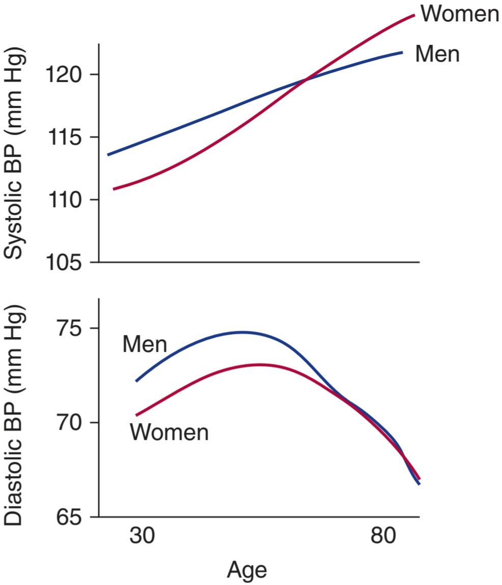
FIGURE 31-29 Effects of age and sex on arterial pressure components in humans. Data are from a large group of individuals who were studied every 2 years throughout their adult lives and who were found not be hypertensive (ie, they had systolic blood pressures < 120 mm Hg) at age 50-60. (Data replotted from Franklin SS, et al: Hemodynamic patterns of age-related changes in blood pressure: The Framingham Heart Study. Circulation 1997; 96:308 as well as the NHANES III study.)
شکل 31-29 اثرات سن و جنس بر اجزای فشار شریانی در انسان. داده ها از گروه بزرگی از افراد است که هر 2 سال یک بار در طول زندگی بزرگسالی خود مورد مطالعه قرار گرفتند و مشخص شد که فشار خون بالا ندارند (یعنی فشار خون سیستولیک کمتر از 120 میلی متر جیوه داشتند) در سن 50-60 سالگی. (داده های بازنگری شده از فرانکلین SS و همکاران: الگوهای همودینامیک تغییرات مرتبط با سن در فشار خون: مطالعه قلب فرامینگام. گردش 1997؛ 96:308 و همچنین مطالعه NHANES III.)
CAPILLARY CIRCULATION
At any one time, only 5% of the circulating blood is in the capillaries, but this 5% is in a sense the most important part of the blood volume because it is the only pool from which O2 and nutrients can enter the interstitial fluid and into which CO2 and waste products can enter the bloodstream. Exchange across the capillary walls is essential to the survival of the tissues.
گردش مویرگی
در هر زمان، تنها 5 درصد از خون در گردش در مویرگ ها است، اما این 5 درصد به یک معنا مهم ترین بخش حجم خون است، زیرا تنها حوضچه ای است که از آن O2 و مواد مغذی می توانند وارد مایع بینابینی شوند و CO2 و مواد زائد می توانند وارد جریان خون شوند. تبادل بین دیواره های مویرگی برای بقای بافت ها ضروری است.
METHODS OF STUDY
It is difficult to obtain accurate measurements of capillary pressures and flows. Experimentally, capillary pressure has been estimated by determining the amount of external pressure necessary to occlude the capillaries or the amount of pressure necessary to make saline start to flow through a micropipette inserted so that its tip faces the arteriolar end of the capillary.
روشهای مطالعه
اندازه گیری دقیق فشار و جریان مویرگی دشوار است. به طور تجربی، فشار مویرگی با تعیین مقدار فشار خارجی لازم برای انسداد مویرگ ها یا مقدار فشار لازم برای شروع جریان آب نمک از طریق میکروپیپت وارد شده به طوری که نوک آن به سمت انتهای شریانی مویرگ قرار گیرد، تخمین زده شده است.
CAPILLARY PRESSURE & FLOW
Capillary pressures vary considerably, but typical values in human nail bed capillaries are 32 mm Hg at the arteriolar end and 15 mm Hg at the venous end. The pulse pressure is approximately 5 mm Hg at the arteriolar end and zero at the venous end. The capillaries are short, but blood moves slowly (about 0.07 cm/s) because the total cross-sectional area of the capillary bed is large. Transit time from the arteriolar to the venular end of an average-sized capillary is 1-2 s.
فشار و جریان مویرگی
فشار مویرگی به طور قابل توجهی متفاوت است، اما مقادیر معمول در مویرگ های بستر ناخن انسان 32 میلی متر جیوه در انتهای شریانی و 15 میلی متر جیوه در انتهای وریدی است. فشار نبض تقریباً 5 میلی متر جیوه در انتهای شریانی و صفر در انتهای وریدی است. مویرگ ها کوتاه هستند، اما خون به کندی حرکت می کند (حدود 0.07 سانتی متر بر ثانیه) زیرا سطح مقطع کل بستر مویرگی زیاد است. زمان انتقال از شریانی به انتهای وریدی یک مویرگ با اندازه متوسط 1-2 ثانیه است.
EQUILIBRATION WITH INTERSTITIAL FLUID
As noted above, the capillary wall is a thin membrane made up of endothelial cells. Substances pass through the junctions between endothelial cells and through fenestrations when they are present. Some also pass through the cells by vesicular transport.
تعادل با سیالات بینابینی
همانطور که در بالا ذکر شد، دیواره مویرگی یک غشای نازک است که از سلول های اندوتلیال تشکیل شده است. مواد از طریق اتصالات بین سلول های اندوتلیال و در صورت وجود از طریق فنسترها عبور می کنند. برخی نیز از طریق انتقال تاولی از سلول ها عبور می کنند.
The factors other than vesicular transport that are responsible for transport across the capillary wall are diffusion and filtration (see Chapter 1). Diffusion is quantitatively much more important. O2 and glucose are in higher concentration in the bloodstream than in the interstitial fluid and diffuse into the interstitial fluid, whereas CO2 diffuses in the opposite direction.
عواملی غیر از انتقال تاولی که مسئول انتقال از طریق دیواره مویرگی هستند، انتشار و فیلتراسیون هستند (به فصل 1 مراجعه کنید). انتشار از نظر کمی بسیار مهمتر است. غلظت O2 و گلوکز در جریان خون بیشتر از مایع بین بافتی است و در مایع بینابینی منتشر می شود، در حالی که CO2 در جهت مخالف منتشر می شود.
The rate of filtration at any point along a capillary depends on a balance of forces sometimes called the Starling forces. One of these is the hydrostatic pressure gradient (the hydrostatic pressure in the capillary minus the hydrostatic pressure of the interstitial fluid). The interstitial fluid pressure varies from one organ to another. It is positive in the liver and kidneys and as high as 6 mm Hg in the brain. The other force is the osmotic pressure gradient across the capillary wall (colloid osmotic pressure of plasma minus colloid osmotic pressure of interstitial fluid). This component is directed inward.
Thus:
where
سرعت فیلتراسیون در هر نقطه در امتداد یک مویرگ به تعادل نیروها بستگی دارد که گاهی اوقات نیروهای استارلینگ نامیده می شود. یکی از اینها گرادیان فشار هیدرواستاتیک (فشار هیدرواستاتیک در مویرگ منهای فشار هیدرواستاتیک مایع بینابینی) است. فشار مایع بینابینی از یک اندام به اندام دیگر متفاوت است. در کبد و کلیه ها مثبت و در مغز تا 6 میلی متر جیوه است. نیروی دیگر، گرادیان فشار اسمزی در سراسر دیواره مویرگ است (فشار اسمزی کلوئیدی پلاسما منهای فشار اسمزی کلوئیدی مایع بینابینی). این جزء به سمت داخل هدایت می شود.
بدین ترتیب:

کجا

Пi is usually negligible, so the osmotic pressure gradient (л – л;) usually equals the oncotic pressure. The capillary filtration coefficient takes into account, and is proportional to, the permeability of the capillary wall and the area available for filtration. The magnitude of the Starling forces along a typical muscle capillary is shown in Figure 31-30. Fluid moves into the interstitial space at the arteriolar end of the capillary and into the capillary at the venular end. In other capillaries, the balance of Starling forces may be different. For example, fluid moves out of almost the entire length of the capillaries in the renal glomeruli. On the other hand, fluid moves into the capillaries through almost their entire length in the intestines. About 24 L of fluid is filtered through the capillaries per day. This is about 0.3% of the cardiac output. About 85% of the filtered fluid is reabsorbed into the capillaries, and the remainder returns to the circulation via the lymphatics.
Пi معمولاً ناچیز است، بنابراین گرادیان فشار اسمزی (л – л;) معمولاً برابر با فشار انکوتیک است. ضریب فیلتراسیون مویرگی، نفوذپذیری دیواره مویرگی و منطقه موجود برای فیلتراسیون را در نظر می گیرد و متناسب با آن است. بزرگی نیروهای استارلینگ در امتداد یک مویرگ عضلانی معمولی در شکل 31-30 نشان داده شده است. مایع به فضای بینابینی در انتهای شریانی مویرگ و به داخل مویرگ در انتهای وریدی حرکت می کند. در مویرگ های دیگر، تعادل نیروهای استارلینگ ممکن است متفاوت باشد. به عنوان مثال، مایع تقریباً از تمام طول مویرگ های گلومرول کلیوی خارج می شود. از طرف دیگر، مایع تقریباً در تمام طول مویرگ ها در روده به داخل مویرگ ها حرکت می کند. حدود 24 لیتر مایع در روز از طریق مویرگ ها فیلتر می شود. این حدود 0.3 درصد برون ده قلبی است. حدود 85 درصد از مایع فیلتر شده مجدداً در مویرگ ها جذب می شود و باقی مانده از طریق لنفاوی به گردش خون باز می گردد.
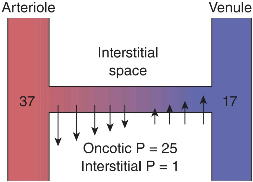
FIGURE 31-30 Schematic representation of pressure gradients across the wall of a muscle capillary. The numbers at the arteriolar and venular ends of the capillary are the hydrostatic pressures in mm Hg at these locations. The arrows indicate the approximate magnitude and direction of fluid movement. In this example, the pressure differential at the arteriolar end of the capillary is 11 mm Hg ([371]-25) outward; at the opposite end, it is 9 mm Hg (25 – [17 – 1]) inward.
شکل 31-30 نمایش شماتیک گرادیان فشار در سراسر دیواره مویرگ ماهیچه ای. اعداد در انتهای شریانی و وریدی مویرگ، فشار هیدرواستاتیک بر حسب میلی متر جیوه در این مکان ها هستند. فلش ها مقدار تقریبی و جهت حرکت سیال را نشان می دهند. در این مثال، اختلاف فشار در انتهای شریانی مویرگ 11 میلی متر جیوه ([371]-25) به سمت بیرون است. در طرف مقابل، 9 میلی متر جیوه (25 – [17-1]) به سمت داخل است.
It is worth noting that small molecules often equilibrate with the tissues near the arteriolar end of each capillary. In this situation, total diffusion can be increased by increasing blood flow; that is, exchange is flow-limited (Figure 31-31). Conversely, transfer of substances that do not reach equilibrium with the tissues during their passage through the capillaries is said to be diffusion- limited.
شایان ذکر است که مولکول های کوچک اغلب با بافت های نزدیک به انتهای شریانی هر مویرگ تعادل برقرار می کنند. در این شرایط، انتشار کل را می توان با افزایش جریان خون افزایش داد. یعنی تبادل با جریان محدود است (شکل 31-31). برعکس، انتقال موادی که در طول عبور از مویرگ ها با بافت ها به تعادل نمی رسند، به انتشار محدود گفته می شود.

FIGURE 31-31 Flow-limited and diffusion-limited exchange across capillary walls. A and V indicate the arteriolar and venular ends of the capillary. Substance X equilibrates with the tissues (movement into the tissues equals movement out) well before the blood leaves the capillary, whereas substance Y does not equilibrate. If other factors stay constant, the amount of X entering the tissues can be increased only by increasing blood flow; that is, it is flow-limited. The movement of Y is diffusion-limited.
شکل 31-31 تبادل محدود با جریان و انتشار محدود در دیواره های مویرگی. A و V انتهای شریانی و وریدی مویرگ را نشان می دهد. ماده X قبل از اینکه خون از مویرگ خارج شود با بافت ها متعادل می شود (حرکت به داخل بافت برابر است با حرکت به بیرون) در حالی که ماده Y تعادل ندارد. اگر سایر عوامل ثابت بمانند، مقدار X وارد شده به بافت ها را می توان تنها با افزایش جریان خون افزایش داد. یعنی محدود به جریان است. حرکت Y از نظر انتشار محدود است.
ACTIVE & INACTIVE CAPILLARIES
In resting tissues, most of the capillaries are collapsed. In active tissues, the metarterioles and the precapillary sphincters dilate. The intracapillary pressure rises, overcoming the critical closing pressure of the vessels, and blood flows through all of the capillaries. Relaxation of the smooth muscle of the metarterioles and precapillary sphincters is due to the action of vasodilator metabolites formed locally in active tissue (see Chapter 32).
مویرگهای فعال و غیر فعال
در بافتهای در حال استراحت، بیشتر مویرگها فرو میروند. در بافت های فعال، متارتریول ها و اسفنکترهای پیش مویرگی گشاد می شوند. فشار داخل مویرگ افزایش می یابد و بر فشار بسته شدن بحرانی عروق غلبه می کند و خون در تمام مویرگ ها جریان می یابد. شل شدن عضله صاف متارتریول ها و اسفنکترهای پیش مویرگی به دلیل عمل متابولیت های گشادکننده عروقی است که به صورت موضعی در بافت فعال تشکیل می شوند (به فصل 32 مراجعه کنید).
After noxious stimulation, substance P released by the axon reflex (see Chapter 33) increases capillary permeability. Bradykinin and histamine also increase capillary permeability. When capillaries are stimulated mechanically, they empty (white reaction; see Chapter 33), probably due to contraction of the precapillary sphincters.
پس از تحریک مضر، ماده P آزاد شده توسط رفلکس آکسون (به فصل 33 مراجعه کنید) نفوذپذیری مویرگی را افزایش می دهد. برادی کینین و هیستامین نیز نفوذپذیری مویرگی را افزایش می دهند. هنگامی که مویرگ ها به صورت مکانیکی تحریک می شوند، آنها خالی می شوند (واکنش سفید؛ به فصل 33 مراجعه کنید)، احتمالاً به دلیل انقباض اسفنکترهای پیش مویرگی.
VENOUS CIRCULATION
Blood flows through the blood vessels, including the veins, primarily because of the pumping action of the heart. However, venous flow is aided by the heartbeat, the increase in the negative intrathoracic pressure during each inspiration, and contractions of skeletal muscles that compress the veins (muscle pump).
گردش خون وریدی
خون از طریق رگ های خونی، از جمله سیاهرگ ها، در درجه اول به دلیل عملکرد پمپاژ قلب جریان دارد. با این حال، جریان وریدی توسط ضربان قلب، افزایش فشار منفی داخل قفسه سینه در طی هر دم و انقباضات ماهیچه های اسکلتی که وریدها را فشرده می کنند (پمپ عضلانی) کمک می کند.
VENOUS PRESSURE & FLOW
The pressure in the venules is 12-18 mm Hg. It falls steadily in the larger veins to about 5.5 mm Hg in the great veins outside the thorax. The pressure in the great veins at their entrance into the right atrium (central venous pressure) averages 4.6 mm Hg, but fluctuates with respiration and heart action.
فشار و جریان وریدی
فشار در ونول ها 12-18 میلی متر جیوه است. به طور پیوسته در وریدهای بزرگتر تا حدود 5.5 میلی متر جیوه در وریدهای بزرگ خارج از قفسه سینه می افتد. فشار وریدهای بزرگ در ورودی آنها به دهلیز راست (فشار ورید مرکزی) به طور متوسط 4.6 میلی متر جیوه است، اما با تنفس و عملکرد قلب در نوسان است.
Peripheral venous pressure, like arterial pressure, is affected by gravity. It is increased by 0.77 mm Hg for each centimeter below the right atrium and decreased by a like amount for each centimeter above the right atrium the pressure is measured (Figure 31-27). Thus, on a proportional basis, gravity has a greater effect on venous than on arterial pressures.
فشار وریدهای محیطی، مانند فشار شریانی، تحت تأثیر گرانش است. به ازای هر سانتی متر زیر دهلیز راست 0.77 میلی متر جیوه افزایش می یابد و به ازای هر سانتی متر بالای دهلیز راست فشار اندازه گیری شده به میزان مشابهی کاهش می یابد (شکل 31-27). بنابراین، بر اساس تناسب، گرانش تأثیر بیشتری بر فشارهای وریدی دارد تا فشارهای شریانی.
When blood flows from the venules to the large veins, its average velocity increases as the total cross-sectional area of the vessels decreases. In the great veins, the velocity of blood is about one-fourth that in the aorta, averaging about 10 cm/s.
هنگامی که خون از وریدها به وریدهای بزرگ جریان می یابد، سرعت متوسط آن با کاهش کل سطح مقطع عروق افزایش می یابد. در وریدهای بزرگ، سرعت خون در حدود یک چهارم آئورت است و به طور متوسط حدود 10 سانتی متر بر ثانیه است.
THORACIC PUMP
During inspiration, the intrapleural pressure falls from -2.5 to -6 mm Hg. This negative pressure is transmitted to the great veins and, to a lesser extent, the atria, so that central venous pressure fluctuates from about 6 mm Hg during expiration to approximately 2 mm Hg during quiet inspiration. The drop in venous pressure during inspiration aids venous return. When the diaphragm descends during inspiration, intra-abdominal pressure rises, and this also squeezes blood toward the heart because backflow into the leg veins is prevented by the venous valves.
پمپ قفسه سینه
در طی دم، فشار داخل جنب از 2.5- تا 6- میلی متر جیوه کاهش می یابد. این فشار منفی به وریدهای بزرگ و به میزان کمتری به دهلیزها منتقل می شود، به طوری که فشار ورید مرکزی از حدود 6 میلی متر جیوه در حین بازدم تا تقریباً 2 میلی متر جیوه در هنگام دمیدن آرام در نوسان است. کاهش فشار وریدی در حین دم به بازگشت وریدی کمک می کند. هنگامی که دیافراگم در حین دم پایین می آید، فشار داخل شکمی افزایش می یابد و این نیز خون را به سمت قلب می فشارد، زیرا دریچه های وریدی از برگشت به وریدهای پا جلوگیری می کنند.
EFFECTS OF HEARTBEAT
The variations in atrial pressure are transmitted to the great veins, producing the a, c, and v waves of the venous pressure-pulse curve (see Chapter 30). Atrial pressure drops sharply during the ejection phase of ventricular systole because the atrioventricular valves are pulled downward, increasing the capacity of the atria. This action sucks blood into the atria from the great veins. The sucking of the blood into the atria during systole contributes appreciably to venous return, especially at rapid heart rates.
اثرات ضربان قلب
تغییرات فشار دهلیزی به سیاهرگهای بزرگ منتقل میشود و امواج a، c و v منحنی فشار-نبض وریدی را تولید میکند (به فصل 30 مراجعه کنید). فشار دهلیزی در مرحله جهش سیستول بطنی به شدت کاهش می یابد زیرا دریچه های دهلیزی به سمت پایین کشیده می شوند و ظرفیت دهلیزها افزایش می یابد. این عمل خون را از وریدهای بزرگ به دهلیز می مکد. مکیدن خون به دهلیزها در طول سیستول به میزان قابل توجهی به بازگشت وریدی کمک می کند، به خصوص در ضربان قلب سریع.
Close to the heart, venous flow becomes pulsatile. When the heart rate is slow, two periods of peak flow are detectable, one during ventricular systole, due to pulling down of the atrioventricular valves, and one in early diastole, during the period of rapid ventricular filling (Figure 31-25).
نزدیک به قلب، جریان وریدی ضربان دار می شود. هنگامی که ضربان قلب کند است، دو دوره اوج جریان قابل تشخیص است، یکی در طول سیستول بطنی، به دلیل پایین کشیدن دریچه های دهلیزی و دیگری در دیاستول اولیه، در طول دوره پر شدن سریع بطن (شکل 31-25).
MUSCLE PUMP
In the limbs, the veins are surrounded by skeletal muscles, and contraction of these muscles during activity compresses the veins. Pulsations of nearby arteries may also compress veins. Because the venous valves prevent reverse flow, the blood moves toward the heart. During quiet standing, when the full effect of gravity is manifest, venous pressure at the ankle is 85-90 mm Hg (Figure 31- 27). Pooling of blood in the leg veins reduces venous return, with the result that cardiac output is reduced, sometimes to the point where fainting occurs. Rhythmic contractions of the leg muscles while the person is standing serve to lower the venous pressure in the legs to less than 30 mm Hg by propelling blood toward the heart. This heartward movement of the blood is decreased in patients with varicose veins because their valves are incompetent. These patients may develop stasis and ankle edema. However, even when the valves are incompetent, muscle contractions continue to produce a basic heartward movement of the blood because the resistance of the larger veins in the direction of the heart is less than the resistance of the small vessels away from the heart.
پمپ عضلانی
در اندام ها، سیاهرگ ها توسط ماهیچه های اسکلتی احاطه شده اند و انقباض این ماهیچه ها در حین فعالیت، رگ ها را فشرده می کند. ضربان شریان های مجاور نیز ممکن است وریدها را فشرده کند. از آنجایی که دریچه های وریدی از جریان معکوس جلوگیری می کنند، خون به سمت قلب حرکت می کند. در هنگام ایستادن آرام، زمانی که اثر کامل جاذبه آشکار می شود، فشار وریدی در مچ پا 90-85 میلی متر جیوه است (شکل 31-27). تجمع خون در وریدهای پا، بازگشت وریدی را کاهش می دهد و در نتیجه برون ده قلبی کاهش می یابد، گاهی اوقات تا حدی که غش رخ می دهد. انقباضات ریتمیک ماهیچه های پا در حالی که فرد ایستاده است، با حرکت خون به سمت قلب، فشار وریدی در پاها را به کمتر از 30 میلی متر جیوه کاهش می دهد. این حرکت خون به سمت قلب در بیماران مبتلا به واریس کاهش می یابد زیرا دریچه های آنها ناکارآمد است. این بیماران ممکن است دچار استاز و ادم مچ پا شوند. با این حال، حتی زمانی که دریچهها ناکارآمد هستند، انقباضات ماهیچهای به ایجاد یک حرکت اساسی به سمت قلب ادامه میدهند، زیرا مقاومت وریدهای بزرگتر در جهت قلب کمتر از مقاومت عروق کوچک دور از قلب است.
VENOUS PRESSURE IN THE HEAD
In the upright position, the venous pressure in the parts of the body above the heart is decreased by the force of gravity. The neck veins collapse above the point where the venous pressure is close to zero. However, the dural sinuses have rigid walls and cannot collapse. The pressure in them in the standing or sitting position is therefore subatmospheric. The magnitude of the negative pressure is proportional to the vertical distance above the top of the collapsed neck veins, and in the superior sagittal sinus may be as much as -10 mm Hg. This fact must be kept in mind by neurosurgeons. Neurosurgical procedures are sometimes performed with the patient seated. If one of the sinuses is opened during such a procedure it sucks air, causing air embolism.
فشار وریدی در سر
در وضعیت عمودی، فشار وریدی در قسمتهای بالای قلب بر اثر نیروی گرانش کاهش مییابد. وریدهای گردن در بالای نقطه ای که فشار وریدی نزدیک به صفر است فرو می ریزند. با این حال، سینوس های دورال دیواره های سفت و سختی دارند و نمی توانند فرو بریزند. بنابراین فشار موجود در آنها در حالت ایستاده یا نشسته زیر اتمسفر است. بزرگی فشار منفی متناسب با فاصله عمودی بالای بالای وریدهای گردن فرو ریخته است و در سینوس ساژیتال فوقانی ممکن است تا 10- میلی متر جیوه باشد. این واقعیت باید توسط جراحان مغز و اعصاب در نظر گرفته شود. گاهی اوقات جراحی های مغز و اعصاب با نشستن بیمار انجام می شود. اگر یکی از سینوس ها در طی چنین روشی باز شود، هوا را می مکد و باعث آمبولی هوا می شود.
AIR EMBOLISM
Because air, unlike fluid, is compressible, its presence in the circulation has serious consequences. The forward movement of the blood depends on the fact that blood is incompressible. Large amounts of air fill the heart and effectively stop the circulation, causing sudden death because most of the air is compressed by the contracting ventricles rather than propelled into the arteries. Small amounts of air are swept through the heart with the blood, but the bubbles lodge in the small blood vessels. The surface capillarity of the bubbles markedly increases the resistance to blood flow, and flow is reduced or abolished. Blockage of small vessels in the brain leads to serious and even fatal neurologic abnormalities. Treatment with hyperbaric oxygen (see Chapter 35) is of value because the pressure reduces the size of the gas emboli. In experimental animals, the amount of air that produces fatal air embolism varies considerably, depending in part on the rate at which it enters the veins. Sometimes as much as 100 mL can be injected without ill effects, whereas at other times as little as 5 mL is lethal.
آمبولیزم هوا
از آنجایی که هوا برخلاف سیال قابل تراکم است، حضور آن در گردش خون عواقب جدی دارد. حرکت خون به سمت جلو به این واقعیت بستگی دارد که خون تراکم ناپذیر است. مقدار زیادی هوا قلب را پر می کند و به طور موثر گردش خون را متوقف می کند و باعث مرگ ناگهانی می شود زیرا بیشتر هوا به جای اینکه به داخل رگ ها هدایت شود توسط بطن های منقبض فشرده می شود. مقدار کمی هوا با خون از طریق قلب عبور می کند، اما حباب ها در رگ های خونی کوچک قرار می گیرند. مویینگی سطحی حباب ها به طور قابل توجهی مقاومت در برابر جریان خون را افزایش می دهد و جریان کاهش می یابد یا از بین می رود. انسداد عروق کوچک در مغز منجر به ناهنجاری های عصبی جدی و حتی کشنده می شود. درمان با اکسیژن هیپرباریک (به فصل 35 مراجعه کنید) ارزشمند است زیرا فشار اندازه آمبولی گاز را کاهش می دهد. در حیوانات آزمایشی، مقدار هوایی که باعث آمبولی هوای کشنده میشود، تا حدودی به سرعت ورود آن به سیاهرگها بستگی دارد. گاهی اوقات تا 100 میلی لیتر می تواند بدون عوارض جانبی تزریق شود، در حالی که در برخی مواقع کمتر از 5 میلی لیتر کشنده است.
MEASURING VENOUS PRESSURE
Central venous pressure can be measured directly by inserting a catheter into the thoracic great veins. Peripheral venous pressure correlates well with central venous pressure in most conditions. To measure peripheral venous pressure, a needle attached to a manometer containing sterile saline is inserted into an arm vein. The peripheral vein should be at the level of the right atrium (a point half the chest diameter from the back in the supine position). The values obtained in millimeters of saline can be converted into millimeters of mercury (mm Hg) by dividing by 13.6 (the density of mercury). The amount by which peripheral venous pressure exceeds central venous pressure increases with the distance from the heart along the veins. The mean pressure in the antecubital vein is normally 7.1 mm Hg, compared with a mean pressure of 4.6 mm Hg in the central veins.
اندازه گیری فشار وریدی
فشار ورید مرکزی را می توان مستقیماً با قرار دادن کاتتر در وریدهای بزرگ قفسه سینه اندازه گیری کرد. فشار ورید محیطی در اکثر شرایط به خوبی با فشار ورید مرکزی ارتباط دارد. برای اندازه گیری فشار ورید محیطی، یک سوزن متصل به مانومتر حاوی سالین استریل در ورید بازو وارد می شود. ورید محیطی باید در سطح دهلیز راست باشد (نقطه ای به اندازه نصف قطر قفسه سینه از پشت در وضعیت خوابیده به پشت). مقادیر بدست آمده بر حسب میلی متر نمک را می توان با تقسیم بر 13.6 (چگالی جیوه) به میلی متر جیوه (mmHg) تبدیل کرد. مقداری که فشار ورید محیطی از فشار ورید مرکزی بیشتر می شود با فاصله از قلب در امتداد وریدها افزایش می یابد. فشار متوسط در ورید جلویی معمولاً 7.1 میلی متر جیوه است، در حالی که فشار متوسط 4.6 میلی متر جیوه در وریدهای مرکزی است.
Central venous pressure is decreased during negative pressure breathing and shock. It is increased by positive pressure breathing, straining, expansion of the blood volume, and heart failure. In advanced heart failure or obstruction of the superior vena cava, the pressure in the antecubital vein may reach values of 20 mm Hg or more.
فشار ورید مرکزی در طی تنفس با فشار منفی و شوک کاهش می یابد. با فشار مثبت تنفس، زور زدن، افزایش حجم خون و نارسایی قلبی افزایش می یابد. در نارسایی قلبی پیشرفته یا انسداد ورید اجوف فوقانی، فشار در ورید جلوی کوبیتال ممکن است به 20 میلی متر جیوه یا بیشتر برسد.
LYMPHATIC CIRCULATION & INTERSTITIAL FLUID VOLUME
LYMPHATIC CIRCULATION
Fluid efflux normally exceeds influx across the capillary walls, but the extra fluid enters the lymphatics and drains through them back into the blood. This keeps the interstitial fluid pressure from rising and promotes the turnover of tissue fluid. The normal 24-h lymph flow is 2-4 L.
گردش لنفاوی و حجم مایعات بینابینی
گردش لنفاوی
جریان مایع معمولاً از هجوم به دیوارههای مویرگی بیشتر است، اما مایع اضافی وارد لنفاوی شده و از طریق آنها به داخل خون تخلیه میشود. این از افزایش فشار مایع بینابینی جلوگیری می کند و باعث افزایش گردش مایع بافتی می شود. جریان طبیعی لنفاوی 24 ساعته 2-4 لیتر است.
Lymphatic vessels can be divided into two types: initial lymphatics and collecting lymphatics (Figure 31-32). The former lack valves and smooth muscle in their walls, and they are found in regions such as the intestine or skeletal muscle. Tissue fluid enters them through loose junctions between the endothelial cells that form their walls. The fluid in them apparently is massaged by muscle contractions of the organs and contraction of arterioles and venules, with which they are often associated. They drain into the collecting lymphatics, which have valves and smooth muscle in their walls and contract in a peristaltic manner, propelling the lymph along the vessels. Flow in the collecting lymphatics is further aided by movements of skeletal muscle, the negative intrathoracic pressure during inspiration, and the suction effect of high velocity flow of blood in the veins in which the lymphatics terminate. However, the contractions are the principal factor propelling the lymph.
عروق لنفاوی را می توان به دو نوع تقسیم کرد: لنفاوی اولیه و لنفاوی جمع کننده (شکل 31-32). اولی فاقد دریچه و ماهیچه صاف در دیواره های خود است و در مناطقی مانند روده یا ماهیچه های اسکلتی یافت می شود. مایع بافتی از طریق اتصالات شل بین سلول های اندوتلیال که دیواره های آنها را تشکیل می دهند وارد آنها می شود. ظاهراً مایع موجود در آنها با انقباضات عضلانی اندام ها و انقباض شریان ها و ونول ها که اغلب با آنها مرتبط هستند، ماساژ داده می شود. آنها به غدد لنفاوی جمع کننده تخلیه می شوند که دریچه ها و ماهیچه های صاف در دیواره آنها وجود دارد و به صورت پریستالتیک منقبض می شوند و لنف را در امتداد عروق به حرکت در می آورند. جریان در غدد لنفاوی جمعآوری شده توسط حرکات ماهیچههای اسکلتی، فشار منفی داخل قفسه سینه در طول دم، و اثر مکش جریان خون با سرعت بالا در وریدهایی که لنفاوی در آن خاتمه مییابد کمک میکند. با این حال، انقباضات عامل اصلی حرکت لنف هستند.

FIGURE 31-32 Schematic of the lymphatic system. Initial lymphatics are highly permeable structures without smooth muscle or valves. Unidirectional flow is accomplished in the collecting lymphatics, which appear like a string of beads due to the regular valves. Lymphatics are often close to blood vessels, whose contractions also encourage flow in the lymphatic system.
شکل 31-32 شماتیک سیستم لنفاوی. لنفاوی اولیه ساختارهایی با نفوذپذیری بالا بدون ماهیچه صاف یا دریچه هستند. جریان یک طرفه در غدد لنفاوی جمع کننده انجام می شود که به دلیل دریچه های منظم مانند رشته ای از مهره ها ظاهر می شوند. لنفاوی اغلب به رگ های خونی نزدیک است که انقباضات آنها جریان را در سیستم لنفاوی تحریک می کند.
OTHER FUNCTIONS OF THE LYMPHATIC SYSTEM
Appreciable quantities of protein enter the interstitial fluid in the liver and intestine, and smaller quantities enter from the blood in other tissues. The macromolecules enter the lymphatics, presumably at the junctions between the endothelial cells, and the proteins are returned to the bloodstream via the lymphatics. The amount of protein returned in this manner in 1 day is equal to 25-50% of the total circulating plasma protein. The transport of lipids absorbed from the intestine via the lymphatics has been discussed in Chapter 26.
سایر عملکردهای سیستم لنفاوی
مقادیر قابل توجهی پروتئین وارد مایع بینابینی در کبد و روده می شود و مقادیر کمتری از خون در سایر بافت ها وارد می شود. ماکرومولکولها احتمالاً در محل اتصال بین سلولهای اندوتلیال وارد لنفاوی میشوند و پروتئینها از طریق غدد لنفاوی به جریان خون بازگردانده میشوند. مقدار پروتئین برگشتی به این روش در 1 روز برابر با 50-25 درصد کل پروتئین پلاسما در گردش است. انتقال لیپیدهای جذب شده از روده از طریق غدد لنفاوی در فصل 26 مورد بحث قرار گرفته است.
INTERSTITIAL FLUID VOLUME
The amount of fluid in the interstitial spaces depends on the capillary pressure, the interstitial fluid pressure, the oncotic pressure, the capillary filtration coefficient, the number of active capillaries, the lymph flow, and the total extracellular fluid (ECF) volume. The ratio of precapillary to postcapillary venular resistance is also important. Precapillary constriction lowers filtration pressure, whereas postcapillary constriction raises it. Changes in any of these variables lead to changes in the volume of interstitial fluid. Factors promoting an increase in this volume are summarized in Table 31-12. Edema is the accumulation of interstitial fluid in abnormally large amounts.
حجم مایعات بینابینی
مقدار مایع در فضاهای بینابینی به فشار مویرگی، فشار مایع بینابینی، فشار انکوتیک، ضریب فیلتراسیون مویرگی، تعداد مویرگ های فعال، جریان لنفاوی و حجم کل مایع خارج سلولی (ECF) بستگی دارد. نسبت مقاومت رگه ای پیش مویرگی به پس مویرگی نیز مهم است. انقباض پیش مویرگ فشار فیلتراسیون را کاهش می دهد، در حالی که انقباض پس از مویرگ آن را افزایش می دهد. تغییر در هر یک از این متغیرها منجر به تغییر در حجم مایع بینابینی می شود. عوامل افزایش این حجم در جدول 31-12 خلاصه شده است. ادم تجمع مایع بینابینی در مقادیر زیاد غیر طبیعی است.
TABLE 31-12 Causes of increased interstitial fluid volume and edema.
جدول 31-12 علل افزایش حجم مایع بینابینی و ادم.
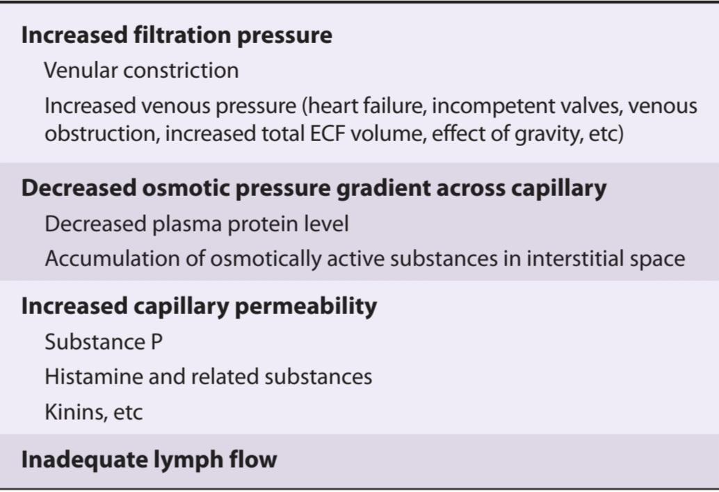
In active tissues, capillary pressure rises, often to the point where it exceeds the oncotic pressure throughout the length of the capillary. In addition, osmotically active metabolites may temporarily accumulate in the interstitial fluid because they cannot be washed away as rapidly as they are formed. To the extent that they accumulate, they exert an osmotic effect that decreases the magnitude of the osmotic gradient due to the oncotic pressure. The amount of fluid leaving the capillaries is therefore markedly increased and the amount entering them reduced. Lymph flow is increased, decreasing the degree to which the fluid would otherwise accumulate, but exercising muscle, for example, still increases in volume by as much as 25%.
در بافتهای فعال، فشار مویرگی افزایش مییابد، اغلب تا جایی که از فشار انکوتیک در طول مویرگ بیشتر میشود. علاوه بر این، متابولیت های فعال اسمزی ممکن است به طور موقت در مایع بینابینی تجمع کنند، زیرا نمی توانند به همان سرعتی که تشکیل می شوند شسته شوند. به میزانی که تجمع می کنند، اثر اسمزی اعمال می کنند که به دلیل فشار انکوتیک، میزان گرادیان اسمزی را کاهش می دهد. بنابراین مقدار مایعی که از مویرگ ها خارج می شود به طور قابل توجهی افزایش یافته و مقدار ورودی به آنها کاهش می یابد. جریان لنف افزایش مییابد و میزان تجمع مایع را کاهش میدهد، اما به عنوان مثال، تمرین ماهیچهها همچنان حجم آن تا ۲۵ درصد افزایش مییابد.
Interstitial fluid tends to accumulate in dependent parts because of the effect of gravity. In the upright position, the capillaries in the legs are protected from the high arterial pressure by the arterioles, but the high venous pressure is transmitted to them through the venules. Skeletal muscle contractions keep the venous pressure low by pumping blood toward the heart (see above) when the individual moves about; however, if one stands still for long periods, fluid accumulates and edema eventually develops. The ankles also swell during long trips when travelers sit for prolonged periods with their feet in a dependent position. Whenever there is abnormal retention of salt in the body, water is also retained. The salt and water are distributed throughout the ECF, and since the interstitial fluid volume is therefore increased, there is a predisposition to edema. Salt and water retention is a factor in the edema seen in heart failure, nephrosis, and cirrhosis, but there are also variations in the mechanisms that govern fluid movement across the capillary walls in these diseases. In heart failure, for example, venous pressure is usually elevated, with a consequent elevation in capillary pressure. In cirrhosis of the liver, oncotic pressure is low because hepatic synthesis of plasma proteins is depressed; and in nephrosis, oncotic pressure is low because large amounts of protein are lost in the urine.
مایع بینابینی به دلیل اثر گرانش تمایل به تجمع در قسمت های وابسته دارد. در حالت عمودی، مویرگ های پاها از فشار شریانی بالا توسط شریان ها محافظت می شوند، اما فشار بالای وریدی از طریق وریدها به آنها منتقل می شود. انقباضات عضلانی اسکلتی با پمپاژ خون به سمت قلب، فشار وریدی را پایین نگه میدارد (به بالا مراجعه کنید) زمانی که فرد حرکت میکند. با این حال، اگر فرد برای مدت طولانی بی حرکت بماند، مایع جمع می شود و در نهایت ادم ایجاد می شود. همچنین مچ پاها در سفرهای طولانی زمانی که مسافران برای مدت طولانی می نشینند و پاهای خود را در وضعیت وابسته قرار می دهند، متورم می شوند. هر گاه احتباس غیرطبیعی نمک در بدن وجود داشته باشد، آب نیز حفظ می شود. نمک و آب در سرتاسر ECF توزیع میشوند و از آنجایی که حجم مایع بینابینی افزایش مییابد، مستعد ادم است. احتباس نمک و آب عاملی در بروز ادم است که در نارسایی قلبی، نفروز و سیروز مشاهده می شود، اما در این بیماری ها مکانیسم هایی نیز وجود دارد که بر حرکت مایع در طول دیواره های مویرگ ها نظارت می کند. به عنوان مثال، در نارسایی قلبی، فشار وریدی معمولاً افزایش مییابد و در نتیجه فشار مویرگی افزایش مییابد. در سیروز کبد، فشار انکوتیک پایین است زیرا سنتز کبدی پروتئین های پلاسما کاهش می یابد. و در نفروز فشار انکوتیک پایین است زیرا مقادیر زیادی پروتئین در ادرار از بین می رود.
Another cause of edema is inadequate lymphatic drainage. Edema caused by lymphatic obstruction is called lymphedema, and the edema fluid has a high protein content. If it persists, it causes a chronic inflammatory condition that can lead to fibrosis of the interstitial tissue. One cause of lymphedema is radical mastectomy, during which removal of the axillary lymph nodes leads to reduced lymph drainage.
یکی دیگر از دلایل ادم، تخلیه ناکافی لنفاوی است. ادم ناشی از انسداد لنفاوی لنف ادم نامیده می شود و مایع ادم دارای محتوای پروتئین بالایی است. اگر ادامه یابد، باعث ایجاد یک بیماری التهابی مزمن می شود که می تواند منجر به فیبروز بافت بینابینی شود. یکی از علل لنف ادم ماستکتومی رادیکال است که طی آن برداشتن غدد لنفاوی زیر بغل منجر به کاهش تخلیه لنفاوی می شود.
CHAPTER SUMMARY
خلاصه فصل
• Blood consists of a suspension of red blood cells (erythrocytes), white blood cells, and platelets. The cells arise in the bone marrow and are subject to regular renewal. Mutations in proteins that make up the cytoskeleton of the red cells make them more fragile and susceptible to lysis.
• خون از تعلیق گلبول های قرمز (گلبول های قرمز)، گلبول های سفید و پلاکت ها تشکیل شده است. سلول ها در مغز استخوان ایجاد می شوند و به طور منظم تجدید می شوند. جهش در پروتئینهایی که اسکلت سلولی گلبولهای قرمز را تشکیل میدهند، آنها را شکنندهتر و حساستر به لیز میکند.
• Hemoglobin, stored in red blood cells, transports oxygen to peripheral tissues. Fetal hemoglobin is specialized to facilitate diffusion of oxygen from mother to fetus during development. Mutated forms of hemoglobin lead to red cell abnormalities and anemia.
• هموگلوبین ذخیره شده در گلبول های قرمز خون، اکسیژن را به بافت های محیطی منتقل می کند. هموگلوبین جنینی برای تسهیل انتشار اکسیژن از مادر به جنین در طول رشد تخصصی است. اشکال جهش یافته هموگلوبین منجر به ناهنجاری های گلبول قرمز و کم خونی می شود.
• The blood cells are suspended in a protein-rich fluid known as plasma. The majority of plasma proteins are synthesized by the liver. Lymph is an exudate of plasma with lower protein concentration that serves to recover fluids from the tissues.
• سلول های خونی در مایع غنی از پروتئین به نام پلاسما معلق هستند. اکثر پروتئین های پلاسما توسط کبد سنتز می شوند. لنف ترشحی از پلاسما با غلظت پروتئین کمتر است که برای بازیابی مایعات از بافت ها عمل می کند.
• Complex oligosaccharide structures on the membrane of red blood cells, specific to groups of individuals, form the basis of the ABO blood group system. AB blood group oligosaccharides, as well as other blood group molecules, can trigger the production of antibodies in naïve individuals following inappropriate transfusions, with potentially serious consequences due to erythrocyte agglutination.
• ساختارهای پیچیده الیگوساکارید روی غشای گلبول های قرمز، مخصوص گروه های افراد، اساس سیستم گروه خونی ABO را تشکیل می دهند. الیگوساکاریدهای گروه خونی AB، و همچنین سایر مولکولهای گروه خونی، میتوانند باعث تولید آنتیبادیها در افراد ساده به دنبال تزریقهای نامناسب شوند، با عواقب بالقوه جدی ناشی از آگلوتیناسیون گلبولهای قرمز.
• When a blood vessel is injured, a complex cascade of molecular interactions among clotting factors ensures that the vessel is sealed and blood loss is minimized (hemostasis). Loss of specific clotting factors may compromise this process and lead to excessive bleeding and bruising. Abnormal clotting that occurs inside an intact vessel is called thrombosis and may occlude blood supply to vital regions.
• هنگامی که یک رگ خونی آسیب می بیند، یک آبشار پیچیده از فعل و انفعالات مولکولی بین فاکتورهای انعقادی تضمین می کند که رگ مهر و موم شده و از دست دادن خون به حداقل می رسد (هموستاز). از دست دادن عوامل انعقادی خاص ممکن است این فرآیند را به خطر بیندازد و منجر به خونریزی و کبودی بیش از حد شود. لخته شدن غیرطبیعی که در داخل یک رگ دست نخورده رخ می دهد، ترومبوز نامیده می شود و ممکن است خون رسانی به نواحی حیاتی را مسدود کند.
• Blood flows from the heart to arteries and arterioles, thence to capillaries, and eventually to venules and veins and back to the heart. Each segment of the vasculature has specific contractile properties and regulatory mechanisms that subserve its physiologic functions. Transfer of oxygen and nutrients from the blood to tissues, as well as collection of metabolic wastes, occurs exclusively in the capillary beds.
• خون از قلب به شریان ها و شریان ها و از آنجا به مویرگ ها و در نهایت به وریدها و سیاهرگ ها و برگشت به قلب جریان می یابد. هر بخش از عروق دارای خواص انقباضی و مکانیسم های تنظیمی خاصی است که عملکردهای فیزیولوژیکی آن را تامین می کند. انتقال اکسیژن و مواد مغذی از خون به بافت ها و همچنین جمع آوری ضایعات متابولیک منحصراً در بسترهای مویرگی انجام می شود.
• Fluid also leaves the circulation across the walls of capillaries. Some is reabsorbed; the remainder enters the lymphatic system, which eventually drains into the subclavian veins to return fluid to the bloodstream.
• مایع همچنین از گردش خون در دیواره های مویرگ ها خارج می شود. مقداری دوباره جذب می شود. باقی مانده وارد سیستم لنفاوی می شود که در نهایت به وریدهای ساب ترقوه تخلیه می شود تا مایع را به جریان خون بازگرداند.
• Physical principles of pressure, wall tension, and vessel caliber govern the flow of blood through each segment of the circulation. These same physical principles can be applied to measure blood flow and blood pressure.
• اصول فیزیکی فشار، کشش دیواره و کالیبر رگ بر جریان خون در هر بخش از گردش خون حاکم است. همین اصول فیزیکی را می توان برای اندازه گیری جریان خون و فشار خون به کار برد.
• Hypertension is an increase in mean blood pressure that is usually chronic and is common in humans. Hypertension can result in serious health consequences if left untreated. The majority of hypertension is of unknown cause, but several gene mutations underlie rare forms of the disease and are informative about mechanisms that control the dynamics of the circulatory system.
• فشار خون بالا افزایش میانگین فشار خون است که معمولا مزمن است و در انسان شایع است. فشار خون بالا در صورت عدم درمان می تواند منجر به عواقب جدی برای سلامتی شود. اکثر فشار خون بالا علت ناشناخته ای دارد، اما چندین جهش ژنی زمینه ساز اشکال نادر این بیماری است و در مورد مکانیسم هایی که دینامیک سیستم گردش خون را کنترل می کند، اطلاعاتی را ارائه می دهد.
MULTIPLE-CHOICE QUESTIONS
For all questions, select the single best answer unless otherwise directed.
سوالات چند گزینه ای
برای همه سؤالات، بهترین پاسخ را انتخاب کنید، مگر اینکه دستور دیگری داده شود.
1. A 2-year-old-African-American boy is brought to his pediatrician because of acute fever, apparent bone pain, and painful swelling of his hands and feet. A physical examination reveals splenomegaly and a blood test reveals the presence of hemoglobin S. After the acute symptoms subside with rest, hydroxyurea is prescribed. This drug would be expected to reduce the risk of further acute symptoms by which mechanism?
A. Increased synthesis of hemoglobin S
B. Increasing the oxygen-carrying capacity of hemoglobin
C. Increasing expression of hypoxia-inducible factor 1 alpha
D. Decreasing the oxygen-carrying capacity of hemoglobin
E. Increasing the synthesis of hemoglobin F
1. یک پسر 2 ساله آفریقایی-آمریکایی به دلیل تب حاد، درد ظاهری استخوان و تورم دردناک دست و پا نزد پزشک اطفال خود آورده شده است. معاینه فیزیکی اسپلنومگالی را نشان می دهد و آزمایش خون وجود هموگلوبین S را نشان می دهد. پس از کاهش علائم حاد با استراحت، هیدروکسی اوره تجویز می شود. انتظار می رود این دارو با کدام مکانیسم خطر بروز علائم حاد بیشتر را کاهش دهد؟
الف. افزایش سنتز هموگلوبین S
ب. افزایش ظرفیت حمل اکسیژن هموگلوبین
ج. افزایش بیان عامل القا کننده هیپوکسی 1 آلفا
د. کاهش ظرفیت حمل اکسیژن هموگلوبین
ی. افزایش سنتز هموگلوبین F
2. A patient is brought to the emergency room suffering from severe blood loss following a motor vehicle accident. Because of a natural disaster, the lab is closed and it is not possible to determine the patient’s blood type before initiating a blood transfusion to save his life. However, banked blood is available. Blood of which type would be least likely to provoke a dangerous hemolytic transfusion reaction?
A. A
B. B
C. AB
D. O
E. A mixture of A and B
2. بیمار به اورژانس آورده می شود که دچار از دست دادن خون شدید به دنبال تصادف وسیله نقلیه موتوری است. به دلیل یک بلای طبیعی، آزمایشگاه بسته است و نمیتوان گروه خونی بیمار را قبل از شروع تزریق خون برای نجات جان او تعیین کرد. با این حال، خون بانکی موجود است. خون از کدام نوع کمتر احتمال دارد که واکنش خطرناک همولیتیک را تحریک کند
الف. A
ب. B
ج. AB
د. O
ی. مخلوطی از A و B
3. Which of the following is not required for hemostasis following injury to a blood vessel?
A. Platelets
B. 5-Hydroxytryptamine
C. Nitric oxide
D. Collagen
E. Fibrin monomers
3. کدام یک از موارد زیر برای هموستاز بدنبال آسیب رگ خونی لازم نیست؟
الف. پلاکت ها
ب. 5-هیدروکسی تریپتامین
ج. اکسید نیتریک
د. کلاژن
ی. مونومرهای فیبرین
4. A pharmacologist discovers a drug that stimulates the production of VEGF receptors. He is excited because the drug might be of value in the treatment of
A. coronary artery disease.
B. cancer.
C. emphysema.
D. diabetes insipidus.
E. dysmenorrhea.
4. یک فارماکولوژیست دارویی را کشف می کند که تولید گیرنده های VEGF را تحریک می کند. او هیجان زده است زیرا این دارو ممکن است در درمان با ارزش باشد
الف. بیماری عروق کرونر.
ب. سرطان.
ج. آمفیزم.
د. دیابت بی مزه.
ی. دیسمنوره.
5. Which of the following has the highest total cross-sectional area in the body?
A. Arteries
B. Arterioles
C. Capillaries
D. Venules
E. Veins
5. کدام یک از موارد زیر دارای بیشترین سطح مقطع کل در بدنه است؟
الف. شریان ها
ب. شریان ها
ج. مویرگها
د. ونول ها
ی. رگها
6. The velocity of blood flow
A. is higher in the capillaries than the arterioles.
B. is higher in the veins than in the venules.
C. is higher in the veins than the arteries.
D. falls to zero in the descending aorta during diastole.
E. is reduced in a constricted area of a blood vessel.
6. سرعت جریان خون
الف. در مویرگ ها بالاتر از شریان ها است.
ب. در وریدها بیشتر از وریدها است.
ج. در سیاهرگ ها بالاتر از شریان ها است.
د. در طول دیاستول در آئورت نزولی به صفر می رسد.
ی. در ناحیه منقبض رگ خونی کاهش می یابد.
7. When the radius of the resistance vessels is increased, which of the following is increased?
A. Systolic blood pressure
B. Diastolic blood pressure
C. Viscosity of the blood
D. Hematocrit
E. Capillary blood flow
7. هنگامی که شعاع مخازن مقاومت افزایش می یابد، کدام یک از موارد زیر افزایش می یابد؟
الف. فشار خون سیستولیک
ب. فشار خون دیاستولیک
ج. ویسکوزیته خون
د. هماتوکریت
ی. جریان خون مویرگی
8. The pressure in a capillary in skeletal muscle is 35 mm Hg at the arteriolar end and 14 mm Hg at the venular end. The interstitial pressure is 0 mm Hg. The colloid osmotic pressure is 25 mm Hg in the capillary and 1 mm Hg in the interstitium. The net force producing fluid movement across the capillary wall at its arteriolar end is
A. 3 mm Hg out of the capillary.
B. 3 mm Hg into the capillary.
C. 10 mm Hg out of the capillary.
D. 11 mm Hg out of the capillary.
E. 11 mm Hg into the capillary.
8. فشار در مویرگ در عضله اسکلتی 35 میلی متر جیوه در انتهای شریانی و 14 میلی متر جیوه در انتهای شریانی است. فشار بینابینی 0 میلی متر جیوه است. فشار اسمزی کلوئیدی در مویرگ 25 میلی متر جیوه و در بینابینی 1 میلی متر جیوه است. نیروی خالص تولید کننده حرکت سیال در سراسر دیواره مویرگی در انتهای شریانی آن است
الف. 3 میلی متر جیوه از مویرگ خارج شود.
ب. 3 میلی متر جیوه به مویرگ.
ج. 10 میلی متر جیوه از مویرگ خارج شود.
د. 11 میلی متر جیوه خارج از مویرگ.
ی. 11 میلی متر جیوه به مویرگ.
9. A 40-year-old man with long-standing diabetes comes to his primary care physician complaining of swelling in his feet and ankles that is so bad that he cannot don his shoes. He states that his urine has developed a foamy appearance and tests reveal that his blood urea nitrogen is decreased. The swelling in this patient is most likely primarily caused by a reduction in which of the following:
A. Lymph flow
B. Barrier function of the capillaries
C. Venous hydrostatic pressure
D. Capillary oncotic pressure
E. Interstitial oncotic pressure
9. مرد 40 ساله مبتلا به دیابت طولانی مدت با شکایت از تورم در پاها و مچ پا که به حدی بد است که نمی تواند کفش هایش را بپوشد به پزشک مراقبت های اولیه مراجعه می کند. او می گوید که ادرارش کف کرده است. ظاهر و آزمایشات نشان می دهد که نیتروژن اوره خون وی کاهش یافته است. تورم در این بیمار به احتمال زیاد عمدتاً ناشی از کاهش یکی از موارد زیر است:
الف. جریان لنفاوی
ب. عملکرد مانع مویرگها
ج. فشار هیدرواستاتیک وریدی
د. فشار انکوتیک مویرگی
ی. فشار انکوتیک بینابینی
10. A 30-year-old patient comes to her primary care clinician complaining of headaches and vertigo. A blood test reveals a hematocrit of 65%, and a diagnosis of polycythemia is made. Which of the following would also be increased?
A. Mean blood pressure
B. Radius of the resistance vessels
C. Radius of the capacitance vessels
D. Central venous pressure
E. Capillary blood flow
10. یک بیمار 30 ساله با شکایت از سردرد و سرگیجه به پزشک مراقبت های اولیه مراجعه می کند. آزمایش خون هماتوکریت 65% را نشان می دهد و تشخیص پلی سیتمی داده می شود. کدام یک از موارد زیر نیز افزایش می یابد؟
الف. فشار خون متوسط
ب. شعاع عروق مقاومت
ج. شعاع مخازن ظرفیت
د. فشار ورید مرکزی
ی. جریان خون مویرگی
11. Lymph flow from the foot is
A. increased when an individual rises from the supine to the standing position.
B. increased by massaging the foot.
C. increased when capillary permeability is decreased.
D. decreased when the valves of the leg veins are incompetent.
E. decreased by exercise.
11. جریان لنفاوی از پا است
الف. هنگامی که فرد از حالت خوابیده به پشت به حالت ایستاده بلند می شود افزایش می یابد.
ب. با ماساژ پا افزایش یافت.
ج. با کاهش نفوذپذیری مویرگی افزایش می یابد.
د. زمانی که دریچه های وریدهای پا ناتوان هستند کاهش می یابد.
ی. با ورزش کاهش یافت.
12. A 60-year-old woman is evaluated for a new finding of chest pain on mild exertion and is incidentally found to be suffering from primary hypertension. Evaluation of her coronary arteries reveals only minimal obstruction. This patient most likely suffers from angina on mild exertion despite only modest coronary artery disease because, compared with a normotensive individual, which of the following would be increased?
A. Oxygen supply to the coronary muscle
B. The size of heart
C. The risk of stroke
D. The propensity to thrombosis
E. Nitric oxide production by the vascular endothelium
12. یک زن 60 ساله از نظر یافته جدیدی از درد قفسه سینه در هنگام فعالیت خفیف مورد ارزیابی قرار می گیرد و اتفاقاً مشخص می شود که از فشار خون اولیه رنج می برد. ارزیابی عروق کرونر او فقط حداقل انسداد را نشان می دهد. این بیمار به احتمال زیاد با فعالیت خفیف با وجود بیماری خفیف عروق کرونر از آنژین رنج می برد زیرا در مقایسه با یک فرد با فشار خون نرمال، کدام یک از موارد زیر افزایش می یابد؟
الف. اکسیژن رسانی به عضله کرونری
ب. اندازه قلب
ج. خطر سکته مغزی
د. تمایل به ترومبوز
ی. تولید اکسید نیتریک توسط اندوتلیوم عروقی
کلیک کنید «منابع»
🚀 با ما همراه شوید!
تازهترین مطالب و آموزشهای مغز و اعصاب را از دست ندهید. با فالو کردن کانال تلگرام آیندهنگاران مغز، از ما حمایت کنید!



