مبانی علوم اعصاب اریک کندل؛ مسیرهای آوران اطلاعات درد؛ چگونگی درک درد


دعای مطالعه [ نمایش ]
بِسْمِ الله الرَّحْمنِ الرَّحیمِ
اَللّهُمَّ اَخْرِجْنى مِنْ ظُلُماتِ الْوَهْمِ
خدايا مرا بيرون آور از تاريكىهاى وهم،
وَ اَكْرِمْنى بِنُورِ الْفَهْمِ
و به نور فهم گرامى ام بدار،
اَللّهُمَّ افْتَحْ عَلَيْنا اَبْوابَ رَحْمَتِكَ
خدايا درهاى رحمتت را به روى ما بگشا،
وَانْشُرْ عَلَيْنا خَزائِنَ عُلُومِكَ بِرَحْمَتِكَ يا اَرْحَمَ الرّاحِمينَ
و خزانههاى علومت را بر ما باز كن به امید رحمتت اى مهربانترين مهربانان.
«مبانی علوم اعصاب» اثر دکتر اریک کندل و همکاران، یکی از جامعترین و معتبرترین منابع در حوزه علوم اعصاب محسوب میشود.
ترجمه دقیق این کتاب، با مدیریت داریوش طاهری و تحت نظارت برند علمی آیندهنگاران مغز، با الهام از تلاشهای علمی و تدریس اساتیدی همچون دکتر محمدتقی جغتایی و دکتر ثریا مهرابی، به فارسی برگردانده شده است.
این ترجمه، دسترسی به دانش عمیق علوم اعصاب را برای دانشجویان و پژوهشگران رشتههای پزشکی، علوم اعصاب، روانشناسی، علوم شناختی، هوش مصنوعی، گفتاردرمانی، مهندسی پزشکی و تمام علاقهمندان به درک عملکرد ذهن و مغز فراهم کرده و گامی در راستای ارتقای دانش و بهبود کیفیت زندگی است.
» کتاب مبانی علوم اعصاب اریک کندل
» » فصل ۲۰: فصل درد؛ قسمت سوم
در حال ویرایش
Principles of Neural Science; Eric R. Kandel
»» Pain
Four Major Ascending Pathways Convey Nociceptive Information From the Spinal Cord to the Brain
چهار مسیر صعودی اصلی، اطلاعات درد را از نخاع به مغز منتقل میکنند
Several Thalamic Nuclei Relay Nociceptive Information to the Cerebral Cortex
چندین هسته تالاموس اطلاعات درد را به قشر مغز منتقل میکنند
The Perception of Pain Arises From and Can Be Controlled by Cortical Mechanisms
درک درد از مکانیسمهای قشر مغز ناشی میشود و میتواند توسط آنها کنترل شود
Anterior cingulate and Insular Cortex Are Associated With the Perception of Pain
سینگولیت قدامی و قشر اینسولا با درک درد مرتبط هستند
Pain Perception Is Regulated by a Balance of Activity in Nociceptive and Nonnociceptive Afferent Fibers
درک درد توسط تعادل فعالیت در فیبرهای آوران دردزا و غیر دردزا تنظیم میشود
Electrical Stimulation of the Brain Produces Analgesia
تحریک الکتریکی مغز باعث ایجاد بیدردی میشود
Four Major Ascending Pathways Convey Nociceptive Information From the Spinal Cord to the Brain
چهار مسیر صعودی اصلی، اطلاعات درد را از نخاع به مغز منتقل میکنند
Four major ascending pathways-the spinothalamic, spinoreticular, spinoparabrachial, and spinohypothalamic tracts contribute sensory information to the central processes that generate pain.
چهار مسیر صعودی اصلی – مسیرهای اسپینوتالامیک، اسپینورتیکولار، اسپینوپارابراکیال و اسپینوهیپوتالامیک – اطلاعات حسی را به فرآیندهای مرکزی تولیدکننده درد ارائه میدهند.
The spinothalamic tract is the most prominent ascending nociceptive pathway in the spinal cord. It includes the axons of nociceptive-specific, thermosensitive, and wide-dynamic-range neurons in laminae I and V through VII of the dorsal horn. These axons cross the midline of the spinal cord near their segment of origin and ascend in the anterolateral white matter before terminating in thalamic nuclei (Figure 20–13). The spinothalamic tract has a crucial role in the transmission of nociceptive information. Cells at the origin of this tract typically have discrete, unilateral receptive fields that underlie our ability to localize painful stimuli. Not surprisingly, electrical stimulation of the tract is sufficient to elicit the sensation of pain; conversely, lesioning this tract (anterolateral cordotomy), a procedure that is generally only used for intractable pain in terminal cancer patients, can result in a marked reduction in pain sensation on the side of the body contralateral to that of the lesion.
مسیر اسپینوتالامیک برجستهترین مسیر صعودی درد در نخاع است. این مسیر شامل آکسونهای نورونهای حساس به درد، حساس به دما و با دامنه دینامیکی وسیع در لامینهای I و V تا VII شاخ خلفی است. این آکسونها از خط میانی نخاع در نزدیکی قطعه مبدا خود عبور میکنند و قبل از پایان یافتن در هستههای تالاموس، در ماده سفید قدامی-جانبی صعود میکنند (شکل 20-13). مسیر اسپینوتالامیک نقش مهمی در انتقال اطلاعات درد دارد. سلولهای مبدا این مسیر معمولاً دارای میدانهای گیرنده مجزا و یک طرفه هستند که زمینهساز توانایی ما در مکانیابی محرکهای دردناک هستند. جای تعجب نیست که تحریک الکتریکی این مسیر برای ایجاد احساس درد کافی است. برعکس، آسیب رساندن به این مسیر (کوردوتومی قدامی-جانبی)، روشی که عموماً فقط برای دردهای غیرقابل تحمل در بیماران سرطانی لاعلاج استفاده میشود، میتواند منجر به کاهش قابل توجه احساس درد در سمت مقابل ضایعه در بدن شود.
The spinoreticular tract contains the axons of projection neurons in laminae VII and VIII. This tract ascends in the anterolateral quadrant of the spinal cord with spinothalamic tract axons, and terminates in both the reticular formation and the thalamus. As neurons at the origin of the spinoreticular tract generally have large, often bilateral receptive fields, this pathway has been implicated more in the processing of diffuse, poorly localized pains.
راه نخاعی-شبکهای شامل آکسونهای نورونهای برآمدگی در لامینای VII و VIII است. این راه در ربع قدامی-جانبی نخاع با آکسونهای راه نخاعی-تالاموسی بالا میرود و در تشکیلات مشبک و تالاموس خاتمه مییابد. از آنجایی که نورونهای مبدأ راه نخاعی-شبکهای عموماً میدانهای گیرنده بزرگ و اغلب دو طرفه دارند، این مسیر بیشتر در پردازش دردهای منتشر و با موضعیت ضعیف نقش دارد.
The spinoparabrachial tract contains the axons of projection neurons in laminae I and V. Information transmitted along this tract is thought to contribute to the affective component of pain. This tract projects in the anterolateral quadrant of the spinal cord to the parabrachial nucleus at the level of the pons (Figure 20-13). This pathway has extensive collaterals to the mesencephalic reticular formation and periaqueductal gray matter. Parabrachial neurons project to the amygdala, a critical nucleus of the limbic system, which regulates emotional states (Chapter 42).
راه نخاعی-پارابراکیال حاوی آکسونهای نورونهای برآمدگی در لامینای I و V است. تصور میشود اطلاعات منتقل شده در امتداد این راه در مؤلفه عاطفی درد نقش دارند. این راه در ربع قدامی-جانبی نخاع به هسته پارابراکیال در سطح پل مغزی امتداد مییابد (شکل 20-13). این مسیر دارای مسیرهای جانبی گستردهای به تشکیلات مشبک مزانسفال و ماده خاکستری اطراف مجرا است. نورونهای پارابراکیال به آمیگدال، هسته حیاتی سیستم لیمبیک که حالات عاطفی را تنظیم میکند، امتداد مییابند (فصل 42).
The spinohypothalamic tract contains the axons of neurons found in spinal cord laminae I, V, VII, and VIII. These axons project to hypothalamic nuclei that serve as autonomic control centers involved in the regulation of the neuroendocrine and cardiovascular responses that accompany pain syndromes (Chapter 41).
راه اسپینوهیپوتالاموس حاوی آکسونهای نورونهای موجود در تیغههای نخاعی I، V، VII و VIII است. این آکسونها به هستههای هیپوتالاموس امتداد مییابند که به عنوان مراکز کنترل خودکار در تنظیم پاسخهای عصبی-غددی و قلبی عروقی همراه با سندرمهای درد نقش دارند (فصل ۴۱).
Several Thalamic Nuclei Relay Nociceptive Information to the Cerebral Cortex
چندین هسته تالاموس اطلاعات درد را به قشر مغز منتقل میکنند
The thalamus contains several relay nuclei that participate in the central processing of nociceptive information. Two of the most important regions of the thalamus are the lateral and medial nuclear groups. The lateral nuclear group comprises the ventroposterolateral (VPL), ventroposteromedial (VPM) and posterior/pulvinar nuclei. The VPL and VPM, respectively, receive inputs via the spinothalamic tract from nociception-specific and wide-dynamic-range neurons in laminae I and V of the dorsal horn and via the trigeminothalamic tract from the trigeminal nucleus caudalis, the trigeminal homolog of the dorsal horn that processes nociceptive information from orofacial regions. The lateral thalamus processes information about the precise location of an injury, information usually conveyed to consciousness as acute pain. Consistent with this view, neurons in the lateral thalamic nuclei have small receptive fields, matching those of the presynaptic spinal neurons.
تالاموس حاوی چندین هسته رله است که در پردازش مرکزی اطلاعات درد شرکت میکنند. دو مورد از مهمترین مناطق تالاموس، گروههای هستهای جانبی و میانی هستند. گروه هستهای جانبی شامل هستههای شکمی-خلفی-جانبی (VPL)، شکمی-خلفی-داخلی (VPM) و خلفی/پولوینار است. VPL و VPM به ترتیب از طریق دستگاه اسپینوتالامیک از نورونهای مخصوص درد و با دامنه دینامیکی وسیع در لامینای I و V شاخ پشتی و از طریق دستگاه تریژمینوتالامیک از هسته تریژمینال کودالیس، همولوگ تریژمینال شاخ پشتی که اطلاعات درد را از نواحی دهانی-صورتی پردازش میکند، ورودی دریافت میکنند. تالاموس جانبی اطلاعات مربوط به محل دقیق آسیب را پردازش میکند، اطلاعاتی که معمولاً به صورت درد حاد به هوشیاری منتقل میشوند. مطابق با این دیدگاه، نورونهای موجود در هستههای تالاموس جانبی دارای میدانهای گیرنده کوچکی هستند که با میدانهای گیرنده نورونهای نخاعی پیشسیناپسی مطابقت دارند.

Figure 20-13 Major ascending pathways that transmit nociceptive information. Sensory discriminative features of the pain experience are transmitted from the spinal cord to the ventroposterolateral thalamus via the spinothalamic tract (brown). From there, information is transmitted predominantly to the somatosensory cortex. A second pathway, (the spinoparabrachial tract (red), carries information from the spinal cord to the parabrachial nucleus of the dorsolateral pons. These neurons in turn target limbic forebrain regions, including the insular and anterior cingulate cortex, which process emotional features of the pain experience.
شکل 20-13 مسیرهای صعودی اصلی که اطلاعات درد را منتقل میکنند. ویژگیهای تمایز حسی تجربه درد از نخاع به تالاموس شکمی-خلفی-جانبی از طریق مسیر اسپینوتالامیک (قهوهای) منتقل میشوند. از آنجا، اطلاعات عمدتاً به قشر حسی-تنی منتقل میشوند. مسیر دوم، (راه اسپینوپارابراکیال (قرمز)، اطلاعات را از نخاع به هسته پارابراکیال پل مغزی پشتی-جانبی منتقل میکند. این نورونها به نوبه خود نواحی مغز قدامی لیمبیک، از جمله قشر اینسولار و قشر کمربندی قدامی را هدف قرار میدهند که ویژگیهای عاطفی تجربه درد را پردازش میکنند.
A cerebrovascular infarct that destroys the lateral thalamus can produce a central neuropathic pain condition called the Dejerine-Roussy (thalamic pain) syndrome. Patients with this syndrome experience spontaneous burning pain as well as abnormal sensations (called dysesthesias) contralateral to the infarct. Electrical stimulation of the thalamus can also result in intense pain. In one dramatic clinical case, electrical stimulation of the thalamus rekindled sensations of angina pectoris that were so realistic that the anesthesiologist thought the patient was experiencing a heart attack. This and other clinical observations suggest that in chronic neuropathic pain conditions there is a fundamental change in thalamic and cortical circuitry. This hypothesis is consistent with studies demonstrating that the topographic map of the body in the thalamus and somatosensory cortex is not fixed, but can change with use and disuse. Loss of a limb can lead to shrinking and even disappearance of the cortical representation of the limb. Abnormal reorganization likely contributes to the phantom limb pain (Figure 20-14).
انفارکتوس مغزی-عروقی که تالاموس جانبی را تخریب میکند، میتواند یک بیماری درد نوروپاتیک مرکزی به نام سندرم دژرین-روسی (درد تالاموس) ایجاد کند. بیماران مبتلا به این سندرم، درد سوزشی خودبهخودی و همچنین احساسات غیرطبیعی (به نام دیسستزی) را در سمت مقابل انفارکتوس تجربه میکنند. تحریک الکتریکی تالاموس نیز میتواند منجر به درد شدید شود. در یک مورد بالینی چشمگیر، تحریک الکتریکی تالاموس احساسات آنژین صدری را دوباره برانگیخت که آنقدر واقعی بودند که متخصص بیهوشی فکر کرد بیمار دچار حمله قلبی شده است. این و سایر مشاهدات بالینی نشان میدهد که در شرایط درد مزمن نوروپاتیک، تغییر اساسی در مدار تالاموس و قشر مغز وجود دارد. این فرضیه با مطالعاتی سازگار است که نشان میدهند نقشه توپوگرافی بدن در تالاموس و قشر حسی-پیکری ثابت نیست، بلکه میتواند با استفاده و عدم استفاده تغییر کند. از دست دادن یک اندام میتواند منجر به کوچک شدن و حتی ناپدید شدن نمایش قشری اندام شود. احتمالاً سازماندهی مجدد غیرطبیعی در درد اندام خیالی نقش دارد (شکل 20-14).
The medial nuclear group of the thalamus comprises the medial dorsal and central lateral nucleus of the thalamus and the intralaminar complex. Its major input is from neurons in laminae VII and VIII of the dorsal horn. The pathway to the medial thalamus was the first spinothalamic projection evident in the evolution of mammals and is therefore known as the paleospinothalamic tract. It is also sometimes referred to as the spinoreticulothalamic tract because it includes indirect connections through the reticular formation of the brain stem. The projection from the lateral thalamus to the ventroposterior lateral and medial nuclei is most developed in primates, and thus is termed the neospinothalamic tract. Many neurons in the medial thalamus respond optimally to noxious stimuli and project to many regions of the limbic system, including the anterior cingulate cortex.
گروه هستهای داخلی تالاموس شامل هسته پشتی داخلی و هسته جانبی مرکزی تالاموس و کمپلکس اینترالامینار است. ورودی اصلی آن از نورونهای لامینای VII و VIII شاخ پشتی است. مسیر منتهی به تالاموس داخلی اولین برآمدگی اسپینوتالامیک مشهود در تکامل پستانداران بود و بنابراین به عنوان راه پالئواسپینوتالامیک شناخته میشود. همچنین گاهی اوقات به عنوان راه اسپینورتیکولوتالامیک شناخته میشود زیرا شامل ارتباطات غیرمستقیم از طریق تشکیل مشبک ساقه مغز است. برآمدگی از تالاموس جانبی به هستههای جانبی و داخلی شکمی-خلفی در نخستیها بیشترین تکامل را دارد و بنابراین راه نئواسپینوتالامیک نامیده میشود. بسیاری از نورونها در تالاموس داخلی به طور مطلوب به محرکهای مضر پاسخ میدهند و به بسیاری از مناطق سیستم لیمبیک، از جمله قشر کمربندی قدامی، امتداد مییابند.
The Perception of Pain Arises From and Can Be Controlled by Cortical Mechanisms
درک درد از مکانیسمهای قشر مغز ناشی میشود و میتواند توسط آنها کنترل شود
Anterior cingulate and Insular Cortex Are Associated With the Perception of Pain
سینگولیت قدامی و قشر اینسولا با درک درد مرتبط هستند
Imaging studies now show that no single area of the cortex is responsible for pain perception. Rather, many regions are activated when an individual experiences pain. In the somatosensory cortex, neurons typically have small receptive fields and may not contribute greatly to the diffuse perception of aches and pains that characterize most clinical syndromes. The anterior cingulate gyrus and insular cortex also contain neurons that are activated strongly and selectively by noxious somatosensory stimuli (Box 20-1).

Figure 20-14 Changes in neural activation in phantom limb pain.
شکل 20-14 تغییرات در فعالسازی عصبی در درد اندام خیالی.
A. The domain of cerebral cortex activated by ascending spinal sensory inputs is expanded in patients with phantom limb pain.
الف) دامنه قشر مغز که توسط ورودیهای حسی صعودی نخاعی فعال میشود، در بیماران مبتلا به درد اندام خیالی گسترش مییابد.
B. Functional magnetic resonance imaging (fMRI) of patients with phantom limb pain and healthy controls during a lip-pursing task. In amputees with phantom limb pain, cortical representation of the mouth has extended into the regions of the hand and arm. In amputees without pain, the areas of primary somatosensory and motor cortices that are activated are similar to those in healthy controls (image not shown). (Adapted, with per- mission, from Flor, Nokolajsen, and Jensen 2006. Copyright © 2006 Springer Nature.)
ب. تصویربرداری تشدید مغناطیسی عملکردی (fMRI) از بیماران مبتلا به درد اندام خیالی و افراد سالم در حین انجام یک آزمایش غنچه کردن لب. در افراد قطع عضو شده با درد اندام خیالی، نمایش قشری دهان به نواحی دست و بازو گسترش یافته است. در افراد قطع عضو شده بدون درد، نواحی قشرهای حسی-پیکری اولیه و حرکتی که فعال میشوند مشابه نواحی افراد سالم است (تصویر نشان داده نشده است). (اقتباس شده، با اجازه، از Flor، Nokolajsen، و Jensen 2006. حق چاپ © 2006 Springer Nature.)
The anterior cingulate gyrus is part of the limbic system and is involved in processing emotional states associated with pain. The insular cortex receives direct projections from the thalamus as well as from the amygdala. Neurons in the insular cortex process information about the internal state of the body and contribute to the autonomic component of pain responses. Importantly, neurosurgical procedures that ablate the cingulate cortex or the pathway from the frontal cortex to the cingulate cortex reduce the affective features of pain without eliminating the ability to recognize the intensity and location of the injury. Patients with lesions of the insular cortex present the striking syndrome of asymbolia for pain. They perceive noxious stimuli as painful and can distinguish sharp from dull pain but fail to display appropriate emotional responses. These observations implicate the insular cortex as an area in which the sensory, affective, and cognitive components of pain are integrated.
Box 20-1 Localizing Illusory Pain in the Cerebral Cortex
کادر 20-1 مکانیابی درد خیالی در قشر مغز
Thunberg’s illusion, first demonstrated in 1896, is a strong, often painful heat felt after placing the hand on a grill of alternating warm and cool bars (Figure 20-15A).
توهم تونبرگ، که اولین بار در سال ۱۸۹۶ نشان داده شد، گرمای شدید و اغلب دردناکی است که پس از قرار دادن دست روی صفحهای از میلههای گرم و سرد متناوب احساس میشود (شکل ۲۰-۱۵A).
One hypothesis proposes that this illusory sensation occurs as a result of differential grill responses of two classes of spinothalamic tract neurons, one sensitive to innocuous and another to noxious cold. This finding has led to a model of pain perception based on a central disinhibition or unmasking process in the cerebral cortex. The model predicts perceptual similarities between grill-evoked and cold-evoked pain, a prediction that has been verified psychophysically. The thalamocortical integration of pain and temperature stimuli may explain the burning sensation felt when nociceptors are activated by cold.
یک فرضیه پیشنهاد میکند که این حس وهمآلود در نتیجه پاسخهای متفاوت گریل دو دسته از نورونهای دستگاه اسپینوتالامیک، یکی حساس به سرمای بیضرر و دیگری حساس به سرمای مضر، رخ میدهد. این یافته منجر به مدلی از درک درد بر اساس یک فرآیند بازداریزدایی یا آشکارسازی مرکزی در قشر مغز شده است. این مدل شباهتهای ادراکی بین درد ناشی از گریل و درد ناشی از سرما را پیشبینی میکند، پیشبینیای که از نظر روانفیزیکی تأیید شده است. ادغام تالاموکورتیکال محرکهای درد و دما ممکن است احساس سوزش احساس شده هنگام فعال شدن گیرندههای درد توسط سرما را توضیح دهد.
To identify the anatomical site of the unmasking phenomenon described above, positron emission tomography (PET) was used to compare the cortical areas activated by Thunberg’s grill with those activated by cool, warm, noxious cold, and noxious heat stimuli separately. All thermal stimuli activate the insula and somatosensory cortices. The anterior cingulate cortex is activated by Thunberg’s grill and by noxious heat and cold, but not by discrete warm and cool stimuli (Figure 20-15B).
برای شناسایی محل آناتومیکی پدیدهی آشکارسازی که در بالا توضیح داده شد، از توموگرافی انتشار پوزیترون (PET) برای مقایسهی نواحی قشری فعال شده توسط گریل تونبرگ با نواحی فعال شده توسط محرکهای سرد، گرم، مضر، سرما و مضر، و مضر، گرما به طور جداگانه استفاده شد. همه محرکهای حرارتی، قشرهای اینسولا و حسی-پیکری را فعال میکنند. قشر کمربندی قدامی توسط گریل تونبرگ و توسط گرما و سرمای مضر فعال میشود، اما توسط محرکهای مجزای گرم و سرد فعال نمیشود (شکل 20-15B).

Figure 20-15A Thunberg’s thermal grill. The stimulus surface (20 x 14 cm) is made of 15 sterling silver bars, each 1 cm wide, set approximately 3 mm apart. Underneath each bar are three longitudinally spaced thermoelectric (Peltier) elements (1 cm2), and on top of each bar is a thermocouple. Alternate (even- and odd-numbered) bars can be controlled independently. (Adapted, with permission, from Craig and Bushnell 1994. Copyright © 1994 AAAS.)
شکل 20-15A گریل حرارتی تونبرگ. سطح محرک (20 در 14 سانتیمتر) از 15 میله نقره استرلینگ، هر کدام به عرض 1 سانتیمتر، با فاصله تقریبی 3 میلیمتر از یکدیگر ساخته شده است. در زیر هر میله، سه عنصر ترموالکتریک (پلتیِر) با فاصله طولی (1 سانتیمتر مربع) قرار دارد و در بالای هر میله یک ترموکوپل قرار دارد. میلههای متناوب (زوج و فرد) را میتوان به طور مستقل کنترل کرد. (اقتباس شده، با اجازه، از کریگ و بوشنل 1994. حق چاپ © 1994 AAAS.)

Figure 20-15B Cortical areas activated by Thunberg’s grill. The anterior cingulate and insula regions of the cerebral cortex are activated when the hand is placed on the grill but not when warm and cool stimuli are applied separately. (Reproduced, with permission, from Craig AD, Reiman EM, Evans A, et al. 1996. Functional imaging of an illusion of pain. Nature 384:258-260. Copyright © 1996 Springer Nature.)
شکل 20-15B نواحی قشری فعال شده توسط گریل تونبرگ. نواحی سینگولیت قدامی و اینسولا قشر مغز هنگامی که دست روی گریل قرار میگیرد فعال میشوند اما هنگامی که محرکهای گرم و سرد به طور جداگانه اعمال میشوند، فعال نمیشوند. (با اجازه، از Craig AD, Reiman EM, Evans A, et al. 1996. تصویرسازی عملکردی از یک توهم درد. Nature 384:258-260. حق چاپ © 1996 Springer Nature.)
Pain Perception Is Regulated by a Balance of Activity in Nociceptive and Nonnociceptive Afferent Fibers
درک درد توسط تعادل فعالیت در فیبرهای آوران دردزا و غیر دردزا تنظیم میشود
Many projection neurons in the dorsal horn of the spinal cord respond selectively to noxious inputs, but others receive convergent inputs from both nociceptive and nonnociceptive afferents. The concept that the convergence of sensory inputs onto spinal projection neurons regulates pain processing first emerged in the 1960s.
بسیاری از نورونهای پروجکشن در شاخ پشتی نخاع به طور انتخابی به ورودیهای مضر پاسخ میدهند، اما برخی دیگر ورودیهای همگرا را از هر دو نوع آوران درد و غیر درد دریافت میکنند. این مفهوم که همگرایی ورودیهای حسی به نورونهای پروجکشن نخاعی، پردازش درد را تنظیم میکند، اولین بار در دهه 1960 پدیدار شد.
Ronald Melzack and Patrick Wall proposed that the relative balance of activity in nociceptive and non- nociceptive afferents might influence the transmission and perception of pain. In particular, they proposed that activation of nonnociceptive sensory neurons, by engaging inhibitory interneurons in the dorsal horn, closes a “gate” for afferent transmission of nociceptive signals that can be opened by the activation of nociceptive sensory neurons. In the original and simplest form of this gate-control theory, the interaction between large and small fibers occurred at the first possible site of convergence on projection neurons in the dorsal horn of the spinal cord (Figure 20-16). We now know that such interactions can also occur at many supraspinal relay centers.
رونالد ملزاک و پاتریک وال پیشنهاد کردند که تعادل نسبی فعالیت در آورانهای دردزا و غیر دردزا ممکن است بر انتقال و درک درد تأثیر بگذارد. به طور خاص، آنها پیشنهاد کردند که فعال شدن نورونهای حسی غیر دردزا، با درگیر کردن نورونهای رابط مهاری در شاخ پشتی، “دروازه” انتقال آوران سیگنالهای دردزا را میبندد که میتواند با فعال شدن نورونهای حسی دردزا باز شود. در شکل اصلی و سادهترین این نظریه کنترل دروازه، تعامل بین فیبرهای بزرگ و کوچک در اولین محل ممکن همگرایی روی نورونهای پروجکشن در شاخ پشتی نخاع رخ میدهد (شکل 20-16). اکنون میدانیم که چنین تعاملاتی میتوانند در بسیاری از مراکز رله فوق نخاعی نیز رخ دهند.
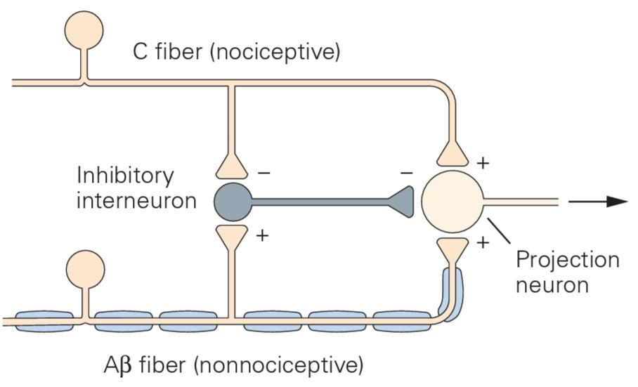
Figure 20-16 The gate control theory of pain. The gate- control hypothesis was proposed in the 1960s to account for the fact that activation of low-threshold primary afferent fibers can attenuate pain. The hypothesis focused on the interaction of neurons in the dorsal horn of the spinal cord: the nociceptive (C) and nonnociceptive (Aβ) sensory neurons, projection neurons, and inhibitory interneurons. In the original version of the model, as shown here, the projection neuron is excited by both classes of sensory neurons and inhibited by interneurons in the superficial dorsal horn. The two classes of sensory fibers also terminate on the inhibitory interneurons; the C fibers indirectly inhibit the interneurons, thus increasing the activity of the projection neurons (thereby “opening the gate”), whereas the AB fibers excite the interneurons, thus suppressing the output of the projection neurons (and “closing the gate”).
شکل 20-16 نظریه کنترل دروازه درد. فرضیه کنترل دروازه در دهه 1960 برای توضیح این واقعیت مطرح شد که فعال شدن فیبرهای آوران اولیه با آستانه پایین میتواند درد را کاهش دهد. این فرضیه بر تعامل نورونها در شاخ پشتی نخاع متمرکز بود: نورونهای حسی دردزا (C) و غیر دردزا (Aβ)، نورونهای برونافکن و نورونهای رابط مهاری. در نسخه اصلی مدل، همانطور که در اینجا نشان داده شده است، نورون برونافکن توسط هر دو دسته نورونهای حسی تحریک و توسط نورونهای رابط در شاخ پشتی سطحی مهار میشود. دو دسته از فیبرهای حسی نیز به نورونهای رابط مهاری ختم میشوند. فیبرهای C به طور غیرمستقیم نورونهای رابط را مهار میکنند و در نتیجه فعالیت نورونهای برونافکن را افزایش میدهند (و در نتیجه “دروازه را باز میکنند”)، در حالی که فیبرهای AB نورونهای رابط را تحریک میکنند و در نتیجه خروجی نورونهای برونافکن را سرکوب میکنند (و “دروازه را میبندند”).
The concept of convergence of different sensory modalities has provided an important basis for the design of new pain therapies. Viewed in its broadest sense, the convergence of high- and low-threshold inputs at spinal or supraspinal sites provided a plausible explanation for several empirical observations about the perception of pain. The shaking of the hand that follows a hammer blow or burn is a reflexive behavior and may alleviate pain by activating large- diameter afferent fibers that suppress the transmission of information about noxious stimuli.
مفهوم همگرایی روشهای حسی مختلف، مبنای مهمی را برای طراحی درمانهای جدید درد فراهم کرده است. در وسیعترین مفهوم خود، همگرایی ورودیهای آستانه بالا و پایین در نواحی نخاعی یا فوق نخاعی، توضیح قابل قبولی برای چندین مشاهده تجربی در مورد درک درد ارائه میدهد. لرزش دست که پس از ضربه چکش یا سوختگی رخ میدهد، یک رفتار انعکاسی است و ممکن است با فعال کردن فیبرهای آوران با قطر بزرگ که انتقال اطلاعات در مورد محرکهای مضر را سرکوب میکنند، درد را کاهش دهد.
The idea of convergence also helped to promote the use of transcutaneous electrical nerve stimulation (TENS) and spinal cord stimulation for the relief of pain. With TENS, stimulating electrodes placed at peripheral locations activate large-diameter afferent fibers that innervate areas that overlap but also sur- round the region of injury and pain. The region of the body in which pain is reduced maps to those segments of the spinal cord in which nociceptive and nonnociceptive afferents from that body region terminate. This makes intuitive sense: You do not shake your left leg to relieve pain in your right arm.
ایده همگرایی همچنین به ترویج استفاده از تحریک الکتریکی عصب از طریق پوست (TENS) و تحریک نخاع برای تسکین درد کمک کرد. با TENS، الکترودهای تحریککننده که در مکانهای محیطی قرار میگیرند، فیبرهای آوران با قطر بزرگ را فعال میکنند که نواحی همپوشانی دارند اما ناحیه آسیب و درد را نیز احاطه کردهاند. ناحیهای از بدن که درد در آن کاهش مییابد، به بخشهایی از نخاع نگاشت میشود که آورانهای درد و غیر درد از آن ناحیه بدن در آنها خاتمه مییابند. این امر به طور شهودی منطقی است: شما پای چپ خود را برای تسکین درد در بازوی راست خود تکان نمیدهید.
Electrical Stimulation of the Brain Produces Analgesia
تحریک الکتریکی مغز باعث ایجاد بیدردی میشود
Several sites of endogenous pain regulation are located in the brain. One effective means of suppressing nociception involves stimulation of the periaqueductal gray region, the area of the midbrain that surrounds the third ventricle and the cerebral aqueduct. In experimental animals, stimulation of this region elicits a profound and selective analgesia. This stimulation-produced analgesia is remarkably modality-specific; animals still respond to touch, pressure, and temperature within the body area that is not sensitive to pain. Stimulation- evoked analgesia has proved to be an effective way of relieving pain in a limited number of human pain conditions.
چندین محل تنظیم درد درونزا در مغز قرار دارند. یکی از روشهای مؤثر برای سرکوب درد، تحریک ناحیه خاکستری اطراف مجرا، ناحیهای از مغز میانی که بطن سوم و مجرای مغزی را احاطه کرده است، میباشد. در حیوانات آزمایشگاهی، تحریک این ناحیه باعث ایجاد بیدردی عمیق و انتخابی میشود. این بیدردی ناشی از تحریک به طور قابل توجهی وابسته به روش است؛ حیوانات هنوز به لمس، فشار و دما در ناحیهای از بدن که به درد حساس نیست، پاسخ میدهند. بیدردی ناشی از تحریک، روشی مؤثر برای تسکین درد در تعداد محدودی از شرایط درد انسان بوده است.
Stimulation of the periaqueductal gray matter blocks spinally mediated withdrawal reflexes that are normally evoked by noxious stimulation. Few of the neurons in the periaqueductal gray matter project directly to the dorsal horn of the spinal cord. Most make excitatory connections with neurons of the rostroventral medulla, including serotonergic neurons in a midline region called the nucleus raphe magnus. The axons of these serotonergic neurons project through the dorsal region of the lateral funiculus to the spinal cord, where they form inhibitory connections with neurons in laminae I, II, and V of the dorsal horn (Figure 20-17). Stimulation of the rostroventral medulla thus inhibits the firing of many classes of dorsal horn neurons, including projection neurons of the major ascending pathways that convey afferent nociceptive signals to the brain.
تحریک ماده خاکستری اطراف قناتی، رفلکسهای عقبنشینی با واسطه نخاعی را که معمولاً توسط تحریک مضر برانگیخته میشوند، مسدود میکند. تعداد کمی از نورونهای ماده خاکستری اطراف قناتی مستقیماً به شاخ پشتی نخاع میروند. اکثر آنها با نورونهای بصل النخاع قدامی، از جمله نورونهای سروتونرژیک در ناحیهای در خط میانی به نام هسته رافه ماگنوس، ارتباط تحریکی برقرار میکنند. آکسونهای این نورونهای سروتونرژیک از طریق ناحیه پشتی طناب جانبی به نخاع میروند، جایی که با نورونهای موجود در لامینای I، II و V شاخ پشتی ارتباط مهاری برقرار میکنند (شکل 20-17). بنابراین تحریک بصل النخاع قدامی، شلیک بسیاری از دستههای نورونهای شاخ پشتی، از جمله نورونهای برآمدگی مسیرهای اصلی صعودی که سیگنالهای درد آوران را به مغز منتقل میکنند، را مهار میکند.

Figure 20-17 Descending monoaminergic pathways regulate nociceptive relay neurons in the spinal cord. A serotonergic pathway arises in the nucleus raphe magnus and projects through the dorsolateral funiculus to the dorsal horn of the spinal cord. A noradrenergic system arises in the locus ceruleus and other nuclei in the pons and medulla. (See Figure 40-11A for the locations and projections of monoaminergic neurons.) In the spinal cord, these descending pathways inhibit nociceptive projection neurons through direct connections as well as through interneurons in the superficial layers of the dorsal horn. Both the serotonergic nucleus raphe magnus and noradrenergic nuclei receive input from neurons in the periaqueductal gray region. Sites of opioid peptide expression and actions of exogenously administered opioids are shown.
شکل 20-17 مسیرهای مونوآمینرژیک نزولی، نورونهای رله درد را در نخاع تنظیم میکنند. یک مسیر سروتونرژیک در هسته رافه ماگنوس ایجاد میشود و از طریق طناب پشتی-جانبی به شاخ پشتی نخاع امتداد مییابد. یک سیستم نورآدرنرژیک در لوکوس سرولئوس و سایر هستهها در پل مغزی و بصل النخاع ایجاد میشود. (برای مکانها و برآمدگیهای نورونهای مونوآمینرژیک به شکل 40-11A مراجعه کنید.) در نخاع، این مسیرهای نزولی، نورونهای پرتابی درد را از طریق اتصالات مستقیم و همچنین از طریق نورونهای رابط در لایههای سطحی شاخ پشتی مهار میکنند. هم هسته سروتونرژیک رافه ماگنوس و هم هستههای نورآدرنرژیک از نورونهای ناحیه خاکستری دور قناتی ورودی دریافت میکنند. محلهای بیان پپتید اوپیوئیدی و عملکرد اوپیوئیدهای تجویز شده خارجی نشان داده شده است.
A second major monoaminergic descending system can also suppress the activity of nociceptive neurons in the dorsal horn. This noradrenergic system originates in the locus ceruleus and other nuclei of the medulla and pons (Figure 20-17). Through direct and indirect synaptic actions, these projections inhibit neurons in laminae I and V of the dorsal horn.
دومین سیستم نزولی مونوآمینرژیک اصلی نیز میتواند فعالیت نورونهای دردزا را در شاخ پشتی سرکوب کند. این سیستم نورآدرنرژیک از لوکوس سرولئوس و سایر هستههای بصل النخاع و پل مغزی منشأ میگیرد (شکل 20-17). این برآمدگیها از طریق اعمال سیناپسی مستقیم و غیرمستقیم، نورونهای موجود در لامینای I و V شاخ پشتی را مهار میکنند.
»» فصل بعد: فصل ماهیت ساختاری پردازش بینایی
»» تمامی کتاب