مبانی علوم اعصاب اریک کندل؛ ماهیت ساختاری پردازش بینایی؛ کدهای عصبی اطلاعات دیداری


دعای مطالعه [ نمایش ]
بِسْمِ الله الرَّحْمنِ الرَّحیمِ
اَللّهُمَّ اَخْرِجْنى مِنْ ظُلُماتِ الْوَهْمِ
خدايا مرا بيرون آور از تاريكىهاى وهم،
وَ اَكْرِمْنى بِنُورِ الْفَهْمِ
و به نور فهم گرامى ام بدار،
اَللّهُمَّ افْتَحْ عَلَيْنا اَبْوابَ رَحْمَتِكَ
خدايا درهاى رحمتت را به روى ما بگشا،
وَانْشُرْ عَلَيْنا خَزائِنَ عُلُومِكَ بِرَحْمَتِكَ يا اَرْحَمَ الرّاحِمينَ
و خزانههاى علومت را بر ما باز كن به امید رحمتت اى مهربانترين مهربانان.
«مبانی علوم اعصاب» اثر دکتر اریک کندل و همکاران، یکی از جامعترین و معتبرترین منابع در حوزه علوم اعصاب محسوب میشود.
ترجمه دقیق این کتاب، با مدیریت داریوش طاهری و تحت نظارت برند علمی آیندهنگاران مغز، با الهام از تلاشهای علمی و تدریس اساتیدی همچون دکتر محمدتقی جغتایی و دکتر ثریا مهرابی، به فارسی برگردانده شده است.
این ترجمه، دسترسی به دانش عمیق علوم اعصاب را برای دانشجویان و پژوهشگران رشتههای پزشکی، علوم اعصاب، روانشناسی، علوم شناختی، هوش مصنوعی، گفتاردرمانی، مهندسی پزشکی و تمام علاقهمندان به درک عملکرد ذهن و مغز فراهم کرده و گامی در راستای ارتقای دانش و بهبود کیفیت زندگی است.
» کتاب مبانی علوم اعصاب اریک کندل
» » فصل ۲۱: فصل ماهیت ساختاری پردازش بینایی؛ قسمت آخر
در حال ویرایش
Principles of Neural Science; Eric R. Kandel
»» Chapter 21: Sensory Coding
The Constructive Nature of Visual Processing
ماهیت سازنده پردازش بصری
Visual Perception Is a Constructive Process
ادراک بصری یک فرآیند سازنده است
Visual Processing Is Mediated by the Geniculostriate Pathway
پردازش بصری توسط مسیر ژنیکولواستریات انجام میشود
Form, Color, Motion, and Depth Are Processed in Discrete Areas of the Cerebral Cortex
فرم، رنگ، حرکت و عمق در نواحی مجزایی از قشر مغز پردازش میشوند
The Receptive Fields of Neurons at Successive Relays in the Visual Pathway Provide Clues to How the Brain Analyzes Visual Form
میدانهای گیرنده نورونها در رلههای متوالی در مسیر بینایی، سرنخهایی از نحوه تجزیه و تحلیل فرم بصری توسط مغز ارائه میدهند
The Visual Cortex Is Organized Into Columns of Specialized Neurons
قشر بینایی به صورت ستونهایی از نورونهای تخصصی سازماندهی شده است
Intrinsic Cortical Circuits Transform Neural Information
مدارهای قشری ذاتی، اطلاعات عصبی را تبدیل میکنند
Visual Information Is Represented by a Variety of Neural Codes
اطلاعات بصری توسط انواع کدهای عصبی نمایش داده میشود
Highlights
نکات برجسته
Intrinsic Cortical Circuits Transform Neural Information
مدارهای قشری ذاتی، اطلاعات عصبی را تبدیل میکنند
Each area of the visual cortex transforms information gathered by the eyes and processed at earlier synaptic relays into a signal that represents the visual scene. This transformation is accomplished by local circuits comprising both excitatory and inhibitory neurons.
هر ناحیه از قشر بینایی، اطلاعات جمعآوریشده توسط چشمها و پردازششده در رلههای سیناپسی قبلی را به سیگنالی تبدیل میکند که صحنه بصری را نشان میدهد. این تبدیل توسط مدارهای موضعی شامل نورونهای تحریکی و مهاری انجام میشود.
The principal input to the primary visual cortex comes from three parallel pathways that originate in the parvocellular, magnocellular, and the blue/ yellow channels of koniocellular layers of the LGN (see Figure 21-12). Neurons in the parvocellular layers project to cortical layers IVCβ and 6, those in the magnocellular layers project to layer IVCα and 6, while the koniocellular neurons project to layer 1 and to the cytochrome oxidase blobs in layers 2 and 3. From there, a sequence of interlaminar connections, mediated by the excitatory spiny stellate neurons, processes visual information over a stereotyped set of connections (Figure 21-15).
ورودی اصلی به قشر بینایی اولیه از سه مسیر موازی ناشی میشود که از کانالهای پارووسلولار، مگنوسلولار و آبی/زرد لایههای کونیوسلولار LGN سرچشمه میگیرند (شکل 21-12 را ببینید). نورونهای لایههای پارووسلولار به لایههای قشری IVCβ و 6، نورونهای لایههای مگنوسلولار به لایههای IVCα و 6، و نورونهای کونیوسلولار به لایه 1 و به حبابهای سیتوکروم اکسیداز در لایههای 2 و 3 متصل میشوند. از آنجا، توالیای از اتصالات بین لایهای، که توسط نورونهای ستارهای خاردار تحریکی واسطهگری میشوند، اطلاعات بصری را از طریق مجموعهای کلیشهای از اتصالات پردازش میکنند (شکل 21-15).
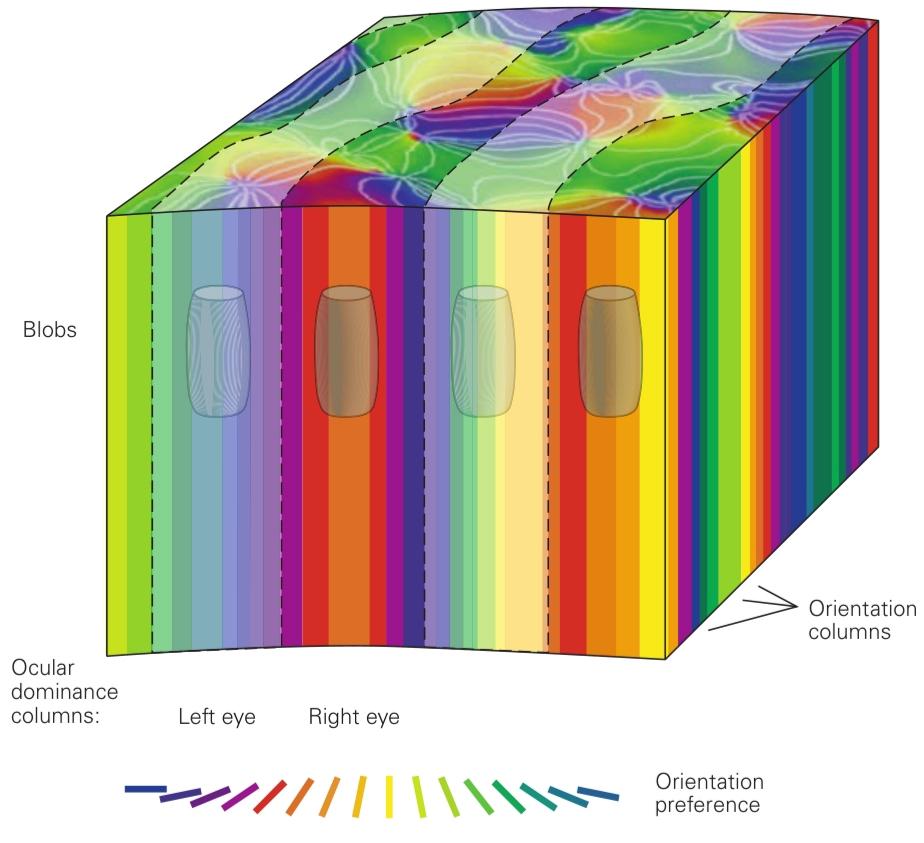
Figure 21-13 A cortical computational module. A chunk of cortical tissue roughly 1 mm square contains an orientation hypercolumn (a full cycle of orientation columns), one cycle of left- and right- eye ocular-dominance columns, and blobs and interblobs. This module would presumably contain all of the functional and anatomical cell types of primary visual cortex, which would be repeated hundreds of times to cover the visual field. (Adapted from Hubel 1988.)
شکل ۲۱-۱۳ یک ماژول محاسباتی قشری. تکهای از بافت قشری تقریباً ۱ میلیمتر مربع شامل یک فوق ستون جهتگیری (یک چرخه کامل از ستونهای جهتگیری)، یک چرخه از ستونهای غالب چشمی چشم چپ و راست، و لکهها و بین لکهها است. این ماژول احتمالاً شامل تمام انواع سلولهای عملکردی و آناتومیکی قشر بینایی اولیه است که صدها بار برای پوشش میدان بینایی تکرار میشوند. (اقتباس از هوبل ۱۹۸۸.)
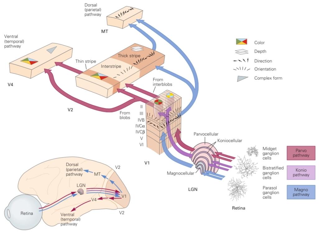
Figure 21-14 Parallel processing in visual pathways. The ventral stream is primarily concerned with object identification, carrying information about form and color. The dorsal pathway is dedicated to visually guided movement, with cells selective for direction of movement. These pathways are not strictly segregated, however, and there is substantial interconnection between them even in the primary visual cortex. (Abbreviations: LGN, lateral geniculate nucleus; MT, middle temporal area.) (Retinal ganglion cell images courtesy of Dennis Dacey, reproduced with permission.)
شکل ۲۱-۱۴ پردازش موازی در مسیرهای بینایی. جریان شکمی در درجه اول مربوط به شناسایی اشیا است و اطلاعاتی در مورد شکل و رنگ را حمل میکند. مسیر پشتی به حرکت هدایتشده بصری اختصاص دارد و سلولهای آن برای جهت حرکت انتخابی هستند. با این حال، این مسیرها کاملاً از هم جدا نیستند و حتی در قشر بینایی اولیه نیز ارتباط متقابل قابل توجهی بین آنها وجود دارد. (اختصارات: LGN، هسته زانویی جانبی؛ MT، ناحیه گیجگاهی میانی.) (تصاویر سلولهای گانگلیون شبکیه با اجازه دنیس دیسی، با اجازه تکثیر شده است.)
This characterization of parallel pathways is only an approximation, as there is considerable interaction between the pathways. This interaction is the means by which various visual features-color, form, depth, and movement—are linked, leading to a unified visual percept. One way this linkage, or binding, may be accomplished is through cells that are tuned to more than one attribute.
این توصیف مسیرهای موازی تنها یک تقریب است، زیرا تعامل قابل توجهی بین مسیرها وجود دارد. این تعامل وسیلهای است که از طریق آن ویژگیهای بصری مختلف – رنگ، شکل، عمق و حرکت – به هم مرتبط میشوند و منجر به یک ادراک بصری یکپارچه میشوند. یکی از راههای انجام این پیوند یا اتصال، از طریق سلولهایی است که به بیش از یک ویژگی تنظیم شدهاند.
At each stage of cortical processing, pyramidal neurons extend output to other brain areas. Superficial-layer cells are responsible for connections to higher-order areas of cortex. Layer V pyramidal neurons project to the superior colliculus and pons in the brain stem. Layer VI cells are responsible for feedback projections, both to the thalamus and to lower-order cortical areas.
در هر مرحله از پردازش قشر مغز، نورونهای هرمی خروجی را به سایر نواحی مغز گسترش میدهند. سلولهای لایه سطحی مسئول ارتباط با نواحی بالاتر قشر مغز هستند. نورونهای هرمی لایه V به کولیکولوس فوقانی و پل مغزی در ساقه مغز منتهی میشوند. سلولهای لایه VI مسئول ارسال بازخورد، هم به تالاموس و هم به نواحی قشری پایینتر هستند.
Neurons in different layers have distinctive receptive- field properties. Neurons in the superficial layer of V1 have small receptive fields, whereas neurons in deeper layers have large ones. The superficial-layer neurons are specialized for high-resolution pattern recognition. Neurons in the deeper layers, such as those in layer V that are selective for the direction of movement, are specialized for the tracking of objects in space.
نورونهای لایههای مختلف، ویژگیهای میدان دریافتی متمایزی دارند. نورونهای لایه سطحی V1 میدانهای دریافتی کوچکی دارند، در حالی که نورونهای لایههای عمیقتر میدانهای دریافتی بزرگی دارند. نورونهای لایه سطحی برای تشخیص الگو با وضوح بالا تخصص یافتهاند. نورونهای لایههای عمیقتر، مانند نورونهای لایه V که برای جهت حرکت انتخابی هستند، برای ردیابی اشیاء در فضا تخصص یافتهاند.

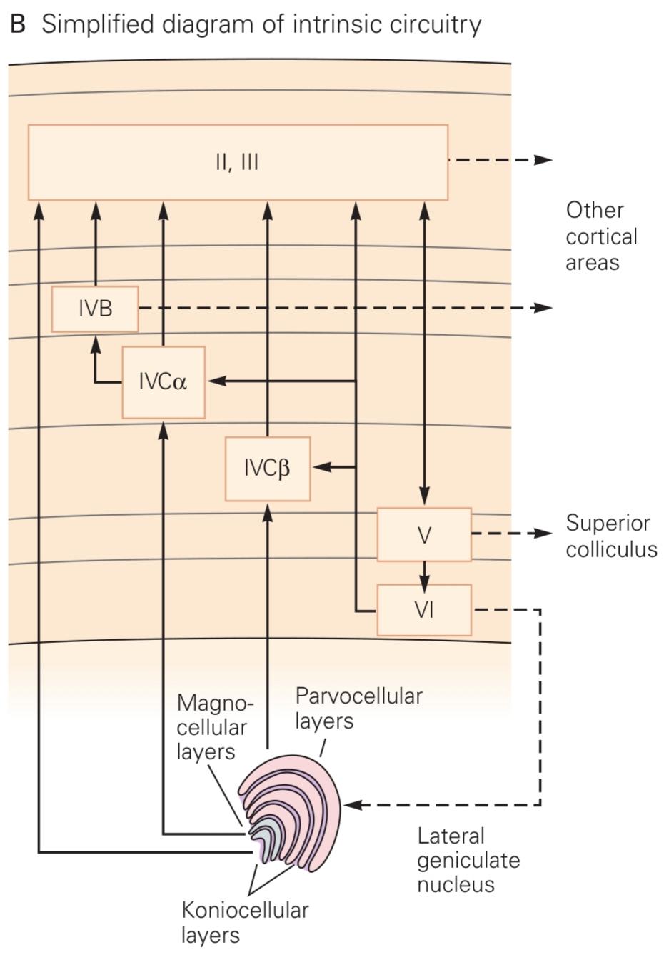
Figure 21-15 The intrinsic circuitry of the primary visual cortex.
شکل 21-15 مدار ذاتی قشر بینایی اولیه.
A. Examples of neurons in different cortical layers responsible for excitatory connections in cortical circuits. Layer IV is the principal layer of input from the lateral geniculate nucleus of the thalamus. Fibers from the parvocellular layer terminate in layer IVCβ, whereas the magnocellular fibers terminate in layer IVCα. The intrinsic cortical excitatory connections are mediated by spiny stellate and pyramidal cells. A variety of γ-aminobutyric acid (GABA)-ergic smooth stellate cells (not shown) are responsible for inhibitory connections. Dendritic arbors are colored blue, and axonal arbors are shown in brown. (Cortical neurons courtesy of E. Callaway, reproduced with permission. Thalamic afferents adapted, with permission, from Blasdel and Lund 1983. Copyright © 1983 Society for Neuroscience.)
الف. نمونههایی از نورونها در لایههای مختلف قشر مغز که مسئول ارتباطات تحریکی در مدارهای قشر مغز هستند. لایه IV لایه اصلی ورودی از هسته زانویی جانبی تالاموس است. فیبرهای لایه پاروسلولار به لایه IVCβ ختم میشوند، در حالی که فیبرهای مگنوسلولار به لایه IVCα ختم میشوند. ارتباطات تحریکی ذاتی قشر مغز توسط سلولهای ستارهای و هرمی خاردار واسطهگری میشوند. انواعی از سلولهای ستارهای صاف γ-آمینوبوتیریک اسید (GABA)-ارژیک (نشان داده نشدهاند) مسئول ارتباطات مهاری هستند. آربورهای دندریتیک به رنگ آبی و آربورهای آکسونی به رنگ قهوهای نشان داده شدهاند. (نورونهای قشر مغز با اجازه E. Callaway، با اجازه تکثیر شدهاند. آورانهای تالاموس با اجازه از Blasdel و Lund 1983 اقتباس شدهاند. حق چاپ © 1983 انجمن علوم اعصاب.)
B. Diagram of excitatory connections within the primary visual cortex. Output to other regions of cortex is sent from every layer of visual cortex.
ب. نمودار ارتباطات تحریکی در قشر بینایی اولیه. خروجی به سایر نواحی قشر از هر لایه قشر بینایی ارسال میشود.

Feedback projections are thought to provide a means whereby higher centers in a pathway can influence lower ones. The number of neurons projecting from the cortex to the LGN is 10-fold the number projecting from the LGN to the cortex. Although this feed- back projection is obviously important, its function is largely unknown.
تصور میشود که بازتابهای بازخوردی وسیلهای را فراهم میکنند که از طریق آن مراکز بالاتر در یک مسیر میتوانند بر مراکز پایینتر تأثیر بگذارند. تعداد نورونهایی که از قشر مغز به LGN منتقل میشوند، ده برابر تعداد نورونهایی است که از LGN به قشر مغز منتقل میشوند. اگرچه این بازتاب بازخوردی آشکارا مهم است، اما عملکرد آن تا حد زیادی ناشناخته است.
The activity of the excitatory pyramidal and spiny stellate neurons that mediate information flow into or out of cortical regions is also tightly controlled by local networks of inhibitory interneurons. The spike rates of excitatory neurons are constantly nonlinearly balanced by matched inhibition that maintains the stability of the neural response to an input. Inhibitory interneurons come in multiple classes distinguished by their morphology and their coexpression of distinct peptides such as parvalbumin, somatostatin, or vasoactive intestinal polypeptide (VIP). Some of these interneurons form cascading circuits where interneurons of one class target interneurons of another class, which then target excitatory neurons. This leads to multistep control mechanisms in the neural circuit whereby increasing activity in the first class of inhibitory interneurons reduces activity in the second class, disinhibiting and increasing responses in the excitatory targets at the end of the cascade. Such motifs of inhibitory control are likely to be common to multiple cortical sensory areas.
فعالیت نورونهای هرمی و ستارهای خاردار تحریکی که واسطه جریان اطلاعات به داخل یا خارج از مناطق قشری هستند، نیز به شدت توسط شبکههای محلی نورونهای رابط مهاری کنترل میشود. نرخهای جهش نورونهای تحریکی دائماً به صورت غیرخطی توسط مهار تطبیقی متعادل میشوند که پایداری پاسخ عصبی به یک ورودی را حفظ میکند. نورونهای رابط مهاری در چندین دسته قرار میگیرند که بر اساس مورفولوژی و بیان همزمان پپتیدهای متمایز مانند پاروالبومین، سوماتوستاتین یا پلیپپتید رودهای وازواکتیو (VIP) از هم متمایز میشوند. برخی از این نورونهای رابط مدارهای آبشاری تشکیل میدهند که در آن نورونهای رابط یک دسته، نورونهای رابط دسته دیگر را هدف قرار میدهند و سپس نورونهای تحریکی را هدف قرار میدهند. این امر منجر به مکانیسمهای کنترل چند مرحلهای در مدار عصبی میشود که در آن افزایش فعالیت در دسته اول نورونهای رابط مهاری، فعالیت در دسته دوم را کاهش میدهد و پاسخها را در اهداف تحریکی در انتهای آبشار مهارزدایی و افزایش میدهد. چنین موتیفهای کنترل مهاری احتمالاً در چندین ناحیه حسی قشری مشترک هستند.
In addition to serial feedforward, feedback, and local recurrent connections, fibers that travel parallel to the cortical surface within each layer provide long- range horizontal connections (Figure 21-16). These connections and their role in the functional architecture of cortex were analyzed by Charles Gilbert and Torsten Wiesel, who used intracellular recordings and dye injection to correlate anatomical features with cortical function. Because the visual cortex is organized visuotopically, the horizontal connections allow target neurons to integrate information over a relatively large area of the visual field and are therefore important in assembling the components of a visual image into a unified percept.
علاوه بر اتصالات سریالیِ پیشخور، پسخور و بازگشتیِ موضعی، فیبرهایی که به موازات سطح قشر مغز در هر لایه حرکت میکنند، اتصالات افقیِ دوربردی را فراهم میکنند (شکل 21-16). این اتصالات و نقش آنها در معماری عملکردی قشر مغز توسط چارلز گیلبرت و تورستن ویزل مورد تجزیه و تحلیل قرار گرفت، که از ثبتهای درون سلولی و تزریق رنگ برای مرتبط کردن ویژگیهای آناتومیکی با عملکرد قشر مغز استفاده کردند. از آنجا که قشر بینایی به صورت بصری-موضعی سازماندهی شده است، اتصالات افقی به نورونهای هدف اجازه میدهند تا اطلاعات را در ناحیه نسبتاً وسیعی از میدان بینایی ادغام کنند و بنابراین در مونتاژ اجزای یک تصویر بصری به یک ادراک واحد مهم هستند.
Integration can also be achieved by other means. The considerable convergence and divergence of connections at the synaptic relays of the afferent visual pathway imply that the receptive fields of neurons are larger and more complex at each successive relay and thus have an integrative function. Feedback connections may also support integration, both because of their divergence and because they originate from cells with larger receptive fields.
ادغام را میتوان از طرق دیگری نیز انجام داد. همگرایی و واگرایی قابل توجه اتصالات در رلههای سیناپسی مسیر بینایی آوران نشان میدهد که میدانهای گیرنده نورونها در هر رله متوالی بزرگتر و پیچیدهتر هستند و بنابراین عملکرد ادغامی دارند. اتصالات بازخوردی نیز ممکن است از ادغام پشتیبانی کنند، هم به دلیل واگرایی آنها و هم به این دلیل که از سلولهایی با میدانهای گیرنده بزرگتر سرچشمه میگیرند.
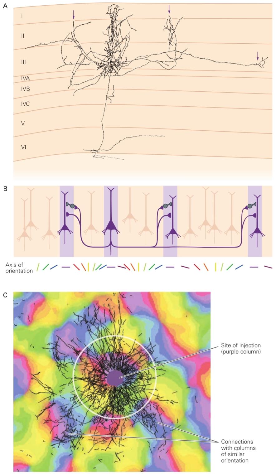
Figure 21-16 Long-range horizontal connections in each layer of the visual cortex integrate information from different parts of the visual field.
شکل ۲۱-۱۶ اتصالات افقی دوربرد در هر لایه از قشر بینایی، اطلاعات را از قسمتهای مختلف میدان بینایی ادغام میکنند.
A. The axons of pyramidal cells extend for many millimeters parallel to the cortical surface. Axon collaterals form connections with other pyramidal cells as well as with inhibitory interneurons. This arrangement enables neurons to integrate information over large parts of the visual field. An important characteristic of these connections is their relationship to the functional columns. The axon collaterals are found in clusters (arrows) at distances greater than 0.5 mm from the cell body. (Reproduced, with permission, from Gilbert and Wiesel 1983. Copyright © 1983 Society for Neuroscience.)
الف) آکسونهای سلولهای هرمی به موازات سطح قشر مغز، به اندازه چندین میلیمتر امتداد دارند. آکسونهای جانبی با سایر سلولهای هرمی و همچنین با نورونهای رابط مهاری ارتباط برقرار میکنند. این ترتیب، نورونها را قادر میسازد تا اطلاعات را در بخشهای بزرگی از میدان بینایی ادغام کنند. یکی از ویژگیهای مهم این ارتباطات، ارتباط آنها با ستونهای عملکردی است. آکسونهای جانبی به صورت خوشهای (فلشها) در فواصل بیش از 0.5 میلیمتر از جسم سلولی یافت میشوند. (با اجازه، از گیلبرت و ویزل 1983 کپیرایت شده است. © 1983 Society for Neuroscience.)
B. Horizontal connections link columns of cells with similar orientation specificity.
ب. اتصالات افقی ستونهایی از سلولها را با جهتگیری خاص مشابه به هم متصل میکنند.
C. The pattern of horizontal connections is visualized by injecting an adenoviral vector containing the gene encoding green fluorescent protein into one orientation column and superimposing the labeled image (black) on an optically imaged map of the orientation columns in the vicinity of the injection. (Diameter of white circle is 1 mm.) (Reproduced, with permission, from Stettler et al. 2002.)
ج. الگوی اتصالات افقی با تزریق یک وکتور آدنوویروسی حاوی ژن کدکننده پروتئین فلورسنت سبز به یک ستون جهتیابی و قرار دادن تصویر برچسبگذاری شده (سیاه) بر روی نقشه تصویربرداری نوری از ستونهای جهتیابی در مجاورت محل تزریق، قابل مشاهده است. (قطر دایره سفید ۱ میلیمتر است.) (با اجازه، از Stettler و همکاران، ۲۰۰۲، بازنشر شده است.)
Visual Information Is Represented by a Variety of Neural Codes
اطلاعات بصری توسط انواع کدهای عصبی نمایش داده میشود
Individual neurons in a sensory pathway respond to a range of stimulus values. For example, a neuron in a color-detection pathway is not limited to responding to one wavelength but is instead tuned to a range of wavelengths. A neuron’s response peaks at a particular value and tails off on either side of that value, forming a bell-shaped tuning curve with a particular bandwidth. Thus, a neuron with a peak response at 650 nm and a bandwidth of 100 nm might give identical responses at 600 nm and 700 nm.
نورونهای منفرد در یک مسیر حسی به طیف وسیعی از مقادیر محرک پاسخ میدهند. به عنوان مثال، یک نورون در یک مسیر تشخیص رنگ محدود به پاسخ به یک طول موج نیست، بلکه در عوض به طیف وسیعی از طول موجها تنظیم میشود. پاسخ یک نورون در یک مقدار خاص به اوج خود میرسد و در دو طرف آن مقدار کاهش مییابد و یک منحنی تنظیم زنگولهای شکل با پهنای باند خاص تشکیل میدهد. بنابراین، یک نورون با حداکثر پاسخ در ۶۵۰ نانومتر و پهنای باند ۱۰۰ نانومتر ممکن است پاسخهای یکسانی در ۶۰۰ نانومتر و ۷۰۰ نانومتر ارائه دهد.
To be able to determine the wavelength from neuronal signals, one needs at least two neurons representing filters centered at different wavelengths. Each neuron can be thought of as a labeled line in which activity signals a stimulus with a given value. When more than one such neuron fires, the convergent signals at the postsynaptic relay represent a stimulus with a wavelength that is the weighted average of the values represented by all the inputs.
برای اینکه بتوان طول موج را از سیگنالهای عصبی تعیین کرد، حداقل به دو نورون نیاز است که نمایانگر فیلترهایی با طول موجهای مختلف باشند. هر نورون را میتوان به عنوان یک خط برچسبگذاری شده در نظر گرفت که در آن فعالیت، محرکی با مقدار معین را نشان میدهد. هنگامی که بیش از یک نورون از این نوع فعال میشود، سیگنالهای همگرا در رله پسسیناپسی، محرکی با طول موجی را نشان میدهند که میانگین وزنی مقادیر نشان داده شده توسط تمام ورودیها است.
A single visual percept is the product of the activity of a number of neurons operating in a specific combinatorial and interactive fashion called a population code. Population coding has been modeled in various ways. The most prevalent model is called vector averaging.
یک ادراک بصری واحد، محصول فعالیت تعدادی نورون است که به شیوهای ترکیبی و تعاملی خاص به نام کد جمعیتی عمل میکنند. کدگذاری جمعیتی به روشهای مختلفی مدلسازی شده است. رایجترین مدل، میانگینگیری برداری نام دارد.
We can illustrate population coding with a population of orientation-selective cells, each of which responds optimally to a line with a specific orientation. Each neuron responds not just to the preferred stimulus but rather to any line that falls within a range of orientations described by a Gaussian tuning curve with a particular bandwidth. A stimulus of a particular orientation most strongly activates cells with tuning curves centered at that orientation; cells with tuning curves centered away from but overlapping that orientation are excited less strongly.
میتوانیم کدگذاری جمعیتی را با جمعیتی از سلولهای جهتیاب نشان دهیم که هر کدام به طور بهینه به خطی با جهتگیری خاص پاسخ میدهند. هر نورون نه تنها به محرک ترجیحی، بلکه به هر خطی که در محدودهای از جهتگیریهای توصیفشده توسط منحنی تنظیم گاوسی با پهنای باند خاص قرار میگیرد، پاسخ میدهد. محرکی با جهتگیری خاص، سلولهایی را که منحنیهای تنظیم آنها در آن جهتگیری متمرکز شده است، به شدت فعال میکند؛ سلولهایی که منحنیهای تنظیم آنها دور از آن جهتگیری متمرکز شده اما با آن همپوشانی دارند، با شدت کمتری تحریک میشوند.
Each cell’s preferred orientation, the line label, is represented as a vector pointing in the direction of that orientation. Each cell’s firing is a “vote” for the cell’s line label, and the cell’s firing rate represents the weighting of the vote. The cell’s signal can thus be represented by a vector pointing in the direction of the cell’s preferred orientation with a length proportional to the strength of the cell’s response. For all the activated cells, one can calculate a vector sum with a direction that represents the value of the stimulus (Figure 21-17).
جهتگیری ترجیحی هر سلول، یعنی برچسب خط، به صورت برداری نشان داده میشود که در جهت آن جهتگیری قرار دارد. شلیک هر سلول یک “رأی” برای برچسب خط سلول است و نرخ شلیک سلول نشان دهنده وزن این رأی است. بنابراین، سیگنال سلول را میتوان با برداری نشان داد که در جهت جهتگیری ترجیحی سلول با طولی متناسب با قدرت پاسخ سلول قرار دارد. برای همه سلولهای فعال شده، میتوان یک جمع برداری با جهتی که نشان دهنده مقدار محرک است، محاسبه کرد (شکل 21-17).
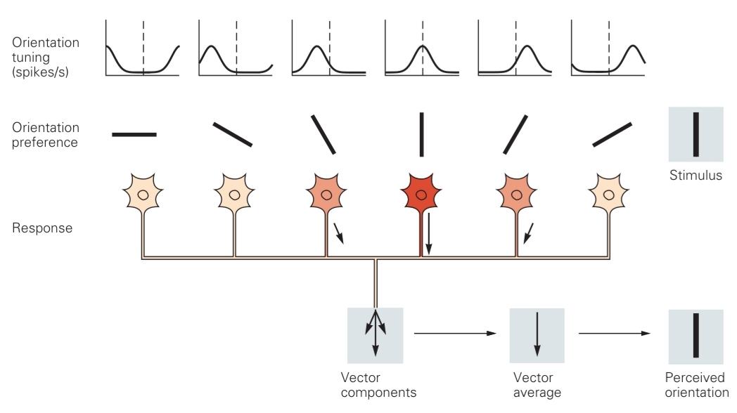
Figure 21-17 Vector averaging is one model for population coding in neural circuits. Vector averages describe the possible relationship between the responses in an ensemble of neurons, the tuning characteristics of individual neurons in the ensemble, and the resultant percept. Individual neurons respond optimally to a particular orientation of a stimulus in the visual field, but also respond at varying rates to a range of orientations. The stimulus orientation to which a neuron fires best can be thought of as a line label-when the cell fires briskly, its activity signifies the presence of a stimulus with that orientation. A number of neurons with different orientation preferences will respond to the same stimulus. Each neuron’s response can be represented as a vector whose length indicates the strength of its response and whose direction represents its preferred orientation, or line label. (Adapted, with permission, from Kapadia, Westheimer, and Gilbert 2000.)
شکل ۲۱-۱۷ میانگینگیری برداری، مدلی برای کدگذاری جمعیت در مدارهای عصبی است. میانگینهای برداری، رابطهی احتمالی بین پاسخها در یک گروه از نورونها، ویژگیهای تنظیم نورونهای منفرد در گروه و ادراک حاصل را توصیف میکنند. نورونهای منفرد به طور بهینه به یک جهتگیری خاص از یک محرک در میدان بینایی پاسخ میدهند، اما همچنین با سرعتهای مختلفی به طیف وسیعی از جهتگیریها پاسخ میدهند. جهتگیری محرکی که یک نورون به بهترین شکل به آن شلیک میکند را میتوان به عنوان یک برچسب خطی در نظر گرفت – وقتی سلول به سرعت شلیک میکند، فعالیت آن نشاندهندهی وجود محرکی با آن جهتگیری است. تعدادی نورون با ترجیحات جهتگیری متفاوت به یک محرک پاسخ میدهند. پاسخ هر نورون را میتوان به عنوان برداری نشان داد که طول آن نشاندهندهی قدرت پاسخ آن و جهت آن نشاندهندهی جهتگیری ترجیحی یا برچسب خطی آن است. (اقتباس شده، با اجازه، از کاپادیا، وستهایمر و گیلبرت ۲۰۰۰.)
Another aspect of the population code is the variability of a neuron’s response to the same stimulus. Repeated presentation of a stimulus to a neuron sensitive to that stimulus will elicit a range of responses. The most sensitive part of a neuron’s tuning curve lies not at the peak but along the flanks, where the tuning curve is steepest. Here, small changes in the value of a stimulus produce the strongest change in response. Changes in stimulus value must, however, be sufficient to elicit a change in response that significantly exceeds the normal variability in the response of the neuron. One can compare that amount of change to the perceptual discrimination threshold. When many neurons contribute to the discrimination, the signal-to-noise ratio increases, a process known as probability summation, and the critical difference in stimulus value required for a significant change in neuronal response is less.
یکی دیگر از جنبههای کد جمعیت، تغییرپذیری پاسخ یک نورون به یک محرک مشابه است. ارائه مکرر یک محرک به نورونی که به آن محرک حساس است، طیف وسیعی از پاسخها را ایجاد میکند. حساسترین بخش منحنی تنظیم یک نورون نه در اوج، بلکه در امتداد کنارهها، جایی که منحنی تنظیم شیبدارترین است، قرار دارد. در اینجا، تغییرات کوچک در مقدار یک محرک، قویترین تغییر در پاسخ را ایجاد میکند. با این حال، تغییرات در مقدار محرک باید برای ایجاد تغییر در پاسخی که به طور قابل توجهی از تغییرپذیری طبیعی در پاسخ نورون فراتر میرود، کافی باشد. میتوان این میزان تغییر را با آستانه تمایز ادراکی مقایسه کرد. هنگامی که بسیاری از نورونها در تمایز مشارکت میکنند، نسبت سیگنال به نویز افزایش مییابد، فرآیندی که به عنوان جمع احتمال شناخته میشود، و تفاوت بحرانی در مقدار محرک مورد نیاز برای تغییر قابل توجه در پاسخ نورونی کمتر است.
When the brain represents a piece of information, an important consideration is the number of neurons that participate in that representation. Although all information about a visual stimulus is present in the retina, the retinal representation is not sufficient for object recognition. At the other end of the visual pathway, some neurons in the temporal lobe are selective for complex objects, such as faces. Can an individual cell represent something as complex as a particular face? Such a hypothetical neuron has been dubbed a “grandmother cell” because it would represent exclusively a person’s grandmother, or a “pontifical cell” because it would represent the apex of a hierarchical cognitive pathway.
وقتی مغز بخشی از اطلاعات را نمایش میدهد، یکی از ملاحظات مهم، تعداد نورونهایی است که در آن نمایش شرکت میکنند. اگرچه تمام اطلاعات مربوط به یک محرک بصری در شبکیه وجود دارد، اما نمایش شبکیه برای تشخیص اشیا کافی نیست. در انتهای دیگر مسیر بصری، برخی از نورونها در لوب گیجگاهی برای اشیاء پیچیده، مانند چهرهها، گزینشی عمل میکنند. آیا یک سلول منفرد میتواند چیزی به پیچیدگی یک چهره خاص را نمایش دهد؟ چنین نورون فرضی «سلول مادربزرگ» نامیده شده است زیرا منحصراً مادربزرگ یک فرد را نشان میدهد، یا «سلول پاپی» نامیده شده است زیرا نمایانگر اوج یک مسیر شناختی سلسله مراتبی است.
The nervous system does not, however, represent entire objects by the activity of single neurons. Instead, some cells represent parts of an object, and an ensemble of neurons represents an entire object. Each member of the ensemble may participate in different ensembles that are activated by different objects. This arrangement is known as a distributed code. Distributed codes can involve a few neurons or many. In any case, a distributed code requires complex connectivity between the cells representing a face and those representing the name and experiences associated with that person.
با این حال، سیستم عصبی کل اشیاء را با فعالیت نورونهای منفرد نشان نمیدهد. در عوض، برخی سلولها بخشهایی از یک شیء را نشان میدهند و مجموعهای از نورونها کل یک شیء را نشان میدهند. هر عضو از این مجموعه ممکن است در گروههای مختلفی که توسط اشیاء مختلف فعال میشوند، شرکت کند. این ترتیب به عنوان یک کد توزیع شده شناخته میشود. کدهای توزیع شده میتوانند شامل چند یا تعداد زیادی نورون باشند. در هر صورت، یک کد توزیع شده نیاز به اتصال پیچیده بین سلولهایی دارد که یک چهره را نشان میدهند و سلولهایی که نام و تجربیات مرتبط با آن شخص را نشان میدهند.
The foregoing discussion assumes that neurons signal information by their firing rate and their line labels. An alternative hypothesis is that the timing of action potentials itself carries information, analogous to Morse code. The code might be read from the synchronous firing of different sets of neurons over time. At one instant, one group of cells might fire together followed by the synchronous firing of another group. Over a single train of action potentials, a single cell could participate in many such ensembles. Whether sensory information is represented this way and whether the nervous system carries more information than that represented by firing rate alone are not known.
بحث فوق فرض میکند که نورونها اطلاعات را با نرخ شلیک و برچسبهای خطی خود ارسال میکنند. یک فرضیه جایگزین این است که زمانبندی پتانسیلهای عمل، خود حامل اطلاعات است، مشابه کد مورس. این کد ممکن است از شلیک همزمان مجموعههای مختلف نورونها در طول زمان خوانده شود. در یک لحظه، یک گروه از سلولها ممکن است با هم شلیک کنند و به دنبال آن شلیک همزمان گروه دیگری انجام شود. در یک قطار از پتانسیلهای عمل، یک سلول واحد میتواند در بسیاری از این مجموعهها شرکت کند. اینکه آیا اطلاعات حسی به این صورت نمایش داده میشوند و اینکه آیا سیستم عصبی اطلاعات بیشتری نسبت به آنچه که تنها با نرخ شلیک نشان داده میشود، حمل میکند، مشخص نیست.
Highlights
نکات برجسته
1. Vision is a constructive process fundamentally different from the mere recording of visual input as in a camera. Rather, the brain uses visual input to infer information about the world around it, including information about objects, such as their sizes, shapes, distances, and identities and how rapidly they are moving.
۱. بینایی یک فرآیند سازنده است که اساساً با ثبت صرف ورودی بصری مانند دوربین متفاوت است. بلکه مغز از ورودی بصری برای استنباط اطلاعات در مورد جهان اطراف خود، از جمله اطلاعات مربوط به اشیاء، مانند اندازه، شکل، فواصل و هویت آنها و سرعت حرکت آنها، استفاده میکند.
2. The tuning of neural circuits for visual features such as contrast, orientation, and motion often matches the distribution of the feature in the natural environment. This suggests an evolutionary, ethologically driven origin for the neural circuitry.
۲. تنظیم مدارهای عصبی برای ویژگیهای بصری مانند کنتراست، جهتگیری و حرکت اغلب با توزیع آن ویژگی در محیط طبیعی مطابقت دارد. این نشان دهنده یک منشأ تکاملی و رفتارشناختی برای مدارهای عصبی است.
3. Visual circuitry, and thus vision, are modulated by individual visual experience.
۳. مدارهای بینایی و در نتیجه بینایی، توسط تجربه بصری فردی تنظیم میشوند.
4. Vision makes extensive use of parallel processing. The higher visual centers form two distinct pathways. The dorsal pathway, located in parietal cortex, is involved in motion perception, attention, and visually guided action. The ventral pathway, located in temporal cortex, processes form and objects. Further subdivisions of the ventral pathway are specialized, for example, for recognizing faces. These pathways, although distinct, communicate with each other; this is likely important for the perception of objects as coherent wholes.
۴. بینایی به طور گسترده از پردازش موازی استفاده میکند. مراکز بینایی بالاتر، دو مسیر مجزا را تشکیل میدهند. مسیر پشتی، که در قشر آهیانه قرار دارد، در ادراک حرکت، توجه و عمل هدایتشده بصری نقش دارد. مسیر شکمی، که در قشر گیجگاهی قرار دارد، فرم و اشیاء را پردازش میکند. زیربخشهای بیشتر مسیر شکمی، به عنوان مثال، برای تشخیص چهرهها تخصصی شدهاند. این مسیرها، اگرچه متمایز هستند، اما با یکدیگر ارتباط برقرار میکنند. این احتمالاً برای درک اشیاء به عنوان کلهای منسجم مهم است.
5. Parallel processing starts at the retina. Distinct retinal circuits analyze each point of the visual input for different local features including local contrasts of achromatic bright versus dark, red versus green, and blue versus yellow. The information is sent out through distinct classes of retinal ganglion cells (magnocelluar, parvo celluar, and koniocellular, respectively, for the three features noted) whose axons form the optic nerves.
۵. پردازش موازی از شبکیه شروع میشود. مدارهای شبکیهای متمایز، هر نقطه از ورودی بصری را برای ویژگیهای محلی مختلف، از جمله تضادهای محلی روشن در مقابل تیره، قرمز در مقابل سبز و آبی در مقابل زرد، تجزیه و تحلیل میکنند. اطلاعات از طریق طبقات متمایز سلولهای گانگلیونی شبکیه (به ترتیب برای سه ویژگی ذکر شده، ماگنوسلولار، پارو سلولی و کونیوسلولار) که آکسونهای آنها اعصاب بینایی را تشکیل میدهند، ارسال میشود.
6. The optic nerves from the two eyes regroup at the optic chiasm such that all fibers from the left visual hemifield project to the right hemisphere of the brain, and vice versa. However, the parallel retinal channels remain anatomically segregated by eye and by visual feature, past a thalamic relay station, the lateral geniculate nucleus (LGN), up to primary visual cortex (V1).
۶. اعصاب بینایی دو چشم در کیاسمای بینایی دوباره گروهبندی میشوند به طوری که تمام فیبرهای نیمکره بینایی چپ به نیمکره راست مغز و برعکس هدایت میشوند. با این حال، کانالهای موازی شبکیه از نظر آناتومیکی بر اساس چشم و ویژگی بصری از هم جدا باقی میمانند و از یک ایستگاه رله تالاموس، هسته زانویی جانبی (LGN)، تا قشر بینایی اولیه (V1) عبور میکنند.
7. The different channels enter V1 at different layers, although primarily they enter at the major input layers 4 and 6. The visual input is recombined to extract new sets of features. These include tuning for orientation, motion, and object depth (obtained by combining left- and right-eye inputs).
۷. کانالهای مختلف در لایههای مختلف وارد V1 میشوند، اگرچه در درجه اول در لایههای ورودی اصلی ۴ و ۶ وارد میشوند. ورودی بصری برای استخراج مجموعههای جدیدی از ویژگیها دوباره ترکیب میشود. این ویژگیها شامل تنظیم جهت، حرکت و عمق شیء (که با ترکیب ورودیهای چشم چپ و راست به دست میآید) میشود.
8. V1 neurons sharing basic properties such as spatial location or orientation preference form columns extending vertically from the pia to the white matter.
۸. نورونهای V1 که ویژگیهای اساسی مانند موقعیت مکانی یا ترجیح جهتگیری را به اشتراک میگذارند، ستونهایی را تشکیل میدهند که به صورت عمودی از نرمشامه تا ماده سفید امتداد یافتهاند.
9. V1 neurons also form systematic horizontal maps of their response properties over cortex. The tuning for location forms a smooth “visuotopic” map of visual space, which changes gradually with distance, and is most finely resolved at the fovea, growing progressively coarser toward the periphery. Superimposed on the spatial map are locally smooth maps of orientation preference and left- versus right-eye preference, with interspersed columns that preferentially process color. These visual response features cycle over relatively short cortical distances, in effect completing one full cycle over each partial shift of the spatial map. Thus, V1 circuits effectively analyze each visual location, in parallel, for the full set of V1 visual features.
۹. نورونهای V1 همچنین نقشههای افقی سیستماتیکی از ویژگیهای پاسخ خود بر روی قشر مغز تشکیل میدهند. تنظیم موقعیت مکانی، یک نقشه “visuotopic” صاف از فضای بصری تشکیل میدهد که به تدریج با فاصله تغییر میکند و در گودی مرکزی (fovea) به ظریفترین شکل خود تفکیک میشود و به تدریج به سمت حاشیه درشتتر میشود. نقشههای صاف محلی از ترجیح جهتگیری و ترجیح چشم چپ در مقابل چشم راست، با ستونهای پراکنده که ترجیحاً رنگ را پردازش میکنند، بر روی نقشه مکانی قرار گرفتهاند. این ویژگیهای پاسخ بصری در فواصل نسبتاً کوتاه قشر مغز چرخه میخورند، در واقع یک چرخه کامل را در هر تغییر جزئی نقشه مکانی تکمیل میکنند. بنابراین، مدارهای V1 به طور مؤثر هر مکان بصری را به طور موازی برای مجموعه کامل ویژگیهای بصری V1 تجزیه و تحلیل میکنند.
10. Neural processing in V1 reflects its architecture, with local vertical processing along columns and lateral processing across columns. In addition, there is long-range processing that spans multiple columns.
۱۰. پردازش عصبی در V1 معماری آن را منعکس میکند، با پردازش عمودی محلی در امتداد ستونها و پردازش جانبی در عرض ستونها. علاوه بر این، پردازش دوربرد وجود دارد که چندین ستون را در بر میگیرد.
11. The output of V1 feeds into progressively higher visual areas comprising more than 30 centers distributed along the dorsal and ventral pathways. The connectivity is reciprocal, with higher loci sending dense feedback targeting lower areas including the LGN.
۱۱. خروجی V1 به نواحی بصری که به تدریج بالاتر میروند و شامل بیش از ۳۰ مرکز هستند که در امتداد مسیرهای پشتی و شکمی توزیع شدهاند، تغذیه میشود. این اتصال متقابل است، به طوری که جایگاههای بالاتر بازخورد متراکمی را به نواحی پایینتر از جمله LGN ارسال میکنند.
12. A useful measure of visual processing is pro- vided by changes in neuronal “receptive fields” along the visual pathway. The receptive field is the region of visual space from which the neuron receives input; it is further characterized by the neuron’s optimal visual stimulus. Receptive fields grow larger and more complex at successive stages along the visual pathway. Their optimal stimuli also increase in complexity from simple pixel-like dots for photoreceptors, to oriented lines for V1, to faces in higher face-selective centers of the ventral pathway.
۱۲. یک معیار مفید برای پردازش بصری، تغییرات در «میدانهای گیرنده» نورونی در امتداد مسیر بصری است. میدان گیرنده، ناحیهای از فضای بصری است که نورون از آن ورودی دریافت میکند؛ این میدان همچنین با محرک بصری بهینه نورون مشخص میشود. میدانهای گیرنده در مراحل متوالی در امتداد مسیر بصری بزرگتر و پیچیدهتر میشوند. محرکهای بهینه آنها نیز از نظر پیچیدگی از نقاط ساده پیکسل مانند برای گیرندههای نوری، به خطوط جهتدار برای V1، و به چهرهها در مراکز بالاتر انتخاب چهره در مسیر شکمی، افزایش مییابند.
13. Looking forward, one of the most important unsolved questions is the interaction between feedforward visual processing through progressively “higher” neural computations and feedback mediated via the dense plexus of connections from higher to lower levels. Understanding this interaction may be the key to understanding how the brain effortlessly forms complex visual percepts.
۱۳. با نگاهی به آینده، یکی از مهمترین سوالات حل نشده، تعامل بین پردازش بصری پیشخور از طریق محاسبات عصبی به تدریج «بالاتر» و بازخورد ناشی از شبکه متراکم ارتباطات از سطوح بالاتر به پایینتر است. درک این تعامل ممکن است کلید درک چگونگی شکلگیری آسان ادراکات بصری پیچیده توسط مغز باشد.
Charles D. Gilbert
Aniruddha Das
کلیک کنید تا Selected Reading نمایش داده شود
Hubel DH, Wiesel TN. 1962. Receptive fields, binocular interaction and functional architecture in the cat’s visual cortex. J Physiol 160:106-154.
Hubel DH, Wiesel TN. 1977. Functional architecture of macaque monkey visual cortex. Proc R Soc Lond B Biol Sci 198:1-59.
Hubener M, Shohan D, Grinvald A, Bonhoeffer T. 1997. Spatial relationships among three columnar systems in cat area 17. J Neurosci 17:9270-9284.
Isaacson JS, Scanziani M. 2011. How inhibition shapes cortical activity. Neuron 72:231-243.
Nassi JJ, Callaway EM. 2009. Parallel processing strategies of the primate visual system. Nat Rev Neurosci 10:360-372.
Orban GA, Van Essen D, Vanduffel W. 2004. Comparative mapping of higher visual areas in monkeys and humans. Trends Cogn Sci 8:315–324.
Stryker MP. 2014. A neural circuit that controls cortical state, plasticity, and the gain of sensory responses in mouse. Cold Spring Harb Symp Quant Biol 79:1-9.
Tsao DY, Moeller S, Freiwald WA. 2008. Comparing face patch systems in macaques and humans. Proc Natl Acad Sci USA 105:19514-19519.
VanEssen DC, Anderson CH, Felleman DJ. 1992. Information processing in the primate visual system: an integrated systems perspective. Science 255:419–423.
Wertheimer M. 1938. Laws of Organization in Perceptual Forms. London: Harcourt, Brace & Jovanovitch.
Wiesel TN, Hubel DH. 1966. Spatial and chromatic interactions in the lateral geniculate body of the rhesus monkey. J Neurophysiol 29:1115–1156.
کلیک کنید تا References نمایش داده شود
Blasdel GG, Lund JS. 1983. Termination of afferent axons in macaque striate cortex. J Neurosci 3:1389-1413.
Callaway EM. 1998. Local circuits in primary visual cortex of the macaque monkey. Annu Rev Neurosci 21:47-74.
Field DJ, Hayes A, Hess RF. 1993. Contour integration by the human visual system: evidence for a local “association field.” Vision Res 33:173–193.
Gilbert CD, Li W. 2012. Adult visual cortical plasticity. Neuron 75:250-264.
Gilbert CD, Li W. 2013. Top-down influences on visual processing. Nat Rev Neurosci 14:350-363.
Gilbert CD, Wiesel TN. 1983. Clustered intrinsic connections in cat visual cortex. J Neurosci 3:1116-1133.
Hartline HK. 1941. The neural mechanisms of vision. Harvey Lect 37:39-68.
Hubel DH. 1988. Eye, Brain and Vision. New York: Scientific American Library.
Hubel DH, Wiesel TN. 1974. Uniformity of monkey striate cortex. A parallel relationship between field size, scatter and magnification factor. J Comp Neurol 158:295–306.
Kapadia MK, Westheimer G, Gilbert CD. 2000. Spatial distribution of contextual interactions in primary visual cortex and in visual perception. J Neurophysiol 84:2048-2062.
Kuffler SF. 1953. Discharge patterns and functional organization of mammalian retina. J Neurophysiol 16:37-68. Porter PB. 1954. Another puzzle-picture. Am J Psychol 67:550-551.
Stettler DD, Das A, Bennett J, Gilbert CD. 2002. Lateral connectivity and contextual interactions in macaque primary visual cortex. Neuron 36:739-750.
»» قسمت اول فصل ماهیت ساختاری پردازش بینایی
»» تمامی کتاب