
فیزیولوژی پزشکی گایتون و هال؛ فیزیولوژی جنین و نوزاد


دعای مطالعه [ نمایش ]
بِسْمِ الله الرَّحْمنِ الرَّحیمِ
اَللّهُمَّ اَخْرِجْنى مِنْ ظُلُماتِ الْوَهْمِ
خدايا مرا بيرون آور از تاريكىهاى وهم،
وَ اَكْرِمْنى بِنُورِ الْفَهْمِ
و به نور فهم گرامى ام بدار،
اَللّهُمَّ افْتَحْ عَلَيْنا اَبْوابَ رَحْمَتِكَ
خدايا درهاى رحمتت را به روى ما بگشا،
وَانْشُرْ عَلَيْنا خَزائِنَ عُلُومِكَ بِرَحْمَتِكَ يا اَرْحَمَ الرّاحِمينَ
و خزانههاى علومت را بر ما باز كن به امید رحمتت اى مهربانترين مهربانان.
کتاب «فیزیولوژی پزشکی گایتون و هال» بهعنوان یکی از جامعترین و معتبرترین منابع در حوزه علوم پزشکی، همچنان مرجع کلیدی برای درک عملکرد پیچیده بدن انسان است. این اثر با تکیه بر تازهترین پژوهشها و توضیحات دقیق از سازوکارهای فیزیولوژیک، پلی میان علوم پایه پزشکی و کاربردهای بالینی ایجاد میکند و نقشی بیبدیل در آموزش، پژوهش و ارتقای دانش سلامت ایفا مینماید.
ترجمه دقیق و علمی این شاهکار توسط برند علمی آیندهنگاران مغز به مدیریت داریوش طاهری، دسترسی فارسیزبانان به مرزهای نوین دانش فیزیولوژی را ممکن ساخته و رسالتی علمی برای ارتقای آموزش پزشکی، فهم عمیقتر سازوکارهای بدن و توسعه روشهای نوین در حوزه سلامت فراهم آورده است.
» Guyton and Hall Textbook of Medical Physiology
»» CHAPTER 84: Fetal and Neonatal Physiology

A complete discussion of fetal development, physiology of the child immediately after birth, and growth and development through the early years of life lies within the province of formal courses in obstetrics and pediatrics. However, many physiological principles are peculiar to the infant, and this chapter discusses the more important of these principles.
بحث کامل در مورد رشد جنین، فیزیولوژی کودک بلافاصله پس از تولد، و رشد و نمو در طی سالهای اولیه زندگی در استان دوره های رسمی در مامایی و اطفال قرار دارد. با این حال، بسیاری از اصول فیزیولوژیکی مخصوص نوزاد هستند و این فصل به مهم تر از این اصول می پردازد.
Growth and Development of the Fetus
The placenta and fetal membranes initially develop far more rapidly than does the fetus. In fact, during the first 2 to 3 weeks after implantation of the blastocyst, the fetus remains almost microscopic, but thereafter, as shown in Figure 84-1, the length of the fetus increases almost in proportion to age. At 12 weeks, the length is about 10 centimeters; at 20 weeks, 25 centimeters; and at term (40 weeks), 53 centimeters (~21 inches). Because the weight of the fetus is approximately proportional to the cube of length, the weight increases almost in proportion to the cube of the age of the fetus.
رشد و تکامل جنین
جفت و غشای جنین در ابتدا بسیار سریعتر از جنین رشد می کنند. در واقع، در طی 2 تا 3 هفته اول پس از کاشت بلاستوسیست، جنین تقریباً میکروسکوپی باقی می ماند، اما پس از آن، همانطور که در شکل 84-1 نشان داده شده است، طول جنین تقریباً متناسب با سن افزایش می یابد. در 12 هفتگی، طول حدود 10 سانتی متر است. در 20 هفتگی، 25 سانتی متر؛ و در ترم (40 هفته)، 53 سانتی متر (~21 اینچ). از آنجایی که وزن جنین تقریباً متناسب با مکعب طول است، وزن تقریباً متناسب با مکعب سن جنین افزایش می یابد.
Note in Figure 84-1 that the weight remains minuscule during the first 12 weeks and reaches 1 pound only at 23 weeks (5.5 months) of gestation. Then, during the last trimester of pregnancy, the fetus gains weight rapidly, so that 2 months before birth, the weight averages 3 pounds; 1 month before birth, the average is 4.5 pounds; and at birth, the average is 7 pounds, with the final birth weight varying from as low as 4.5 pounds to as high as 11 pounds in normal infants with normal gestational periods.
در شکل 84-1 توجه داشته باشید که وزن در طول 12 هفته اول کم می ماند و تنها در هفته 23 (5.5 ماهگی) بارداری به 1 پوند می رسد. سپس، در سه ماهه آخر بارداری، وزن جنین به سرعت افزایش می یابد، به طوری که 2 ماه قبل از تولد، وزن به طور متوسط 3 پوند است. 1 ماه قبل از تولد، میانگین 4.5 پوند است. و در هنگام تولد، میانگین 7 پوند است، با وزن نهایی تولد از 4.5 پوند تا 11 پوند در نوزادان طبیعی با دوره های بارداری طبیعی متغیر است.

Figure 84-1. Growth of the fetus.
شکل 84-1. رشد جنین.
Development of the Organ Systems
Within 1 month after fertilization of the ovum, the gross characteristics of all the different organs of the fetus have already begun to develop, and during the next 2 to 3 months, most of the details of the different organs are established. Beyond month 4, the organs of the fetus are grossly the same as those of the neonate. However, cellular development in each organ is usually far from complete and requires the full remaining 5 months of pregnancy for complete development. Even at birth, certain structures lack full development, particularly in the nervous system, the kidneys, and the liver, as discussed later in this chapter.
توسعه سیستم های اندام
در عرض 1 ماه پس از لقاح تخمک، ویژگی های ناخالص همه اندام های مختلف جنین شروع به رشد کرده است و طی 2 تا 3 ماه آینده، بیشتر جزئیات اندام های مختلف مشخص می شود. بعد از ماه 4، اندام های جنین کاملاً مشابه اندام های نوزاد است. با این حال، رشد سلولی در هر اندام معمولاً کامل نیست و برای رشد کامل به 5 ماه باقی مانده از بارداری نیاز دارد. حتی در هنگام تولد، ساختارهای خاصی فاقد رشد کامل هستند، به ویژه در سیستم عصبی، کلیه ها و کبد، همانطور که در ادامه این فصل مورد بحث قرار گرفت.
Circulatory System. The human heart begins beating during the fourth week after fertilization, contracting at a rate of about 65 beats/min. This rate increases steadily to about 140 beats/min immediately before birth.
سیستم گردش خون. قلب انسان در هفته چهارم پس از لقاح شروع به تپیدن می کند و با سرعتی در حدود 65 ضربه در دقیقه منقبض می شود. این میزان بلافاصله قبل از تولد به حدود 140 ضربه در دقیقه افزایش می یابد.
Formation of Blood Cells. Nucleated red blood cells be- gin to be formed in the yolk sac and mesothelial layers of the placenta at about the third week of fetal development. This is followed 1 week later (at 4-5 weeks) by formation of non-nucleated red blood cells by the fetal mesenchyme and also by the endothelium of fetal blood vessels. At 6 weeks, the liver begins to form blood cells, and in the third month, the spleen and other lymphoid tissues of the body begin forming blood cells. Finally, from the third month on, the bone marrow gradually becomes the principal source of red blood cells, as well as most of the white blood cells, except for continued lymphocyte and plasma cell production in lymphoid tissue.
تشکیل سلول های خونی. گلبول های قرمز هسته دار در کیسه زرده و لایه های مزوتلیال جفت در حدود هفته سوم رشد جنین شروع به تشکیل می کنند. این امر یک هفته بعد (در 4-5 هفتگی) با تشکیل گلبول های قرمز بدون هسته توسط مزانشیم جنین و همچنین توسط اندوتلیوم رگ های خونی جنین دنبال می شود. در 6 هفتگی، کبد شروع به تشکیل سلول های خونی می کند و در ماه سوم، طحال و سایر بافت های لنفاوی بدن شروع به تشکیل سلول های خونی می کنند. سرانجام، از ماه سوم به بعد، مغز استخوان به تدریج به منبع اصلی گلبول های قرمز و همچنین بیشتر گلبول های سفید تبدیل می شود، به جز ادامه تولید لنفوسیت و پلاسما سلول در بافت لنفاوی.
Respiratory System. Respiration cannot occur during fetal life because there is no air to breathe in the amniotic cavity. However, attempted respiratory movements do take place beginning at the end of the first trimester of pregnancy. Tactile stimuli and fetal asphyxia especially cause these attempted respiratory movements.
سیستم تنفسی. تنفس در طول زندگی جنینی امکان پذیر نیست زیرا در حفره آمنیوتیک هوایی برای تنفس وجود ندارد. با این حال، تلاش برای حرکات تنفسی در پایان سه ماهه اول بارداری انجام می شود. محرک های لمسی و خفگی جنین به ویژه باعث این حرکات تنفسی می شوند.
During the last 3 to 4 months of pregnancy, the respiratory movements of the fetus are mainly inhibited, for reasons unknown, and the lungs remain almost completely deflated. The inhibition of respiration during the later months of fetal life prevents filling of the lungs with fluid and debris from the meconium excreted by the fetus’s gastrointestinal tract into the amniotic fluid. Also, small amounts of fluid are secreted into the lungs by the alveolar epithelium up until the moment of birth, thus keeping only clean fluid in the lungs.
در طول 3 تا 4 ماه آخر بارداری، حرکات تنفسی جنین به دلایل ناشناخته عمدتاً مهار می شود و ریه ها تقریباً کاملاً تخلیه می شوند. مهار تنفس در ماههای آخر زندگی جنینی از پر شدن ریهها با مایعات و زبالههای مکونیومی که توسط دستگاه گوارش جنین به داخل مایع آمنیوتیک دفع میشود، جلوگیری میکند. همچنین مقدار کمی مایع تا لحظه تولد توسط اپیتلیوم آلوئول به ریه ها ترشح می شود و بنابراین فقط مایع تمیز در ریه ها باقی می ماند.
Nervous System. Most of the reflexes of the fetus that involve the spinal cord and even the brain stem are present by the third to fourth months of pregnancy. However, nervous system functions that involve the cerebral cortex are still only in the early stages of development, even at birth. Indeed, myelinization of some major tracts of the brain be- comes complete only after about 1 year of postnatal life.
سیستم عصبی. اکثر رفلکس های جنین که نخاع و حتی ساقه مغز را درگیر می کند در ماه های سوم تا چهارم بارداری وجود دارد. با این حال، عملکردهای سیستم عصبی که قشر مغز را درگیر می کنند، هنوز در مراحل اولیه رشد هستند، حتی در بدو تولد. در واقع، میلینیزاسیون برخی از بخش های اصلی مغز تنها پس از حدود 1 سال از زندگی پس از زایمان کامل می شود.
Gastrointestinal Tract. By midpregnancy, the fetus be- gins to ingest and absorb large quantities of amniotic fluid, and during the last 2 to 3 months, gastrointestinal function approaches that of the normal neonate. By that time, small quantities of meconium are continually formed in the gastrointestinal tract and excreted from the anus into the amniotic fluid. Meconium is composed partly of residue from swallowed amniotic fluid and partly of mucus, epithelial cells, and other residues of excretory products from the gastrointestinal mucosa and glands.
دستگاه گوارش. در اواسط بارداری، جنین شروع به مصرف و جذب مقادیر زیادی مایع آمنیوتیک می کند و در طی 2 تا 3 ماه گذشته، عملکرد دستگاه گوارش به عملکرد نوزاد طبیعی نزدیک می شود. در آن زمان، مقادیر کمی مکونیوم به طور مداوم در دستگاه گوارش تشکیل شده و از مقعد به مایع آمنیوتیک دفع می شود. مکونیوم تا حدی از بقایای مایع آمنیوتیک بلعیده شده و بخشی از مخاط، سلول های اپیتلیال و سایر بقایای محصولات دفعی غدد مخاطی دستگاه گوارش تشکیل شده است.
Kidneys. The fetal kidneys begin to excrete urine during the second trimester, and fetal urine accounts for about 70% to 80% of the amniotic fluid. Abnormal kidney development or severe impairment of kidney function in the fetus greatly reduces formation of amniotic fluid (oligohydramnios) and can lead to fetal death.
کلیه ها. کلیه های جنین در سه ماهه دوم شروع به دفع ادرار می کنند و ادرار جنین حدود 70 تا 80 درصد مایع آمنیوتیک را تشکیل می دهد. رشد غیر طبیعی کلیه یا اختلال شدید عملکرد کلیه در جنین، تشکیل مایع آمنیوتیک (الیگوهیدرآمنیوس) را تا حد زیادی کاهش می دهد و می تواند منجر به مرگ جنین شود.
Although fetal kidneys form urine, the renal control systems for regulating fetal extracellular fluid volume and electrolyte balances, and especially acid-base balance, are almost nonexistent until late fetal life and do not reach full development until a few months after birth.
اگرچه کلیههای جنین ادرار را تشکیل میدهند، اما سیستمهای کنترل کلیه برای تنظیم حجم مایع خارج سلولی جنین و تعادل الکترولیتها و بهویژه تعادل اسید و باز تقریباً تا اواخر عمر جنین وجود ندارد و تا چند ماه پس از تولد به رشد کامل نمیرسد.
Fetal Metabolism
The fetus mainly uses glucose for energy. The fetus has a high capability of storing fat and protein, with much if not most of the fat being synthesized from glucose rather than being absorbed directly from the mother’s blood. In addition to these generalities, there are special problems of fetal metabolism related to calcium, phosphate, iron, and some vitamins.
متابولیسم جنین
جنین عمدتاً از گلوکز برای انرژی استفاده می کند. جنین توانایی بالایی در ذخیرهسازی چربی و پروتئین دارد، بهطوریکه بیشتر چربیها به جای جذب مستقیم از خون مادر، از گلوکز سنتز میشوند. علاوه بر این کلیات، مشکلات خاصی در متابولیسم جنین در ارتباط با کلسیم، فسفات، آهن و برخی ویتامین ها وجود دارد.
Metabolism of Calcium and Phosphate
Figure 84-2 shows the rates of calcium and phosphate accumulation in the fetus, demonstrating that about 22.5 grams of calcium and 13.5 grams of phosphorus are accumulated in the average fetus during gestation. About one-half of these accumulate during the last 4 weeks of gestation, which is coincident with the period of rapid ossification of the fetal bones and with the period of rapid weight gain of the fetus.
متابولیسم کلسیم و فسفات
شکل 84-2 میزان تجمع کلسیم و فسفات در جنین را نشان می دهد و نشان می دهد که حدود 22.5 گرم کلسیم و 13.5 گرم فسفر در جنین متوسط در طول بارداری انباشته می شود. تقریباً نیمی از اینها در 4 هفته آخر بارداری تجمع می یابند که همزمان با دوره استخوانی شدن سریع استخوان های جنین و با دوره افزایش وزن سریع جنین است.
During the earlier part of fetal life, the bones are relatively unossified and have mainly a cartilaginous matrix. Ossification usually does not occur until after the fourth month of pregnancy.
در مراحل اولیه زندگی جنینی، استخوانها نسبتاً غیر استخوانی هستند و عمدتاً دارای ماتریکس غضروفی هستند. استخوان بندی معمولاً تا بعد از ماه چهارم بارداری اتفاق نمی افتد.
Note especially that the total amounts of calcium and phosphate needed by the fetus during gestation represent only about 2% of the quantities of these substances in the mother’s bones, and thus the drain of these substances from the mother is minimal. A much greater drain occurs after birth during lactation.
به ویژه توجه داشته باشید که کل کلسیم و فسفات مورد نیاز جنین در دوران بارداری تنها حدود 2 درصد از مقادیر این مواد در استخوان های مادر را تشکیل می دهد و بنابراین تخلیه این مواد از مادر به حداقل می رسد. تخلیه بسیار بیشتر پس از تولد در دوران شیردهی اتفاق می افتد.
Accumulation of Iron
Figure 84-2 also shows that iron accumulates in the fetus even more rapidly than does calcium and phosphate. Most of the iron is in the form of hemoglobin, which begins to be formed as early as the third week after fertilization of the ovum.
تجمع آهن
شکل 84-2 همچنین نشان می دهد که آهن در جنین حتی سریعتر از کلسیم و فسفات تجمع می یابد. بیشتر آهن به شکل هموگلوبین است که از هفته سوم پس از لقاح تخمک شروع به تشکیل میکند.
Small amounts of iron are concentrated in the mother’s uterine progestational endometrium even before implantation of the ovum; this iron is ingested into the embryo by the trophoblastic cells and is used to form the very early red blood cells. About one-third of the iron in a fully developed fetus is normally stored in the liver. This iron can then be used by the neonate to form additional hemoglobin for several months after birth.
حتی قبل از لانه گزینی تخمک، مقادیر کمی آهن در اندومتر پروژستاتیک رحم مادر متمرکز می شود. این آهن توسط سلول های تروفوبلاستیک وارد جنین می شود و برای تشکیل گلبول های قرمز بسیار اولیه استفاده می شود. حدود یک سوم آهن در یک جنین کاملاً رشد یافته به طور معمول در کبد ذخیره می شود. سپس نوزاد می تواند از این آهن برای تشکیل هموگلوبین اضافی تا چند ماه پس از تولد استفاده کند.

Figure 84-2. Iron, calcium, and phosphorus storage in the fetus at different stages of gestation.
شکل 84-2. ذخیره آهن، کلسیم و فسفر در جنین در مراحل مختلف بارداری.
Utilization and Storage of Vitamins
The fetus needs vitamins equally as much as the adult and in some instances to a far greater extent. In general, the vitamins function the same in the fetus as in the adult, as discussed in Chapter 72. Special functions of several vita- mins should be mentioned, however.
استفاده و ذخیره ویتامین ها
جنین به اندازه بزرگسالان و در برخی موارد به میزان بسیار بیشتری به ویتامین ها نیاز دارد. به طور کلی، عملکرد ویتامین ها در جنین همانند بزرگسالان است، همانطور که در فصل 72 بحث شد.
The B vitamins, especially vitamin B12 and folic acid, are necessary for the formation of red blood cells and nervous tissue, as well as for overall growth of the fetus.
ویتامین های گروه B به خصوص ویتامین B12 و اسید فولیک برای تشکیل گلبول های قرمز خون و بافت عصبی و همچنین برای رشد کلی جنین ضروری هستند.
Vitamin C is necessary for appropriate formation of intercellular substances, especially the bone matrix and fibers of connective tissue.
ویتامین C برای تشکیل مناسب مواد بین سلولی به ویژه ماتریکس استخوان و الیاف بافت همبند ضروری است.
Vitamin D is necessary for normal bone growth in the fetus, but even more important, the mother needs it for adequate absorption of calcium from her gastrointestinal tract. If the mother has plenty of vitamin D in her body fluids, large quantities of the vitamin will be stored by the fetal liver to be used by the neonate for several months after birth.
ویتامین D برای رشد طبیعی استخوان در جنین ضروری است، اما مهمتر از آن، مادر برای جذب کافی کلسیم از دستگاه گوارش خود به آن نیاز دارد. اگر مادر مقدار زیادی ویتامین D در مایعات بدن خود داشته باشد، مقدار زیادی از این ویتامین در کبد جنین ذخیره می شود تا نوزاد تا چند ماه پس از تولد از آن استفاده کند.
The mechanisms of the functions of vitamin E are not entirely clear, but it is necessary for normal development of the early embryo. In its absence in laboratory animals, spontaneous abortion usually occurs at an early stage of pregnancy.
مکانیسم عملکرد ویتامین E کاملاً مشخص نیست، اما برای رشد طبیعی جنین اولیه ضروری است. در غیاب آن در حیوانات آزمایشگاهی، سقط خود به خودی معمولا در مراحل اولیه بارداری اتفاق می افتد.
Vitamin K is used by the fetal liver for formation of Factor VII, prothrombin, and several other blood coagulation factors. When vitamin K is insufficient in the mother, Factor VII and prothrombin become deficient in the fetus and the mother. Because most vitamin K is formed by bacterial action in the mother’s colon, the neonate has no adequate source of vitamin K for the first week or so of life after birth until normal colonic bacterial flora become established in the newborn infant. Therefore, prenatal storage in the fetal liver of at least small amounts of vitamin K derived from the mother is helpful in preventing fetal hemorrhage, particularly hemorrhage in the brain when the head is traumatized by squeezing through the birth canal.
ویتامین K توسط کبد جنین برای تشکیل فاکتور VII، پروترومبین و چندین فاکتور دیگر انعقاد خون استفاده می شود. هنگامی که ویتامین K در مادر کافی نباشد، فاکتور VII و پروترومبین در جنین و مادر دچار کمبود می شوند. از آنجایی که بیشتر ویتامین K در اثر عملکرد باکتری در روده بزرگ مادر ایجاد می شود، نوزاد در هفته اول زندگی پس از تولد تا زمانی که فلور باکتریایی کولون طبیعی در نوزاد تازه متولد شده ایجاد نشود، منبع کافی ویتامین K ندارد. بنابراین، ذخیره حداقل مقادیر کمی از ویتامین K مشتق شده از مادر در کبد جنین، برای جلوگیری از خونریزی جنین، به ویژه خونریزی در مغز هنگامی که سر با فشار دادن از طریق کانال زایمان آسیب می بیند، مفید است.
Adjustments of the Infant to Extrauterine Life
Onset of Breathing. The most obvious effect of birth on the baby is loss of the placental connection with the mother and, therefore, loss of metabolic support. One of the most important immediate adjustments required of the infant is to begin breathing.
تعدیل نوزاد در زندگی خارج رحمی
شروع تنفس. بارزترین تأثیر تولد بر نوزاد از دست دادن ارتباط جفت با مادر و در نتیجه از دست دادن حمایت متابولیک است. یکی از مهمترین تنظیمات فوری مورد نیاز نوزاد، شروع تنفس است.
Cause of Breathing at Birth. After normal delivery from a mother whose system has not been depressed by anesthetics, the child ordinarily begins to breathe within seconds and has a normal respiratory rhythm within less than 1 minute after birth. The promptness with which the fetus begins to breathe indicates that breathing is initiated by sudden exposure to the exterior world, probably resulting from a slightly asphyxiated state that is incident to the birth process and from sensory impulses that originate in the suddenly cooled skin. In an infant who does not breathe immediately, the body becomes progressively more hypoxic and hypercapnic, which provides additional stimulus to the respiratory center and usually causes breathing within an additional minute after birth.
علت تنفس هنگام تولد. پس از زایمان طبیعی از مادری که سیستم او توسط داروهای بیهوشی افسرده نشده است، کودک معمولاً در عرض چند ثانیه شروع به تنفس می کند و در کمتر از 1 دقیقه پس از تولد ریتم تنفسی طبیعی دارد. سرعتی که جنین با آن شروع به نفس کشیدن می کند نشان می دهد که تنفس با قرار گرفتن ناگهانی در دنیای بیرون آغاز می شود، که احتمالاً ناشی از حالت خفگی کمی است که در فرآیند تولد اتفاق می افتد و از تکانه های حسی که از پوست ناگهانی سرد شده منشأ می گیرند. در نوزادی که بلافاصله نفس نمیکشد، بدن به تدریج هیپوکسیکتر و هیپرکاپنیکتر میشود، که محرک اضافی برای مرکز تنفسی فراهم میکند و معمولاً در عرض یک دقیقه بعد از تولد باعث تنفس میشود.
Delayed or Abnormal Breathing at Birth-Danger of Hypoxia. If the mother’s system has been depressed by a general anesthetic during delivery, which at least partially anesthetizes the fetus as well, the onset of respiration is likely to be delayed for several minutes, thus demonstrating the importance of using as little anesthesia as feasible. Also, many infants who have had head trauma during delivery or who undergo prolonged delivery are slow to breathe or sometimes do not breathe at all. This can result from two possible effects: (1) in a few infants, intracranial hemorrhage or brain contusion causes a concussion syndrome, with a greatly depressed respiratory center; (2) probably much more important, prolonged fetal hypoxia during delivery can cause serious depression of the respiratory center.
تنفس تاخیری یا غیر طبیعی در هنگام تولد – خطر هیپوکسی. اگر سیستم مادر در حین زایمان توسط یک بیهوشی عمومی افسرده شده باشد، که حداقل تا حدی جنین را نیز بیهوش می کند، شروع تنفس احتمالا برای چند دقیقه به تعویق می افتد، بنابراین اهمیت استفاده از بیهوشی کم تا حد امکان را نشان می دهد. همچنین، بسیاری از نوزادانی که در حین زایمان دچار ضربه به سر شده اند یا زایمان طولانی مدت دارند، کند نفس می کشند یا گاهی اصلاً نفس نمی کشند. این می تواند ناشی از دو اثر احتمالی باشد: (1) در تعداد کمی از نوزادان، خونریزی داخل جمجمه یا کوفتگی مغزی باعث ایجاد سندرم ضربه مغزی می شود که مرکز تنفسی به شدت افسرده است. (2) احتمالاً بسیار مهمتر، هیپوکسی طولانی مدت جنین در طول زایمان می تواند باعث افسردگی جدی مرکز تنفسی شود.
Hypoxia may occur during delivery because of (1) com- pression of the umbilical cord; (2) premature separation of the placenta; (3) excessive contraction of the uterus, which can cut off the mother’s blood flow to the placenta; or (4) excessive anesthesia of the mother, which depresses oxygenation even of her blood.
هیپوکسی ممکن است در حین زایمان به دلیل (1) فشردگی بند ناف رخ دهد. (2) جدا شدن زودرس جفت. (3) انقباض بیش از حد رحم که می تواند جریان خون مادر را به جفت قطع کند. یا (4) بیهوشی بیش از حد مادر، که باعث کاهش اکسیژن رسانی حتی خون او می شود.
Degree of Hypoxia That an Infant Can Tolerate. In adults, failure to breathe for only 4 minutes often causes death, but neonates may survive as long as 10 minutes without breathing after birth. Permanent and serious brain impairment often ensues if breathing is delayed more than 8 to 10 minutes. Indeed, actual lesions develop mainly in the thalamus, in the inferior colliculi, and in other brain stem areas, thus permanently affecting many of the motor functions of the body.
درجه هیپوکسی که یک نوزاد می تواند تحمل کند. در بزرگسالان، عدم تنفس تنها به مدت 4 دقیقه اغلب باعث مرگ می شود، اما نوزادان ممکن است تا 10 دقیقه بدون تنفس پس از تولد زنده بمانند. اگر تنفس بیش از 8 تا 10 دقیقه به تعویق بیفتد، اغلب نارسایی مغزی دائمی و جدی ایجاد می شود. در واقع، ضایعات واقعی عمدتاً در تالاموس، در کولیکولهای تحتانی و در سایر نواحی ساقه مغز ایجاد میشوند، بنابراین به طور دائم بسیاری از عملکردهای حرکتی بدن را تحت تأثیر قرار میدهند.
Expansion of the Lungs at Birth. At birth, the walls of the alveoli are at first collapsed because of the surface tension of the viscid fluid that fills them. More than 25 mm Hg of negative inspiratory pressure in the lungs is usually required to oppose the effects of this surface tension and to open the alveoli for the first time. Once the alveoli open, however, further respiration can be effected with relatively weak respiratory movements. Fortunately, the first inspirations of the normal neonate are extremely powerful; they are usually capable of creating as much as 60 mm Hg negative pressure in the intrapleural space.
انبساط ریه ها در بدو تولد. در بدو تولد، دیواره آلوئول ها در ابتدا به دلیل کشش سطحی مایع چسبناکی که آنها را پر می کند، فرو می ریزند. معمولاً برای مقابله با اثرات این کشش سطحی و باز کردن آلوئول ها برای اولین بار، بیش از 25 میلی متر جیوه فشار دمی منفی در ریه ها مورد نیاز است. با این حال، هنگامی که آلوئول ها باز می شوند، تنفس بیشتر با حرکات تنفسی نسبتا ضعیف انجام می شود. خوشبختانه، اولین الهامات نوزاد عادی بسیار قدرتمند است. آنها معمولاً قادر به ایجاد فشار منفی 60 میلی متر جیوه در فضای داخل جنب هستند.
Figure 84-3 shows the tremendous negative intrapleural pressures required to open the lungs at the onset of breathing. At the top of the figure, the pressure-volume curve (“compliance” curve) for the first breath after birth is shown. Observe, first, that the lower part of the curve begins at the zero pressure point and moves to the right. The curve shows that the volume of air in the lungs remains almost exactly zero until the negative pressure has reached -40 centimeters of water (-30 mm Hg). Then, as the negative pressure increases to -60 centimeters of water, about 40 ml of air enters the lungs. To deflate the lungs, considerable positive pressure, about +40 centimeters of water, is required because of viscous resistance offered by the fluid in the bronchioles.
شکل 84-3 فشارهای منفی شدید داخل جنب مورد نیاز برای باز کردن ریه ها در شروع تنفس را نشان می دهد. در بالای شکل، منحنی فشار-حجم (منحنی “انطباق”) برای اولین نفس پس از تولد نشان داده شده است. ابتدا توجه کنید که قسمت پایین منحنی از نقطه فشار صفر شروع شده و به سمت راست حرکت می کند. این منحنی نشان میدهد که حجم هوا در ریهها تا زمانی که فشار منفی به 40- سانتیمتر آب (30- میلیمتر جیوه) برسد، تقریباً صفر میماند. سپس با افزایش فشار منفی به 60- سانتی متر آب، حدود 40 میلی لیتر هوا وارد ریه ها می شود. برای تخلیه ریه ها، فشار مثبت قابل توجهی، حدود 40+ سانتی متر آب، به دلیل مقاومت چسبناکی که مایع موجود در برونشیول ها ارائه می دهد، مورد نیاز است.
Note that the second breath is much easier, requiring far less negative and positive pressures. Breathing does not become completely normal until about 40 minutes after birth, as shown by the third compliance curve, the shape of which compares favorably with that for the normal adult, as shown in Chapter 38.
توجه داشته باشید که نفس دوم بسیار آسان تر است و به فشارهای منفی و مثبت بسیار کمتری نیاز دارد. تنفس تا حدود 40 دقیقه پس از تولد کاملاً عادی نمی شود، همانطور که در منحنی انطباق سوم نشان داده شده است، شکلی که در مقایسه با شکل آن برای بزرگسالان طبیعی مطلوب است، همانطور که در فصل 38 نشان داده شده است.
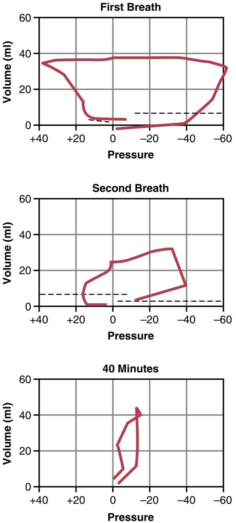
Figure 84-3. Pressure-volume curves of the lungs (“compliance” curves) of a neonate immediately after birth, showing the extreme forces required for breathing during the first two breaths of life and development of a nearly normal compliance curve within 40 minutes after birth. (Modified from Smith CA: The first breath. Sci Am 209:32, 1963. Copyright 1963 by Scientific American, Inc.)
شکل 84-3. منحنیهای فشار-حجم ریهها (منحنیهای «انطباق») یک نوزاد بلافاصله پس از تولد، نشاندهنده نیروهای شدید مورد نیاز برای تنفس در طی دو نفس اول زندگی و ایجاد یک منحنی سازگاری تقریباً طبیعی در 40 دقیقه پس از تولد است. (اصلاح شده از Smith CA: The first breath. Sci Am 209:32، 1963. حق چاپ 1963 توسط Scientific American, Inc.)
Respiratory Distress Syndrome Occurs When Surfactant Secretion Is Deficient. In a small number of infants, especially premature infants and infants born of mothers with diabetes mellitus, severe respiratory distress develops in the early hours to the first several days after birth, and some infants die within the next day or so. The alveoli of these infants at death contain large quantities of proteinceous fluid, almost as if pure plasma had leaked out of the capillaries into the alveoli. The fluid also contains desquamated alveolar epithelial cells. This condition is called hyaline membrane disease because microscopic slides of the lung show that the material filling the alveoli looks like a hyaline membrane.
سندرم دیسترس تنفسی زمانی رخ می دهد که ترشح سورفکتانت کم باشد. در تعداد کمی از نوزادان، به ویژه نوزادان نارس و نوزادان متولد شده از مادران مبتلا به دیابت، دیسترس تنفسی شدید در ساعات اولیه تا چند روز اول پس از تولد ایجاد می شود و برخی از نوزادان در یک روز بعد یا بیشتر می میرند. آلوئول های این نوزادان در هنگام مرگ حاوی مقادیر زیادی مایع پروتئینی است، تقریباً انگار پلاسمای خالص از مویرگ ها به داخل آلوئول ها نشت کرده است. این مایع همچنین حاوی سلول های اپیتلیال آلوئولی پوسته پوسته شده است. این بیماری را بیماری غشای هیالین می نامند زیرا اسلایدهای میکروسکوپی ریه نشان می دهد که مواد پرکننده آلوئول ها شبیه یک غشای هیالین هستند.
A characteristic finding in respiratory distress syndrome is failure of the respiratory epithelium to secrete adequate quantities of surfactant, a substance normally secreted into the alveoli that decreases the surface tension of the alveolar fluid, therefore allowing the alveoli to open easily during inspiration. The surfactant-secreting cells (type II alveolar epithelial cells) do not begin to secrete surfactant until the last 1 to 3 months of gestation. Therefore, many premature babies and a few full-term babies are born without the capability to secrete sufficient surfactant, which causes both a collapse tendency of the alveoli and development of pulmonary edema. The role of surfactant in preventing these effects is discussed in Chapter 38.
یک یافته مشخص در سندرم دیسترس تنفسی، ناتوانی اپیتلیوم تنفسی در ترشح مقادیر کافی سورفکتانت است، ماده ای که معمولاً در آلوئول ترشح می شود که کشش سطحی مایع آلوئول را کاهش می دهد، بنابراین به آلوئول ها اجازه می دهد به راحتی در طول دم باز شوند. سلول های ترشح کننده سورفکتانت (سلول های اپیتلیال آلوئولی نوع II) تا 1 تا 3 ماه آخر بارداری شروع به ترشح سورفکتانت نمی کنند. بنابراین، بسیاری از نوزادان نارس و تعداد کمی از نوزادان ترم بدون توانایی ترشح سورفکتانت کافی به دنیا می آیند که هم باعث تمایل به فروپاشی آلوئول ها و هم ایجاد ادم ریوی می شود. نقش سورفکتانت در جلوگیری از این اثرات در فصل 38 مورد بحث قرار گرفته است.
Circulatory Readjustments at Birth
Equally as essential as the onset of breathing at birth are immediate circulatory adjustments that allow adequate blood flow through the lungs. In addition, circulatory adjustments during the first few hours of life cause more and more blood flow through the baby’s liver, which up to this point has had little blood flow. To describe these readjustments, we first consider the anatomical structure of the fetal circulation.
تنظیم مجدد گردش خون هنگام تولد
به همان اندازه که شروع تنفس در بدو تولد ضروری است، تنظیمات فوری گردش خون است که جریان خون کافی را از طریق ریه ها امکان پذیر می کند. علاوه بر این، تنظیم گردش خون در چند ساعت اول زندگی باعث جریان بیشتر و بیشتر خون در کبد نوزاد می شود که تا این لحظه جریان خون کمی داشته است. برای توصیف این تنظیمات مجدد، ابتدا ساختار آناتومیکی گردش خون جنین را در نظر می گیریم.
Specific Anatomical Structure of the Fetal Circulation
Because the lungs are mainly nonfunctional during fetal life and because the liver is only partially functional, it is not necessary for the fetal heart to pump much blood through either the lungs or the liver. However, the fetal heart must pump large quantities of blood through the placenta. Therefore, special anatomical arrangements cause the fetal circulatory system to operate much differently from that of the newborn baby.
ساختار تشریحی خاص گردش خون جنین
از آنجایی که ریه ها در طول زندگی جنینی عمدتاً غیرفعال هستند و از آنجا که کبد فقط تا حدی کار می کند، لازم نیست قلب جنین خون زیادی را از طریق ریه ها یا کبد پمپاژ کند. با این حال، قلب جنین باید مقادیر زیادی خون را از طریق جفت پمپاژ کند. بنابراین، ترتیبات خاص تشریحی باعث می شود که سیستم گردش خون جنین بسیار متفاوت از نوزاد تازه متولد شده عمل کند.
First, as shown in Figure 84-4, blood returning from the placenta through the umbilical vein passes through the ductus venosus, mainly bypassing the liver. Then most of the blood entering the right atrium from the inferior vena cava is directed in a straight pathway across the posterior aspect of the right atrium and through the foramen ovale directly into the left atrium. Thus, the well-oxygenated blood from the placenta enters mainly the left side of the heart, rather than the right side, and is pumped by the left ventricle mainly into the arteries of the head and forelimbs.
ابتدا، همانطور که در شکل 84-4 نشان داده شده است، خون برگشتی از جفت از طریق ورید ناف از مجرای وریدی عبور می کند، عمدتاً کبد را دور می زند. سپس بیشتر خونی که از ورید اجوف تحتانی وارد دهلیز راست می شود در یک مسیر مستقیم در قسمت خلفی دهلیز راست و از طریق فورامن اوال مستقیماً به دهلیز چپ هدایت می شود. بنابراین، خون با اکسیژن خوب از جفت به جای سمت راست، عمدتاً به سمت چپ قلب وارد می شود و توسط بطن چپ عمدتاً به شریان های سر و اندام های جلویی پمپ می شود.
The blood entering the right atrium from the superior vena cava is directed downward through the tricuspid valve into the right ventricle. This blood is mainly deoxygenated blood from the head region of the fetus. It is pumped by the right ventricle into the pulmonary artery and then mainly through the ductus arteriosus into the descending aorta, then through the two umbilical arteries into the placenta, where the deoxygenated blood becomes oxygenated.
خونی که از ورید اجوف فوقانی وارد دهلیز راست می شود از طریق دریچه سه لتی به سمت پایین به داخل بطن راست هدایت می شود. این خون عمدتاً خون بدون اکسیژن از ناحیه سر جنین است. توسط بطن راست به شریان ریوی و سپس عمدتاً از طریق مجرای شریانی به آئورت نزولی پمپ می شود، سپس از طریق دو شریان نافی به جفت، جایی که خون بدون اکسیژن اکسیژن می شود.
Figure 84-5 shows the relative percentages of the total blood pumped by the heart that pass through the different vascular circuits of the fetus. Approximately 55% of all the blood goes through the placenta, leaving only 45% to pass through all the tissues of the fetus. Furthermore, during fetal life, only 12% of the blood flows through the lungs, whereas immediately after birth, virtually all the blood flows through the lungs.
شکل 84-5 درصد نسبی کل خون پمپ شده توسط قلب را نشان می دهد که از مدارهای مختلف عروقی جنین عبور می کند. تقریباً 55٪ از کل خون از طریق جفت می رود و تنها 45٪ از تمام بافت های جنین عبور می کند. علاوه بر این، در طول زندگی جنینی، تنها 12 درصد از خون از طریق ریه ها جریان می یابد، در حالی که بلافاصله پس از تولد، تقریباً تمام خون از طریق ریه ها جریان می یابد.

Figure 84-4. Organization of the fetal circulation.
شکل 84-4. سازماندهی گردش خون جنین.

Figure 84-5. Diagram of the fetal circulatory system, showing relative distribution of blood flow to the different vascular areas. The numerals represent the percentage of the total output from both sides of the heart flowing through each particular area.
شکل 84-5. نمودار سیستم گردش خون جنین، توزیع نسبی جریان خون را در نواحی مختلف عروقی نشان می دهد. اعداد نشان دهنده درصد کل خروجی از هر دو طرف قلب است که در هر ناحیه خاص جریان دارد.
Changes in Fetal Circulation at Birth
The basic changes in fetal circulation at birth are discussed in Chapter 23 in relation to congenital anomalies of the ductus arteriosus and foramen ovale that persist throughout life in a few persons. These changes are briefly described in the following sections.
تغییرات در گردش خون جنین در بدو تولد
تغییرات اساسی در گردش خون جنین در بدو تولد در فصل 23 در رابطه با ناهنجاری های مادرزادی مجرای شریانی و فورامن اوال که در طول زندگی در تعداد کمی از افراد باقی می مانند، مورد بحث قرار می گیرد. این تغییرات به اختصار در بخش های بعدی توضیح داده شده است.
Decreased Pulmonary and Increased Systemic Vascular Resistances at Birth. The primary changes in the circulation at birth are, first, loss of the tremendous blood flow through the placenta, which approximately doubles the systemic vascular resistance at birth. This doubling of the systemic vascular resistance increases the aortic pressure, as well as the pressures in the left ventricle and left atrium.
کاهش ریوی و افزایش مقاومت عروق سیستمیک در بدو تولد. تغییرات اولیه در گردش خون هنگام تولد، اولاً از دست دادن جریان خون فوق العاده از طریق جفت است که تقریباً مقاومت عروقی سیستمیک را در بدو تولد دو برابر می کند. این دو برابر شدن مقاومت عروقی سیستمیک باعث افزایش فشار آئورت و همچنین فشار در بطن چپ و دهلیز چپ می شود.
Second, the pulmonary vascular resistance greatly de- creases as a result of expansion of the lungs. In the unexpanded fetal lungs, the blood vessels are compressed be- cause of the small volume of the lungs. Immediately on expansion, these vessels are no longer compressed, and the resistance to blood flow decreases severalfold. Also, in fetal life, the hypoxia of the lungs causes considerable tonic vasoconstriction of the lung blood vessels, but vasodilation takes place when aeration of the lungs eliminates the hypoxia. All these changes together reduce the resistance to blood flow through the lungs as much as 5-fold, which reduces the pulmonary arterial pressure, right ventricular pressure, and right atrial pressure.
دوم، مقاومت عروق ریوی در نتیجه انبساط ریه ها به شدت کاهش می یابد. در ریه های منبسط نشده جنین، رگ های خونی به دلیل حجم کم ریه ها فشرده می شوند. بلافاصله پس از انبساط، این عروق دیگر فشرده نمی شوند و مقاومت در برابر جریان خون چندین برابر کاهش می یابد. همچنین، در زندگی جنینی، هیپوکسی ریه ها باعث انقباض عروق خونی تونیک قابل توجهی در رگ های خونی ریه می شود، اما زمانی که هوادهی ریه ها هیپوکسی را از بین می برد، اتساع عروق رخ می دهد. همه این تغییرات با هم مقاومت در برابر جریان خون از طریق ریه ها را تا 5 برابر کاهش می دهد که باعث کاهش فشار شریان ریوی، فشار بطن راست و فشار دهلیز راست می شود.
Closure of the Foramen Ovale. The low right atrial pressure and the high left atrial pressure that occur secondarily to the changes in pulmonary and systemic resistances at birth cause blood to now attempt to flow backward through the foramen ovale, from the left atrium into the right atrium, rather than in the other direction, as occurred during fetal life. Consequently, the small valve that lies over the foramen ovale on the left side of the atrial septum closes over this opening, thereby preventing further flow through the foramen ovale.
بسته شدن فورامن بیضی. فشار پایین دهلیز راست و فشار بالای دهلیز چپ که بهعنوان ثانویه به دلیل تغییرات در مقاومتهای ریوی و سیستمیک در بدو تولد اتفاق میافتد، باعث میشود که خون در حال حاضر تلاش کند تا از طریق فورامن اوال به سمت عقب، از دهلیز چپ به دهلیز راست، جریان یابد تا در جهت دیگر، همانطور که در طول زندگی جنین اتفاق افتاد. در نتیجه، دریچه کوچکی که بر روی فورامن اوال در سمت چپ سپتوم دهلیزی قرار دارد، روی این دهانه بسته میشود و در نتیجه از جریان بیشتر در فورامن اوال جلوگیری میکند.
In two-thirds of all people, the valve becomes adherent over the foramen ovale within a few months to a few years and forms a permanent closure. However, even if permanent closure does not occur-a condition called patent foramen ovale throughout life the left atrial pressure normally remains 2 to 4 mm Hg greater than the right atrial pressure, and the backpressure keeps the valve closed.
در دو سوم افراد، دریچه در عرض چند ماه تا چند سال روی سوراخ بیضی چسبیده و یک بسته دائمی ایجاد می کند. با این حال، حتی اگر بسته شدن دائمی اتفاق نیفتد – وضعیتی به نام فورامن اوال ثبت شده در طول زندگی، فشار دهلیز چپ معمولاً 2 تا 4 میلیمتر جیوه بیشتر از فشار دهلیز راست باقی میماند و فشار برگشتی دریچه را بسته نگه میدارد.
Closure of the Ductus Arteriosus. The ductus arteriosus also closes, but for different reasons. First, the increased systemic resistance elevates the aortic pressure while the decreased pulmonary resistance reduces the pulmonary arterial pressure. As a consequence, after birth, blood begins to flow backward from the aorta into the pulmonary artery through the ductus arteriosus, rather than in the other di- rection, as in fetal life. However, after only a few hours, the muscle wall of the ductus arteriosus constricts markedly and, within 1 to 8 days, the constriction is usually sufficient to stop all blood flow. This is called functional closure of the ductus arteriosus. Then, during the next 1 to 4 months, the ductus arteriosus ordinarily becomes anatomically occluded by growth of fibrous tissue into its lumen.
بسته شدن مجرای شریانی. مجرای شریانی نیز بسته می شود، اما به دلایل مختلف. اول، افزایش مقاومت سیستمیک فشار آئورت را افزایش می دهد در حالی که کاهش مقاومت ریوی فشار شریان ریوی را کاهش می دهد. در نتیجه، پس از تولد، خون از طریق مجرای شریانی به سمت عقب از آئورت به شریان ریوی جریان می یابد، نه در جهت دیگر، مانند زندگی جنینی. با این حال، تنها پس از چند ساعت، دیواره عضلانی مجرای شریانی به طور قابل توجهی منقبض می شود و در عرض 1 تا 8 روز، انقباض معمولا برای متوقف کردن جریان خون کافی است. به این حالت بسته شدن عملکردی مجرای شریانی می گویند. سپس، در طی 1 تا 4 ماه آینده، مجرای شریانی معمولاً از نظر تشریحی با رشد بافت فیبری در لومن آن مسدود می شود.
The cause of ductus arteriosus closure relates to the increased oxygenation of the blood flowing through the ductus, as well as loss of the vascular relaxing effects of prostaglandin E2 (PGE2). In fetal life, the partial pressure of oxygen (Po2) of the ductus blood is only 15 to 20 mm Hg, but it increases to about 100 mm Hg within a few hours after birth. Furthermore, experiments have shown that the degree of contraction of the smooth muscle in the ductus wall is highly related to this availability of oxygen.
علت بسته شدن مجرای شریانی به افزایش اکسیژن رسانی خونی که در مجرای جریان دارد و همچنین از دست دادن اثرات آرام بخش عروقی پروستاگلاندین E2 (PGE2) مربوط می شود. در زندگی جنینی، فشار جزئی اکسیژن (Po2) مجرای خون تنها 15 تا 20 میلی متر جیوه است، اما در عرض چند ساعت پس از تولد به حدود 100 میلی متر جیوه افزایش می یابد. علاوه بر این، آزمایشها نشان دادهاند که میزان انقباض عضله صاف در دیواره مجرای ارتباط زیادی با این در دسترس بودن اکسیژن دارد.
In one of several thousand infants, the ductus fails to close, resulting in a patent ductus arteriosus, the consequences of which are discussed in Chapter 23. The failure of closure has been postulated to result from excessive ductus dilation caused by vasodilating prostaglandins, especially PGE2, in the ductus wall. In fact, administration of the drug indomethacin, which blocks synthesis of prostaglandins, often leads to closure.
در یکی از چندین هزار نوزاد، مجرای بسته نمی شود و در نتیجه مجرای شریانی باز می شود که عواقب آن در فصل 23 مورد بحث قرار می گیرد. فرض بر این است که شکست بسته شدن ناشی از اتساع بیش از حد مجرای ناشی از پروستاگلاندین های گشادکننده عروق، به ویژه PGE2، در دیواره مجرا است. در واقع، تجویز داروی ایندومتاسین، که سنتز پروستاگلاندین ها را مسدود می کند، اغلب منجر به بسته شدن آن می شود.
Closure of the Ductus venosus. In fetal life the portal blood from the fetus’s abdomen joins the blood from the umbilical vein, and these together pass by way of the ductus venosus directly into the vena cava immediately below the heart but above the liver, thus bypassing the liver.
بسته شدن مجرای وریدی. در زندگی جنینی، خون باب از شکم جنین به خون ورید نافی می پیوندد و اینها با هم از طریق مجرای وریدی مستقیماً به داخل ورید اجوف بلافاصله در زیر قلب اما بالای کبد می روند و در نتیجه کبد را دور می زنند.
Immediately after birth, blood flow through the umbilical vein ceases, but most of the portal blood still flows through the ductus venosus, with only a small amount passing through the channels of the liver. However, within 1 to 3 hours the muscle wall of the ductus venosus contracts strongly and closes this avenue of flow. As a consequence, the portal venous pressure rises from near 0 to 6 to 10 mm Hg, which is enough to force portal venous blood flow through the liver sinuses. Although the ductus venosus rarely fails to close, the mechanisms that cause its closure are uncertain.
بلافاصله پس از تولد، جریان خون از طریق ورید ناف متوقف می شود، اما بیشتر خون پورتال همچنان از مجرای وریدی جریان دارد و تنها مقدار کمی از کانال های کبد عبور می کند. اما در عرض 1 تا 3 ساعت دیواره ماهیچه ای مجرای وریدی به شدت منقبض می شود و این مسیر جریان را می بندد. در نتیجه، فشار ورید پورتال از نزدیک به 0 تا 6 تا 10 میلیمتر جیوه افزایش مییابد که برای وادار کردن جریان خون ورید پورتال از طریق سینوسهای کبد کافی است. اگرچه مجرای وریدی به ندرت نمی تواند بسته شود، مکانیسم هایی که باعث بسته شدن آن می شوند نامشخص هستند.
Nutrition of the Neonate
Before birth, the fetus derives almost all its energy from glucose obtained from the mother’s blood. After birth, the amount of glucose stored in the infant’s body in the form of liver and muscle glycogen is sufficient to supply the infant’s needs for only a few hours. The liver of the neonate is still far from functionally adequate at birth, which prevents significant gluconeogenesis. Therefore, the infant’s blood glucose concentration frequently falls the first day to as low as 30 to 40 mg/dl of plasma, which is less than half the normal value. Fortunately, appropriate mechanisms are available that allow the infant to use its stored fats and proteins for metabolism until mother’s milk can be provided 2 to 3 days later.
تغذیه نوزاد
جنین قبل از تولد تقریبا تمام انرژی خود را از گلوکز به دست آمده از خون مادر به دست می آورد. پس از تولد، مقدار گلوکز ذخیره شده در بدن نوزاد به شکل گلیکوژن کبدی و ماهیچه ای تنها برای چند ساعت برای تامین نیاز نوزاد کافی است. کبد نوزاد در هنگام تولد هنوز از نظر عملکردی کافی نیست، که از گلوکونئوژنز قابل توجه جلوگیری می کند. بنابراین، غلظت گلوکز خون نوزاد اغلب در روز اول به 30 تا 40 میلی گرم در دسی لیتر پلاسما می رسد که کمتر از نصف مقدار طبیعی است. خوشبختانه، مکانیسمهای مناسبی وجود دارد که به نوزاد اجازه میدهد تا از چربیها و پروتئینهای ذخیرهشده خود برای متابولیسم استفاده کند تا اینکه ۲ تا ۳ روز بعد شیر مادر تامین شود.
Special problems are also frequently associated with getting an adequate fluid supply to the neonate because the infant’s rate of body fluid turnover averages seven times that of an adult, and the mother’s milk supply requires several days to develop. Ordinarily, the infant’s weight de- creases 5% to 10% and sometimes as much as 20% within the first 2 to 3 days of life. Most of this weight loss is loss of fluid rather than of body solids.
مشکلات خاصی نیز اغلب با تامین مایعات کافی برای نوزاد همراه است، زیرا میزان گردش مایعات بدن نوزاد به طور متوسط هفت برابر بزرگسالان است و تامین شیر مادر به چندین روز نیاز دارد تا رشد کند. به طور معمول، وزن نوزاد در 2 تا 3 روز اول زندگی 5 تا 10 درصد و گاهی تا 20 درصد کاهش می یابد. بیشتر این کاهش وزن به جای از دست دادن مواد جامد بدن، از دست دادن مایعات است.
Special Functional Problems in the Neonate
An important characteristic of the neonate is instability of the various hormonal and neurogenic control systems. This instability results partly from immature development of the different organs of the body and partly from the fact that the control systems simply have not become adjusted to the new way of life.
مشکلات عملکردی ویژه در نوزادان
یکی از ویژگی های مهم نوزاد، بی ثباتی سیستم های مختلف کنترل هورمونی و عصبی است. این بی ثباتی تا حدی ناشی از رشد نابالغ اندام های مختلف بدن و تا حدی از این واقعیت است که سیستم های کنترل به سادگی با روش جدید زندگی سازگار نشده اند.
Respiratory System
The normal rate of respiration in a neonate is about 40 breaths per minute, and tidal air with each breath averages 16 ml, which results in a total minute respiratory volume of 640 ml/min-about twice as great in relation to the body weight as that of an adult. The functional residual capacity of the infant’s lungs is only one-half that of an adult in relation to body weight. This difference causes excessive cyclical increases and decreases in the newborn baby’s blood gas concentrations if the respiratory rate becomes slowed, be- cause it is the residual air in the lungs that smooths out the blood gas variations.
سیستم تنفسی
سرعت طبیعی تنفس در یک نوزاد حدود 40 تنفس در دقیقه است و هوای جزر و مدی با هر تنفس به طور متوسط 16 میلی لیتر است که منجر به حجم تنفسی در دقیقه 640 میلی لیتر در دقیقه می شود – تقریباً دو برابر وزن بدن یک بزرگسال. ظرفیت باقیمانده عملکردی ریه های نوزاد تنها نصف یک بزرگسال نسبت به وزن بدن است. این تفاوت باعث افزایش و کاهش بیش از حد چرخه ای در غلظت گاز خون نوزاد تازه متولد شده در صورت کاهش سرعت تنفس می شود، زیرا این هوای باقی مانده در ریه ها است که تغییرات گاز خون را صاف می کند.
Circulation
Blood Volume. The blood volume of a neonate immediately after birth averages about 300 ml, but if the infant is left attached to the placenta for a few minutes after birth or if the umbilical cord is stripped to force blood out of its vessels into the baby, an additional 75 ml of blood enters the infant, to make a total of 375 ml. Then, during the ensuing few hours, fluid is lost into the neonate’s tissue spaces from this blood, which increases the hematocrit but returns the blood volume once again to the normal value of about 300 ml. Some pediatricians believe that this extra blood volume that results from stripping the umbilical cord can lead to mild pulmonary edema with some degree of respiratory distress, but the extra red blood cells may also be valuable to the infant.
گردش
حجم خون. حجم خون نوزاد بلافاصله پس از تولد به طور متوسط حدود 300 میلی لیتر است، اما اگر نوزاد برای چند دقیقه پس از تولد به جفت چسبیده بماند یا بند ناف کنده شود تا خون از رگ های آن به داخل نوزاد خارج شود، 75 میلی لیتر خون اضافی وارد نوزاد می شود که در مجموع 375 میلی لیتر می شود. سپس در طی چند ساعت بعد، مایعی از این خون به داخل فضاهای بافتی نوزاد از دست میرود که هماتوکریت را افزایش میدهد اما حجم خون را بار دیگر به مقدار طبیعی حدود 300 میلیلیتر برمیگرداند. برخی از متخصصان اطفال بر این باورند که این حجم خون اضافی که از برداشتن بند ناف حاصل میشود، میتواند به ادم ریوی خفیف همراه با درجاتی از ناراحتی تنفسی منجر شود، اما گلبولهای قرمز اضافی نیز ممکن است برای نوزاد ارزشمند باشد.
Cardiac output. The cardiac output of the neonate aver- ages 500 ml/min, which, like respiration and body metabolism, is about twice as much in relation to body weight as in the adult. Occasionally a child is born with an especially low cardiac output caused by hemorrhage of much of its blood volume from the placenta at birth.
برون ده قلبی. برون ده قلبی نوزاد به طور متوسط 500 میلی لیتر در دقیقه است که مانند تنفس و متابولیسم بدن، تقریباً دو برابر وزن بدن بزرگسالان است. گاهی اوقات کودکی با برون ده قلبی پایین به دنیا می آید که ناشی از خونریزی بیشتر حجم خون آن از جفت در بدو تولد است.
Arterial Pressure. The arterial pressure during the first day after birth averages about 70 mm Hg systolic and 50 mm Hg diastolic and increases slowly during the next several months to about 90/60 mm Hg. A much slower rise then occurs during the subsequent years until the adult pressure of 115/70 mm Hg is attained at adolescence.
فشار شریانی. فشار شریانی در روز اول پس از تولد به طور متوسط حدود 70 میلی متر جیوه سیستولیک و 50 میلی متر جیوه دیاستولیک است و در طی چند ماه آینده به آرامی افزایش می یابد و به حدود 90/60 میلی متر جیوه می رسد. سپس افزایش بسیار آهستهتری در طول سالهای بعدی اتفاق میافتد تا زمانی که فشار بزرگسالان 115/70 میلیمتر جیوه در نوجوانی به دست آید.
Blood Characteristics. The red blood cell count in the neonate averages about 4 million per cubic millimeter. If blood is stripped from the cord into the infant, the red blood cell count rises an additional 0.5 to 0.75 million during the first few hours of life, giving a red blood cell count of about 4.75 million per cubic millimeter, as shown in Figure 84-6. Subsequently, however, few new red blood cells are formed in the infant during the first few weeks of life, presumably because the hypoxic stimulus of fetal life is no longer present to stimulate red blood cell production. Thus, as shown in Figure 84-6, the average red blood cell count falls to less than 4 million per cubic millimeter by about 6 to 8 weeks of age. From that time on, increasing activity by the baby provides the appropriate stimulus for the red blood cell count to return to normal within another 2 to 3 months. Immediately after birth, the white blood cell count of the neonate is approximately 45,000 per cubic millimeter, which is about five times as great as that of the normal adult.
خصوصیات خونی. تعداد گلبول های قرمز خون در نوزادان به طور متوسط حدود 4 میلیون در میلی متر مکعب است. اگر خون از بند ناف به داخل نوزاد برداشته شود، تعداد گلبولهای قرمز خون در چند ساعت اول زندگی 0.5 تا 0.75 میلیون افزایش مییابد و تعداد گلبولهای قرمز خون حدود 4.75 میلیون در میلیمتر مکعب است، همانطور که در شکل 84-6 نشان داده شده است. با این حال، متعاقباً، تعداد کمی گلبول قرمز جدید در نوزاد در چند هفته اول زندگی تشکیل میشود، احتمالاً به این دلیل که محرک هیپوکسیک زندگی جنین دیگر برای تحریک تولید گلبولهای قرمز وجود ندارد. بنابراین، همانطور که در شکل 84-6 نشان داده شده است، میانگین تعداد گلبول های قرمز خون در حدود 6 تا 8 هفتگی به کمتر از 4 میلیون در میلی متر مکعب می رسد. از آن زمان به بعد، افزایش فعالیت نوزاد، محرک مناسبی را برای بازگشت تعداد گلبول های قرمز خون به حالت طبیعی در طی 2 تا 3 ماه دیگر فراهم می کند. بلافاصله پس از تولد، تعداد گلبولهای سفید خون نوزاد تقریباً 45000 در میلیمتر مکعب است که تقریباً پنج برابر بزرگسالان عادی است.
Neonatal Jaundice and Erythroblastosis Fetalis. Bilirubin formed in the fetus can cross the placenta into the mother and be excreted through the liver of the mother. Immediately after birth, the only means for rid- ding the neonate of bilirubin is through the neonate’s own liver, which for the first week or so of life functions poorly and is incapable of conjugating significant quantities of bilirubin with glucuronic acid for excretion into the bile. Consequently, the plasma bilirubin concentration rises from a normal value of less than 1 mg/dl to an average of 5 mg/dl during the first 3 days of life and then gradually falls back to normal as the liver becomes functional. This effect, called physiological hyperbilirubinemia, is shown in Figure 84-6, and it is associated with mild jaundice (yellowness) of the infant’s skin and especially of the sclerae of its eyes for a week or two.
زردی نوزادان و اریتروبلاستوز جنینی. بیلی روبین تشکیل شده در جنین می تواند از جفت وارد مادر شده و از طریق کبد مادر دفع شود. بلافاصله پس از تولد، تنها راه خلاصی از بیلی روبین نوزاد از طریق کبد خود نوزاد است که در هفته اول زندگی ضعیف عمل می کند و قادر به ترکیب مقادیر قابل توجهی از بیلی روبین با اسید گلوکورونیک برای دفع در صفرا نیست. در نتیجه، غلظت بیلی روبین پلاسما از مقدار طبیعی کمتر از 1 میلی گرم در دسی لیتر به میانگین 5 میلی گرم در دسی لیتر در طول 3 روز اول زندگی افزایش می یابد و سپس با فعال شدن کبد به تدریج به حالت عادی باز می گردد. این اثر که هیپربیلی روبینمی فیزیولوژیکی نامیده می شود در شکل 6-84 نشان داده شده است و با زردی خفیف (زردی) پوست نوزاد و به ویژه صلبیه چشم به مدت یک یا دو هفته همراه است.
However, by far the most important abnormal cause of serious neonatal jaundice is erythroblastosis fetalis, which is discussed in detail in Chapters 33 and 36 in relation to Rh factor incompatibility between the fetus and mother. Briefly, the erythroblastotic baby inherits Rh-positive red blood cells from the father, while the mother is Rh-negative. The mother then becomes immunized against the Rh-positive factor (a protein) in the fetus’s blood cells, and her antibodies destroy fetal red blood cells, releasing extreme quantities of bilirubin into the fetus’s plasma and often causing fetal death because of a lack of adequate red blood cells. Before the advent of modern obstetrical therapeutics, mild or serious cases of this condition occurred in 1 of every 50 to 100 neonates.
با این حال، تا حد زیادی مهمترین علت غیرطبیعی زردی جدی نوزاد، اریتروبلاستوز جنینی است که در فصلهای 33 و 36 در رابطه با ناسازگاری فاکتور Rh بین جنین و مادر به تفصیل مورد بحث قرار گرفته است. به طور خلاصه، نوزاد اریتروبلاستاتیک گلبول های قرمز Rh مثبت را از پدر به ارث می برد، در حالی که مادر Rh منفی است. سپس مادر در برابر فاکتور Rh مثبت (پروتئینی) در گلبولهای خون جنین ایمن میشود و آنتیبادیهای او گلبولهای قرمز جنین را از بین میبرند و مقادیر زیادی بیلی روبین را در پلاسمای جنین آزاد میکنند و اغلب به دلیل کمبود گلبولهای قرمز کافی باعث مرگ جنین میشوند. قبل از ظهور درمان های مدرن مامایی، از هر 50 تا 100 نوزاد، یک مورد خفیف یا جدی از این بیماری رخ می داد.

Figure 84-6. Changes in the red blood cell count and in serum bilirubin concentration during the first 16 weeks of life, showing physiological anemia at 6 to 12 weeks of life and physiological hyperbilirubinemia during the first 2 weeks of life.
شکل 84-6. تغییرات در تعداد گلبول های قرمز و غلظت بیلی روبین سرم در 16 هفته اول زندگی، نشان دهنده کم خونی فیزیولوژیک در 6 تا 12 هفته زندگی و هیپربیلی روبینمی فیزیولوژیکی در طول 2 هفته اول زندگی.
Fluid Balance, Acid-Base Balance, and Renal Function
The rate of fluid intake and fluid excretion in the newborn infant is seven times as great in relation to weight as in the adult, which means that alteration of even a slight percent- age in fluid intake or fluid output can cause rapidly developing abnormalities.
تعادل مایع، تعادل اسید و باز و عملکرد کلیه
میزان مصرف مایعات و دفع مایعات در نوزاد تازه متولد شده نسبت به وزن هفت برابر بزرگسالان است، به این معنی که تغییر حتی درصدی جزئی در مصرف مایعات یا خروجی مایعات میتواند باعث ایجاد ناهنجاریهایی شود که به سرعت در حال رشد هستند.
The rate of metabolism in the infant is also twice as great in relation to body mass as in the adult, which means that twice as much acid is normally formed, creating a tendency toward acidosis in the infant. Functional development of the kidneys is not complete until the end of about the first month of life. For example, the kidneys of the neonate can concentrate urine to only 1.5 times the osmolality of the plasma, whereas the adult can concentrate the urine to three to four times the plasma osmolarity. Therefore, considering the immaturity of the kidneys, together with the marked fluid turnover in the infant and the rapid formation of acid, one can readily understand that among the most important problems of infancy are acidosis, dehydration, and, more rarely, overhydration.
سرعت متابولیسم در نوزاد نسبت به توده بدنی دو برابر بیشتر از بزرگسالان است، به این معنی که دو برابر بیشتر اسید در حالت عادی تشکیل می شود و باعث ایجاد تمایل به اسیدوز در نوزاد می شود. رشد عملکردی کلیه ها تا پایان تقریباً ماه اول زندگی کامل نمی شود. به عنوان مثال، کلیه نوزادان می توانند ادرار را تنها 1.5 برابر اسمولالیته پلاسما متمرکز کنند، در حالی که بزرگسالان می توانند ادرار را تا سه تا چهار برابر اسمولاریته پلاسما متمرکز کنند. بنابراین، با توجه به نارس بودن کلیه ها، همراه با گردش مایع مشخص در نوزاد و تشکیل سریع اسید، به راحتی می توان دریافت که از مهمترین مشکلات دوران شیرخوارگی می توان به اسیدوز، کم آبی بدن و به ندرت آب بیش از حد اشاره کرد.
Liver Function
During the first few days of life, liver function in the neonate may be quite deficient, as evidenced by the following effects:
عملکرد کبد
در طی چند روز اول زندگی، عملکرد کبد در نوزاد ممکن است کاملاً ناقص باشد، همانطور که توسط اثرات زیر مشهود است:
1. The liver of the neonate conjugates bilirubin with glucuronic acid poorly and therefore excretes only a slight amount of bilirubin during the first few days of life.
1. کبد نوزاد بیلی روبین را با اسید گلوکورونیک ضعیف ترکیب می کند و بنابراین فقط مقدار کمی بیلی روبین را در چند روز اول زندگی دفع می کند.
2. Because the liver of the neonate is deficient in forming plasma proteins, the plasma protein concentration falls during the first weeks of life to 15% to 20% less than that for older children. Occasionally the protein concentration falls so low that hypo protein emic edema develops.
2. از آنجایی که کبد نوزاد در تشکیل پروتئین های پلاسما کمبود دارد، غلظت پروتئین پلاسما در هفته های اول زندگی به 15 تا 20 درصد کمتر از کودکان بزرگتر می رسد. گاهی اوقات غلظت پروتئین آنقدر پایین می آید که ادم امیک هیپو پروتئین ایجاد می شود.
3. The gluconeogenesis function of the neonate’s liver is particularly deficient. As a result, the blood glucose level of the unfed neonate falls to about 30 to 40 mg/dl (~40% of normal), and the infant must depend mainly on its stored fats for energy until sufficient feeding can occur.
3. عملکرد گلوکونئوژنز کبد نوزاد ناقص است. در نتیجه، سطح گلوکز خون نوزاد تغذیه نشده به حدود 30 تا 40 میلی گرم در دسی لیتر (تقریباً 40 درصد از حد طبیعی) کاهش می یابد و شیرخوار باید عمدتاً برای انرژی به چربی های ذخیره شده خود وابسته باشد تا زمانی که تغذیه کافی حاصل شود.
4. The liver of the neonate usually also forms too little of the blood factors needed for normal blood coagulation.
4. کبد نوزاد نیز معمولاً تعداد کمی از فاکتورهای خونی مورد نیاز برای انعقاد خون طبیعی را تشکیل می دهد.
Digestion, Absorption, and Metabolism of Energy Foods and Nutrition
In general, the ability of the neonate to digest, absorb, and metabolize foods is no different from that of the older child, with the following three exceptions:
هضم، جذب و متابولیسم مواد غذایی انرژی و تغذیه
به طور کلی، توانایی نوزاد در هضم، جذب و متابولیسم غذاها با کودک بزرگتر تفاوتی ندارد، به استثنای سه مورد زیر:
1. Secretion of pancreatic amylase in the neonate is deficient, so the neonate uses starches less adequately than do older children.
1. ترشح آمیلاز پانکراس در نوزاد کم است، بنابراین نوزاد به اندازه کافی کمتر از کودکان بزرگتر از نشاسته استفاده می کند.
2. Absorption of fats from the gastrointestinal tract is some- what less than that in the older child. Consequently, milk with a high fat content, such as cow’s milk, is often inadequately absorbed.
2. جذب چربی از دستگاه گوارش تا حدودی کمتر از جذب چربی در کودک بزرگتر است. در نتیجه، شیر با محتوای چربی بالا، مانند شیر گاو، اغلب به اندازه کافی جذب نمی شود.
3. Because the liver functions imperfectly during at least the first week of life, glucose concentration in the blood is unstable and low. The neonate is especially capable of synthesizing and storing proteins. Indeed, with an adequate diet, up to 90% of the ingested amino acids is used for formation of body proteins, which is a much higher percentage than in adults.
3. از آنجایی که کبد حداقل در هفته اول زندگی ناقص عمل می کند، غلظت گلوکز در خون ناپایدار و کم است. نوزاد به ویژه قادر به سنتز و ذخیره پروتئین است. در واقع، با یک رژیم غذایی کافی، تا 90٪ از اسیدهای آمینه مصرف شده برای تشکیل پروتئین های بدن استفاده می شود، که درصد بسیار بالاتری نسبت به بزرگسالان است.
Increased Metabolic Rate and Poor Body Temperature Regulation. The normal metabolic rate of the neonate in relation to body weight is about twice that of the adult, which also accounts for the fact that cardiac output and minute respiratory volume are twice as great in relation to body weight in the infant.
افزایش نرخ متابولیک و تنظیم ضعیف دمای بدن. نرخ متابولیک طبیعی نوزاد نسبت به وزن بدن حدود دو برابر بزرگسالان است، که همچنین دلیل این واقعیت است که برون ده قلبی و حجم دقیقه تنفسی نسبت به وزن بدن نوزاد دو برابر بیشتر است.
Because the body surface area is large in relation to body mass, heat is readily lost from the body. As a result, the body temperature of the neonate, particularly of premature infants, falls easily. Figure 84-7 shows that the body temperature of even a normal infant often falls several degrees during the first few hours after birth but returns to normal in 7 to 10 hours. Still, the body temperature regulatory mechanisms remain poor during the early days of life, allowing marked deviations in temperature, which are also shown in Figure 84-7.
از آنجایی که سطح بدن نسبت به جرم بدن بزرگ است، گرما به راحتی از بدن خارج می شود. در نتیجه دمای بدن نوزاد، به ویژه نوزادان نارس، به راحتی کاهش می یابد. شکل 84-7 نشان می دهد که دمای بدن حتی یک نوزاد عادی اغلب در چند ساعت اول پس از تولد چندین درجه کاهش می یابد اما در 7 تا 10 ساعت به حالت عادی باز می گردد. با این حال، مکانیسمهای تنظیم دمای بدن در روزهای اولیه زندگی ضعیف باقی میمانند و انحرافات قابل توجهی را در دما ممکن میسازند، که در شکل 84-7 نیز نشان داده شده است.

Figure 84-7. Fall in body temperature of the neonate immediately after birth, and instability of body temperature during the first few days of life.
شکل 84-7. کاهش دمای بدن نوزاد بلافاصله پس از تولد و بی ثباتی دمای بدن در چند روز اول زندگی.
Nutritional Needs During the Early Weeks of Life
At birth, a neonate is usually in complete nutritional balance, provided the mother has had an adequate diet. Furthermore, the function of the gastrointestinal system is usually more than adequate to digest and assimilate all the nutritional needs of the infant if appropriate nutrients are provided in the diet. However, three specific problems occur in the early nutrition of the infant.
نیازهای تغذیه ای در هفته های اولیه زندگی
در هنگام تولد، نوزاد معمولاً در تعادل تغذیه ای کامل است، مشروط بر اینکه مادر رژیم غذایی کافی داشته باشد. علاوه بر این، عملکرد دستگاه گوارش معمولاً برای هضم و جذب تمام نیازهای تغذیهای شیرخوار در صورت تأمین مواد مغذی مناسب در رژیم غذایی، بیش از اندازه کافی است. با این حال، سه مشکل خاص در تغذیه اولیه نوزاد رخ می دهد.
Need for Calcium and Vitamin D. Because the neonate is in a stage of rapid ossification of its bones at birth, a ready supply of calcium throughout infancy is necessary. This is ordinarily supplied adequately by the usual diet of milk. However, absorption of calcium by the gastrointestinal tract is poor in the absence of vitamin D. Therefore, within only a few weeks, severe rickets can develop in infants who have vitamin D deficiency. This is particularly true in premature babies because their gastrointestinal tracts absorb calcium even less effectively than do those of normal infants.
نیاز به کلسیم و ویتامین D. از آنجایی که نوزاد در بدو تولد در مرحله استخوان سازی سریع استخوان های خود قرار دارد، تامین کلسیم آماده در تمام دوران نوزادی ضروری است. این معمولاً به اندازه کافی از طریق رژیم غذایی معمول شیر تامین می شود. با این حال، جذب کلسیم توسط دستگاه گوارش در غیاب ویتامین D ضعیف است. بنابراین، تنها در عرض چند هفته، راشیتیسم شدید می تواند در نوزادانی که کمبود ویتامین D دارند ایجاد شود. این امر به ویژه در نوزادان نارس صادق است زیرا دستگاه گوارش آنها حتی کمتر از نوزادان عادی کلسیم را جذب می کند.
Need for Iron in the Diet. If the mother has had adequate amounts of iron in her diet, the infant’s liver usually has stored enough iron to keep forming blood cells for 4 to 6 months after birth. However, if the mother has had insufficient iron in her diet, severe anemia is likely to occur in the infant after about 3 months of life. To prevent this possibility, early feeding of the infant with egg yolk, which contains reasonably large quantities of iron, or the administration of iron in some other form is desirable by the second or third month of life.
نیاز به آهن در رژیم غذایی. اگر مادر مقادیر کافی آهن در رژیم غذایی خود داشته باشد، کبد نوزاد معمولاً آهن کافی برای تشکیل سلول های خونی را برای 4 تا 6 ماه پس از تولد ذخیره می کند. با این حال، اگر مادر در رژیم غذایی خود آهن کافی نداشته باشد، احتمالاً کم خونی شدید پس از حدود 3 ماه زندگی در نوزاد رخ می دهد. برای جلوگیری از این احتمال، تغذیه زودهنگام شیرخوار با زرده تخم مرغ که حاوی مقادیر نسبتاً زیادی آهن است یا تجویز آهن به شکل دیگری تا ماه دوم یا سوم زندگی مطلوب است.
Vitamin C Deficiency in Infants. Ascorbic acid (vitamin C) is not stored in significant quantities in the fetal tissues, yet it is required for proper formation of cartilage, bone, and other intercellular structures of the infant. However, adequate amounts of vitamin C are normally provided in the mother’s breast milk unless the mother has severe vitamin C deficiency. Cow’s milk has only one-fourth as much vitamin C as human milk. In some cases, orange juice or other sources of ascorbic acid are prescribed for infants with vitamin C deficiency.
کمبود ویتامین C در نوزادان. اسید اسکوربیک (ویتامین C) به مقدار قابل توجهی در بافت های جنین ذخیره نمی شود، اما برای شکل گیری مناسب غضروف، استخوان و سایر ساختارهای بین سلولی نوزاد مورد نیاز است. با این حال، به طور معمول مقادیر کافی ویتامین C در شیر مادر تامین می شود، مگر اینکه مادر کمبود شدید ویتامین C داشته باشد. شیر گاو تنها یک چهارم ویتامین C شیر انسان دارد. در برخی موارد، آب پرتقال یا سایر منابع اسید اسکوربیک برای نوزادان مبتلا به کمبود ویتامین C تجویز می شود.
Immunity
The neonate inherits a great degree of immunity from the mother because many protein antibodies diffuse from the mother’s blood through the placenta into the fetus. However, the neonate does not form antibodies of its own to a significant extent. By the end of the first month, the baby’s gamma globulins, which contain the antibodies, have decreased to less than half the original level, with a corresponding decrease in immunity. Thereafter, the baby’s own immune system begins to form antibodies, and the gamma globulin concentration returns essentially to normal by the age of 12 to 20 months.
مصونیت
نوزاد درجه بالایی از ایمنی را از مادر به ارث می برد زیرا بسیاری از آنتی بادی های پروتئینی از خون مادر از طریق جفت به جنین منتشر می شود. با این حال، نوزاد به میزان قابل توجهی آنتی بادی های خود را تشکیل نمی دهد. در پایان ماه اول، گاما گلوبولینهای نوزاد که حاوی آنتیبادیها هستند، به کمتر از نصف سطح اولیه کاهش یافته است و در نتیجه ایمنی بدن نیز کاهش مییابد. پس از آن، سیستم ایمنی بدن کودک شروع به تشکیل آنتی بادی می کند و غلظت گاما گلوبولین اساساً در سن 12 تا 20 ماهگی به حالت عادی باز می گردد.
Despite the decrease in gamma globulins soon after birth, the antibodies inherited from the mother protect the infant for about 6 months against most major childhood infectious diseases, including diphtheria, measles, and polio. Therefore, immunization against these diseases before 6 months is usually not necessary. However, the inherited antibodies against whooping cough are normally insufficient to protect the neonate; therefore, for full safety, the infant requires immunization against this disease beginning the second month or so of life. Health care professionals recommend that children receive five “shots” that combine vaccines for diphtheria, tetanus, and pertussis (DTaP) at 2, 4, 6, and 15 to 18 months, and then at 4 to 6 years of age.
علیرغم کاهش گاما گلوبولینها بلافاصله پس از تولد، آنتیبادیهای به ارث رسیده از مادر به مدت حدود 6 ماه از نوزاد در برابر اکثر بیماریهای عفونی عمده دوران کودکی از جمله دیفتری، سرخک و فلج اطفال محافظت میکنند. بنابراین معمولاً ایمن سازی در برابر این بیماری ها قبل از 6 ماهگی ضروری نیست. با این حال، آنتی بادی های ارثی علیه سیاه سرفه معمولاً برای محافظت از نوزاد ناکافی هستند. بنابراین، برای ایمنی کامل، نوزاد نیاز به ایمن سازی در برابر این بیماری از ماه دوم زندگی دارد. متخصصان مراقبت های بهداشتی توصیه می کنند که کودکان در 2، 4، 6، و 15 تا 18 ماهگی، و سپس در 4 تا 6 سالگی، پنج “واکسن” دریافت کنند که ترکیبی از واکسن های دیفتری، کزاز و سیاه سرفه (DTaP) است.
Allergy. Newborn infants are seldom subject to allergies. Several months later, however, when the infant’s own antibodies first begin to form, extreme allergic states can develop, sometimes resulting in serious eczema, gastrointestinal abnormalities, and even anaphylaxis. As the child grows older and still higher degrees of immunity develop, these allergic manifestations usually disappear. This relation of immunity to allergy is discussed in Chapter 35.
آلرژی. نوزادان تازه متولد شده به ندرت در معرض آلرژی هستند. با این حال، چندین ماه بعد، زمانی که آنتی بادی های خود نوزاد برای اولین بار شروع به تشکیل می کنند، حالت های آلرژیک شدید ممکن است ایجاد شود که گاهی منجر به اگزما جدی، ناهنجاری های گوارشی و حتی آنافیلاکسی می شود. همانطور که کودک بزرگتر می شود و هنوز درجات بالاتری از ایمنی ایجاد می شود، این تظاهرات آلرژیک معمولا ناپدید می شوند. این رابطه ایمنی نسبت به آلرژی در فصل 35 مورد بحث قرار گرفته است.
Endocrine Problems
Ordinarily, the endocrine system of the infant is highly developed at birth, and infants seldom exhibit any immediate endocrine abnormalities. However, the endocrinology of infancy is important in the following special circumstances:
مشکلات غدد درون ریز
به طور معمول، سیستم غدد درون ریز نوزاد در بدو تولد بسیار توسعه یافته است و نوزادان به ندرت هر گونه ناهنجاری غدد درون ریز فوری را نشان می دهند. با این حال، غدد درون ریز دوران نوزادی در شرایط خاص زیر مهم است:
1. If a pregnant mother bearing a female child is treated with an androgenic hormone or if an androgenic tumor develops during pregnancy, the child will be born with a high degree of masculinization of her sexual organs, thus resulting in a type of hermaphroditism.
1. اگر مادر باردار دارای فرزند دختر با هورمون آندروژنیک درمان شود یا تومور آندروژن در دوران بارداری ایجاد شود، کودک با درجه بالایی از مردانه شدن اندام های جنسی خود متولد می شود و در نتیجه نوعی هرمافرودیتیسم ایجاد می شود.
2. The sex hormones secreted by the placenta and by the mother’s glands during pregnancy occasionally cause the neonate’s breasts to form milk during the first days of life. Sometimes the breasts then become inflamed, or infectious mastitis develops.
2. هورمون های جنسی که در دوران بارداری از جفت و غدد مادر ترشح می شود، گهگاه باعث تشکیل شیر در سینه های نوزاد در روزهای اول زندگی می شود. گاهی اوقات سینه ها ملتهب می شوند یا ورم پستان عفونی ایجاد می شود.
3. An infant born of an untreated diabetic mother will have considerable hypertrophy and hyperfunction of the islets of Langerhans in the pancreas. As a consequence, the infant’s blood glucose concentration may fall to lower than 20 mg/dl shortly after birth. Fortunately, in the neonate-unlike in the adult-insulin shock or coma from this low level of blood glucose concentration only rarely develops. Maternal type 2 diabetes is the most common cause of large babies. Type 2 diabetes in the mother is associated with resistance to the metabolic effects of insulin and compensatory increases in plasma insulin concentration. The high levels of insulin are believed to stimulate fetal growth and contribute to increased birth weight. An increased supply of glucose and other nutrients to the fetus may also contribute to increased fetal growth. However, most of the increased fetal weight is due to increased body fat; there is usually little increase in body length, although the size of some organs may be increased (organomegaly). When a mother has uncontrolled type 1 diabetes (caused by lack of insulin secretion), fetal growth may be stunted because of metabolic deficits in the mother, and growth and tissue maturation of the neonate are often stunted. Also, there is a high rate of intrauterine mortality. Among the fetuses who do come to term, there is still a high mortality rate. Two- thirds of the infants who die succumb to respiratory dis- tress syndrome, which is described earlier in this chapter.
3. نوزادی که از مادر دیابتی درمان نشده متولد شود، هیپرتروفی و عملکرد بیش از حد جزایر لانگرهانس در پانکراس خواهد داشت. در نتیجه، غلظت گلوکز خون نوزاد ممکن است کمی پس از تولد به کمتر از 20 میلی گرم در دسی لیتر برسد. خوشبختانه، در نوزادان – برخلاف بزرگسالان – شوک انسولین یا کما از این سطح پایین غلظت گلوکز خون فقط به ندرت ایجاد می شود. دیابت نوع 2 مادر شایع ترین علت نوزادان بزرگ است. دیابت نوع 2 در مادر با مقاومت در برابر اثرات متابولیکی انسولین و افزایش جبرانی غلظت انسولین پلاسما همراه است. اعتقاد بر این است که سطوح بالای انسولین باعث تحریک رشد جنین می شود و به افزایش وزن هنگام تولد کمک می کند. افزایش عرضه گلوکز و سایر مواد مغذی به جنین نیز ممکن است به افزایش رشد جنین کمک کند. با این حال، بیشتر افزایش وزن جنین به دلیل افزایش چربی بدن است. معمولاً افزایش کمی در طول بدن وجود دارد، اگرچه اندازه برخی از اندام ها ممکن است افزایش یابد (ارگانومگالی). هنگامی که مادری دیابت نوع 1 کنترل نشده دارد (ناشی از کمبود ترشح انسولین)، رشد جنین ممکن است به دلیل نقص متابولیک در مادر متوقف شود و رشد و بلوغ بافتی نوزاد اغلب متوقف می شود. همچنین میزان مرگ و میر داخل رحمی بالاست. در بین جنین هایی که به دنیا می آیند، هنوز میزان مرگ و میر بالایی وجود دارد. دو سوم از نوزادانی که می میرند به سندرم زجر تنفسی تسلیم می شوند که قبلا در این فصل توضیح داده شد.
4. Occasionally a child is born with hypofunctional adrenal cortices, often resulting from agenesis of the adrenal glands or exhaustion atrophy, which can occur when the adrenal glands have been vastly overstimulated.
4. گهگاه کودکی با قشر آدرنال کم کار به دنیا می آید که اغلب ناشی از آژنزیس غدد فوق کلیوی یا آتروفی خستگی است که می تواند زمانی رخ دهد که غدد فوق کلیوی بیش از حد تحریک شده باشند.
5. If a pregnant woman has hyperthyroidism or is treated with excess thyroid hormone, the infant is likely to be born with a temporarily hyposecreting thyroid gland. Conversely, if before pregnancy a woman has had her thyroid gland removed, her pituitary gland may secrete great quantities of thyrotropin during gestation, and the child might be born with temporary hyperthyroidism.
5. اگر یک زن باردار پرکاری تیروئید داشته باشد یا با هورمون تیروئید اضافی درمان شود، نوزاد احتمالاً با غده تیروئید کم ترشح موقت متولد می شود. برعکس، اگر زنی قبل از بارداری غده تیروئید خود را برداشته باشد، غده هیپوفیز او ممکن است مقادیر زیادی تیروتروپین در دوران بارداری ترشح کند و ممکن است کودک با پرکاری تیروئید موقت متولد شود.
6. In a fetus lacking thyroid hormone secretion, the bones grow poorly, and there is mental retardation, resulting in the condition called cretin dwarfism, which is dis- cussed in Chapter 77.
6. در جنینی که فاقد ترشح هورمون تیروئید است، استخوانها ضعیف رشد میکنند و عقب ماندگی ذهنی ایجاد میشود و در نتیجه بیماری به نام کوتولگی کرتین ایجاد میشود که در فصل 77 مورد بحث قرار گرفته است.
Special Problems of Prematurity
All the problems in neonatal life just noted are severely ex- acerbated in prematurity. They can be categorized under the following two headings: (1) immaturity of certain organ systems and (2) instability of the different homeostatic control systems. Advances in medical care have greatly improved outcomes for preterm babies in recent years. The survival rate for “extremely preterm” (born at less than 28 weeks of gestation) infants is about 80% to 90% with modern medical care. However, with each week of shortened gestation below 28 weeks, the survival rate declines; at 22 weeks or less gestational age, the preterm baby rarely survives.
مشکلات خاص نارس بودن
تمام مشکلاتی که در زندگی نوزادی ذکر شد در نارس بودن به شدت تشدید می شود. آنها را می توان تحت دو عنوان زیر دسته بندی کرد: (1) نابالغی سیستم های اندام خاص و (2) ناپایداری سیستم های کنترل هموستاتیک مختلف. پیشرفت های مراقبت های پزشکی در سال های اخیر نتایج را برای نوزادان نارس بسیار بهبود بخشیده است. نرخ بقای نوزادان “بسیار نارس” (متولد شده در کمتر از هفته 28 بارداری) با مراقبت های پزشکی مدرن حدود 80٪ تا 90٪ است. با این حال، با هر هفته کوتاه شدن بارداری کمتر از 28 هفته، میزان بقا کاهش می یابد. در 22 هفته یا کمتر از سن حاملگی، نوزاد نارس به ندرت زنده می ماند.
Immature Development of the Premature Infant
Almost all the organ systems of the body are immature in the premature infant and require particular attention if the life of the premature baby is to be saved.
رشد نابالغ نوزاد نارس
تقریباً تمام سیستم های اندام بدن در نوزاد نارس نابالغ هستند و برای نجات جان نوزاد نارس نیاز به توجه خاصی دارند.
Respiration. The respiratory system is especially likely to be underdeveloped in the extremely premature infant. The vital capacity and the functional residual capacity of the lungs are especially small in relation to the size of the infant. In addition, surfactant secretion is depressed or absent. As a consequence, respiratory distress syndrome is a common cause of death. Also, the low functional residual capacity in the premature infant is often associated with periodic breathing of the Cheyne-Stokes type.
تنفس. سیستم تنفسی به ویژه در نوزادان نارس بسیار توسعه نیافته است. ظرفیت حیاتی و ظرفیت باقیمانده عملکردی ریه ها به خصوص نسبت به اندازه نوزاد کوچک است. علاوه بر این، ترشح سورفکتانت کاهش یافته یا وجود ندارد. در نتیجه، سندرم دیسترس تنفسی یک علت شایع مرگ است. همچنین، ظرفیت باقیمانده عملکردی پایین در نوزاد نارس اغلب با تنفس دوره ای از نوع Cheyne-Stokes همراه است.
Gastrointestinal Function. Another major problem of the premature infant is ingesting and absorbing adequate food. In infants who are more than 2 months premature, the digestive and absorptive systems are almost always in- adequate. The absorption of fat is so poor that the premature infant must have a low-fat diet. Furthermore, because the absorption of calcium is unusually difficult in premature infants, severe rickets can develop before the difficulty is recognized. For this reason, special attention to adequate calcium and vitamin D intake is necessary.
عملکرد دستگاه گوارش. یکی دیگر از مشکلات عمده نوزاد نارس، خوردن و جذب غذای کافی است. در نوزادانی که بیش از 2 ماه نارس هستند، سیستم گوارشی و جذبی تقریباً همیشه ناکافی است. جذب چربی به حدی ضعیف است که نوزاد نارس باید رژیم غذایی کم چرب داشته باشد. علاوه بر این، از آنجایی که جذب کلسیم در نوزادان نارس به طور غیرعادی مشکل است، راشیتیسم شدید می تواند قبل از تشخیص مشکل ایجاد شود. به همین دلیل توجه ویژه به دریافت کافی کلسیم و ویتامین D ضروری است.
Function of Other Organs. Immaturity of other organ systems that frequently causes serious difficulties in the premature infant includes (1) immaturity of the liver, which results in poor intermediary metabolism and often a bleeding tendency as a result of poor formation of coagulation factors; (2) immaturity of the kidneys, which are particularly deficient in their ability to rid the body of acids, thereby predisposing the infant to acidosis and to serious fluid balance abnormalities; (3) immaturity of the blood-forming mechanism of the bone marrow, which allows rapid development of anemia; and (4) depressed formation of gamma globulin by the lymphoid system, which often leads to serious infection.
عملکرد سایر اندام ها. نابالغی سایر سیستم های اندام که اغلب باعث مشکلات جدی در نوزاد نارس می شود شامل (1) نابالغی کبد است که منجر به متابولیسم واسطه ای ضعیف و اغلب تمایل به خونریزی در نتیجه تشکیل ضعیف عوامل انعقادی می شود. (2) نابالغی کلیه ها، که به ویژه در توانایی آنها برای خلاص کردن بدن از اسیدها کمبود دارند، در نتیجه نوزاد را مستعد اسیدوز و ناهنجاری های جدی تعادل مایعات می کند. (3) نابالغی مکانیسم خون سازی مغز استخوان، که امکان توسعه سریع کم خونی را فراهم می کند. و (4) تشکیل افسرده گاما گلوبولین توسط سیستم لنفاوی، که اغلب منجر به عفونت جدی می شود.
Instability of the Homeostatic Control Systems in Premature Infants
Immaturity of the different organ systems in the premature infant creates a high degree of instability in the homeostatic mechanisms of the body. For example, the acid-base balance can vary tremendously, particularly when the rate of food intake varies from time to time. Likewise, the blood protein concentration is usually low because of immature liver development, often leading to hypo protein emic edema. Inability of the infant to regulate its calcium ion concentration may bring on hypo calcemic tetany. Also, the blood glucose concentration can vary between the extremely wide limits of 20 to more than 100 mg/dl, depending principally on the regularity of feeding.
ناپایداری سیستم های کنترل هومئوستاتیک در نوزادان نارس
نابالغی سیستم های مختلف اندام در نوزاد نارس درجه بالایی از بی ثباتی را در مکانیسم های هموستاتیک بدن ایجاد می کند. به عنوان مثال، تعادل اسید و باز می تواند بسیار متفاوت باشد، به ویژه زمانی که میزان دریافت غذا از زمان به زمان متفاوت باشد. به همین ترتیب، غلظت پروتئین خون معمولاً به دلیل رشد نابالغ کبد پایین است که اغلب منجر به ادم امیک هیپو پروتئین می شود. ناتوانی نوزاد در تنظیم غلظت یون کلسیم خود ممکن است باعث تتانی هیپوکلسمیک شود. همچنین، غلظت گلوکز خون می تواند بین محدوده بسیار وسیع 20 تا بیش از 100 میلی گرم در دسی لیتر، بسته به منظم بودن تغذیه متفاوت باشد.
Instability of Body Temperature. One important problem of the premature infant is the inability to maintain a normal body temperature. The premature infant’s temperature tends to approach that of its surroundings. At normal room temperature, the infant’s temperature (in degrees Fahrenheit) may stabilize in the low 90s or even in the 80s. Studies show that a body temperature maintained below 96°F (35.5°C) is associated with a particularly high incidence of death, which explains the almost mandatory use of the incubator when treating prematurity.
بی ثباتی دمای بدن. یکی از مشکلات مهم نوزاد نارس ناتوانی در حفظ دمای طبیعی بدن است. دمای نوزاد نارس به دمای محیط اطراف نزدیک می شود. در دمای معمولی اتاق، دمای نوزاد (بر حسب درجه فارنهایت) ممکن است در دهه 90 و یا حتی در دهه 80 تثبیت شود. مطالعات نشان میدهد که دمای بدن زیر 96 درجه فارنهایت (35.5 درجه سانتیگراد) با میزان مرگ و میر بالا همراه است، که استفاده تقریباً اجباری از دستگاه جوجهکشی را برای درمان نارس توضیح میدهد.
Danger of Blindness Caused by Excess Oxygen Therapy in the Premature Infant
Because premature infants frequently experience respiratory distress, oxygen therapy has often been used to treat these infants. However, excess use of oxygen in treating premature infants, especially in early prematurity, can lead to blindness because too much oxygen stops the growth of new blood vessels in the retina. Then, when oxygen therapy is stopped, the blood vessels try to make up for lost time and burst forth with a great mass of vessels that grow all through the vitreous humor, blocking light from the pupil to the retina. Later, the vessels are replaced with a mass of fibrous tissue where the eye’s clear vitreous humor should be located.
خطر نابینایی ناشی از اکسیژن درمانی بیش از حد در نوزاد نارس
از آنجایی که نوزادان نارس اغلب دچار ناراحتی تنفسی می شوند، اکسیژن درمانی اغلب برای درمان این نوزادان استفاده شده است. با این حال، استفاده بیش از حد از اکسیژن در درمان نوزادان نارس، به ویژه در نوزادان نارس، می تواند منجر به نابینایی شود، زیرا اکسیژن بیش از حد رشد رگ های خونی جدید در شبکیه را متوقف می کند. سپس، هنگامی که اکسیژن درمانی متوقف می شود، رگ های خونی سعی می کنند زمان از دست رفته را جبران کنند و با توده بزرگی از رگ ها که در سراسر زجاجیه رشد می کنند، منفجر می شوند و نور را از مردمک به شبکیه مسدود می کنند. بعداً، رگها با تودهای از بافت فیبری جایگزین میشوند که زجاجیه شفاف چشم باید در آن قرار گیرد.
This condition, known as retrolental fibroplasias, causes permanent blindness. For this reason, it is particularly important to avoid treatment of premature infants with high concentrations of respiratory oxygen. Physiological studies indicate that premature infants are usually safe with up to 40% oxygen in the air breathed, but some child physiologists believe that complete safety can be achieved only at normal oxygen concentration in the air breathed.
این بیماری که به عنوان فیبروپلازی رترولنتال شناخته می شود، باعث کوری دائمی می شود. به همین دلیل، اجتناب از درمان نوزادان نارس با غلظت بالای اکسیژن تنفسی بسیار مهم است. مطالعات فیزیولوژیکی نشان می دهد که نوزادان نارس معمولاً با حداکثر 40 درصد اکسیژن موجود در هوای تنفس شده ایمن هستند، اما برخی از فیزیولوژیست های کودک معتقدند که ایمنی کامل تنها با غلظت طبیعی اکسیژن در هوای تنفسی حاصل می شود.
Growth and Development of the Child
The major physiological problems of the child beyond the neonatal period are related to special metabolic needs for growth, which have been fully covered in the sections of this book on metabolism and endocrinology.
رشد و تکامل کودک
عمده مشکلات فیزیولوژیکی کودک پس از دوره نوزادی مربوط به نیازهای متابولیک ویژه رشد است که در بخش های این کتاب در مورد متابولیسم و غدد درون ریز به طور کامل به آن پرداخته شده است.
Figure 84-8 shows the changes in heights of boys and girls from the time of birth until the age of 20 years. Note especially that these heights parallel each other almost exactly until the end of the first decade of life. Between the ages of 11 and 13 years, the female estrogens begin to be formed and cause rapid growth in height but early uniting of the epiphyses of the long bones at about the 14th to 16th year of life, so growth in height then ceases. In contrast, the effect of testosterone in the male causes extra growth at a slightly later age-mainly between ages 13 and 17 years. The male, however, undergoes more prolonged growth be- cause of delayed uniting of the epiphyses, so his final height is considerably greater than that of the female.
شکل 8-84 تغییرات قد پسران و دختران را از زمان تولد تا سن 20 سالگی نشان می دهد. به خصوص توجه داشته باشید که این ارتفاعات تقریباً تا پایان دهه اول زندگی به موازات یکدیگر هستند. بین سنین 11 تا 13 سالگی، استروژنهای زنانه شروع به تشکیل میکنند و باعث رشد سریع قد میشوند، اما اپیفیزهای استخوانهای بلند در حدود سال 14 تا 16 زندگی به هم میرسند، بنابراین رشد قد متوقف میشود. در مقابل، تأثیر تستوسترون در مردان باعث رشد بیشتر در سنین کمی دیرتر می شود – عمدتاً بین سنین 13 تا 17 سالگی. با این حال، نر به دلیل تاخیر در اتحاد اپی فیزها، رشد طولانی تری را تجربه می کند، بنابراین قد نهایی او به طور قابل توجهی بیشتر از قد ماده است.

Figure 84-8. Average height of boys (blue curve) and girls (red curve) from infancy to 20 years of age.
شکل 84-8. میانگین قد پسران (منحنی آبی) و دختران (منحنی قرمز) از نوزادی تا 20 سالگی.
Behavioral Growth
Behavioral growth is principally related to maturity of the nervous system. It is difficult to dissociate maturity of the anatomical structures of the nervous system from maturity caused by training. Anatomical studies show that certain major tracts in the central nervous system are not completely myelinated until the end of the first year of life. For this reason, it is frequently stated that the nervous system is not fully functional at birth. The brain cortex and its associated functions, such as vision, seem to require several months after birth for final functional development to occur.
رشد رفتاری
رشد رفتاری اساساً با بلوغ سیستم عصبی مرتبط است. تفکیک بلوغ ساختارهای آناتومیکی سیستم عصبی از بلوغ ناشی از تمرین دشوار است. مطالعات تشریحی نشان می دهد که برخی از دستگاه های اصلی در سیستم عصبی مرکزی تا پایان سال اول زندگی کاملاً میلین نمی شوند. به همین دلیل، اغلب گفته می شود که سیستم عصبی در بدو تولد به طور کامل کار نمی کند. به نظر می رسد که قشر مغز و عملکردهای مرتبط با آن، مانند بینایی، به چند ماه پس از تولد نیاز دارند تا رشد عملکردی نهایی رخ دهد.
At birth, the infant brain mass is only 26% of the adult brain mass and 55% at 1 year, but it reaches almost adult proportions by the end of the second year. This process is also associated with closure of the fontanels and sutures of the skull, which allows only 20% additional growth of the brain beyond the first 2 years of life. Figure 84-9 shows a normal progress chart for the infant during the first year of life. Comparison of this chart with the baby’s actual development is used for clinical assessment of mental and behavioral growth.
در بدو تولد، توده مغزی نوزاد تنها 26 درصد از توده مغزی بزرگسالان و 55 درصد در یک سالگی است، اما در پایان سال دوم تقریباً به نسبت بزرگسالان می رسد. این فرآیند همچنین با بسته شدن فونتانلها و بخیههای جمجمه همراه است، که اجازه میدهد تنها 20 درصد رشد بیشتر مغز بعد از 2 سال اول زندگی را افزایش دهد. شکل 9-84 نمودار پیشرفت طبیعی نوزاد را در طول سال اول زندگی نشان می دهد. مقایسه این نمودار با رشد واقعی نوزاد برای ارزیابی بالینی رشد ذهنی و رفتاری مورد استفاده قرار می گیرد.

Figure 84-9. Behavioral development of the infant during the first year of life.
شکل 84-9. رشد رفتاری نوزاد در سال اول زندگی.
کلیک کنید «Bibliography: فهرست کتب مربوطه»
Alexander BT, Dasinger JH, Intapad S: Fetal programming and cardio- vascular pathology. Compr Physiol 5:997, 2015.
Alvarez SGV, McBrien A: Ductus arteriosus and fetal echocardiography: implications for practice. Semin Fetal Neonatal Med 23:285, 2018.
Burton GJ, Jauniaux E: Pathophysiology of placental-derived fetal growth restriction. Am J Obstet Gynecol 218(25):S745, 2018.
Di Fiore JM, Vento M: Intermittent hypoxemia and oxidative stress in preterm infants. Respir Physiol Neurobiol 266:121, 2019.
Ducsay CA, Goyal R, Pearce WJ, Wilson S, Hu XQ, Zhang L: Gestational hypoxia and developmental plasticity. Physiol Rev 98:1241, 2018.
Finken MJJ, van der Steen M, Smeets CCJ, et al: Children born small for gestational age: differential diagnosis, molecular genetic evaluation, and implications. Endocr Rev 39:851, 2018.
Gao Y, Raj JU: Regulation of the pulmonary circulation in the fetus and newborn. Physiol Rev 90:1291, 2010.
Gentle SJ, Abman SH, Ambalavanan N: Oxygen therapy and pulmonary hypertension in preterm infants. Clin Perinatol 46:611, 2019.
Gilmore JH, Knickmeyer RC, Gao W: Imaging structural and functional brain development in early childhood. Nat Rev Neurosci 19:123, 2018.
McDonald FB, Dempsey EM, O’Halloran KD: The impact of preterm adversity on cardiorespiratory function. Exp Physiol 105;17, 2020.
Muglia LJ, Katz M: The enigma of spontaneous preterm birth. N Engl J Med 362:529, 2010.
Perico N, Askenazi D, Cortinovis M, Remuzzi G: Maternal and environmental risk factors for neonatal AKI and its long-term conse- quences. Nat Rev Nephrol 14:688, 2018.
Ream MA, Lehwald L: Neurologic consequences of preterm birth. Curr Neurol Neurosci Rep 2018 Jun 16;18(8):48. doi: 10.1007/ s11910-018-0862-2.
Reynolds LA, Finlay BB: Early life factors that affect allergy development. Nat Rev Immunol 17:518, 2017.
Ringholm L, Damm P, Mathiesen ER: Improving pregnancy outcomes in women with diabetes mellitus: modern management. Nat Rev Endocrinol 15:406, 2019.
Sferruzzi-Perri AN, Sandovici I, Constancia M, Fowden AL: Placental phenotype and the insulin-like growth factors: resource allocation to fetal growth. J Physiol 595:5057, 2017.
Short KM, Smyth IM: The contribution of branching morphogenesis to kidney development and disease. Nat Rev Nephrol 12:754, 2016.
Zhang X, Zhivaki D, Lo-Man R: Unique aspects of the perinatal immune system. Nat Rev Immunol 17:495, 2017.
🚀 با ما همراه شوید!
تازهترین مطالب و آموزشهای مغز و اعصاب را از دست ندهید. با فالو کردن کانال تلگرام آیندهنگاران مغز، از ما حمایت کنید!


