
فیزیولوژی پزشکی گایتون و هال؛ بارداری و شیردهی


دعای مطالعه [ نمایش ]
بِسْمِ الله الرَّحْمنِ الرَّحیمِ
اَللّهُمَّ اَخْرِجْنى مِنْ ظُلُماتِ الْوَهْمِ
خدايا مرا بيرون آور از تاريكىهاى وهم،
وَ اَكْرِمْنى بِنُورِ الْفَهْمِ
و به نور فهم گرامى ام بدار،
اَللّهُمَّ افْتَحْ عَلَيْنا اَبْوابَ رَحْمَتِكَ
خدايا درهاى رحمتت را به روى ما بگشا،
وَانْشُرْ عَلَيْنا خَزائِنَ عُلُومِكَ بِرَحْمَتِكَ يا اَرْحَمَ الرّاحِمينَ
و خزانههاى علومت را بر ما باز كن به امید رحمتت اى مهربانترين مهربانان.
کتاب «فیزیولوژی پزشکی گایتون و هال» بهعنوان یکی از جامعترین و معتبرترین منابع در حوزه علوم پزشکی، همچنان مرجع کلیدی برای درک عملکرد پیچیده بدن انسان است. این اثر با تکیه بر تازهترین پژوهشها و توضیحات دقیق از سازوکارهای فیزیولوژیک، پلی میان علوم پایه پزشکی و کاربردهای بالینی ایجاد میکند و نقشی بیبدیل در آموزش، پژوهش و ارتقای دانش سلامت ایفا مینماید.
ترجمه دقیق و علمی این شاهکار توسط برند علمی آیندهنگاران مغز به مدیریت داریوش طاهری، دسترسی فارسیزبانان به مرزهای نوین دانش فیزیولوژی را ممکن ساخته و رسالتی علمی برای ارتقای آموزش پزشکی، فهم عمیقتر سازوکارهای بدن و توسعه روشهای نوین در حوزه سلامت فراهم آورده است.
» Guyton and Hall Textbook of Medical Physiology
»» CHAPTER 83: Pregnancy and Lactation
In Chapters 81 and 82, the sexual functions of the male and female are described to the point of fertilization of the ovum. If the ovum becomes fertilized, a new sequence of events called gestation or pregnancy takes place, and the fertilized ovum eventually develops into a full-term fetus. The purpose of this chapter is to discuss the early stages of ovum development after fertilization and then to discuss the physiology of pregnancy. In Chapter 84, some special aspects of fetal and early childhood physiology are discussed.
در فصل های 81 و 82، اعمال جنسی نر و ماده تا نقطه لقاح تخمک توضیح داده شده است. اگر تخمک بارور شود، توالی جدیدی از وقایع به نام حاملگی یا بارداری رخ می دهد و تخمک بارور شده در نهایت به یک جنین کامل تبدیل می شود. هدف از این فصل بحث در مورد مراحل اولیه رشد تخمک پس از لقاح و سپس بحث در مورد فیزیولوژی بارداری است. در فصل 84، برخی از جنبه های خاص فیزیولوژی جنین و دوران کودکی مورد بحث قرار گرفته است.
MATURATION AND FERTILIZATION OF THE OVUM
While still in the ovary, the ovum is in the primary oocyte stage. Shortly before it is released from the ovarian follicle, its nucleus divides by meiosis and a first polar body is expelled from the nucleus of the oocyte (see Figure 82-3). The primary oocyte then becomes the secondary oocyte. In this process, each of the 23 pairs of chromosomes loses one of its partners, which becomes incorporated in a polar body that is expelled. This leaves 23 unpaired chromosomes in the secondary oocyte. It is at this time that the ovum, which is still in the secondary oocyte stage, is ovulated into the abdominal cavity. Then, almost immediately, it enters the fimbriated end of one of the fallopian tubes.
بلوغ و لقاح تخمک
در حالی که تخمک هنوز در تخمدان است، در مرحله تخمک اولیه است. اندکی قبل از رها شدن از فولیکول تخمدان، هسته آن با میوز تقسیم می شود و اولین جسم قطبی از هسته تخمک بیرون می رود (شکل 82-3 را ببینید). سپس تخمک اولیه به تخمک ثانویه تبدیل می شود. در این فرآیند، هر یک از 23 جفت کروموزوم یکی از شرکای خود را از دست می دهد، که در جسم قطبی که خارج می شود، ترکیب می شود. این باعث می شود 23 کروموزوم جفت نشده در تخمک ثانویه باقی بماند. در این زمان است که تخمک که هنوز در مرحله تخمک ثانویه است به داخل حفره شکمی تخمک گذاری می شود. سپس، تقریباً بلافاصله، وارد انتهای فیمبریایی یکی از لولههای فالوپ میشود.
Entry of the Ovum Into the Fallopian Tube (Uterine Tube). When ovulation occurs, the ovum, along with a hundred or more attached granulosa cells that constitute the corona radiata, is expelled directly into the peritoneal cavity and must then enter one of the fallopian tubes (also called uterine tubes) to reach the cavity of the uterus. The fimbriated ends of each fallopian tube fall naturally around the ovaries. The inner surfaces of the fimbriated tentacles are lined with ciliated epithelium, and the cilia are activated by estrogen from the ovaries, which causes the cilia to beat toward the opening, or ostium, of the involved fallopian tube. One can actually see a slow fluid current flowing toward the ostium. By this means, the ovum enters one of the fallopian tubes.
ورود تخمک به لوله فالوپ (لوله رحم). هنگامی که تخمک گذاری اتفاق می افتد، تخمک به همراه صدها یا بیشتر سلول های گرانولوزای متصل که تاج رادیاتا را تشکیل می دهند، مستقیماً به داخل حفره صفاقی خارج می شود و سپس باید وارد یکی از لوله های فالوپ (که لوله های رحمی نیز نامیده می شود) به حفره رحم برسد. انتهای فیمبریایی هر لوله فالوپ به طور طبیعی در اطراف تخمدان ها قرار می گیرد. سطوح داخلی شاخک های فیمبره دار با اپیتلیوم مژک دار پوشانده شده اند و مژک ها توسط استروژن تخمدان ها فعال می شوند که باعث می شود مژک ها به سمت دهانه یا استیوم لوله فالوپ درگیر کوبیده شوند. در واقع می توان جریان سیال آهسته ای را دید که به سمت دهانه می رود. به این ترتیب تخمک وارد یکی از لوله های فالوپ می شود.
Although one might suspect that many ova fail to enter the fallopian tubes, conception studies suggest that up to 98% of ova succeed in this task. Indeed, in some recorded cases, women with one ovary removed and the opposite fallopian tube removed have had several children with relative ease of conception, thus demonstrating that ova can even enter the opposite fallopian tube.
اگرچه ممکن است تصور شود که بسیاری از تخمک ها وارد لوله های فالوپ نمی شوند، اما مطالعات لقاح نشان می دهد که تا 98٪ از تخمک ها در این کار موفق می شوند. در واقع، در برخی از موارد ثبت شده، زنانی که یک تخمدان برداشته شده و لوله فالوپ مخالف آن برداشته شده است، چندین فرزند با سهولت نسبی داشته اند، بنابراین نشان می دهد که تخمک ها حتی می توانند وارد لوله فالوپ مقابل شوند.
Fertilization of the Ovum. After the male ejaculates semen into the vagina during intercourse, a few sperm are transported within 5 to 10 minutes upward from the vagina and through the uterus and fallopian tubes to the ampullae of the fallopian tubes near the ovarian ends of the tubes. This transport of the sperm is aided by contractions of the uterus and fallopian tubes stimulated by prostaglandins in the male seminal fluid and also by oxytocin released from the posterior pituitary gland of the female during her orgasm. Of the almost half a billion sperm deposited in the vagina, a few thousand succeed in reaching each ampulla.
لقاح تخمک. پس از انزال مرد منی به داخل واژن در حین مقاربت، چند اسپرم در عرض 5 تا 10 دقیقه از واژن به بالا و از طریق رحم و لوله های فالوپ به آمپول های لوله های فالوپ در نزدیکی انتهای تخمدانی لوله ها منتقل می شود. این انتقال اسپرم توسط انقباضات رحم و لوله های فالوپ که توسط پروستاگلاندین ها در مایع منی مرد تحریک می شود و همچنین توسط اکسی توسین آزاد شده از غده هیپوفیز خلفی زن در طول ارگاسم کمک می کند. از تقریباً نیم میلیارد اسپرم ته نشین شده در واژن، چند هزار نفر موفق به رسیدن به هر آمپول می شوند.
Fertilization of the ovum (Figure 83-1) normally takes place in the ampulla of one of the fallopian tubes soon after both the sperm and the ovum enter the ampulla. Before a sperm can enter the ovum, however, it must first penetrate the multiple layers of granulosa cells attached to the outside of the ovum (the corona radiata) and then bind to and penetrate the zona pellucida surrounding the ovum. The mechanisms used by the sperm for these purposes are presented in Chapter 81.
لقاح تخمک (شکل 83-1) معمولاً در آمپول یکی از لوله های فالوپ بلافاصله پس از ورود اسپرم و تخمک به آمپول انجام می شود. با این حال، قبل از اینکه اسپرم بتواند وارد تخمک شود، ابتدا باید به لایههای متعدد سلولهای گرانولوزا متصل به خارج تخمک (کرونا رادیاتا) نفوذ کند و سپس به زونا شفاف اطراف تخمک متصل شده و به آن نفوذ کند. مکانیسم های استفاده شده توسط اسپرم برای این اهداف در فصل 81 ارائه شده است.
Once a sperm has entered the ovum (which is still in the secondary oocyte stage of development), the oocyte divides again to form the mature ovum plus a second polar body that is expelled (see Figure 82-3). The mature ovum still carries in its nucleus (now called the female pronucleus) 23 chromosomes. One of these chromosomes is the female chromosome, known as the X chromosome.
هنگامی که یک اسپرم وارد تخمک شد (که هنوز در مرحله رشد تخمک ثانویه است)، تخمک مجدداً تقسیم می شود تا تخمک بالغ به اضافه یک جسم قطبی دوم که خارج می شود را تشکیل دهد (شکل 82-3 را ببینید). تخمک بالغ هنوز 23 کروموزوم را در هسته خود (که اکنون پیش هسته ماده نامیده می شود) حمل می کند. یکی از این کروموزوم ها کروموزوم ماده است که به کروموزوم X معروف است.
In the meantime, the fertilizing sperm has also changed. On entering the ovum, its head swells to form a male pronucleus, shown in Figure 83-1D. Later, the 23 unpaired chromosomes of the male pronucleus and the 23 unpaired chromosomes of the female pronucleus align themselves to re-form a complete complement of 46 chromosomes (23 pairs) in the fertilized ovum or zygote (see Figure 83-1E).
در این بین، اسپرم لقاح نیز تغییر کرده است. با ورود به تخمک، سر آن متورم می شود و یک پیش هسته نر تشکیل می دهد که در شکل 83-1D نشان داده شده است. بعداً، 23 کروموزوم جفت نشده پیش هسته نر و 23 کروموزوم جفت نشده پیش هسته ماده، خود را در یک راستا قرار می دهند تا مکمل کاملی از 46 کروموزوم (23 جفت) در تخمک یا زیگوت بارور شده را تشکیل دهند (شکل 83-1E را ببینید).

Figure 83-1. Fertilization of the ovum. A, The mature ovum surrounded by the corona radiata. B, Dispersal of the corona radiata. C, Entry of the sperm. D, Formation of the male and female pronuclei. E, Reorganization of a full complement of chromosomes and beginning division of the ovum. (Modified from Arey LB: Developmental Anatomy: A Textbook and Laboratory Manual of Embryology, 7th ed. Philadelphia: WB Saunders, 1974.)
شکل 83-1. لقاح تخمک. الف، تخمک بالغ که توسط تاج رادیاتا احاطه شده است. ب، پراکندگی تاج رادیاتا. ج، ورود اسپرم. د، تشکیل پیش هسته های نر و ماده. ی، سازماندهی مجدد یک مکمل کامل از کروموزوم ها و شروع تقسیم تخمک. (اصلاح شده از Arey LB: Developmental Anatomy: A Textbook and Laboratory Manual of Embryology, 7th ed. Philadelphia: WB Saunders, 1974.)
WHAT DETERMINES THE SEX OF THE FETUS THAT IS CREATED?
Half of the mature sperm carry in their genome an X chromosome (the female chromosome) and half carry a Y chromosome (the male chromosome). Therefore, if an X chromosome from a sperm combines with an X chromosome from an ovum, giving an XX combination, a female child will be born, as explained in Chapter 81. If a Y chromosome from a sperm is paired with an X chromosome from an ovum, giving an XY combination, a male child will be born.
چه چیزی جنسیت جنین ایجاد شده را تعیین می کند؟
نیمی از اسپرم های بالغ یک کروموزوم X (کروموزوم ماده) و نیمی دیگر حامل کروموزوم Y (کروموزوم مرد) در ژنوم خود دارند. بنابراین، اگر یک کروموزوم X از یک اسپرم با یک کروموزوم X از یک تخمک ترکیب شود، یک ترکیب XX ایجاد می کند، همانطور که در فصل 81 توضیح داده شد، یک فرزند دختر متولد می شود. اگر یک کروموزوم Y از یک اسپرم با یک کروموزوم X از یک تخمک جفت شود، یک فرزند ترکیبی XY به دنیا می آید، یک پسر به دنیا می آید.
TRANSPORT OF THE FERTILIZED OVUM IN THE FALLOPIAN TUBE
After fertilization has occurred, an additional 3 to 5 days is normally required for transport of the fertilized ovum through the remainder of the fallopian tube into the cavity of the uterus (Figure 83-2). This transport is effected mainly by a feeble fluid current in the tube resulting from epithelial secretion plus action of the ciliated epithelium that lines the tube; the cilia always beat toward the uterus. Weak contractions of the fallopian tube may also aid pas- sage of the ovum.
انتقال تخمک بارور شده در لوله فالوپ
پس از وقوع لقاح، معمولاً 3 تا 5 روز اضافی برای انتقال تخمک بارور شده از طریق باقیمانده لوله فالوپ به داخل حفره رحم لازم است (شکل 83-2). این انتقال عمدتاً توسط جریان ضعیف مایع در لوله حاصل از ترشح اپیتلیال به علاوه عملکرد اپیتلیوم مژکدار که لوله را می پوشاند، انجام می شود. مژک ها همیشه به سمت رحم می زنند. انقباضات ضعیف لوله فالوپ نیز ممکن است به عبور تخمک کمک کند.
The fallopian tubes are lined with a rugged cryptoid surface that impedes passage of the ovum despite the fluid current. Also, the isthmus of the fallopian tube (the last 2 centimeters before the tube enters the uterus) remains spastically contracted for about the first 3 days after ovulation. After this time, the rapidly increasing progesterone secreted by the ovarian corpus luteum first promotes increasing progesterone receptors on the fallopian tube smooth muscle cells; then the progesterone activates the receptors, relaxing the tubules and allowing entry of the ovum into the uterus.
لوله های فالوپ با یک سطح کریپتوئید ناهموار پوشانده شده اند که با وجود جریان سیال مانع از عبور تخمک می شود. همچنین تنگه لوله فالوپ (2 سانتی متر آخر قبل از ورود لوله به رحم) حدود 3 روز اول پس از تخمک گذاری به صورت اسپاستیک منقبض می ماند. پس از این مدت، افزایش سریع پروژسترون ترشح شده توسط جسم زرد تخمدان ابتدا باعث افزایش گیرنده های پروژسترون در سلول های ماهیچه صاف لوله فالوپ می شود. سپس پروژسترون گیرنده ها را فعال می کند، لوله ها را شل می کند و اجازه ورود تخمک به رحم را می دهد.
This delayed transport of the fertilized ovum through the fallopian tube allows several stages of cell division to occur before the dividing ovum-now called a blastocyst, with about 100 cells-enters the uterus. During this time, the fallopian tube secretory cells produce large quantities of secretions used for nutrition of the developing blastocyst.
این انتقال تاخیری تخمک بارور شده از طریق لوله فالوپ به چندین مرحله تقسیم سلولی اجازه می دهد تا قبل از اینکه تخمک در حال تقسیم – که اکنون بلاستوسیست نامیده می شود – با حدود 100 سلول وارد رحم شود، رخ دهد. در طی این مدت، سلول های ترشحی لوله فالوپ مقادیر زیادی ترشحات تولید می کنند که برای تغذیه بلاستوسیست در حال رشد استفاده می شود.

Figure 83-2. A, Ovulation, fertilization of the ovum in the fallopian tube, and implantation of the blastocyst in the uterus. B, The action of trophoblast cells in implantation of the blastocyst in the uterine endometrium.
شکل 83-2. A، تخمک گذاری، لقاح تخمک در لوله فالوپ و لانه گزینی بلاستوسیست در رحم. ب، اثر سلول های تروفوبلاست در لانه گزینی بلاستوسیست در آندومتر رحم.
IMPLANTATION OF THE BLASTOCYST IN THE UTERUS
After reaching the uterus, the developing blastocyst usually remains in the uterine cavity an additional 1 to 3 days before it implants in the endometrium; thus, implantation ordinarily occurs on about the fifth to seventh day after ovulation. Before implantation, the blastocyst obtains its nutrition from the uterine endometrial secretions, called “uterine milk.”
کاشت بلاستوسیست در رحم
پس از رسیدن به رحم، بلاستوسیست در حال رشد معمولاً 1 تا 3 روز قبل از کاشت در آندومتر در حفره رحم باقی میماند. بنابراین، لانه گزینی معمولاً در روز پنجم تا هفتم پس از تخمک گذاری اتفاق می افتد. قبل از لانه گزینی، بلاستوسیست تغذیه خود را از ترشحات آندومتر رحم به نام “شیر رحم” به دست می آورد.
Implantation results from the action of trophoblast cells that develop over the surface of the blastocyst. These cells secrete proteolytic enzymes that digest and liquefy the adjacent cells of the uterine endometrium. Some of the fluid and nutrients released are actively transported by the same trophoblast cells into the blastocyst, adding more sustenance for growth. Figure 83-3 shows an early implanted human blastocyst with a small embryo. Once implantation has taken place, the trophoblast cells and other adjacent cells (from the blastocyst and the uterine endometrium) proliferate rapidly, forming the placenta and the various membranes of pregnancy.
کاشت در نتیجه عمل سلول های تروفوبلاست که در سطح بلاستوسیست ایجاد می شوند، ایجاد می شود. این سلول ها آنزیم های پروتئولیتیک ترشح می کنند که سلول های مجاور آندومتر رحم را هضم و مایع می کند. برخی از مایعات و مواد مغذی آزاد شده به طور فعال توسط همان سلولهای تروفوبلاست به داخل بلاستوسیست منتقل میشوند و به رشد بیشتر کمک میکنند. شکل 83-3 یک بلاستوسیست انسانی کاشته شده اولیه با یک جنین کوچک را نشان می دهد. هنگامی که لانه گزینی انجام شد، سلول های تروفوبلاست و سایر سلول های مجاور (از بلاستوسیست و آندومتر رحم) به سرعت تکثیر می شوند و جفت و غشاهای مختلف بارداری را تشکیل می دهند.

Figure 83-3. Implantation of the early human embryo, showing trophoblastic digestion and invasion of the endometrium. (Courtesy Dr. Arthur Hertig.)
شکل 83-3. لانه گزینی جنین اولیه انسان، نشان دهنده هضم تروفوبلاستیک و تهاجم به آندومتر. (با احترام از دکتر آرتور هرتیگ.)
EARLY NUTRITION OF THE EMBRYO
In Chapter 82, we pointed out that the progesterone secreted by the ovarian corpus luteum during the latter half of each monthly sexual cycle has an effect on the uterine endometrium, converting the endometrial stromal cells into large swollen cells containing extra quantities of glycogen, proteins, lipids, and even some minerals necessary for development of the conceptus (the embryo and its adjacent parts or associated membranes). Then, when the conceptus implants in the endometrium, continued secretion of progesterone causes the endometrial cells to swell further and to store even more nutrients. These cells are now called decidual cells, and the total mass of cells is called the decidua.
تغذیه اولیه جنین
در فصل 82، اشاره کردیم که پروژسترون ترشح شده توسط جسم زرد تخمدان در نیمه دوم هر چرخه جنسی ماهانه بر روی آندومتر رحم تأثیر می گذارد و سلول های استرومای آندومتر را به سلول های متورم بزرگ حاوی مقادیر اضافی گلیکوژن، پروتئین ها، لیپیدها و حتی برخی از مواد معدنی مرتبط با آن تبدیل می کند. غشاها). سپس، هنگامی که کانسپتوس در آندومتر کاشته می شود، ترشح مداوم پروژسترون باعث می شود که سلول های آندومتر متورم شوند و حتی مواد مغذی بیشتری ذخیره کنند. این سلولها اکنون سلولهای دسیدوال نامیده میشوند و جرم کل سلولها را دسیدوا میگویند.
As the trophoblast cells invade the decidua, digesting and imbibing it, the stored nutrients in the decidua are used by the embryo for growth and development. During the first week after implantation, this is the only means by which the embryo can obtain nutrients; the embryo continues to obtain at least some of its nutrition in this way for up to 8 weeks, although the placenta also begins to provide nutrition after about the 16th day beyond fertilization (a little more than 1 week after implantation). Figure 83-4 shows this trophoblastic period of nutrition, which gradually gives way to placental nutrition.
همانطور که سلول های تروفوبلاست به دسیدو حمله می کنند، آن را هضم و جذب می کنند، مواد مغذی ذخیره شده در دسیدوا توسط جنین برای رشد و نمو استفاده می شود. در هفته اول پس از لانه گزینی، این تنها وسیله ای است که جنین می تواند مواد مغذی را به دست آورد. جنین تا 8 هفته حداقل مقداری از تغذیه خود را از این طریق به دست می آورد، اگرچه جفت نیز پس از حدود 16 روز پس از لقاح (کمی بیش از 1 هفته پس از لانه گزینی) شروع به تغذیه می کند. شکل 83-4 این دوره تروفوبلاستیک تغذیه را نشان می دهد که به تدریج جای خود را به تغذیه جفتی می دهد.

Figure 83-4. Nutrition of the fetus. Most of the early nutrition is due to trophoblastic digestion and absorption of nutrients from the endometrial decidua, and essentially all the later nutrition results from diffusion through the placental membrane.
شکل 83-4. تغذیه جنین. بیشتر تغذیه اولیه به دلیل هضم تروفوبلاستیک و جذب مواد مغذی از دسیدوای آندومتر است و اساساً تمام تغذیه بعدی از انتشار از طریق غشای جفت ناشی می شود.
ANATOMY AND FUNCTION OF THE PLACENTA
While the trophoblastic cords from the blastocyst are attaching to the uterus, blood capillaries grow into the cords from the vascular system of the newly forming embryo. About 21 days after fertilization, blood also begins to be pumped by the heart of the human embryo.Simultaneously, blood sinuses supplied with blood from the mother develop around the outsides of the trophoblastic cords. The trophoblast cells send out more and more projections, which become placental villi into which fetal capillaries grow. Thus, the villi, carrying fetal blood, are surrounded by sinuses that contain maternal blood.
آناتومی و عملکرد جفت
در حالی که طنابهای تروفوبلاستیک از بلاستوسیست به رحم میچسبند، مویرگهای خونی از سیستم عروقی جنین تازه تشکیل شده به طنابها رشد میکنند. حدود 21 روز پس از لقاح، خون نیز شروع به پمپاژ توسط قلب جنین انسان می کند. به طور همزمان، سینوس های خونی که با خون مادر تامین می شود، در اطراف بیرونی تارهای تروفوبلاستیک ایجاد می شوند. سلولهای تروفوبلاست برجستگیهای بیشتری را ارسال میکنند که به پرزهای جفتی تبدیل میشوند که مویرگهای جنین در آن رشد میکنند. بنابراین، پرزها، حامل خون جنین، توسط سینوس هایی احاطه شده اند که حاوی خون مادر هستند.
The final structure of the placenta is shown in Figure 83-5. Note that the blood of the fetus flows through two umbilical arteries, then into the capillaries of the villi, and finally back through a single umbilical vein into the fetus. At the same time, the mother’s blood flows from her uterine arteries into large maternal sinuses that surround the villi and then back into the uterine veins of the mother. The lower part of Figure 83-5 shows the relationship between the fetal blood of each fetal placental villus and the blood of the mother surrounding the out- sides of the villus in the fully developed placenta.
ساختار نهایی جفت در شکل 83-5 نشان داده شده است. توجه داشته باشید که خون جنین از طریق دو شریان نافی، سپس به مویرگهای پرزها و در نهایت از طریق یک ورید نافی منفرد به داخل جنین جریان می یابد. همزمان، خون مادر از شریانهای رحمی به سینوسهای بزرگ مادری که پرزها را احاطه کردهاند، جریان مییابد و سپس به وریدهای رحمی مادر بازمیگردد. قسمت پایینی شکل 83-5 رابطه بین خون جنین هر پرز جفت جنینی و خون مادر اطراف پرزهای بیرونی در جفت کاملاً توسعه یافته را نشان می دهد.
The total surface area of all the villi of the mature placenta is only a few square meters-many times less than the area of the pulmonary membrane in the lungs. Nevertheless, nutrients and other substances pass through this placental membrane mainly by diffusion in much the same manner that diffusion occurs through the alveolar membranes of the lungs and the capillary membranes elsewhere in the body.
مساحت کل تمام پرزهای جفت بالغ تنها چند متر مربع است – بسیار کمتر از مساحت غشای ریوی در ریه ها. با این وجود، مواد مغذی و سایر مواد عمدتاً از طریق انتشار از این غشای جفت عبور می کنند، تقریباً به همان روشی که انتشار از طریق غشای آلوئولی ریه ها و غشای مویرگی در سایر نقاط بدن رخ می دهد.

Figure 83-5. Top, Organization of the mature placenta. Bottom, Relationship of the fetal blood in the villus capillaries to the mother’s blood in the intervillous spaces.
شکل 83-5. بالا، سازمان جفت بالغ. پایین، ارتباط خون جنین در مویرگ های پرز با خون مادر در فضاهای بین پرز.
PLACENTAL PERMEABILITY AND MEMBRANE DIFFUSION CONDUCTANCE
The major function of the placenta is to provide for diffusion of foodstuffs and oxygen from the mother’s blood into the fetus’s blood and diffusion of excretory products from the fetus back into the mother.
نفوذپذیری جفت و رسانایی انتشار غشاء
وظیفه اصلی جفت، انتشار مواد غذایی و اکسیژن از خون مادر به خون جنین و انتشار محصولات دفعی از جنین به مادر است.
In the early months of pregnancy, the placental membrane is still thick because it is not fully developed. There- fore, its permeability is low. Further, the surface area is small because the placenta has not grown significantly.
در ماه های اولیه بارداری، غشای جفت هنوز ضخیم است زیرا به طور کامل رشد نکرده است. بنابراین نفوذپذیری آن کم است. علاوه بر این، سطح کوچک است زیرا جفت به طور قابل توجهی رشد نکرده است.
Therefore, the total diffusion conductance is minuscule at first. In later pregnancy, the permeability increases because of thinning of the membrane diffusion layers and because the surface area expands many times over, thus giving the tremendous increase in placental diffusion shown in Figure 83-4.
بنابراین، هدایت انتشار کل در ابتدا ناچیز است. در حاملگی بعدی، نفوذپذیری به دلیل نازک شدن لایههای انتشار غشایی و به دلیل افزایش چند برابری سطح، افزایش مییابد، بنابراین افزایش فوقالعادهای در انتشار جفت نشان داده شده در شکل 83-4 ایجاد میکند.
Rarely, “breaks” occur in the placental membrane, which allows fetal blood cells to pass into the mother or, even less commonly, the mother’s cells to pass into the fetus. Fortunately, it is rare for the fetus to bleed severely into the mother’s circulation because of a ruptured placental membrane.
به ندرت، “شکستگی” در غشای جفت رخ می دهد، که اجازه می دهد تا سلول های خونی جنین به مادر یا حتی در موارد کمتر، سلول های مادر به جنین منتقل شوند. خوشبختانه، به ندرت اتفاق می افتد که جنین به دلیل پارگی غشای جفت، به شدت وارد گردش خون مادر شود.
Diffusion of Oxygen Through the Placental Membrane. Almost the same principles for diffusion of oxygen through the pulmonary membrane (discussed in detail in Chapter 40) are applicable for diffusion of oxygen through the placental membrane. The dissolved oxygen in the blood of the large maternal sinuses passes into the fetal blood by simple diffusion, driven by an oxygen pressure gradient from the mother’s blood to the fetus’s blood. Near the end of pregnancy, the mean partial pressure of oxygen (Po2) of the mother’s blood in the placental sinuses is about 50 mm Hg, and the mean Po2 in the fetal blood after it becomes oxygenated in the placenta is about 30 mm Hg. Therefore, the mean pressure gradient for diffusion of oxygen through the placental membrane is about 20 mm Hg.
انتشار اکسیژن از طریق غشای جفت. تقریباً همان اصول برای انتشار اکسیژن از طریق غشای ریوی (به تفصیل در فصل 40 مورد بحث قرار گرفته است) برای انتشار اکسیژن از طریق غشای جفت نیز قابل استفاده است. اکسیژن محلول در خون سینوسهای بزرگ مادری با انتشار ساده به خون جنین منتقل میشود که توسط شیب فشار اکسیژن از خون مادر به خون جنین هدایت میشود. در اواخر بارداری، میانگین فشار جزئی اکسیژن (Po2) خون مادر در سینوسهای جفت حدود 50 میلیمتر جیوه و میانگین Po2 در خون جنین پس از اکسیژنگیری در جفت حدود 30 میلیمتر جیوه است. بنابراین، میانگین گرادیان فشار برای انتشار اکسیژن از طریق غشای جفت حدود 20 میلی متر جیوه است.
One might wonder how it is possible for a fetus to obtain sufficient oxygen when the fetal blood leaving the placenta has a Po2 of only 30 mm Hg. There are three reasons why even this low Po2 is capable of allowing the fetal blood to transport almost as much oxygen to the fetal tissues as is transported by the mother’s blood to her tissues.
ممکن است تعجب کنید که چگونه ممکن است یک جنین اکسیژن کافی به دست آورد، زمانی که خون جنینی که از جفت خارج می شود تنها 30 میلی متر جیوه Po2 دارد. سه دلیل وجود دارد که چرا حتی همین Po2 کم میتواند به خون جنین اجازه دهد تقریباً همان مقدار اکسیژن را به بافتهای جنین منتقل کند که توسط خون مادر به بافتهای او منتقل میشود.
First, the hemoglobin of the fetus is mainly fetal hemoglobin, which is a type of hemoglobin synthesized in the fetus before birth. Figure 83-6 shows the comparative oxygen dissociation curves for maternal hemoglobin and fetal hemoglobin, demonstrating that the curve for fetal hemoglobin is shifted to the left of that for maternal hemoglobin. This means that at the low Po2 levels in fetal blood, the fetal hemoglobin can carry 20% to 50% more oxygen than can maternal hemoglobin.
اول اینکه هموگلوبین جنین عمدتاً هموگلوبین جنینی است که نوعی هموگلوبین است که قبل از تولد در جنین سنتز می شود. شکل 83-6 منحنی های تفکیک اکسیژن مقایسه ای را برای هموگلوبین مادر و هموگلوبین جنین نشان می دهد، که نشان می دهد منحنی هموگلوبین جنین به سمت چپ منحنی هموگلوبین مادر منتقل شده است. این بدان معناست که در سطوح پایین Po2 در خون جنین، هموگلوبین جنین می تواند 20 تا 50 درصد بیشتر از هموگلوبین مادر حمل کند.
Second, the hemoglobin concentration of fetal blood is about 50% greater than that of the mother, which is an even more important factor in enhancing the amount of oxygen transported to the fetal tissues.
دوم، غلظت هموگلوبین خون جنین حدود 50 درصد بیشتر از هموگلوبین مادر است که عامل مهم تری در افزایش میزان اکسیژن انتقال یافته به بافت های جنین است.
Third, the Bohr effect, which is explained in relation to the exchange of carbon dioxide and oxygen in the lung in Chapter 41, provides another mechanism to enhance the transport of oxygen by fetal blood. That is, hemoglobin can carry more oxygen at a low Pco2 than it can at a high Pco2. The fetal blood entering the placenta carries large amounts of carbon dioxide, but much of this carbon dioxide diffuses from the fetal blood into the maternal blood. Loss of the carbon dioxide makes the fetal blood more alkaline, whereas the increased carbon dioxide in the maternal blood makes it more acidic.
سوم، اثر بور، که در رابطه با تبادل دی اکسید کربن و اکسیژن در ریه در فصل 41 توضیح داده شده است، مکانیسم دیگری برای افزایش انتقال اکسیژن توسط خون جنین ارائه می دهد. یعنی هموگلوبین می تواند اکسیژن بیشتری را در Pco2 پایین حمل کند تا در Pco2 بالا. خون جنینی که وارد جفت می شود مقادیر زیادی دی اکسید کربن را حمل می کند، اما بیشتر این دی اکسید کربن از خون جنین به خون مادر منتشر می شود. از دست دادن دی اکسید کربن خون جنین را قلیایی تر می کند، در حالی که افزایش دی اکسید کربن در خون مادر، آن را اسیدی تر می کند.
These changes increase the capacity of fetal blood to combine with oxygen and decrease oxygen binding of maternal blood, which forces still more oxygen from the maternal blood while enhancing oxygen uptake by the fetal blood. Thus, the Bohr shift operates in one direction in the maternal blood and in the other direction in the fetal blood. These two effects make the Bohr shift twice as important here as it is for oxygen exchange in the lungs; therefore, it is called the double Bohr effect.
این تغییرات ظرفیت خون جنین را برای ترکیب با اکسیژن افزایش میدهد و اتصال اکسیژن به خون مادر را کاهش میدهد که در عین حال باعث افزایش جذب اکسیژن توسط خون جنین میشود. بنابراین، شیفت بور در یک جهت در خون مادر و در جهت دیگر در خون جنین عمل می کند. این دو اثر باعث میشود که جابجایی بور در اینجا دو برابر مهمتر از تبادل اکسیژن در ریهها باشد. بنابراین به آن اثر دوگانه بور می گویند.
By these three means, the fetus is capable of receiving more than adequate oxygen through the placental membrane, despite the fact that the fetal blood leaving the placenta has a Po2 of only 30 mm Hg.
با این سه روش، جنین قادر است بیش از مقدار کافی اکسیژن را از طریق غشای جفت دریافت کند، علیرغم این واقعیت که خون جنینی که از جفت خارج می شود تنها 30 میلی متر جیوه دارد.
The total diffusing capacity of the entire placenta for oxygen at term is about 1.2 ml of oxygen per minute per mm Hg oxygen pressure difference across the membrane, which compares favorably with that of the lungs of the newborn baby.
ظرفیت انتشار کل جفت برای اکسیژن در ترم حدود 1.2 میلی لیتر اکسیژن در دقیقه به ازای هر میلی متر جیوه اختلاف فشار اکسیژن در سراسر غشاء است که به طور مطلوبی با ریه های نوزاد تازه متولد شده مقایسه می شود.

Figure 83-6. Oxyhemoglobin dissociation curves for maternal (blue curve) and fetal (red curve) blood, showing that fetal blood can carry a greater quantity of oxygen than can maternal blood for a given blood Poz. (Data from Metcalfe J, Moll W, Bartels H: Gas exchange across the placenta. Fed Proc 23:775, 1964.)
شکل 83-6. منحنی تفکیک اکسی هموگلوبین برای خون مادر (منحنی آبی) و جنین (منحنی قرمز)، نشان میدهد که خون جنین میتواند مقدار بیشتری از اکسیژن را نسبت به خون مادر برای یک خون معین حمل کند. (داده های Metcalfe J, Moll W, Bartels H: تبادل گاز در سراسر جفت. Fed Proc 23:775, 1964.)
Diffusion of Carbon Dioxide Through the Placental Membrane. Carbon dioxide is continually formed in the fetal tissues in the same way that it is formed in maternal tissues, and the only means for excreting the carbon dioxide from the fetus is through the placenta into the mother’s blood. The partial pressure of carbon dioxide (Pco2) of the fetal blood is 2 to 3 mm Hg higher than that of the maternal blood. This small pressure gradient for carbon dioxide across the membrane is more than sufficient to allow adequate diffusion of carbon dioxide because the extreme solubility of carbon dioxide in the placental membrane allows carbon dioxide to diffuse about 20 times as rapidly as oxygen.
انتشار دی اکسید کربن از طریق غشای جفت. دی اکسید کربن به طور مداوم در بافت های جنینی به همان شکلی که در بافت های مادر تشکیل می شود تشکیل می شود و تنها وسیله دفع دی اکسید کربن از جنین از طریق جفت به خون مادر است. فشار جزئی دی اکسید کربن (Pco2) خون جنین 2 تا 3 میلی متر جیوه بیشتر از فشار خون مادر است. این گرادیان فشار کوچک برای دی اکسید کربن در سراسر غشاء برای اجازه انتشار کافی دی اکسید کربن بیش از حد کافی است زیرا حلالیت شدید دی اکسید کربن در غشای جفت اجازه می دهد تا دی اکسید کربن حدود 20 برابر سریعتر از اکسیژن منتشر شود.
Diffusion of Foodstuffs Through the Placental Membrane. Other metabolic substrates needed by the fetus diffuse into the fetal blood in the same manner as oxygen. For example, in the late stages of pregnancy, the fetus of- ten uses as much glucose as is used by the entire body of the mother. To provide this much glucose, the trophoblast cells lining the placental villi provide for facilitated diffusion of glucose through the placental membrane-that is, the glucose is transported by carrier molecules in the trophoblast cells of the membrane. Even so, the glucose level in fetal blood is 20% to 30% lower than that in maternal blood.
انتشار مواد غذایی از طریق غشای جفت. سایر بسترهای متابولیک مورد نیاز جنین به همان روشی که اکسیژن در خون جنین منتشر می شود. به عنوان مثال، در مراحل پایانی بارداری، جنین اغلب به اندازه کل بدن مادر از گلوکز استفاده می کند. برای تامین این میزان گلوکز، سلول های تروفوبلاست پوشاننده پرزهای جفت، انتشار آسان گلوکز را از طریق غشای جفت فراهم می کنند، یعنی گلوکز توسط مولکول های حامل در سلول های تروفوبلاست غشاء منتقل می شود. با این حال، سطح گلوکز در خون جنین 20 تا 30 درصد کمتر از خون مادر است.
Because of the high solubility of fatty acids in cell membranes, these fatty acids also diffuse from the maternal blood into the fetal blood, but more slowly than glucose, so glucose is used more easily by the fetus for nutrition. Also, such substances as ketone bodies and potassium, sodium, and chloride ions diffuse with relative ease from the maternal blood into the fetal blood.
به دلیل حلالیت زیاد اسیدهای چرب در غشای سلولی، این اسیدهای چرب نیز از خون مادر به خون جنین منتشر میشوند، اما کندتر از گلوکز، بنابراین گلوکز راحتتر توسط جنین برای تغذیه استفاده میشود. همچنین موادی مانند اجسام کتون و یون های پتاسیم، سدیم و کلرید با سهولت نسبی از خون مادر به خون جنین پخش می شوند.
Excretion of Waste Products Through the Placental Membrane. In the same manner that carbon dioxide diffuses from the fetal blood into the maternal blood, other excretory products formed in the fetus also diffuse through the placental membrane into the maternal blood and are then excreted along with the excretory products of the mother. These products include especially the nonprotein nitrogens such as urea, uric acid, and creatinine. The level of urea in fetal blood is only slightly greater than that in maternal blood because urea diffuses through the placental membrane with great ease. However, creatinine, which does not diffuse as easily, has a fetal blood concentration considerably higher than that in the mother’s blood. Therefore, excretion from the fetus depends mainly, if not entirely, on the diffusion gradients across the placental membrane and its permeability and surface area. Because there are higher concentrations of the excretory products in the fetal blood than in the maternal blood, there is continual diffusion of these sub- stances from the fetal blood to the maternal blood.
دفع مواد زائد از طریق غشای جفت. همانطور که دی اکسید کربن از خون جنین به خون مادر منتشر می شود، سایر محصولات دفعی تشکیل شده در جنین نیز از طریق غشای جفت به خون مادر منتشر می شود و سپس همراه با محصولات دفعی مادر دفع می شود. این محصولات به ویژه شامل نیتروژن های غیر پروتئینی مانند اوره، اسید اوریک و کراتینین هستند. سطح اوره در خون جنین فقط کمی بیشتر از خون مادر است زیرا اوره به راحتی از طریق غشای جفت منتشر می شود. با این حال، کراتینین، که به راحتی منتشر نمی شود، غلظت خون جنین به طور قابل توجهی بالاتر از خون مادر است. بنابراین، دفع از جنین عمدتاً، اگر نه به طور کامل، به گرادیان های انتشار در سراسر غشای جفت و نفوذپذیری و سطح آن بستگی دارد. از آنجایی که غلظت های بالاتری از محصولات دفعی در خون جنین نسبت به خون مادر وجود دارد، انتشار مداوم این مواد از خون جنین به خون مادر وجود دارد.
HORMONAL FACTORS IN PREGNANCY
In pregnancy, the placenta forms especially large quantities of human chorionic gonadotropin, estrogens, progesterone, and human chorionic somatomammotropin, the first three of which, and probably the fourth as well, are all essential to a normal pregnancy.
عوامل هورمونی در بارداری
در بارداری، جفت مقادیر زیادی گنادوتروپین جفتی انسانی، استروژن، پروژسترون و سوماتوماموتروپین جفتی انسان را تشکیل می دهد که سه مورد اول و احتمالاً چهارمی نیز برای بارداری طبیعی ضروری هستند.
HUMAN CHORIONIC GONADOTROPIN CAUSES PERSISTENCE OF THE CORPUS LUTEUM AND PREVENTS MENSTRUATION
Menstruation normally occurs in a nonpregnant woman about 14 days after ovulation, at which time most of the endometrium of the uterus sloughs away from the uterine wall and is expelled to the exterior. If this happens after an ovum has implanted, the pregnancy will terminate. However, this sloughing is prevented by secretion of human chorionic gonadotropin by the newly developing embryonic tissues.
گنادوتروپین جفتی انسان باعث تداوم جسم زرد و جلوگیری از قاعدگی می شود
قاعدگی به طور معمول در یک زن غیرباردار حدود 14 روز پس از تخمک گذاری اتفاق می افتد، در این زمان بیشتر آندومتر رحم از دیواره رحم جدا می شود و به بیرون دفع می شود. اگر این اتفاق پس از کاشت تخمک رخ دهد، بارداری خاتمه می یابد. با این حال، با ترشح گنادوتروپین جفتی انسان توسط بافتهای جنینی که به تازگی در حال رشد هستند، از این ریزش جلوگیری میشود.
Coincidental with the development of the trophoblast cells from the early fertilized ovum, human chorionic gonadotropin is secreted by the syncytial trophoblast cells into the fluids of the mother, as shown in Figure 83-7. Secretion of this hormone can first be measured in the blood 8 to 9 days after ovulation, shortly after the blastocyst implants in the endometrium. Then, the secretion rate rises rapidly to reach a maximum at about 10 to 12 weeks of pregnancy and decreases back to a lower value by 16 to 20 weeks. It continues at this level for the remainder of the pregnancy.
همزمان با رشد سلول های تروفوبلاست از تخمک لقاح یافته اولیه، گنادوتروپین جفتی انسان توسط سلول های تروفوبلاست سنسیشیال به مایعات مادر ترشح می شود، همانطور که در شکل 83-7 نشان داده شده است. ترشح این هورمون را می توان ابتدا 8 تا 9 روز پس از تخمک گذاری، مدت کوتاهی پس از کاشت بلاستوسیست در آندومتر، در خون اندازه گیری کرد. سپس، میزان ترشح به سرعت افزایش مییابد تا در هفتههای 10 تا 12 بارداری به حداکثر میرسد و در هفتههای 16 تا 20 به مقدار کمتری کاهش مییابد. تا پایان بارداری در این سطح ادامه می یابد.
Function of Human Chorionic Gonadotropin. Human chorionic gonadotropin is a glycoprotein having a molecular weight of about 39,000 and much the same molecular structure and function as luteinizing hormone secreted by the pituitary gland. The most important function of human chorionic gonadotropin is to prevent involution of the corpus luteum at the end of the monthly female sexual cycle. Instead, it causes the corpus luteum to secrete even larger quantities of its sex hormones-progesterone and estrogens-for the next few months. These sex hormones prevent menstruation and cause the endometrium to continue to grow and store large amounts of nutrients rather than being shed in the menstruum. As a result, the decidua-like cells that develop in the endometrium during the normal female sexual cycle become actual decidual cells-greatly swollen and nutritious-at about the time that the blastocyst implants.
عملکرد گنادوتروپین کوریونی انسانی. گنادوتروپین جفتی انسانی یک گلیکوپروتئین با وزن مولکولی حدود 39000 و ساختار و عملکرد مولکولی مشابه هورمون لوتئینیزه کننده ترشح شده از غده هیپوفیز است. مهمترین عملکرد گنادوتروپین جفتی انسانی جلوگیری از انحلال جسم زرد در پایان چرخه جنسی ماهانه زن است. درعوض، باعث می شود جسم زرد مقادیر بیشتری از هورمون های جنسی خود – پروژسترون و استروژن – را برای چند ماه آینده ترشح کند. این هورمونهای جنسی از قاعدگی جلوگیری میکنند و باعث میشوند که اندومتر به رشد خود ادامه دهد و مقادیر زیادی از مواد مغذی را به جای ریختن در قاعدگی ذخیره کند. در نتیجه، سلولهای دسیدو مانند که در آندومتر در طول چرخه جنسی طبیعی زنان ایجاد میشوند، تقریباً در زمانی که بلاستوسیست کاشته میشود، به سلولهای دسیدوال واقعی تبدیل میشوند – به شدت متورم و مغذی میشوند.
Under the influence of human chorionic gonadotropin, the corpus luteum in the mother’s ovary grows to about twice its initial size by a month or so after pregnancy begins. Its continued secretion of estrogens and progesterone maintains the decidual nature of the uterine endometrium, which is necessary for early development of the fetus.
تحت تأثیر گنادوتروپین جفتی انسان، جسم زرد در تخمدان مادر تا یک ماه یا بیشتر پس از شروع بارداری به دو برابر اندازه اولیه خود می رسد. ترشح مداوم استروژن و پروژسترون، ماهیت دسیدیو آندومتر رحم را حفظ می کند، که برای رشد اولیه جنین ضروری است.
If the corpus luteum is removed before approximately the seventh week of pregnancy, spontaneous abortion almost always occurs, sometimes even up to the 12th week. After that time, the placenta secretes sufficient quantities of progesterone and estrogens to maintain pregnancy for the remainder of the gestation period. The corpus luteum involutes slowly after the 13th to 17th week of gestation.
اگر جسم زرد تقریباً قبل از هفته هفتم بارداری برداشته شود، سقط خود به خود تقریباً همیشه اتفاق می افتد، حتی گاهی اوقات تا هفته دوازدهم. پس از آن زمان، جفت مقادیر کافی پروژسترون و استروژن ترشح می کند تا بارداری را برای باقیمانده دوران بارداری حفظ کند. جسم زرد بعد از هفته سیزدهم تا هفدهم بارداری به آرامی پیچ می خورد.

Figure 83-7. Rates of secretion of estrogens (blue curve) and progesterone (black curve) and con- centration of human chorionic gonadotropin (red curve) at different stages of pregnancy.
شکل 83-7. میزان ترشح استروژن (منحنی آبی) و پروژسترون (منحنی سیاه) و غلظت گنادوتروپین جفتی انسانی (منحنی قرمز) در مراحل مختلف بارداری.
Human Chorionic Gonadotropin Stimulates the Male Fetal Testes to Produce Testosterone. Human chorionic gonadotropin also exerts an interstitial cell-stimulating effect on the testes of the male fetus, resulting in production of testosterone in male fetuses until the time of birth. This small secretion of testosterone during gestation is what causes the fetus to grow male sex organs instead of female organs. Near the end of pregnancy, testosterone secreted by the fetal testes also causes the testes to descend into the scrotum.
گنادوتروپین کوریونی انسان بیضه های جنین مرد را برای تولید تستوسترون تحریک می کند. گنادوتروپین کوریونیک انسانی همچنین یک اثر تحریک کننده سلولی بینابینی بر بیضه جنین پسر اعمال می کند، که منجر به تولید تستوسترون در جنین های پسر تا زمان تولد می شود. همین ترشح اندک تستوسترون در دوران بارداری باعث می شود که جنین به جای اندام های زنانه، اندام های جنسی مردانه رشد کند. در اواخر بارداری، تستوسترون ترشح شده از بیضه های جنین نیز باعث می شود که بیضه ها به داخل کیسه بیضه فرود آیند.
SECRETION OF ESTROGENS BY THE PLACENTA
The placenta, like the corpus luteum, secretes estrogens and progesterone. Histochemical and physiological studies show that these two hormones, like most other placental hormones, are secreted by the syncytial trophoblast cells of the placenta.
ترشح استروژن توسط جفت
جفت مانند جسم زرد، استروژن و پروژسترون ترشح می کند. مطالعات هیستوشیمیایی و فیزیولوژیکی نشان می دهد که این دو هورمون مانند سایر هورمون های جفتی توسط سلول های تروفوبلاست سنسیشیال جفت ترشح می شوند.
Figure 83-7 shows that toward the end of pregnancy, the daily production of placental estrogens increases to about 30 times the mother’s normal level of production. However, secretion of estrogens by the placenta is quite different from secretion by the ovaries. Most important, the estrogens secreted by the placenta are not synthesized de novo from basic substrates in the placenta. Instead, they are formed almost entirely from androgenic steroid compounds, dehydroepiandrosterone and 16-hydroxyde-hydroepiandrosterone, which are formed in the mother’s adrenal glands and in the fetus’s adrenal glands. These weak androgens are transported by the blood to the placenta and converted by the trophoblast cells into estradiol, estrone, and estriol. The cortices of the fetal adrenal glands are extremely large, and about 80% consists of a so- called fetal zone, the primary function of which seems to be to secrete dehydroepiandrosterone during pregnancy.
شکل 83-7 نشان می دهد که در اواخر بارداری، تولید روزانه استروژن جفتی به حدود 30 برابر سطح تولید طبیعی مادر افزایش می یابد. با این حال، ترشح استروژن توسط جفت با ترشح تخمدان ها کاملاً متفاوت است. مهمتر از همه، استروژنهای ترشح شده توسط جفت بهطور جدید از بسترهای اصلی جفت سنتز نمیشوند. در عوض، آنها تقریباً به طور کامل از ترکیبات استروئیدی آندروژنیک، دهیدرواپی آندروسترون و 16-هیدروکسید-هیدرواپی آندروسترون، که در غدد فوق کلیوی مادر و در غدد فوق کلیوی جنین تشکیل می شوند، تشکیل می شوند. این آندروژن های ضعیف توسط خون به جفت منتقل می شوند و توسط سلول های تروفوبلاست به استرادیول، استرون و استریول تبدیل می شوند. قشر غدد فوق کلیوی جنین بسیار بزرگ است و حدود 80 درصد آن از ناحیه ای به نام جنین تشکیل شده است که به نظر می رسد وظیفه اصلی آن ترشح دهیدرواپی آندروسترون در دوران بارداری است.
Function of Estrogen in Pregnancy. In Chapter 82, we pointed out that estrogens exert mainly a proliferative function on most reproductive and associated organs of the mother. During pregnancy, the extreme quantities of estrogens cause (1) enlargement of the mother’s uterus, (2) enlargement of the mother’s breasts and growth of the breast ductal structure, and (3) enlargement of the mother’s female external genitalia.
عملکرد استروژن در بارداری.در فصل 82، اشاره کردیم که استروژنها عمدتاً یک عملکرد تکثیری بر روی اکثر اندامهای تولید مثلی و مرتبط با مادر اعمال میکنند. در دوران بارداری، مقادیر زیاد استروژن باعث (1) بزرگ شدن رحم مادر، (2) بزرگ شدن سینه های مادر و رشد ساختار مجرای سینه، و (3) بزرگ شدن اندام تناسلی خارجی زن مادر می شود.
The estrogens also relax the pelvic ligaments of the mother, so the sacroiliac joints become relatively limber, and the symphysis pubis becomes elastic. These changes allow easier passage of the fetus through the birth canal. There is reason to believe that estrogens also affect many general aspects of fetal development during pregnancyfor example, by affecting the rate of cell reproduction in the early embryo.
استروژن ها همچنین رباط های لگنی مادر را شل می کنند، بنابراین مفاصل ساکروایلیاک نسبتاً نرم می شوند و سمفیز پوبیس حالت ارتجاعی پیدا می کند. این تغییرات باعث می شود جنین راحت تر از کانال زایمان عبور کند. دلیلی وجود دارد که باور کنیم استروژنها بر بسیاری از جنبههای کلی رشد جنین در دوران بارداری تأثیر میگذارند – برای مثال، با تأثیر بر سرعت تولید مثل سلولی در جنین اولیه.
SECRETION OF PROGESTERONE BY THE PLACENTA
Progesterone is just as essential as estrogen for a successful pregnancy. In addition to being secreted in moderate quantities by the corpus luteum at the beginning of pregnancy, progesterone is secreted later in tremendous quantities by the placenta, as shown in Figure 83-7.
ترشح پروژسترون توسط جفت
پروژسترون به اندازه استروژن برای بارداری موفق ضروری است. همانطور که در شکل 83-7 نشان داده شده است، علاوه بر ترشح مقادیر متوسطی از جسم زرد در ابتدای بارداری، پروژسترون بعداً در مقادیر بسیار زیادی توسط جفت ترشح می شود.
The following special effects of progesterone are essential for the normal progression of pregnancy:
اثرات ویژه پروژسترون برای پیشرفت طبیعی بارداری ضروری است:
1. Progesterone causes decidual cells to develop in the uterine endometrium. These cells play an important role in nutrition of the early embryo.
1. پروژسترون باعث ایجاد سلول های دسیدوال در آندومتر رحم می شود. این سلول ها نقش مهمی در تغذیه جنین اولیه دارند.
2. Progesterone decreases contractility of the pregnant uterus, thus preventing uterine contractions from causing spontaneous abortion.
2. پروژسترون انقباض رحم باردار را کاهش می دهد، بنابراین از انقباضات رحمی که باعث سقط خود به خودی می شود جلوگیری می کند.
3. Progesterone contributes to development of the conceptus even before implantation because it specifically increases secretions of the mother’s fallopian tubes and uterus to provide appropriate nutrition for the developing morula (the spherical mass of 16 to 32 blastomeres formed before the blastula) and blastocyst. Progesterone may also affect cell cleavage in the early developing embryo.
3. پروژسترون حتی قبل از لانه گزینی به رشد مفهوم کمک می کند زیرا به طور خاص ترشحات لوله های فالوپ و رحم مادر را افزایش می دهد تا تغذیه مناسبی برای مورولای در حال رشد (توده کروی 16 تا 32 بلاستومر تشکیل شده قبل از بلاستولا) و بلاستوسیست فراهم کند. پروژسترون همچنین ممکن است بر برش سلولی در جنین اولیه تأثیر بگذارد.
4. The progesterone secreted during pregnancy helps estrogen prepare the mother’s breasts for lactation, which is discussed later in this chapter.
4. پروژسترون ترشح شده در دوران بارداری به استروژن کمک می کند تا سینه های مادر را برای شیردهی آماده کند که در ادامه این فصل به آن پرداخته می شود.
HUMAN CHORIONIC SOMATOMAMMOTROPIN
Human chorionic somatomammotropin, a protein hormone with a molecular weight of about 22,000, begins to be secreted by the placenta at about the fifth week of pregnancy. Secretion of this hormone increases progressively throughout the remainder of pregnancy in direct proportion to the weight of the placenta. Although the functions of chorionic somatomammotropin are uncertain, it is secreted in quantities several times greater than that of all the other pregnancy hormones combined. It has several possible important effects.
سوماتوماموتروپین جفتی انسان
سوماتوماموتروپین کوریونی انسانی، یک هورمون پروتئینی با وزن مولکولی حدود 22000، در حدود هفته پنجم بارداری شروع به ترشح توسط جفت می کند. ترشح این هورمون در طول باقیمانده بارداری متناسب با وزن جفت به تدریج افزایش می یابد. اگرچه عملکرد سوماتوماموتروپین کوریونی نامشخص است، اما در مقادیری چندین برابر بیشتر از سایر هورمون های بارداری ترشح می شود. چندین اثر مهم احتمالی دارد.
First, when administered to several types of animals, human chorionic somatomammotropin causes at least partial development of the animal’s breasts and in some cases causes lactation. Because this was the first function of the hormone that was discovered, it was first named human placental lactogen and was believed to have functions similar to those of prolactin. However, attempts to use it to promote lactation in humans have not been successful.
اولاً، زمانی که سوماتوماموتروپین جفتی انسان در چندین نوع حیوان تجویز می شود، حداقل باعث رشد جزئی سینه های حیوان و در برخی موارد باعث شیردهی می شود. از آنجایی که این اولین عملکرد هورمونی بود که کشف شد، ابتدا لاکتوژن جفت انسانی نام گرفت و اعتقاد بر این بود که عملکردهایی مشابه عملکرد پرولاکتین دارد. با این حال، تلاش برای استفاده از آن برای تقویت شیردهی در انسان موفقیت آمیز نبوده است.
Second, this hormone has weak actions similar to those of growth hormone, causing formation of tissue proteins in the same way that growth hormone does. It also has a chemical structure similar to that of growth hormone, but 100 times as much human chorionic somatomammotropin as growth hormone is required to promote growth.
دوم، این هورمون عملکرد ضعیفی شبیه به هورمون رشد دارد و باعث تشکیل پروتئین های بافتی به همان روشی که هورمون رشد می کند، می شود. همچنین ساختار شیمیایی مشابه هورمون رشد دارد، اما 100 برابر هورمون سوماتوماموتروپین جفتی انسانی برای تقویت رشد مورد نیاز است.
Third, human chorionic somatomammotropin causes decreased insulin sensitivity and decreased utilization of glucose in the mother, thereby making larger quantities of glucose available to the fetus. Because glucose is the major substrate used by the fetus to energize its growth, the possible importance of such a hormonal effect is obvious.
سوم، سوماتوماموتروپین کوریونی انسانی باعث کاهش حساسیت به انسولین و کاهش استفاده از گلوکز در مادر می شود و در نتیجه مقادیر بیشتری از گلوکز را در دسترس جنین قرار می دهد. از آنجایی که گلوکز بستر اصلی مورد استفاده جنین برای تقویت رشد است، اهمیت احتمالی چنین اثر هورمونی آشکار است.
Further, the hormone promotes the release of free fatty acids from fat stores of the mother, thus providing this alternative source of energy for the mother’s metabolism during pregnancy. Therefore, it appears that human chorionic somatomammotropin is a general metabolic hormone that has specific nutritional implications for the mother and the fetus.
علاوه بر این، این هورمون باعث آزاد شدن اسیدهای چرب آزاد از ذخایر چربی مادر می شود، بنابراین این منبع جایگزین انرژی برای متابولیسم مادر در دوران بارداری فراهم می کند. بنابراین، به نظر می رسد که سوماتوماموتروپین کوریونی انسانی یک هورمون متابولیک عمومی است که پیامدهای تغذیه ای خاصی برای مادر و جنین دارد.
Other Hormonal Factors in Pregnancy
Almost all the nonsexual endocrine glands of the mother also react markedly to pregnancy. This reaction results mainly from the increased metabolic load on the mother but also, to some extent, from the effects of placental hormones on the pituitary and other glands. The following effects are some of the most notable.
سایر عوامل هورمونی در بارداری
تقریباً تمام غدد درون ریز غیرجنسی مادر نیز به طور قابل توجهی به بارداری واکنش نشان می دهند. این واکنش عمدتاً از افزایش بار متابولیک بر روی مادر و همچنین تا حدی از تأثیر هورمون های جفت بر هیپوفیز و سایر غدد ناشی می شود. اثرات زیر برخی از برجسته ترین آنها هستند.
Pituitary Secretion. The anterior pituitary gland of the mother enlarges at least 50% during pregnancy and increases its production of adrenocorticotropic hormone (ACTH), thyrotropin, and prolactin. Conversely, pituitary secretion of follicle-stimulating hormone and luteinizing hormone is almost totally suppressed as a result of the inhibitory effects of estrogens and progesterone from the placenta.
ترشح هیپوفیز. غده هیپوفیز قدامی مادر در دوران بارداری حداقل 50 درصد بزرگ می شود و تولید هورمون آدرنوکورتیکوتروپیک (ACTH)، تیروتروپین و پرولاکتین را افزایش می دهد. برعکس، ترشح هیپوفیز هورمون محرک فولیکول و هورمون لوتئینه کننده تقریباً به طور کامل در نتیجه اثرات مهاری استروژن ها و پروژسترون از جفت سرکوب می شود.
Increased Corticosteroid Secretion. The rate of adrenocortical secretion of glucocorticoids is moderately increased throughout pregnancy. It is possible that these glucocorticoids help mobilize amino acids from the mother’s tissues to be used for synthesis of fetal tissues.
افزایش ترشح کورتیکواستروئید. میزان ترشح گلوکوکورتیکوئیدها از قشر آدرنال در طول بارداری به طور متوسط افزایش می یابد. ممکن است این گلوکوکورتیکوئیدها به بسیج اسیدهای آمینه از بافت های مادر کمک کنند تا برای سنتز بافت های جنین استفاده شوند.
Pregnant women usually have about a 2-fold increase in aldosterone secretion, reaching a peak at the end of gestation. This increase, along with the actions of estrogens, causes a tendency for even a normal pregnant woman to reabsorb excess sodium from her renal tubules and, there- fore, to retain fluid.
ترشح آلدوسترون در زنان باردار معمولاً 2 برابر افزایش می یابد و در پایان بارداری به اوج خود می رسد. این افزایش، همراه با اعمال استروژن، باعث می شود حتی یک زن باردار عادی تمایل به جذب مجدد سدیم اضافی از لوله های کلیوی خود و در نتیجه حفظ مایعات داشته باشد.
Increased Thyroid Gland Secretion. The mother’s thyroid gland ordinarily enlarges up to 50% during pregnancy and increases its production of thyroxine a corresponding amount. The increased thyroxine production is caused at least partly by a thyrotropic effect of human chorionic gonadotropin secreted by the placenta and by small quantities of a specific thyroid-stimulating hormone, human chorionic thyrotropin, also secreted by the placenta.
افزایش ترشح غده تیروئید. غده تیروئید مادر معمولاً در دوران بارداری تا 50 درصد بزرگ میشود و تولید تیروکسین را به همان میزان افزایش میدهد. افزایش تولید تیروکسین حداقل تا حدودی ناشی از اثر تیروتروپیک گنادوتروپین جفتی انسانی است که از جفت ترشح میشود و مقدار کمی از هورمون محرک تیروئید خاص، تیروتروپین جفتی انسان، که از جفت نیز ترشح میشود.
Increased Parathyroid Gland Secretion. The mother’s parathyroid glands usually enlarge during pregnancy, especially if her diet is deficient in calcium. Enlargement of these glands causes calcium absorption from the mother’s bones, thereby maintaining normal calcium ion concentration in the mother’s extracellular fluid, even while the fetus removes calcium to ossify its own bones. This secretion of parathyroid hormone is even greater during lactation after the baby’s birth because the growing baby requires many times more calcium than does the fetus.
افزایش ترشح غده پاراتیروئید. غدد پاراتیروئید مادر معمولاً در دوران بارداری بزرگ می شوند، به خصوص اگر رژیم غذایی او کمبود کلسیم داشته باشد. بزرگ شدن این غدد باعث جذب کلسیم از استخوان های مادر می شود و در نتیجه غلظت یون کلسیم طبیعی در مایع خارج سلولی مادر حفظ می شود، حتی در حالی که جنین کلسیم را خارج می کند تا استخوان های خود را استخوان بندی کند. این ترشح هورمون پاراتیروئید در دوران شیردهی پس از تولد نوزاد حتی بیشتر است زیرا نوزاد در حال رشد چندین برابر بیشتر از جنین به کلسیم نیاز دارد.
Secretion of “Relaxin” by the Ovaries and Placenta. A hormone called relaxin is also secreted by the corpus luteum of the ovary and by placental tissues. Its secretion is increased by a stimulating effect of human chorionic gonadotropin at the same time that the corpus luteum and the placenta secrete large quantities of estrogens and progesterone.
ترشح “Relaxin” توسط تخمدان ها و جفت. هورمونی به نام ریلکسین نیز توسط جسم زرد تخمدان و بافت جفت ترشح می شود. ترشح آن با اثر تحریک کننده گنادوتروپین جفتی انسان در همان زمان که جسم زرد و جفت مقادیر زیادی استروژن و پروژسترون ترشح می کنند افزایش می یابد.
Relaxin is a 48-amino acid polypeptide with a molecular weight of about 9000. This hormone, when injected, causes relaxation of the ligaments of the symphysis pubis in the estrous rat and guinea pig. This effect is weak or possibly even absent in pregnant women. Instead, this role is probably played mainly by the estrogens, which also cause relaxation of the pelvic ligaments. It has also been claimed that relaxin softens the cervix of the pregnant woman at the time of delivery. Relaxin is also thought to serve as a vasodilator, contributing to increased blood flow in various tissues, including the kidneys, and increasing venous return and cardiac output in pregnancy.
Relaxin یک پلی پپتید 48 اسید آمینه با وزن مولکولی حدود 9000 است. این هورمون وقتی تزریق می شود باعث شل شدن رباط های سمفیز پوبیس در موش صحرایی فحلی و خوکچه هندی می شود. این اثر در زنان باردار ضعیف و یا حتی وجود ندارد. در عوض، احتمالاً این نقش عمدتاً توسط استروژن ها انجام می شود که باعث شل شدن رباط های لگن نیز می شود. همچنین ادعا شده است که ریلکسین دهانه رحم زن باردار را در زمان زایمان نرم می کند. Relaxin همچنین به عنوان یک گشاد کننده عروق عمل می کند و به افزایش جریان خون در بافت های مختلف از جمله کلیه ها و افزایش بازگشت وریدی و برون ده قلبی در بارداری کمک می کند.
Response of the Mother’s Body to Pregnancy
Most apparent among the many reactions of the mother to the fetus and to the higher levels of pregnancy hormones is the increased size of the various sexual organs. For example, the uterus increases from about 50 to 1100 grams, and the breasts approximately double in size. At the same time, the vagina enlarges and the introitus opens more widely. Also, the various hormones can cause marked changes in a pregnant woman’s appearance, sometimes resulting in the development of edema, acne, and masculine or acromegalic features.
واکنش بدن مادر به بارداری
آشکارترین واکنش مادر به جنین و سطوح بالاتر هورمون های بارداری، افزایش اندازه اندام های جنسی مختلف است. به عنوان مثال، رحم از حدود 50 به 1100 گرم افزایش می یابد و اندازه سینه ها تقریباً دو برابر می شود. در عین حال، واژن بزرگ می شود و داخل رحم بیشتر باز می شود. همچنین، هورمون های مختلف می توانند تغییرات قابل توجهی در ظاهر زن باردار ایجاد کنند که گاهی اوقات منجر به ایجاد ادم، آکنه و ویژگی های مردانه یا آکرومگالی می شود.
Weight Gain in the Pregnant Woman
The average weight gain during pregnancy is about 25 to 35 pounds, with most of this gain occurring during the last two trimesters. Of this added weight, about 8 pounds is fetus and 4 pounds is amniotic fluid, placenta, and fetal membranes. The uterus increases about 3 pounds and the breasts another 2 pounds, still leaving an average weight increase of 8 to 18 pounds. About 5 pounds of this added weight is extra fluid in the blood and extracellular fluid, and the remaining 3 to 13 pounds is generally fat accumulation. The extra fluid is excreted in the urine during the first few days after birth—that is, after loss of the fluid-retaining hormones from the placenta.
افزایش وزن در خانم باردار
میانگین افزایش وزن در دوران بارداری حدود 25 تا 35 پوند است که بیشتر این افزایش وزن در دو سه ماهه آخر اتفاق می افتد. از این وزن اضافه حدود 8 پوند جنین و 4 پوند مایع آمنیوتیک، جفت و غشای جنین است. رحم حدود 3 پوند افزایش می یابد و سینه ها 2 پوند دیگر افزایش می یابد و همچنان وزن متوسط 8 تا 18 پوند افزایش می یابد. حدود 5 پوند از این وزن اضافه شده مایع اضافی در خون و مایع خارج سلولی است و 3 تا 13 پوند باقیمانده به طور کلی تجمع چربی است. مایع اضافی در چند روز اول پس از تولد، یعنی پس از از دست دادن هورمون های نگهدارنده مایع از جفت، از طریق ادرار دفع می شود.
During pregnancy, a woman often has a greatly in- creased desire for food, partly as a result of removal of food substrates from the mother’s blood by the fetus and partly because of hormonal factors. Without appropriate prenatal control of diet, the mother’s weight gain can be as great as 75 pounds instead of the usual 25 to 35 pounds.
در دوران بارداری، یک زن اغلب به دلیل حذف بسترهای غذایی از خون مادر توسط جنین و تا حدی به دلیل عوامل هورمونی، تمایل زیادی به غذا دارد. بدون کنترل مناسب رژیم غذایی قبل از تولد، افزایش وزن مادر می تواند به 75 پوند به جای 25 تا 35 پوند معمول برسد.
Metabolism During Pregnancy
As a consequence of the increased secretion of many hormones during pregnancy, including thyroxine, adrenocortical hormones, and the sex hormones, the basal metabolic rate of the pregnant woman increases about 15% during the latter half of pregnancy. As a result, she frequently has sensations of becoming overheated. Also, owing to the extra load she is carrying, greater amounts of energy than normal must be expended for muscle activity.
متابولیسم در دوران بارداری
در نتیجه افزایش ترشح بسیاری از هورمون ها در دوران بارداری از جمله تیروکسین، هورمون های قشر آدرنال و هورمون های جنسی، میزان متابولیسم پایه زن باردار در نیمه دوم بارداری حدود 15 درصد افزایش می یابد. در نتیجه، او اغلب احساس گرم شدن بیش از حد دارد. همچنین، به دلیل بار اضافی که او حمل می کند، باید مقادیر بیشتری انرژی نسبت به حالت عادی برای فعالیت عضلانی صرف شود.
Nutrition During Pregnancy
By far the greatest growth of the fetus occurs during the last trimester of pregnancy; its weight almost doubles during the last 2 months of pregnancy. Ordinarily, the mother does not absorb sufficient protein, calcium, phosphates, and iron from her diet during the last months of pregnancy to supply these extra needs of the fetus. However, in anticipation of these extra needs, the mother’s body has already been storing these substances-some in the placenta, but most in the normal storage depots of the mother.
تغذیه در دوران بارداری
بیشترین رشد جنین در سه ماهه آخر بارداری اتفاق می افتد. وزن آن در 2 ماه آخر بارداری تقریبا دو برابر می شود. به طور معمول، مادر در ماه های آخر بارداری، پروتئین، کلسیم، فسفات و آهن کافی را از رژیم غذایی خود برای تامین این نیازهای اضافی جنین جذب نمی کند. با این حال، با پیش بینی این نیازهای اضافی، بدن مادر قبلاً این مواد را ذخیره می کند – برخی در جفت، اما بیشتر آنها در انبارهای طبیعی ذخیره سازی مادر.
If appropriate nutritional elements are not present in a pregnant woman’s diet, several maternal deficiencies can occur, especially in calcium, phosphates, iron, and the vitamins. For example, the fetus needs about 375 milligrams of iron to form its blood, and the mother needs an additional 600 milligrams to form her own extra blood. The normal store of non hemoglobin iron in the mother at the outset of pregnancy is often only 100 milligrams and almost never more than 700 milligrams. Therefore, without sufficient iron in her food, a pregnant woman may develop hypochromic anemia. Also, it is especially important that she receive vitamin D, because although the total quantity of calcium used by the fetus is small, calcium is normally poorly absorbed by the mother’s gastrointestinal tract without vitamin D. Finally, shortly before birth of the baby, vitamin K is often added to the mother’s diet so the baby will have sufficient prothrombin to prevent hemorrhage, particularly brain hemorrhage, caused by the birth process.
اگر عناصر غذایی مناسب در رژیم غذایی یک زن باردار وجود نداشته باشد، ممکن است کمبودهای متعددی در مادر ایجاد شود، به ویژه در کلسیم، فسفات، آهن و ویتامین ها. به عنوان مثال، جنین برای تشکیل خون خود به حدود 375 میلی گرم آهن نیاز دارد و مادر برای تشکیل خون اضافی خود به 600 میلی گرم آهن اضافی نیاز دارد. ذخیره طبیعی آهن غیر هموگلوبین در مادر در ابتدای بارداری اغلب تنها 100 میلی گرم است و تقریباً هرگز بیش از 700 میلی گرم نیست. بنابراین، بدون آهن کافی در غذای خود، یک زن باردار ممکن است به کم خونی هیپوکرومیک مبتلا شود. همچنین دریافت ویتامین D از اهمیت ویژهای برخوردار است، زیرا اگرچه مقدار کل کلسیم مصرفی جنین کم است، اما به طور معمول کلسیم بدون ویتامین D توسط دستگاه گوارش مادر جذب ضعیفی میشود. در نهایت، اندکی قبل از تولد نوزاد، اغلب ویتامین K به رژیم غذایی مادر اضافه میشود، بنابراین نوزاد برای جلوگیری از خونرسانی کافی به مغز، خونرسانی یا خونرسانی کافی در مغز ایجاد میکند. روند تولد
Changes in the Maternal Circulatory System During Pregnancy
Blood Flow Through the Placenta and Maternal Cardiac Output Increase During Pregnancy. About 625 ml of blood flows through the maternal circulation of the placenta each minute during the last month of pregnancy. This flow, plus the general increase in the mother’s metabolism, increases the mother’s cardiac output to 30% to 40% above normal by the 27th week of pregnancy; then, for unexplained reasons, the cardiac output falls to only a little above normal during the last 8 weeks of pregnancy, despite the high uterine blood flow, indicating that blood flow in some other tissue(s) may be reduced.
تغییرات در سیستم گردش خون مادر در دوران بارداری
جریان خون از طریق جفت و برون ده قلب مادر در دوران بارداری افزایش می یابد. در ماه آخر بارداری در هر دقیقه حدود 625 میلی لیتر خون از طریق گردش خون مادر در جفت جریان می یابد. این جریان به علاوه افزایش کلی در متابولیسم مادر، برون ده قلبی مادر را تا هفته بیست و هفتم بارداری به 30 تا 40 درصد بالاتر از حد طبیعی می رساند. سپس، به دلایل غیرقابل توضیح، برون ده قلبی در طول 8 هفته آخر بارداری، علیرغم جریان خون بالای رحم، تنها کمی بالاتر از حد طبیعی قرار می گیرد، که نشان می دهد جریان خون در برخی بافت(های) دیگر ممکن است کاهش یابد.
Maternal Blood Volume Increases During Pregnancy. The maternal blood volume shortly before term is about 30% above normal. This increase occurs mainly during the latter half of pregnancy, as shown in Figure 83-8. The cause of the increased volume is likely due, at least in part, to aldosterone and estrogens, which are greatly increased in pregnancy, and to increased fluid retention by the kidneys. In addition, the bone marrow becomes increasingly active and produces extra red blood cells to go with the excess fluid volume. Therefore, at the time of the birth of the baby, the mother has about 1 to 2 liters of extra blood in her circulatory system. Only about one-fourth of this amount is normally lost through bleeding during delivery of the baby, thereby allowing a considerable safety factor for the mother.
حجم خون مادر در دوران بارداری افزایش می یابد. حجم خون مادر کمی قبل از ترم حدود 30 درصد بالاتر از حد طبیعی است. این افزایش عمدتاً در نیمه دوم بارداری رخ می دهد، همانطور که در شکل 83-8 نشان داده شده است. علت افزایش حجم احتمالاً حداقل تا حدی به دلیل آلدوسترون و استروژن است که در بارداری بسیار افزایش مییابد و به دلیل افزایش احتباس مایعات توسط کلیهها. علاوه بر این، مغز استخوان به طور فزاینده ای فعال می شود و گلبول های قرمز اضافی تولید می کند تا با حجم مایع اضافی همراه شود. بنابراین در زمان تولد نوزاد، مادر حدود 1 تا 2 لیتر خون اضافی در دستگاه گردش خون خود دارد. تنها حدود یک چهارم این مقدار معمولاً از طریق خونریزی در حین زایمان از بین می رود و در نتیجه فاکتور ایمنی قابل توجهی را برای مادر فراهم می کند.
Maternal Respiration Increases During Pregnancy. Be- cause of the increased basal metabolic rate of a pregnant woman and because of her greater size, the total amount of oxygen used by the mother shortly before the birth of the baby is about 20% above normal, and a commensurate amount of carbon dioxide is formed. These effects cause the mother’s minute ventilation to increase. It is also believed that the high levels of progesterone during pregnancy increase the minute ventilation even more because progesterone increases the sensitivity of the respiratory center to carbon dioxide. The net result is an increase in minute ventilation of about 50% and a decrease in arterial Pco2 to several mm Hg below that in a nonpregnant woman. Simultaneously, the growing uterus presses upward against the abdominal contents, which press upward against the diaphragm, so the total excursion of the diaphragm is decreased. Consequently, the respiratory rate is increased to maintain the extra ventilation.
تنفس مادر در دوران بارداری افزایش می یابد. به دلیل افزایش متابولیسم پایه یک زن باردار و به دلیل جثه بزرگتر، مقدار کل اکسیژن مصرفی مادر کمی قبل از تولد نوزاد حدود 20 درصد بالاتر از حد طبیعی است و مقدار متناسبی دی اکسید کربن تشکیل می شود. این اثرات باعث می شود تهویه دقیقه مادر افزایش یابد. همچنین اعتقاد بر این است که سطوح بالای پروژسترون در دوران بارداری تهویه دقیق را حتی بیشتر می کند زیرا پروژسترون حساسیت مرکز تنفسی را به دی اکسید کربن افزایش می دهد. نتیجه خالص افزایش تهویه دقیقه ای در حدود 50٪ و کاهش Pco2 شریانی به چندین میلی متر جیوه کمتر از آن در یک زن غیرباردار است. همزمان، رحم در حال رشد به محتویات شکمی به سمت بالا فشار میآورد که روی دیافراگم به سمت بالا فشار میآورد، بنابراین حرکت کلی دیافراگم کاهش مییابد. در نتیجه، تعداد تنفس برای حفظ تهویه اضافی افزایش می یابد.
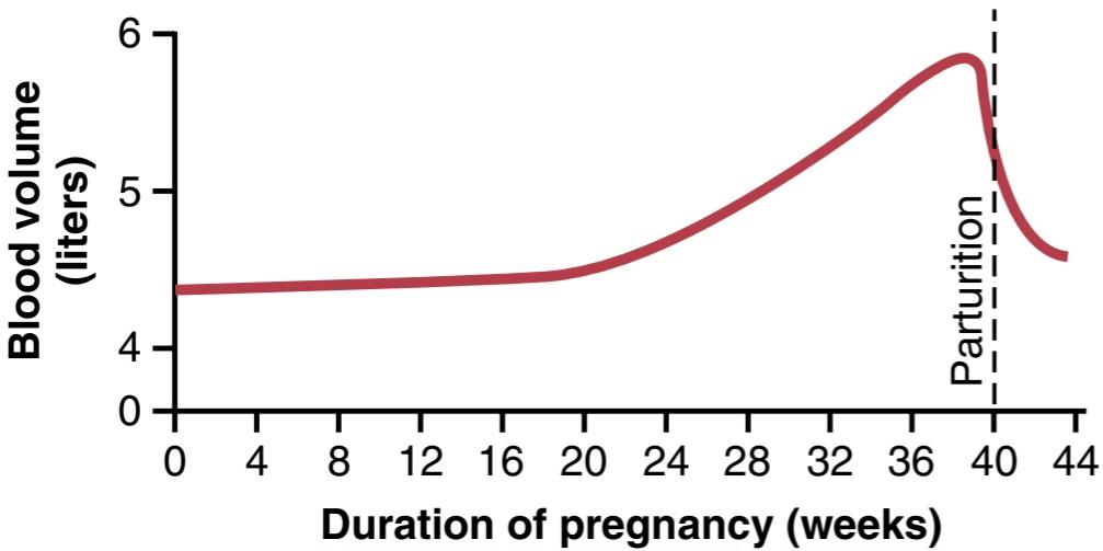
Figure 83-8. Effect of pregnancy on increasing the mother’s blood volume.
شکل 83-8. تاثیر بارداری بر افزایش حجم خون مادر.
Maternal Kidney Function During Pregnancy
The rate of urine formation by a pregnant woman is usually slightly increased because of increased fluid intake and increased load of excretory products. In addition, several special alterations of kidney function occur.
عملکرد کلیه مادر در دوران بارداری
سرعت تشکیل ادرار توسط یک زن باردار معمولاً به دلیل افزایش مصرف مایعات و افزایش بار محصولات دفعی کمی افزایش می یابد. علاوه بر این، چندین تغییر خاص در عملکرد کلیه رخ می دهد.
First, the renal tubules’ reabsorptive capacity for sodium, chloride, and water is increased as much as 50% as a consequence of increased production of salt and water- retaining hormones, especially steroid hormones by the placenta and adrenal cortex.
اول، ظرفیت بازجذب توبولهای کلیوی برای سدیم، کلرید و آب تا 50 درصد افزایش مییابد که نتیجه آن افزایش تولید نمک و هورمونهای نگهدارنده آب، بهویژه هورمونهای استروئیدی توسط جفت و قشر آدرنال است.
Second, the renal blood flow and glomerular filtration rate increase up to 50% during normal pregnancy as a result of renal vasodilation. Although the mechanisms that cause renal vasodilation in pregnancy are still unclear, some studies suggest that increased levels of nitric oxide or the ovarian hormone relaxin may contribute to these changes. The increased glomerular filtration rate likely occurs, at least in part, as a compensation for increased tubular reabsorption of salt and water. Thus, the normal pregnant woman ordinarily accumulates only about 5 pounds of extra water and salt.
دوم، جریان خون کلیوی و نرخ فیلتراسیون گلومرولی در طول بارداری طبیعی در نتیجه اتساع عروق کلیه تا 50٪ افزایش می یابد. اگرچه مکانیسم هایی که باعث اتساع عروق کلیه در بارداری می شوند هنوز نامشخص است، برخی مطالعات نشان می دهد که افزایش سطح اکسید نیتریک یا هورمون ریلکسین تخمدان ممکن است به این تغییرات کمک کند. افزایش نرخ فیلتراسیون گلومرولی احتمالاً حداقل تا حدی به عنوان جبران افزایش بازجذب لولهای نمک و آب رخ میدهد. بنابراین، زن باردار معمولی معمولاً فقط حدود 5 پوند آب و نمک اضافی جمع می کند.
Amniotic Fluid and Its Formation
Normally, the volume of amniotic fluid (the fluid inside the uterus in which the fetus floats) is between 500 ml and 1 liter, but it can be only a few milliliters or as much as several liters. On average, the water in amniotic fluid is replaced once every 3 hours and the electrolytes sodium and potassium are replaced an average of once every 15 hours. A large portion of the fluid is derived from renal excretion by the fetus. Likewise, a certain amount of absorption occurs by way of the gastrointestinal tract and lungs of the fetus. However, even after in utero death of a fetus, some turnover of the amniotic fluid still occurs, which indicates that some of the fluid is formed and absorbed directly through the amniotic membranes.
مایع آمنیوتیک و تشکیل آن
به طور معمول حجم مایع آمنیوتیک (مایع داخل رحمی که جنین در آن شناور است) بین 500 میلی لیتر تا 1 لیتر است، اما می تواند تنها چند میلی لیتر یا تا چند لیتر باشد. به طور متوسط، آب موجود در مایع آمنیوتیک هر 3 ساعت یک بار و الکترولیت های سدیم و پتاسیم به طور متوسط هر 15 ساعت یک بار جایگزین می شوند. بخش بزرگی از مایع از دفع کلیوی توسط جنین به دست می آید. به همین ترتیب، مقدار معینی از جذب از طریق دستگاه گوارش و ریه های جنین اتفاق می افتد. با این حال، حتی پس از مرگ داخل رحمی جنین، مقداری گردش مایع آمنیوتیک هنوز رخ می دهد، که نشان می دهد مقداری از مایع به طور مستقیم از طریق غشای آمنیوتیک تشکیل شده و جذب می شود.
Preeclampsia and Eclampsia
About 5% of all pregnant women experience pregnancy- induced hypertension, a rapid rise in arterial blood pressure to hypertensive levels during the last few months of pregnancy that may also be associated with leakage of large amounts of protein into the urine. This condition is called preeclampsia or toxemia of pregnancy. It is often characterized by excess salt and water retention by the mother’s kidneys and by weight gain and development of edema and hypertension in the mother. In addition, function of the vascular endothelium is impaired, and arterial spasm occurs in many parts of the mother’s body, most significantly in the kidneys, brain, and liver. Renal blood flow and the glomerular filtration rate are decreased, which is exactly opposite to the changes that occur in the normal pregnant woman. The renal effects also include thickened glomerular tufts that contain a protein deposit in the basement membranes.
پره اکلامپسی و اکلامپسی
حدود 5 درصد از تمام زنان باردار فشار خون ناشی از بارداری را تجربه می کنند، افزایش سریع فشار خون شریانی به سطوح فشار خون بالا در چند ماه آخر بارداری که ممکن است با نشت مقادیر زیادی پروتئین به ادرار نیز همراه باشد. این حالت پره اکلامپسی یا مسمومیت بارداری نامیده می شود. اغلب با احتباس بیش از حد نمک و آب توسط کلیه های مادر و افزایش وزن و ایجاد ادم و فشار خون در مادر مشخص می شود. علاوه بر این، عملکرد اندوتلیوم عروقی مختل می شود و اسپاسم شریانی در بسیاری از قسمت های بدن مادر رخ می دهد، به ویژه در کلیه ها، مغز و کبد. جریان خون کلیوی و میزان فیلتراسیون گلومرولی کاهش می یابد که دقیقاً برخلاف تغییراتی است که در یک زن باردار طبیعی رخ می دهد. اثرات کلیوی همچنین شامل توفت های گلومرولی ضخیم شده است که حاوی رسوب پروتئین در غشای پایه است.
Various attempts have been made to prove that preeclampsia is caused by excessive secretion of placental or adrenal hormones, but proof of a hormonal basis is still lacking. Another theory is that preeclampsia results from some type of autoimmunity or allergy in the mother caused by the presence of the fetus. In support of this theory, the acute symptoms usually disappear within a few days after birth of the baby.
تلاشهای مختلفی برای اثبات اینکه پره اکلامپسی در اثر ترشح بیش از حد هورمونهای جفت یا آدرنال ایجاد میشود، انجام شده است، اما هنوز اثباتی مبنی بر مبنای هورمونی وجود ندارد. نظریه دیگر این است که پره اکلامپسی ناشی از نوعی خودایمنی یا آلرژی در مادر ناشی از حضور جنین است. در تأیید این نظریه، علائم حاد معمولاً در عرض چند روز پس از تولد نوزاد ناپدید می شوند.
Evidence also indicates that preeclampsia is initiated by insufficient blood supply to the placenta, resulting in the placenta’s release of substances that cause widespread dysfunction of the maternal vascular endothelium. During normal placental development, the trophoblasts invade the spiral arteries of the uterine endometrium and completely remodel the maternal arteries into much larger blood vessels with low resistance to blood flow (Figure 83-9). In women with preeclampsia, the maternal spiral arteries fail to undergo these adaptive changes, for reasons that are still unclear, and blood supply to the placenta is insufficient. This insufficient blood supply, in turn, causes the placenta to release various substances that enter the mother’s circulation and cause impaired vascular endothelial function, decreased blood flow to the kidneys, excess salt and water retention, and increased blood pressure.
شواهد همچنین نشان می دهد که پره اکلامپسی با خون رسانی ناکافی به جفت آغاز می شود و در نتیجه جفت موادی آزاد می کند که باعث اختلال گسترده در اندوتلیوم عروقی مادر می شود. در طول رشد طبیعی جفت، تروفوبلاستها به شریانهای مارپیچی آندومتر رحم حمله میکنند و به طور کامل شریانهای مادری را به عروق خونی بزرگتر با مقاومت کم در برابر جریان خون تغییر میدهند (شکل 83-9). در زنان مبتلا به پره اکلامپسی، شریانهای مارپیچی مادری به دلایلی که هنوز مشخص نیست، دچار این تغییرات تطبیقی نمیشوند و خونرسانی به جفت ناکافی است. این خون رسانی ناکافی به نوبه خود باعث می شود جفت مواد مختلفی را آزاد کند که وارد گردش خون مادر شده و باعث اختلال در عملکرد اندوتلیال عروقی، کاهش جریان خون به کلیه ها، احتباس بیش از حد نمک و آب و افزایش فشار خون می شود.
Although the factors that link reduced placental blood supply with maternal endothelial dysfunction are still un- certain, some experimental studies suggest a role for in- creased levels of inflammatory cytokines such as tumor necrosis factor-a and interleukin-6. Placental factors that impede angiogenesis (blood vessel growth) have also been shown to contribute to increased inflammatory cytokines and preeclampsia. For example, the antiangiogenic proteins soluble fmsrelated tyrosine kinase 1 (s-Flt1) and soluble endoglin are increased in the blood of women with preeclampsia. These substances are released by the placenta into the maternal circulation in response to ischemia and hypoxia of the placenta. Soluble endoglin and s-Flt1 have multiple effects that may impair function of the maternal vascular endothelium and cause hypertension, proteinuria, and the other systemic manifestations of preeclampsia. However, the precise role of the various factors released from the ischemic placenta in causing the multiple cardiovascular and renal abnormalities in women with preeclampsia is still uncertain.
اگرچه عواملی که کاهش عرضه خون جفت را با اختلال عملکرد اندوتلیال مادر مرتبط میسازند هنوز مشخص نیستند، برخی از مطالعات تجربی نقش افزایش سطوح سایتوکینهای التهابی مانند فاکتور نکروز تومور-a و اینترلوکین-6 را نشان میدهند. فاکتورهای جفتی که مانع از رگ زایی (رشد عروق خونی) می شوند نیز در افزایش سیتوکین های التهابی و پره اکلامپسی نقش دارند. به عنوان مثال، پروتئینهای ضد رگزایی محلول با fms مربوط به تیروزین کیناز 1 (s-Flt1) و اندوگلین محلول در خون زنان مبتلا به پره اکلامپسی افزایش مییابد. این مواد در پاسخ به ایسکمی و هیپوکسی جفت توسط جفت وارد گردش خون مادر می شوند. اندوگلین محلول و s-Flt1 اثرات متعددی دارند که ممکن است عملکرد اندوتلیوم عروقی مادر را مختل کند و باعث فشار خون بالا، پروتئینوری و سایر تظاهرات سیستمیک پره اکلامپسی شود. با این حال، نقش دقیق عوامل مختلف آزاد شده از جفت ایسکمیک در ایجاد ناهنجاری های قلبی عروقی و کلیوی متعدد در زنان مبتلا به پره اکلامپسی هنوز نامشخص است.
Eclampsia is an extreme degree of preeclampsia characterized by vascular spasm throughout the body; clonic seizures in the mother, sometimes followed by coma; greatly decreased kidney output; malfunction of the liver; often extreme hypertension; and a generalized toxic condition of the body. It usually occurs shortly before the birth of the baby. Without treatment, a high percentage of mothers with eclampsia die. However, with optimal and immediate use of rapidly acting vasodilating drugs to reduce the arterial pressure to normal, followed by immediate termination of pregnancy-by cesarean section if necessary-the mortality even in mothers with eclampsia has been reduced to 1% or less.
اکلامپسی یک درجه شدید از پره اکلامپسی است که با اسپاسم عروقی در سراسر بدن مشخص می شود. تشنج های کلونیک در مادر، گاهی اوقات به دنبال کما. کاهش شدید برون ده کلیه؛ اختلال در عملکرد کبد؛ اغلب فشار خون شدید؛ و یک وضعیت سمی عمومی بدن. معمولاً کمی قبل از تولد نوزاد رخ می دهد. بدون درمان، درصد بالایی از مادران مبتلا به اکلامپسی می میرند. با این حال، با استفاده بهینه و فوری از داروهای گشادکننده عروق سریع برای کاهش فشار شریانی به حالت طبیعی و به دنبال آن خاتمه فوری بارداری – در صورت لزوم با سزارین – مرگ و میر حتی در مادران مبتلا به اکلامپسی به 1٪ یا کمتر کاهش یافته است.

Figure 83-9. Remodeling of the spiral arteries of the uterine endometrium during normal pregnancy and failure of the spiral arteries to remodel adequately in preeclampsia. In normal pregnancy, the trophoblasts migrate into the maternal uterine spiral arteries and transform them into much larger, low-resistance, high-flow vessels. In preeclampsia, the trophoblasts fail to invade the endothelium of the spiral arteries adequately, resulting in narrow placental vessels and relative placental ischemia.
شکل 83-9. بازسازی شریان های مارپیچی آندومتر رحم در دوران بارداری طبیعی و شکست عروق مارپیچی در بازسازی مناسب در پره اکلامپسی. در بارداری طبیعی، تروفوبلاستها به شریانهای مارپیچی رحمی مادر مهاجرت میکنند و آنها را به عروق بسیار بزرگتر، کممقاومت و جریان بالا تبدیل میکنند. در پره اکلامپسی، تروفوبلاست ها نمی توانند به اندازه کافی به اندوتلیوم شریان های مارپیچی حمله کنند و در نتیجه عروق جفت باریک و ایسکمی نسبی جفت ایجاد می شود.
PARTURITION
INCREASED UTERINE EXCITABILITY NEAR TERM
Parturition means birth of the baby. Toward the end of pregnancy, the uterus becomes progressively more excitable, until finally it develops such strong rhythmic con- tractions that the baby is expelled. The exact cause of the increased activity of the uterus is not known, but at least two major categories of effects lead up to the intense contractions responsible for parturition: (1) progressive hormonal changes that cause increased excitability of the uterine musculature and (2) progressive mechanical changes.
زایمان
افزایش تحریک پذیری رحم نزدیک ترم
زایمان به معنای تولد نوزاد است. در اواخر بارداری، رحم به تدریج تحریک پذیرتر می شود، تا این که در نهایت چنان انقباضات ریتمیک قوی ایجاد می کند که نوزاد بیرون می رود. علت دقیق افزایش فعالیت رحم مشخص نیست، اما حداقل دو دسته اصلی از اثرات منجر به انقباضات شدید مسئول زایمان می شود: (1) تغییرات هورمونی پیشرونده که باعث افزایش تحریک پذیری ماهیچه های رحم می شود و (2) تغییرات مکانیکی پیشرونده.
HORMONAL FACTORS THAT INCREASE UTERINE CONTRACTILITY
Increased Ratio of Estrogens to Progesterone. Progesterone inhibits uterine contractility during pregnancy, thereby helping to prevent expulsion of the fetus. Conversely, estrogens have tend to increase the degree of uterine contractility, partly because estrogens increase the number of gap junctions between the adjacent uterine smooth muscle cells, but also because of other poorly understood effects. Both progesterone and estrogen are secreted in progressively greater quantities throughout most of pregnancy, but from the seventh month onward, estrogen secretion continues to increase while progesterone secretion remains constant or perhaps even decreases slightly. Therefore, it has been postulated that the estrogen-to-progesterone ratio increases sufficiently toward the end of pregnancy to be at least partly responsible for the increased contractility of the uterus.
عوامل هورمونی که باعث افزایش انقباض رحم می شوند
افزایش نسبت استروژن به پروژسترون. پروژسترون از انقباض رحم در دوران بارداری جلوگیری می کند و در نتیجه به جلوگیری از خروج جنین کمک می کند. برعکس، استروژنها تمایل به افزایش درجه انقباض رحم دارند، تا حدی به این دلیل که استروژنها تعداد اتصالات شکاف بین سلولهای عضله صاف رحم مجاور را افزایش میدهند، اما همچنین به دلیل سایر اثرات ناشناخته است. هم پروژسترون و هم استروژن در بیشتر دوران بارداری به تدریج بیشتر ترشح میشوند، اما از ماه هفتم به بعد، ترشح استروژن همچنان افزایش مییابد در حالی که ترشح پروژسترون ثابت میماند یا حتی ممکن است اندکی کاهش یابد. بنابراین، فرض شده است که نسبت استروژن به پروژسترون به اندازه کافی در پایان بارداری افزایش می یابد تا حداقل تا حدی مسئول افزایش انقباض رحم باشد.
Oxytocin Causes Contraction of the Uterus. Oxytocin, a hormone secreted by the neurohypophysis, specifically causes uterine contraction (see Chapter 76). There are four reasons to believe that oxytocin is important in increasing the contractility of the uterus near term:
اکسی توسین باعث انقباض رحم می شود. اکسی توسین، هورمونی که توسط نوروهیپوفیز ترشح می شود، به طور خاص باعث انقباض رحم می شود (به فصل 76 مراجعه کنید). چهار دلیل برای این باور وجود دارد که اکسی توسین در افزایش انقباض رحم در کوتاه مدت مهم است:
1. The uterine muscle increases its oxytocin receptors and therefore increases its responsiveness to a given dose of oxytocin during the latter few months of pregnancy.
1. عضله رحم گیرنده های اکسی توسین خود را افزایش می دهد و بنابراین واکنش خود را به دوز معین اکسی توسین در چند ماه آخر بارداری افزایش می دهد.
2. Oxytocin secretion rate by the neurohypophysis is considerably increased at the time of labor.
2. میزان ترشح اکسی توسین توسط نوروهیپوفیز به طور قابل توجهی در زمان زایمان افزایش می یابد.
3. Although hypophysectomized animals can still de- liver their young at term, labor is prolonged.
3. اگرچه حیوانات هیپوفیزیکتومی شده هنوز هم می توانند بچه های خود را در ترم به دنیا بیاورند، زایمان طولانی می شود.
4. Experiments in animals indicate that irritation or stretching of the uterine cervix, as occurs during labor, can cause a neurogenic reflex through the paraventricular and supraoptic nuclei of the hypothalamus that causes the posterior pituitary gland (the neurohypophysis) to increase its secretion of oxytocin.
4. آزمایشها روی حیوانات نشان میدهد که تحریک یا کشیدگی دهانه رحم، همانطور که در طول زایمان اتفاق میافتد، میتواند باعث ایجاد یک رفلکس عصبی از طریق هستههای پارا بطنی و فوقاپتیک هیپوتالاموس شود که باعث میشود غده هیپوفیز خلفی (هیپوفیز عصبی) ترشح اکسی توسین خود را افزایش دهد.
Effect of Fetal Hormones on the Uterus. The fetus’s pituitary gland secretes increasing quantities of oxytocin, which might play a role in exciting the uterus. Also, the fetus’s adrenal glands secrete large quantities of cortisol, another possible uterine stimulant. In addition, the fetal membranes release prostaglandins in high concentration at the time of labor. These prostaglandins, too, can increase the intensity of uterine con- tractions.
تاثیر هورمون های جنینی بر رحم. غده هیپوفیز جنین مقادیر فزاینده ای اکسی توسین ترشح می کند که ممکن است در تحریک رحم نقش داشته باشد. همچنین، غدد فوق کلیوی جنین مقادیر زیادی کورتیزول ترشح می کنند که یکی دیگر از محرک های احتمالی رحم است. علاوه بر این، غشای جنین در زمان زایمان پروستاگلاندین ها را با غلظت بالایی آزاد می کند. این پروستاگلاندین ها نیز می توانند شدت انقباضات رحمی را افزایش دهند.
Mechanical Factors That Increase Uterine Contractility
Stretch of the Uterine Musculature. Simply stretching smooth muscles usually increases their contractility. Further, intermittent stretch, which occurs repeatedly in the uterus because of fetal movements, can also elicit smooth muscle contraction. Note especially that twins are born, on average, 19 days earlier than a single child, which emphasizes the importance of mechanical stretch in eliciting uterine contractions.
عوامل مکانیکی که انقباض رحم را افزایش می دهند
کشش عضلات رحم. کشش ساده عضلات صاف معمولاً انقباض آنها را افزایش می دهد. علاوه بر این، کشش متناوب، که به طور مکرر در رحم به دلیل حرکات جنین رخ می دهد، همچنین می تواند باعث انقباض عضلات صاف شود. به خصوص توجه داشته باشید که دوقلوها به طور متوسط 19 روز زودتر از یک فرزند مجرد به دنیا می آیند که بر اهمیت کشش مکانیکی در ایجاد انقباضات رحم تاکید دارد.
Stretch or Irritation of the Cervix. There is reason to believe that stretching or irritating the uterine cervix is particularly important in eliciting uterine contractions. For example, obstetricians frequently induce labor by rupturing the membranes so the head of the baby stretch- es the cervix more forcefully than usual or irritates it in other ways.
کشش یا تحریک دهانه رحم. دلایلی وجود دارد که باور کنیم کشش یا تحریک دهانه رحم در ایجاد انقباضات رحمی مهم است. به عنوان مثال، متخصصان زنان و زایمان اغلب با پارگی غشاها، زایمان را القا می کنند تا سر نوزاد دهانه رحم را با شدت بیشتری نسبت به معمول کشیده یا به روش های دیگر آن را تحریک می کند.
The mechanism whereby cervical irritation excites the body of the uterus is not known. It has been suggested that stretching or irritation of nerves in the cervix initiates reflexes to the body of the uterus, but the effect could also result simply from myogenic transmission of signals from the cervix to the body of the uterus.
مکانیسمی که در آن تحریک دهانه رحم بدن رحم را تحریک می کند شناخته شده نیست. پیشنهاد شده است که کشش یا تحریک اعصاب در دهانه رحم بازتاب هایی را به بدن رحم آغاز می کند، اما این اثر می تواند به سادگی از انتقال میوژنیک سیگنال ها از دهانه رحم به بدن رحم باشد.
ONSET OF LABOR-A POSITIVE FEEDBACK MECHANISM FOR ITS INITIATION
During pregnancy, the uterus undergoes periodic episodes of weak and slow rhythmic contractions called Braxton Hicks contractions. These contractions are usually not felt until the second or third trimester and become progressively stronger toward the end of pregnancy; then they change suddenly, within hours, to become exceptionally strong contractions that start stretching the cervix and later force the baby through the birth canal, thereby causing parturition. This process is called labor, and the strong contractions that result in final parturition are called labor contractions.
شروع کار – مکانیزم بازخورد مثبت برای شروع آن
در دوران بارداری، رحم دورههایی از انقباضات ریتمیک ضعیف و آهسته به نام انقباضات براکستون هیکس را تجربه میکند. این انقباضات معمولاً تا سه ماهه دوم یا سوم احساس نمی شوند و در اواخر بارداری به تدریج قوی تر می شوند. سپس در عرض چند ساعت ناگهان تغییر میکنند و تبدیل به انقباضات فوقالعاده شدیدی میشوند که شروع به کشش دهانه رحم میکند و بعداً نوزاد را از کانال زایمان مجبور میکند و در نتیجه باعث زایمان میشود. این فرآیند زایمان نامیده می شود و انقباضات قوی که منجر به زایمان نهایی می شود انقباضات زایمان نامیده می شود.
We do not know what suddenly changes the slow, weak rhythmicity of the uterus into strong labor contractions. However, the positive feedback theory suggests that stretching of the cervix by the fetus’s head finally becomes great enough to elicit a strong reflex increase in contractility of the uterine body. This pushes the baby for- ward, which stretches the cervix more and initiates more positive feedback to the uterine body. Thus, the process repeats until the baby is expelled. This theory is shown in Figure 83-10, and the following observations support this theory.
ما نمی دانیم چه چیزی به طور ناگهانی ریتمیک ضعیف و آهسته رحم را به انقباضات شدید زایمان تبدیل می کند. با این حال، نظریه بازخورد مثبت نشان می دهد که کشش دهانه رحم توسط سر جنین در نهایت به اندازه کافی بزرگ می شود که باعث افزایش رفلکس قوی در انقباض بدن رحم شود. این امر نوزاد را به سمت جلو هل می دهد که دهانه رحم را بیشتر کشیده و بازخورد مثبت بیشتری را به بدن رحم آغاز می کند. بنابراین، این روند تا زمانی که کودک بیرون رانده شود، تکرار می شود. این نظریه در شکل 83-10 نشان داده شده است و مشاهدات زیر این نظریه را تایید می کند.
First, labor contractions obey all the principles of positive feedback. That is, once the strength of uterine contraction becomes greater than a critical value, each contraction leads to subsequent contractions that become stronger and stronger until maximum effect is achieved. By referring to the discussion in Chapter 1 of positive feedback in control systems, one can see that this is the precise nature of all positive feedback mechanisms when the feedback gain becomes greater than a critical value.
اول، انقباضات زایمان از تمام اصول بازخورد مثبت پیروی می کنند. یعنی زمانی که قدرت انقباض رحم از مقدار بحرانی بیشتر شود، هر انقباض منجر به انقباضات بعدی می شود که قوی تر و قوی تر می شوند تا حداکثر اثر حاصل شود. با مراجعه به بحث در فصل 1 بازخورد مثبت در سیستم های کنترلی، می توان دریافت که این ماهیت دقیق همه مکانیسم های بازخورد مثبت است زمانی که سود بازخورد بیشتر از یک مقدار بحرانی می شود.
Second, two known types of positive feedback increase uterine contractions during labor: (1) stretching of the cervix causes the entire body of the uterus to contract, and this contraction stretches the cervix even more because of the downward thrust of the baby’s head, and (2) cervical stretching also causes the pituitary gland to secrete oxytocin, which is another means for increasing uterine contractility.
دوم، دو نوع شناخته شده بازخورد مثبت باعث افزایش انقباضات رحم در حین زایمان می شود: (1) کشش دهانه رحم باعث می شود تمام بدن رحم منقبض شود و این انقباض دهانه رحم را به دلیل فشار به پایین سر نوزاد بیشتر می کند و (2) کشش دهانه رحم همچنین باعث می شود غده هیپوفیز که به معنای افزایش انقباض ترشح اکسی توسین است.
To summarize, multiple factors increase the contractility of the uterus toward the end of pregnancy. Eventually a uterine contraction becomes strong enough to irritate the uterus, especially at the cervix, and this irritation increases uterine contractility still more because of positive feedback, resulting in a second uterine contraction stronger than the first, a third stronger than the second, and so forth. Once these contractions become strong enough to cause this type of feedback, with each succeeding contraction greater than the preceding one, the process proceeds to completion. One might ask about the many cases of false labor, in which the contractions become stronger and stronger and then fade away. Remember that for a positive feedback to continue, each new cycle of the positive feedback must be stronger than the previous one. If at any time after labor starts some contractions fail to re-excite the uterus sufficiently, the positive feedback could go into a retrograde decline, and the labor contractions would fade away.
به طور خلاصه، عوامل متعددی باعث افزایش انقباض رحم در اواخر بارداری می شود. در نهایت یک انقباض رحم به اندازه ای قوی می شود که رحم را به خصوص در دهانه رحم تحریک کند و این تحریک به دلیل بازخورد مثبت انقباض رحم را بیشتر افزایش می دهد و در نتیجه انقباض رحم دوم قوی تر از اولی، یک سوم قوی تر از دومی و غیره می شود. هنگامی که این انقباضات به اندازه کافی قوی می شوند که باعث ایجاد این نوع بازخورد شوند، با هر انقباض بعدی بزرگتر از قبلی، فرآیند به پایان می رسد. ممکن است در مورد موارد متعدد زایمان کاذب بپرسید که در آن انقباضات قویتر و قویتر میشوند و سپس محو میشوند. به یاد داشته باشید که برای ادامه بازخورد مثبت، هر چرخه جدید بازخورد مثبت باید قوی تر از دوره قبلی باشد. اگر در هر زمانی پس از شروع زایمان، برخی از انقباضات نتوانند به اندازه کافی رحم را تحریک کنند، بازخورد مثبت می تواند به سمت کاهش برگشتی رفته و انقباضات زایمان از بین برود.

Figure 83-10. Theory for the onset of intensely strong contractions during labor.
شکل 83-10. تئوری برای شروع انقباضات شدید در طول زایمان.
ABDOMINAL MUSCLE CONTRACTIONS DURING LABOR
Once uterine contractions become strong during labor, pain signals originate both from the uterus and from the birth canal. These signals, in addition to causing suffering, elicit neurogenic reflexes in the spinal cord to the abdominal muscles, causing intense contractions of these muscles. The abdominal contractions add greatly to the force that causes expulsion of the baby.
انقباضات عضلات شکم در حین زایمان
هنگامی که انقباضات رحم در حین زایمان قوی می شوند، سیگنال های درد هم از رحم و هم از کانال زایمان منشا می گیرند. این سیگنال ها علاوه بر ایجاد رنج، رفلکس های عصبی در نخاع را به عضلات شکم برمی انگیزند و باعث انقباضات شدید این عضلات می شوند. انقباضات شکمی به شدت به نیرویی که باعث بیرون راندن نوزاد می شود می افزاید.
Mechanics of Parturition
The uterine contractions during labor begin mainly at the top of the uterine fundus and spread downward over the body of the uterus. Also, the intensity of contraction is great in the top and body of the uterus but weak in the lower segment of the uterus adjacent to the cervix. Therefore, each uterine contraction tends to force the baby downward toward the cervix.
مکانیک زایمان
انقباضات رحم در حین زایمان عمدتاً از بالای فوندوس رحم شروع می شود و به سمت پایین روی بدنه رحم پخش می شود. همچنین شدت انقباض در بالا و بدنه رحم زیاد است اما در قسمت پایینی رحم مجاور دهانه رحم ضعیف است. بنابراین، هر انقباض رحمی تمایل دارد که کودک را به سمت پایین به سمت دهانه رحم فشار دهد.
In the early part of labor, the contractions might occur only once every 30 minutes. As labor progresses, the con- tractions finally appear as often as once every 1 to 3 minutes and the intensity of contraction increases greatly, with only a short period of relaxation between contractions. The combined contractions of the uterine and abdominal musculature during delivery of the baby cause a downward force on the fetus of about 25 pounds during each strong contraction.
در اوایل زایمان، انقباضات ممکن است تنها یک بار در هر 30 دقیقه رخ دهد. با پیشرفت زایمان، انقباضات در نهایت هر 1 تا 3 دقیقه یک بار ظاهر می شوند و شدت انقباض به شدت افزایش می یابد، تنها با یک دوره کوتاه آرامش بین انقباضات. انقباضات ترکیبی ماهیچه های رحم و شکم در هنگام زایمان باعث می شود که در هر انقباض قوی حدود 25 پوند نیروی رو به پایین روی جنین وارد شود.
It is fortunate that the contractions of labor occur intermittently, because strong contractions impede or some- times even stop blood flow through the placenta and would cause death of the fetus if the contractions were continuous. Indeed, overuse of various uterine stimulants, such as oxytocin, can cause uterine spasm rather than rhythmic contractions and can lead to death of the fetus.
خوشبختانه انقباضات زایمان به طور متناوب رخ می دهد، زیرا انقباضات شدید جریان خون را در جفت مختل می کند یا گاهی اوقات حتی متوقف می کند و اگر انقباضات مداوم باشد باعث مرگ جنین می شود. در واقع، استفاده بیش از حد از محرک های رحمی مختلف، مانند اکسی توسین، می تواند باعث اسپاسم رحم به جای انقباضات ریتمیک شود و می تواند منجر به مرگ جنین شود.
In more than 95% of births, the head is the first part of the baby to be expelled and, in most of the remaining cases, the buttocks are presented first. Entering the birth canal with the buttocks or feet first is called a breech presentation.
در بیش از 95 درصد از تولدها، سر اولین قسمتی از نوزاد است که خارج می شود و در بیشتر موارد باقی مانده، ابتدا باسن ظاهر می شود. ورود بریچ ابتدا با باسن یا پا وارد کانال زایمان می شود.
The head acts as a wedge to open the structures of the birth canal as the fetus is forced downward. The first major obstruction to expulsion of the fetus is the uterine cervix. Toward the end of pregnancy, the cervix becomes soft, which allows it to stretch when labor contractions begin in the uterus. The so-called first stage of labor is a period of progressive cervical dilation, lasting until the cervical opening is as large as the head of the fetus. This stage usually lasts for 8 to 24 hours in the first pregnancy but often only a few minutes after many pregnancies.
سر به عنوان یک گوه عمل می کند تا ساختارهای کانال زایمان را در حالی که جنین مجبور به پایین آمدن می شود باز کند. اولین انسداد اصلی خروج جنین، دهانه رحم است. در اواخر بارداری، دهانه رحم نرم می شود، که به آن اجازه می دهد با شروع انقباضات زایمان در رحم کشیده شود. به اصطلاح اولین مرحله زایمان، یک دوره گشاد شدن پیشرونده دهانه رحم است که تا زمانی که دهانه رحم به اندازه سر جنین بزرگ شود طول می کشد. این مرحله معمولاً 8 تا 24 ساعت در بارداری اول طول می کشد اما اغلب تنها چند دقیقه بعد از بسیاری از بارداری ها ادامه می یابد.
Once the cervix has dilated fully, the fetal membranes usually rupture and the amniotic fluid is lost suddenly through the vagina. Then the head of the fetus moves rapidly into the birth canal, and with additional force from above, it continues to wedge its way through the canal until delivery occurs. This is called the second stage of labor, and it may last from as little as 1 minute after many pregnancies to 30 minutes or more in the first pregnancy.
هنگامی که دهانه رحم به طور کامل گشاد شد، معمولاً غشای جنین پاره می شود و مایع آمنیوتیک به طور ناگهانی از طریق واژن از بین می رود. سپس سر جنین به سرعت به داخل کانال زایمان حرکت می کند و با نیروی اضافی از بالا، مسیر خود را از طریق کانال تا زمان زایمان ادامه می دهد. این مرحله دوم زایمان نامیده می شود و ممکن است از 1 دقیقه بعد از بسیاری از بارداری ها تا 30 دقیقه یا بیشتر در بارداری اول طول بکشد.
Separation and Delivery of the Placenta. For 10 to 45 minutes after birth of the baby, the uterus continues to contract to a smaller and smaller size, which causes a shearing effect between the walls of the uterus and the placenta, thus separating the placenta from its implantation site. Separation of the placenta opens the placental sinuses and causes bleeding. The amount of bleeding is usually limited to an average of 350 ml by the following mechanism:
جداسازی و تحویل جفت. به مدت 10 تا 45 دقیقه پس از تولد نوزاد، رحم به اندازه کوچکتر و کوچکتر منقبض می شود که باعث ایجاد اثر برشی بین دیواره های رحم و جفت می شود و در نتیجه جفت از محل لانه گزینی خود جدا می شود. جدا شدن جفت سینوس های جفت را باز می کند و باعث خونریزی می شود. مقدار خونریزی معمولاً با مکانیسم زیر به طور متوسط 350 میلی لیتر محدود می شود:
• The smooth muscle fibers of the uterine musculature are arranged in figures of eight around the blood vessels as the vessels pass through the uterine wall.
• فیبرهای عضلانی صاف ماهیچه رحم به شکل هشت تایی در اطراف رگهای خونی قرار گرفتهاند که رگها از دیواره رحم عبور میکنند.
• Therefore, contraction of the uterus after delivery of the baby constricts the vessels that had previously supplied blood to the placenta.
• بنابراین، انقباض رحم پس از زایمان باعث انقباض عروقی می شود که قبلاً خون جفت را تامین می کردند.
• In addition, it is believed that vasoconstrictor prostaglandins formed at the placental separation site cause additional blood vessel spasm.
• علاوه بر این، اعتقاد بر این است که پروستاگلاندین های منقبض کننده عروق تشکیل شده در محل جدا شدن جفت باعث اسپاسم عروق خونی اضافی می شود.
Labor Pains
With each uterine contraction, the mother experiences considerable pain. The cramping pain in early labor is probably caused mainly by hypoxia of the uterine muscle resulting from compression of the blood vessels in the uterus. This pain is not felt when the visceral sensory hypogastric nerves, which carry the visceral sensory fibers leading from the uterus, have been sectioned.
درد زایمان
با هر انقباض رحم، مادر درد قابل توجهی را تجربه می کند. درد گرفتگی در اوایل زایمان احتمالاً عمدتاً ناشی از هیپوکسی عضله رحم است که در نتیجه فشرده شدن رگ های خونی در رحم ایجاد می شود. زمانی که اعصاب هیپوگاستریک حسی احشایی که رشته های حسی احشایی منتهی به رحم را حمل می کنند، بریده شده باشند، این درد احساس نمی شود.
During the second stage of labor, when the fetus is being expelled through the birth canal, much more severe pain is caused by cervical stretching, perineal stretching, and stretching or tearing of structures in the vaginal canal. This pain is conducted to the mother’s spinal cord and brain by somatic nerves instead of by the visceral sensory nerves.
در مرحله دوم زایمان، زمانی که جنین از طریق کانال زایمان خارج می شود، درد بسیار شدیدتری ناشی از کشش دهانه رحم، کشش پرینه و کشیدگی یا پارگی ساختارهای کانال واژن است. این درد به جای اعصاب حسی احشایی توسط اعصاب جسمی به نخاع و مغز مادر منتقل می شود.
Involution of the Uterus After Parturition
During the first 4 to 5 weeks after parturition, the uterus involutes. Its weight becomes less than half its immediate postpartum weight within 1 week, and in 4 weeks, if the mother lactates, the uterus may become as small as it was before pregnancy. This effect of lactation results from the suppression of pituitary gonadotropin and ovarian hormone secretion during the first few months of lactation, as dis- cussed later. During early involution of the uterus, the placental site on the endometrial surface autolyzes, causing a vaginal discharge known as lochia, which is first bloody and then serous in nature and continues for a total of about 10 days. After this time, the endometrial surface becomes re- epithelialized and ready for normal, nongravid sex life again.
چرخش رحم پس از زایمان
در طی 4 تا 5 هفته اول پس از زایمان، رحم پیچ خورده است. وزن آن در عرض 1 هفته کمتر از نصف وزن بلافاصله پس از زایمان می شود و در 4 هفته، اگر مادر شیردهی داشته باشد، رحم ممکن است به اندازه قبل از بارداری کوچک شود. این اثر شیردهی ناشی از سرکوب گنادوتروپین هیپوفیز و ترشح هورمون تخمدان در چند ماه اول شیردهی است، همانطور که بعداً بحث شد. در طول دوران اولیه رحم، محل جفت روی سطح آندومتر اتولیز می شود و باعث ترشحات واژن معروف به لوچیا می شود که ابتدا خونی و سپس سروز بوده و در مجموع حدود 10 روز ادامه می یابد. پس از این مدت، سطح آندومتر مجدداً اپیتلیالیزه می شود و دوباره برای زندگی جنسی عادی و بدون زایمان آماده می شود.
LACTATION
شیردهی
DEVELOPMENT OF THE BREASTS
The breasts, shown in Figure 83-11, begin to develop at puberty. This development is stimulated by the estrogens of the monthly female sexual cycle; estro- gens stimulate growth of the breasts’ mammary glands plus the deposition of fat to give the breasts mass. In addition, far greater growth occurs during the high- estrogen state of pregnancy, and only then does the glandular tissue become completely developed for pro- duction of milk.
رشد سینه ها
سینه ها که در شکل 11-83 نشان داده شده اند، در دوران بلوغ شروع به رشد می کنند. این رشد توسط استروژن چرخه جنسی ماهانه زن تحریک می شود. استروژن ها باعث تحریک رشد غدد پستانی سینه به علاوه رسوب چربی برای ایجاد حجم به سینه ها می شود. بعلاوه، رشد بسیار بیشتری در دوران بارداری با استروژن بالا رخ می دهد و تنها در این صورت است که بافت غده برای تولید شیر به طور کامل توسعه می یابد.
Estrogens Stimulate Growth of the Ductal System of the Breasts. All through pregnancy, the large quantities of estrogens secreted by the placenta cause the ductal system of the breasts to grow and branch. Simultaneously, the stroma of the breasts increases in quantity, and large quantities of fat are laid down in the stroma.
استروژن ها رشد سیستم مجرای سینه ها را تحریک می کنند. در تمام دوران بارداری، مقادیر زیادی استروژن ترشح شده توسط جفت باعث رشد و انشعاب سیستم مجرای سینه ها می شود. به طور همزمان، استرومای سینه ها از نظر کمیت افزایش می یابد و مقادیر زیادی چربی در استروما قرار می گیرد.
Also important for growth of the ductal system are at least four other hormones: growth hormone, prolactin, adrenal glucocorticoids, and insulin. Each of these hormones is known to play at least some role in protein metabolism, which presumably explains their function in the development of the breasts.
همچنین برای رشد سیستم مجرای حداقل چهار هورمون دیگر مهم هستند: هورمون رشد، پرولاکتین، گلوکوکورتیکوئیدهای آدرنال و انسولین. هر یک از این هورمون ها حداقل نقشی در متابولیسم پروتئین ایفا می کنند که احتمالاً عملکرد آنها را در رشد سینه ها توضیح می دهد.

Figure 83-11. A, The breast and its secretory lobules, alveoli, and lactiferous ducts (milk ducts) that constitute its mammary gland. B, The enlargements show a lobule and milk-secreting cells (C) of an alveolus.
شکل 83-11. الف، پستان و لوبول های ترشحی، آلوئول ها و مجاری شیری (مجرای شیری) که غده پستانی آن را تشکیل می دهند. ب، بزرگ شدن یک لوبول و سلول های ترشح کننده شیر (C) آلوئول را نشان می دهد.
Progesterone Is Required for Full Development of the Lobule-Alveolar System. Final development of the breasts into milk-secreting organs also requires progesterone. Once the ductal system has developed, progesterone-acting synergistically with estrogen, as well as with the other hormones just mentioned-causes additional growth of the breast lobules, with budding of alveoli and development of secretory characteristics in the cells of the alveoli. These changes are analogous to the secretory effects of progesterone on the endometrium of the uterus during the latter half of the female menstrual cycle.
پروژسترون برای رشد کامل سیستم لوبول-آلوئولار مورد نیاز است. رشد نهایی سینه ها به اندام های ترشح کننده شیر نیز به پروژسترون نیاز دارد. هنگامی که سیستم مجرای رشد کرد، پروژسترون با استروژن، و همچنین با هورمون های دیگر که ذکر شد، به صورت هم افزایی عمل می کند، باعث رشد بیشتر لوبول های سینه، با جوانه زدن آلوئول ها و ایجاد ویژگی های ترشحی در سلول های آلوئول می شود. این تغییرات مشابه اثرات ترشحی پروژسترون بر روی آندومتر رحم در نیمه دوم سیکل قاعدگی زنان است.
PROLACTIN PROMOTES LACTATION
Although estrogen and progesterone are essential for physical development of the breasts during pregnancy, a specific effect of both these hormones is to inhibit the actual secretion of milk. Conversely, the hormone prolactin has the opposite effect and promotes milk secretion. Prolactin is secreted by the mother’s anterior pituitary gland, and its concentration in her blood rises steadily from the fifth week of pregnancy until birth of the baby, at which time it has risen to 10 to 20 times the normal nonpregnant level. This high level of prolactin at the end of pregnancy is shown in Figure 83-12.
پرولاکتین باعث افزایش شیردهی می شود
اگرچه استروژن و پروژسترون برای رشد فیزیکی سینه ها در دوران بارداری ضروری هستند، اما اثر خاص هر دو این هورمون ها مهار ترشح واقعی شیر است. برعکس، هورمون پرولاکتین اثر معکوس دارد و باعث ترشح شیر می شود. پرولاکتین توسط غده هیپوفیز قدامی مادر ترشح می شود و غلظت آن در خون او از هفته پنجم بارداری تا تولد نوزاد به طور پیوسته افزایش می یابد و در این زمان به 10 تا 20 برابر سطح طبیعی غیرباردار افزایش یافته است. این سطح بالای پرولاکتین در پایان بارداری در شکل 83-12 نشان داده شده است.
In addition, the placenta secretes large quantities of human chorionic somatomammotropin, which probably has lactogenic properties, thus supporting the prolactin from the mother’s pituitary during pregnancy. Even so, because of the suppressive effects of estrogen and progesterone, no more than a few milliliters of fluid are secreted each day until after the baby is born. The fluid secreted during the last few days before and the first few days after parturition is called colostrum; it contains essentially the same concentrations of proteins and lactose as milk, but it has almost no fat, and its maximum rate of production is about 1/100 the subsequent rate of milk production.
علاوه بر این، جفت مقادیر زیادی سوماتوماموتروپین جفتی انسانی ترشح می کند که احتمالاً دارای خواص لاکتوژنیک است، بنابراین از پرولاکتین هیپوفیز مادر در دوران بارداری حمایت می کند. با این حال، به دلیل اثرات سرکوبکننده استروژن و پروژسترون، روزانه بیش از چند میلیلیتر مایع تا پس از تولد نوزاد ترشح نمیشود. مایع ترشح شده در چند روز آخر قبل و چند روز اول پس از زایمان، آغوز نامیده می شود. در اصل حاوی همان غلظت پروتئین و لاکتوز شیر است، اما تقریباً بدون چربی است و حداکثر میزان تولید آن حدود 1/100 میزان تولید شیر بعدی است.
Immediately after the baby is born, the sudden loss of both estrogen and progesterone secretion from the placenta allows the lactogenic effect of prolactin from the mother’s pituitary gland to assume its natural milk- promoting role, and during the next 1 to 7 days, the breasts begin to secrete copious quantities of milk instead of colostrum. This secretion of milk requires an adequate background secretion of most of the mother’s other hormones as well, but most important are growth hormone, cortisol, parathyroid hormone, and insulin. These hormones are necessary to provide the amino acids, fatty acids, glucose, and calcium required for the formation of milk.
بلافاصله پس از تولد نوزاد، از دست دادن ناگهانی ترشح استروژن و پروژسترون از جفت به اثر لاکتوژنیک پرولاکتین از غده هیپوفیز مادر اجازه می دهد تا نقش طبیعی تولید شیر خود را ایفا کند و در طی 1 تا 7 روز آینده، پستان ها شروع به ترشح مقادیر زیادی شیر به جای شیر می کنند. این ترشح شیر مستلزم ترشح کافی پس زمینه بیشتر هورمون های دیگر مادر نیز هست، اما مهم ترین آنها هورمون رشد، کورتیزول، هورمون پاراتیروئید و انسولین است. این هورمون ها برای تامین اسیدهای آمینه، اسیدهای چرب، گلوکز و کلسیم مورد نیاز برای تشکیل شیر ضروری هستند.
After the birth of the baby, the basal level of prolactin secretion returns to the nonpregnant level during the next few weeks, as shown in Figure 83-12. However, each time the mother nurses her baby, nervous signals from the nipples to the hypothalamus cause a 10- to 20-fold surge in prolactin secretion that lasts for about 1 hour, which is also shown in Figure 83-12. This prolactin acts on the mother’s breasts to keep the mammary glands secreting milk into the alveoli for the subsequent nursing periods. If this prolactin surge is absent or blocked as a result of hypothalamic or pituitary damage or if nursing does not continue, the breasts lose their ability to produce milk within 1 week or so. However, milk production can continue for several years if the child continues to suckle, although the rate of milk formation normally decreases considerably after 7 to 9 months.
پس از تولد نوزاد، سطح پایه ترشح پرولاکتین در طی چند هفته آینده به سطح غیر باردار باز می گردد، همانطور که در شکل 83-12 نشان داده شده است. با این حال، هر بار که مادر به نوزادش شیر می دهد، سیگنال های عصبی از نوک سینه ها به هیپوتالاموس باعث افزایش 10 تا 20 برابری ترشح پرولاکتین می شود که حدود 1 ساعت طول می کشد، که در شکل 83-12 نیز نشان داده شده است. این پرولاکتین بر روی سینههای مادر تأثیر میگذارد تا غدد پستانی ترشح شیر را برای دورههای شیردهی بعدی به داخل آلوئولها نگه دارد. اگر این افزایش پرولاکتین در نتیجه آسیب هیپوتالاموس یا هیپوفیز وجود نداشته باشد یا مسدود شود یا شیردهی ادامه پیدا نکند، سینه ها توانایی خود را برای تولید شیر در عرض یک هفته یا بیشتر از دست می دهند. با این حال، در صورتی که کودک به شیر خوردن ادامه دهد، تولید شیر می تواند چندین سال ادامه یابد، اگرچه سرعت تشکیل شیر معمولاً پس از 7 تا 9 ماه به میزان قابل توجهی کاهش می یابد.
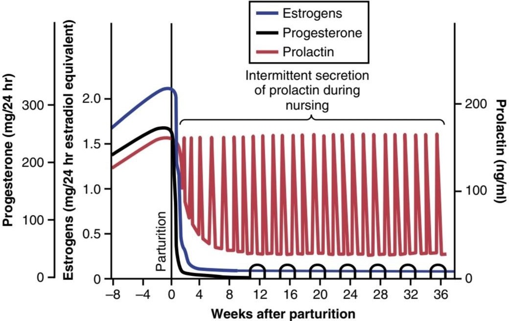
Figure 83-12. Changes in rates of secretion of estrogens, progesterone, and prolactin for 8 weeks before parturition and 36 weeks thereafter. Note especially the decrease of prolactin secretion back to basal levels within a few weeks after parturition, but also the intermittent periods of marked prolactin secretion (for about 1 hour at a time) during and after periods of nursing.
شکل 83-12. تغییرات در میزان ترشح استروژن، پروژسترون و پرولاکتین به مدت 8 هفته قبل از زایمان و 36 هفته پس از آن. به ویژه به کاهش ترشح پرولاکتین به سطح پایه در طی چند هفته پس از زایمان، اما همچنین دوره های متناوب ترشح مشخص پرولاکتین (حدود 1 ساعت در هر بار) در طول و بعد از دوره های شیردهی توجه کنید.
The Hypothalamus Secretes Prolactin Inhibitory Hormone. The hypothalamus plays an essential role in controlling prolactin secretion, as it does for almost all the other anterior pituitary hormones. How- ever, this control is different in one aspect: The hypothalamus mainly stimulates production of all the other hormones, but it mainly inhibits prolactin production. Consequently, damage to the hypothalamus or blockage of the hypothalamic-hypophysial portal system often increases prolactin secretion while it depresses secretion of the other anterior pituitary hormones.
هیپوتالاموس هورمون بازدارنده پرولاکتین ترشح می کند. هیپوتالاموس مانند سایر هورمون های هیپوفیز قدامی نقش اساسی در کنترل ترشح پرولاکتین دارد. با این حال، این کنترل از یک جنبه متفاوت است: هیپوتالاموس عمدتاً تولید همه هورمونهای دیگر را تحریک میکند، اما عمدتاً تولید پرولاکتین را مهار میکند. در نتیجه، آسیب به هیپوتالاموس یا انسداد سیستم پورتال هیپوتالاموس-هیپوفیزیال اغلب ترشح پرولاکتین را افزایش می دهد در حالی که ترشح سایر هورمون های هیپوفیز قدامی را کاهش می دهد.
Therefore, it is believed that anterior pituitary secretion of prolactin is controlled either entirely or almost entirely by an inhibitory factor formed in the hypothalamus and transported through the hypothalamic-hypophysial portal system to the anterior pituitary gland. This factor is sometimes called prolactin inhibitory hormone, but it is almost certainly the same as the catecholamine dopamine, which is known to be secreted by the arcuate nuclei of the hypothalamus and can decrease prolactin secretion as much as 10-fold.
بنابراین، اعتقاد بر این است که ترشح قدامی هیپوفیز پرولاکتین به طور کامل یا تقریباً به طور کامل توسط یک عامل بازدارنده تشکیل شده در هیپوتالاموس کنترل می شود و از طریق سیستم پورتال هیپوتالاموس-هیپوفیزیال به غده هیپوفیز قدامی منتقل می شود. این عامل گاهی اوقات هورمون بازدارنده پرولاکتین نامیده می شود، اما تقریباً به طور قطع مشابه کاتکول آمین دوپامین است که شناخته شده است که توسط هسته های قوسی هیپوتالاموس ترشح می شود و می تواند ترشح پرولاکتین را تا 10 برابر کاهش دهد.
Suppression of the Female Ovarian Cycles in Nursing Mothers for Many Months After Delivery. In most nursing mothers, the ovarian cycle (and ovulation) does not resume until a few weeks after cessation of nursing. The reason seems to be that the same nervous signals from the breasts to the hypothalamus that cause prolactin secretion during suckling-either because of the nervous signals or because of a subsequent effect of increased prolactin-inhibit secretion of gonadotropin- releasing hormone by the hypothalamus. This inhibition, in turn, suppresses formation of the pituitary gonadotropic hormones-luteinizing hormone and follicle- stimulating hormone. However, after several months of lactation, in some mothers (especially those who nurse their babies only some of the time), the pituitary begins to secrete sufficient gonadotropic hormones to reinstate the monthly sexual cycle, even though nursing continues.
سرکوب چرخه های تخمدان زن در مادران شیرده برای چندین ماه پس از زایمان. در اکثر مادران شیرده، چرخه تخمدان (و تخمک گذاری) تا چند هفته پس از قطع شیردهی از سر گرفته نمی شود. به نظر می رسد دلیل آن این است که همان سیگنال های عصبی از سینه ها به هیپوتالاموس که باعث ترشح پرولاکتین در حین شیردهی می شود – چه به دلیل سیگنال های عصبی و چه به دلیل اثر بعدی افزایش پرولاکتین – ترشح هورمون آزاد کننده گنادوتروپین توسط هیپوتالاموس را مهار می کند. این مهار، به نوبه خود، تشکیل هورمون های گنادوتروپیک هیپوفیز – هورمون لوتئینه کننده و هورمون محرک فولیکول را سرکوب می کند. با این حال، پس از چندین ماه شیردهی، در برخی از مادران (به ویژه در آنهایی که فقط برخی از اوقات از نوزادان خود شیر می دهند)، هیپوفیز شروع به ترشح هورمون های گنادوتروپیک کافی برای بازگرداندن چرخه جنسی ماهانه می کند، حتی اگر شیردهی ادامه یابد.
EJECTION (OR “LET-DOWN”) PROCESS IN MILK SECRETION-FUNCTION OF OXYTOCIN
Milk is secreted continuously into the alveoli of the breasts, but it does not flow easily from the alveoli into the ductal system and, therefore, does not continually leak from the nipples. Instead, the milk must be ejected from the alveoli into the ducts before the baby can obtain it. This ejection is caused by a combined neurogenic and hormonal reflex that involves the posterior pituitary hormone oxytocin.
فرآیند خروج (یا “کاهش”) در ترشح شیر – عملکرد اکسی توسین
شیر به طور مداوم به داخل آلوئول سینه ها ترشح می شود، اما به راحتی از آلوئول ها به سیستم مجرای جریان نمی یابد و بنابراین به طور مداوم از نوک سینه ها نشت نمی کند. در عوض، شیر باید از آلوئول ها به داخل مجاری خارج شود تا نوزاد بتواند آن را به دست آورد. این خروج توسط یک رفلکس عصبی و هورمونی ترکیبی ایجاد می شود که هورمون اکسی توسین خلفی هیپوفیز را درگیر می کند.
When the baby suckles, it receives virtually no milk for the first half minute or so. Sensory impulses must first be transmitted through somatic nerves from the nipples to the mother’s spinal cord and then to her hypothalamus, where they cause nerve signals that promote oxytocin secretion at the same time that they cause prolactin secretion. The oxytocin is carried in the blood to the breasts, where it causes myoepithelial cells (which surround the outer walls of the alveoli) to con- tract, thereby expressing the milk from the alveoli into the ducts at a pressure of +10 to 20 mm Hg. Then the baby’s suckling becomes effective in removing the milk. Thus, within 30 seconds to 1 minute after a baby begins to suckle, milk begins to flow. This process is called milk ejection or milk let-down.
هنگامی که کودک شیر می خورد، تقریباً در نیم دقیقه اول شیری دریافت نمی کند. تکانه های حسی ابتدا باید از طریق اعصاب جسمانی از نوک سینه ها به نخاع مادر و سپس به هیپوتالاموس او منتقل شوند، جایی که باعث ایجاد سیگنال های عصبی می شوند که همزمان با ترشح پرولاکتین باعث ترشح اکسی توسین می شوند. اکسی توسین در خون به سینهها منتقل میشود، جایی که باعث انقباض سلولهای میواپیتلیال (که دیوارههای بیرونی آلوئولها را احاطه کردهاند)، در نتیجه شیر را از آلوئولها به مجاری با فشار 10+ تا 20 میلیمتر جیوه خارج میکند. سپس شیر دادن نوزاد در خروج شیر موثر می شود. بنابراین، در عرض 30 ثانیه تا 1 دقیقه پس از شروع شیر خوردن نوزاد، شیر شروع به جریان می کند. این فرآیند را تخلیه شیر یا رها شدن شیر می نامند.
Suckling on one breast causes milk flow not only in that breast but also in the opposite breast. It is especially interesting that fondling of the baby by the mother or hearing the baby crying often gives enough of an emotional signal to the hypothalamus to cause milk ejection.
مکیدن یک سینه باعث جریان یافتن شیر نه تنها در آن سینه بلکه در سینه مقابل نیز می شود. به خصوص جالب است که نوازش نوزاد توسط مادر یا شنیدن گریه کودک اغلب به اندازه کافی سیگنال احساسی به هیپوتالاموس می دهد که باعث دفع شیر می شود.
Inhibition of Milk Ejection. A particular problem in nursing a baby comes from the fact that many psychogenic factors or even generalized sympathetic nervous system stimulation throughout the mother’s body can inhibit oxytocin secretion and consequently depress milk ejection. For this reason, many mothers must have an undisturbed period of adjustment after childbirth if they are to be successful in nursing their babies.
مهار خروج شیر. یک مشکل خاص در شیردهی نوزاد از این واقعیت ناشی میشود که بسیاری از عوامل روانزا یا حتی تحریک عمومی سیستم عصبی سمپاتیک در سراسر بدن مادر میتوانند ترشح اکسی توسین را مهار کرده و در نتیجه خروج شیر را کاهش دهند. به همین دلیل، بسیاری از مادران برای موفقیت در شیردهی نوزادان خود، پس از زایمان باید یک دوره سازگاری بدون مزاحمت داشته باشند.
MILK COMPOSITION AND THE METABOLIC DRAIN ON THE MOTHER CAUSED BY LACTATION
Table 83-1 lists the approximate composition of human milk and cow’s milk. The concentration of lactose in human milk is about 50% greater than in cow’s milk, but the concentration of protein in cow’s milk is ordinarily two or more times greater than in human milk. Finally, only one-third as much ash, which contains calcium and other minerals, is found in human milk compared with cow’s milk.
ترکیب شیر و تخلیه متابولیک روی مادر ناشی از شیردهی
جدول 83-1 ترکیب تقریبی شیر انسان و شیر گاو را فهرست می کند. غلظت لاکتوز در شیر انسان حدود 50 درصد بیشتر از شیر گاو است، اما غلظت پروتئین در شیر گاو معمولاً دو یا چند برابر بیشتر از شیر انسان است. در نهایت، تنها یک سوم خاکستر حاوی کلسیم و سایر مواد معدنی در مقایسه با شیر گاو در شیر انسان یافت می شود.
At the height of lactation in the human mother, 1.5 liters of milk may be formed each day (and even more if the mother has twins). With this degree of lactation, great quantities of energy are drained from the mother; approximately 650 to 750 kilocalories per liter (or 19 to 22 kilocalories per ounce) are contained in breast milk, although the composition and caloric content of the milk depends on the mother’s diet and other factors such as the fullness of the breasts.
در اوج شیردهی در مادر انسان، ممکن است هر روز 1.5 لیتر شیر تشکیل شود (و حتی اگر مادر دوقلو داشته باشد). با این درجه از شیردهی، مقادیر زیادی انرژی از مادر تخلیه می شود. تقریباً 650 تا 750 کیلو کالری در لیتر (یا 19 تا 22 کیلو کالری در هر اونس) در شیر مادر وجود دارد، اگرچه ترکیب و محتوای کالری شیر به رژیم غذایی مادر و عوامل دیگری مانند پر بودن سینه ها بستگی دارد.
Large amounts of metabolic substrates are also lost from the mother. For example, about 50 grams of fat enter the milk each day, as well as about 100 grams of lactose, which must be derived by conversion from the mother’s glucose. Also, 2 to 3 grams of calcium phosphate may be lost each day; unless the mother is drinking large quantities of milk and has an adequate intake of vitamin D, the output of calcium and phosphate by the lactating mammae will often be much greater than the intake of these substances. To supply the needed calcium and phosphate, the parathyroid glands enlarge greatly, and the bones become progressively decalcified. The mother’s bone decalcification is usually not a big problem during pregnancy, but it can become more important during lactation.
مقادیر زیادی از سوبستراهای متابولیک نیز از مادر از بین می رود. به عنوان مثال، روزانه حدود 50 گرم چربی وارد شیر می شود و همچنین حدود 100 گرم لاکتوز که باید از طریق تبدیل از گلوکز مادر به دست آید. همچنین ممکن است هر روز 2 تا 3 گرم کلسیم فسفات از دست برود. مگر اینکه مادر مقادیر زیادی شیر بنوشد و ویتامین D کافی دریافت کند، خروجی کلسیم و فسفات توسط مادران شیرده اغلب بسیار بیشتر از دریافت این مواد خواهد بود. برای تامین کلسیم و فسفات مورد نیاز، غدد پاراتیروئید به شدت بزرگ می شوند و استخوان ها به تدریج کلسیم زدایی می شوند. کلسیفیکاسیون استخوان مادر معمولاً در دوران بارداری مشکل بزرگی نیست، اما در دوران شیردهی می تواند اهمیت بیشتری پیدا کند.
Table 83-1 Composition of Milk
جدول 83-1 ترکیب شیر.

Antibodies and Other Anti-infectious Agents in Milk. Not only does milk provide the newborn baby with needed nutrients, but it also provides important protection against infection. For example, multiple types of antibodies and other anti-infectious agents are secreted in milk along with nutrients. Also, several different types of white blood cells are secreted, including both neutrophils and macrophages, some of which are especially lethal to bacteria that could cause deadly infections in newborn babies. Particularly important are antibodies and macrophages that destroy Escherichia coli bacteria, which can cause lethal diarrhea in newborns.
آنتی بادی ها و سایر عوامل ضد عفونی در شیر. شیر نه تنها مواد مغذی مورد نیاز نوزاد را تامین می کند، بلکه محافظت مهمی در برابر عفونت نیز دارد. به عنوان مثال، انواع مختلفی از آنتی بادی ها و سایر عوامل ضد عفونی همراه با مواد مغذی در شیر ترشح می شوند. همچنین، چندین نوع مختلف گلبول سفید ترشح میشود، از جمله نوتروفیلها و ماکروفاژها، که برخی از آنها به ویژه برای باکتریهایی که میتوانند باعث عفونتهای کشنده در نوزادان تازه متولد شده شوند، کشنده هستند. آنتی بادی ها و ماکروفاژهایی که باکتری اشریشیا کلی را از بین می برند و می توانند باعث اسهال کشنده در نوزادان شوند، بسیار مهم هستند.
When cow’s milk is used to supply nutrition for the baby in place of mother’s milk, the protective agents in it are usually of little value because they are normally destroyed within minutes in the internal environment of the human being.
هنگامی که از شیر گاو برای تامین غذای کودک به جای شیر مادر استفاده می شود، عوامل محافظتی موجود در آن معمولاً ارزش کمی دارند زیرا معمولاً در عرض چند دقیقه در محیط داخلی انسان از بین می روند.
کلیک کنید «Bibliography: فهرست کتب مربوطه»
Berkane N, Liere P, Oudinet JP, et al: From pregnancy to preeclampsia: a key role for estrogens. Endocr Rev 38:123, 2017.
Bernard V, Young J, Binart N: Prolactin – a pleiotropic factor in health and disease. Nat Rev Endocrinol 15:356, 2019.
Burton GJ, Redman CW, Roberts JM, Moffett A: Pre-eclampsia: pathophysiology and clinical implications. BMJ 366:12381, 2019.
Fleming TP, Watkins AJ, Velazquez MA, et al: Origins of lifetime health around the time of conception: causes and consequences. Lancet 391:1842, 2018.
Goldstein RF, Abell SK, Ranasinha S, et al: Association of gestational weight gain with maternal and infant outcomes: a systematic re- view and meta-analysis. JAMA 317:2207, 2017.
Hill JW, Elias CF: Neuroanatomical framework of the metabolic control of reproduction. Physiol Rev 98:2349, 2018.
Jelinic M, Marshall SA, Stewart D, Unemori E, et al: Peptide hormone relaxin: from bench to bedside. Am J Physiol Regul Integr Comp Physiol 314:R753, 2018.
Jurek B, Neumann ID: The oxytocin receptor: from intracellular signaling to behavior. Physiol Rev 98:1805, 2018.
Kelleher AM, DeMayo FJ, Spencer TE: Uterine glands: developmental biology and functional roles in pregnancy. Endocr Rev 40:1424, 2019.
Kovacs CS: Maternal mineral and bone metabolism during pregnancy, lactation, and post-weaning recovery. Physiol Rev 96:449, 2016.
Rana S, Lemoine E, Granger J, Karumanchi SA: Preeclampsia. Circ Res 124:1094, 2019.
Robertson SA, Care AS, Moldenhauer LM: Regulatory T cells in embryo implantation and the immune response to pregnancy. J Clin Invest 128:4224, 2018.
Shahbazi MN, Siggia ED, Zernicka-Goetz M: Self-organization of stem cells into embryos: a window on early mammalian development. Science 364:948, 2019.
Shennan DB, Peaker M: Transport of milk constituents by the mammary gland. Physiol Rev 80:925, 2000.
Silver RM, Branch DW: Placenta accreta spectrum. N Engl J Med 378:1529, 2018.
Wiles KS, Nelson-Piercy C, Bramham K: Reproductive health and pregnancy in women with chronic kidney disease. Nat Rev Nephrol 14:165, 2018.
🚀 با ما همراه شوید!
تازهترین مطالب و آموزشهای مغز و اعصاب را از دست ندهید. با فالو کردن کانال تلگرام آیندهنگاران مغز، از ما حمایت کنید!



