
فیزیولوژی پزشکی گایتون و هال؛ فیزیولوژی زنان قبل از بارداری و هورمون های زنانه


دعای مطالعه [ نمایش ]
بِسْمِ الله الرَّحْمنِ الرَّحیمِ
اَللّهُمَّ اَخْرِجْنى مِنْ ظُلُماتِ الْوَهْمِ
خدايا مرا بيرون آور از تاريكىهاى وهم،
وَ اَكْرِمْنى بِنُورِ الْفَهْمِ
و به نور فهم گرامى ام بدار،
اَللّهُمَّ افْتَحْ عَلَيْنا اَبْوابَ رَحْمَتِكَ
خدايا درهاى رحمتت را به روى ما بگشا،
وَانْشُرْ عَلَيْنا خَزائِنَ عُلُومِكَ بِرَحْمَتِكَ يا اَرْحَمَ الرّاحِمينَ
و خزانههاى علومت را بر ما باز كن به امید رحمتت اى مهربانترين مهربانان.
کتاب «فیزیولوژی پزشکی گایتون و هال» بهعنوان یکی از جامعترین و معتبرترین منابع در حوزه علوم پزشکی، همچنان مرجع کلیدی برای درک عملکرد پیچیده بدن انسان است. این اثر با تکیه بر تازهترین پژوهشها و توضیحات دقیق از سازوکارهای فیزیولوژیک، پلی میان علوم پایه پزشکی و کاربردهای بالینی ایجاد میکند و نقشی بیبدیل در آموزش، پژوهش و ارتقای دانش سلامت ایفا مینماید.
ترجمه دقیق و علمی این شاهکار توسط برند علمی آیندهنگاران مغز به مدیریت داریوش طاهری، دسترسی فارسیزبانان به مرزهای نوین دانش فیزیولوژی را ممکن ساخته و رسالتی علمی برای ارتقای آموزش پزشکی، فهم عمیقتر سازوکارهای بدن و توسعه روشهای نوین در حوزه سلامت فراهم آورده است.
» کتاب فیزیولوژی پزشکی گایتون و هال
» » فصل ۸۲: فیزیولوژی زنان قبل از بارداری و هورمون های زنانه
در حال ویرایش
» Guyton and Hall Textbook of Medical Physiology
»» CHAPTER 82: Female Physiology Before Pregnancy and Female Hormones
Female reproductive functions can be divided into two major phases: (1) preparation of the female body for conception and pregnancy and (2) the period of pregnancy itself. This chapter is concerned with preparation of the female body for pregnancy, and Chapter 83 presents the physiology of pregnancy and childbirth.
عملکرد تولید مثل زنان را می توان به دو مرحله عمده تقسیم کرد: (1) آماده سازی بدن زن برای لقاح و بارداری و (2) خود دوره بارداری. این فصل به آماده سازی بدن زن برای بارداری می پردازد و فصل 83 فیزیولوژی بارداری و زایمان را ارائه می کند.
PHYSIOLOGIC ANATOMY OF THE FEMALE SEXUAL ORGANS
Figures 82-1 and 82-2 show the principal organs of the human female reproductive tract, including the ovaries, fallopian tubes (also called uterine tubes), uterus, and vagina. Reproduction begins with the development of ova in the ovaries. In the middle of each monthly sexual cycle, a single ovum is expelled from an ovarian follicle into the abdominal cavity near the open fimbriated ends of the two fallopian tubes. This ovum then passes through one of the fallopian tubes into the uterus; if it has been fertilized by a sperm, it implants in the uterus, where it develops into a fetus, a placenta, and fetal membranes- and eventually into a baby.
آناتومی فیزیولوژیک اندام های جنسی زنان
شکلهای 82-1 و 82-2 اندامهای اصلی دستگاه تناسلی زن انسان، از جمله تخمدانها، لولههای فالوپ (که لولههای رحمی نیز نامیده میشوند)، رحم و واژن را نشان میدهند. تولید مثل با رشد تخمدان ها در تخمدان ها شروع می شود. در اواسط هر چرخه جنسی ماهانه، یک تخمک منفرد از فولیکول تخمدان به داخل حفره شکمی در نزدیکی انتهای باز دو لوله فالوپ خارج می شود. سپس این تخمک از طریق یکی از لوله های فالوپ به داخل رحم می رود. اگر توسط یک اسپرم بارور شده باشد، در رحم کاشته می شود، جایی که به جنین، جفت و غشای جنین تبدیل می شود و در نهایت به نوزاد تبدیل می شود.

Figure 82-1. The female reproductive organs.
شکل 82-1. اندام های تناسلی زنانه.
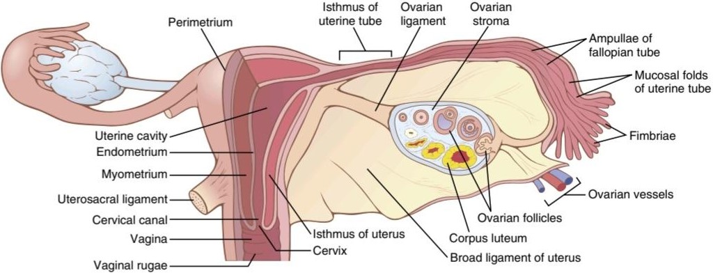
Figure 82-2. Internal structures of the uterus, ovary, and a uterine tube.
شکل 82-2. ساختارهای داخلی رحم، تخمدان و لوله رحم.
OOGENESIS AND FOLLICULAR DEVELOPMENT IN THE OVARIES
A developing egg (oocyte) differentiates into a mature egg (ovum) through a series of steps called oogenesis (Figure 82-3). During early embryonic development, primordial germ cells from the dorsal endoderm of the yolk sac migrate along the mesentery of the hindgut to the outer surface of the ovary, which is covered by a germinal epithelium, derived embryologically from the epithelium of the germinal ridges. During this migration, the germ cells divide repeatedly. Once these primordial germ cells reach the germinal epithelium, they migrate into the substance of the ovarian cortex and become oogonia or primordial ova.
OOGENESIS و رشد فولیکولی در تخمدان ها
یک تخمک در حال رشد (اووسیت) از طریق یک سری مراحل به نام اووژنز به یک تخمک بالغ (تخمک) تمایز می یابد (شکل 82-3). در طی رشد اولیه جنینی، سلول های زایای اولیه از آندودرم پشتی کیسه زرده در امتداد مزانتر روده عقبی به سطح بیرونی تخمدان مهاجرت می کنند که توسط یک اپیتلیوم ژرمینال پوشیده شده است که به طور جنینی از اپیتلیوم برآمدگی های ژرمینال گرفته شده است. در طول این مهاجرت، سلول های زاینده به طور مکرر تقسیم می شوند. هنگامی که این سلول های زایای اولیه به اپیتلیوم ژرمینال می رسند، به درون ماده قشر تخمدان مهاجرت می کنند و تبدیل به اووگونیا یا تخمدان اولیه می شوند.
Each primordial ovum then collects around it a layer of spindle cells from the ovarian stroma (the supporting tissue of the ovary) and causes them to take on epithelioid characteristics; these epithelioid-like cells are then called granulosa cells. The ovum surrounded by a single layer of granulosa cells is called a primordial follicle. At this stage, the ovum is still immature and is called a primary oocyte, requiring two more cell divisions before it can be fertilized by a sperm.
سپس هر تخمک اولیه لایهای از سلولهای دوکی را از استرومای تخمدان (بافت پشتیبان تخمدان) در اطراف خود جمع میکند و باعث میشود که ویژگیهای اپیتلیوئیدی به خود بگیرند. این سلول های اپیتلیوئید مانند سلول های گرانولوزا نامیده می شوند. تخمک که توسط یک لایه سلول گرانولوزا احاطه شده است، فولیکول اولیه نامیده می شود. در این مرحله، تخمک هنوز نابالغ است و اووسیت اولیه نامیده می شود و قبل از اینکه بتواند توسط اسپرم بارور شود، به دو تقسیم سلولی دیگر نیاز دارد.
The oogonia in the embryonic ovary complete mitotic replication, and the first stage of meiosis starts by the fifth month of fetal development. The germ cell mitosis then ceases and no additional oocytes are formed. At birth the ovary contains about 1 to 2 million primary oocytes.
اوگونیا در تخمدان جنینی تکثیر میتوزی را کامل می کند و اولین مرحله میوز در ماه پنجم رشد جنین شروع می شود. سپس میتوز سلول زاینده متوقف می شود و تخمک اضافی تشکیل نمی شود. در هنگام تولد تخمدان دارای حدود 1 تا 2 میلیون تخمک اولیه است.
The first stage of meiosis starts during fetal development but is arrested in the late stage of prophase I until puberty, which usually occurs between ages 10 and 14 in females. The first meiotic division of the oocyte is completed after puberty. Each oocyte divides into two cells, a large ovum (secondary oocyte) and a small first polar body. Each of these cells contains 23 duplicated chromosomes. The first polar body may or may not undergo a second meiotic division and then disintegrates. The ovum undergoes a second meiotic division, and after the sister chromatids separate, there is a pause in meiosis. If the ovum is fertilized, the final step in meiosis occurs and the sister chromatids in the ovum go to separate cells.
اولین مرحله میوز در طول رشد جنین شروع می شود، اما در اواخر مرحله پروفاز I تا بلوغ متوقف می شود، که معمولا بین 10 تا 14 سالگی در زنان رخ می دهد. اولین تقسیم میوز تخمک پس از بلوغ کامل می شود. هر تخمک به دو سلول تقسیم می شود، یک تخمک بزرگ (تخمک ثانویه) و یک جسم قطبی اول کوچک. هر یک از این سلول ها حاوی 23 کروموزوم تکراری است. جسم قطبی اول ممکن است تحت یک تقسیم میوز دوم قرار گیرد یا نباشد و سپس متلاشی شود. تخمک تحت یک تقسیم میوز دوم قرار می گیرد و پس از جدا شدن کروماتیدهای خواهر، یک مکث در میوز ایجاد می شود. اگر تخمک بارور شود، مرحله نهایی میوز رخ می دهد و کروماتیدهای خواهر در تخمک به سلول های جدا می روند.
When the ovary releases the ovum (ovulation), and if the ovum is fertilized, the final meiosis occurs. Half of the sister chromatids remain in the fertilized ovum, and the other half are released in a second polar body, which then disintegrates.
هنگامی که تخمدان تخمک را آزاد می کند (تخمک گذاری) و اگر تخمک بارور شود، میوز نهایی رخ می دهد. نیمی از کروماتیدهای خواهر در تخمک بارور شده باقی می مانند و نیمی دیگر در جسم قطبی دوم آزاد می شوند که سپس متلاشی می شود.
At puberty, only about 300,000 oocytes remain in the ovaries, and only a small percentage of these oocytes become mature. The many thousands of oocytes that do not mature degenerate. During all the reproductive years of adult life, between about 13 and 46 years of age on average, only 400 to 500 of the primordial follicles develop enough to expel their ova, one each month; the remainder degenerate (i.e., become atretic). At the end of reproductive capability (at menopause), only a few primordial follicles remain in the ovaries, and even these follicles degenerate soon thereafter.
در دوران بلوغ، تنها حدود 300000 تخمک در تخمدان ها باقی می ماند و تنها درصد کمی از این تخمک ها بالغ می شوند. هزاران تخمک که بالغ نمی شوند، تحلیل می روند. در طول تمام سالهای تولیدمثلی زندگی بزرگسالی، به طور متوسط بین 13 تا 46 سالگی، تنها 400 تا 500 فولیکول اولیه به اندازه کافی رشد می کنند تا تخمک های خود را خارج کنند، هر ماه یک عدد. انحطاط باقی مانده (یعنی آترتیک می شوند). در پایان توانایی تولید مثل (در دوران یائسگی)، تنها چند فولیکول اولیه در تخمدان ها باقی می ماند و حتی این فولیکول ها به زودی پس از آن تحلیل می روند.

Figure 82-3. Oogenesis and follicle development.
شکل 82-3. اووژنز و رشد فولیکول.
FEMALE HORMONAL SYSTEM
The female hormonal system, like that of the male hormonal system, consists of three hierarchies of hormones, as follows:
سیستم هورمونی زنانه
سیستم هورمونی زنانه مانند سیستم هورمونی مردانه از سه سلسله مراتب هورمونی به شرح زیر تشکیل شده است:
1. A hypothalamic releasing hormone, called gonadotropin-releasing hormone (GnRH)
1. یک هورمون آزاد کننده هیپوتالاموس، به نام هورمون آزاد کننده گنادوتروپین (GnRH)
2. The anterior pituitary sex hormones, follicle- stimulating hormone (FSH) and luteinizing hormone (LH), both of which are secreted in response to release of GnRH from the hypothalamus
2. هورمون های جنسی هیپوفیز قدامی، هورمون محرک فولیکول (FSH) و هورمون لوتئین کننده (LH)، که هر دو در پاسخ به آزاد شدن GnRH از هیپوتالاموس ترشح می شوند.
3. The ovarian hormones, estrogen and progesterone, which are secreted by the ovaries in response to the two female sex hormones from the anterior pituitary gland
3. هورمون های تخمدان، استروژن و پروژسترون، که توسط تخمدان ها در پاسخ به دو هورمون جنسی زنانه از غده هیپوفیز قدامی ترشح می شوند.
These various hormones are secreted at drastically differing rates during different parts of the female monthly sexual cycle. Figure 82-4 shows the approximate changing concentrations of the anterior pituitary gonadotropic hormones FSH and LH (bottom two curves) and of the ovarian hormones estradiol (estrogen) and progesterone (top two curves).
این هورمونهای مختلف با سرعتهای بسیار متفاوتی در طول دورههای مختلف چرخه جنسی ماهانه زنان ترشح میشوند. شکل 82-4 غلظت تغییر تقریبی هورمون های گنادوتروپیک هیپوفیز قدامی FSH و LH (دو منحنی پایین) و هورمون های تخمدانی استرادیول (استروژن) و پروژسترون (دو منحنی بالا) را نشان می دهد.
The amount of GnRH released from the hypothalamus increases and decreases much less drastically during the monthly sexual cycle. It is secreted in short pulses averaging once every 90 minutes, as occurs in males.
مقدار GnRH آزاد شده از هیپوتالاموس در طول چرخه جنسی ماهانه به شدت کاهش می یابد. به طور متوسط هر 90 دقیقه یک بار در نبض های کوتاه ترشح می شود، همانطور که در مردان اتفاق می افتد.

Figure 82-4. Approximate plasma concentrations of the gonadotropins and ovarian hormones during the normal female sexual cycle. FSH, Follicle-stimulating hormone; LH, luteinizing hormone.
شکل 82-4. غلظت تقریبی پلاسمایی گنادوتروپین ها و هورمون های تخمدانی در طول چرخه جنسی طبیعی زنان. FSH، هورمون محرک فولیکول؛ LH، هورمون لوتئینه کننده.
MONTHLY OVARIAN CYCLE AND FUNCTION OF GONADOTROPIC HORMONES
The normal reproductive years of the female are characterized by monthly rhythmic changes in the rates of secretion of the female hormones and corresponding physical changes in the ovaries and other sexual organs. This rhythmic pattern is called the female monthly sexual cycle (or, less accurately, the menstrual cycle). The duration of the cycle averages 28 days. It may be as short as 20 days or as long as 45 days in some women, although abnormal cycle length is frequently associated with decreased fertility.
چرخه ماهانه تخمدانی و عملکرد هورمون های گنادوتروپیک
سالهای تولید مثلی طبیعی زنان با تغییرات ریتمیک ماهانه در میزان ترشح هورمونهای زنانه و تغییرات فیزیکی مربوطه در تخمدانها و سایر اندامهای جنسی مشخص میشود. به این الگوی ریتمیک، چرخه ماهانه جنسی زنان (یا به عبارت دقیق تر، چرخه قاعدگی) می گویند. مدت چرخه به طور متوسط 28 روز است. ممکن است در برخی از زنان 20 روز یا 45 روز طول بکشد، اگرچه طول چرخه غیرطبیعی اغلب با کاهش باروری همراه است.
The female sexual cycle has two significant results. First, only a single ovum is normally released from the ovaries each month, so normally only a single fetus will begin to grow at a time. Second, the uterine endometrium is prepared in advance for implantation of the fertilized ovum at the required time of the month.
چرخه جنسی زنان دو نتیجه قابل توجه دارد. اولاً، هر ماه به طور معمول تنها یک تخمک از تخمدان ها آزاد می شود، بنابراین به طور معمول تنها یک جنین در یک زمان شروع به رشد می کند. دوم، آندومتر رحم از قبل برای لانه گزینی تخمک بارور شده در زمان مورد نیاز ماه آماده می شود.
GONADOTROPIC HORMONES AND THEIR EFFECTS ON THE OVARIES
The ovarian changes that occur during the sexual cycle depend completely on the gonadotropic hormones FSH and LH, which are secreted by the anterior pituitary gland. Both FSH and LH are small glycoproteins that have molecular weights of about 30,000. In the absence of these hormones, the ovaries remain inactive, which is the case throughout childhood, when almost no pituitary gonadotropic hormones are secreted. At age 9 to 12 years, the pituitary begins to secrete progressively more FSH and LH, which leads to the onset of normal monthly sexual cycles beginning between the ages of 11 and 15 years. This period of change is called puberty, and the time of the first menstrual cycle is called menarche. During each month of the female sexual cycle, there is a cyclical increase and decrease of FSH and LH, as shown in the bottom of Figure 82-4. These cyclical variations cause cyclical ovarian changes, which are explained in the following sections.
هورمون های گنادوتروپیک و تأثیر آنها بر تخمدان ها
تغییرات تخمدانی که در طول چرخه جنسی رخ می دهد کاملاً به هورمون های گنادوتروپیک FSH و LH بستگی دارد که توسط غده هیپوفیز قدامی ترشح می شوند. هر دو FSH و LH گلیکوپروتئین های کوچکی هستند که وزن مولکولی آنها حدود 30000 است. در غیاب این هورمونها، تخمدانها غیرفعال میمانند، که در تمام دوران کودکی، زمانی که تقریباً هیچ هورمون گنادوتروپیک هیپوفیز ترشح نمیشود، وجود دارد. در سن 9 تا 12 سالگی، هیپوفیز شروع به ترشح تدریجی FSH و LH می کند که منجر به شروع چرخه های جنسی طبیعی ماهانه بین 11 تا 15 سالگی می شود. این دوره تغییر را بلوغ و زمان اولین سیکل قاعدگی را قاعدگی می نامند. در طول هر ماه از چرخه جنسی زنان، افزایش و کاهش چرخه ای FSH و LH وجود دارد، همانطور که در پایین شکل 82-4 نشان داده شده است. این تغییرات چرخه ای باعث تغییرات چرخه ای تخمدان می شود که در قسمت های زیر توضیح داده شده است.
Both FSH and LH stimulate their ovarian target cells by combining with highly specific FSH and LH receptors in the ovarian target cell membranes. In turn, the activated receptors increase the cells’ secretion rates and usually the growth and proliferation of the cells as well. Almost all these stimulatory effects result from activation of the cyclic adenosine monophosphate second messenger system in the cell cytoplasm, which causes formation of protein kinase and multiple phosphorylations of key enzymes that stimulate sex hormone synthesis, as explained in Chapter 75.
هر دو FSH و LH سلول های هدف تخمدان خود را با ترکیب با گیرنده های بسیار اختصاصی FSH و LH در غشای سلول هدف تخمدان تحریک می کنند. به نوبه خود، گیرنده های فعال شده، میزان ترشح سلول ها و معمولاً رشد و تکثیر سلول ها را نیز افزایش می دهند. تقریباً تمام این اثرات تحریکی ناشی از فعال شدن سیستم پیام رسان دوم آدنوزین مونوفسفات حلقوی در سیتوپلاسم سلولی است که باعث تشکیل پروتئین کیناز و فسفوریلاسیون های متعدد آنزیم های کلیدی می شود که سنتز هورمون های جنسی را تحریک می کند، همانطور که در فصل 75 توضیح داده شد.
OVARIAN FOLLICLE GROWTH-THE FOLLICULAR PHASE OF THE OVARIAN CYCLE
Figure 82-5 shows the progressive stages of follicular growth in the ovaries. When a female child is born, each ovum is surrounded by a single layer of granulosa cells; the ovum, with this granulosa cell sheath, is called a primordial follicle, as shown in the figure. Throughout childhood, the granulosa cells are believed to provide nourishment for the ovum and to secrete an oocyte maturation inhibiting factor that keeps the ovum suspended in its primordial state in the prophase stage of meiotic division. Then, after puberty, when FSH and LH from the anterior pituitary gland begin to be secreted in significant quantities, the ovaries (together with some of the follicles within them) begin to grow.
رشد فولیکول تخمدان – فاز فولیکولی چرخه تخمدان
شکل 82-5 مراحل پیشرونده رشد فولیکولی در تخمدان ها را نشان می دهد. هنگامی که یک کودک دختر به دنیا می آید، هر تخمک توسط یک لایه واحد از سلول های گرانولوزا احاطه شده است. همانطور که در شکل نشان داده شده است، تخمک با این غلاف سلولی گرانولوزا، فولیکول اولیه نامیده می شود. اعتقاد بر این است که در طول دوران کودکی، سلولهای گرانولوزا برای تخمک تغذیه میکنند و یک عامل بازدارنده بلوغ تخمک ترشح میکنند که تخمک را در حالت اولیهاش در مرحله پیشفرض تقسیم میوز معلق نگه میدارد. سپس، پس از بلوغ، هنگامی که FSH و LH از غده هیپوفیز قدامی به مقدار قابل توجهی شروع به ترشح می کنند، تخمدان ها (به همراه برخی از فولیکول های درون آنها) شروع به رشد می کنند.
The first stage of follicular growth is moderate enlargement of the ovum, which increases in diameter 2-fold to 3-fold. That stage is followed by growth of additional layers of granulosa cells in some of the follicles. These follicles are known as primary follicles.
اولین مرحله رشد فولیکولی بزرگ شدن متوسط تخمک است که قطر آن 2 برابر تا 3 برابر افزایش می یابد. این مرحله با رشد لایه های اضافی سلول های گرانولوزا در برخی از فولیکول ها دنبال می شود. این فولیکول ها به عنوان فولیکول های اولیه شناخته می شوند.
Development of Antral and Vesicular Follicles. During the first few days of each monthly female sexual cycle, the concentrations of FSH and LH secreted by the anterior pituitary gland increase slightly to moderately, with the increase in FSH slightly greater than that of LH and preceding it by a few days. These hormones, especially FSH, cause accelerated growth of 6 to 12 primary follicles each month. The initial effect is rapid proliferation of the granulosa cells, giving rise to many more layers of these cells. In addition, spindle cells derived from the ovary interstitium collect in several layers outside the granulosa cells, giving rise to a second mass of cells called the theca. The theca is divided into two layers. In the theca interna, the cells take on epithelioid characteristics similar to those of the granulosa cells and develop the ability to secrete additional steroid sex hormones (estrogen and progesterone). The outer layer, the theca externa, develops into a highly vascular connective tissue capsule that becomes the capsule of the developing follicle.
رشد فولیکول های آنترال و تاولی. در طی چند روز اول هر چرخه ماهانه جنسی زن، غلظت FSH و LH ترشح شده توسط غده هیپوفیز قدامی کمی تا متوسط افزایش می یابد، با افزایش FSH کمی بیشتر از LH و چند روز قبل از آن. این هورمون ها به ویژه FSH باعث رشد سریع 6 تا 12 فولیکول اولیه در هر ماه می شوند. اثر اولیه تکثیر سریع سلول های گرانولوزا است که باعث ایجاد لایه های بیشتری از این سلول ها می شود. علاوه بر این، سلولهای دوکی مشتق شده از بینابینی تخمدان در چندین لایه خارج از سلولهای گرانولوزا جمع میشوند و توده دومی از سلولها به نام theca را ایجاد میکنند. theca به دو لایه تقسیم می شود. در theca interna، سلولها ویژگیهای اپیتلیوئیدی مشابه سلولهای گرانولوزا میگیرند و توانایی ترشح هورمونهای جنسی استروئیدی اضافی (استروژن و پروژسترون) را ایجاد میکنند. لایه بیرونی، theca externa، به یک کپسول بافت همبند بسیار عروقی تبدیل می شود که به کپسول فولیکول در حال رشد تبدیل می شود.
After the early proliferative phase of growth, which lasts for a few days, the mass of granulosa cells secretes a follicular fluid that contains a high concentration of estrogen, one of the important female sex hormones (dis- cussed later). Accumulation of this fluid causes an antrum to appear within the mass of granulosa cells, as shown in Figure 82-5.
پس از مرحله تکثیر اولیه رشد، که برای چند روز طول می کشد، توده سلول های گرانولوزا مایع فولیکولی ترشح می کند که حاوی غلظت بالایی از استروژن، یکی از هورمون های جنسی زنانه مهم است (در ادامه بحث خواهد شد). تجمع این مایع باعث می شود که یک آنتروم در داخل توده سلول های گرانولوزا ظاهر شود، همانطور که در شکل 82-5 نشان داده شده است.

Figure 82-5. Stages of follicular growth in the ovary, also showing formation of the corpus luteum.
شکل 82-5. مراحل رشد فولیکولی در تخمدان، همچنین تشکیل جسم زرد را نشان می دهد.
The early growth of the primary follicle up to the antral stage is stimulated mainly by FSH alone. Greatly accelerated growth then occurs, leading to still larger follicles called vesicular follicles. This accelerated growth is caused by the following mechanisms:
رشد اولیه فولیکول اولیه تا مرحله آنترال عمدتاً توسط FSH به تنهایی تحریک می شود. سپس رشد بسیار سریعی رخ می دهد که منجر به فولیکول های بزرگتر به نام فولیکول های تاولی می شود. این رشد سریع توسط مکانیسم های زیر ایجاد می شود:
1. Estrogen is secreted into the follicle and causes the granulosa cells to form increasing numbers of FSH receptors, which causes a positive feedback effect because it makes the granulosa cells even more sensitive to FSH.
1. استروژن به داخل فولیکول ترشح میشود و باعث میشود سلولهای گرانولوزا تعداد فزایندهای گیرندههای FSH را تشکیل دهند که باعث یک اثر بازخورد مثبت میشود زیرا سلولهای گرانولوزا را حتی به FSH حساستر میکند.
2. The pituitary FSH and the estrogens combine to promote LH receptors on the original granulosa cells, thus allowing LH stimulation to occur in addition to FSH stimulation and creating an even more rapid increase in follicular secretion.
2. FSH هیپوفیز و استروژن ها ترکیب می شوند تا گیرنده های LH را بر روی سلول های گرانولوزای اصلی تقویت کنند، بنابراین اجازه می دهند تحریک LH علاوه بر تحریک FSH رخ دهد و افزایش حتی سریعتر در ترشح فولیکولی ایجاد می کند.
3. The increasing estrogens from the follicle plus the increasing LH from the anterior pituitary gland act together to cause proliferation of the follicular thecal cells and increase their secretion.
3. افزایش استروژن از فولیکول به علاوه افزایش LH از غده هیپوفیز قدامی با هم عمل می کنند و باعث تکثیر سلول های موضعی فولیکولی و افزایش ترشح آنها می شوند.
Once the antral follicles begin to grow, their growth occurs almost explosively. The ovum also enlarges in diameter another 3-fold to 4-fold, giving a total ovum diameter increase up to 10-fold, or a mass increase of 1000-fold. As the follicle enlarges, the ovum remains embedded in a mass of granulosa cells located at one pole of the follicle.
هنگامی که فولیکول های آنترال شروع به رشد می کنند، رشد آنها تقریباً انفجاری رخ می دهد. همچنین قطر تخمک 3 برابر تا 4 برابر افزایش می یابد که باعث افزایش کل قطر تخمک تا 10 برابر یا افزایش جرم 1000 برابری می شود. با بزرگ شدن فولیکول، تخمک در تودهای از سلولهای گرانولوزا واقع در یک قطب فولیکول قرار میگیرد.
Only One Follicle Fully Matures Each Month, and the Remainder Undergo Atresia. After a week or more of growth-but before ovulation occurs-one of the follicles begins to outgrow all the others, and the remaining 5 to 11 developing follicles involute (a process called atresia).
هر ماه فقط یک فولیکول به طور کامل بالغ می شود و باقیمانده دچار آترزی می شود. پس از یک هفته یا بیشتر از رشد – اما قبل از تخمکگذاری – یکی از فولیکولها شروع به رشد بیشتر از فولیکولهای دیگر میکند و 5 تا 11 فولیکول باقی مانده در حال رشد در پیچ میشوند (فرآیندی به نام آترزی).
The cause of the atresia is unclear, but it has been postulated to be the following: The large amounts of estrogen from the most rapidly growing follicle act on the hypo- thalamus to depress further enhancement of FSH secretion by the anterior pituitary gland, in this way blocking further growth of the less well-developed follicles. Therefore, the largest follicle continues to grow because of its intrinsic positive feedback effects, while all the other follicles stop growing and actually involute.
علت آترزی نامشخص است، اما فرض بر این است که به شرح زیر است: مقادیر زیادی استروژن از فولیکولهایی که سریعتر در حال رشد هستند، روی هیپوتالاموس تأثیر میگذارد تا از افزایش بیشتر ترشح FSH توسط غده هیپوفیز قدامی جلوگیری کند، به این ترتیب رشد بیشتر فولیکولهای کمتر توسعهیافته را مسدود میکند. بنابراین، بزرگترین فولیکول به دلیل اثرات بازخورد مثبت ذاتی خود به رشد خود ادامه می دهد، در حالی که تمام فولیکول های دیگر رشد نمی کنند و در واقع درهم می مانند.
This process of atresia is important because it normally allows only one of the follicles to grow large enough each month to ovulate, which usually prevents more than one child from developing with each pregnancy. The single follicle reaches a diameter of 1 to 1.5 centimeters at the time of ovulation and is called the mature follicle.
این فرآیند آترزی مهم است زیرا به طور معمول به تنها یکی از فولیکولها اجازه میدهد تا هر ماه به اندازه کافی بزرگ شوند تا تخمک گذاری کنند، که معمولاً از رشد بیش از یک کودک در هر بارداری جلوگیری میکند. قطر تک فولیکول در زمان تخمک گذاری به 1 تا 1.5 سانتی متر می رسد و فولیکول بالغ نامیده می شود.
Ovulation
Ovulation in a woman who has a normal 28-day female sexual cycle occurs 14 days after the onset of menstruation. Shortly before ovulation, the protruding outer wall of the follicle swells rapidly, and a small area in the center of the follicular capsule, called the stigma, protrudes like a nipple. In another 30 minutes or so, fluid begins to ooze from the follicle through the stigma, and about 2 minutes later, the stigma ruptures widely, allowing a more viscous fluid, which has occupied the central portion of the follicle, to evaginate outward. This viscous fluid carries with it the ovum surrounded by a mass of several thousand small granulosa cells, called the corona radiata.
تخمک گذاری
تخمک گذاری در خانمی که دارای چرخه جنسی طبیعی 28 روزه است، 14 روز پس از شروع قاعدگی رخ می دهد. کمی قبل از تخمک گذاری، دیواره بیرونی فولیکول به سرعت متورم می شود و ناحیه کوچکی در مرکز کپسول فولیکولی به نام کلاله مانند نوک پستان بیرون زده است. در حدود 30 دقیقه دیگر، مایع از فولیکول از طریق کلاله شروع به ترشح می کند و حدود 2 دقیقه بعد، کلاله به طور گسترده ای پاره می شود و به مایع چسبناک تری که قسمت مرکزی فولیکول را اشغال کرده است، اجازه می دهد تا به سمت بیرون خارج شود. این مایع چسبناک، تخمک احاطه شده توسط توده ای متشکل از چندین هزار سلول گرانولوزای کوچک به نام تاج رادیاتا را با خود حمل می کند.
A Surge of Luteinizing Hormone Is Necessary for Ovulation. LH is necessary for final follicular growth and ovulation. Without this hormone, even when large quantities of FSH are available, the follicle will not progress to the ovulation stage.
افزایش هورمون لوتئینه کننده برای تخمک گذاری ضروری است. LH برای رشد فولیکولی نهایی و تخمک گذاری ضروری است. بدون این هورمون، حتی زمانی که مقادیر زیادی FSH در دسترس باشد، فولیکول به مرحله تخمک گذاری پیش نمی رود.
About 2 days before ovulation, the rate of secretion of LH by the anterior pituitary gland increases markedly, rising 6 to 10-fold and peaking about 16 hours before ovulation. FSH also increases about 2-fold to 3-fold at the same time, and the FSH and LH act synergistically to cause rapid swelling of the follicle during the last few days before ovulation. The LH also has a specific effect on the granulosa and theca cells, converting them mainly to progesterone-secreting cells. Therefore, the rate of estro- gen secretion begins to fall about 1 day before ovulation, while increasing amounts of progesterone begin to be secreted.
حدود 2 روز قبل از تخمک گذاری، میزان ترشح LH توسط غده هیپوفیز قدامی به طور قابل توجهی افزایش می یابد و 6 تا 10 برابر افزایش می یابد و حدود 16 ساعت قبل از تخمک گذاری به اوج خود می رسد. FSH همچنین در همان زمان حدود 2 برابر تا 3 برابر افزایش می یابد و FSH و LH به صورت هم افزایی عمل می کنند و باعث تورم سریع فولیکول در چند روز گذشته قبل از تخمک گذاری می شوند. LH همچنین تأثیر خاصی بر روی سلول های گرانولوزا و theca دارد و آنها را عمدتاً به سلول های ترشح کننده پروژسترون تبدیل می کند. بنابراین، میزان ترشح استروژن حدود 1 روز قبل از تخمک گذاری شروع به کاهش می کند، در حالی که مقادیر فزاینده ای پروژسترون شروع به ترشح می کند.
It is in this environment of (1) rapid growth of the follicle, (2) diminishing estrogen secretion after a prolonged phase of excessive estrogen secretion, and (3) initiation of secretion of progesterone that ovulation occurs. With- out the initial preovulatory surge of LH, ovulation will not take place.
در این محیط (1) رشد سریع فولیکول، (2) کاهش ترشح استروژن پس از یک فاز طولانی ترشح بیش از حد استروژن، و (3) شروع ترشح پروژسترون است که تخمک گذاری رخ می دهد. بدون افزایش اولیه LH قبل از تخمک گذاری، تخمک گذاری انجام نمی شود.
Initiation of Ovulation. Figure 82-6 provides a schema for the initiation of ovulation, showing the role of the large quantity of LH secreted by the anterior pituitary gland. This LH causes rapid secretion of follicular steroid hormones that contain progesterone. Within a few hours, two events occur, both of which are necessary for ovulation:
شروع تخمک گذاری. شکل 82-6 طرحی را برای شروع تخمک گذاری ارائه می دهد که نقش مقدار زیادی LH ترشح شده توسط غده هیپوفیز قدامی را نشان می دهد. این LH باعث ترشح سریع هورمون های استروئیدی فولیکولی می شود که حاوی پروژسترون هستند. در عرض چند ساعت، دو رویداد رخ می دهد که هر دو برای تخمک گذاری ضروری هستند:
1. The theca externa (i.e., the capsule of the follicle) begins to release proteolytic enzymes from lysosomes. These enzymes cause dissolution of the follicular capsular wall and consequent weakening of the wall, resulting in further swelling of the entire follicle and degeneration of the stigma.
1. theca externa (یعنی کپسول فولیکول) شروع به آزادسازی آنزیم های پروتئولیتیک از لیزوزوم می کند. این آنزیم ها باعث انحلال دیواره کپسولی فولیکولی و در نتیجه ضعیف شدن دیواره و در نتیجه تورم بیشتر کل فولیکول و دژنراسیون کلاله می شود.
2. Simultaneously there is rapid growth of new blood vessels into the follicle wall. At the same time, prostaglandins (local hormones that cause vasodilation) are secreted into the follicular tissues.
2. همزمان رشد سریع عروق خونی جدید در دیواره فولیکول وجود دارد. در همان زمان، پروستاگلاندین ها (هورمون های محلی که باعث اتساع عروق می شوند) در بافت های فولیکولی ترشح می شوند.
These two effects cause plasma transudation into the follicle, which contributes to follicle swelling. Finally, the combination of follicle swelling and simultaneous degeneration of the stigma causes follicle rupture, with dis- charge of the ovum.
این دو اثر باعث انتقال پلاسما به داخل فولیکول می شود که به تورم فولیکول کمک می کند. در نهایت، ترکیبی از تورم فولیکول و دژنراسیون همزمان کلاله باعث پارگی فولیکول و تخلیه تخمک می شود.

Figure 82-6. The postulated mechanism of ovulation.
شکل 82-6. مکانیسم فرضی تخمک گذاری
CORPUS LUTEUM-THE LUTEAL PHASE OF THE OVARIAN CYCLE
During the first few hours after expulsion of the ovum from the follicle, the remaining granulosa and theca interna cells change rapidly into lutein cells. They enlarge in diameter two or more times and become filled with lipid inclusions that give them a yellowish appearance. This process is called luteinization, and the total mass of cells together is called the corpus luteum, which is shown in Figure 82-5. A well-developed vascular supply also grows into the corpus luteum.
جسم زرد – فاز لوتئال چرخه تخمدان
در چند ساعت اول پس از بیرون راندن تخمک از فولیکول، سلول های گرانولوزا و theca interna باقی مانده به سرعت به سلول های لوتئین تبدیل می شوند. قطر آنها دو یا چند بار بزرگ می شود و پر از مواد لیپیدی می شود که ظاهری مایل به زرد به آنها می دهد. به این فرآیند لوتئینیزاسیون و مجموع جرم سلول ها با هم جسم زرد می گویند که در شکل 82-5 نشان داده شده است. یک منبع عروقی به خوبی توسعه یافته نیز به جسم زرد رشد می کند.
The granulosa cells in the corpus luteum develop extensive intracellular smooth endoplasmic reticula that form large amounts of the female sex hormones progesterone and estrogen (with more progesterone than estrogen during the luteal phase). The theca cells form mainly the androgens androstenedione and testosterone rather than female sex hormones. However, most of these hormones are also converted by the enzyme aromatase in the granulosa cells into estrogens.
سلول های گرانولوزا در جسم زرد شبکه آندوپلاسمی صاف داخل سلولی گسترده ای ایجاد می کنند که مقادیر زیادی از هورمون های جنسی زنانه پروژسترون و استروژن (با پروژسترون بیشتر از استروژن در طول فاز لوتئال) را تشکیل می دهند. سلولهای theca عمدتاً آندروژنهای آندروستندیون و تستوسترون را تشکیل میدهند تا هورمونهای جنسی زنانه. با این حال، بیشتر این هورمون ها نیز توسط آنزیم آروماتاز در سلول های گرانولوزا به استروژن تبدیل می شوند.
The corpus luteum normally grows to about 1.5 centimeters in diameter, reaching this stage of development 7 to 8 days after ovulation. Then the corpus luteum begins to involute and eventually loses its secretory function and its yellowish, lipid characteristic about 12 days after ovulation, becoming the corpus albicans; during the ensuing few weeks, the corpus albicans is replaced by connective tissue and over months is absorbed.
قطر جسم زرد معمولاً به حدود 1.5 سانتی متر می رسد و 7 تا 8 روز پس از تخمک گذاری به این مرحله از رشد می رسد. سپس جسم زرد شروع به پیچیدن می کند و در نهایت عملکرد ترشحی و ویژگی زرد رنگ و لیپیدی خود را حدود 12 روز پس از تخمک گذاری از دست می دهد و به جسم آلبیکنس تبدیل می شود. در طی چند هفته بعد، جسم آلبیکنس با بافت همبند جایگزین شده و طی ماه ها جذب می شود.
Luteinizing Function of Luteinizing Hormone. The change of granulosa and theca interna cells into lutein cells depends mainly on LH secreted by the anterior pituitary gland. In fact, this function gives LH its name- “luteinizing,” for “yellowing” Luteinization also depends on extrusion of the ovum from the follicle. A yet uncharacterized factor in the follicular fluid, called luteinization- inhibiting factor, seems to hold the luteinization process in check until after ovulation.
عملکرد لوتئینه کننده هورمون لوتئینه کننده. تغییر سلول های گرانولوزا و theca interna به سلول های لوتئین عمدتاً به LH ترشح شده توسط غده هیپوفیز قدامی بستگی دارد. در واقع، این تابع به LH نام خود را می دهد – “لوتئین کننده”، برای “زرد شدن” لوتئینیزاسیون نیز به اکستروژن تخمک از فولیکول بستگی دارد. به نظر می رسد یک عامل هنوز مشخص نشده در مایع فولیکولی، به نام عامل بازدارنده لوتئینیزاسیون، فرآیند لوتئین شدن را تا پس از تخمک گذاری کنترل می کند.
Secretion by the Corpus Luteum: An Additional Func- tion of Luteinizing Hormone. The corpus luteum is a highly secretory organ, secreting large amounts of progesterone and estrogen. Once LH (mainly that secreted during the ovulatory surge) has acted on the granulosa and theca cells to cause luteinization, the newly formed lutein cells go through a sequence of (1) proliferation, (2) enlargement, and (3) secretion, followed by (4) degeneration. All this occurs in about 12 days. As discussed in Chapter 83, another hormone with almost exactly the same properties as LH, chorionic gonadotropin, which is secreted by the placenta, can act on the corpus luteum to prolong its life-usually maintaining it for at least the first 2 to 4 months of pregnancy.
ترشح توسط جسم زرد: یک عملکرد اضافی هورمون لوتئین کننده. جسم زرد یک اندام بسیار ترشحی است که مقادیر زیادی پروژسترون و استروژن ترشح می کند. هنگامی که LH (عمدتاً آن چیزی که در طول موج تخمک گذاری ترشح می شود) روی سلول های گرانولوزا و theca عمل کرد و باعث لوتئینیزه شدن شد، سلول های لوتئین تازه تشکیل شده از طریق دنباله ای از (1) تکثیر، (2) بزرگ شدن و (3) ترشح و به دنبال آن (4) دژنراسیون می شوند. همه اینها در حدود 12 روز اتفاق می افتد. همانطور که در فصل 83 بحث شد، هورمون دیگری با تقریباً دقیقاً همان خواص LH، گنادوتروپین جفتی، که توسط جفت ترشح میشود، میتواند بر روی جسم زرد تأثیر بگذارد و عمر آن را طولانیتر کند – معمولاً حداقل در 2 تا 4 ماه اول بارداری آن را حفظ میکند.
Involution of the Corpus Luteum and Onset of the Next Ovarian Cycle.
Estrogen in particular and progesterone to a lesser extent, secreted by the corpus luteum during the luteal phase of the ovarian cycle, have strong feedback effects on the anterior pituitary gland to maintain low secretory rates of FSH and LH.
چرخش جسم زرد و شروع چرخه تخمدان بعدی.
استروژن به طور خاص و پروژسترون به میزان کمتری که توسط جسم زرد در طول فاز لوتئال چرخه تخمدان ترشح می شود، اثرات بازخورد قوی بر غده هیپوفیز قدامی برای حفظ میزان ترشح پایین FSH و LH دارند.
In addition, the lutein cells secrete small amounts of the hormone inhibin, the same as the inhibin secreted by the Sertoli cells of the male testes. This hormone inhibits FSH secretion by the anterior pituitary gland. Low blood concentrations of FSH and LH result, and loss of these hormones finally causes the corpus luteum to degenerate completely, a process called involution of the corpus luteum.
علاوه بر این، سلولهای لوتئین مقادیر کمی از هورمون اینهیبین ترشح میکنند، همان هورمونی که توسط سلولهای سرتولی بیضههای نر ترشح میشود. این هورمون ترشح FSH توسط غده هیپوفیز قدامی را مهار می کند. غلظت پایین FSH و LH در خون نتیجه میشود و از دست دادن این هورمونها در نهایت باعث تحلیل رفتن کامل جسم زرد میشود، فرآیندی که به آن چرخش جسم زرد میگویند.
Final involution normally occurs at the end of almost exactly 12 days of corpus luteum life, which is around the 26th day of the normal female sexual cycle, 2 days before menstruation begins. At this time, the sudden cessation of estrogen, progesterone, and inhibin secretion by the corpus luteum removes the feedback inhibition of the anterior pituitary gland, allowing it to begin secreting increasing amounts of FSH and LH again. FSH and LH initiate the growth of new follicles, beginning a new ovarian cycle. The paucity of progesterone and estrogen secretion at this time also leads to menstruation by the uterus, which will be explained later.
انفولشن نهایی معمولاً در پایان تقریباً 12 روز از زندگی جسم زرد اتفاق میافتد، یعنی حدود 26 روز از چرخه جنسی طبیعی زنان، 2 روز قبل از شروع قاعدگی. در این زمان، توقف ناگهانی ترشح استروژن، پروژسترون و اینهیبین توسط جسم زرد، مهار بازخورد غده هیپوفیز قدامی را از بین می برد و به آن اجازه می دهد تا دوباره شروع به ترشح مقادیر فزاینده FSH و LH کند. FSH و LH رشد فولیکول های جدید را آغاز می کنند و چرخه تخمدانی جدیدی را آغاز می کنند. کمبود ترشح پروژسترون و استروژن در این زمان نیز منجر به قاعدگی توسط رحم می شود که در ادامه توضیح داده خواهد شد.
SUMMARY
About every 28 days, gonadotropic hormones from the anterior pituitary gland cause 8 to 12 new follicles to begin to grow in the ovaries. One of these follicles finally becomes “mature” and ovulates on the 14th day of the cycle. During growth of the follicles, estrogen is mainly secreted.
خلاصه
تقریباً هر 28 روز، هورمون های گنادوتروپیک از غده هیپوفیز قدامی باعث می شوند که 8 تا 12 فولیکول جدید در تخمدان ها شروع به رشد کنند. یکی از این فولیکول ها در نهایت در روز چهاردهم سیکل ” بالغ” می شود و تخمک گذاری می کند. در طول رشد فولیکول ها، استروژن عمدتا ترشح می شود.
After ovulation, the secretory cells of the ovulating follicle develop into a corpus luteum that secretes large quantities of progesterone and estrogen. After another 2 weeks, the corpus luteum degenerates, whereupon the ovarian hormones estrogen and progesterone decrease greatly, and menstruation begins. A new ovarian cycle then follows.
پس از تخمک گذاری، سلول های ترشحی فولیکول در حال تخمک گذاری به جسم زرد تبدیل می شوند که مقادیر زیادی پروژسترون و استروژن ترشح می کند. پس از 2 هفته دیگر، جسم زرد تحلیل می رود، در نتیجه هورمون های استروژن و پروژسترون تخمدان به شدت کاهش می یابد و قاعدگی شروع می شود. سپس یک چرخه تخمدانی جدید دنبال می شود.
FUNCTIONS OF OVARIAN HORMONES- ESTRADIOL AND PROGESTERONE
The two types of ovarian sex hormones are the estrogens and the progestins. By far the most important of the estrogens is estradiol, and by far the most important progestin is progesterone. The estrogens mainly promote proliferation and growth of specific cells in the body that are responsible for development of most secondary sexual characteristics of females. The progestins function mainly to prepare the uterus for pregnancy and the breasts for lactation.
عملکرد هورمون های تخمدان – استرادیول و پروژسترون
دو نوع هورمون جنسی تخمدان، استروژن و پروژستین هستند. تا حد زیادی مهمترین استروژن استرادیول است و تا حد زیادی مهمترین پروژسترون پروژسترون است. استروژنها عمدتاً تکثیر و رشد سلولهای خاصی را در بدن که مسئول ایجاد بیشتر ویژگیهای جنسی ثانویه زنان هستند، ترویج میکنند. پروژستین ها عمدتاً برای آماده سازی رحم برای بارداری و سینه ها برای شیردهی عمل می کنند.
CHEMISTRY OF THE SEX HORMONES
Estrogens. In the normal nonpregnant female, estrogens are secreted in significant quantities only by the ovaries, although minute amounts are also secreted by the adrenal cortices. During pregnancy, large quantities of estrogens are also secreted by the placenta, as discussed in Chapter 83.
شیمی هورمون های جنسی
استروژن ها در زنان غیرباردار طبیعی، استروژن ها به مقدار قابل توجهی فقط از تخمدان ها ترشح می شود، اگرچه مقادیر بسیار کمی از قشر آدرنال نیز ترشح می شود. همانطور که در فصل 83 بحث شد، در طول بارداری، مقادیر زیادی استروژن نیز توسط جفت ترشح می شود.
Only three estrogens are present in significant quantities in the plasma of the human female-ẞ-estradiol, estrone, and estriol, the formulas for which are shown in Figure 82-7. The principal estrogen secreted by the ovaries is ẞ-estradiol. Small amounts of estrone are also secreted, but most of this is formed in peripheral tissues from androgens secreted by the adrenal cortices and by ovarian thecal cells. Estriol is a weak estrogen; it is an oxidative product derived from both estradiol and estrone, with the conversion occurring mainly in the liver.
فقط سه استروژن به مقدار قابل توجهی در پلاسمای ماده انسان وجود دارد – استرادیول، استرون و استریول که فرمول آنها در شکل 82-7 نشان داده شده است. استروژن اصلی ترشح شده از تخمدان ها، استرادیول است. مقادیر کمی استرون نیز ترشح میشود، اما بیشتر آن در بافتهای محیطی از آندروژنهای ترشح شده از قشر آدرنال و سلولهای پوستی تخمدان تشکیل میشود. استریول یک استروژن ضعیف است. این یک محصول اکسیداتیو است که از هر دو استرادیول و استرون مشتق می شود و تبدیل آن عمدتاً در کبد اتفاق می افتد.
The estrogenic potency of ẞ-estradiol is 12 times that of estrone and 80 times that of estriol. Considering these relative potencies, one can see that the total estrogenic effect of ẞ-estradiol is usually many times that of the other two together. For this reason, ẞ-estradiol is considered the major estrogen, although the estrogenic effects of estrone are not negligible.
قدرت استروژنیک ẞ-estradiol 12 برابر استرون و 80 برابر استریول است. با در نظر گرفتن این قدرت های نسبی، می توان دریافت که اثر استروژنی کل ẞ-استرادیول معمولاً چندین برابر دو اثر دیگر با هم است. به همین دلیل، ẞ-استرادیول استروژن اصلی در نظر گرفته می شود، اگرچه اثرات استروژنی استرون قابل چشم پوشی نیست.
Progestins. By far the most important of the progestins is progesterone. However, small amounts of another progestin, 17α-hydroxyprogesterone, are secreted along with progesterone and have essentially the same effects. Yet, for practical purposes, progesterone is usually considered to be the only important progestin.
پروژستین ها. تا کنون مهمترین پروژستین پروژسترون است. با این حال، مقادیر کمی از پروژستین دیگر، 17α-هیدروکسی پروژسترون، همراه با پروژسترون ترشح می شود و اساساً اثرات مشابهی دارد. با این حال، برای اهداف عملی، معمولاً پروژسترون تنها پروژسترون مهم در نظر گرفته می شود.
In nonpregnant females, progesterone is usually secreted in significant amounts only during the latter half of each ovarian cycle, when it is secreted by the corpus luteum.
در زنان غیر باردار، پروژسترون معمولاً به مقدار قابل توجهی فقط در نیمه دوم هر چرخه تخمدانی ترشح می شود، زمانی که توسط جسم زرد ترشح می شود.
As discussed in Chapter 83, large amounts of progesterone are also secreted by the placenta during pregnancy, especially after the fourth month of gestation.
همانطور که در فصل 83 بحث شد، مقادیر زیادی پروژسترون نیز در دوران بارداری، به ویژه پس از ماه چهارم بارداری، توسط جفت ترشح می شود.
Synthesis of the Estrogens and Progestins. Note from the chemical formulas of the estrogens and progesterone in Figure 82-7 that they are all steroids. They are synthesized in the ovaries mainly from cholesterol derived from the blood but also to a slight extent from acetyl coenzyme A, multiple molecules of which can combine to form the appropriate steroid nucleus.
سنتز استروژن ها و پروژستین ها. از فرمول های شیمیایی استروژن ها و پروژسترون در شکل 82-7 توجه کنید که همه آنها استروئید هستند. آنها در تخمدان ها عمدتاً از کلسترول مشتق شده از خون و همچنین تا حدودی از استیل کوآنزیم A سنتز می شوند که مولکول های متعدد آن می توانند برای تشکیل هسته استروئید مناسب ترکیب شوند.
During synthesis, mainly progesterone and androgens (testosterone and androstenedione) are synthesized first; then, during the follicular phase of the ovarian cycle, before these two initial hormones can leave the ovaries, almost all the androgens and much of the progesterone are converted into estrogens by the enzyme aromatase in the granulosa cells. Because the theca cells lack aromatase, they cannot convert androgens to estrogens. However, androgens diffuse out of the theca cells into the adjacent granulosa cells, where they are converted to estrogens by aromatase, the activity of which is stimulated by FSH (Figure 82-8).
در طول سنتز، ابتدا پروژسترون و آندروژن ها (تستوسترون و آندروستندیون) سنتز می شوند. سپس، در طول فاز فولیکولی چرخه تخمدان، قبل از اینکه این دو هورمون اولیه بتوانند از تخمدان ها خارج شوند، تقریباً تمام آندروژن ها و بسیاری از پروژسترون توسط آنزیم آروماتاز در سلول های گرانولوزا به استروژن تبدیل می شوند. از آنجایی که سلول های تکا فاقد آروماتاز هستند، نمی توانند آندروژن ها را به استروژن تبدیل کنند. با این حال، آندروژنها از سلولهای theca به سلولهای گرانولوزای مجاور منتشر میشوند، جایی که توسط آروماتاز به استروژن تبدیل میشوند که فعالیت آن توسط FSH تحریک میشود (شکل 82-8).
During the luteal phase of the cycle, far too much progesterone is formed for all of it to be converted, which accounts for the large secretion of progesterone into the circulating blood at this time. Also, about one-fifteenth as much testosterone is secreted into the plasma of the female by the ovaries as is secreted into the plasma of the male by the testes.
در طول فاز لوتئال چرخه، پروژسترون بسیار زیادی برای تبدیل شدن همه آن تشکیل می شود، که باعث ترشح زیاد پروژسترون به خون در حال گردش در این زمان می شود. همچنین حدود یک پانزدهم تستوسترونی که توسط تخمدان ها به پلاسمای جنس نر ترشح می شود، ترشح می شود.
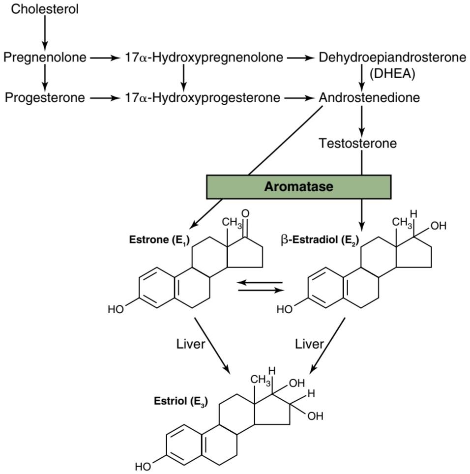
Figure 82-7. Synthesis of the principal female hormones. The chemical structures of the precursor hormones, including progesterone, are shown in Figure 78-2.
شکل 82-7. سنتز هورمون های زنانه اصلی ساختارهای شیمیایی هورمون های پیش ساز، از جمله پروژسترون، در شکل 78-2 نشان داده شده است.
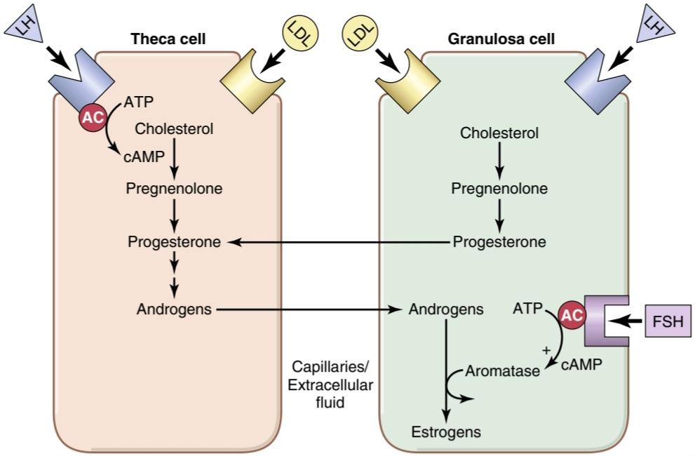
Figure 82-8. Interaction of follicular theca and granulosa cells for production of estrogens. The theca cells, under the control of luteinizing hormone (LH), produce androgens that diffuse into the granulosa cells. In mature follicles, follicle-stimulating hormone (FSH) acts on granulosa cells to stimulate aromatase activity, which converts the androgens to estrogens. AC, Adenylate cyclase; ATP, adenosine triphosphate; cAMP, cyclic adeno- sine monophosphate; LDL, low- density lipoproteins.
شکل 82-8. برهمکنش سلولهای فولیکولی تکا و گرانولوزا برای تولید استروژن. سلول های theca، تحت کنترل هورمون لوتئینیزه کننده (LH)، آندروژن تولید می کنند که در سلول های گرانولوزا منتشر می شود. در فولیکول های بالغ، هورمون محرک فولیکول (FSH) روی سلول های گرانولوزا برای تحریک فعالیت آروماتاز عمل می کند که آندروژن ها را به استروژن تبدیل می کند. AC، آدنیلات سیکلاز؛ ATP، آدنوزین تری فسفات؛ cAMP، آدنوزین مونوفسفات حلقوی. LDL، لیپوپروتئین های با چگالی کم.
Estrogens and Progesterone Are Transported in the Blood Bound to Plasma Proteins. Both estrogens and progesterone are transported in the blood bound mainly with plasma albumin and with specific estrogen- and progesterone-binding globulins. The binding between these hormones and the plasma proteins is loose enough that they are rapidly released to the tissues over a period of 30 minutes or so.
استروژن و پروژسترون در خون به پروتئین های پلاسما متصل می شوند. هر دو استروژن و پروژسترون در خون منتقل می شوند که عمدتاً با آلبومین پلاسما و با گلوبولین های خاص متصل شونده به استروژن و پروژسترون متصل می شوند. اتصال بین این هورمون ها و پروتئین های پلاسما به قدری شل است که به سرعت در مدت 30 دقیقه یا بیشتر به بافت ها آزاد می شوند.
Functions of the Liver in Estrogen Degradation. The liver conjugates estrogens to form glucuronides and sulfates, and about one-fifth of these conjugated products is excreted in the bile; most of the remainder is excreted in the urine. Also, the liver converts the potent estrogens estradiol and estrone into the almost totally impotent estrogen estriol. Therefore, diminished liver function actually increases the activity of estrogens in the body, sometimes causing hyper estrin ism.
عملکرد کبد در تخریب استروژن. کبد استروژن ها را برای تشکیل گلوکورونیدها و سولفات ها ترکیب می کند و حدود یک پنجم این محصولات مزدوج در صفرا دفع می شود. بیشتر باقی مانده از طریق ادرار دفع می شود. همچنین، کبد استروژنهای قوی استرادیول و استرون را به استروژن استروژن تقریباً ناتوان تبدیل میکند. بنابراین، کاهش عملکرد کبد در واقع باعث افزایش فعالیت استروژن در بدن می شود و گاهی اوقات باعث ایجاد هیپر استرینیسم می شود.
Fate of Progesterone. Within a few minutes after secretion, almost all the progesterone is degraded to other steroids that have no progestational effect. As with the estrogens, the liver is especially important for this metabolic degradation.
سرنوشت پروژسترون. در عرض چند دقیقه پس از ترشح، تقریباً تمام پروژسترون به سایر استروئیدها که هیچ اثر پروژستاتیکی ندارند، تجزیه می شود. همانند استروژن ها، کبد برای این تخریب متابولیک اهمیت ویژه ای دارد.
The major end product of progesterone degradation is pregnanediol. About 10% of the original progesterone is excreted in the urine in this form. Therefore, one can estimate the rate of progesterone formation in the body from the rate of this excretion.
محصول نهایی اصلی تخریب پروژسترون پرگناندیول است. حدود 10 درصد از پروژسترون اصلی به این شکل از طریق ادرار دفع می شود. بنابراین می توان سرعت تشکیل پروژسترون در بدن را از روی میزان این دفع تخمین زد.
FUNCTIONS OF THE ESTROGENS-THEIR EFFECTS ON THE PRIMARY AND SECONDARY FEMALE SEX CHARACTERISTICS
A primary function of the estrogens is to cause cellular proliferation and growth of the tissues of the sex organs and other tissues related to reproduction.
عملکرد استروژن ها – تأثیر آنها بر ویژگی های جنسی اولیه و ثانویه زن
عملکرد اصلی استروژن ها ایجاد تکثیر سلولی و رشد بافت های اندام های جنسی و سایر بافت های مرتبط با تولید مثل است.
Effect of Estrogens on the Uterus and External Fe- male Sex Organs. During childhood, estrogens are secreted only in minute quantities, but at puberty, the quantity secreted in the female under the influence of the pituitary gonadotropic hormones increases 20-fold or more. At this time, the female sex organs change from those of a child to those of an adult. The ovaries, fallopian tubes, uterus, and vagina all increase several times in size. Also, the external genitalia enlarge, with deposition of fat in the mons pubis and labia majora and enlargement of the labia minora.
اثر استروژن ها بر رحم و اندام های جنسی خارجی زن. در دوران کودکی، استروژنها فقط در مقادیر کم ترشح میشوند، اما در سن بلوغ، مقدار ترشح شده در زنان تحت تأثیر هورمونهای گنادوتروپیک هیپوفیز 20 برابر یا بیشتر افزایش مییابد. در این زمان اندام های جنسی زنانه از یک کودک به یک بزرگسال تغییر می کند. تخمدان ها، لوله های فالوپ، رحم و واژن همگی چندین برابر بزرگ می شوند. همچنین اندام تناسلی خارجی با رسوب چربی در مونس پوبیس و لابیا بزرگ و بزرگ شدن لب های کوچک بزرگ می شود.
In addition, estrogens change the vaginal epithelium from a cuboidal into a stratified type, which is consider- ably more resistant to trauma and infection than is the prepubertal cuboidal cell epithelium. Vaginal infections in children can often be cured by the administration of estrogens simply because of the resulting increased resistance of the vaginal epithelium.
علاوه بر این، استروژن ها اپیتلیوم واژن را از یک مکعبی به یک نوع طبقه بندی شده تغییر می دهند که به طور قابل توجهی در برابر ضربه و عفونت مقاوم تر از اپیتلیوم سلولی مکعبی قبل از بلوغ است. عفونتهای واژن در کودکان را میتوان با تجویز استروژن به سادگی به دلیل افزایش مقاومت اپیتلیوم واژن درمان کرد.
During the first few years after puberty, the size of the uterus increases 2-fold to 3-fold, but more important than the increase in uterus size are the changes that take place in the uterine endometrium under the influence of estrogens. Estrogens cause marked proliferation of the endometrial stroma and greatly increased development of the endometrial glands, which will later aid in providing nutrition to the implanted ovum. These effects are discussed later in the chapter in connection with the endometrial cycle.
در چند سال اول پس از بلوغ، اندازه رحم 2 تا 3 برابر می شود، اما مهمتر از افزایش اندازه رحم، تغییراتی است که تحت تأثیر استروژن در آندومتر رحم رخ می دهد. استروژن ها باعث تکثیر مشخص استرومای آندومتر و رشد شدید غدد آندومتر می شوند که بعداً به تغذیه تخمک کاشته شده کمک می کند. این اثرات بعداً در فصل مربوط به چرخه آندومتر مورد بحث قرار می گیرد.
Effect of Estrogens on the Fallopian Tubes. The estrogens’ effects on the mucosal lining of the fallopian tubes are similar to their effects on the uterine endometrium. They cause the glandular tissues of this lining to proliferate, and especially important, they cause the number of ciliated epithelial cells that line the fallopian tubes to in- crease. Also, activity of the cilia is considerably enhanced. These cilia always beat toward the uterus, which helps propel the fertilized ovum in that direction.
اثر استروژن بر لوله های فالوپ. اثرات استروژن بر روی پوشش مخاطی لوله های فالوپ مشابه اثرات آنها بر روی آندومتر رحم است. آنها باعث تکثیر بافت های غده ای این پوشش می شوند و به ویژه مهم این است که باعث می شوند تعداد سلول های اپیتلیال مژک دار که لوله های فالوپ را پوشانده اند افزایش یابد. همچنین فعالیت مژه ها به طور قابل توجهی افزایش می یابد. این مژکها همیشه به سمت رحم میزنند که به حرکت تخمک بارور شده به آن سمت کمک میکند.
Effect of Estrogens on the Breasts. The primordial breasts of females and males are exactly alike. In fact, under the influence of appropriate hormones, the masculine breast during the first 2 decades of life can develop sufficiently to produce milk in the same manner as the female breast.
تاثیر استروژن بر سینه ها. سینه های اولیه مونث و نر دقیقا شبیه هم هستند. در واقع، تحت تأثیر هورمونهای مناسب، سینه مردانه در طی 2 دهه اول زندگی میتواند به اندازهای رشد کند که به همان روشی که سینه زن تولید میکند، شیر تولید کند.
Estrogens cause (1) development of the stromal tissues of the breasts, (2) growth of an extensive ductile system, and (3) deposition of fat in the breasts. The lobules and alveoli of the breast develop to a slight extent under the influence of estrogens alone, but it is progesterone and prolactin that ultimately complete the growth and function of these structures.
استروژن ها باعث (1) رشد بافت های استرومایی سینه ها، (2) رشد یک سیستم انعطاف پذیر گسترده و (3) رسوب چربی در سینه ها می شود. لوبولها و آلوئولهای پستان تا حدودی تحت تأثیر استروژنها به تنهایی رشد میکنند، اما پروژسترون و پرولاکتین هستند که در نهایت رشد و عملکرد این ساختارها را تکمیل میکنند.
In summary, the estrogens initiate growth of the breasts and of the milk-producing apparatus. They are also responsible for the characteristic growth and external appearance of the mature female breast. However, they do not complete the job of converting the breasts into milkproducing organs.
به طور خلاصه، استروژن ها شروع به رشد سینه ها و دستگاه تولید شیر می کنند. آنها همچنین مسئول رشد مشخصه و ظاهر خارجی سینه بالغ زن هستند. با این حال، آنها کار تبدیل سینه ها به اندام های تولید کننده شیر را کامل نمی کنند.
Effect of Estrogens on the Skeleton. Estrogens inhibit osteoclastic activity in the bones and therefore stimulate bone growth. As discussed in Chapter 80, at least part of this effect is due to stimulation of osteoprotegerin, which is also called osteoclastogenesis inhibitory factor, a cytokine that inhibits bone resorption.
اثر استروژن بر روی اسکلت. استروژن ها فعالیت استئوکلاستیک را در استخوان ها مهار می کنند و بنابراین رشد استخوان را تحریک می کنند. همانطور که در فصل 80 بحث شد، حداقل بخشی از این اثر به دلیل تحریک استئوپروتجرین است که به آن عامل مهارکننده استئوکلاستوژنز نیز میگویند، سیتوکینی که از جذب استخوان جلوگیری میکند.
At puberty, when the female enters her reproductive years, her growth in height becomes rapid for several years. However, estrogens also cause uniting of the epiphyses with the shafts of the long bones. This effect of estrogen in the female is much stronger than the similar effect of testosterone in the male. As a result, growth of the female usually ceases several years earlier than growth of the male. A female eunuch who is devoid of estrogen production usually grows several inches taller than a normal mature female because her epiphyses do not unite at the normal time.
در سن بلوغ، زمانی که ماده وارد سال های تولید مثل می شود، رشد قد او برای چندین سال سریع می شود. با این حال، استروژن ها همچنین باعث اتحاد اپی فیزها با محورهای استخوان های بلند می شوند. این اثر استروژن در زنان بسیار قوی تر از تأثیر مشابه تستوسترون در مردان است. در نتیجه رشد ماده معمولا چند سال زودتر از رشد نر متوقف می شود. یک خواجه مونث که فاقد تولید استروژن است معمولاً چندین اینچ از یک ماده بالغ طبیعی بلندتر می شود زیرا اپی فیزهای او در زمان عادی با هم متحد نمی شوند.
Osteoporosis of the Bones Caused by Estrogen Deficiency in Old Age. After menopause, almost no estrogens are secreted by the ovaries. This estrogen deficiency leads to (1) increased osteoclastic activity in the bones, (2) decreased bone matrix, and (3) decreased deposition of bone calcium and phosphate. In some women this effect is extremely severe, and the resulting condition is called osteoporosis, described in Chapter 80. Because osteoporosis can greatly weaken the bones and lead to bone fracture, especially fracture of the vertebrae, many postmenopausal women are treated prophylactically with estrogen replacement to prevent the osteoporotic effects.
پوکی استخوان ناشی از کمبود استروژن در سنین بالا. پس از یائسگی، تقریبا هیچ استروژنی از تخمدان ترشح نمی شود. این کمبود استروژن منجر به (1) افزایش فعالیت استئوکلاستیک در استخوان ها، (2) کاهش ماتریکس استخوان و (3) کاهش رسوب کلسیم و فسفات استخوان می شود. در برخی از زنان این اثر بسیار شدید است و وضعیت ناشی از آن پوکی استخوان نامیده می شود که در فصل 80 توضیح داده شده است. از آنجایی که پوکی استخوان می تواند استخوان ها را تا حد زیادی ضعیف کرده و منجر به شکستگی استخوان، به ویژه شکستگی مهره ها شود، بسیاری از زنان یائسه به طور پیشگیرانه با جایگزینی استروژن برای جلوگیری از اثرات پوکی استخوان درمان می شوند.
Estrogens Slightly Increase Protein Deposition. Estrogens cause a slight increase in total body protein, which is evidenced by a slight positive nitrogen balance when estrogens are administered. This effect mainly results from the growthpromoting effect of estrogen on the sexual organs, the bones, and a few other tissues of the body. The enhanced protein deposition caused by testosterone is much more general and much more powerful than that caused by estrogens.
استروژن ها رسوب پروتئین را اندکی افزایش می دهند. استروژن ها باعث افزایش جزئی در کل پروتئین بدن می شوند که با یک تعادل مثبت جزئی نیتروژن در هنگام تجویز استروژن مشخص می شود. این اثر عمدتاً ناشی از اثر محرک رشد استروژن بر اندامهای جنسی، استخوانها و چند بافت دیگر بدن است. افزایش رسوب پروتئین ناشی از تستوسترون بسیار عمومی تر و بسیار قوی تر از استروژن است.
Estrogens Increase Body Metabolism and Fat Deposition. Estrogens increase the whole-body metabolic rate slightly, but only about one-third as much as the increase caused by testosterone. Estrogens also cause deposition of increased quantities of fat in the subcutaneous tissues. As a result, the percentage of body fat in females is considerably greater than that in the males whose bodies contain more protein. In addition to deposition of fat in the breasts and subcutaneous tissues, estrogens cause deposition of fat in the buttocks and thighs, which is characteristic of the feminine figure.
استروژن ها متابولیسم بدن و رسوب چربی را افزایش می دهند. استروژن ها میزان متابولیسم کل بدن را اندکی افزایش می دهند، اما فقط یک سوم به اندازه افزایش ناشی از تستوسترون. استروژن ها همچنین باعث رسوب مقادیر زیاد چربی در بافت های زیر جلدی می شوند. در نتیجه، درصد چربی بدن در زنان به طور قابل توجهی بیشتر از مردانی است که بدن آنها پروتئین بیشتری دارد. استروژنها علاوه بر رسوب چربی در سینهها و بافتهای زیر جلدی باعث رسوب چربی در باسن و رانها میشوند که مشخصهی اندام زنانه است.
Estrogens Have Little Effect on Hair Distribution. Estrogens do not greatly affect hair distribution. However, hair does develop in the pubic region and in the axillae after puberty. Androgens formed in increased quantities by the female adrenal glands after puberty are mainly responsible for this development of hair.
استروژن ها تأثیر کمی بر توزیع مو دارند. استروژن ها تاثیر زیادی روی توزیع مو ندارند. با این حال، پس از بلوغ، مو در ناحیه شرمگاهی و زیر بغل رشد می کند. آندروژن هایی که بعد از بلوغ به مقدار زیاد توسط غدد فوق کلیوی زنانه تشکیل می شوند، عمدتاً مسئول این رشد مو هستند.
Effect of Estrogens on the Skin. Estrogens cause the skin to develop a texture that is soft and usually smooth, but even so, the skin of a woman is thicker than that of a child or a castrated female. Estrogens also cause the skin to become more vascular, which is often associated with increased warmth of the skin and greater bleeding of cut surfaces than is observed in men.
تاثیر استروژن بر پوست. استروژن ها باعث می شوند پوست بافتی نرم و معمولاً صاف ایجاد کند، اما با این وجود، پوست زنان ضخیم تر از پوست کودکان یا زنان اخته است. استروژن ها همچنین باعث عروقی شدن پوست می شوند که اغلب با افزایش گرمای پوست و خونریزی بیشتر از سطوح بریده شده نسبت به مردان همراه است.
Effect of Estrogens on Electrolyte Balance. The chemical similarity of estrogenic hormones to adrenocortical hormones has been discussed previously. Estrogens, like aldosterone and some other adrenocortical hormones, cause sodium and water retention by the kidney tubules. This effect of estrogens is normally slight and rarely of significance, but during pregnancy the tremendous formation of estrogens by the placenta may contribute to body fluid retention, as discussed in Chapter 83.
اثر استروژن بر تعادل الکترولیت. شباهت شیمیایی هورمون های استروژنی به هورمون های قشر آدرنال قبلاً مورد بحث قرار گرفته است. استروژن ها مانند آلدوسترون و برخی دیگر از هورمون های قشر آدرنال باعث احتباس سدیم و آب توسط لوله های کلیه می شوند. این اثر استروژن معمولاً خفیف است و به ندرت مهم است، اما در طول بارداری تشکیل شدید استروژن توسط جفت ممکن است به احتباس مایعات بدن کمک کند، همانطور که در فصل 83 بحث شد.
FUNCTIONS OF PROGESTERONE
Progesterone Promotes Secretory Changes in the Uterus. A major function of progesterone is to promote secretory changes in the uterine endometrium during the latter half of the monthly female sexual cycle, thus preparing the uterus for implantation of the fertilized ovum. This function is discussed later in connection with the endometrial cycle of the uterus.
عملکرد پروژسترون
پروژسترون باعث ایجاد تغییرات ترشحی در رحم می شود. عملکرد اصلی پروژسترون این است که تغییرات ترشحی در آندومتر رحم را در نیمه دوم چرخه جنسی ماهانه زنان افزایش دهد، بنابراین رحم را برای لانه گزینی تخمک بارور شده آماده می کند. این عملکرد بعداً در ارتباط با چرخه آندومتر رحم مورد بحث قرار می گیرد.
In addition to this effect on the endometrium, progesterone decreases the frequency and intensity of uterine contractions, thereby helping to prevent expulsion of the implanted ovum.
علاوه بر این تأثیر بر روی آندومتر، پروژسترون فراوانی و شدت انقباضات رحمی را کاهش می دهد و در نتیجه به جلوگیری از دفع تخمک کاشته شده کمک می کند.
Progesterone Promotes Secretion by the Fallopian Tubes. Progesterone also promotes increased secretion by the mucosal lining of the fallopian tubes. These secretions are necessary for nutrition of the fertilized, dividing ovum as it traverses the fallopian tube before implantation. Progesterone Promotes Development of the Breasts. Progesterone promotes development of the lobules and alveoli of the breasts, causing the alveolar cells to proliferate, enlarge, and become secretory. However, progesterone does not cause the alveoli to secrete milk; as discussed in Chapter 83, milk is secreted only after the prepared breast is further stimulated by prolactin from the anterior pituitary gland.
پروژسترون ترشح توسط لوله های فالوپ را افزایش می دهد. پروژسترون همچنین باعث افزایش ترشح توسط پوشش مخاطی لوله های فالوپ می شود. این ترشحات برای تغذیه تخمک لقاح یافته و تقسیم شده هنگام عبور از لوله فالوپ قبل از لانه گزینی ضروری هستند. پروژسترون باعث رشد سینه ها می شود. پروژسترون باعث رشد لوبول ها و آلوئول های سینه می شود و باعث تکثیر، بزرگ شدن و ترشح سلول های آلوئولی می شود. با این حال، پروژسترون باعث ترشح شیر از آلوئول نمی شود. همانطور که در فصل 83 بحث شد، شیر تنها پس از تحریک بیشتر سینه آماده شده توسط پرولاکتین از غده هیپوفیز قدامی ترشح می شود.
Progesterone also causes the breasts to swell. Part of this swelling is due to the secretory development in the lobules and alveoli, but part also results from increased fluid in the tissue.
پروژسترون همچنین باعث تورم سینه ها می شود. بخشی از این تورم به دلیل رشد ترشحی در لوبولها و آلوئولها است، اما بخشی نیز ناشی از افزایش مایع در بافت است.
MONTHLY ENDOMETRIAL CYCLE AND MENSTRUATION
Associated with the monthly cyclical production of estrogens and progesterone by the ovaries is an endometrial cycle in the lining of the uterus that operates through the following stages: (1) proliferation of the uterine endometrium; (2) development of secretory changes in the endometrium; and (3) desquamation of the endometrium, which is known as menstruation. The various phases of this endometrial cycle are shown in Figure 82-9. See Video 82-1.
سیکل ماهانه آندومتریال و قاعدگی
در ارتباط با تولید چرخه ای ماهانه استروژن و پروژسترون توسط تخمدان ها، یک چرخه آندومتر در پوشش داخلی رحم است که در مراحل زیر عمل می کند: (1) تکثیر آندومتر رحم. (2) ایجاد تغییرات ترشحی در آندومتر. و (3) لایه برداری آندومتر که به نام قاعدگی شناخته می شود. مراحل مختلف این چرخه آندومتر در شکل 82-9 نشان داده شده است. ویدیو 82-1 را ببینید.

Figure 82-9. Phases of endometrial growth and menstruation during each monthly female sexual cycle.
شکل 82-9. مراحل رشد آندومتر و قاعدگی در طول هر سیکل ماهانه جنسی زن.
Proliferative Phase (Estrogen Phase) of the Endometrial Cycle Occurs Before Ovulation. At the beginning of each monthly cycle, most of the endometrium has been desquamated by menstruation. After menstruation, only a thin layer of endometrial stroma remains, and the only epithelial cells that are left are those located in the remaining deeper portions of the glands and crypts of the endometrium. Under the influence of estrogens, secreted in increasing quantities by the ovary during the first part of the monthly ovarian cycle, the stromal cells and the epithelial cells proliferate rapidly. The endometrial surface is re-epithelialized within 4 to 7 days after the beginning of menstruation.
فاز پرولیفراتیو (فاز استروژن) چرخه آندومتر قبل از تخمک گذاری اتفاق می افتد. در ابتدای هر سیکل ماهانه، قسمت اعظم آندومتر در اثر قاعدگی پوسته پوسته شده است. پس از قاعدگی، تنها یک لایه نازک از استرومای آندومتر باقی میماند و تنها سلولهای اپیتلیال باقیمانده آنهایی هستند که در قسمتهای عمیقتر باقیمانده غدد و کریپتهای آندومتر قرار دارند. تحت تأثیر استروژنهایی که در اولین بخش از چرخه ماهانه تخمدان به مقدار زیاد توسط تخمدان ترشح میشوند، سلولهای استرومایی و سلولهای اپیتلیال به سرعت تکثیر میشوند. سطح آندومتر ظرف 4 تا 7 روز پس از شروع قاعدگی دوباره اپیتلیال می شود.
Then, during the next week and a half, before ovulation occurs, the endometrium increases greatly in thickness, owing to increasing numbers of stromal cells and to progressive growth of the endometrial glands and new blood vessels into the endometrium. At the time of ovulation, the endometrium is 3 to 5 millimeters thick.
سپس، در طول یک هفته و نیم آینده، قبل از تخمک گذاری، ضخامت آندومتر به شدت افزایش می یابد که به دلیل افزایش تعداد سلول های استرومایی و رشد پیشرونده غدد آندومتر و عروق خونی جدید به داخل آندومتر است. در زمان تخمک گذاری، آندومتر 3 تا 5 میلی متر ضخامت دارد.
The endometrial glands, especially those of the cervical region, secrete thin, stringy mucus. The mucus strings actually align themselves along the length of the cervical canal, forming channels that help guide sperm in the proper direction from the vagina into the uterus.
غدد آندومتر، به ویژه غدد ناحیه دهانه رحم، مخاط نازک و رشته ای ترشح می کنند. رشته های مخاطی در واقع خود را در امتداد کانال دهانه رحم قرار می دهند و کانال هایی را تشکیل می دهند که به هدایت اسپرم در جهت مناسب از واژن به داخل رحم کمک می کنند.
Secretory Phase (Progestational Phase) of the Endometrial Cycle Occurs After Ovulation. During most of the latter half of the monthly cycle, after ovulation has occurred, progesterone and estrogen together are secreted in large quantities by the corpus luteum. The estrogens cause slight additional cellular proliferation in the endometrium during this phase of the cycle, whereas progesterone causes marked swelling and secretory development of the endometrium. The glands increase in tortuosity, and excess secretory substances accumulate in the glandular epithelial cells. In addition, the cytoplasm of the stromal cells increases, lipid and glycogen deposits increase greatly in the stromal cells, and the blood supply to the endometrium further increases in proportion to the developing secretory activity, with the blood vessels becoming highly tortuous. At the peak of the secretory phase, about 1 week after ovulation, the endometrium has a thickness of 5 to 6 millimeters.
فاز ترشحی (فاز پروژستاتیو) چرخه آندومتر پس از تخمک گذاری اتفاق می افتد. در اکثر نیمه دوم چرخه ماهانه، پس از تخمک گذاری، پروژسترون و استروژن با هم در مقادیر زیادی توسط جسم زرد ترشح می شوند. استروژن ها باعث تکثیر سلولی اضافی جزئی در آندومتر در این مرحله از چرخه می شوند، در حالی که پروژسترون باعث تورم مشخص و توسعه ترشحی آندومتر می شود. غدد در پیچ خوردگی زیاد می شوند و مواد ترشحی اضافی در سلول های اپیتلیال غدد تجمع می یابند. علاوه بر این، سیتوپلاسم سلولهای استرومایی افزایش مییابد، رسوبات لیپید و گلیکوژن در سلولهای استرومایی به میزان زیادی افزایش مییابد، و خون رسانی به آندومتر متناسب با فعالیت ترشحی در حال رشد بیشتر میشود و رگهای خونی بسیار پرپیچوخم میشوند. در اوج فاز ترشحی، حدود 1 هفته پس از تخمک گذاری، اندومتر ضخامتی بین 5 تا 6 میلی متر دارد.
The purpose of all these endometrial changes is to produce a highly secretory endometrium that contains large amounts of stored nutrients to provide appropriate conditions for implantation of a fertilized ovum during the latter half of the monthly cycle. From the time a fertilized ovum enters the uterine cavity from the fallopian tube (which occurs 3 to 4 days after ovulation) until the time the ovum implants (7 to 9 days after ovulation), the uterine secretions, called “uterine milk,” provide nutrition for the early dividing ovum. Then, once the ovum implants in the endometrium, the trophoblastic cells on the surface of the implanting ovum (in the blastocyst stage) begin to digest the endometrium and absorb the endometrial stored substances, thus making great quantities of nutrients available to the early implanting embryo.
هدف از همه این تغییرات آندومتر، تولید یک آندومتر بسیار ترشحی است که حاوی مقادیر زیادی مواد مغذی ذخیره شده است تا شرایط مناسب برای کاشت تخمک بارور شده در نیمه دوم چرخه ماهانه را فراهم کند. از زمانی که یک تخمک بارور شده از لوله فالوپ وارد حفره رحم می شود (که 3 تا 4 روز پس از تخمک گذاری اتفاق می افتد) تا زمان لانه گزینی تخمک (7 تا 9 روز پس از تخمک گذاری)، ترشحات رحمی که “شیر رحم” نامیده می شود، تغذیه تخمک در حال تقسیم اولیه را فراهم می کند. سپس، هنگامی که تخمک در آندومتر لانه گزینی می کند، سلول های تروفوبلاست روی سطح تخمک کاشت (در مرحله بلاستوسیست) شروع به هضم آندومتر و جذب مواد ذخیره شده در آندومتر می کنند، بنابراین مقادیر زیادی مواد مغذی در دسترس جنین اولیه قرار می گیرند.
Menstruation. If the ovum is not fertilized, about 2 days before the end of the monthly cycle, the corpus luteum in the ovary involutes, and the ovarian hormones (estrogens and progesterone) decrease to low levels of secretion, as shown in Figure 82-9. Menstruation follows.
قاعدگی. اگر تخمک بارور نشود، حدود 2 روز قبل از پایان چرخه ماهانه، جسم زرد در تخمدان به هم می پیچد و هورمون های تخمدان (استروژن و پروژسترون) به سطوح پایین ترشح کاهش می یابد، همانطور که در شکل 82-9 نشان داده شده است. قاعدگی به دنبال دارد.
Menstruation is caused by the reduction of estrogens and progesterone, especially progesterone, at the end of the monthly ovarian cycle. The first effect is decreased stimulation of the endometrial cells by these two hormones, followed rapidly by involution of the endometrium to about 65% of its previous thickness. Then, during the 24 hours preceding the onset of menstruation, the tortuous blood vessels leading to the mucosal layers of the endometrium become vasospastic, presumably because of some effect of involution, such as release of a vasoconstrictor material-possibly one of the vasoconstrictor types of prostaglandins that are present in abundance at this time.
قاعدگی به دلیل کاهش استروژن و پروژسترون به خصوص پروژسترون در پایان چرخه ماهانه تخمدان ایجاد می شود. اولین اثر کاهش تحریک سلول های آندومتر توسط این دو هورمون است و به دنبال آن اندومتر به سرعت در حدود 65 درصد ضخامت قبلی خود چرخش می یابد. سپس، در طی 24 ساعت قبل از شروع قاعدگی، عروق خونی پرپیچ و خم منتهی به لایههای مخاطی آندومتر، احتمالاً به دلیل برخی از تأثیرات انفولشن، مانند آزاد شدن یک ماده منقبض کننده عروق – احتمالاً یکی از انواع منقبض کننده عروق پروستاگلاندینها که در این زمان به وفور وجود دارد، وازواسپاستیک میشوند.
The vasospasm, the decrease in nutrients to the endometrium, and the loss of hormonal stimulation initiate necrosis in the endometrium, especially of the blood vessels. As a result, blood at first seeps into the vascular layer of the endometrium and the hemorrhagic areas grow rapidly over a period of 24 to 36 hours. Gradually, the necrotic outer layers of the endometrium separate from the uterus at the sites of the hemorrhages until, about 48 hours after the onset of menstruation, all the superficial layers of the endometrium have desquamated. The mass of desquamated tissue and blood in the uterine cavity, plus contractile effects of prostaglandins or other substances in the decaying desquamate, all acting together, initiate uterine contractions that expel the uterine contents.
وازواسپاسم، کاهش مواد مغذی به آندومتر و از دست دادن تحریک هورمونی باعث شروع نکروز در آندومتر، به ویژه رگ های خونی می شود. در نتیجه، خون ابتدا به لایه عروقی آندومتر نفوذ می کند و نواحی خونریزی دهنده به سرعت در یک دوره 24 تا 36 ساعته رشد می کنند. به تدریج لایههای نکروزه خارجی آندومتر در محلهای خونریزی از رحم جدا میشوند تا اینکه حدود 48 ساعت پس از شروع قاعدگی، تمام لایههای سطحی آندومتر پوسته پوسته میشوند. توده بافت پوسته پوسته شده و خون در حفره رحم، به علاوه اثرات انقباضی پروستاگلاندین ها یا سایر مواد موجود در دسکومات در حال پوسیدگی، همه با هم عمل می کنند، انقباضات رحمی را آغاز می کنند که محتویات رحم را خارج می کند.
During normal menstruation, approximately 40 milliliters of blood and an additional 35 milliliters of serous fluid are lost. The menstrual fluid is normally nonclotting because a fibrinolysin is released along with the necrotic endometrial material. If heavy bleeding occurs from the uterine surface, the quantity of fibrinolysin may be insufficient to prevent clotting, resulting in the passage of blood clots. Menstrual blood clots are not uncommon and usually occur during the first couple of days of menstruation, when bleeding is greatest; however, excessive bleeding and large clots during menstruation can be clinical evidence of uterine disease.
در طول قاعدگی طبیعی، تقریباً 40 میلی لیتر خون و 35 میلی لیتر مایع سروزی اضافی از دست می رود. مایع قاعدگی معمولاً لخته نمی شود زیرا یک فیبرینولیزین همراه با ماده نکروزه آندومتر آزاد می شود. اگر خونریزی شدید از سطح رحم اتفاق بیفتد، ممکن است مقدار فیبرینولیزین برای جلوگیری از لخته شدن کافی نباشد و در نتیجه لخته های خون عبور کنند. لخته شدن خون قاعدگی غیرمعمول نیست و معمولاً در چند روز اول قاعدگی، زمانی که خونریزی بیشتر است، رخ می دهد. با این حال، خونریزی بیش از حد و لخته های بزرگ در طول قاعدگی می تواند شواهد بالینی بیماری رحم باشد.
Within 4 to 7 days after menstruation starts, the loss of blood ceases because, by this time, the endometrium has become re-epithelialized.
در عرض 4 تا 7 روز پس از شروع قاعدگی، از دست دادن خون متوقف می شود زیرا، در این زمان، آندومتر دوباره اپیتلیال شده است.
Leukorrhea During Menstruation. During menstruation, large numbers of leukocytes are released, along with the necrotic material and blood. A substance liberated by the endometrial necrosis likely causes this outflow of leukocytes. As a result of the presence of these leukocytes and possibly other factors, the uterus is highly resistant to infection during menstruation, even though the endometrial surfaces are denuded. This resistance to infection is of extreme protective value.
لکوره در دوران قاعدگی. در طول قاعدگی، تعداد زیادی لکوسیت همراه با مواد نکروزه و خون آزاد می شود. ماده ای که توسط نکروز آندومتر آزاد می شود احتمالاً باعث این خروج لکوسیت ها می شود. در نتیجه وجود این لکوسیت ها و احتمالاً عوامل دیگر، رحم در برابر عفونت در دوران قاعدگی بسیار مقاوم است، حتی اگر سطوح آندومتر برهنه شده باشد. این مقاومت در برابر عفونت از ارزش محافظتی فوق العاده ای برخوردار است.
REGULATION OF FEMALE MONTHLY RHYTHM-INTERPLAY BETWEEN OVAR- IAN AND HYPOTHALAMIC-PITUITARY HORMONES
Now that we have presented the major cyclical changes that occur during the monthly female sexual cycle, we can explain the basic rhythmic mechanism that causes the cyclical variations.
تنظیم ریتم ماهانه زنان – تداخل بین هورمون های تخمدان و هیپوتالاموس – هیپوفیز
اکنون که تغییرات چرخهای عمدهای را که در طول چرخه جنسی ماهانه زنان رخ میدهد، ارائه کردهایم، میتوانیم مکانیسم اصلی ریتمیک را که باعث تغییرات چرخهای میشود، توضیح دهیم.
THE HYPOTHALAMUS SECRETES GNRH, WHICH STIMULATES THE ANTERIOR PITUITARY GLAND TO SECRETE LH AND FSH
As discussed in Chapter 75, secretion of most of the anterior pituitary hormones is controlled by “releasing hormones” formed in the hypothalamus and then transported to the anterior pituitary gland by way of the hypothalamic-hypophysial portal system. In the case of the gonadotropins, one releasing hormone, GnRH, is important. This hormone has been purified and has been found to be a decapeptide with the following formula:
هیپوتالاموس GNRH را ترشح می کند که غده هیپوفیز قدامی را برای ترشح LH و FSH تحریک می کند.
همانطور که در فصل 75 بحث شد، ترشح بیشتر هورمون های هیپوفیز قدامی توسط “هورمون های آزاد کننده” تشکیل شده در هیپوتالاموس کنترل می شود و سپس از طریق سیستم پورتال هیپوتالاموس-هیپوفیزیال به غده هیپوفیز قدامی منتقل می شود. در مورد گنادوتروپین ها، یکی از هورمون های آزاد کننده، GnRH، مهم است. این هورمون خالص شده است و مشخص شده است که یک دکاپپتید با فرمول زیر است:
Intermittent, Pulsatile Secretion of GnRH by the Hypothalamus Stimulates Pulsatile Release of LH From the Anterior Pituitary Gland. The hypothalamus does not secrete GnRH continuously but instead secretes it in pulses lasting 5 to 25 minutes that occur every 1 to 2 hours. The lower curve in Figure 82-10 shows the electrical pulsatile signals in the hypothalamus that cause the hypothalamic pulsatile output of GnRH.
ترشح متناوب و ضربانی GnRH توسط هیپوتالاموس باعث تحریک آزادسازی ضربانی LH از غده هیپوفیز قدامی می شود. هیپوتالاموس GnRH را به طور مداوم ترشح نمی کند، بلکه آن را در پالس هایی به مدت 5 تا 25 دقیقه که هر 1 تا 2 ساعت اتفاق می افتد ترشح می کند. منحنی پایین در شکل 82-10 سیگنال های ضربانی الکتریکی در هیپوتالاموس را نشان می دهد که باعث خروجی ضربانی هیپوتالاموس GnRH می شود.
It is intriguing that when GnRH is infused continuously so that it is available all the time rather than in pulses, its ability to cause release of LH and FSH by the anterior pituitary gland is lost. Therefore, the pulsatile nature of GnRH release is essential to its function.
جالب است که وقتی GnRH به طور مداوم تزریق می شود به طوری که در تمام مدت به جای پالس در دسترس باشد، توانایی آن در ایجاد ترشح LH و FSH توسط غده هیپوفیز قدامی از بین می رود. بنابراین، ماهیت ضربانی انتشار GnRH برای عملکرد آن ضروری است.
The pulsatile release of GnRH also causes intermittent output of LH secretion about every 90 minutes, which is shown by the upper curve in Figure 82-10.
انتشار ضربانی GnRH همچنین باعث برون ده متناوب ترشح LH در حدود هر 90 دقیقه می شود که با منحنی بالایی در شکل 82-10 نشان داده شده است.
Hypothalamic Centers for Release of Gonadotropin- Releasing Hormone. The neuronal activity that causes pulsatile release of GnRH in humans occurs primarily in the medio basal hypothalamus, especially in the arcuate nuclei of this area. Neurons located in the preoptic area of the anterior hypothalamus also secrete GnRH in moderate amounts. Multiple neuronal centers in the higher brain’s “limbic” system (the system for psychic control) transmit signals into the hypothalamus to modify the intensity of GnRH release and the frequency of the pulses, thus providing a partial explanation of why psychic factors often modify female sexual function.
مراکز هیپوتالاموس برای آزادسازی هورمون آزادکننده گنادوتروپین. فعالیت عصبی که باعث آزادسازی ضربانی GnRH در انسان می شود، عمدتاً در هیپوتالاموس پایه میانی، به ویژه در هسته های کمانی این ناحیه رخ می دهد. نورون های واقع در ناحیه پره اپتیک هیپوتالاموس قدامی نیز GnRH را در مقادیر متوسط ترشح می کنند. مراکز عصبی متعدد در سیستم لیمبیک مغز بالاتر (سیستم کنترل روانی) سیگنالهایی را به هیپوتالاموس منتقل میکنند تا شدت آزاد شدن GnRH و فرکانس پالسها را تغییر دهند، بنابراین توضیحی جزئی از این که چرا عوامل روانی اغلب عملکرد جنسی زنان را تغییر میدهند، ارائه میدهند.

Figure 82-10. Shown is a pulsatile change in luteinizing hormone (LH) in the peripheral circulation of a pentobarbitalanesthetized ovariectomized rhesus monkey (red line) and a minute by minute recording of multiunit electrical activity (MUA) in the medio basal hypothalamus (blue line). (Data from Wilson RC, Kesner JS, Kaufman JM, et al: Central electrophysiologic correlates of pulsatile luteinizing hormone secretion. Neuroendocrinology 39:256, 1984.)
شکل 82-10. یک تغییر ضربانی در هورمون لوتئینیزه کننده (LH) در گردش خون محیطی میمون رزوس بیهوش شده با پنتوباربیتال (خط قرمز) و ثبت دقیقه به دقیقه فعالیت الکتریکی چند واحدی (MUA) در هیپوتالاموس پایه میانی (خط آبی) نشان داده شده است. (داده های Wilson RC، Kesner JS، Kaufman JM، و همکاران: همبستگی های الکتروفیزیولوژیک مرکزی ترشح هورمون لوتئینیزه کننده ضربانی. Neuroendocrinology 39:256، 1984.)
NEGATIVE FEEDBACK EFFECTS OF ESTROGEN AND PROGESTERONE TO DECREASE LH AND FSH SECRETION
Estrogen in small amounts has a strong inhibitory effect on production of LH and FSH. Also, when progesterone is available, the inhibitory effect of estrogen is multiplied, even though progesterone by itself has little effect (Figure 82-11).
اثرات بازخورد منفی استروژن و پروژسترون برای کاهش ترشح LH و FSH
استروژن در مقادیر کم اثر مهاری قوی بر تولید LH و FSH دارد. همچنین، هنگامی که پروژسترون در دسترس باشد، اثر مهاری استروژن چند برابر می شود، حتی اگر پروژسترون به خودی خود اثر کمی دارد (شکل 82-11).
These feedback effects seem to operate mainly on the anterior pituitary gland directly, but they also operate to a lesser extent on the hypothalamus to decrease secretion of GnRH, especially by altering the frequency of the GnRH pulses.
به نظر می رسد این اثرات بازخوردی عمدتاً بر روی غده هیپوفیز قدامی به طور مستقیم عمل می کنند، اما به میزان کمتری بر روی هیپوتالاموس برای کاهش ترشح GnRH، به ویژه با تغییر فرکانس پالس های GnRH، عمل می کنند.
Inhibin From the Corpus Luteum Inhibits FSH and LH Secretion. In addition to the feedback effects of estro- gen and progesterone, other hormones are involved, especially inhibin, which is secreted along with the steroid sex hormones by the granulosa cells of the ovarian corpus luteum in the same way that Sertoli cells secrete inhibin in the male testes (see Figure 82-11). This hormone has the same effect in the female as in the male-it inhibits secretion of FSH and, to a lesser extent, LH by the anterior pituitary gland. Therefore, inhibin may be especially important in causing the decrease in secretion of FSH and LH at the end of the monthly female sexual cycle.
Inhibin از جسم زرد ترشح FSH و LH را مهار می کند. علاوه بر اثرات بازخوردی استروژن و پروژسترون، هورمونهای دیگری نیز دخیل هستند، بهویژه اینهیبین، که همراه با هورمونهای جنسی استروئیدی توسط سلولهای گرانولوزای جسم زرد تخمدان ترشح میشود، به همان روشی که سلولهای سرتولی اینهیبین را در بیضههای مردانه ترشح میکنند (شکل 82-11 را ببینید). این هورمون در زنان تأثیر مشابهی با مردان دارد – ترشح FSH و به میزان کمتری LH توسط غده هیپوفیز قدامی را مهار می کند. بنابراین، اینهیبین ممکن است در کاهش ترشح FSH و LH در پایان چرخه جنسی ماهانه زنان اهمیت ویژه ای داشته باشد.
POSITIVE FEEDBACK EFFECT OF ESTROGEN BEFORE OVULATION-THE PREOVULATORY LUTEINIZING HORMONE SURGE
The anterior pituitary gland secretes greatly increased amounts of LH for 1 to 2 days beginning 24 to 48 hours before ovulation. This effect is demonstrated in Figure 82-4. The figure shows a much smaller preovulatory surge of FSH as well.
اثر بازخورد مثبت استروژن قبل از تخمک گذاری – افزایش هورمون لوتئینیزه کننده قبل از تخمک گذاری
غده هیپوفیز قدامی به مدت 1 تا 2 روز از 24 تا 48 ساعت قبل از تخمک گذاری، مقادیر زیادی LH ترشح می کند. این اثر در شکل 82-4 نشان داده شده است. شکل، افزایش بسیار کوچکتری از FSH قبل از تخمک گذاری را نیز نشان می دهد.
Experiments have shown that estrogen infusion into a female above a critical rate for 2 to 3 days during the latter part of the first half of the ovarian cycle will cause rap- idly accelerating growth of the ovarian follicles, as well as rapidly accelerating secretion of ovarian estrogens. During this period, secretions of FSH and LH by the anterior pituitary gland are at first slightly suppressed. Secretion of LH then increases abruptly 6-fold to 8-fold, and secretion of FSH increases about 2-fold. The greatly increased LH secretion causes ovulation to occur.
آزمایشها نشان دادهاند که تزریق استروژن به یک ماده بیش از حد بحرانی به مدت 2 تا 3 روز در طول نیمه اول چرخه تخمدان باعث تسریع رشد سریع فولیکولهای تخمدان و همچنین تسریع ترشح استروژنهای تخمدانی میشود. در این دوره، ترشح FSH و LH توسط غده هیپوفیز قدامی در ابتدا کمی سرکوب می شود. سپس ترشح LH به طور ناگهانی 6 برابر تا 8 برابر افزایش می یابد و ترشح FSH حدود 2 برابر افزایش می یابد. افزایش شدید ترشح LH باعث تخمک گذاری می شود.
The cause of this abrupt surge in LH secretion is not known. However, the following explanations are possible:
علت این افزایش ناگهانی ترشح LH مشخص نیست. با این حال، توضیحات زیر ممکن است:
1. It has been suggested that at this point in the cycle, estrogen has a peculiar positive feedback effect of stimulating pituitary secretion of LH and, to a lesser extent, FSH (see Figure 82-11), which is in sharp contrast to the normal negative feedback effect of estrogen that occurs during the remainder of the female monthly cycle.
1. پیشنهاد شده است که در این مرحله از چرخه، استروژن دارای یک اثر بازخورد مثبت خاص برای تحریک ترشح هیپوفیز LH و به میزان کمتری FSH است (شکل 82-11 را ببینید)، که در تضاد شدید با اثر بازخورد منفی طبیعی استروژن است که در طول باقیمانده چرخه ماهانه زن رخ می دهد.
2. The granulosa cells of the follicles begin to secrete small but increasing quantities of progesterone a day or so before the preovulatory LH surge, and it has been suggested that this secretion might be the factor that stimulates the excess LH secretion. Without this normal preovulatory surge of LH, ovulation will not occur.
2. سلولهای گرانولوزای فولیکولها شروع به ترشح مقادیر کم اما افزایشی پروژسترون در روز قبل از افزایش LH قبل از تخمکگذاری میکنند، و پیشنهاد شده است که این ترشح ممکن است عاملی باشد که ترشح LH اضافی را تحریک میکند. بدون این افزایش طبیعی LH قبل از تخمک گذاری، تخمک گذاری اتفاق نمی افتد.

Figure 82-11. Feedback regulation of the hypothalamic-pituitary- ovarian axis in females. Stimulatory effects are shown by plus signs (+); negative feedback inhibitory effects are shown by minus signs (-). Estrogens and progestins exert negative and positive feedback effects on the anterior pituitary and hypothalamus, depending on the stage of the ovarian cycle. Inhibin has a negative feedback effect on the anterior pituitary, whereas activin has the opposite effect, stimulating follicle-stimulating hormone (FSH) secretion by the anterior pituitary. CNS, Central nervous system; GnRH, gonadotropin-releasing hormone; LH, luteinizing hormone.
شکل 82-11. تنظیم بازخورد محور هیپوتالاموس – هیپوفیز – تخمدان در زنان اثرات محرک با علائم مثبت (+) نشان داده می شود. اثرات بازدارنده بازخورد منفی با علائم منفی (-) نشان داده می شود. استروژن ها و پروژستین ها بسته به مرحله چرخه تخمدان، اثرات بازخورد منفی و مثبتی بر هیپوفیز قدامی و هیپوتالاموس دارند. Inhibin اثر بازخورد منفی بر هیپوفیز قدامی دارد، در حالی که اکتیوین اثر معکوس دارد و ترشح هورمون محرک فولیکول (FSH) توسط هیپوفیز قدامی را تحریک می کند. CNS، سیستم عصبی مرکزی؛ GnRH، هورمون آزاد کننده گنادوتروپین؛ LH، هورمون لوتئینه کننده.
FEEDBACK OSCILLATION OF THE HYPOTHALAMIC-PITUITARY-OVARIAN SYSTEM
Now that we have discussed the interrelations of the different components of the female hormonal system, we can explain the feedback oscillation that controls the rhythm of the female sexual cycle. It seems to operate in approximately the following sequence of three events.
نوسانات بازخوردی سیستم هیپوتالاموس – هیپوفیز – تخمدان
اکنون که روابط متقابل اجزای مختلف سیستم هورمونی زنانه را مورد بحث قرار دادیم، میتوانیم نوسانات بازخوردی را توضیح دهیم که ریتم چرخه جنسی زنان را کنترل میکند. به نظر می رسد که تقریباً در توالی زیر از سه رویداد عمل می کند.
1. Postovulatory secretion of the ovarian hormones and depression of the pituitary gonadotropins. Between ovulation and the beginning of menstruation, the corpus luteum secretes large quantities of progesterone and estrogen, as well as inhibin. All these hormones together have a combined negative feedback effect on the anterior pituitary gland and hypothalamus, causing suppression of FSH and LH secretion and decreasing them to their lowest levels about 3 to 4 days before the onset of menstruation. These effects are shown in Figure 82-4.
1. ترشح هورمون های تخمدانی پس از تخمک گذاری و افسردگی گنادوتروپین های هیپوفیز. بین تخمک گذاری و شروع قاعدگی، جسم زرد مقادیر زیادی پروژسترون و استروژن و همچنین اینهیبین ترشح می کند. همه این هورمون ها با هم اثر بازخورد منفی ترکیبی بر غده هیپوفیز قدامی و هیپوتالاموس دارند و باعث سرکوب ترشح FSH و LH و کاهش آنها به پایین ترین سطح خود در حدود 3 تا 4 روز قبل از شروع قاعدگی می شوند. این اثرات در شکل 82-4 نشان داده شده است.
2. Follicular growth phase. Two to 3 days before menstruation, the corpus luteum has regressed to al- most total involution and secretion of estrogen, progesterone, and inhibin from the corpus luteum decreases to a low ebb, which releases the hypothalamus and anterior pituitary from the negative feedback effect of these hormones. Therefore, a day or so later, at about the time that menstruation begins, pituitary secretion of FSH begins to increase again, as much as 2-fold; then, several days after menstruation begins, LH secretion increases slightly as well. These hormones initiate new ovarian follicle growth and a progressive increase in the secretion of estrogen, reaching a peak estrogen secretion at about 12.5 to 13 days after the onset of the new female monthly sexual cycle. During the first 11 to 12 days of this follicle growth, the rates of pituitary secretion of the gonadotropins FSH and LH decrease slightly because of the negative feedback effect, mainly of estrogen, on the anterior pituitary gland. Then there is a sudden, marked increase in the secretion of LH and, to a lesser extent, FSH. This increased secretion is the preovulatory surge of LH and FSH, which is followed by ovulation.
2. فاز رشد فولیکولی. دو تا سه روز قبل از قاعدگی، جسم زرد تقریباً به انحلال کامل بازگشته است و ترشح استروژن، پروژسترون و اینهیبین از جسم زرد تا حد کم کاهش می یابد که هیپوتالاموس و هیپوفیز قدامی را از اثر بازخورد منفی این هورمون ها آزاد می کند. بنابراین، یک روز بعد، تقریباً در زمانی که قاعدگی شروع می شود، ترشح FSH هیپوفیز دوباره شروع به افزایش می کند، تا 2 برابر. سپس، چند روز پس از شروع قاعدگی، ترشح LH نیز اندکی افزایش می یابد. این هورمون ها رشد فولیکول های تخمدانی جدید و افزایش تدریجی ترشح استروژن را آغاز می کنند و در حدود 12.5 تا 13 روز پس از شروع چرخه جنسی ماهانه جدید زنان به حداکثر ترشح استروژن می رسند. در طی 11 تا 12 روز اول رشد این فولیکول، میزان ترشح هیپوفیز گنادوتروپین های FSH و LH به دلیل اثر بازخورد منفی، عمدتاً استروژن، بر غده هیپوفیز قدامی، اندکی کاهش می یابد. سپس یک افزایش ناگهانی و مشخص در ترشح LH و به میزان کمتری FSH وجود دارد. این افزایش ترشح، افزایش LH و FSH قبل از تخمک گذاری است که به دنبال آن تخمک گذاری می شود.
3. The preovulatory surge of LH and FSH causes ovulation. About 11.5 to 12 days after the onset of the monthly cycle, the decline in FSH and LH secretion comes to an abrupt halt. The high level of estrogens at this time (or the beginning of progesterone secretion by the follicles) is believed to cause a positive feedback stimulatory effect on the anterior pituitary, as explained earlier, which leads to a large surge in the secretion of LH and, to a lesser extent, FSH. Whatever the cause of this preovulatory LH and FSH surge, the great excess of LH leads to both ovulation and subsequent development of and secretion by the corpus luteum. Thus, the hormonal system begins its new round of secretions until the next instance of ovulation.
3. افزایش LH و FSH قبل از تخمک گذاری باعث تخمک گذاری می شود. حدود 11.5 تا 12 روز پس از شروع چرخه ماهانه، کاهش ترشح FSH و LH ناگهان متوقف می شود. اعتقاد بر این است که سطح بالای استروژن در این زمان (یا شروع ترشح پروژسترون توسط فولیکول ها) باعث ایجاد یک اثر محرک بازخورد مثبت بر روی هیپوفیز قدامی می شود، همانطور که قبلا توضیح داده شد، که منجر به افزایش زیادی در ترشح LH و به میزان کمتری FSH می شود. علت این افزایش LH و FSH قبل از تخمک گذاری هرچه باشد، بیش از حد زیاد LH منجر به تخمک گذاری و متعاقب آن ایجاد و ترشح جسم زرد می شود. بنابراین، سیستم هورمونی دور جدید ترشحات خود را تا زمان بعدی تخمک گذاری آغاز می کند.
Anovulatory Cycles-Sexual Cycles at Puberty
If the preovulatory surge of LH is not of sufficient magnitude, ovulation will not occur, and the cycle is said to be “anovulatory.” The phases of the sexual cycle continue, but they are altered in the following ways:
چرخه های عدم تخمک گذاری – چرخه های جنسی در بلوغ
اگر افزایش LH قبل از تخمک گذاری به اندازه کافی نباشد، تخمک گذاری اتفاق نمی افتد و گفته می شود که این چرخه “تک تخمک گذاری” است. مراحل چرخه جنسی ادامه می یابد، اما آنها به روش های زیر تغییر می کنند:
1. Lack of ovulation causes failure of development of the corpus luteum, so there is almost no secretion of progesterone during the latter portion of the cycle.
1. عدم تخمک گذاری باعث نارسایی رشد جسم زرد می شود، بنابراین تقریباً هیچ ترشح پروژسترون در بخش دوم چرخه وجود ندارد.
2. The cycle is shortened by several days, but the rhythm continues.
2. چرخه چند روز کوتاه می شود، اما ریتم ادامه دارد.
Therefore, it is likely that progesterone is not required for maintenance of the cycle, although it can alter the cycle’s rhythm.
بنابراین، احتمالاً پروژسترون برای حفظ چرخه مورد نیاز نیست، اگرچه می تواند ریتم چرخه را تغییر دهد.
The first few cycles after the onset of puberty are usually anovulatory, as are the cycles occurring several months to years before menopause, presumably because the LH surge is not potent enough at these times to cause ovulation.
چند چرخه اول پس از شروع بلوغ معمولاً بدون تخمک گذاری هستند، مانند چرخه هایی که چندین ماه تا سال ها قبل از یائسگی رخ می دهند، احتمالاً به این دلیل که افزایش LH در این زمان ها به اندازه کافی قوی نیست که باعث تخمک گذاری شود.
PUBERTY AND MENARCHE
Puberty means the onset of adult sexual life, and men- arche means the beginning of the cycle of menstruation. The period of puberty is caused by a gradual increase in gonadotropic hormone secretion by the pituitary, beginning in about the eighth year of life, as shown in Figure 82-12, and usually culminating in the onset of puberty and menstruation between ages 10 and 14 years in girls (average, 12 years).
بلوغ و منارک
بلوغ به معنای شروع زندگی جنسی بزرگسالان و قاعدگی به معنای آغاز چرخه قاعدگی است. دوره بلوغ ناشی از افزایش تدریجی ترشح هورمون گنادوتروپیک توسط هیپوفیز است که از حدود سال هشتم زندگی آغاز می شود، همانطور که در شکل 82-12 نشان داده شده است و معمولاً با شروع بلوغ و قاعدگی بین 10 تا 14 سالگی در دختران به اوج می رسد (متوسط 12 سال).
In the female, as in the male, the infantile pituitary gland and ovaries are capable of full function if they are appropriately stimulated. However, as is also true in the male, and for reasons that are not understood, the hypothalamus does not secrete significant quantities of GnRH during childhood. Experiments have shown that the hypothalamus is capable of secreting this hormone, but the appropriate signal from some other area of the brain to cause secretion is lacking. Therefore, it is now believed that the onset of puberty is initiated by some maturation process that occurs elsewhere in the brain, perhaps somewhere in the limbic system.
در زنان، مانند مردان، غده هیپوفیز و تخمدان های شیرخوار اگر به طور مناسب تحریک شوند، قادر به عملکرد کامل هستند. با این حال، همانطور که در مردان نیز صادق است و به دلایلی که مشخص نیست، هیپوتالاموس مقادیر قابل توجهی از GnRH را در دوران کودکی ترشح نمی کند. آزمایشات نشان داده است که هیپوتالاموس قادر به ترشح این هورمون است، اما سیگنال مناسب از ناحیه دیگری از مغز برای ایجاد ترشح وجود ندارد. بنابراین، اکنون اعتقاد بر این است که شروع بلوغ توسط برخی از فرآیندهای بلوغ آغاز می شود که در جای دیگری از مغز، شاید جایی در سیستم لیمبیک رخ می دهد.
Figure 82-13 shows (1) the increasing levels of estrogen secretion at puberty, (2) the cyclical variation during the monthly sexual cycle, (3) the further increase in estrogen secretion during the first few years of reproductive life, (4) the progressive decrease in estrogen secretion toward the end of reproductive life, and, finally, (5) almost no estrogen or progesterone secretion beyond menopause.
شکل 82-13 (1) افزایش سطح ترشح استروژن در بلوغ، (2) تغییرات چرخه ای در طول چرخه جنسی ماهانه، (3) افزایش بیشتر در ترشح استروژن در طی چند سال اول زندگی باروری، (4) کاهش تدریجی ترشح استروژن به سمت پایان عمر باروری، و، در نهایت، عدم ترشح استروژن (5) را نشان می دهد.
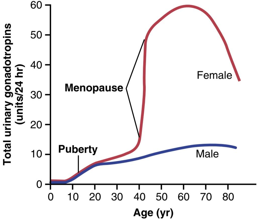
Figure 82-12. Total rates of secretion of gonadotropic hormones throughout the sexual lives of female and male human beings, showing an especially abrupt increase in gonadotropic hormones at menopause in the female.
شکل 82-12. میزان کل ترشح هورمون های گنادوتروپیک در طول زندگی جنسی انسان های زن و مرد، نشان دهنده افزایش شدید هورمون های گنادوتروپیک در دوران یائسگی در زنان است.
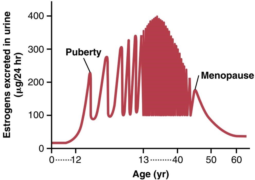
Figure 82-13. Estrogen secretion throughout the sexual life of the female human being.
شکل 82-13. ترشح استروژن در طول زندگی جنسی انسان زن.
MENOPAUSE
At age 40 to 50 years, the sexual cycle usually becomes irregular, and ovulation often fails to occur. After a few months to a few years, the cycle ceases altogether, as shown in Figure 82-13. The period during which the cycle ceases and the female sex hormones diminish to almost none is called menopause.
یائسگی
در سنین 40 تا 50 سالگی، چرخه جنسی معمولاً نامنظم می شود و اغلب تخمک گذاری انجام نمی شود. همانطور که در شکل 82-13 نشان داده شده است، پس از چند ماه تا چند سال، چرخه به طور کلی متوقف می شود. دورهای که در طی آن چرخه متوقف میشود و هورمونهای جنسی زنانه تقریباً به هیچوجه کاهش نمییابد، یائسگی نامیده میشود.
The cause of menopause is “burning out” of the ovaries. Throughout a woman’s reproductive life, about 400 of the primordial follicles grow into mature follicles and ovulate, and hundreds of thousands of ova degenerate. At about age 45 years, only a few primordial follicles remain to be stimulated by FSH and LH and, as shown in Figure 82-13, the production of estrogens by the ovaries decreases as the number of primordial follicles approaches zero. When estrogen production falls below a critical value, the estrogens can no longer inhibit the production of FSH and LH. Instead, as shown in Figure 82-12, the gonadotropins FSH and LH (mainly FSH) are produced after menopause in large and continuous quantities, but as the remaining primordial follicles become atretic, production of estrogens by the ovaries falls virtually to zero.
علت یائسگی “سوختن” تخمدان ها است. در طول زندگی باروری یک زن، حدود 400 فولیکول ازلی به فولیکولهای بالغ تبدیل میشوند و تخمکگذاری میکنند و صدها هزار تخمک تحلیل میروند. در حدود سن 45 سالگی، تنها تعداد کمی از فولیکول های اولیه باقی می مانند که توسط FSH و LH تحریک می شوند و همانطور که در شکل 82-13 نشان داده شده است، با نزدیک شدن تعداد فولیکول های اولیه به صفر، تولید استروژن توسط تخمدان ها کاهش می یابد. وقتی تولید استروژن به زیر یک مقدار بحرانی میرسد، استروژنها دیگر نمیتوانند تولید FSH و LH را مهار کنند. در عوض، همانطور که در شکل 82-12 نشان داده شده است، گنادوتروپین های FSH و LH (عمدتا FSH) پس از یائسگی در مقادیر زیاد و مداوم تولید می شوند، اما با آترتیک شدن فولیکول های اولیه باقی مانده، تولید استروژن توسط تخمدان ها تقریباً به صفر می رسد.
At the time of menopause, a woman must readjust her life from one that has been physiologically stimulated by estrogen and progesterone production to one devoid of these hormones. The loss of estrogens often causes marked physiological changes in the function of the body, including (1) “hot flushes” characterized by extreme flushing of the skin, (2) psychic sensations of dyspnea, (3) irritability, (4) fatigue, (5) anxiety, and (6) decreased strength and calcification of bones throughout the body. These symptoms are of sufficient magnitude in about 15% of women to warrant treatment. Daily administration of estrogen in small quantities usually reverses the symptoms, and by gradually decreasing the dose, postmenopausal women may avoid severe symptoms.
در زمان یائسگی، یک زن باید زندگی خود را از حالتی که از نظر فیزیولوژیکی توسط تولید استروژن و پروژسترون تحریک شده است، به زندگی بدون این هورمون ها تغییر دهد. از دست دادن استروژن اغلب باعث تغییرات فیزیولوژیکی قابل توجهی در عملکرد بدن می شود، از جمله (1) “گرگرفتگی” که با گرگرفتگی شدید پوست، (2) احساسات روانی تنگی نفس، (3) تحریک پذیری، (4) خستگی، (5) اضطراب، و (6) کاهش قدرت و کلسیفیه شدن استخوان ها مشخص می شود. این علائم در حدود 15 درصد از زنان به اندازه کافی برای درمان ضروری است. تجویز روزانه استروژن در مقادیر کم معمولاً علائم را معکوس می کند و با کاهش تدریجی دوز، زنان یائسه ممکن است از علائم شدید اجتناب کنند.
Large clinical trials have provided evidence that administration of estrogen after menopause, although ameliorating many of the symptoms of menopause, may increase the risk for cardiovascular disease. As a result, hormone replacement therapy with estrogen is no longer routinely prescribed for postmenopausal women. Some studies, however, suggest that estrogen therapy may actually reduce the risk for cardiovascular disease if it is begun early in the postmenopausal years. Therefore, it is currently recommended that postmenopausal women who are considering hormone replacement therapy should discuss with their physicians whether the benefits outweigh the risks.
آزمایشهای بالینی بزرگ شواهدی را ارائه کردهاند که نشان میدهد تجویز استروژن پس از یائسگی، اگرچه بسیاری از علائم یائسگی را بهبود میبخشد، اما ممکن است خطر ابتلا به بیماریهای قلبی عروقی را افزایش دهد. در نتیجه، درمان جایگزین هورمونی با استروژن دیگر به طور معمول برای زنان یائسه تجویز نمی شود. با این حال، برخی از مطالعات نشان میدهند که درمان با استروژن ممکن است خطر ابتلا به بیماریهای قلبی عروقی را در صورتی که در اوایل سالهای پس از یائسگی آغاز شود، کاهش دهد. بنابراین، در حال حاضر توصیه میشود که زنان یائسهای که در حال بررسی درمان جایگزینی هورمون هستند، با پزشک خود درمورد اینکه آیا فواید آن بیشتر از خطرات است، صحبت کنند.
Abnormalities of Secretion by the Ovaries
Hypogonadism-Reduced Secretion by the Ovaries. Less than normal secretion by the ovaries can result from poorly formed ovaries, lack of ovaries, or genetically abnormal ovaries that secrete the wrong hormones because of missing enzymes in the secretory cells. When ovaries are absent from birth or when they become nonfunctional before puberty, female eunuchism occurs. In this condition the usual secondary sexual characteristics do not appear, and the sexual organs remain infantile. Especially characteristic of this condition is prolonged growth of the long bones because the epiphyses do not unite with the shafts as early as they do in a normal woman. Consequently, the female eunuch is essentially as tall as or perhaps even slightly taller than her male counterpart of similar genetic background.
اختلالات ترشح توسط تخمدان ها
ترشح کاهش یافته هیپوگنادیسم توسط تخمدان ها. ترشح کمتر از حد طبیعی توسط تخمدان ها می تواند ناشی از شکل گیری ضعیف تخمدان ها، فقدان تخمدان ها یا تخمدان های ژنتیکی غیرطبیعی باشد که به دلیل کمبود آنزیم در سلول های ترشحی، هورمون های اشتباه ترشح می کنند. هنگامی که تخمدان ها از بدو تولد وجود نداشته باشند یا قبل از بلوغ غیرعملکردی شوند، خواجه زن رخ می دهد. در این حالت خصوصیات جنسی ثانویه معمول ظاهر نمی شود و اندام های جنسی نوزادی باقی می مانند. مشخصه این بیماری رشد طولانی مدت استخوان های بلند است زیرا اپی فیزها مانند یک زن عادی با شفت ها یکی نمی شوند. در نتیجه، خواجه زن اساساً به اندازه یا شاید حتی کمی بلندتر از همتای مرد خود با پیشینه ژنتیکی مشابه است.
When the ovaries of a fully developed woman are re- moved, the sexual organs regress to some extent so that the uterus becomes almost infantile in size, the vagina becomes smaller, and the vaginal epithelium becomes thin and easily damaged. The breasts atrophy and become pendulous, and the pubic hair becomes thinner. The same changes occur in women after menopause.
هنگامی که تخمدان های یک زن کاملاً رشد یافته برداشته می شود، اندام های جنسی تا حدی پسرفت می کنند به طوری که اندازه رحم تقریباً نوزادی می شود، واژن کوچکتر می شود و اپیتلیوم واژن نازک می شود و به راحتی آسیب می بیند. سینه ها آتروفی می شوند و آویزان می شوند و موهای ناحیه تناسلی نازک می شوند. همین تغییرات در زنان پس از یائسگی رخ می دهد.
Irregularity of Menses, and Amenorrhea Caused by Hypogonadism. As pointed out in the preceding discussion of menopause, the quantity of estrogens produced by the ovaries must rise above a critical value to cause rhythmic sexual cycles. Consequently, in hypogonadism or when the gonads are secreting small quantities of estrogens as a result of other factors, such as hypothyroidism, the ovarian cycle often does not occur normally. Instead, several months may elapse between menstrual periods, or menstruation may cease altogether (amenorrhea). Prolonged ovarian cycles are frequently associated with failure of ovulation, presumably because of insufficient secretion of LH at the time of the preovulatory surge of LH, which is necessary for ovulation.
بی نظمی قاعدگی و آمنوره ناشی از هیپوگنادیسم. همانطور که در بحث قبلی در مورد یائسگی اشاره شد، مقدار استروژن تولید شده توسط تخمدان ها برای ایجاد چرخه های جنسی ریتمیک باید از یک مقدار بحرانی بالاتر برود. در نتیجه، در هیپوگنادیسم یا زمانی که غدد جنسی در نتیجه عوامل دیگر، مانند کم کاری تیروئید، مقادیر کمی استروژن ترشح می کنند، چرخه تخمدان اغلب به طور طبیعی رخ نمی دهد. در عوض، ممکن است چند ماه بین دوره های قاعدگی فاصله باشد یا قاعدگی به طور کلی متوقف شود (آمنوره). چرخه های تخمدانی طولانی مدت اغلب با شکست تخمک گذاری همراه است، احتمالاً به دلیل ترشح ناکافی LH در زمان افزایش قبل از تخمک گذاری LH، که برای تخمک گذاری ضروری است.
Hypersecretion by the Ovaries. Extreme hypersecretion of ovarian hormones by the ovaries is a rare clinical entity because excessive secretion of estrogens automatically decreases production of gonadotropins by the pituitary, which limits production of ovarian hormones. Consequently, hypersecretion of feminizing hormones is usually recognized clinically only when a feminizing tumor develops.
ترشح بیش از حد توسط تخمدان ها. ترشح بیش از حد هورمون های تخمدان توسط تخمدان ها یک موجود بالینی نادر است زیرا ترشح بیش از حد استروژن به طور خودکار تولید گنادوتروپین ها را توسط هیپوفیز کاهش می دهد که تولید هورمون های تخمدانی را محدود می کند. در نتیجه، ترشح بیش از حد هورمون های زنانه معمولاً از نظر بالینی تنها زمانی تشخیص داده می شود که یک تومور زنانه ایجاد شود.
A rare granulosa cell tumor can develop in an ovary; development of this tumor occurs more often after menopause than before menopause. These tumors secrete large quantities of estrogens, which exert the usual estrogenic effects, including hypertrophy of the uterine endometrium and irregular bleeding from this endometrium. In fact, bleeding is often the first and only indication that such a tumor exists.
یک تومور نادر سلولی گرانولوزا می تواند در تخمدان ایجاد شود. توسعه این تومور اغلب بعد از یائسگی نسبت به قبل از یائسگی رخ می دهد. این تومورها مقادیر زیادی استروژن ترشح می کنند که اثرات معمول استروژنی از جمله هیپرتروفی آندومتر رحم و خونریزی نامنظم از این اندومتر را اعمال می کند. در واقع، خونریزی اغلب اولین و تنها نشانه وجود چنین توموری است.
FEMALE SEXUAL ACT
Stimulation of the Female Sexual Act. As is true in the male sexual act, successful performance of the female sexual act depends on both psychic stimulation and local sexual stimulation.
کنش جنسی زنانه
تحریک قانون جنسی زنان. همانطور که در کنش جنسی مردانه صادق است، انجام موفقیت آمیز کنش جنسی زنانه هم به تحریک روانی و هم به تحریک جنسی موضعی بستگی دارد.
Thinking sexual thoughts can lead to female sexual desire, and this aids greatly in the performance of the female sexual act. Such desire is based on psychological and physiological drive, although sexual desire does increase in proportion to the level of sex hormones secreted. Desire also changes during the monthly sexual cycle, reaching a peak near the time of ovulation, probably because of the high levels of estrogen secretion during the preovulatory period.
فکر کردن به افکار جنسی می تواند به میل جنسی زنان منجر شود و این به انجام عمل جنسی زنان کمک زیادی می کند. چنین میل بر انگیزه روانی و فیزیولوژیکی است، اگرچه میل جنسی متناسب با سطح هورمون های جنسی ترشح شده افزایش می یابد. میل نیز در طول چرخه جنسی ماهانه تغییر می کند و در نزدیکی زمان تخمک گذاری به اوج خود می رسد، احتمالاً به دلیل سطوح بالای ترشح استروژن در طول دوره قبل از تخمک گذاری.
Local sexual stimulation in women occurs in more or less the same manner as in men because massage and other types of stimulation of the vulva, vagina, and other perineal regions can create sexual sensations. The glans of the clitoris is especially sensitive for initiating sexual sensations.
تحریک جنسی موضعی در زنان کمابیش مانند مردان اتفاق می افتد زیرا ماساژ و انواع دیگر تحریکات فرج، واژن و سایر نواحی پرینه می تواند احساسات جنسی ایجاد کند. غده کلیتوریس به ویژه برای شروع احساسات جنسی حساس است.
As in the male, the sexual sensory signals are trans- mitted to the sacral segments of the spinal cord through the pudendal nerve and sacral plexus. Once these signals have entered the spinal cord, they are transmitted to the cerebrum. Also, local reflexes integrated in the sacral and lumbar spinal cord are at least partly responsible for some of the reactions in the female sexual organs.
همانند مردان، سیگنالهای حسی جنسی از طریق عصب پودندال و شبکه خاجی به بخشهای خاجی نخاع منتقل میشوند. هنگامی که این سیگنال ها وارد نخاع شدند، به مغز منتقل می شوند. همچنین، رفلکس های موضعی ادغام شده در طناب نخاعی ساکرال و کمر حداقل تا حدودی مسئول برخی از واکنش ها در اندام های جنسی زنانه هستند.
Female Erection and Lubrication. Located around the introitus and extending into the clitoris is erectile tissue almost identical to the erectile tissue of the penis. This erectile tissue, like that of the penis, is controlled by the parasympathetic nerves that pass through the nervi erigentes from the sacral plexus to the external genitalia. In the early phases of sexual stimulation, parasympathetic signals dilate the arteries of the erectile tissue, probably resulting from release of acetylcholine, nitric oxide, and vasoactive intestinal polypeptide at the nerve endings. This allows rapid accumulation of blood in the erectile tis- sue so that the introitus tightens around the penis, which aids the male in his attainment of sufficient sexual stimulation for ejaculation to occur.
نعوظ و روانکاری زنانه. بافت نعوظی که در اطراف ناحیه داخلی قرار گرفته و به داخل کلیتوریس کشیده شده است تقریباً مشابه بافت نعوظ آلت تناسلی است. این بافت نعوظ، مانند بافت آلت تناسلی، توسط اعصاب پاراسمپاتیک کنترل می شود که از طریق عصب erigentes از شبکه خاجی به اندام تناسلی خارجی عبور می کنند. در مراحل اولیه تحریک جنسی، سیگنالهای پاراسمپاتیک عروق بافت نعوظ را گشاد میکنند که احتمالاً ناشی از آزاد شدن استیل کولین، اکسید نیتریک و پلی پپتید وازواکتیو رودهای در انتهای عصبی است. این امکان تجمع سریع خون در بافت نعوظ را فراهم می کند به طوری که اندرونیوس در اطراف آلت تناسلی منقبض می شود، که به مرد کمک می کند تا به تحریک جنسی کافی برای انزال دست یابد.
Parasympathetic signals also pass to the bilateral Bartholin glands located beneath the labia minora and cause them to secrete mucus immediately inside the introitus. This mucus is responsible for much of the lubrication during sexual intercourse, although much lubrication is also provided by mucus secreted by the vaginal epithelium, and a small amount is provided from the male urethral glands. This lubrication is necessary during intercourse to establish a satisfactory massaging sensation rather than an irritative sensation, which may be provoked by a dry vagina. A massaging sensation constitutes the optimal stimulus for evoking the appropriate reflexes that culminate in both the male and female climaxes.
سیگنالهای پاراسمپاتیک نیز به غدد بارتولین دو طرفه که در زیر لبهای کوچک قرار دارند میرسند و باعث میشوند که بلافاصله در داخل دهانه مخاط ترشح کنند. این مخاط مسئول بسیاری از روانکاری در طول مقاربت جنسی است، اگرچه مقدار زیادی روانکاری نیز توسط مخاط ترشح شده از اپیتلیوم واژن انجام می شود و مقدار کمی از غدد مجرای ادرار مردانه تامین می شود. این روانکاری در حین مقاربت برای ایجاد یک احساس رضایت بخش ماساژ به جای یک احساس تحریکی که ممکن است توسط خشکی واژن تحریک شود، ضروری است. یک احساس ماساژ محرک بهینه برای برانگیختن رفلکس های مناسب است که در هر دو نقطه اوج مرد و زن به اوج می رسد.
Female Orgasm. When local sexual stimulation reaches maximum intensity, and especially when the local sensations are supported by appropriate psychic conditioning signals from the cerebrum, reflexes are initiated that cause the female orgasm, also called the female climax. The female orgasm is analogous to emission and ejaculation in the male, and it may help promote fertilization of the ovum. Indeed, the human female is known to be some- what more fertile when inseminated by normal sexual intercourse rather than by artificial methods, thus indicating an important function of the female orgasm. Possible reasons for this phenomenon are as follows.
ارگاسم زنانه. هنگامی که تحریک جنسی موضعی به حداکثر شدت می رسد، و به ویژه هنگامی که احساسات محلی توسط سیگنال های شرطی سازی روانی مناسب از مغز پشتیبانی می شود، رفلکس هایی آغاز می شود که باعث ارگاسم زنانه می شود که به اوج زن نیز می گویند. ارگاسم زنانه مشابه ترشح و انزال در مرد است و ممکن است به لقاح تخمک کمک کند. در واقع، زن انسان زمانی که با آمیزش جنسی معمولی به جای روشهای مصنوعی لقاح مییابد، تا حدودی بارورتر است، بنابراین نشاندهنده عملکرد مهم ارگاسم زن است. دلایل احتمالی این پدیده به شرح زیر است.
First, during the orgasm, the perineal muscles of the female contract rhythmically, which results from spinal cord reflexes similar to those that cause ejaculation in the male. It is possible that these reflexes increase uterine and fallopian tube motility during the orgasm, thus helping to transport the sperm upward through the uterus toward the ovum; information on this subject is scanty, however. Also, the orgasm seems to cause dilation of the cervical canal for up to 30 minutes, thus allowing easy transport of the sperm.
اول، در طول ارگاسم، ماهیچه های پرینه زن به طور ریتمیک منقبض می شوند، که ناشی از رفلکس های نخاعی مشابه آن هایی است که باعث انزال در مرد می شود. ممکن است این رفلکس ها باعث افزایش تحرک رحم و لوله فالوپ در طول ارگاسم شود، بنابراین به انتقال اسپرم به سمت بالا از طریق رحم به سمت تخمک کمک می کند. با این حال، اطلاعات در مورد این موضوع کم است. همچنین، به نظر می رسد ارگاسم باعث گشاد شدن کانال دهانه رحم به مدت 30 دقیقه می شود و بنابراین امکان انتقال آسان اسپرم را فراهم می کند.
Second, in many animals, copulation causes the posterior pituitary gland to secrete oxytocin; this effect is probably mediated through the brain amygdaloid nuclei and then through the hypothalamus to the pituitary. The oxytocin causes increased rhythmic contractions of the uterus, which may increase transport of the sperm. A few sperm have been shown to traverse the entire length of the fallopian tube in the cow in about 5 minutes, a rate at least 10 times as fast as that which the swimming motions of the sperm could possibly achieve. Whether this effect occurs in the human female is unknown.
دوم، در بسیاری از حیوانات، جفت شدن باعث ترشح اکسی توسین در غده هیپوفیز خلفی می شود. این اثر احتمالاً از طریق هستههای آمیگدالوئید مغز و سپس از طریق هیپوتالاموس به هیپوفیز انجام میشود. اکسی توسین باعث افزایش انقباضات ریتمیک رحم می شود که ممکن است انتقال اسپرم را افزایش دهد. نشان داده شده است که تعداد کمی از اسپرم ها تمام طول لوله فالوپ را در حدود 5 دقیقه در گاو طی می کنند، سرعتی که حداقل 10 برابر سریعتر از سرعتی است که حرکات شنای اسپرم ممکن است به دست آورد. اینکه آیا این اثر در زن انسان رخ می دهد ناشناخته است.
In addition to possible effects of the orgasm on fertilization, the intense sexual sensations that develop during the orgasm also pass to the cerebrum and cause intense muscle tension throughout the body. After culmination of the sexual act, this tension gives way during the succeeding minutes to a sense of satisfaction characterized by relaxed peacefulness, an effect called resolution.
علاوه بر تأثیرات احتمالی ارگاسم بر لقاح، احساسات شدید جنسی که در طول ارگاسم ایجاد می شود به مغز نیز منتقل می شود و باعث تنش شدید عضلانی در سراسر بدن می شود. پس از اوج گیری کنش جنسی، این تنش در طی دقایق بعدی جای خود را به احساس رضایت می دهد که با آرامش آرام مشخص می شود، اثری که حل و فصل نامیده می شود.
Female Fertility
Fertile Period of Each Sexual Cycle. The ovum remains viable and capable of being fertilized probably no longer than 24 hours after it is expelled from the ovary. Therefore, sperm must be available soon after ovulation if fertilization is to take place. A few sperm can remain fertile in the female reproductive tract for up to 5 days. Therefore, for fertilization to take place, intercourse must occur sometime between 4 and 5 days before ovulation up to a few hours after ovulation. Thus, the period of female fertility during each month is short, about 4 to 5 days.
باروری زنانه
دوره باروری هر چرخه جنسی. تخمک احتمالاً بیش از 24 ساعت پس از خروج از تخمدان زنده می ماند و قادر به بارور شدن است. بنابراین، اگر قرار است لقاح انجام شود، اسپرم باید بلافاصله پس از تخمک گذاری در دسترس باشد. تعداد کمی از اسپرم ها می توانند تا 5 روز در دستگاه تناسلی زن بارور باقی بمانند. بنابراین، برای انجام لقاح، مقاربت باید بین 4 تا 5 روز قبل از تخمک گذاری تا چند ساعت پس از تخمک گذاری انجام شود. بنابراین دوره باروری زنان در طول هر ماه کوتاه و حدود 4 تا 5 روز است.
Rhythm Method of Contraception. One commonly practiced method of contraception is to avoid intercourse near the time of ovulation. The difficulty with this method of contraception is predicting the exact time of ovulation. Yet, the interval from ovulation until the next succeeding onset of menstruation is almost always between 13 and 15 days. Therefore, if the menstrual cycle is regular, with an exact periodicity of 28 days, ovulation usually occurs within 1 day of the 14th day of the cycle. If, in contrast, the periodicity of the cycle is 40 days, ovulation usually occurs within 1 day of the 26th day of the cycle. Finally, if the periodicity of the cycle is 21 days, ovulation usually occurs within 1 day of the seventh day of the cycle. Therefore, it is usually stated that avoidance of intercourse for 4 days before the calculated day of ovulation and 3 days afterward prevents conception. However, such a method of contraception can be used only when the periodicity of the menstrual cycle is regular. The failure rate of this method of contraception, resulting in an unintentional pregnancy, may be as high as 20% to 25% per year.
روش ریتم پیشگیری از بارداری. یکی از روش های رایج پیشگیری از بارداری، اجتناب از نزدیکی نزدیک به زمان تخمک گذاری است. مشکل این روش پیشگیری از بارداری پیش بینی زمان دقیق تخمک گذاری است. با این حال، فاصله بین تخمک گذاری تا شروع بعدی قاعدگی تقریباً همیشه بین 13 تا 15 روز است. بنابراین، اگر چرخه قاعدگی منظم باشد، با تناوب دقیق 28 روز، تخمک گذاری معمولا در 1 روز از روز چهاردهم سیکل اتفاق می افتد. در مقابل، اگر تناوب چرخه 40 روز باشد، تخمک گذاری معمولاً در 1 روز از روز بیست و ششم سیکل اتفاق می افتد. در نهایت، اگر دوره تناوب سیکل 21 روز باشد، تخمک گذاری معمولا در 1 روز از روز هفتم سیکل اتفاق می افتد. بنابراین، معمولاً بیان میشود که پرهیز از آمیزش به مدت 4 روز قبل از روز محاسبه شده تخمکگذاری و 3 روز پس از آن از بارداری جلوگیری میکند. با این حال، چنین روشی برای پیشگیری از بارداری تنها زمانی قابل استفاده است که تناوب چرخه قاعدگی منظم باشد. میزان شکست این روش پیشگیری از بارداری که منجر به بارداری ناخواسته می شود، ممکن است به 20 تا 25 درصد در سال برسد.
Hormonal Suppression of Fertility-“The Pill”
Administration of either estrogen or progesterone, if given in appropriate quantities during the first half of the monthly cycle, can inhibit ovulation. The reason for this is that appropriate administration of either of these hormones can prevent the preovulatory surge of LH secretion by the pituitary gland, which is essential in causing ovulation.
سرکوب هورمونی باروری – “قرص”
تجویز استروژن یا پروژسترون، اگر در مقادیر مناسب در نیمه اول چرخه ماهانه داده شود، می تواند تخمک گذاری را مهار کند. دلیل این امر این است که تجویز مناسب هر یک از این هورمون ها می تواند از افزایش ترشح LH قبل از تخمک گذاری توسط غده هیپوفیز جلوگیری کند که در ایجاد تخمک گذاری ضروری است.
It is not fully understood why administration of estro- gen or progesterone prevents the preovulatory surge of LH secretion. However, experimental work has suggested that immediately before the surge occurs, a sudden depression of estrogen secretion by the ovarian follicles probably occurs, which might be the necessary signal that causes the subsequent feedback effect on the anterior pituitary that leads to the LH surge. The administration of sex hormones (estrogens or progesterone) could prevent the initial ovarian hormonal depression that might be the initiating signal for ovulation.
به طور کامل درک نشده است که چرا تجویز استروژن یا پروژسترون از افزایش ترشح LH قبل از تخمک گذاری جلوگیری می کند. با این حال، کار تجربی نشان میدهد که بلافاصله قبل از وقوع موج، کاهش ناگهانی ترشح استروژن توسط فولیکولهای تخمدان احتمالاً رخ میدهد، که ممکن است سیگنال لازمی باشد که باعث اثر بازخورد بعدی در هیپوفیز قدامی میشود که منجر به افزایش LH میشود. تجویز هورمون های جنسی (استروژن یا پروژسترون) می تواند از افسردگی اولیه هورمونی تخمدان که ممکن است سیگنال آغاز کننده تخمک گذاری باشد، جلوگیری کند.
The challenge in devising methods for the hormonal suppression of ovulation has been in developing appropriate combinations of estrogens and progestins that suppress ovulation but do not cause other, unwanted effects. For example, too much of either hormone can cause abnormal menstrual bleeding patterns. However, use of certain synthetic progestins in place of progesterone, especially the 19-norsteroids, along with small amounts of estrogens, usually prevents ovulation yet allows an almost normal pattern of menstruation. Therefore, almost all “pills” used for the control of fertility consist of some combination of synthetic estrogens and synthetic progestins. The main reason for using synthetic estrogens and progestins is that the natural hormones are almost entirely destroyed by the liver within a short time after they are absorbed from the gastrointestinal tract into the portal circulation. However, many of the synthetic hormones can resist this destructive propensity of the liver, thus allowing oral administration.
چالش در ابداع روشهایی برای سرکوب هورمونی تخمکگذاری، ایجاد ترکیبهای مناسب از استروژنها و پروژستینها است که تخمکگذاری را سرکوب میکنند، اما اثرات ناخواسته دیگری ایجاد نمیکنند. به عنوان مثال، مصرف بیش از حد هر دو هورمون می تواند باعث الگوهای خونریزی غیر طبیعی قاعدگی شود. با این حال، استفاده از پروژستین های مصنوعی خاص به جای پروژسترون، به ویژه 19-نورستروئیدها، همراه با مقادیر کمی استروژن، معمولاً از تخمک گذاری جلوگیری می کند، اما یک الگوی تقریباً طبیعی قاعدگی را امکان پذیر می کند. بنابراین، تقریباً تمام «قرصهایی» که برای کنترل باروری استفاده میشوند از ترکیبی از استروژنهای مصنوعی و پروژستینهای مصنوعی تشکیل شدهاند. دلیل اصلی استفاده از استروژن ها و پروژستین های مصنوعی این است که هورمون های طبیعی تقریباً به طور کامل توسط کبد در مدت کوتاهی پس از جذب از دستگاه گوارش به گردش خون پورتال از بین می روند. با این حال، بسیاری از هورمونهای مصنوعی میتوانند در برابر این تمایل مخرب کبد مقاومت کنند، بنابراین اجازه تجویز خوراکی را میدهند.
Two of the most commonly used synthetic estrogens are ethinyl estradiol and mestranol. Among the most commonly used progestins are norethindrone, norethynodrel, ethynodiol, and norgestrel. The drug is usually begun in the early stages of the monthly cycle and continued beyond the time that ovulation would normally occur. Then the drug is stopped, allowing menstruation to occur and a new cycle to begin.
دو مورد از متداول ترین استروژن های مصنوعی اتینیل استرادیول و مسترانول هستند. از رایج ترین پروژستین های مورد استفاده می توان به نورتیندرون، نورتینودرل، اتینودیول و نورژسترل اشاره کرد. این دارو معمولاً در مراحل اولیه چرخه ماهانه شروع می شود و بعد از زمانی که تخمک گذاری به طور معمول رخ می دهد ادامه می یابد. سپس دارو قطع می شود و اجازه می دهد قاعدگی رخ دهد و چرخه جدیدی شروع شود.
The failure rate, resulting in an unintentional pregnancy, for hormonal suppression of fertility using various forms of the “pill” is about 8% to 9% per year.
میزان شکست، که منجر به بارداری ناخواسته می شود، برای سرکوب هورمونی باروری با استفاده از اشکال مختلف “قرص” حدود 8٪ تا 9٪ در سال است.
Abnormal Conditions That Cause Female Sterility
About 5% to 10% of women are infertile. Occasionally, no abnormality can be discovered in the female genital organs, in which case the infertility is assumed to be due to either abnormal physiological function of the genital system or abnormal genetic development of the ova.
شرایط غیر طبیعی که باعث عقیمی زنان می شود
حدود 5 تا 10 درصد زنان نابارور هستند. گاهی اوقات، هیچ گونه ناهنجاری در اندام تناسلی زنان کشف نمی شود، در این صورت فرض می شود که ناباروری به دلیل عملکرد فیزیولوژیکی غیر طبیعی دستگاه تناسلی یا رشد ژنتیکی غیرطبیعی تخمک ها باشد.
The most common cause of female sterility is failure to ovulate. This failure can result from hyposecretion of gonadotropic hormones, in which case the intensity of the hormonal stimuli is simply insufficient to cause ovulation, or it can result from abnormal ovaries that do not allow ovulation. For example, thick ovarian capsules occasionally exist on the outsides of the ovaries, making ovulation difficult.
شایع ترین علت نازایی زنان عدم تخمک گذاری است. این نارسایی می تواند ناشی از ترشح کم هورمون های گنادوتروپیک باشد، در این صورت شدت محرک های هورمونی برای ایجاد تخمک گذاری کافی نیست یا می تواند ناشی از تخمدان های غیرطبیعی باشد که اجازه تخمک گذاری را نمی دهند. به عنوان مثال، کپسول های تخمدان ضخیم گاهی اوقات در خارج تخمدان ها وجود دارد که تخمک گذاری را دشوار می کند.
Because of the high incidence of anovulation in sterile women, special methods are often used to determine whether ovulation occurs. These methods are based mainly on the effects of progesterone on the body because the nor- mal increase in progesterone secretion usually does not occur during the latter half of anovulatory cycles. In the absence of progestational effects, the cycle can be assumed to be anovulatory.
به دلیل شیوع بالای عدم تخمک گذاری در زنان عقیم، اغلب از روش های خاصی برای تعیین اینکه آیا تخمک گذاری اتفاق می افتد استفاده می شود. این روش ها عمدتاً بر اساس تأثیرات پروژسترون بر بدن است، زیرا افزایش طبیعی ترشح پروژسترون معمولاً در نیمه دوم چرخه های تخمک گذاری رخ نمی دهد. در غیاب اثرات پروژستاتیک، سیکل را می توان بدون تخمک گذاری فرض کرد.
One of these tests is simply to analyze the urine for a surge in pregnanediol, the end product of progesterone metabolism, during the latter half of the sexual cycle; the lack of this substance indicates failure of ovulation.
یکی از این آزمایشها صرفاً آنالیز ادرار برای افزایش پرگناندیول، محصول نهایی متابولیسم پروژسترون، در نیمه دوم چرخه جنسی است. فقدان این ماده نشان دهنده شکست تخمک گذاری است.
Another common test is for the woman to chart her body temperature throughout the cycle. Secretion of progesterone during the latter half of the cycle raises the body temperature about 0.5°F, with the temperature rise coming abruptly at the time of ovulation. Such a temperature chart, showing the point of ovulation, is illustrated in Figure 82-14.
یکی دیگر از آزمایشات رایج این است که زن دمای بدن خود را در طول چرخه نشان می دهد. ترشح پروژسترون در نیمه دوم چرخه دمای بدن را حدود 0.5 درجه فارنهایت افزایش می دهد و در زمان تخمک گذاری به طور ناگهانی افزایش می یابد. چنین نمودار دمایی که نقطه تخمک گذاری را نشان می دهد، در شکل 82-14 نشان داده شده است.

Figure 82-14. Elevation in body temperature shortly after ovulation.
شکل 82-14. افزایش دمای بدن در مدت کوتاهی پس از تخمک گذاری.
Lack of ovulation caused by hyposecretion of the pituitary gonadotropic hormones can sometimes be treated by appropriately timed administration of human chorionic gonadotropin, a hormone (discussed in Chapter 83) that is extracted from the human placenta. This hormone, although secreted by the placenta, has almost the same effects as LH and is therefore a powerful stimulator of ovulation. However, excess use of this hormone can cause ovulation from many follicles simultaneously, which results in multiple births, an effect that has caused as many as eight babies (stillborn in many cases) to be born to mothers treated for infertility with this hormone.
عدم تخمک گذاری ناشی از افت ترشح هورمون های گنادوتروپیک هیپوفیز گاهی اوقات می تواند با تجویز به موقع گنادوتروپین جفتی انسانی، هورمونی (که در فصل 83 بحث شد) که از جفت انسان استخراج می شود، درمان شود. این هورمون، اگرچه از جفت ترشح می شود، اما تقریباً همان اثرات LH را دارد و بنابراین یک محرک قدرتمند تخمک گذاری است. با این حال، استفاده بیش از حد از این هورمون می تواند باعث تخمک گذاری از بسیاری از فولیکول ها به طور همزمان شود که منجر به چند قلوزایی می شود، اثری که باعث شده است تا هشت نوزاد (در بسیاری از موارد مرده به دنیا آمده) از مادرانی که به دلیل ناباروری با این هورمون درمان شده اند به دنیا بیایند.
One of the most common causes of female sterility is endometriosis, a common condition in which endometrial tissue almost identical to that of the normal uterine endometrium grows and even menstruates in the pelvic cavity surrounding the uterus, fallopian tubes, and ovaries. Endometriosis causes fibrosis throughout the pelvis, and this fibrosis sometimes so enshrouds the ovaries that an ovum cannot be released into the abdominal cavity. Often, endometriosis occludes the fallopian tubes, either at the fimbriated ends or elsewhere along their extent.
یکی از شایع ترین علل عقیمی زنان آندومتریوز است، یک بیماری شایع که در آن بافت آندومتر تقریباً مشابه آندومتر طبیعی رحم رشد می کند و حتی در حفره لگن اطراف رحم، لوله های فالوپ و تخمدان ها قاعدگی می شود. اندومتریوز باعث ایجاد فیبروز در سراسر لگن می شود و این فیبروز گاهی اوقات آنقدر تخمدان ها را می پوشاند که تخمک نمی تواند در حفره شکمی آزاد شود. اغلب، آندومتریوز لولههای فالوپ را چه در انتهای فیمبریه یا در سایر نقاط در امتداد وسعت آنها مسدود میکند.
Another common cause of female infertility is salpingitis, that is, inflammation of the fallopian tubes; this inflammation causes fibrosis in the tubes, thereby occluding them. In the past, such inflammation occurred mainly as a result of gonococcal infection. However, with modern therapy, salpingitis is becoming a less prevalent cause of female infertility.
یکی دیگر از علل شایع ناباروری زنان سالپنژیت است، یعنی التهاب لوله های فالوپ. این التهاب باعث فیبروز در لوله ها می شود و در نتیجه آنها را مسدود می کند. در گذشته، چنین التهابی عمدتاً در نتیجه عفونت گنوکوکی رخ می داد. با این حال، با درمان مدرن، سالپنژیت در حال تبدیل شدن به یک علت کمتر شایع ناباروری زنان است.
Still another cause of infertility is secretion of abnormal mucus by the uterine cervix. Ordinarily, at the time of ovulation, the hormonal environment of estrogen causes secretion of mucus with special characteristics that allow rapid mobility of sperm into the uterus and actually guide the sperm up along mucous “threads.” Abnormalities of the cervix, such as low-grade infection or inflammation, or abnormal hormonal stimulation of the cervix, can lead to a viscous mucous plug that prevents fertilization.
یکی دیگر از دلایل ناباروری ترشح مخاط غیرطبیعی توسط دهانه رحم است. به طور معمول، در زمان تخمک گذاری، محیط هورمونی استروژن باعث ترشح مخاطی با ویژگی های خاص می شود که امکان تحرک سریع اسپرم به داخل رحم را فراهم می کند و در واقع اسپرم را در امتداد “نخ های مخاطی” به سمت بالا هدایت می کند. ناهنجاریهای دهانه رحم، مانند عفونت یا التهاب با درجه پایین، یا تحریک هورمونی غیرطبیعی دهانه رحم، میتواند منجر به یک پلاگ مخاطی چسبناک شود که از لقاح جلوگیری میکند.
کلیک کنید «Bibliography: فهرست کتب مربوطه»
Abbara A, Clarke SA, Dhillo WS: Novel concepts for inducing final oocyte maturation in in vitro fertilization treatment. Endocr Rev 39:593, 2018.
Almeida M, Laurent MR, Dubois V, et al: Estrogens and androgens in skeletal physiology and pathophysiology. Physiol Rev 97:135, 2017.
Anderson RC, Newton CL, Anderson RA, Millar RP: Gonadotropins and their analogs: current and potential clinical applications. En- docr Rev 39:911, 2018.
Arnal JF, Lenfant F, Metivier R, et al: Membrane and nuclear estrogen receptor alpha actions: from tissue specificity to medical implica- tions. Physiol Rev 97:1045, 2017.
Berkane N, Liere P, Oudinet JP, et al: From pregnancy to preeclamp- sia: a key role for estrogens. Endocr Rev 38:123, 2017.
Chapron C, Marcellin L, Borghese B, Santulli P: Rethinking mecha- nisms, diagnosis and management of endometriosis. Nat Rev En- docrinol 15:666, 2019.
Duffy DM, Ko C, Jo M, Brannstrom M, Curry TE: Ovulation: parallels with inflammatory processes. Endocr Rev 40:369, 2019.
Escobar-Morreale HF: Polycystic ovary syndrome: definition, aetiol- ogy, diagnosis and treatment. Nat Rev Endocrinol 14:270, 2018.
Gartlehner G, Patel SV, Feltner C, et al: Hormone therapy for the pri- mary prevention of chronic conditions in postmenopausal women: evidence report and systematic review for the US Preventive Ser- vices Task Force. JAMA 318:2234, 2017.
Gordon CM, Ackerman KE, Berga SL, et al: Functional hypothalamic amenorrhea: an Endocrine Society clinical practice guideline. J Clin Endocrinol Metab 102:1413, 2017.
Hart RJ: Physiological aspects of female fertility: role of the environment, modern lifestyle, and genetics. Physiol Rev 96:873, 2016.
Herbison AE: The gonadotropin-releasing hormone pulse generator. Endocrinology 159:3723, 2018.
Hewitt SC, Korach KS: Estrogen receptors: new directions in the new millennium. Endocr Rev 39:664, 2018.
Monteleone P, Mascagni G, Giannini A, et al: Symptoms of meno- pause – global prevalence, physiology and implications. Nat Rev Endocrinol 14:199, 2018.
Richards JS, Ascoli M: Endocrine, paracrine, and autocrine signal- ing pathways that regulate ovulation. Trends Endocrinol Metab 29:313, 2018.
Richards JS, Ren YA, Candelaria N, et al: Ovarian follicular theca cell recruitment, differentiation, and impact on fertility. Endocr Rev 39:1, 2018.
Robker RL, Hennebold JD, Russell DL: Coordination of ovulation and oocyte maturation: a good egg at the right time. Endocrinology 159:3209, 2018.
Stilley JAW, Segaloff DL: FSH actions and pregnancy: looking beyond ovarian FSH receptors. Endocrinology 159:4033, 2018.
🚀 با ما همراه شوید!
تازهترین مطالب و آموزشهای مغز و اعصاب را از دست ندهید. با فالو کردن کانال تلگرام آیندهنگاران مغز، از ما حمایت کنید!



