
فیزیولوژی پزشکی گایتون و هال؛ عملکردهای تولید مثلی و هورمونی مردان و عملکرد غده پینهال


دعای مطالعه [ نمایش ]
بِسْمِ الله الرَّحْمنِ الرَّحیمِ
اَللّهُمَّ اَخْرِجْنى مِنْ ظُلُماتِ الْوَهْمِ
خدايا مرا بيرون آور از تاريكىهاى وهم،
وَ اَكْرِمْنى بِنُورِ الْفَهْمِ
و به نور فهم گرامى ام بدار،
اَللّهُمَّ افْتَحْ عَلَيْنا اَبْوابَ رَحْمَتِكَ
خدايا درهاى رحمتت را به روى ما بگشا،
وَانْشُرْ عَلَيْنا خَزائِنَ عُلُومِكَ بِرَحْمَتِكَ يا اَرْحَمَ الرّاحِمينَ
و خزانههاى علومت را بر ما باز كن به امید رحمتت اى مهربانترين مهربانان.
کتاب «فیزیولوژی پزشکی گایتون و هال» بهعنوان یکی از جامعترین و معتبرترین منابع در حوزه علوم پزشکی، همچنان مرجع کلیدی برای درک عملکرد پیچیده بدن انسان است. این اثر با تکیه بر تازهترین پژوهشها و توضیحات دقیق از سازوکارهای فیزیولوژیک، پلی میان علوم پایه پزشکی و کاربردهای بالینی ایجاد میکند و نقشی بیبدیل در آموزش، پژوهش و ارتقای دانش سلامت ایفا مینماید.
ترجمه دقیق و علمی این شاهکار توسط برند علمی آیندهنگاران مغز به مدیریت داریوش طاهری، دسترسی فارسیزبانان به مرزهای نوین دانش فیزیولوژی را ممکن ساخته و رسالتی علمی برای ارتقای آموزش پزشکی، فهم عمیقتر سازوکارهای بدن و توسعه روشهای نوین در حوزه سلامت فراهم آورده است.
» کتاب فیزیولوژی پزشکی گایتون و هال
» » فصل ۸۱: عملکردهای تولید مثلی و هورمونی مردان (و عملکرد غده پینهال)
در حال ویرایش
» Guyton and Hall Textbook of Medical Physiology
»» CHAPTER 81: Reproductive and Hormonal Functions of the Male (and Function of the Pineal Gland)
Male reproductive functions can be divided into three major subdivisions: (1) formation of sperm-spermatogenesis; (2) performance of the male sexual act; and (3) regulation of male reproductive functions by the various hormones. Associated with these reproductive functions are the effects of the male sex hormones on the accessory sexual organs, cellular metabolism, growth, and other functions of the body.
عملکردهای تولید مثل مردان را می توان به سه زیربخش عمده تقسیم کرد: (1) تشکیل اسپرم-اسپرماتوژنز. (2) انجام عمل جنسی مردانه؛ و (3) تنظیم عملکرد تولید مثل مردان توسط هورمون های مختلف. با این عملکردهای تولید مثلی، تأثیر هورمون های جنسی مردانه بر اندام های جنسی جانبی، متابولیسم سلولی، رشد و سایر عملکردهای بدن مرتبط است.
Physiological Anatomy of the Male Sexual Organs
Figure 81-14 shows various portions of the male reproductive system, and Figure 81-1B gives a more detailed structure of the testis and epididymis. The testis is composed of up to 900 coiled seminiferous tubules, each averaging more than one-half meter long, in which the sperm are formed. The sperm then empty into the epididymis, which is another coiled tube about 6 meters long. The epididymis leads into the vas deferens, which enlarges into the ampulla of the vas deferens immediately before the vas enters the body of the prostate gland.
آناتومی فیزیولوژیکی اندام های جنسی مردانه
شکل 81-14 بخش های مختلف سیستم تولیدمثلی نر را نشان می دهد و شکل 81-1B ساختار دقیق تری از بیضه و اپیدیدیم را نشان می دهد. بیضه از 900 لوله اسپرم ساز مارپیچ تشکیل شده است که هر کدام به طور متوسط بیش از نیم متر طول دارند و در آنها اسپرم تشکیل می شود. سپس اسپرم به داخل اپیدیدیم تخلیه میشود که یک لوله پیچدار دیگری به طول حدود 6 متر است. اپیدیدیم به داخل مجرای دفران منتهی می شود، که بلافاصله قبل از اینکه مجرا وارد بدن غده پروستات شود، به آمپول مجرای دفران بزرگ می شود.
Two seminal vesicles, one located on each side of the prostate, empty into the prostatic end of the ampulla, and the contents from both the ampulla and the seminal vesicles pass into an ejaculatory duct leading through the body of the prostate gland and then emptying into the internal urethra. Prostatic ducts also empty from the prostate gland into the ejaculatory duct and from there into the prostatic urethra.
دو وزیکول منی، یکی در هر طرف پروستات، به انتهای پروستات آمپول تخلیه میشوند و محتویات آمپول و وزیکولهای منی به مجرای انزالی که از بدن غده پروستات منتهی میشود و سپس به مجرای ادرار داخلی تخلیه میشود. مجاری پروستات نیز از غده پروستات به مجرای انزال و از آنجا به مجرای ادرار پروستات تخلیه می شود.
Finally, the urethra is the last connecting link from the testis to the exterior. The urethra is supplied with mucus derived from a large number of minute urethral glands located along its entire extent and even more so from bilateral bulbourethral glands (Cowper glands) located near the origin of the urethra.
در نهایت، مجرای ادرار آخرین حلقه اتصال بیضه به بیرون است. مجرای ادرار حاوی مخاطی است که از تعداد زیادی غدد ریز مجرای ادراری واقع در تمام وسعت آن و حتی بیشتر از آن از غدد بولبورترال دو طرفه (غدد Cowper) واقع در نزدیکی مبدا مجرای ادرار به دست می آید.
SPERMATOGENESIS
During formation of the embryo, the primordial germ cells migrate into the testes and become immature germ cells called spermatogonia, which lie in two or three layers of the inner surfaces of the seminiferous tubules (a cross section of a tubule is shown in Figure 81-24). At puberty the spermatogonia begin to undergo mitotic division and continually proliferate and differentiate through definite stages of development to form sperm, as shown in Figure 81-2B.
اسپرماتوژنز
در طول تشکیل جنین، سلول های زایای اولیه به بیضه ها مهاجرت می کنند و تبدیل به سلول های زایای نابالغ به نام اسپرماتوگونیا می شوند که در دو یا سه لایه از سطوح داخلی لوله های اسپرم ساز قرار دارند (مقطع یک لوله در شکل 24-81 نشان داده شده است). همانطور که در شکل 81-2B نشان داده شده است، در دوران بلوغ، اسپرماتوگونیا شروع به تقسیم میتوزی می کند و به طور مداوم تکثیر می شود و در طی مراحل مشخص رشد، تمایز پیدا می کند تا اسپرم را تشکیل دهد.
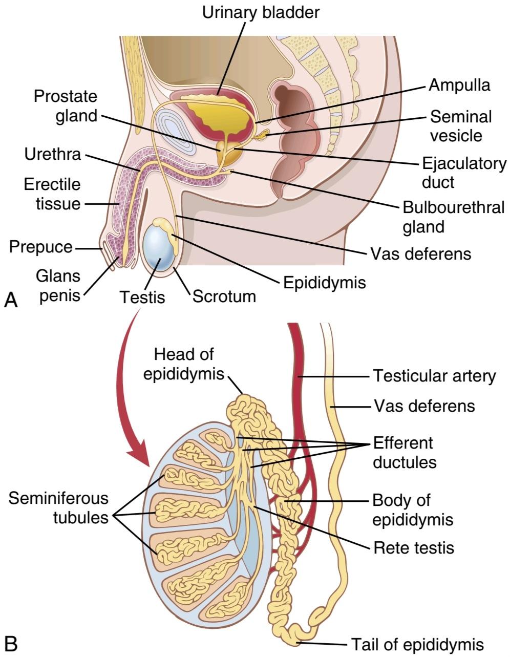
Figure 81-1 A, The male reproductive system. B, The internal structure of the testis and the relation of the testis to the epididymis. (A, Modified from Bloom V, Fawcett DW: Textbook of Histology, 10th ed. Philadelphia: WB Saunders, 1975. B, Modified from Guyton AC: Anatomy and Physiology. Philadelphia: Saunders College Publishing, 1985.)
شکل 81-1 الف، سیستم تولید مثل مرد. ب، ساختار داخلی بیضه و ارتباط بیضه با اپیدیدیم. (A, Modified from Bloom V, Fawcett DW: Textbook of Histology, ed. Philadelphia: WB Saunders, 1975. B, Modified from Guyton AC: Anatomy and Physiology. Philadelphia: Saunders College Publishing, 1985.)
STEPS OF SPERMATOGENESIS
Spermatogenesis occurs in the seminiferous tubules during active sexual life as the result of stimulation by anterior pituitary gonadotropic hormones. Spermatogenesis begins in the male at an average age of 13 years and continues throughout most of the remainder of life but decreases markedly in old age.
مراحل اسپرماتوژنز
اسپرماتوژنز در لوله های منی ساز در طول زندگی جنسی فعال در نتیجه تحریک هورمون های گنادوتروپیک هیپوفیز قدامی رخ می دهد. اسپرماتوژنز در مردان به طور متوسط در سن 13 سالگی شروع می شود و در بیشتر مابقی عمر ادامه می یابد، اما در سنین بالا به میزان قابل توجهی کاهش می یابد.
In the first stage of spermatogenesis, the spermatogonia migrate among Sertoli cells toward the central lumen of the seminiferous tubule. The Sertoli cells are large, with overflowing cytoplasmic envelopes that surround the developing spermatogonia all the way to the central lumen of the tubule.
در مرحله اول اسپرماتوژنز، اسپرماتوگونی ها از بین سلول های سرتولی به سمت لومن مرکزی لوله اسپرم ساز مهاجرت می کنند. سلول های سرتولی بزرگ هستند، با پوشش های سیتوپلاسمی سرریز که اسپرماتوگونی در حال رشد را تا لومن مرکزی توبول احاطه کرده است.

Figure 81-2 A, Cross section of a seminiferous tubule. B, Stages in the development of sperm from spermatogonia.
شکل 81-2 A، مقطع لوله منی ساز. ب، مراحل رشد اسپرم از اسپرماتوگونی.
Meiosis. Spermatogonia that cross the barrier into the Sertoli cell layer become progressively modified and enlarged to form large primary spermatocytes (Figure 81-3). Each of these primary spermatocytes, in turn, undergoes meiotic division to form two secondary spermatocytes. After another few days, these secondary spermatocytes also divide to form spermatids that are eventually modified to become spermatozoa (sperm).
میوز. اسپرماتوگونی هایی که از سد عبور می کنند و وارد لایه سلولی سرتولی می شوند به تدریج تغییر یافته و بزرگ می شوند و اسپرماتوسیت های اولیه بزرگی را تشکیل می دهند (شکل 81-3). هر یک از این اسپرماتوسیت های اولیه به نوبه خود تحت تقسیم میوز قرار می گیرند تا دو اسپرماتوسیت ثانویه را تشکیل دهند. پس از چند روز دیگر، این اسپرماتوسیت های ثانویه نیز تقسیم می شوند و اسپرماتیدهایی را تشکیل می دهند که در نهایت اصلاح می شوند تا به اسپرم (اسپرم) تبدیل شوند.
During the change from the spermatocyte stage to the spermatid stage, the 46 chromosomes (23 pairs of chromosomes) of the spermatocyte are divided, and thus 23 chromosomes go to one spermatid and the other 23 go to the second spermatid. The chromosomal genes are also divided so that only one-half of the genetic characteristics of the eventual fetus are provided by the father, with the other half being derived from the oocyte provided by the mother.
در طی تغییر از مرحله اسپرماتوسیت به مرحله اسپرماتید، 46 کروموزوم (23 جفت کروموزوم) اسپرماتوسیت تقسیم می شود و به این ترتیب 23 کروموزوم به یک اسپرماتید و 23 کروموزوم دیگر به اسپرماتید دوم می روند. ژن های کروموزومی نیز به گونه ای تقسیم می شوند که تنها نیمی از خصوصیات ژنتیکی جنین نهایی توسط پدر تامین می شود و نیمی دیگر از تخمک ارائه شده توسط مادر به دست می آید.
The entire period of spermatogenesis, from spermatogonia to spermatozoa, takes about 74 days.
کل دوره اسپرماتوژنز، از اسپرماتوگونی تا اسپرم، حدود 74 روز طول می کشد.

Figure 81-3 Cell divisions during spermatogenesis. During embryonic development, the primordial germ cells migrate to the testis, where they become spermatogonia. At puberty (usually 12 to 14 years after birth), the spermatogonia proliferate rapidly by mitosis. Some begin meiosis to become primary spermatocytes and continue through meiotic division I to become secondary spermatocytes. After completion of meiotic division II, the secondary spermatocytes produce spermatids, which differentiate to form spermatozoa.
شکل 81-3 تقسیمات سلولی در طی اسپرم زایی. در طول رشد جنینی، سلول های زایای اولیه به بیضه مهاجرت می کنند و در آنجا تبدیل به اسپرماتوگونی می شوند. در دوران بلوغ (معمولاً 12 تا 14 سال پس از تولد)، اسپرماتوگونی ها به سرعت توسط میتوز تکثیر می شوند. برخی از آنها میوز را شروع می کنند تا به اسپرماتوسیت های اولیه تبدیل شوند و از طریق تقسیم میوز I ادامه پیدا می کنند تا به اسپرماتوسیت های ثانویه تبدیل شوند. پس از اتمام تقسیم میوز II، اسپرماتوسیتهای ثانویه اسپرماتید تولید میکنند که تمایز یافته و اسپرماتوزوا را تشکیل میدهند.
Sex Chromosomes. In each spermatogonium, one of the 23 pairs of chromosomes carries the genetic information that determines the sex of each eventual offspring. This pair is composed of one X chromosome, which is called the female chromosome, and one Y chromosome, the male chromosome. During meiotic division, the male Y chromosome goes to one spermatid that then becomes a male sperm, and the female X chromosome goes to another spermatid that becomes a female sperm. The sex of the eventual offspring is determined by which of these two types of sperm fertilizes the ovum. This process is discussed further in Chapter 83.
کروموزوم های جنسی. در هر اسپرماتوگونیوم، یکی از 23 جفت کروموزوم حامل اطلاعات ژنتیکی است که جنسیت هر فرزند نهایی را تعیین می کند. این جفت از یک کروموزوم X که کروموزوم ماده نامیده می شود و یک کروموزوم Y به نام کروموزوم مرد تشکیل شده است. در طی تقسیم میوز، کروموزوم مرد Y به یک اسپرماتید می رود که سپس به اسپرم مرد تبدیل می شود و کروموزوم X ماده به اسپرماتید دیگری می رود که به اسپرم ماده تبدیل می شود. جنسیت فرزند نهایی با توجه به اینکه کدام یک از این دو نوع اسپرم تخمک را بارور می کند تعیین می شود. این فرآیند در فصل 83 بیشتر مورد بحث قرار گرفته است.

Figure 81-4 Structure of the human spermatozoon.
شکل 81-4 ساختار اسپرم انسان.
Formation of Sperm. When the spermatids are first formed, they still have the usual characteristics of epithelioid cells, but soon they begin to differentiate and elongate into spermatozoa. As shown in Figure 81-4, each spermatozoon is composed of a head and a tail. The head comprises the condensed nucleus of the cell, with only a thin cytoplasmic and cell membrane layer around its surface. On the outside of the anterior two-thirds of the head is a thick cap called the acrosome that is formed mainly from the Golgi apparatus. The acrosome contains several enzymes similar to those found in lysosomes of the typical cell, including hyaluronidase (which can digest proteoglycan filaments of tissues) and powerful proteolytic enzymes (which can digest proteins). These enzymes play important roles in allowing the sperm to enter the ovum and fertilize it.
تشکیل اسپرم. هنگامی که اسپرماتیدها برای اولین بار تشکیل می شوند، هنوز ویژگی های معمول سلول های اپیتلیوئیدی را دارند، اما به زودی شروع به تمایز و طویل شدن به اسپرم می کنند. همانطور که در شکل 81-4 نشان داده شده است، هر اسپرم از یک سر و یک دم تشکیل شده است. سر شامل هسته متراکم سلول است که فقط یک لایه سیتوپلاسمی نازک و غشای سلولی در اطراف سطح آن دارد. در قسمت بیرونی دو سوم قدامی سر یک کلاه ضخیم به نام آکروزوم وجود دارد که عمدتاً از دستگاه گلژی تشکیل شده است. آکروزوم حاوی چندین آنزیم مشابه آنزیمهای موجود در لیزوزومهای سلول معمولی است، از جمله هیالورونیداز (که میتواند رشتههای پروتئوگلیکان بافتها را هضم کند) و آنزیمهای پروتئولیتیک قدرتمند (که میتوانند پروتئینها را هضم کنند). این آنزیم ها نقش مهمی در اجازه دادن به اسپرم برای ورود به تخمک و بارور کردن آن دارند.
The tail of the sperm, called the flagellum, has three major components: (1) a central skeleton constructed of 11 microtubules, collectively called the axoneme; the structure of the axoneme is similar to that of cilia found on the surfaces of other types of cells described in Chapter 2; (2) a thin cell membrane covering the axoneme; and (3) a collection of mitochondria surrounding the axoneme in the proximal portion of the tail, called the body of the tail.
دم اسپرم که فلاژلوم نامیده می شود دارای سه جزء اصلی است: (1) یک اسکلت مرکزی از 11 میکروتوبول ساخته شده است که مجموعاً آکسونم نامیده می شود. ساختار آکسونم شبیه به مژک های موجود در سطوح دیگر انواع سلول های شرح داده شده در فصل 2 است. (2) یک غشای سلولی نازک که آکسونم را می پوشاند. و (3) مجموعه ای از میتوکندری ها که آکسونم را در قسمت پروگزیمال دم احاطه کرده اند که بدنه دم نامیده می شود.
Back-and-forth movement of the tail (flagellar movement) provides motility for the sperm. This movement results from a rhythmic longitudinal sliding motion between the anterior and posterior tubules that make up the axoneme. The energy for this process is supplied in the form of adenosine triphosphate, which is synthesized by the mitochondria in the body of the tail.
حرکت عقب و جلو دم (حرکت تاژک) باعث تحرک اسپرم می شود. این حرکت ناشی از یک حرکت لغزشی ریتمیک طولی بین لوله های قدامی و خلفی است که آکسونم را تشکیل می دهند. انرژی این فرآیند به شکل آدنوزین تری فسفات تامین میشود که توسط میتوکندریهای موجود در بدنه دم سنتز میشود.
Normal sperm move in a fluid medium at a velocity of 1 to 4 mm/min, which allows them to move through the female genital tract in quest of the ovum.
اسپرم های طبیعی در یک محیط مایع با سرعت 1 تا 4 میلی متر در دقیقه حرکت می کنند که به آنها اجازه می دهد در دستگاه تناسلی زن در جستجوی تخمک حرکت کنند.
Hormonal Factors That Stimulate Spermatogenesis
The role of hormones in reproduction is discussed later in detail; for now, note that several hormones play essential roles in spermatogenesis. Some of these roles are as follows:
عوامل هورمونی که باعث تحریک اسپرماتوژنز می شوند
نقش هورمون ها در تولید مثل بعداً به تفصیل مورد بحث قرار می گیرد. در حال حاضر، توجه داشته باشید که چندین هورمون در تولید اسپرم نقش اساسی دارند. برخی از این نقش ها به شرح زیر است:
1. Testosterone, secreted by the Leydig cells located in the interstitium of the testis (see Figure 81-2), is essential for growth and division of the testicular germinal cells, which is the first stage in forming sperm.
1. تستوسترون که توسط سلول های لیدیگ واقع در بینابینی بیضه ترشح می شود (شکل 81-2 را ببینید) برای رشد و تقسیم سلول های زایایی بیضه که اولین مرحله در تشکیل اسپرم است ضروری است.
2. Luteinizing hormone, secreted by the anterior pituitary gland, stimulates the Leydig cells to secrete testosterone.
2. هورمون لوتئینیزه کننده که توسط غده هیپوفیز قدامی ترشح می شود، سلول های لیدیگ را برای ترشح تستوسترون تحریک می کند.
3. Follicle-stimulating hormone, also secreted by the anterior pituitary gland, stimulates the Sertoli cells; without this stimulation, conversion of the spermatids to sperm (the process of spermiogenesis) will not occur.
3. هورمون محرک فولیکول که از غده هیپوفیز قدامی نیز ترشح می شود، سلول های سرتولی را تحریک می کند. بدون این تحریک، تبدیل اسپرماتیدها به اسپرم (فرایند اسپرم زایی) اتفاق نمی افتد.
4. Estrogens, formed from testosterone by the Sertoli cells when they are stimulated by follicle-stimulating hormone, are probably also essential for spermiogenesis.
4. استروژنهایی که از تستوسترون توسط سلولهای سرتولی تشکیل میشوند، زمانی که توسط هورمون محرک فولیکول تحریک میشوند، احتمالاً برای اسپرمزایی نیز ضروری هستند.
5. Growth hormone (as well as most of the other body hormones) is necessary for controlling background metabolic functions of the testes. Growth hormone specifically promotes early division of the spematogonia; in its absence, as in pituitary dwarfs, spermatogenesis is severely deficient or absent, thus causing infertility.
5. هورمون رشد (و همچنین بسیاری از هورمون های دیگر بدن) برای کنترل عملکرد متابولیک پس زمینه بیضه ها ضروری است. هورمون رشد به طور خاص تقسیم اولیه اسپماتوگونیا را ترویج می کند. در غیاب آن، مانند کوتوله های هیپوفیز، اسپرم زایی به شدت کمبود یا وجود ندارد، در نتیجه باعث ناباروری می شود.
Maturation of Sperm in the Epididymis
After formation in the seminiferous tubules, the sperm require several days to pass through the 6-meter-long tubule of the epididymis. Sperm removed from the seminiferous tubules and from the early portions of the epididymis are nonmotile and cannot fertilize an ovum. However, after the sperm have been in the epididymis for 18 to 24 hours, they develop the capability of motility, even though several inhibitory proteins in the epididymal fluid still prevent final motility until after ejaculation.
بلوغ اسپرم در اپیدیدیم
پس از تشکیل در لوله های اسپرم ساز، اسپرم به چند روز نیاز دارد تا از لوله 6 متری اپیدیدیم عبور کند. اسپرم خارج شده از لوله های اسپرم ساز و از قسمت های اولیه اپیدیدیم غیر متحرک است و نمی تواند تخمک را بارور کند. با این حال، پس از اینکه اسپرم ها به مدت 18 تا 24 ساعت در اپیدیدیم بودند، توانایی تحرک را توسعه می دهند، حتی اگر چندین پروتئین بازدارنده در مایع اپیدیدیم هنوز از تحرک نهایی تا پس از انزال جلوگیری می کنند.
Storage of Sperm in the Testes. The two testes of the human adult form up to 120 million sperm each day. Most of these sperm are stored in the epididymis, although a small quantity is stored in the vas deferens. They can remain stored, while maintaining their fertility, for at least a month. During this time, they are kept in a deeply suppressed, inactive state by multiple inhibitory substances in the secretions of the ducts. Conversely, with a high level of sexual activity and ejaculations, they may be stored no longer than a few days.
ذخیره اسپرم در بیضه ها. دو بیضه انسان بالغ روزانه 120 میلیون اسپرم تشکیل می دهند. بیشتر این اسپرم ها در اپیدیدیم ذخیره می شوند، اگرچه مقدار کمی در مجرای دفران ذخیره می شود. آنها می توانند در حالی که باروری خود را حفظ می کنند، حداقل برای یک ماه ذخیره شوند. در طول این مدت، آنها در یک حالت عمیق سرکوب شده و غیرفعال توسط مواد بازدارنده متعدد در ترشحات مجاری نگهداری می شوند. برعکس، با سطح بالای فعالیت جنسی و انزال، ممکن است بیش از چند روز ذخیره نشوند.
After ejaculation, the sperm become motile and capable of fertilizing the ovum, a process called maturation. The Sertoli cells and the epithelium of the epididymis secrete a special nutrient fluid that is ejaculated along with the sperm. This fluid contains hormones (including both testosterone and estrogens), enzymes, and special nutrients that are essential for sperm maturation.
پس از انزال، اسپرم متحرک می شود و می تواند تخمک را بارور کند، فرآیندی که بلوغ نامیده می شود. سلول های سرتولی و اپیتلیوم اپیدیدیم مایع غذایی خاصی ترشح می کنند که همراه با اسپرم انزال می شود. این مایع حاوی هورمون ها (از جمله تستوسترون و استروژن)، آنزیم ها و مواد مغذی خاصی است که برای بلوغ اسپرم ضروری هستند.
Physiology of the Mature Sperm. The normal motile, fertile sperm are capable of flagellated movement through the fluid medium at velocities of 1 to 4 mm/min. The activity of sperm is greatly enhanced in a neutral and slightly alkaline medium, as exists in the ejaculated semen, but it is greatly depressed in a mildly acidic medium. A strong acidic medium can cause the rapid death of sperm.
فیزیولوژی اسپرم بالغ. اسپرم های نرمال متحرک و بارور قادر به حرکت تاژک دار در محیط مایع با سرعت 1 تا 4 میلی متر در دقیقه هستند. فعالیت اسپرم در یک محیط خنثی و کمی قلیایی، همانطور که در مایع منی انزالی وجود دارد، بسیار افزایش می یابد، اما در یک محیط اسیدی خفیف به شدت کاهش می یابد. یک محیط اسیدی قوی می تواند باعث مرگ سریع اسپرم شود.
The activity of sperm increases markedly with increasing temperature, but so does the rate of metabolism, causing the life of the sperm to be considerably shortened. Although sperm can live for many weeks in the suppressed state in the genital ducts of the testes, the life expectancy of ejaculated sperm in the female genital tract is only 1 to 2 days.
فعالیت اسپرم با افزایش دما به طور قابل توجهی افزایش می یابد، اما سرعت متابولیسم نیز افزایش می یابد و باعث می شود که عمر اسپرم به طور قابل توجهی کوتاه شود. اگرچه اسپرم می تواند هفته ها در حالت سرکوب شده در مجاری تناسلی بیضه ها زندگی کند، امید به زندگی اسپرم انزالی در دستگاه تناسلی زنان تنها 1 تا 2 روز است.
FUNCTION OF THE SEMINAL VESICLES
Each seminal vesicle is a tortuous, loculated tube lined with a secretory epithelium that secretes a mucoid mate- rial containing an abundance of fructose, citric acid, and other nutrient substances, as well as large quantities of prostaglandins and fibrinogen. During the process of emission and ejaculation, each seminal vesicle empties its contents into the ejaculatory duct shortly after the vas deferens empties the sperm. This action adds greatly to the bulk of the ejaculated semen, and the fructose and other substances in the seminal fluid are of considerable nutrient value for the ejaculated sperm until one of the sperm fertilizes the ovum.
عملکرد وزیکول های منی
هر وزیکول منی یک لوله پرپیچ و خم و لوکول شده است که با یک اپیتلیوم ترشحی پوشانده شده است که یک ماده مخاطی حاوی مقدار زیادی فروکتوز، اسید سیتریک و سایر مواد مغذی و همچنین مقادیر زیادی پروستاگلاندین و فیبرینوژن ترشح می کند. در طی فرآیند انتشار و انزال، هر وزیکول منی، اندکی پس از تخلیه اسپرم، محتویات خود را در مجرای انزال تخلیه می کند. این عمل به مقدار زیادی به حجم منی انزال شده اضافه می کند و فروکتوز و سایر مواد موجود در مایع منی از ارزش غذایی قابل توجهی برای اسپرم انزال شده برخوردار هستند تا زمانی که یکی از اسپرم ها تخمک را بارور کند.
Prostaglandins are believed to aid fertilization in two ways: (1) by reacting with the female cervical mucus to make it more receptive to sperm movement and (2) by possibly causing backward, reverse peristaltic contractions in the uterus and fallopian tubes to move the ejaculated sperm toward the ovaries (a few sperm reach the upper ends of the fallopian tubes within 5 minutes).
اعتقاد بر این است که پروستاگلاندین ها به دو طریق به لقاح کمک می کنند: (1) با واکنش با مخاط دهانه رحم زن برای پذیرش بیشتر حرکت اسپرم و (2) احتمالاً با ایجاد انقباضات پریستالتیک معکوس در رحم و لوله های فالوپ برای حرکت دادن اسپرم های انزال شده به سمت چند انتها اسپرم. لوله های فالوپ در عرض 5 دقیقه).
FUNCTION OF THE PROSTATE GLAND
The prostate gland secretes a thin, milky fluid that contains calcium, citrate ion, phosphate ion, a clotting enzyme, and a profibrinolysin. During emission, the capsule of the prostate gland contracts simultaneously with the contractions of the vas deferens so that the thin, milky fluid of the prostate gland adds further to the bulk of the semen. A slightly alkaline characteristic of the prostatic fluid may be quite important for successful fertilization of the ovum because the fluid of the vas deferens is relatively acidic owing to the presence of citric acid and metabolic end products of the sperm and, consequently, helps inhibit sperm fertility. Also, the vaginal secretions of the female are acidic (with a pH of 3.5-4.0). Sperm do not become optimally motile until the pH of the surrounding fluids rises to about 6.0 to 6.5. Consequently, it is probable that the slightly alkaline prostatic fluid helps neutralize the acidity of the other seminal fluids during ejaculation and thus enhances the motility and fertility of the sperm.
عملکرد غده پروستات
غده پروستات مایع رقیق و شیری رنگی ترشح می کند که حاوی کلسیم، یون سیترات، یون فسفات، یک آنزیم لخته کننده و یک پروفیبرینولیزین است. در حین انتشار، کپسول غده پروستات همزمان با انقباضات مجرای دفران منقبض می شود به طوری که مایع رقیق و شیری رنگ غده پروستات به قسمت عمده مایع منی اضافه می کند. ویژگی کمی قلیایی مایع پروستات ممکن است برای لقاح موفقیت آمیز تخمک بسیار مهم باشد زیرا مایع مجرای دفران به دلیل وجود اسید سیتریک و محصولات نهایی متابولیک اسپرم نسبتاً اسیدی است و در نتیجه به مهار باروری اسپرم کمک می کند. همچنین ترشحات واژن ماده اسیدی است (با pH 3.5-4.0). تا زمانی که PH مایعات اطراف به حدود 6.0 تا 6.5 برسد، اسپرم به طور مطلوب تحرک ندارد. در نتیجه، این احتمال وجود دارد که مایع پروستات کمی قلیایی به خنثی کردن اسیدیته سایر مایعات منی در حین انزال کمک می کند و در نتیجه تحرک و باروری اسپرم را افزایش می دهد.
SEMEN
Semen, which is ejaculated during the male sexual act, is composed of the fluid and sperm from the vas deferens (~10% of the total), fluid from the seminal vesicles (almost 60%), fluid from the prostate gland (~30%), and small amounts from the mucous glands, especially the bulbourethral glands. Thus, the bulk of the semen is seminal vesicle fluid, which is the last to be ejaculated and serves to wash the sperm through the ejaculatory duct and urethra.
سمن
منی که در حین عمل جنسی مردانه انزال میشود، از مایع و اسپرم از مجرای دفران (حدود 10٪ از کل)، مایع از وزیکولهای منی (تقریبا 60٪)، مایع از غده پروستات (حدود 30٪) و مقادیر کمی از غدد مخاطی، بهویژه غده غده تشکیل شده است. بنابراین، قسمت عمده مایع منی مایع کیسه منی است که آخرین انزال است و برای شستشوی اسپرم از طریق مجرای انزال و مجرای ادرار استفاده می شود.
The average pH of the combined semen is about 7.5, with the alkaline prostatic fluid having more than neutralized the mild acidity of the other portions of the semen. The prostatic fluid gives the semen a milky appearance, and fluid from the seminal vesicles and mucous glands gives the semen a mucoid consistency. Also, a clotting enzyme from the prostatic fluid causes the fibrinogen of the seminal vesicle fluid to form a weak fibrin coagulum that holds the semen in the deeper regions of the vagina where the uterine cervix lies. The coagulum then dissolves during the next 15 to 30 minutes because of lysis by fibrinolysin formed from the prostatic profibrinolysin. In the early minutes after ejaculation, the sperm remain relatively immobile, possibly because of the viscosity of the coagulum. As the coagulum dissolves, the sperm simultaneously become highly motile.
میانگین pH مایع منی ترکیبی حدود 7.5 است، با مایع قلیایی پروستات که اسیدیته خفیف سایر بخشهای منی را خنثی میکند. مایع پروستات به مایع منی ظاهری شیری رنگ می دهد و مایع حاصل از وزیکول های منی و غدد مخاطی به مایع منی قوام مخاطی می بخشد. همچنین، یک آنزیم لختهکننده از مایع پروستات باعث میشود که فیبرینوژن مایع وزیکول منی یک منعقد ضعیف فیبرینی تشکیل دهد که مایع منی را در نواحی عمیقتر واژن، جایی که دهانه رحم قرار دارد، نگه میدارد. سپس انعقاد در 15 تا 30 دقیقه بعدی به دلیل لیز شدن توسط فیبرینولیزین تشکیل شده از پروفیبرینولیزین پروستات حل می شود. در دقایق اولیه پس از انزال، اسپرم احتمالاً به دلیل ویسکوزیته انعقاد نسبتاً بی حرکت می ماند. با حل شدن انعقاد، اسپرم به طور همزمان بسیار تحرک می شود.
Although sperm can live for many weeks in the male genital ducts, once they are ejaculated in the semen, their maximal life span is only 24 to 48 hours at body temperature. At lowered temperatures, however, semen can be stored for several weeks, and when frozen at temperatures below -100°C, sperm have been preserved for years.
اگرچه اسپرم ها می توانند هفته های زیادی در مجاری تناسلی مردانه زندگی کنند، پس از انزال در منی، حداکثر طول عمر آنها در دمای بدن تنها 24 تا 48 ساعت است. با این حال، در دماهای پایینتر، منی را میتوان برای چند هفته ذخیره کرد و وقتی در دمای زیر 100- درجه سانتیگراد منجمد شود، اسپرم برای سالها حفظ میشود.
“Capacitation” of Spermatozoa Is Required for Fertilization of the Ovum
Although spermatozoa are said to be “mature” when they leave the epididymis, their activity is held in check by multiple inhibitory factors secreted by the genital duct epithelia. Therefore, when they are first expelled in the semen, they are unable to fertilize the ovum. However, on coming in contact with the fluids of the female genital tract, multiple changes occur that activate the sperm for the final processes of fertilization. These collective changes are called capacitation of the spermatozoa, which normally requires from 1 to 10 hours. The following changes are believed to occur:
“ظرفیت” اسپرم برای لقاح تخمک مورد نیاز است
اگرچه گفته می شود اسپرم ها هنگام خروج از اپیدیدیم ” بالغ” می شوند، اما فعالیت آنها توسط عوامل بازدارنده متعددی که توسط اپیتلیوم مجرای تناسلی ترشح می شود، کنترل می شود. بنابراین، هنگامی که برای اولین بار در مایع منی خارج می شوند، قادر به بارور کردن تخمک نیستند. با این حال، در تماس با مایعات دستگاه تناسلی زنان، تغییرات متعددی رخ می دهد که اسپرم را برای فرآیندهای نهایی لقاح فعال می کند. این تغییرات جمعی ظرفیت اسپرم نامیده می شود که معمولاً بین 1 تا 10 ساعت نیاز دارد. اعتقاد بر این است که تغییرات زیر رخ می دهد:
1. The uterine and fallopian tube fluids wash away the various inhibitory factors that suppress sperm activity in the male genital ducts.
1. مایعات رحم و لوله فالوپ عوامل بازدارنده مختلفی را که فعالیت اسپرم را در مجاری تناسلی مردانه سرکوب می کنند، از بین می برد.
2. While the spermatozoa remain in the fluid of the male genital ducts, they are continually exposed to many floating vesicles from the seminiferous tubules containing large amounts of cholesterol. This cholesterol is continually added to the cellular membrane covering the sperm acrosome, toughening this membrane and preventing release of its enzymes. After ejaculation, the sperm deposited in the vagina swim away from the cholesterol vesicles upward into the uterine cavity, and they gradually lose much of their other excess cholesterol during the next few hours. In so doing, the membrane at the head of the sperm (the acrosome) becomes much weaker.
2. در حالی که اسپرم ها در مایع مجاری تناسلی مردانه باقی می مانند، آنها به طور مداوم در معرض بسیاری از وزیکول های شناور از لوله های اسپرم ساز حاوی مقادیر زیادی کلسترول قرار می گیرند. این کلسترول به طور مداوم به غشای سلولی پوشاننده آکروزوم اسپرم اضافه می شود و این غشاء را سفت می کند و از آزاد شدن آنزیم های آن جلوگیری می کند. پس از انزال، اسپرمهای رسوبشده در واژن از وزیکولهای کلسترول دور میشوند و به سمت حفره رحم میروند و در طی چند ساعت آینده به تدریج مقدار زیادی از کلسترول اضافی خود را از دست میدهند. با انجام این کار، غشای سر اسپرم (آکروزوم) بسیار ضعیف تر می شود.
3. The membrane of the sperm also becomes much more permeable to calcium ions, so calcium now enters the sperm in abundance and changes the activity of the flagellum, giving it a powerful whiplash motion in contrast to its previously weak undulating motion. In addition, the calcium ions cause changes in the cellular membrane that cover the leading edge of the acrosome, making it possible for the acrosome to release its enzymes rapidly and easily as the sperm penetrates the granulosa cell mass surrounding the ovum, and even more so as it attempts to penetrate the zona pellucida of the ovum.
3. غشای اسپرم نیز نسبت به یونهای کلسیم بسیار نفوذپذیرتر میشود، بنابراین کلسیم اکنون به وفور وارد اسپرم میشود و فعالیت تاژک را تغییر میدهد و بر خلاف حرکت مواج ضعیف قبلی، حرکت شلاقی قدرتمندی به آن میدهد. علاوه بر این، یونهای کلسیم باعث ایجاد تغییراتی در غشای سلولی میشوند که لبه جلویی آکروزوم را میپوشاند، و این امکان را برای آکروزوم فراهم میکند که آنزیمهای خود را به سرعت و به راحتی آزاد کند، زیرا اسپرم به توده سلولی گرانولوزا اطراف تخمک نفوذ میکند و حتی بیشتر از آن تلاش میکند تا به زونا شفافسازی نفوذ کند.
Thus, multiple changes occur during the process of capacitation. Without these changes, the sperm can- not make its way to the interior of the ovum to cause fertilization.
بنابراین، تغییرات متعددی در طول فرآیند ظرفیت سازی رخ می دهد. بدون این تغییرات، اسپرم نمی تواند به داخل تخمک راه پیدا کند و باعث لقاح شود.
Acrosome Enzymes, the “Acrosome Reaction,” and Penetration of the Ovum
Stored in the acrosome of the sperm are large quantities of hyaluronidase and proteolytic enzymes. Hyaluronidase depolymerizes the hyaluronic acid polymers in the intercellular cement that holds the ovarian granulosa cells together. The proteolytic enzymes digest proteins in the structural elements of tissue cells that still adhere to the ovum.
آنزیم های آکروزوم، “واکنش آکروزوم” و نفوذ تخمک
در آکروزوم اسپرم مقادیر زیادی هیالورونیداز و آنزیم های پروتئولیتیک ذخیره می شود. هیالورونیداز پلیمرهای اسید هیالورونیک را در سیمان بین سلولی که سلول های گرانولوزای تخمدان را در کنار هم نگه می دارد، پلیمریزه می کند. آنزیم های پروتئولیتیک پروتئین ها را در عناصر ساختاری سلول های بافتی که هنوز به تخمک می چسبند هضم می کنند.
When the ovum is expelled from the ovarian follicle into the fallopian tube, it still carries with it multiple layers of granulosa cells. Before a sperm can fertilize the ovum, it must dissolute these granulosa cell layers, and then it must penetrate through the zona pellucida, the thick covering of the ovum. To achieve this penetration, the stored enzymes in the acrosome begin to be released. The hyaluronidase among these enzymes is especially important in opening pathways between the granulosa cells so that the sperm can reach the ovum.
هنگامی که تخمک از فولیکول تخمدان به داخل لوله فالوپ خارج می شود، همچنان لایه های متعددی از سلول های گرانولوزا را با خود حمل می کند. قبل از اینکه اسپرم بتواند تخمک را بارور کند، باید این لایه های سلولی گرانولوزا را حل کند و سپس باید از طریق زونا پلوسیدا، پوشش ضخیم تخمک، نفوذ کند. برای رسیدن به این نفوذ، آنزیم های ذخیره شده در آکروزوم شروع به آزاد شدن می کنند. هیالورونیداز در بین این آنزیم ها به ویژه در باز کردن مسیرهای بین سلول های گرانولوزا به طوری که اسپرم بتواند به تخمک برسد، مهم است.
When the sperm reaches the zona pellucida of the ovum, the anterior membrane of the sperm binds specifically with receptor proteins in the zona pellucida. Next, the entire acrosome rapidly dissolves, and all the acrosomal enzymes are released. Within minutes, these enzymes open a penetrating pathway for passage of the sperm head through the zona pellucida to the inside of the ovum. Within another 30 minutes, the cell membranes of the sperm head and of the oocyte fuse with each other to form a single cell. At the same time, the genetic material of the sperm and the oocyte combine to form a completely new cell genome, containing equal numbers of chromosomes and genes from mother and father. This is the process of fertilization; the embryo then begins to develop, as discussed in Chapter 83.
هنگامی که اسپرم به زونا شفاف تخمک می رسد، غشای قدامی اسپرم به طور خاص با پروتئین های گیرنده در زونا پلوسیدا متصل می شود. سپس، کل آکروزوم به سرعت حل می شود و تمام آنزیم های آکروزومی آزاد می شوند. در عرض چند دقیقه، این آنزیم ها مسیر نفوذی را برای عبور سر اسپرم از طریق زونا شفاف به داخل تخمک باز می کنند. در عرض 30 دقیقه دیگر، غشای سلولی سر اسپرم و تخمک با یکدیگر ترکیب می شوند و یک سلول واحد را تشکیل می دهند. در عین حال، مواد ژنتیکی اسپرم و تخمک با هم ترکیب میشوند و ژنوم سلولی کاملاً جدیدی را تشکیل میدهند که حاوی تعداد مساوی کروموزوم و ژنهای مادر و پدر است. این فرآیند لقاح است. همانطور که در فصل 83 بحث شد، جنین سپس شروع به رشد می کند.
Why Does Only One Sperm Enter the Oocyte? With as many sperm as there are, why does only one enter the oocyte? The reason is not entirely known, but within a few minutes after the first sperm penetrates the zona pellucida of the ovum, calcium ions diffuse inward through the oocyte membrane and cause multiple cortical granules to be released by exocytosis from the oocyte into the perivitelline space. These granules contain substances that permeate all portions of the zona pellucida and prevent binding of additional sperm, and they even cause any sperm that have already begun to bind to fall off. Thus, almost never does more than one sperm enter the oocyte during fertilization.
چرا فقط یک اسپرم وارد تخمک می شود؟ با تعداد اسپرم های موجود، چرا فقط یک اسپرم وارد تخمک می شود؟ دلیل آن کاملاً مشخص نیست، اما در عرض چند دقیقه پس از نفوذ اولین اسپرم به زونا شفاف تخمک، یونهای کلسیم از طریق غشای تخمک به داخل منتشر میشوند و باعث میشوند گرانولهای متعدد قشری با اگزوسیتوز از تخمک به فضای پریویتلین رها شوند. این گرانول ها حاوی موادی هستند که در تمام قسمت های زونا پلوسیدا نفوذ می کنند و از اتصال اسپرم های اضافی جلوگیری می کنند و حتی باعث می شوند هر اسپرمی که قبلاً شروع به اتصال کرده باشد از بین برود. بنابراین، تقریباً هرگز بیش از یک اسپرم در طول لقاح وارد تخمک نمی شود.
Abnormal Spermatogenesis and Male Fertility
The seminiferous tubular epithelium can be destroyed by several diseases. For example, bilateral orchitis (inflammation) of the testes resulting from mumps causes sterility in some affected males. Also, some male infants are born with degenerate tubular epithelia as a result of strictures in the genital ducts or other abnormalities. Finally, another cause of sterility, usually temporary, is excessive temperature of the testes.
اسپرم زایی غیر طبیعی و باروری مردان
اپیتلیوم لوله منی ساز می تواند توسط چندین بیماری از بین برود. به عنوان مثال، ارکیت دو طرفه (التهاب) بیضه های ناشی از اوریون باعث عقیمی در برخی از مردان مبتلا می شود. همچنین برخی از نوزادان پسر با اپیتلیوم لولهای دژنره شده در نتیجه تنگی مجاری تناسلی یا سایر ناهنجاریها متولد میشوند. در نهایت، یکی دیگر از علل عقیمی، معمولاً موقتی، دمای بیش از حد بیضه ها است.
Effect of Temperature on Spermatogenesis. Increasing the temperature of the testes can prevent spermatogenesis by causing degeneration of most cells of the seminiferous tubules besides the spermatogonia. It has often been stated that the reason the testes are located in the dangling scrotum is to maintain the temperature of these glands below the internal temperature of the body, although usually only about 2°C below the internal temperature. On cold days, scrotal reflexes cause the musculature of the scrotum to contract, pulling the testes close to the body to maintain this 2°C differential. Thus, the scrotum acts as a cooling mechanism for the testes (but a controlled cooling), without which spermatogenesis might be deficient during hot weather.
اثر دما بر اسپرماتوژنز. افزایش دمای بیضه ها می تواند با ایجاد انحطاط بیشتر سلول های لوله های اسپرم ساز علاوه بر اسپرماتوگونی، از اسپرم زایی جلوگیری کند. اغلب گفته شده است که دلیل قرار گرفتن بیضه ها در کیسه بیضه آویزان، حفظ دمای این غدد زیر دمای داخلی بدن است، اگرچه معمولاً فقط حدود 2 درجه سانتیگراد کمتر از دمای داخلی بدن است. در روزهای سرد، رفلکس های کیسه بیضه باعث انقباض عضلانی کیسه بیضه می شود و بیضه ها را به بدن نزدیک می کند تا این اختلاف 2 درجه سانتی گراد حفظ شود. بنابراین، کیسه بیضه به عنوان یک مکانیسم خنک کننده برای بیضه ها عمل می کند (اما یک خنک کننده کنترل شده)، که بدون آن ممکن است اسپرم زایی در هوای گرم ناقص باشد.
Cryptorchidism. Cryptorchidism means failure of a testis to descend from the abdomen into the scrotum at or near the time of birth of a fetus. During development of the male fetus, the testes are derived from the genital ridges in the abdomen. However, at about 3 weeks to 1 month before birth of the baby, the testes normally descend through the inguinal canals into the scrotum. Occasionally this descent does not occur or occurs incompletely, and as a result one or both testes remain in the abdomen, in the inguinal canal, or elsewhere along the route of descent.
کریپتورکیدیسم. کریپتورکیدیسم به معنای عدم نزول بیضه از شکم به داخل کیسه بیضه در زمان تولد جنین یا نزدیک به آن است. در طول رشد جنین پسر، بیضه ها از برجستگی های تناسلی در شکم مشتق می شوند. با این حال، حدود 3 هفته تا یک ماه قبل از تولد نوزاد، بیضه ها به طور معمول از طریق کانال های مغبنی به داخل کیسه بیضه فرود می آیند. گاهی اوقات این نزول رخ نمی دهد یا به طور ناقص رخ می دهد و در نتیجه یک یا هر دو بیضه در شکم، کانال مغبنی یا هر جای دیگری در طول مسیر نزول باقی می مانند.
A testis that remains in the abdominal cavity throughout life is incapable of forming sperm. The tubular epithelium becomes degenerate, leaving only the interstitial structures of the testis. It has been claimed that even the few degrees higher temperature in the abdomen than in the scrotum is sufficient to cause this degeneration of the tubular epithelium and, consequently, to cause sterility, although this effect is not certain. Nevertheless, for this reason, operations to re- locate the cryptorchid testes from the abdominal cavity into the scrotum before the beginning of adult sexual life can be performed on boys who have undescended testes.
بیضه ای که در طول زندگی در حفره شکمی باقی می ماند، قادر به تشکیل اسپرم نیست. اپیتلیوم لوله ای تحلیل رفته و تنها ساختارهای بینابینی بیضه باقی می ماند. ادعا شده است که حتی چند درجه دمای بالاتر در شکم نسبت به کیسه بیضه برای ایجاد این انحطاط اپیتلیوم لوله ای و در نتیجه باعث عقیمی کافی است، اگرچه این تأثیر قطعی نیست. با این وجود، به همین دلیل، عملیات جابجایی بیضه های کریپتورکید از حفره شکمی به داخل کیسه بیضه قبل از شروع زندگی جنسی بزرگسالان می تواند در پسرانی که بیضه های نزول نکرده دارند انجام شود.
Testosterone secretion by the fetal testes is the normal stimulus that causes the testes to descend into the scrotum from the abdomen. Therefore, many, if not most, cases of cryptorchidism are caused by abnormally formed testes that are unable to secrete enough testosterone. The surgical operation for cryptorchidism in these patients is unlikely to be successful.
ترشح تستوسترون توسط بیضه های جنین محرک طبیعی است که باعث می شود بیضه ها از شکم به داخل کیسه بیضه نزول کنند. بنابراین، بسیاری از موارد کریپتورکیدیسم، اگر نگوییم بیشتر، ناشی از بیضههایی است که بهطور غیرطبیعی تشکیل شدهاند که قادر به ترشح کافی تستوسترون نیستند. عمل جراحی کریپتورکیدیسم در این بیماران بعید است که موفقیت آمیز باشد.
Effect of Sperm Count on Fertility. The usual quantity of semen ejaculated during each coitus averages about 3.5 milliliters, and each milliliter of semen contains an average of about 120 million sperm, although even in “normal” males this quantity can vary from 35 million to 200 million. This means an average total of about 400 million sperm are usually present in the several milliliters of each ejaculate. When the number of sperm in each milliliter falls below about 20 million, the person is likely to be infertile. Thus, even though only a single sperm is necessary to fertilize the ovum, for reasons that are not understood, the ejaculate usually must contain a tremendous number of sperm for only one sperm to fertilize the ovum.
تاثیر تعداد اسپرم بر باروری. مقدار معمول منی انزال شده در طول هر رابطه جنسی به طور متوسط حدود 3.5 میلی لیتر است و هر میلی لیتر منی به طور متوسط حاوی حدود 120 میلیون اسپرم است، اگرچه حتی در مردان “عادی” این مقدار می تواند از 35 میلیون تا 200 میلیون متغیر باشد. این بدان معناست که به طور متوسط در مجموع حدود 400 میلیون اسپرم معمولاً در چندین میلی لیتر از هر انزال وجود دارد. وقتی تعداد اسپرم در هر میلی لیتر به کمتر از 20 میلیون برسد، احتمال ناباروری فرد وجود دارد. بنابراین، حتی اگر تنها یک اسپرم برای بارور کردن تخمک لازم است، به دلایلی که مشخص نیست، انزال معمولاً باید حاوی تعداد زیادی اسپرم باشد تا فقط یک اسپرم بتواند تخمک را بارور کند.
Effect of Sperm Morphology and Motility on Fertility. Occasionally, a man has a normal number of sperm but is still infertile. When this situation occurs, some- times as many as one-half of the sperm are found to be abnormal physically, having two heads, abnormally shaped heads, or abnormal tails, as shown in Figure 81-5. At other times, the sperm appear to be structurally normal, but for reasons not understood, they are either entirely nonmotile or relatively nonmotile. When- ever most of the sperm are morphologically abnormal or are nonmotile, the person is likely to be infertile, even though the remainder of the sperm appear to be normal.
تأثیر مورفولوژی و تحرک اسپرم بر باروری. گاهی اوقات، مردی دارای تعداد طبیعی اسپرم است اما هنوز نابارور است. هنگامی که این وضعیت رخ می دهد، گاهی اوقات تقریباً نیمی از اسپرم ها از نظر فیزیکی غیرعادی هستند، دارای دو سر، شکل غیرعادی سر، یا دم غیر طبیعی هستند، همانطور که در شکل 81-5 نشان داده شده است. در زمانهای دیگر، اسپرمها از نظر ساختاری طبیعی به نظر میرسند، اما به دلایلی که مشخص نیست، آنها کاملاً غیر متحرک یا نسبتاً غیر متحرک هستند. زمانی که بیشتر اسپرم ها از نظر مورفولوژیکی غیرطبیعی یا غیر متحرک باشند، فرد احتمالاً نابارور است، حتی اگر بقیه اسپرم ها طبیعی به نظر می رسند.

Figure 81-5 Abnormal infertile sperm, compared with a normal sperm on the right.
شکل 81-5 اسپرم نابارور غیرطبیعی، در مقایسه با اسپرم طبیعی در سمت راست.
MALE SEXUAL ACT
NEURONAL STIMULUS FOR PERFORMANCE OF THE MALE SEXUAL ACT
The most important source of sensory nerve signals for initiating the male sexual act is the glans penis. The glans contains an especially sensitive sensory end- organ system that transmits into the central nervous system that special modality of sensation called sexual sensation. The slippery massaging action of intercourse on the glans stimulates the sensory end organs, and the sexual signals in turn pass through the pudendal nerve, then through the sacral plexus into the sacral portion of the spinal cord, and finally up the cord to the brain.
کنش جنسی مردانه
محرک عصبی برای انجام عمل جنسی مردانه
مهمترین منبع سیگنالهای عصبی حسی برای شروع عمل جنسی مردانه، گلانس آلت تناسلی است. غده حاوی یک سیستم اندام انتهایی حسی حساس است که روش خاصی از احساس را به نام حس جنسی به سیستم عصبی مرکزی منتقل می کند. عمل ماساژ لغزنده مقاربت بر روی سر، اندام های انتهایی حسی را تحریک می کند و سیگنال های جنسی به نوبه خود از طریق عصب پودندال، سپس از طریق شبکه خاجی به قسمت خاجی نخاع و در نهایت از طناب به سمت مغز عبور می کنند.
Impulses may also enter the spinal cord from areas adjacent to the penis to aid in stimulating the sexual act. For example, stimulation of the anal epithelium, the scrotum, and perineal structures in general can send signals into the cord that add to the sexual sensation. Sexual sensations can even originate in internal structures, such as in areas of the urethra, bladder, prostate, seminal vesicles, testes, and vas deferens. Indeed, one of the causes of “sexual drive” is filling of the sexual organs with secretions. Mild infection and inflammation of these sexual organs may sometimes stimulate sexual desire, and some “aphrodisiac” drugs, such as cantharidin, irritate the bladder and urethral mucosa, inducing inflammation and vascular congestion.
همچنین ممکن است تکانه ها از نواحی مجاور آلت تناسلی وارد نخاع شوند تا به تحریک عمل جنسی کمک کنند. به عنوان مثال، تحریک اپیتلیوم مقعد، کیسه بیضه و به طور کلی ساختارهای پرینه می تواند سیگنال هایی را به بند ناف ارسال کند که به حس جنسی می افزاید. احساسات جنسی حتی می تواند در ساختارهای داخلی مانند نواحی مجرای ادرار، مثانه، پروستات، وزیکول های منی، بیضه ها و مجرای دفران منشاء بگیرد. در واقع یکی از علل «میل جنسی» پر شدن اندام های جنسی از ترشحات است. عفونت و التهاب خفیف این اندام های جنسی ممکن است گاهی اوقات میل جنسی را تحریک کند و برخی از داروهای “افرودیزیاک” مانند کانتاریدین، مثانه و مخاط مجرای ادرار را تحریک کرده و باعث التهاب و احتقان عروق می شود.
Psychic Element of Male Sexual Stimulation. Appropriate psychic stimuli can greatly enhance the ability of a person to perform the sexual act. Simply thinking sexual thoughts or even dreaming that the act of intercourse is being performed can initiate the male act, culminating in ejaculation. Indeed, nocturnal emissions during dreams, often called “wet dreams,” occur in many males during some stages of sexual life, especially during the teens.
عنصر روانی تحریک جنسی مردان. محرک های روانی مناسب می تواند توانایی فرد را در انجام عمل جنسی به میزان زیادی افزایش دهد. صرفاً فکر کردن به افکار جنسی یا حتی دیدن اینکه عمل مقاربت در حال انجام است، می تواند شروع کننده عمل مردانه باشد و به انزال منجر شود. در واقع، تشعشعات شبانه در طول رویاها، که اغلب “رویاهای مرطوب” نامیده می شود، در بسیاری از مردان در برخی از مراحل زندگی جنسی، به ویژه در دوران نوجوانی رخ می دهد.
Integration of the Male Sexual Act in the Spinal Cord. Although psychic factors usually play an important part in the male sexual act and can initiate or inhibit it, brain function is probably not necessary for its performance because appropriate genital stimulation can cause ejaculation in some animals and occasionally in humans after their spinal cords have been cut above the lumbar region. The male sexual act results from inherent reflex mechanisms integrated in the sacral and lumbar spinal cord, and these mechanisms can be initiated by either psychic stimulation from the brain or actual sexual stimulation from the sex organs, but usually it is a combination of both.
ادغام قانون جنسی مردانه در نخاع. اگرچه عوامل روانی معمولاً نقش مهمی در کنش جنسی مرد دارند و می توانند آن را شروع یا مهار کنند، عملکرد مغز احتمالاً برای عملکرد آن ضروری نیست زیرا تحریک تناسلی مناسب می تواند باعث انزال در برخی از حیوانات و گاهی اوقات در انسان پس از بریده شدن طناب نخاعی بالای ناحیه کمر شود. عمل جنسی مردانه ناشی از مکانیسمهای انعکاسی ذاتی است که در طناب نخاعی ساکرال و کمری ادغام شدهاند و این مکانیسمها میتوانند با تحریک روانی از مغز یا تحریک جنسی واقعی از اندامهای جنسی آغاز شوند، اما معمولاً ترکیبی از هر دو است.
STAGES OF THE MALE SEXUAL ACT
Penile Erection-Role of the Parasympathetic Nerves. Penile erection is the first effect of male sexual stimulation, and the degree of erection is proportional to the degree of stimulation, whether psychic or physical. Erection is caused by parasympathetic impulses that pass from the sacral portion of the spinal cord through the pelvic nerves to the penis. These parasympathetic nerve fibers, in contrast to most other parasympathetic fibers, are believed to release nitric oxide and/or vasoactive intestinal peptide in addition to acetylcholine. Nitric oxide activates the enzyme guanylyl cyclase, causing increased formation of cyclic guanosine monophosphate (GMP). The cyclic GMP especially relaxes the arteries of the penis and the trabecular meshwork of smooth muscle fibers in the erectile tissue of the corpora cavernosa and corpus spongiosum in the shaft of the penis, shown in Figure 81-6. As the vascular smooth muscles relax, blood flow into the penis increases, causing release of nitric oxide from the vascular endothelial cells and further vasodilation.
مراحل عمل جنسی مردانه
نعوظ آلت تناسلی – نقش اعصاب پاراسمپاتیک. نعوظ آلت تناسلی اولین اثر تحریک جنسی مردان است و میزان نعوظ متناسب با میزان تحریک، چه روحی و چه فیزیکی است. نعوظ ناشی از تکانه های پاراسمپاتیک است که از قسمت خاجی نخاع از طریق اعصاب لگن به آلت تناسلی می گذرد. اعتقاد بر این است که این رشته های عصبی پاراسمپاتیک، برخلاف اکثر رشته های پاراسمپاتیک دیگر، علاوه بر استیل کولین، اکسید نیتریک و/یا پپتید وازواکتیو روده ای را آزاد می کنند. اکسید نیتریک آنزیم گوانیلیل سیکلاز را فعال می کند و باعث افزایش تشکیل گوانوزین مونوفسفات حلقوی (GMP) می شود. GMP حلقوی به ویژه شریان های آلت تناسلی و شبکه ترابکولار فیبرهای عضلانی صاف را در بافت نعوظ اجسام غارنوردی و جسم اسفنجی در ساقه آلت تناسلی که در شکل 81-6 نشان داده شده است، شل می کند. با شل شدن ماهیچه های صاف عروق، جریان خون به آلت تناسلی افزایش می یابد و باعث آزاد شدن اکسید نیتریک از سلول های اندوتلیال عروق و اتساع بیشتر عروق می شود.
The erectile tissue of the penis consists of large cavernous sinusoids that are normally relatively empty of blood but become dilated tremendously when arterial blood flows rapidly into them under pressure while the venous outflow is partially occluded. Also, the erectile bodies, especially the two corpora cavernosa, are surrounded by strong fibrous coats; therefore, high pressure within the sinusoids causes ballooning of the erectile tissue to such an extent that the penis becomes hard and elongated, which is the phenomenon of erection.
بافت نعوظ آلت تناسلی متشکل از سینوسهای غاردار بزرگی است که معمولاً نسبتاً خالی از خون هستند، اما زمانی که خون شریانی به سرعت به داخل آنها جریان مییابد تحت فشار در حالی که جریان خروجی وریدی تا حدی مسدود میشود، به شدت گشاد میشوند. همچنین، بدن های نعوظ، به ویژه دو جسم غار، توسط پوشش های فیبری قوی احاطه شده اند. بنابراین فشار زیاد داخل سینوسی باعث بالون شدن بافت نعوظ به حدی می شود که آلت تناسلی سفت و کشیده می شود که این پدیده نعوظ است.
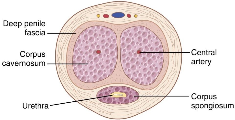
Figure 81-6 Erectile tissue of the penis.
شکل 81-6 بافت نعوظ آلت تناسلی.
Lubrication Is a Parasympathetic Function. During sexual stimulation, the parasympathetic impulses, in addition to promoting erection, cause the urethral glands and the bulbourethral glands to secrete mucus. This mucus flows through the urethra during intercourse to aid in lubrication during coitus. However, most of the lubrication of coitus is provided by the female sexual organs rather than by the male organs. Without satisfactory lubrication, the male sexual act is seldom successful because unlubricated intercourse causes grating, painful sensations that inhibit rather than excite sexual sensations.
روانکاری یک عملکرد پاراسمپاتیک است. در هنگام تحریک جنسی، تکانه های پاراسمپاتیک علاوه بر تقویت نعوظ، باعث ترشح مخاط از غدد مجرای ادرار و غدد بولبورترال می شود. این مخاط در طول مقاربت از طریق مجرای ادرار جریان می یابد تا به روان سازی در هنگام مقاربت کمک کند. با این حال، بیشتر روانکاری کویتیشن توسط اندام های جنسی زنانه به جای اندام های مردانه انجام می شود. بدون روانکاری رضایت بخش، عمل جنسی مردانه به ندرت موفقیت آمیز است، زیرا مقاربت بدون روغن باعث ایجاد احساسات دردناک و رنده می شود که به جای تحریک احساسات جنسی، آنها را مهار می کند.
Emission and Ejaculation Are Functions of the Sympathetic Nerves. Emission and ejaculation are the culmination of the male sexual act. When the sexual stimulus becomes extremely intense, the reflex centers of the spinal cord begin to emit sympathetic impulses that leave the cord at T12 to L2 and pass to the genital organs through the hypogastric and pelvic sympathetic nerve plexuses to initiate emission, the forerunner of ejaculation.
انتشار و انزال از وظایف اعصاب سمپاتیک هستند. ترشح و انزال اوج عمل جنسی مردانه است. هنگامی که محرک جنسی بسیار شدید می شود، مراکز رفلکس نخاع شروع به انتشار تکانه های سمپاتیک می کنند که طناب را در T12 تا L2 ترک می کند و از طریق شبکه عصبی سمپاتیک هیپوگاستریک و لگن به اندام های تناسلی می رسد تا انتشار را آغاز کند، پیشرو انزال.
Emission begins with contraction of the vas deferens and the ampulla to cause expulsion of sperm into the internal urethra. Then, contractions of the muscular coat of the prostate gland followed by contraction of the seminal vesicles expel prostatic and seminal fluid also into the urethra, forcing the sperm forward. All these fluids mix in the internal urethra with mucus already secreted by the bulbourethral glands to form the semen. The process to this point is emission.
انتشار با انقباض مجرای دفران و آمپول شروع می شود تا باعث بیرون راندن اسپرم به داخل مجرای ادراری داخلی شود. سپس، انقباضات پوشش عضلانی غده پروستات و به دنبال آن انقباض وزیکول های منی، پروستات و مایع منی را نیز به داخل مجرای ادرار خارج می کند و اسپرم را به جلو می برد. همه این مایعات در مجرای ادرار داخلی با مخاطی که قبلاً توسط غدد بولبورترال ترشح می شود مخلوط می شوند و منی را تشکیل می دهند. فرآیند تا این مرحله انتشار است.
The filling of the internal urethra with semen elicits sensory signals that are transmitted through the pudendal nerves to the sacral regions of the cord, giving the feeling of sudden fullness in the internal genital organs. Also, these sensory signals further excite rhythmic contraction of the internal genital organs and cause contraction of the ischiocavernosus and bulbocavernosus muscles that compress the bases of the penile erectile tissue. These effects together cause rhythmic wavelike increases in pressure in the erectile tissue of the penis and the genital ducts and urethra, which “ejaculate” the semen from the urethra to the exterior. This final process is called ejaculation. At the same time, rhythmic contractions of the pelvic muscles and even of some of the muscles of the body trunk cause thrusting movements of the pelvis and penis, which also help propel the semen into the deepest recesses of the vagina and perhaps even slightly into the cervix of the uterus.
پر شدن مجرای ادرار داخلی با مایع منی سیگنالهای حسی را ایجاد میکند که از طریق اعصاب پودندال به نواحی خاجی طناب منتقل میشود و احساس پری ناگهانی در اندامهای تناسلی داخلی را ایجاد میکند. همچنین این سیگنالهای حسی انقباض ریتمیک اندامهای تناسلی داخلی را تحریک میکنند و باعث انقباض ماهیچههای ایسکیوکاورنوس و بولبوکاورنوس میشوند که پایههای بافت نعوظ آلت تناسلی را فشرده میکنند. این اثرات با هم باعث افزایش موجی ریتمیک در فشار در بافت نعوظ آلت تناسلی و مجاری تناسلی و مجرای ادرار می شوند که منی را از مجرای ادرار به بیرون “انزال” می کند. این فرآیند نهایی انزال نامیده می شود. در عین حال، انقباضات ریتمیک عضلات لگن و حتی برخی از عضلات تنه بدن باعث حرکات فشاری لگن و آلت تناسلی می شود که همچنین به حرکت مایع منی به عمیق ترین فرورفتگی های واژن و شاید حتی اندکی به داخل دهانه رحم کمک می کند.
This entire period of emission and ejaculation is called the male orgasm. At its termination, the male sexual excitement disappears almost entirely within 1 to 2 minutes and erection ceases, a process called resolution.
تمام این دوره از انتشار و انزال، ارگاسم مردانه نامیده می شود. با پایان یافتن، هیجان جنسی مردان تقریباً به طور کامل در عرض 1 تا 2 دقیقه ناپدید می شود و نعوظ متوقف می شود، فرآیندی به نام حل شدن.
TESTOSTERONE AND OTHER MALE SEX HORMONES
SECRETION, METABOLISM, AND CHEMISTRY OF THE MALE SEX HORMONES
Secretion of Testosterone by the Interstitial Cells of Leydig in the Testes. The testes secrete several male sex hormones, which are collectively called androgens, including testosterone, dihydrotestosterone, and androstenedione. Testosterone is so much more abundant than the others that one can consider it to be the primary testicular hormone, although much of the testosterone is eventually converted into the more active hormone dihydrotestosterone in target tissues.
تستوسترون و سایر هورمون های جنسی مردانه
ترشح، متابولیسم و شیمی هورمون های جنسی مردانه
ترشح تستوسترون توسط سلول های بینابینی لیدیگ در بیضه ها. بیضه ها چندین هورمون جنسی مردانه ترشح می کنند که در مجموع آندروژن نامیده می شوند، از جمله تستوسترون، دی هیدروتستوسترون و آندروستندیون. تستوسترون بسیار بیشتر از سایرین است که می توان آن را هورمون اولیه بیضه در نظر گرفت، اگرچه بسیاری از تستوسترون در نهایت به هورمون فعال تر دی هیدروتستوسترون در بافت های هدف تبدیل می شود.
Testosterone is formed by the interstitial cells of Leydig, which lie in the interstices between the seminiferous tubules and constitute about 20% of the mass of the adult testes, as shown in Figure 81-7. Leydig cells are almost nonexistent in the testes during childhood, when the testes secrete almost no testosterone, but they are numerous in the newborn male infant for the first few months of life and in the adult male after puberty; at both these times the testes secrete large quantities of testosterone. Furthermore, when tumors develop from the interstitial cells of Leydig, great quantities of testosterone are secreted. When the germinal epithelium of the testes is destroyed by x-ray treatment or excessive heat, the Leydig cells, which are less easily destroyed, often continue to produce testosterone.
همانطور که در شکل 81-7 نشان داده شده است، تستوسترون توسط سلول های بینابینی لیدیگ، که در فاصله بین لوله های اسپرم ساز قرار دارند، تشکیل می شود. سلولهای لیدیگ در دوران کودکی تقریباً در بیضهها وجود ندارند، زمانی که بیضهها تقریباً هیچ تستوسترون ترشح نمیکنند، اما این سلولها در نوزاد پسر نوزاد در چند ماه اول زندگی و در پسر بالغ پس از بلوغ زیاد هستند. در هر دو این زمان بیضه ها مقادیر زیادی تستوسترون ترشح می کنند. علاوه بر این، هنگامی که تومورها از سلول های بینابینی لیدیگ ایجاد می شوند، مقادیر زیادی تستوسترون ترشح می شود. هنگامی که اپیتلیوم ژرمینال بیضه ها توسط اشعه ایکس یا گرمای بیش از حد از بین می رود، سلول های لیدیگ که به راحتی تخریب می شوند، اغلب به تولید تستوسترون ادامه می دهند.
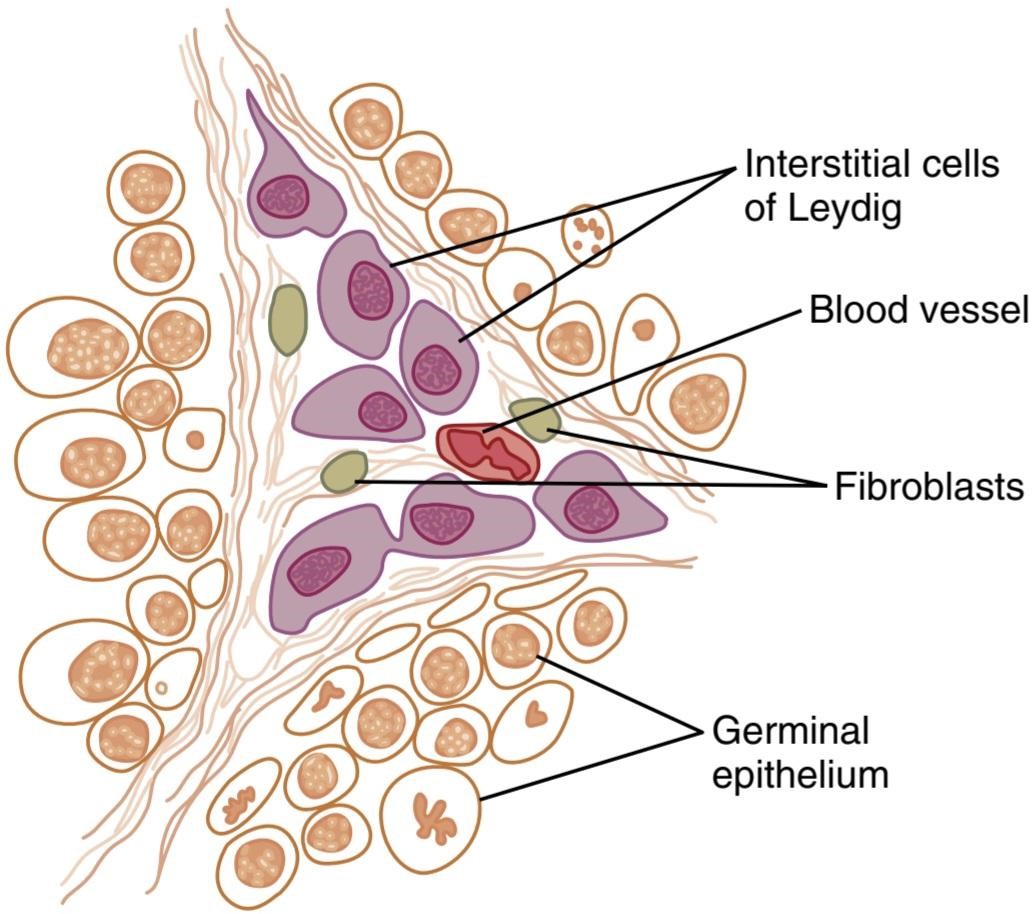
Figure 81-7 Interstitial cells of Leydig, the cells that secrete testosterone, located in the interstices between the seminiferous tubules.
شکل 81-7 سلول های بینابینی لیدیگ، سلول هایی که تستوسترون ترشح می کنند، در فواصل بین لوله های اسپرم ساز قرار دارند.
Secretion of Androgens Elsewhere in the Body. The term “androgen” means any steroid hormone that has masculinizing effects, including testosterone; it also includes male sex hormones produced elsewhere in the body besides the testes. For example, the adrenal glands secrete at least five androgens, although the total masculinizing activity of all these androgens is normally so slight (<5% of the total in the adult male) that even in women they do not cause significant masculine characteristics, except for causing growth of pubic and axillary hair. However, when a tumor of the adrenal androgen-producing cells occurs, the quantity of androgenic hormones may then become great enough to cause all the usual male secondary sexual characteristics to occur, even in females. These effects are described in connection with the adrenogenital syndrome in Chapter 78.
ترشح آندروژن در جاهای دیگر بدن. اصطلاح “آندروژن” به معنای هر هورمون استروئیدی است که دارای اثرات مردانه است، از جمله تستوسترون. همچنین شامل هورمونهای جنسی مردانه میشود که به غیر از بیضهها در جای دیگری از بدن تولید میشوند. به عنوان مثال، غدد فوق کلیوی حداقل پنج آندروژن ترشح میکنند، اگرچه کل فعالیت مردانهسازی همه این آندروژنها معمولاً آنقدر ناچیز است (کمتر از 5٪ از کل در مردان بالغ) که حتی در زنان نیز ویژگیهای مردانه قابل توجهی ایجاد نمیکنند، مگر اینکه باعث رشد موهای ناحیه تناسلی و زیر بغل شوند. با این حال، زمانی که تومور سلولهای تولیدکننده آندروژن آدرنال رخ میدهد، مقدار هورمونهای آندروژنی ممکن است آنقدر زیاد شود که باعث شود تمام خصوصیات جنسی ثانویه معمول مردانه حتی در زنان رخ دهد. این اثرات در ارتباط با سندرم آدرنوژنیتال در فصل 78 توضیح داده شده است.
Rarely, embryonic crest cells in the ovary can develop into a tumor that produces excessive quantities of andro- gens in women; one such tumor is the arrhenoblastoma. The normal ovary also produces minute quantities of androgens, but they are not significant.
به ندرت، سلول های تاج جنینی در تخمدان می توانند به توموری تبدیل شوند که مقادیر زیادی آندروژن در زنان تولید می کند. یکی از این تومورها آرنوبلاستوم است. تخمدان طبیعی نیز مقادیر اندکی آندروژن تولید می کند، اما آنها قابل توجه نیستند.
Androgens Are Steroids. All androgens are steroid compounds, as shown by the formulas in Figure 81-8 for testosterone and dihydrotestosterone. Both in the testes and in the adrenals, the androgens can be synthesized either from cholesterol or directly from acetyl coenzyme A.
آندروژن ها استروئید هستند. همه آندروژن ها ترکیبات استروئیدی هستند، همانطور که فرمول های موجود در شکل 8-81 برای تستوسترون و دی هیدروتستوسترون نشان داده شده است. هم در بیضه ها و هم در غده فوق کلیوی، آندروژن ها را می توان از کلسترول یا مستقیماً از استیل کوآنزیم A سنتز کرد.
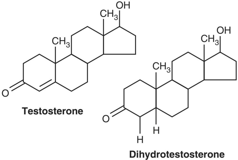
Figure 81-8 Testosterone and dihydrotestosterone.
شکل 81-8 تستوسترون و دی هیدروتستوسترون.
Metabolism of Testosterone. After secretion by the testes, about 97% of the testosterone becomes either loosely bound with plasma albumin or more tightly bound with a beta globulin called sex hormone-binding globulin and circulates in the blood in these states for 30 minutes to several hours. By that time, the testosterone is either transferred to the tissues or degraded into inactive products that are subsequently excreted.
متابولیسم تستوسترون. پس از ترشح توسط بیضه ها، حدود 97 درصد از تستوسترون یا به صورت سست با آلبومین پلاسما متصل می شود یا با یک بتا گلوبولین به نام گلوبولین اتصال به هورمون جنسی پیوند محکم تری پیدا می کند و در این حالت ها به مدت 30 دقیقه تا چند ساعت در خون گردش می کند. در آن زمان، تستوسترون یا به بافت ها منتقل می شود یا به محصولات غیرفعال تجزیه می شود که متعاقباً دفع می شوند.
Much of the testosterone that becomes fixed to the tissues is converted within the tissue cells to dihydrotestosterone, especially in certain target organs such as the prostate gland in the adult and the external genitalia of the male fetus. Some but not all actions of testosterone depend on this conversion. The intracellular functions are discussed later in this chapter.
بخش اعظم تستوسترونی که به بافتها ثابت میشود، در داخل سلولهای بافتی به دی هیدروتستوسترون تبدیل میشود، بهویژه در اندامهای هدف خاصی مانند غده پروستات در بزرگسالان و اندام تناسلی خارجی جنین پسر. برخی اما نه همه اعمال تستوسترون به این تبدیل بستگی دارد. عملکردهای درون سلولی بعداً در این فصل مورد بحث قرار خواهند گرفت.
Degradation and Excretion of Testosterone. The testosterone that does not become fixed to the tissues is rap- idly converted, mainly by the liver, into androsterone and dehydroepiandrosterone and simultaneously conjugated as either glucuronides or sulfates (glucuronides, particularly). These substances are excreted either into the gut by way of the liver bile or into the urine through the kidneys.
تخریب و دفع تستوسترون. تستوسترونی که به بافت ها ثابت نمی شود، به سرعت، عمدتاً توسط کبد، به آندروسترون و دهیدرواپی آندروسترون تبدیل می شود و به طور همزمان به صورت گلوکورونیدها یا سولفات ها (به ویژه گلوکورونیدها) کونژوگه می شود. این مواد یا از طریق صفرا به داخل روده یا از طریق کلیه ها به داخل ادرار دفع می شوند.
Production of Estrogen in the Male. In addition to testosterone, small amounts of estrogens are formed in the male (about one fifth the amount in the nonpregnant female), and a reasonable quantity of estrogens can be recovered from a man’s urine. The exact source of estrogens in the male is unclear, but the following information is known:
تولید استروژن در مردان. علاوه بر تستوسترون، مقادیر کمی استروژن در مردان (حدود یک پنجم مقدار در زنان غیرباردار) تشکیل میشود و مقدار معقولی از استروژن را میتوان از ادرار مرد بازیابی کرد. منبع دقیق استروژن در مردان نامشخص است، اما اطلاعات زیر شناخته شده است:
1. The concentration of estrogens in the fluid of the seminiferous tubules is quite high and probably plays an important role in spermiogenesis. This estrogen is believed to be formed by the Sertoli cells by converting testosterone to estradiol.
1. غلظت استروژن در مایع لوله های اسپرم ساز کاملاً زیاد است و احتمالاً نقش مهمی در اسپرم زایی دارد. اعتقاد بر این است که این استروژن توسط سلول های سرتولی با تبدیل تستوسترون به استرادیول تشکیل می شود.
2. Much larger amounts of estrogens are formed from testosterone and androstanediol in other tissues of the body, especially the liver, probably accounting for as much as 80% of the total male estrogen production.
2. مقادیر بسیار بیشتری استروژن از تستوسترون و آندروستندیول در سایر بافت های بدن، به ویژه کبد، تشکیل می شود که احتمالاً 80 درصد از کل تولید استروژن مردانه را تشکیل می دهد.
FUNCTIONS OF TESTOSTERONE
In general, testosterone is responsible for the distinguishing characteristics of the masculine body. Even during fetal life, the testes are stimulated by chorionic gonadotropin from the placenta to produce moderate quantities of testosterone throughout the entire period of fetal development and for 10 or more weeks after birth; thereafter, essentially no testosterone is produced during childhood until about the ages of 10 to 13 years. Testosterone production then increases rapidly under the stimulus of anterior pituitary gonadotropic hormones at the onset of puberty and lasts throughout most of the remainder of life, as shown in Figure 81-9, dwindling somewhat beyond age 50 to 60 years. Some studies, however, suggest that plasma testosterone levels may remain in the normal range in most healthy males as they age; only about 10% to 20% of men over 60 years of age and 50% of men over 80 years of age have “low” testosterone levels below 3 ng/ml (usually expressed clinically as <300 ng/dl or <10.4 nmol/L). In recent years, it has become clear that there is a clear association between late onset reductions in testosterone and metabolic disorders such as obesity and type 2 diabetes mellitus.
توابع تستوسترون
به طور کلی، تستوسترون مسئول ویژگی های متمایز بدن مردانه است. حتی در طول زندگی جنینی، بیضهها توسط گنادوتروپین کوریونی از جفت تحریک میشوند تا مقادیر متوسطی از تستوسترون را در کل دوره رشد جنین و 10 هفته یا بیشتر پس از تولد تولید کنند. پس از آن، اساساً هیچ تستوسترونی در دوران کودکی تا سن 10 تا 13 سالگی تولید نمی شود. سپس تولید تستوسترون تحت تحریک هورمون های گنادوتروپیک هیپوفیز قدامی در شروع بلوغ به سرعت افزایش می یابد و در تمام طول عمر باقی می ماند، همانطور که در شکل 81-9 نشان داده شده است و تا حدودی فراتر از سن 50 تا 60 سالگی کاهش می یابد. با این حال، برخی از مطالعات نشان می دهد که سطح تستوسترون پلاسما ممکن است در محدوده طبیعی در اکثر مردان سالم با افزایش سن باقی بماند. تنها حدود 10 تا 20 درصد از مردان بالای 60 سال و 50 درصد از مردان بالای 80 سال سطح تستوسترون «پایین» زیر 3 نانوگرم در میلی لیتر دارند (معمولاً از نظر بالینی کمتر از ng/dl 300 یا کمتر از 10.4 نانومول در لیتر بیان می شود). در سالهای اخیر، مشخص شده است که ارتباط واضحی بین کاهش دیررس تستوسترون و اختلالات متابولیک مانند چاقی و دیابت نوع 2 وجود دارد.

Figure 81-9 The different stages of male sexual function as reflected by average plasma testosterone concentrations (red line) and sperm production (blue line) at different ages. (Modified from Griffin JF, Wilson JD: The testis. In: Bondy PK, Rosenberg LE [eds]: Metabolic Control and Disease, 8th ed. Philadelphia: WB Saunders, 1980.)
شکل 9-81 مراحل مختلف عملکرد جنسی مردان که توسط میانگین غلظت تستوسترون پلاسما (خط قرمز) و تولید اسپرم (خط آبی) در سنین مختلف منعکس شده است. (اصلاح شده از Griffin JF, Wilson JD: The testis. In: Bondy PK, Rosenberg LE [eds]: Metabolic Control and Disease, 8th ed. Philadelphia: WB Saunders, 1980.)
Functions of Testosterone During Fetal Development
Testosterone begins to be elaborated by the male fetal testes at about the seventh week of embryonic life. Indeed, one of the major functional differences between the female and the male sex chromosome is that the male chromosome has the sex-determining region Y (SRY) gene that encodes a protein called the testis determining factor (also called the SRY protein). The SRY protein initiates a cascade of gene activations that cause the genital ridge cells to differentiate into cells that eventually become the testes and secrete testosterone, whereas the female chromosome causes this ridge to differentiate into cells that secrete estrogens.
عملکرد تستوسترون در طول رشد جنین
تستوسترون در حدود هفته هفتم زندگی جنینی توسط بیضه های جنین پسر شروع به بسط می کند. در واقع، یکی از تفاوت های عملکردی عمده بین کروموزوم جنسی زن و مرد این است که کروموزوم مرد دارای ژن ناحیه تعیین کننده جنسی Y (SRY) است که پروتئینی به نام فاکتور تعیین کننده بیضه (که پروتئین SRY نیز نامیده می شود) را کد می کند. پروتئین SRY یک آبشار از فعالسازی ژن را آغاز میکند که باعث تمایز سلولهای برآمدگی تناسلی به سلولهایی میشود که در نهایت به بیضه تبدیل میشوند و تستوسترون ترشح میکنند، در حالی که کروموزوم ماده باعث تمایز این برجستگی به سلولهایی میشود که استروژن ترشح میکنند.
Injection of large quantities of male sex hormone into pregnant animals causes development of male sexual organs, even though the fetus is female. Also, early removal of the testes in the male fetus causes development of female sexual organs.
تزریق مقادیر زیادی هورمون جنسی مردانه به حیوانات باردار باعث رشد اندام های جنسی نر می شود، حتی اگر جنین ماده باشد. همچنین برداشتن زودهنگام بیضه ها در جنین پسر باعث رشد اندام های جنسی زنانه می شود.
Thus, testosterone secreted by the fetal testes is responsible for development of the male body characteristics, including the formation of a penis and a scrotum rather than formation of a clitoris and a vagina. It also causes formation of the prostate gland, seminal vesicles, and male genital ducts, while at the same time suppressing formation of female genital organs.
بنابراین، تستوسترون ترشح شده توسط بیضه های جنین مسئول رشد ویژگی های بدن مرد است، از جمله تشکیل آلت تناسلی و کیسه بیضه به جای ایجاد کلیتوریس و واژن. همچنین باعث تشکیل غده پروستات، وزیکولهای منی و مجاری تناسلی مردانه میشود و در عین حال تشکیل اندامهای تناسلی زنان را سرکوب میکند.
Effect of Testosterone to Cause Descent of the Testes. The testes usually descend into the scrotum during the last 2 to 3 months of gestation when the testes begin secreting reasonable quantities of testosterone. If a male child is born with undescended but otherwise normal testes, administration of testosterone usually causes the testes to descend in the usual manner if the inguinal canals are large enough to allow the testes to pass.
تاثیر تستوسترون در نزول بیضه ها. زمانی که بیضه ها شروع به ترشح مقادیر معقول تستوسترون می کنند، بیضه ها معمولاً در طی 2 تا 3 ماه آخر بارداری به داخل کیسه بیضه فرو می روند. اگر کودک پسری با بیضههای نزول نکرده اما در غیر این صورت طبیعی به دنیا بیاید، تجویز تستوسترون معمولاً باعث پایین آمدن بیضهها به روش معمول میشود، اگر کانالهای مغبنی به اندازهای بزرگ باشد که اجازه عبور بیضهها را بدهد.
Administration of gonadotropic hormones, which stimulate the Leydig cells of the newborn child’s testes to produce testosterone, can also cause the testes to descend. Thus, the stimulus for descent of the testes is testosterone, indicating again that testosterone is an important hormone for male sexual development during fetal life.
تجویز هورمونهای گنادوتروپیک که سلولهای لیدیگ بیضههای نوزاد را برای تولید تستوسترون تحریک میکنند، همچنین میتواند باعث نزول بیضهها شود. بنابراین، محرک نزول بیضه ها تستوسترون است، که دوباره نشان می دهد که تستوسترون یک هورمون مهم برای رشد جنسی مردان در طول زندگی جنینی است.
Effect of Testosterone on Development of Adult Primary and Secondary Sexual Characteristics
After puberty, increasing amounts of testosterone secretion cause the penis, scrotum, and testes to enlarge about eightfold before the age of 20 years. In addition, testosterone causes the secondary sexual characteristics of the male to develop, beginning at puberty and ending at maturity. These secondary sexual characteristics, in addition to the sexual organs, distinguish the male from the female as follows.
تأثیر تستوسترون بر رشد ویژگیهای جنسی اولیه و ثانویه بزرگسالان
پس از بلوغ، افزایش میزان ترشح تستوسترون باعث می شود آلت تناسلی، کیسه بیضه و بیضه ها قبل از 20 سالگی حدود 8 برابر بزرگ شوند. علاوه بر این، تستوسترون باعث می شود که خصوصیات جنسی ثانویه در مرد ایجاد شود که از بلوغ شروع می شود و در بلوغ پایان می یابد. این خصوصیات جنسی ثانویه، علاوه بر اندام های جنسی، نر را از ماده به شرح زیر متمایز می کند.
Effect on the Distribution of Body Hair. Testosterone causes growth of hair (1) over the pubis, (2) upward along the linea alba of the abdomen sometimes to the umbilicus and above, (3) on the face, (4) usually on the chest, and (5) less often on other regions of the body, such as the back. It also causes the hair on most other portions of the body to become more prolific.
تأثیر بر توزیع موهای بدن. تستوسترون باعث رشد مو (1) روی ناحیه شرمگاهی، (2) به سمت بالا در امتداد خط آلبای شکم گاهی به سمت ناف و بالاتر، (3) روی صورت، (4) معمولاً روی سینه و (5) کمتر در سایر نواحی بدن مانند پشت می شود. همچنین باعث می شود موهای بیشتر قسمت های دیگر بدن پربارتر شوند.
Male Pattern Baldness. Testosterone decreases the growth of hair on the top of the head; a man who does not have functional testes does not become bald. However, many virile men never become bald because baldness is a result of two factors: first, a genetic background for the development of baldness and, second, superimposed on this genetic background, large quantities of androgenic hormones. When a long-sustained androgenic tumor develops in a woman who has the appropriate genetic back- ground, she becomes bald in the same manner as does a man.
طاسی الگوی مردانه. تستوسترون رشد موهای بالای سر را کاهش می دهد. مردی که بیضه های عملکردی ندارد کچل نمی شود. با این حال، بسیاری از مردان مردانه هرگز طاسی نمی شوند زیرا طاسی نتیجه دو عامل است: اول، زمینه ژنتیکی برای ایجاد طاسی و دوم، قرار گرفتن بر روی این زمینه ژنتیکی، مقادیر زیادی هورمون های آندروژنیک. هنگامی که یک تومور آندروژنی طولانی مدت در زنی که دارای زمینه ژنتیکی مناسب است ایجاد می شود، او مانند یک مرد طاسی می شود.
Effect on the Voice. Testosterone secreted by the testes or injected into the body causes hypertrophy of the laryngeal mucosa and enlargement of the larynx. The effects at first cause a relatively discordant, “cracking” voice that gradually changes into the typical adult masculine voice.
تاثیر بر صدا. تستوسترون ترشح شده از بیضه ها یا تزریق به بدن باعث هیپرتروفی مخاط حنجره و بزرگ شدن حنجره می شود. این اثرات در ابتدا باعث ایجاد صدای نسبتاً ناهماهنگ و “ترک” می شود که به تدریج به صدای معمولی مردانه بزرگسالان تبدیل می شود.
Testosterone Increases Thickness of the Skin and Can Contribute to Development of Acne. Testosterone in- creases the thickness of the skin over the entire body and the ruggedness of the subcutaneous tissues. Testosterone also increases the rate of secretion by some or perhaps all of the body’s sebaceous glands. Especially important is excessive secretion by the sebaceous glands of the face, which can result in acne. Therefore, acne is one of the most common features of male adolescence when the body is first becoming introduced to increased testosterone. After several years of testosterone secretion, the skin normally adapts to the testosterone in a way that allows it to overcome the acne.
تستوسترون ضخامت پوست را افزایش می دهد و می تواند به ایجاد آکنه کمک کند. تستوسترون ضخامت پوست را در کل بدن و ناهمواری بافت های زیر جلدی را افزایش می دهد. تستوسترون همچنین میزان ترشح برخی یا شاید همه غدد چربی بدن را افزایش می دهد. ترشح بیش از حد غدد چربی صورت که می تواند منجر به آکنه شود، اهمیت ویژه ای دارد. بنابراین، آکنه یکی از شایعترین ویژگیهای نوجوانی مردان است، زمانی که بدن برای اولین بار با افزایش تستوسترون آشنا میشود. پس از چندین سال ترشح تستوسترون، پوست به طور معمول با تستوسترون سازگار می شود به گونه ای که به آن اجازه می دهد بر آکنه غلبه کند.
Testosterone Increases Protein Formation and Muscle Development. One of the most important male characteristics is development of increasing musculature after puberty, averaging about a 50% increase in muscle mass over that in the female. This increase in muscle mass is also associated with increased protein in the nonmuscle parts of the body. Many of the changes in the skin are due to deposition of proteins in the skin, and changes in the voice also result partly from this protein anabolic function of testosterone.
تستوسترون تشکیل پروتئین و رشد عضلات را افزایش می دهد. یکی از مهمترین ویژگیهای مردانه، رشد عضلانی بعد از بلوغ است که به طور متوسط حدود 50 درصد افزایش توده عضلانی نسبت به زنان دارد. این افزایش توده عضلانی همچنین با افزایش پروتئین در قسمت های غیر عضلانی بدن همراه است. بسیاری از تغییرات در پوست به دلیل رسوب پروتئین ها در پوست است و تغییرات در صدا نیز تا حدی ناشی از عملکرد آنابولیک پروتئین تستوسترون است.
Because of the great effect that testosterone and other androgens have on the body’s musculature, synthetic androgens are widely used by athletes to improve their muscular performance. This practice is to be severely deprecated because of prolonged harmful effects of excess androgens, as discussed in Chapter 85 in relation to sports physiology. Testosterone or synthetic androgens are also occasionally used in old age as a “youth hormone” to improve muscle strength and vigor, but with questionable results. Some studies suggest that testosterone replacement therapy in elderly men may increase the risk of adverse cardiovascular events.
به دلیل تأثیر زیادی که تستوسترون و سایر آندروژن ها روی ماهیچه های بدن دارند، آندروژن های مصنوعی به طور گسترده توسط ورزشکاران برای بهبود عملکرد عضلانی خود استفاده می شود. این عمل به دلیل اثرات مضر طولانی مدت آندروژن های اضافی، همانطور که در فصل 85 در رابطه با فیزیولوژی ورزشی مورد بحث قرار گرفت، باید به شدت منسوخ شود. تستوسترون یا آندروژن های مصنوعی نیز گهگاه در سنین بالا به عنوان “هورمون جوانی” برای بهبود قدرت و قوت عضلانی استفاده می شود، اما نتایج مشکوکی دارد. برخی از مطالعات نشان می دهد که درمان جایگزینی تستوسترون در مردان مسن ممکن است خطر عوارض قلبی عروقی را افزایش دهد.
Testosterone Increases Bone Matrix and Causes Calcium Retention. After the great increase in circulating testosterone that occurs at puberty (or after prolonged injection of testosterone), the bones grow considerably thicker and deposit considerable additional calcium salts. Thus, testosterone increases the total quantity of bone matrix and causes calcium retention. The increase in bone matrix is believed to result from the general protein anabolic function of testosterone plus deposition of calcium salts in response to the increased protein.
تستوسترون ماتریکس استخوان را افزایش می دهد و باعث احتباس کلسیم می شود. پس از افزایش زیاد تستوسترون در گردش که در دوران بلوغ (یا پس از تزریق طولانی مدت تستوسترون) اتفاق می افتد، استخوان ها به طور قابل توجهی ضخیم تر می شوند و نمک های کلسیم اضافی قابل توجهی را رسوب می دهند. بنابراین، تستوسترون مقدار کل ماتریکس استخوان را افزایش می دهد و باعث احتباس کلسیم می شود. اعتقاد بر این است که افزایش ماتریکس استخوان ناشی از عملکرد کلی پروتئین آنابولیک تستوسترون به علاوه رسوب نمک های کلسیم در پاسخ به پروتئین افزایش یافته است.
Testosterone has a specific effect on the pelvis to (1) narrow the pelvic outlet, (2) lengthen it, (3) cause a funnel-like shape instead of the broad ovoid shape of the female pelvis, and (4) greatly increase the strength of the entire pelvis for load bearing. In the absence of testosterone, the male pelvis develops into a pelvis that is similar to that of the female.
تستوسترون اثر خاصی بر روی لگن دارد تا (1) دهانه خروجی لگن را باریک کند، (2) آن را طولانی کند، (3) به جای شکل تخم مرغی پهن لگن زنان، یک شکل قیف مانند ایجاد کند، و (4) استحکام کل لگن را برای تحمل بار بسیار افزایش دهد. در غیاب تستوسترون، لگن مردانه به لگنی شبیه لگن زنان تبدیل می شود.
Because of the ability of testosterone to increase the size and strength of bones, it is sometimes used in older men to treat osteoporosis.
به دلیل توانایی تستوسترون در افزایش اندازه و استحکام استخوان ها، گاهی اوقات در مردان مسن برای درمان پوکی استخوان استفاده می شود.
When great quantities of testosterone (or any other androgen) are secreted abnormally in the still-growing child, the rate of bone growth increases markedly, causing a spurt in total body height. However, the testosterone also causes the epiphyses of the long bones to unite with the shafts of the bones at an early age. Therefore, despite the rapidity of growth, this early uniting of the epiphyses prevents the person from growing as tall as he would have grown had testosterone not been secreted at all. Even in normal men, the final adult height is slightly less than that which occurs in males castrated before puberty.
هنگامی که مقادیر زیادی تستوسترون (یا هر آندروژن دیگر) به طور غیر طبیعی در کودک در حال رشد ترشح می شود، سرعت رشد استخوان به طور قابل توجهی افزایش می یابد و باعث افزایش قد در کل بدن می شود. با این حال، تستوسترون همچنین باعث می شود که اپی فیز استخوان های بلند در سنین پایین با شفت استخوان ها متحد شود. بنابراین، علیرغم سرعت رشد، این اتحاد زودهنگام اپی فیزها مانع از آن می شود که فرد به اندازه ای رشد کند که اگر تستوسترون اصلا ترشح نمی شد، رشد کند. حتی در مردان عادی، قد نهایی بزرگسالان کمی کمتر از مردانی است که قبل از بلوغ اخته شده اند.
Testosterone Increases the Basal Metabolic Rate. Injection of large quantities of testosterone can increase the basal metabolic rate by as much as 15%. Also, even the usual quantity of testosterone secreted by the testes during adolescence and early adult life increases the rate of metabolism some 5% to 10% above the value that it would be were the testes not active. This increased metabolic rate is possibly an indirect result of the effect of testosterone on protein anabolism, with the increased quantity of proteins the enzymes, especially-increasing activities of all cells.
تستوسترون میزان متابولیسم پایه را افزایش می دهد. تزریق مقادیر زیاد تستوسترون می تواند میزان متابولیسم پایه را تا 15 درصد افزایش دهد. همچنین، حتی مقدار معمول تستوسترون ترشح شده از بیضه ها در دوران نوجوانی و اوایل زندگی، میزان متابولیسم را حدود 5 تا 10 درصد بیشتر از مقداری که در صورت فعال نبودن بیضه ها افزایش می یابد، افزایش می دهد. این افزایش سرعت متابولیسم احتمالاً نتیجه غیرمستقیم اثر تستوسترون بر آنابولیسم پروتئین است، با افزایش مقدار پروتئین آنزیم ها، به ویژه فعالیت های فزاینده همه سلول ها.
Testosterone Increases Red Blood Cells. When normal quantities of testosterone are injected into a castrated adult, the number of red blood cells per cubic millimeter of blood increases 15% to 20%. Also, the average man has about 700,000 more red blood cells per cubic millimeter than the average woman. Despite the strong association of testosterone and increased hematocrit, testosterone does not appear to directly increase erythropoietin levels or have a direct effect on red blood cell production. The effect of testosterone to increase red blood cell production may be at least partly indirect because of the increased metabolic rate that occurs after testosterone administration.
تستوسترون گلبول های قرمز خون را افزایش می دهد. هنگامی که مقادیر نرمال تستوسترون به یک فرد بالغ اخته شده تزریق می شود، تعداد گلبول های قرمز خون در هر میلی متر مکعب خون بین 15 تا 20 درصد افزایش می یابد. همچنین، یک مرد به طور متوسط حدود ۷۰۰ هزار گلبول قرمز در هر میلی متر مکعب بیشتر از یک زن معمولی دارد. علیرغم ارتباط قوی تستوسترون و افزایش هماتوکریت، به نظر نمی رسد که تستوسترون به طور مستقیم سطح اریتروپویتین را افزایش دهد یا تأثیر مستقیمی بر تولید گلبول های قرمز داشته باشد. اثر تستوسترون برای افزایش تولید گلبول های قرمز ممکن است حداقل تا حدی غیرمستقیم باشد زیرا میزان متابولیک افزایش یافته پس از تجویز تستوسترون رخ می دهد.
Effect on Electrolyte and Water Balance. As pointed out in Chapter 78, many steroid hormones can increase the reabsorption of sodium in the distal tubules of the kidneys. Testosterone also has such an effect, but only to a minor degree in comparison with the adrenal mineralocorticoids. Nevertheless, after puberty, the blood and extracellular fluid volumes of the male in relation to body weight increase as much as 5% to 10%.
اثر بر تعادل الکترولیت و آب. همانطور که در فصل 78 اشاره شد، بسیاری از هورمون های استروئیدی می توانند بازجذب سدیم را در لوله های انتهایی کلیه ها افزایش دهند. تستوسترون نیز چنین تأثیری دارد، اما در مقایسه با مینرالوکورتیکوئیدهای آدرنال، فقط به میزان جزئی. با این وجود، پس از بلوغ، حجم خون و مایعات خارج سلولی مرد نسبت به وزن بدن بین 5 تا 10 درصد افزایش می یابد.
BASIC INTRACELLULAR MECHANISM OF ACTION OF TESTOSTERONE
Most of the effects of testosterone result basically from increased rate of protein formation in the target cells. In the prostate gland, for example, testosterone enters the prostatic cells within a few minutes after secretion. Then it is most often converted, under the influence of the intracellular enzyme 5a-reductase, to dihydrotestosterone which binds with a cytoplasmic “receptor protein.” This combination migrates to the cell nucleus where it binds with a nuclear protein and induces DNA-RNA transcription. Within 30 minutes, RNA polymerase has become activated, and the concentration of RNA begins to increase in the prostatic cells, which is followed by a progressive increase in cellular protein. After several days, the quantity of DNA in the prostate gland has also increased, and a simultaneous increase in the number of prostatic cells has occurred.
مکانیسم بین سلولی اساسی عمل تستوسترون
بیشتر اثرات تستوسترون اساساً ناشی از افزایش سرعت تشکیل پروتئین در سلول های هدف است. به عنوان مثال، در غده پروستات، تستوسترون در عرض چند دقیقه پس از ترشح وارد سلول های پروستات می شود. سپس اغلب تحت تأثیر آنزیم داخل سلولی 5a-ردوکتاز به دی هیدروتستوسترون تبدیل می شود که با یک “پروتئین گیرنده” سیتوپلاسمی متصل می شود. این ترکیب به هسته سلول مهاجرت می کند، جایی که با پروتئین هسته ای متصل می شود و رونویسی DNA-RNA را القا می کند. در عرض 30 دقیقه، RNA پلیمراز فعال می شود و غلظت RNA در سلول های پروستات شروع به افزایش می کند که به دنبال آن افزایش تدریجی پروتئین سلولی به دنبال دارد. پس از چند روز، مقدار DNA در غده پروستات نیز افزایش یافته است و همزمان افزایش تعداد سلول های پروستات رخ داده است.
Testosterone stimulates production of proteins virtually everywhere in the body, although more specifically affecting the proteins in “target” organs or tissues responsible for the development of primary and secondary male sexual characteristics.
تستوسترون تولید پروتئینها را تقریباً در همه جای بدن تحریک میکند، اگرچه به طور خاص بر پروتئینهای موجود در اندامها یا بافتهای «هدف» که مسئول ایجاد ویژگیهای جنسی اولیه و ثانویه مردانه هستند، تأثیر میگذارد.
Some studies suggest that testosterone, like other steroidal hormones, may also exert some rapid, nongenomic effects that do not require synthesis of new proteins. The physiological role of these nongenomic actions of testosterone, however, has yet to be determined.
برخی از مطالعات نشان می دهد که تستوسترون، مانند سایر هورمون های استروئیدی، ممکن است برخی از اثرات سریع و غیر ژنومیک را نیز اعمال کند که نیازی به سنتز پروتئین های جدید ندارد. نقش فیزیولوژیکی این اعمال غیر ژنومیک تستوسترون، با این حال، هنوز مشخص نشده است.
CONTROL OF MALE SEXUAL FUNCTIONS BY HORMONES FROM THE HYPOTHALAMUS AND ANTERIOR PITUITARY GLAND
A major share of the control of sexual functions in both the male and the female begins with secretion of gonadotropin-releasing hormone (GnRH) by the hypothalamus (Figure 81-10). This hormone stimulates the anterior pituitary gland to secrete two other hormones called gonadotropic hormones: (1) luteinizing hormone (LH) and (2) follicle-stimulating hormone (FSH). In turn, LH is the primary stimulus for the secretion of testosterone by the testes, and FSH mainly stimulates spermatogenesis.
کنترل عملکردهای جنسی مردان توسط هورمون های هیپوتالاموس و هیپوفیز قدامی
بخش عمده ای از کنترل عملکردهای جنسی در مردان و زنان با ترشح هورمون آزاد کننده گنادوتروپین (GnRH) توسط هیپوتالاموس شروع می شود (شکل 81-10). این هورمون غده هیپوفیز قدامی را تحریک می کند تا دو هورمون دیگر به نام هورمون های گنادوتروپیک ترشح کند: (1) هورمون لوتئینه کننده (LH) و (2) هورمون محرک فولیکول (FSH). به نوبه خود، LH محرک اولیه برای ترشح تستوسترون توسط بیضه ها است و FSH عمدتاً باعث تحریک اسپرماتوژنز می شود.
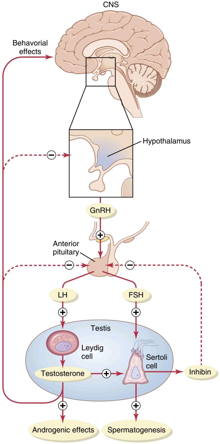
Figure 81-10 Feedback regulation of the hypothalamic-pituitary- testicular axis in males. Stimulatory effects are shown by plus signs, and negative feedback inhibitory effects are shown by minus signs. CNS, Central nervous system; FSH, follicle-stimulating hormone; GnRH, gonadotropin-releasing hormone; LH, luteinizing hormone.
شکل 81-10 تنظیم بازخورد محور هیپوتالاموس-هیپوفیز-بیضه در مردان. اثرات محرک با علائم مثبت و اثرات بازدارنده بازخورد منفی با علائم منفی نشان داده می شود. CNS، سیستم عصبی مرکزی؛ FSH، هورمون محرک فولیکول؛ GnRH، هورمون آزاد کننده گنادوتروپین؛ LH، هورمون لوتئینه کننده.
Gonadotropin-Releasing Hormone Increases Secretion of Luteinizing Hormone and Follicle-Stimulating Hormone
GnRH is a 10-amino acid peptide secreted by neurons whose cell bodies are located in the arcuate nuclei of the hypothalamus. The endings of these neurons terminate mainly in the median eminence of the hypothalamus, where they release GnRH into the hypothalamic-hypophysial portal vascular system. The GnRH is then transported to the anterior pituitary gland in the hypophysial portal blood and stimulates release of the two gonadotropins, LH and FSH.
هورمون آزاد کننده گنادوتروپین ترشح هورمون لوتئینیزه کننده و هورمون محرک فولیکول را افزایش می دهد.
GnRH یک پپتید 10 اسید آمینه است که توسط سلول های عصبی ترشح می شود که بدنه سلولی آن در هسته کمانی هیپوتالاموس قرار دارد. انتهای این نورونها عمدتاً به برجستگی میانی هیپوتالاموس ختم میشوند، جایی که GnRH را در سیستم عروقی پورتال هیپوتالاموس-هیپوفیزیال آزاد میکنند. سپس GnRH در خون پورتال هیپوفیزیال به غده هیپوفیز قدامی منتقل می شود و آزادسازی دو گنادوتروپین LH و FSH را تحریک می کند.
GnRH is secreted intermittently a few minutes at a time once every 1 to 3 hours. The intensity of this hormone’s stimulus is determined in two ways: (1) by the frequency of these cycles of secretion and (2) by the quantity of GnRH released with each cycle.
GnRH هر 1 تا 3 ساعت یک بار به طور متناوب چند دقیقه در یک زمان ترشح می شود. شدت محرک این هورمون به دو صورت تعیین می شود: (1) با فراوانی این چرخه های ترشح و (2) با مقدار GnRH آزاد شده در هر چرخه.
Secretion of LH by the anterior pituitary gland is also cyclical, with LH following fairly faithfully the pulsatile release of GnRH. In contrast, FSH secretion increases and decreases only slightly with each fluctuation of GnRH secretion; instead, it changes more slowly over a period of many hours in response to longer-term changes in GnRH. Because of the much closer relation between GnRH secretion and LH secretion, GnRH is also widely known as LH-releasing hormone.
ترشح LH توسط غده هیپوفیز قدامی نیز چرخه ای است، با LH که نسبتاً صادقانه از انتشار ضربانی GnRH پیروی می کند. در مقابل، ترشح FSH با هر نوسان ترشح GnRH فقط اندکی کاهش مییابد. در عوض، در واکنش به تغییرات طولانیمدت در GnRH، طی یک دوره چند ساعته آهستهتر تغییر میکند. به دلیل ارتباط بسیار نزدیکتر بین ترشح GnRH و ترشح LH، GnRH به طور گسترده به عنوان هورمون آزاد کننده LH نیز شناخته می شود.
Gonadotropic Hormones: Luteinizing Hormone and Follicle-Stimulating Hormone
Both of the gonadotropic hormones, LH and FSH, are secreted by the same cells, called gonadotropes, in the anterior pituitary gland. In the absence of GnRH secretion from the hypothalamus, the gonadotropes in the pituitary gland secrete almost no LH or FSH.
هورمون های گنادوتروپیک: هورمون لوتئینه کننده و هورمون محرک فولیکول
هر دو هورمون گنادوتروپیک LH و FSH توسط سلول های مشابهی به نام گنادوتروپ در غده هیپوفیز قدامی ترشح می شوند. در غیاب ترشح GnRH از هیپوتالاموس، گنادوتروپ ها در غده هیپوفیز تقریبا هیچ LH یا FSH ترشح نمی کنند.
LH and FSH are glycoproteins. They exert their effects on their target tissues in the testes mainly by activating the cyclic adenosine monophosphate second messenger system, which in turn activates specific enzyme systems in the respective target cells.
LH و FSH گلیکوپروتئین هستند. آنها عمدتاً با فعال کردن سیستم پیام رسان دوم آدنوزین مونوفسفات حلقوی، که به نوبه خود سیستم های آنزیمی خاصی را در سلول های هدف مربوطه فعال می کند، اثرات خود را بر روی بافت های هدف خود در بیضه ها اعمال می کنند.
Regulation of Testosterone Production by Luteinizing Hormone. Testosterone is secreted by the interstitial cells of Leydig in the testes, but only when they are stimulated by LH from the anterior pituitary gland. Furthermore, the quantity of testosterone that is secreted increases approximately in direct proportion to the amount of LH that is available.
تنظیم تولید تستوسترون توسط هورمون لوتئینه کننده. تستوسترون توسط سلول های بینابینی لیدیگ در بیضه ها ترشح می شود، اما تنها زمانی که توسط LH از غده هیپوفیز قدامی تحریک شوند. علاوه بر این، مقدار تستوسترون ترشح شده تقریباً به نسبت مستقیم با مقدار LH موجود افزایش می یابد.
Mature Leydig cells are normally found in a child’s testes for a few weeks after birth but then disappear until after the age of about 10 years. However, injection of purified LH into a child at any age or secretion of LH at puberty causes testicular interstitial cells that look like fibroblasts to evolve into functioning Leydig cells.
سلولهای بالغ لیدیگ معمولاً چند هفته پس از تولد در بیضههای کودک یافت میشوند، اما تا بعد از حدود 10 سالگی ناپدید میشوند. با این حال، تزریق LH خالص شده به کودک در هر سنی یا ترشح LH در دوران بلوغ باعث می شود سلول های بینابینی بیضه که شبیه فیبروبلاست هستند به سلول های لیدیگ فعال تبدیل شوند.
Inhibition of Anterior Pituitary Secretion of Luteinizing and Follicle-Stimulating Hormones by Testosterone-Negative Feedback Control of Testosterone Secretion. Testosterone secreted by the testes in response to LH has the reciprocal effect of inhibiting anterior pituitary secretion of LH (see Figure 81-10). Most of this inhibition probably results from a direct effect of testosterone on the hypothalamus to decrease secretion of GnRH. This effect in turn causes a corresponding decrease in secretion of LH and FSH by the anterior pituitary, and the de- crease in LH reduces secretion of testosterone by the testes. Thus, whenever testosterone secretion becomes too great, this automatic negative feedback effect, operating through the hypothalamus and anterior pituitary gland, reduces testosterone secretion back toward the desired operating level. Conversely, too little testosterone allows the hypothalamus to secrete large amounts of GnRH, with a corresponding increase in anterior pituitary LH and FSH secretion and consequent increase in testicular testosterone secretion.
مهار ترشح هیپوفیز قدامی هورمون های لوتئینه کننده و محرک فولیکول توسط کنترل بازخورد منفی تستوسترون ترشح تستوسترون. تستوسترون ترشح شده توسط بیضه ها در پاسخ به LH اثر متقابل مهار ترشح هیپوفیز قدامی LH را دارد (شکل 81-10 را ببینید). بیشتر این مهار احتمالاً ناشی از تأثیر مستقیم تستوسترون بر هیپوتالاموس برای کاهش ترشح GnRH است. این اثر به نوبه خود باعث کاهش متناظر در ترشح LH و FSH توسط هیپوفیز قدامی می شود و کاهش LH باعث کاهش ترشح تستوسترون توسط بیضه ها می شود. بنابراین، هر زمان که ترشح تستوسترون بیش از حد زیاد شود، این اثر بازخورد منفی خودکار، که از طریق هیپوتالاموس و غده هیپوفیز قدامی عمل میکند، ترشح تستوسترون را به سطح مورد نظر کاهش میدهد. برعکس، تستوسترون بسیار کم به هیپوتالاموس اجازه می دهد تا مقادیر زیادی GnRH ترشح کند، با افزایش متناظر در ترشح LH و FSH هیپوفیز قدامی و در نتیجه افزایش ترشح تستوسترون بیضه.
Regulation of Spermatogenesis by Follicle-Stimulating Hormone and Testosterone
FSH binds with specific FSH receptors attached to the Sertoli cells in the seminiferous tubules, which causes the Sertoli cells to grow and secrete various spermatogenic substances. Simultaneously, testosterone (and dihydrotestosterone) diffusing into the seminiferous tubules from the Leydig cells in the interstitial spaces also has a strong tropic effect on spermatogenesis. Thus, both FSH and testosterone are necessary to initiate spermatogenesis.
تنظیم اسپرماتوژنز توسط هورمون محرک فولیکول و تستوسترون
FSH با گیرنده های خاص FSH متصل به سلول های سرتولی در لوله های اسپرم ساز متصل می شود که باعث رشد سلول های سرتولی و ترشح مواد مختلف اسپرم زا می شود. به طور همزمان، انتشار تستوسترون (و دی هیدروتستوسترون) به داخل لوله های منی ساز از سلول های لیدیگ در فضاهای بینابینی نیز تأثیر استوایی قوی بر اسپرماتوژنز دارد. بنابراین، FSH و تستوسترون هر دو برای شروع اسپرماتوژنز ضروری هستند.
Role of Inhibin in Negative Feedback Control of Seminiferous Tubule Activity. When the seminiferous tubules fail to produce sperm, secretion of FSH by the anterior pituitary gland increases markedly. Conversely, when spermatogenesis proceeds too rapidly, pituitary secretion of FSH diminishes. The cause of this negative feedback effect on the anterior pituitary is believed to be secretion by the Sertoli cells of still another hormone called inhibin (see Figure 81-10). This hormone has a strong direct effect on the anterior pituitary gland to inhibit FSH secretion.
نقش اینهیبین در کنترل بازخورد منفی فعالیت لوله منی ساز. هنگامی که لوله های اسپرم ساز نتوانند اسپرم تولید کنند، ترشح FSH توسط غده هیپوفیز قدامی به طور قابل توجهی افزایش می یابد. برعکس، وقتی اسپرمزایی خیلی سریع پیش میرود، ترشح FSH از هیپوفیز کاهش مییابد. اعتقاد بر این است که علت این اثر بازخورد منفی بر هیپوفیز قدامی ترشح هورمون دیگری به نام اینهیبین توسط سلول های سرتولی است (شکل 81-10 را ببینید). این هورمون تأثیر مستقیم قوی بر غده هیپوفیز قدامی برای مهار ترشح FSH دارد.
Inhibin is a glycoprotein, like LH and FSH, with a molecular weight between 10,000 and 30,000. It has been isolated from cultured Sertoli cells. Its potent inhibitory effect on the anterior pituitary gland provides an important negative feedback mechanism for control of spermatogenesis, operating simultaneously with and in parallel to the negative feedback mechanism for control of testosterone secretion.
Inhibin یک گلیکوپروتئین است، مانند LH و FSH، با وزن مولکولی بین 10000 تا 30000. از سلول های سرتولی کشت شده جدا شده است. اثر مهاری قوی آن بر روی غده هیپوفیز قدامی یک مکانیسم بازخورد منفی مهم برای کنترل اسپرمزایی فراهم میکند که همزمان با مکانیسم بازخورد منفی برای کنترل ترشح تستوسترون و به موازات آن عمل میکند.
Human Chorionic Gonadotropin Secreted by the Placenta During Pregnancy Stimulates Testosterone Secretion by the Fetal Testes
During pregnancy, the hormone human chorionic gonadotropin (hCG) is secreted by the placenta and circulates in the mother and in the fetus. This hormone has almost the same effects on the sexual organs as LH.
گنادوتروپین کوریونی انسان که توسط جفت در دوران بارداری ترشح می شود، ترشح تستوسترون را در بیضه های جنینی تحریک می کند.
در دوران بارداری، هورمون گنادوتروپین جفتی انسانی (hCG) توسط جفت ترشح می شود و در مادر و جنین گردش می کند. این هورمون تقریباً همان اثرات LH را روی اندام های جنسی دارد.
During pregnancy, if the fetus is a male, hCG from the placenta causes the testes of the fetus to secrete testosterone. This testosterone is critical for promoting formation of the male sexual organs, as pointed out earlier. We dis-cuss hCG and its functions during pregnancy in greater detail in Chapter 83.
در دوران بارداری، اگر جنین مرد باشد، hCG از جفت باعث ترشح تستوسترون در بیضه های جنین می شود. همانطور که قبلاً اشاره شد، این تستوسترون برای تقویت تشکیل اندام های جنسی مردانه حیاتی است. ما hCG و عملکردهای آن در دوران بارداری را با جزئیات بیشتری در فصل 83 مورد بحث قرار می دهیم.
Puberty and Regulation of Its Onset
Initiation of the onset of puberty has long been a mystery, but it has now been determined that during childhood the hypothalamus does not secrete significant amounts of GnRH. One of the reasons for this is that, during child- hood, the slightest secretion of any sex steroid hormones exerts a strong inhibitory effect on hypothalamic secretion of GnRH. Yet,for reasons still not fully understood, at the time of puberty, the secretion of hypothalamic GnRH breaks through the childhood inhibition and adult sexual life begins.
بلوغ و تنظیم شروع آن
شروع بلوغ مدتها یک راز بوده است، اما اکنون مشخص شده است که در دوران کودکی هیپوتالاموس مقادیر قابل توجهی GnRH ترشح نمیکند. یکی از دلایل این امر این است که در دوران کودکی، کوچکترین ترشح هر هورمون استروئیدی جنسی، اثر مهاری قوی بر ترشح هیپوتالاموس GnRH دارد. با این حال، به دلایلی که هنوز به طور کامل شناخته نشده است، در زمان بلوغ، ترشح GnRH هیپوتالاموس از طریق مهار دوران کودکی شکسته می شود و زندگی جنسی بزرگسالان آغاز می شود.
Male Adult Sexual Life and Male Climacteric. After puberty, gonadotropic hormones are produced by the male pituitary gland for the remainder of life, and at least some spermatogenesis usually continues until death. Many men, however, begin to exhibit slowly decreasing sexual functions in their late 50s or 60s, especially if they smoke or are obese and have associated cardiovascular and metabolic disorders, such as hypertension, atherosclerosis, and type 2 diabetes mellitus. There is considerable variation in the decline of sexual function, with healthy men continuing to be virile until their 80s and 90s.
زندگی جنسی بزرگسالان مرد و اقلیم مرد. پس از بلوغ، هورمونهای گنادوتروپیک توسط غده هیپوفیز مردانه تا پایان عمر تولید میشوند و حداقل مقداری اسپرمزایی معمولاً تا زمان مرگ ادامه مییابد. با این حال، بسیاری از مردان در اواخر دهه 50 یا 60 زندگی خود به آرامی کاهش عملکرد جنسی را نشان می دهند، به خصوص اگر سیگار می کشند یا چاق هستند و اختلالات قلبی عروقی و متابولیک مرتبط، مانند فشار خون بالا، آترواسکلروز، و دیابت نوع 2 دارند. تغییرات قابل توجهی در کاهش عملکرد جنسی وجود دارد، به طوری که مردان سالم تا دهه 80 و 90 سالگی به زور بودن ادامه می دهند.
The slow, gradual decline in sexual function is also related, in part, to a decrease in testosterone secretion, as shown in Figure 81-9. The decrease in male sexual function is called the male climacteric.
کاهش تدریجی و آهسته عملکرد جنسی نیز تا حدی با کاهش ترشح تستوسترون مرتبط است، همانطور که در شکل 9-81 نشان داده شده است. به کاهش عملکرد جنسی مردان، کلیماکتریک مردانه می گویند.
Abnormalities of Male Sexual Function
The Prostate Gland and Its Abnormalities
The prostate gland remains relatively small throughout childhood and begins to grow at puberty under the stimulus of testosterone. This gland reaches an almost stationary size by the age of 20 years and remains at this size up to the age of about 50 years. At that time, in some men it begins to involute, along with decreased production of testosterone by the testes.
ناهنجاری های عملکرد جنسی مردان
غده پروستات و ناهنجاری های آن
غده پروستات در طول دوران کودکی نسبتا کوچک باقی می ماند و در دوران بلوغ تحت محرک تستوسترون شروع به رشد می کند. این غده در سن 20 سالگی به اندازه ای تقریبا ثابت می رسد و تا حدود 50 سالگی در این اندازه باقی می ماند. در آن زمان، در برخی از مردان، همراه با کاهش تولید تستوسترون توسط بیضه ها، شروع به پیچیدن می کند.
A benign prostatic fibroadenoma frequently develops in the prostate in many older men and can cause urinary obstruction. This hypertrophy is caused not by testosterone but instead by abnormal overgrowth of prostate tissue.
فیبروآدنوم خوش خیم پروستات اغلب در بسیاری از مردان مسن در پروستات ایجاد می شود و می تواند باعث انسداد ادرار شود. این هیپرتروفی نه به دلیل تستوسترون بلکه به دلیل رشد بیش از حد غیر طبیعی بافت پروستات ایجاد می شود.
Cancer of the prostate gland is a different problem that accounts for about 2% to 3% of all male deaths. Once cancer of the prostate gland occurs, the cancerous cells are usually stimulated to more rapid growth by testosterone and are inhibited by removal of both testes so that testosterone cannot be formed. Prostatic cancer usually can be inhibited by administration of estrogens. Even some patients who have prostatic cancer that has already metastasized to almost all the bones of the body can be successfully treated for a few months to years by removal of the testes, estrogen therapy, or both; after initiation of this therapy, the metastases usually diminish in size, and the bones partially heal. This treatment does not stop the cancer but slows it and sometimes greatly diminishes the severe bone pain.
سرطان غده پروستات یک مشکل متفاوت است که حدود 2 تا 3 درصد از کل مرگ و میرهای مردان را تشکیل می دهد. هنگامی که سرطان غده پروستات رخ می دهد، سلول های سرطانی معمولاً توسط تستوسترون برای رشد سریع تر تحریک می شوند و با برداشتن هر دو بیضه مهار می شوند به طوری که تستوسترون نمی تواند تشکیل شود. سرطان پروستات معمولاً با تجویز استروژن قابل مهار است. حتی برخی از بیمارانی که سرطان پروستات دارند و تقریباً به تمام استخوانهای بدن متاستاز دادهاند، میتوانند با برداشتن بیضهها، استروژن درمانی یا هر دو، برای چند ماه تا سالها با موفقیت درمان شوند. پس از شروع این درمان، متاستازها معمولاً کاهش مییابند و استخوانها تا حدی بهبود مییابند. این درمان سرطان را متوقف نمی کند، بلکه آن را کند می کند و گاهی اوقات درد شدید استخوان را تا حد زیادی کاهش می دهد.
Hypogonadism in the Male
When the testes of a male fetus are nonfunctional during fetal life, none of the male sexual characteristics develop in the fetus. Instead, female organs are formed. The reason for this is that the basic genetic characteristic of the fetus, whether male or female, is to form female sexual organs if there are no sex hormones. However, in the presence of testosterone, formation of female sexual organs is suppressed and male organs are induced instead.
هیپوگنادیسم در مردان
هنگامی که بیضه های جنین پسر در طول زندگی جنینی کار نمی کند، هیچ یک از خصوصیات جنسی مرد در جنین ایجاد نمی شود. در عوض، اندام های زنانه تشکیل می شود. دلیل این امر این است که ویژگی اصلی ژنتیکی جنین، اعم از زن یا مرد، تشکیل اندام جنسی زنانه در صورت عدم وجود هورمون جنسی است. با این حال، در حضور تستوسترون، تشکیل اندام های جنسی زنانه سرکوب می شود و به جای آن اندام های مردانه القا می شوند.
When a boy loses his testes before puberty, a state of eunuchism ensues in which he continues to have infantile sex organs and other infantile sexual characteristics through- out life. The height of an adult eunuch is slightly greater than that of a normal man because the bone epiphyses are slow to unite, although the bones are quite thin and the muscles are considerably weaker than those of a normal man. The voice is childlike, there is no loss of hair on the head, and the normal adult masculine hair distribution on the face and elsewhere does not occur.
وقتی پسری قبل از بلوغ بیضههای خود را از دست میدهد، حالت خواجهخوانی به وجود میآید که در آن اعضای جنسی نوزاد و سایر ویژگیهای جنسی نوزادان در طول زندگی ادامه مییابد. قد یک خواجه بالغ کمی بیشتر از قد یک مرد عادی است زیرا اپی فیزهای استخوانی دیر به هم می پیوندند، اگرچه استخوان ها کاملاً نازک هستند و ماهیچه ها به طور قابل توجهی ضعیف تر از یک مرد عادی است. صدا کودکانه است، ریزش مو در سر وجود ندارد و توزیع موی مردانه طبیعی بزرگسالان در صورت و جاهای دیگر رخ نمی دهد.
When a man is castrated after puberty, some of his male secondary sexual characteristics revert to those of a child, and others remain of adult masculine character. The sexual organs regress slightly in size but not to a childlike state, and the voice regresses from the bass quality only slightly. However, there is loss of masculine hair production, loss of the thick masculine bones, and loss of the musculature of the virile male.
هنگامی که مردی پس از بلوغ اخته می شود، برخی از ویژگی های جنسی ثانویه مرد او به ویژگی های یک کودک باز می گردد و برخی دیگر از ویژگی های مردانه بزرگسالی باقی می ماند. اندامهای جنسی کمی از نظر اندازه عقب میروند، اما نه به حالت کودکانه، و صدا از کیفیت باس فقط کمی پسرفت میکند. با این حال، از دست دادن تولید موی مردانه، از دست دادن استخوان های ضخیم مردانه و از دست دادن ماهیچه های مردانه مردانه وجود دارد.
Also in a castrated adult male, sexual desires are decreased but not lost, provided sexual activities have been practiced previously. Erection can still occur as before, although with less ease, but it is rare that ejaculation can take place, primarily because the semen-forming organs degenerate and there has been a loss of the testosterone-driven psychic desire.
همچنین در یک مرد بالغ اخته شده، تمایلات جنسی کاهش مییابد اما از بین نمیرود، مشروط بر اینکه قبلاً فعالیتهای جنسی انجام شده باشد. نعوظ همچنان میتواند مانند قبل رخ دهد، اگرچه با سهولت کمتر، اما به ندرت اتفاق میافتد که انزال اتفاق بیفتد، عمدتاً به این دلیل که اندامهای سازنده منی تحلیل میروند و میل روانی ناشی از تستوسترون از بین رفته است.
Some cases of hypogonadism are caused by a genetic inability of the hypothalamus to secrete normal amounts of GnRH. This condition is often associated with a simultaneous abnormality of the feeding center of the hypothalamus, causing the person to greatly overeat. Consequently, obesity occurs along with eunuchism. A patient with this condition is shown in Figure 81-11; the condition is called adiposogenital syndrome, Fröhlich’s syndrome, or hypothalamic eunuchism.
برخی از موارد هیپوگنادیسم ناشی از ناتوانی ژنتیکی هیپوتالاموس در ترشح مقادیر طبیعی GnRH است. این وضعیت اغلب با یک ناهنجاری همزمان در مرکز تغذیه هیپوتالاموس همراه است که باعث می شود فرد به شدت پرخوری کند. در نتیجه چاقی همراه با خواجه رخ می دهد. بیمار با این وضعیت در شکل 81-11 نشان داده شده است. این بیماری سندرم چربی تناسلی، سندرم فرولیش یا خواجه شناسی هیپوتالاموس نامیده می شود.

Figure 81-11 Adiposogenital syndrome in an adolescent male. Note the obesity and childlike sexual organs. (Courtesy Dr. Leonard Posey.)
شکل 81-11 سندرم چربی تناسلی در یک پسر نوجوان. به چاقی و اندام های جنسی کودکانه توجه کنید. (با احترام از دکتر لئونارد پوزی.)
Testicular Tumors and Hypergonadism in the Male
Interstitial Leydig cell tumors develop in rare cases in the testes. These tumors sometimes produce as much as 100 times the normal quantities of testosterone. When such tumors develop in young children, they cause rapid growth of the musculature and bones but also early uniting of the epiphyses, so that the eventual adult height is actually considerably less than that which would have been achieved otherwise. Such interstitial cell tumors also cause excessive development of the male sexual organs, all skeletal muscles, and other male sexual characteristics. In the adult male, small interstitial cell tumors are difficult to diagnose because masculine features are already present.
تومورهای بیضه و هیپرگنادیسم در مردان
تومورهای بینابینی سلول لیدیگ در موارد نادری در بیضه ها ایجاد می شوند. این تومورها گاهی اوقات تا 100 برابر مقادیر طبیعی تستوسترون تولید می کنند. هنگامی که چنین تومورهایی در کودکان خردسال ایجاد میشوند، باعث رشد سریع ماهیچهها و استخوانها میشوند، اما باعث اتحاد زودهنگام اپیفیزها میشوند، به طوری که قد نهایی بزرگسالان در واقع به میزان قابل توجهی کمتر از آن چیزی است که در غیر این صورت به دست میآمد. چنین تومورهای سلولی بینابینی باعث رشد بیش از حد اندام های جنسی مردانه، تمام ماهیچه های اسکلتی و سایر خصوصیات جنسی مردانه می شوند. در مردان بالغ، تشخیص تومورهای سلول بینابینی کوچک دشوار است زیرا ویژگی های مردانه در حال حاضر وجود دارد.
Much more common than interstitial Leydig cell tumors are tumors of the germinal epithelium. Because germinal cells are capable of differentiating into almost any type of cell, many of these tumors contain multiple tis- sues, such as placental tissue, hair, teeth, bone, skin, and so forth, all found together in the same tumorous mass called a teratoma. These tumors often secrete few hormones, but if a significant quantity of placental tissue develops in the tumor, it may secrete large quantities of hCG with functions similar to those of LH. Also, estrogenic hormones are sometimes secreted by these tumors and cause the condition called gynecomastia (overgrowth of the breasts).
بسیار شایع تر از تومورهای بینابینی سلول لیدیگ، تومورهای اپیتلیوم ژرمینال هستند. از آنجایی که سلولهای ژرمینال تقریباً به هر نوع سلولی تمایز مییابند، بسیاری از این تومورها حاوی بافتهای متعددی مانند بافت جفت، مو، دندان، استخوان، پوست و غیره هستند که همه با هم در یک توده توموری به نام تراتوم یافت میشوند. این تومورها اغلب هورمون های کمی ترشح می کنند، اما اگر مقدار قابل توجهی از بافت جفت در تومور ایجاد شود، ممکن است مقادیر زیادی hCG با عملکردهایی مشابه LH ترشح کند. همچنین گاهی اوقات هورمون های استروژنی توسط این تومورها ترشح می شود و باعث ایجاد بیماری به نام ژنیکوماستی (رشد بیش از حد سینه ها) می شود.
Erectile Dysfunction in the Male
Erectile dysfunction, also called “impotence,” is characterized by an inability of the man to develop or maintain an erection of sufficient rigidity for satisfactory sexual inter- course. Neurological problems, such as trauma to the para- sympathetic nerves from prostate surgery, deficient levels of testosterone, and some drugs (e.g., nicotine, alcohol, and antidepressants) can also contribute to erectile dysfunction.
اختلال نعوظ در مردان
اختلال نعوظ که “ناتوانی جنسی” نیز نامیده می شود، با ناتوانی مرد در ایجاد یا حفظ نعوظ با سفتی کافی برای رابطه جنسی رضایت بخش مشخص می شود. مشکلات عصبی، مانند ضربه به اعصاب پاراسمپاتیک ناشی از جراحی پروستات، کمبود سطح تستوسترون، و برخی داروها (مانند نیکوتین، الکل و داروهای ضد افسردگی) نیز می توانند در ایجاد اختلال نعوظ نقش داشته باشند.
In men older than 40 years, erectile dysfunction is most often caused by underlying vascular disease. As discussed previously, adequate blood flow and nitric oxide formation are essential for penile erection. Vascular disease, which can occur as a result of uncontrolled hypertension, diabetes, and atherosclerosis, reduces the ability of the body’s blood vessels, including those in the penis, to dilate. Part of this impaired vasodilation is due to decreased release of nitric oxide.
در مردان بالای 40 سال، اختلال نعوظ اغلب به دلیل بیماری زمینه ای عروقی ایجاد می شود. همانطور که قبلاً گفته شد، جریان خون کافی و تشکیل اکسید نیتریک برای نعوظ آلت تناسلی ضروری است. بیماری عروقی که می تواند در نتیجه فشار خون کنترل نشده، دیابت و تصلب شرایین رخ دهد، توانایی رگ های خونی بدن، از جمله رگ های آلت تناسلی، را کاهش می دهد. بخشی از این اختلال اتساع عروقی به دلیل کاهش انتشار اکسید نیتریک است.
Erectile dysfunction caused by vascular disease can often be successfully treated with phosphodiesterase-5 (PDE- 5) inhibitors such as sildenafil (Viagra), vardenafil (Levitra), or tadalafil (Cialis). These drugs increase cyclic GMP levels in the erectile tissue by inhibiting the enzyme phosphodiesterase-5, which rapidly degrades cyclic GMP. Thus, by inhibiting the degradation of cyclic GMP, the PDE-5 inhibitors enhance and prolong the effect of cyclic GMP to cause erection.
اختلال نعوظ ناشی از بیماری عروقی اغلب با مهارکننده های فسفودی استراز 5 (PDE-5) مانند سیلدنافیل (ویاگرا)، واردنافیل (لویترا) یا تادالافیل (سیالیس) با موفقیت درمان می شود. این داروها سطح GMP حلقوی را در بافت نعوظ با مهار آنزیم فسفودی استراز-5 افزایش می دهند که به سرعت GMP حلقوی را تخریب می کند. بنابراین، با مهار تخریب GMP حلقوی، مهارکنندههای PDE-5 اثر GMP حلقوی را برای ایجاد نعوظ افزایش داده و طولانیتر میکنند.
Function of the Pineal Gland in Controlling Seasonal Fertility in Some Animals
For as long as the pineal gland has been known to exist, myriad functions have been ascribed to it, including (1) enhancing sex, (2) staving off infection, (3) promoting sleep, (4) enhancing mood, and (5) increasing longevity (as much as 10% to 25%). It is known from comparative anatomy that the pineal gland is a vestigial remnant of what was a third eye located high in the back of the head in some lower animals. Many physiologists have been content with the idea that this gland is a nonfunctional remnant, but others have claimed for many years that it plays important roles in the control of sexual activities and reproduction.
عملکرد غده صنوبری در کنترل باروری فصلی در برخی از حیوانات
تا زمانی که غده صنوبری وجود دارد، عملکردهای بی شماری به آن نسبت داده شده است، از جمله (1) افزایش رابطه جنسی، (2) جلوگیری از عفونت، (3) بهبود خواب، (4) بهبود خلق و خو و (5) افزایش طول عمر (از 10٪ تا 25٪). از آناتومی مقایسه ای مشخص شده است که غده صنوبری بقایای چشم سومی است که در بالا در پشت سر در برخی از حیوانات پایین تر قرار دارد. بسیاری از فیزیولوژیست ها از این ایده که این غده یک بقایای غیرعملکردی است راضی بوده اند، اما برخی دیگر برای سال ها ادعا کرده اند که نقش مهمی در کنترل فعالیت های جنسی و تولید مثل دارد.
Now, after years of research, it appears that the pineal gland does indeed play a regulatory role in sexual and reproductive function. In animals that bear their young at certain seasons of the year and in which the pineal gland has been removed or the nervous circuits to the pineal gland have been sectioned, the normal periods of seasonal fertility are lost. To these animals, such seasonal fertility is important because it allows birth of the offspring at the time of year, usually springtime or early summer, when survival is most likely. The mechanism of this effect is not entirely clear, but it seems to be the following.
اکنون، پس از سال ها تحقیق، به نظر می رسد که غده صنوبری در واقع نقش تنظیم کننده ای در عملکرد جنسی و تولید مثل ایفا می کند. در حیواناتی که در فصول خاصی از سال بچههای خود را به دنیا میآورند و در آنها غده صنوبری برداشته شده یا مدارهای عصبی غده صنوبری بریده شده است، دورههای طبیعی باروری فصلی از بین میرود. برای این حیوانات، چنین باروری فصلی مهم است زیرا امکان تولد فرزندان را در زمان سال، معمولاً بهار یا اوایل تابستان، زمانی که احتمال زنده ماندن بیشتر است، میدهد. مکانیسم این اثر کاملاً مشخص نیست، اما به نظر می رسد که به شرح زیر است.
First, the pineal gland is controlled by the amount of light or “time pattern” of light seen by the eyes each day. For example, in the hamster, more than 13 hours of dark- ness each day activates the pineal gland, whereas less than that amount of darkness fails to activate it, with a critical balance between activation and nonactivation. The nervous pathway involves the passage of light signals from the eyes to the suprachiasmal nucleus of the hypothalamus and then to the pineal gland, activating pineal secretion.
اول، غده صنوبری توسط مقدار نور یا “الگوی زمانی” نور که هر روز توسط چشم ها دیده می شود، کنترل می شود. به عنوان مثال، در همستر، بیش از 13 ساعت تاریکی در هر روز غده صنوبری را فعال می کند، در حالی که کمتر از این مقدار تاریکی قادر به فعال کردن آن نیست، با یک تعادل مهم بین فعال شدن و غیرفعال شدن. مسیر عصبی شامل عبور سیگنال های نوری از چشم به هسته فوق کیاسمال هیپوتالاموس و سپس به غده صنوبری است و ترشح پینه آل را فعال می کند.
Second, the pineal gland secretes melatonin and several other similar substances. Either melatonin or one of the other substances is believed to pass either by way of the blood or through the fluid of the third ventricle to the anterior pituitary gland to decrease gonadotropic hormone secretion.
دوم، غده صنوبری ملاتونین و چندین ماده مشابه دیگر ترشح می کند. اعتقاد بر این است که ملاتونین یا یکی از مواد دیگر یا از طریق خون یا از طریق مایع بطن سوم به غده هیپوفیز قدامی منتقل می شود تا ترشح هورمون گنادوتروپیک را کاهش دهد.
Thus, in the presence of pineal gland secretion, gonadotropic hormone secretion is suppressed in some species of animals, and the gonads become inhibited and even partly involuted, which is what presumably occurs during the early winter months when there is increasing darkness. However, after about 4 months of dysfunction, gonadotropic hormone secretion breaks through the inhibitory effect of the pineal gland and the gonads become functional once more, ready for a full springtime of activity.
بنابراین، در حضور ترشح غده صنوبری، ترشح هورمون گنادوتروپیک در برخی از گونههای حیوانات سرکوب میشود و غدد جنسی مهار میشوند و حتی تا حدی درگیر میشوند، که احتمالاً در اوایل ماههای زمستان که تاریکی فزاینده وجود دارد، رخ میدهد. با این حال، پس از حدود 4 ماه اختلال، ترشح هورمون گنادوتروپیک از طریق اثر مهاری غده صنوبری شکسته می شود و غدد جنسی یک بار دیگر فعال می شوند و برای یک بهار کامل فعالیت آماده می شوند.
Does the pineal gland have a similar function for control of reproduction in humans? The answer to this question is unknown. However, tumors sometimes occur in the region of the pineal gland. Some of these tumors secrete excessive quantities of pineal hormones, whereas others are tumors of surrounding tissue and press on the pineal gland to destroy it. Both types of tumors are often associated with hypogonadal or hyper gonadal function, so perhaps the pineal gland does play at least some role in controlling sexual drive and reproduction in humans.
آیا غده صنوبری عملکرد مشابهی برای کنترل تولید مثل در انسان دارد؟ پاسخ این سوال ناشناخته است. با این حال، گاهی اوقات تومورها در ناحیه غده صنوبری ایجاد می شوند. برخی از این تومورها مقادیر بیش از حد هورمون های صنوبری ترشح می کنند، در حالی که برخی دیگر تومورهای بافت اطراف هستند و بر غده صنوبری فشار می آورند تا آن را از بین ببرند. هر دو نوع تومور اغلب با عملکرد هیپوگنادال یا هیپرگنادال همراه هستند، بنابراین شاید غده صنوبری حداقل نقشی در کنترل میل جنسی و تولید مثل در انسان ایفا کند.
کلیک کنید «Bibliography: فهرست کتب مربوطه»
Allen MS: Physical activity as an adjunct treatment for erectile dysfunction. Nat Rev Urol 16:553, 2019.
Cipolla-Neto J, Amaral FGD: Melatonin as a hormone: new physiological and clinical insights. Endocr Rev 39:990, 2018.
Darszon A, Nishigaki T, Beltran C, Treviño CL: Calcium channels in the development, maturation, and function of spermatozoa. Physiol Rev 91:1305, 2011.
Goldman A, Basaria S: Adverse health effects of androgen use. Mol
Cell Endocrinol 464:46, 2018.
Grinspon RP, Urrutia M, Rey RA: Male central hypogonadism in paediatrics – the relevance of follicle-stimulating hormone and sertoli cell markers. Eur Endocrinol 14:67, 2018.
Griswold MD: Spermatogenesis: the commitment to meiosis. Physiol Rev 96:1, 2016.
Hammes SR, Levin ER: Impact of estrogens in males and androgens in females. J Clin Invest 129:1818, 2019.
Matsushita S, Suzuki K, Murashima A, et al: Regulation of masculinization: androgen signalling for external genitalia development. Nat Rev Urol 15:358, 2018.
Oatley JM, Brinster RL: The germline stem cell niche unit in mammalian testes. Physiol Rev 92:577, 2012.
Skakkebaek NE, Rajpert-De Meyts E, Buck Louis GM, et al: Male reproductive disorders and fertility trends: influences of environment and genetic susceptibility. Physiol Rev 96:55, 2016
Stamatiades GA, Kaiser UB: Gonadotropin regulation by pulsatile GnRH: signaling and gene expression. Mol Cell Endocrinol 463:131, 2018.
Tchernof A, Brochu D, Maltais-Payette I, et al: Androgens and the regulation of adiposity and body fat distribution in humans. Compr Physiol 8:1253, 2018.
Tournaye H, Krausz C, Oates RD: Concepts in diagnosis and therapy for male reproductive impairment. Lancet Diabetes Endocrinol 5:554, 2017.
Tournaye H, Krausz C, Oates RD: Novel concepts in the aetiology of male reproductive impairment. Lancet Diabetes Endocrinol 5:544, 2017.
Yeap BB, Page ST, Grossmann M. Testosterone treatment in older men: clinical implications and unresolved questions from the tes- tosterone trials. Lancet Diabetes Endocrinol 6:659, 2018.
Wilhelm D, Palmer S, Koopman P: Sex determination and gonadal development in mammals. Physiol Rev 87:1, 2007.
🚀 با ما همراه شوید!
تازهترین مطالب و آموزشهای مغز و اعصاب را از دست ندهید. با فالو کردن کانال تلگرام آیندهنگاران مغز، از ما حمایت کنید!


