
فیزیولوژی پزشکی گایتون و هال؛ هورمون پاراتیروئید، کلسی تونین، متابولیسم کلسیم و فسفات، ویتامین D، استخوان و دندان


دعای مطالعه [ نمایش ]
بِسْمِ الله الرَّحْمنِ الرَّحیمِ
اَللّهُمَّ اَخْرِجْنى مِنْ ظُلُماتِ الْوَهْمِ
خدايا مرا بيرون آور از تاريكىهاى وهم،
وَ اَكْرِمْنى بِنُورِ الْفَهْمِ
و به نور فهم گرامى ام بدار،
اَللّهُمَّ افْتَحْ عَلَيْنا اَبْوابَ رَحْمَتِكَ
خدايا درهاى رحمتت را به روى ما بگشا،
وَانْشُرْ عَلَيْنا خَزائِنَ عُلُومِكَ بِرَحْمَتِكَ يا اَرْحَمَ الرّاحِمينَ
و خزانههاى علومت را بر ما باز كن به امید رحمتت اى مهربانترين مهربانان.
کتاب «فیزیولوژی پزشکی گایتون و هال» بهعنوان یکی از جامعترین و معتبرترین منابع در حوزه علوم پزشکی، همچنان مرجع کلیدی برای درک عملکرد پیچیده بدن انسان است. این اثر با تکیه بر تازهترین پژوهشها و توضیحات دقیق از سازوکارهای فیزیولوژیک، پلی میان علوم پایه پزشکی و کاربردهای بالینی ایجاد میکند و نقشی بیبدیل در آموزش، پژوهش و ارتقای دانش سلامت ایفا مینماید.
ترجمه دقیق و علمی این شاهکار توسط برند علمی آیندهنگاران مغز به مدیریت داریوش طاهری، دسترسی فارسیزبانان به مرزهای نوین دانش فیزیولوژی را ممکن ساخته و رسالتی علمی برای ارتقای آموزش پزشکی، فهم عمیقتر سازوکارهای بدن و توسعه روشهای نوین در حوزه سلامت فراهم آورده است.
» کتاب فیزیولوژی پزشکی گایتون و هال
» » فصل ۸۰: هورمون پاراتیروئید، کلسی تونین، متابولیسم کلسیم و فسفات، ویتامین D، استخوان و دندان
در حال ویرایش
» Guyton and Hall Textbook of Medical Physiology
»» CHAPTER 80: Parathyroid Hormone, Calcitonin, Calcium and Phosphate Metabolism, Vitamin D, Bone, and Teeth
 The physiology of calcium and phosphate metabolism, formation of bone and teeth, and regulation of vitamin D, parathyroid hormone (PTH), and calcitonin are all closely intertwined. The extracellular calcium ion concentration, for example, is determined by the interplay of calcium absorption from the intestine, renal excretion of calcium, and bone uptake and release of calcium, each of which is regulated by the hormones just noted. Because phosphate homeostasis and calcium homeostasis are closely associated, they are discussed together in this chapter.
فیزیولوژی متابولیسم کلسیم و فسفات، تشکیل استخوان و دندان، و تنظیم ویتامین D، هورمون پاراتیروئید (PTH) و کلسی تونین همگی از نزدیک در هم تنیده هستند. به عنوان مثال، غلظت یون کلسیم خارج سلولی با تأثیر متقابل جذب کلسیم از روده، دفع کلیوی کلسیم، و جذب استخوان و آزادسازی کلسیم تعیین میشود که هر کدام توسط هورمونهایی که قبلاً ذکر شد تنظیم میشود. از آنجایی که هموستاز فسفات و هموستاز کلسیم ارتباط نزدیکی با هم دارند، در این فصل با هم مورد بحث قرار می گیرند.
OVERVIEW OF CALCIUM AND PHOSPHATE REGULATION IN EXTRACELLULAR FLUID AND PLASMA
Extracellular fluid calcium concentration is normally regulated precisely; it seldom rises or falls more than a few per- cent from the normal value of about 9.4 mg/dl, which is equivalent to 2.4 millimoles of calcium per liter. This precise control is essential because calcium plays a key role in many physiological processes, including contraction of skeletal, cardiac, and smooth muscles, blood clotting, and transmission of nerve impulses, to name just a few. Excitable cells such as neurons are sensitive to changes in calcium ion concentrations, and increases above normal (hypercalcemia) cause progressive depression of the nervous system; conversely, decreases in calcium concentration (hypocalcemia) cause the nervous system to become more excited.
مروری بر تنظیم کلسیم و فسفات در مایعات خارج سلولی و پلاسما
غلظت کلسیم مایع خارج سلولی معمولاً دقیقاً تنظیم می شود. به ندرت بیش از چند درصد از مقدار طبیعی حدود 9.4 میلی گرم در دسی لیتر، که معادل 2.4 میلی مول کلسیم در لیتر است، افزایش یا کاهش می یابد. این کنترل دقیق ضروری است زیرا کلسیم در بسیاری از فرآیندهای فیزیولوژیکی از جمله انقباض ماهیچه های اسکلتی، قلبی و صاف، لخته شدن خون و انتقال تکانه های عصبی نقش کلیدی ایفا می کند. سلول های تحریک پذیر مانند نورون ها به تغییرات در غلظت یون کلسیم حساس هستند و افزایش بیش از حد طبیعی (هیپرکلسمی) باعث افسردگی پیشرونده سیستم عصبی می شود. برعکس، کاهش غلظت کلسیم (هیپوکلسمی) باعث تحریک بیشتر سیستم عصبی می شود.
An important feature of extracellular calcium regulation is that only about 0.1% of the total body calcium is in the extracellular fluid, about 1% is in the cells and its organelles, and the rest is stored in bones. Therefore, the bones can serve as large reservoirs, storing excess calcium and releasing calcium when extracellular fluid concentration decreases.
یکی از ویژگی های مهم تنظیم کلسیم خارج سلولی این است که تنها حدود 0.1 درصد از کل کلسیم بدن در مایع خارج سلولی، حدود 1 درصد در سلول ها و اندامک های آن و بقیه در استخوان ها ذخیره می شود. بنابراین، استخوان ها می توانند به عنوان مخازن بزرگی عمل کنند، کلسیم اضافی را ذخیره کرده و زمانی که غلظت مایع خارج سلولی کاهش می یابد، کلسیم آزاد می شود.
Approximately 85% of the body’s phosphate is stored in bones, 14% to 15% is in the cells, and less than 1% is in the extracellular fluid. Although extracellular fluid phosphate concentration is not nearly as well regulated as calcium concentration, phosphate serves several important functions and is controlled by many of the same factors that regulate calcium.
تقریباً 85 درصد از فسفات بدن در استخوان ها، 14 تا 15 درصد در سلول ها و کمتر از 1 درصد در مایع خارج سلولی ذخیره می شود. اگرچه غلظت فسفات مایع خارج سلولی تقریباً به اندازه غلظت کلسیم تنظیم نمی شود، فسفات چندین عملکرد مهم را انجام می دهد و توسط بسیاری از عواملی که کلسیم را تنظیم می کنند کنترل می شود.
CALCIUM IN THE PLASMA AND INTERSTITIAL FLUID
Calcium in the plasma is present in three forms, as shown in Figure 80-1: (1) About 41% (1 mmol/L) of the calcium is combined with plasma proteins and in this form is nondiffusible through the capillary membrane; (2) about 9% of the calcium (0.2 mmol/L) is diffusible through the capillary membrane but is combined with anionic substances of the plasma and interstitial fluids (e.g., citrate and phosphate) in such a manner that it is not ionized; and (3) the remaining 50% of the calcium in plasma is diffusible through the capillary membrane and ionized.
کلسیم در پلاسما و مایع بینابینی
کلسیم در پلاسما به سه شکل وجود دارد، همانطور که در شکل 80-1 نشان داده شده است: (1) حدود 41٪ (1 میلی مول در لیتر) از کلسیم با پروتئین های پلاسما ترکیب شده و در این شکل از طریق غشای مویرگی غیر قابل انتشار است. (2) حدود 9 درصد از کلسیم (0.2 میلی مول در لیتر) از طریق غشای مویرگی قابل انتشار است اما با مواد آنیونی پلاسما و مایعات بینابینی (مانند سیترات و فسفات) ترکیب می شود به گونه ای که یونیزه نمی شود. و (3) 50 درصد باقیمانده کلسیم پلاسما از طریق غشای مویرگی قابل انتشار و یونیزه می شود.
Thus, the plasma and interstitial fluids have a normal calcium ion concentration of about 1.2 mmol/L (or 2.4 mEq/L, because it is a divalent ion), a level only onehalf the total plasma calcium concentration. This ionic calcium is the form that is important for most functions of calcium in the body, including the effect of calcium on the heart, the nervous system, and bone formation.
بنابراین، پلاسما و مایعات بینابینی دارای غلظت نرمال یون کلسیم در حدود 1.2 mmol/L (یا 2.4 mEq/L، زیرا یک یون دو ظرفیتی است)، سطحی فقط نصف غلظت کل کلسیم پلاسما است. این کلسیم یونی شکلی است که برای اکثر عملکردهای کلسیم در بدن از جمله تأثیر کلسیم بر قلب، سیستم عصبی و تشکیل استخوان مهم است.
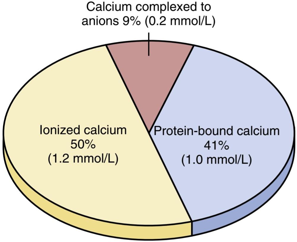
Figure 80-1. Distribution of ionized calcium (Ca2+), diffusible but un-ionized calcium complexed to anions, and non diffusible protein- bound calcium in blood plasma.
شکل 80-1. توزیع کلسیم یونیزه شده (+Ca2)، کلسیم قابل انتشار اما غیریونیزه کمپلکس به آنیون ها و کلسیم غیر قابل انتشار متصل به پروتئین در پلاسمای خون.
INORGANIC PHOSPHATE IN THE EXTRACELLULAR FLUIDS
Inorganic phosphate in the plasma is mainly in two forms, HPO4 and H2PO4. The concentration of HPO4 is about 1.05 mmol/L, and the concentration of H2PO4 is about 0.26 mmol/L. When the total quantity of phosphate in the extracellular fluid rises, so does the quantity of each of these two types of phosphate ions. Furthermore, when the pH of the extracellular fluid becomes more acidic, there is a relative increase in H2PO4 and a decrease in HPO4 whereas the opposite occurs when the extracellular fluid becomes alkaline. These relations were presented in the discussion of acid-base balance in Chapter 31.
فسفات غیر آلی در مایعات خارج سلولی
فسفات معدنی موجود در پلاسما عمدتاً به دو شکل HPO4 و H2PO4 است. غلظت HPO4 حدود 1.05 mmol/L و غلظت H2PO4 حدود 0.26 mmol/L است. هنگامی که مقدار کل فسفات در مایع خارج سلولی افزایش می یابد، مقدار هر یک از این دو نوع یون فسفات نیز افزایش می یابد. علاوه بر این، هنگامی که PH مایع خارج سلولی اسیدی تر می شود، افزایش نسبی در H2PO4 و کاهش HPO4 وجود دارد در حالی که برعکس زمانی که مایع خارج سلولی قلیایی می شود اتفاق می افتد. این روابط در بحث تعادل اسید و باز در فصل 31 ارائه شد.
Because it is difficult to determine chemically the exact quantities of HPO4 and H2PO4 in the blood, ordinarily the total quantity of phosphate is expressed in terms of milligrams of phosphorus per deciliter (100 milliliters) of blood. The average total quantity of inorganic phosphorus represented by both phosphate ions is about 4 mg/dl, varying between normal limits of 3 to 4 mg/dl in adults and 4 to 5 mg/dl in children.
از آنجایی که از نظر شیمیایی تعیین مقادیر دقیق HPO4 و H2PO4 در خون دشوار است، معمولاً مقدار کل فسفات بر حسب میلی گرم فسفر در دسی لیتر (100 میلی لیتر) خون بیان می شود. مقدار متوسط کل فسفر معدنی که توسط هر دو یون فسفات نشان داده می شود حدود 4 میلی گرم در دسی لیتر است که بین حد نرمال 3 تا 4 میلی گرم در دسی لیتر در بزرگسالان و 4 تا 5 میلی گرم در دسی لیتر در کودکان متغیر است.
NONBONE PHYSIOLOGICAL EFFECTS OF ALTERED CALCIUM AND PHOSPHATE CONCENTRATIONS IN THE BODY FLUIDS
Changing the level of phosphate in the extracellular fluid from far below normal to two to three times normal does not cause major immediate effects on the body. In contrast, even slight increases or decreases of calcium ion in the extracellular fluid can cause extreme immediate physiological effects. In addition, chronic hypocalcemia or hypophosphatemia greatly decreases bone mineralization, as is explained later in the chapter.
اثرات فیزیولوژیکی غیر استخوانی تغییر غلظت کلسیم و فسفات در مایعات بدن
تغییر سطح فسفات در مایع خارج سلولی از بسیار کمتر از حد طبیعی به دو تا سه برابر طبیعی، اثرات فوری فوری بر بدن ایجاد نمی کند. در مقابل، حتی افزایش یا کاهش جزئی یون کلسیم در مایع خارج سلولی می تواند اثرات فیزیولوژیکی شدید و فوری ایجاد کند. علاوه بر این، هیپوکلسمی مزمن یا هیپوفسفاتمی تا حد زیادی معدنی شدن استخوان را کاهش می دهد، همانطور که در ادامه این فصل توضیح داده شد.
Hypocalcemia Causes Nervous System Excitement and Tetany.
When the extracellular fluid concentration of calcium ions falls below normal, the nervous system becomes progressively more excitable because of in- creased neuronal membrane permeability to sodium ions, allowing easy initiation of action potentials. At plasma calcium ion concentrations about 50% below normal, the peripheral nerve fibers become so excitable that they begin to discharge spontaneously, initiating trains of nerve impulses that pass to the peripheral skeletal muscles to elicit tetanic muscle contraction. Consequently, hypocalcemia causes tetany. It also occasionally causes seizures because of its action of increasing excitability in the brain.
هیپوکلسمی باعث تحریک سیستم عصبی و کزاز می شود.
هنگامی که غلظت مایع خارج سلولی یونهای کلسیم به زیر نرمال میرسد، سیستم عصبی به دلیل افزایش نفوذپذیری غشای عصبی به یونهای سدیم، به تدریج تحریکپذیرتر میشود و امکان شروع آسان پتانسیل عمل را فراهم میکند. در غلظتهای یون کلسیم پلاسما حدود 50 درصد کمتر از حد طبیعی، رشتههای عصبی محیطی آنقدر تحریکپذیر میشوند که بهطور خود به خود شروع به تخلیه میکنند و قطارهایی از تکانههای عصبی را آغاز میکنند که برای ایجاد انقباض عضلانی کزاز به عضلات اسکلتی محیطی میرسند. در نتیجه هیپوکلسمی باعث کزاز می شود. همچنین گاهی اوقات به دلیل افزایش تحریک پذیری در مغز باعث تشنج می شود.
Figure 80-2 shows tetany in the hand, which usually occurs before tetany develops in most other parts of the body. This is called carpopedal spasm.
شکل 80-2 کزاز را در دست نشان می دهد که معمولاً قبل از ایجاد کزاز در اکثر قسمت های دیگر بدن رخ می دهد. این اسپاسم کارپوپدال نامیده می شود.
Tetany ordinarily occurs when the blood concentration of calcium falls from its normal level of 9.4 mg/dl to about 6 mg/dl, which is only 35% below the normal calcium concentration, and it is usually lethal at about 4 mg/dl.
کزاز معمولاً زمانی اتفاق میافتد که غلظت کلسیم خون از سطح طبیعی خود یعنی mg/dl 9.4 به حدود 6 mg/dl برسد که تنها 35 درصد کمتر از غلظت طبیعی کلسیم است و معمولاً در حدود 4 mg/dl کشنده است.
In laboratory animals, extreme hypocalcemia can cause other effects that are seldom evident in patients, such as marked dilation of the heart, changes in cellular enzyme activities, increased membrane permeability in some cells (in addition to nerve cells), and impaired blood clotting.
در حیوانات آزمایشگاهی، هیپوکلسمی شدید می تواند عوارض دیگری ایجاد کند که به ندرت در بیماران مشهود است، مانند اتساع قابل توجه قلب، تغییر در فعالیت آنزیم های سلولی، افزایش نفوذپذیری غشاء در برخی سلول ها (علاوه بر سلول های عصبی)، و اختلال در لخته شدن خون.
Hypercalcemia Depresses Nervous System and Muscle Activity. When calcium concentration in the body fluids rises above normal, the nervous system becomes depressed and reflex activities of the central nervous system are sluggish. Also, increased calcium ion concentration decreases the QT interval of the heart and causes lack of appetite and constipation, probably because of depressed contractility of the muscle walls of the gastrointestinal tract.
هیپرکلسمی سیستم عصبی و فعالیت عضلانی را کاهش می دهد. هنگامی که غلظت کلسیم در مایعات بدن از حد طبیعی بالاتر می رود، سیستم عصبی افسرده می شود و فعالیت های بازتابی سیستم عصبی مرکزی کند می شود. همچنین، افزایش غلظت یون کلسیم، فاصله QT قلب را کاهش می دهد و باعث بی اشتهایی و یبوست می شود که احتمالاً به دلیل کاهش انقباض دیواره های عضلانی دستگاه گوارش است.
These depressive effects begin to appear when the blood level of calcium rises above about 12 mg/dl, and they can become marked as the calcium level rises above 15 mg/dl. When the calcium concentration rises above about 17 mg/dl in the blood, calcium phosphate crystals are likely to precipitate throughout the body; this condition is discussed later in connection with parathyroid poisoning.
این اثرات افسردگی زمانی ظاهر می شوند که سطح کلسیم خون از حدود 12 میلی گرم در دسی لیتر بالاتر می رود و با افزایش سطح کلسیم به بالای 15 میلی گرم در دسی لیتر می توانند مشخص شوند. هنگامی که غلظت کلسیم در خون از حدود 17 میلی گرم در دسی لیتر بالاتر می رود، کریستال های فسفات کلسیم احتمالاً در سراسر بدن رسوب می کنند. این وضعیت بعداً در رابطه با مسمومیت پاراتیروئید مورد بحث قرار می گیرد.
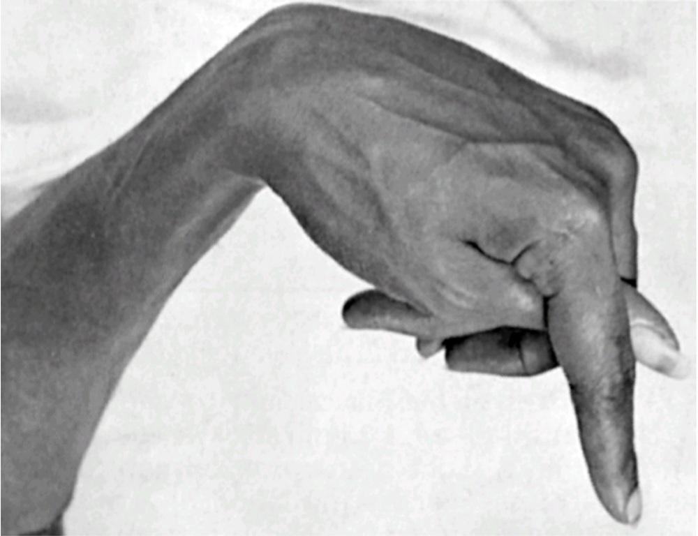
Figure 80-2. Hypocalcemic tetany in the hand, called carpopedal spasm.
شکل 80-2. کزاز هیپوکلسمیک در دست، به نام اسپاسم کارپوپدال.
ABSORPTION AND EXCRETION OF CALCIUM AND PHOSPHATE
Intestinal Absorption and Fecal Excretion of Calcium and Phosphate. The usual rates of intake are approximately 1000 mg/day each for calcium and phosphorus, about the amounts in 1 liter of milk. Normally, divalent cations such as calcium ions are poorly absorbed from the intestines. However, as discussed later, vitamin D promotes calcium absorption by the intestines, and about 35% (350 mg/day) of the ingested calcium is usually absorbed; the remaining calcium in the intestine is excreted in the feces. An additional 250 mg/day of calcium enters the intestines via secreted gastrointestinal juices and sloughed mucosal cells. Thus, about 90% (900 mg/day) of the daily intake of calcium is excreted in the feces (Figure 80-3).
جذب و دفع کلسیم و فسفات
جذب روده ای و دفع مدفوع کلسیم و فسفات. نرخ معمول مصرف تقریباً 1000 میلی گرم در روز برای کلسیم و فسفر، تقریباً در 1 لیتر شیر است. به طور معمول، کاتیون های دو ظرفیتی مانند یون های کلسیم به خوبی از روده ها جذب نمی شوند. با این حال، همانطور که بعداً بحث شد، ویتامین D جذب کلسیم توسط روده ها را تقویت می کند و حدود 35٪ (350 میلی گرم در روز) از کلسیم مصرفی معمولاً جذب می شود. کلسیم باقی مانده در روده از طریق مدفوع دفع می شود. روزانه 250 میلی گرم کلسیم اضافی از طریق شیره های ترشح شده گوارشی و سلول های مخاطی ترشح شده وارد روده می شود. بنابراین، حدود 90٪ (900 میلی گرم در روز) از کلسیم دریافتی روزانه از طریق مدفوع دفع می شود (شکل 80-3).
Intestinal absorption of phosphate occurs easily. Except for the portion of phosphate that is excreted in the feces in combination with non absorbed calcium, almost all the dietary phosphate is absorbed into the blood from the gut and later excreted in the urine.
جذب روده ای فسفات به راحتی انجام می شود. به جز قسمتی از فسفات که از طریق مدفوع همراه با کلسیم غیر جذبی دفع می شود، تقریباً تمام فسفات رژیم غذایی از روده جذب خون شده و بعداً از طریق ادرار دفع می شود.
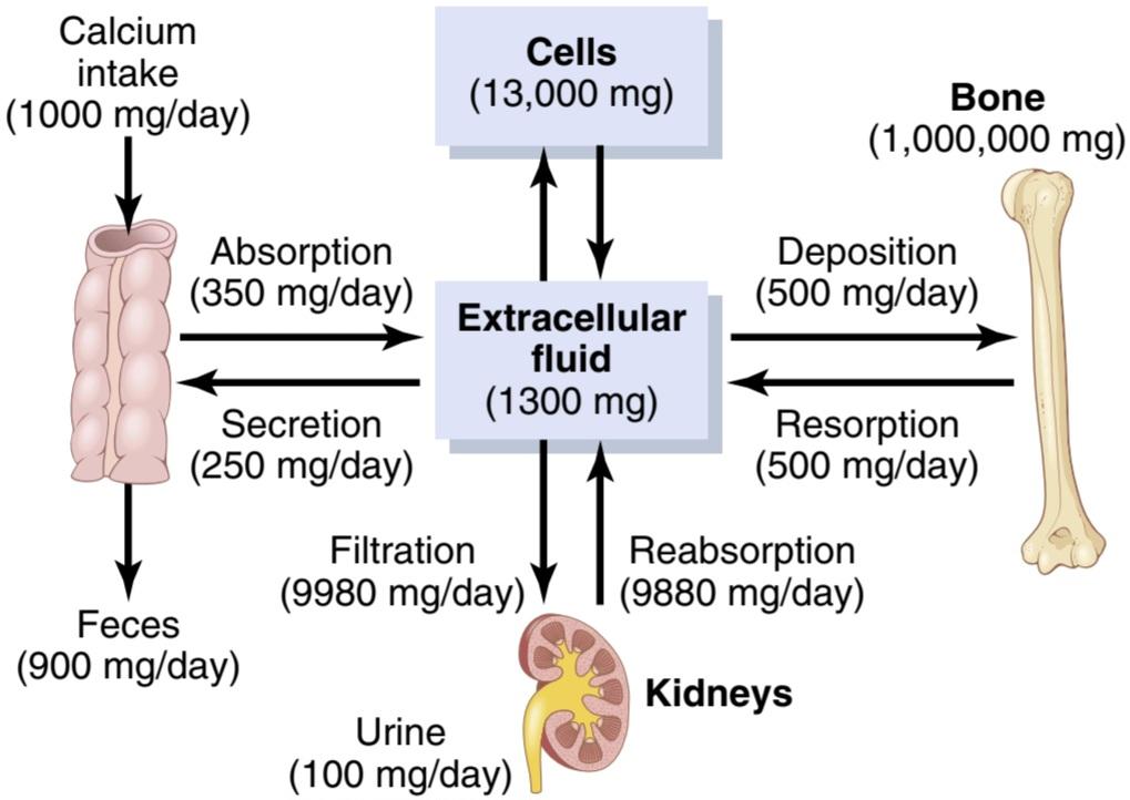
Figure 80-3. Overview of calcium exchange between different tissue compartments in a person ingesting 1000 mg of calcium per day. Note that most of the ingested calcium is normally eliminated in the feces, although the kidneys have the capacity to excrete large amounts by reducing tubular reabsorption of calcium.
شکل 80-3. مروری بر تبادل کلسیم بین بخش های مختلف بافت در فردی که روزانه 1000 میلی گرم کلسیم مصرف می کند. توجه داشته باشید که بیشتر کلسیم خورده شده معمولاً از طریق مدفوع دفع می شود، اگرچه کلیه ها ظرفیت دفع مقادیر زیادی را با کاهش بازجذب لوله ای کلسیم دارند.
Renal Excretion of Calcium and Phosphate. Approximately 10% (100 mg/day) of the ingested calcium is excreted in the urine. About 41% of the plasma calcium is bound to plasma proteins and is therefore not filtered by the glomerular capillaries. The remainder is combined with anions such as phosphate (9%) or ionized (50%) and filtered through the glomeruli into the renal tubules.
دفع کلیوی کلسیم و فسفات. تقریباً 10٪ (100 میلی گرم در روز) از کلسیم مصرف شده از طریق ادرار دفع می شود. حدود 41 درصد از کلسیم پلاسما به پروتئین های پلاسما متصل می شود و بنابراین توسط مویرگ های گلومرولی فیلتر نمی شود. باقیمانده با آنیون هایی مانند فسفات (9٪) یا یونیزه شده (50٪) ترکیب می شود و از طریق گلومرول ها به داخل لوله های کلیوی فیلتر می شود.
Normally, the renal tubules reabsorb 99% of the filtered calcium, and about 100 mg/day are excreted in the urine (see Chapter 30 for further discussion of renal calcium excretion). Approximately 90% of the calcium in the glomerular filtrate is reabsorbed in the proximal tubules, loops of Henle, and early distal tubules.
به طور معمول، لوله های کلیوی 99 درصد کلسیم فیلتر شده را بازجذب می کنند و حدود 100 میلی گرم در روز از طریق ادرار دفع می شود (برای بحث بیشتر در مورد دفع کلسیم کلیوی به فصل 30 مراجعه کنید). تقریباً 90 درصد کلسیم موجود در فیلتر گلومرولی در لولههای پروگزیمال، حلقههای هنله و لولههای دیستال اولیه بازجذب میشود.
In the late distal tubules and early collecting ducts, reabsorption of the remaining 10% is more variable, depending on the calcium ion concentration in the blood.
در لوله های انتهایی دیستال و مجاری جمع آوری اولیه، بازجذب 10 درصد باقیمانده بسته به غلظت یون کلسیم در خون متغیرتر است.
When calcium concentration is low, this reabsorption is great, and almost no calcium is lost in the urine. Conversely, even a minute increase in blood calcium ion concentration above normal increases calcium excretion markedly. We shall see later in this chapter that the most important factor controlling this reabsorption of calcium in the distal portions of the nephron, and therefore controlling the rate of calcium excretion, is PTH.
وقتی غلظت کلسیم کم باشد، این بازجذب عالی است و تقریباً هیچ کلسیمی در ادرار از دست نمیرود. برعکس، حتی یک دقیقه افزایش غلظت یون کلسیم خون بالاتر از حد طبیعی، دفع کلسیم را به طور قابل توجهی افزایش می دهد. بعداً در این فصل خواهیم دید که مهم ترین عامل کنترل کننده این بازجذب کلسیم در قسمت های انتهایی نفرون و در نتیجه کنترل سرعت دفع کلسیم، PTH است.
Renal phosphate excretion is controlled by an overflow mechanism, as explained in Chapter 30. That is, when phosphate concentration in the plasma is below the critical value of about 1 mmol/L, all the phosphate in the glomerular filtrate is reabsorbed and no phosphate is lost in the urine. Above this critical concentration, however, the rate of phosphate loss is directly proportional to the additional increase. Thus, the kidneys regulate the phosphate concentration in the extracellular fluid by altering the rate of phosphate excretion in accordance with the plasma phosphate concentration and the rate of phosphate filtration by the kidneys.
همانطور که در فصل 30 توضیح داده شد، دفع فسفات کلیوی توسط مکانیسم سرریز کنترل می شود. یعنی زمانی که غلظت فسفات در پلاسما کمتر از مقدار بحرانی حدود 1 میلی مول در لیتر باشد، تمام فسفات موجود در فیلتر گلومرولی دوباره جذب می شود و هیچ فسفاتی در ادرار از دست نمی رود. با این حال، بالاتر از این غلظت بحرانی، نرخ از دست دادن فسفات به طور مستقیم با افزایش اضافی متناسب است. بنابراین، کلیه ها با تغییر سرعت دفع فسفات مطابق با غلظت فسفات پلاسما و سرعت فیلتراسیون فسفات توسط کلیه ها، غلظت فسفات را در مایع خارج سلولی تنظیم می کنند.
However, as discussed later in this chapter, PTH can greatly increase phosphate excretion by the kidneys, thereby playing an important role in the control of plasma phosphate and calcium concentrations.
با این حال، همانطور که بعداً در این فصل مورد بحث قرار گرفت، PTH می تواند دفع فسفات توسط کلیه ها را تا حد زیادی افزایش دهد و در نتیجه نقش مهمی در کنترل غلظت فسفات و کلسیم پلاسما ایفا کند.
BONE AND ITS RELATIONSHIP TO EXTRACELLULAR CALCIUM AND PHOSPHATE
There are two general type of bony tissue-cortical (compact) and trabecular (spongy) bone (Figure 80-4). Cortical bone forms the hard outer (cortex) layer, is much denser than trabecular bone, and accounts for about 80% of the total bone mass of the human skeleton. Cortical bone is especially thick in the shaft of long bones, such as those in the legs, which support the weight of the entire body.
استخوان و ارتباط آن با کلسیم و فسفات خارج سلولی
دو نوع کلی از بافت استخوانی وجود دارد – استخوان قشر (فشرده) و ترابکولار (اسفنجی) (شکل 80-4). استخوان کورتیکال لایه بیرونی سخت (قشر) را تشکیل می دهد، بسیار متراکم تر از استخوان ترابکولار است و حدود 80 درصد از کل توده استخوانی اسکلت انسان را تشکیل می دهد. استخوان کورتیکال مخصوصاً در ساقه استخوانهای بلند، مانند استخوانهای پا که وزن کل بدن را تحمل میکنند، ضخیم است.
Trabecular bone accounts for about 20% of bone mass and is found in the interior of skeletal bones. It is much more porous than cortical bone and is usually located at the ends of long bones, near joints and in the interior of vertebrae. Trabecular bone contains lattice-shaped units with bony spicules (trabeculae) that branch and unite with one another form an irregular meshwork. The spaces between the trabeculae are filled with red bone mar- row where hematopoiesis, the production of blood cells, occurs. The rates of synthesis and resorption, and therefore bone turnover rate, are much higher for trabecular bone than for cortical bone.
استخوان ترابکولار حدود 20 درصد از توده استخوانی را تشکیل می دهد و در داخل استخوان های اسکلتی یافت می شود. این استخوان بسیار متخلخل تر از استخوان قشر مغز است و معمولاً در انتهای استخوان های بلند، نزدیک مفاصل و در قسمت داخلی مهره ها قرار دارد. استخوان ترابکولار شامل واحدهای شبکهای شکل با اسپیکولهای استخوانی (ترابکول) است که منشعب شده و با یکدیگر متحد میشوند و شبکهای نامنظم را تشکیل میدهند. فضاهای بین ترابکول ها با مغز استخوان قرمز پر می شود که در آن خون سازی، تولید سلول های خونی رخ می دهد. نرخ سنتز و جذب، و در نتیجه نرخ گردش استخوان، برای استخوان ترابکولار بسیار بیشتر از استخوان قشر مغز است.
Bone is composed of a tough organic matrix that is greatly strengthened by deposits of calcium salts. Average cortical bone contains by weight about 30% matrix and 70% salts. Newly formed bone may have a considerably higher percentage of matrix in relation to salts.
استخوان از یک ماتریکس آلی سخت تشکیل شده است که توسط رسوبات نمک های کلسیم بسیار تقویت می شود. متوسط استخوان کورتیکال از نظر وزنی حدود 30 درصد ماتریکس و 70 درصد نمک دارد. استخوان تازه تشکیل شده ممکن است درصد ماتریکس بسیار بالاتری نسبت به نمک ها داشته باشد.

Figure 80-4. Cortical (compact) and trabecular (spongy) bone.
شکل 80-4. استخوان کورتیکال (فشرده) و ترابکولار (اسفنجی).
Organic Matrix of Bone. The organic matrix of bone is 90% to 95% collagen fibers, and the remainder is a homogeneous gelatinous medium called ground sub- stance. The collagen fibers extend primarily along the lines of tensional force and give bone its powerful tensile strength.
ماتریکس ارگانیک استخوان. ماتریکس آلی استخوان 90 تا 95 درصد فیبرهای کلاژن است و باقیمانده یک محیط ژلاتینی همگن به نام ماده زمینی است. الیاف کلاژن عمدتاً در امتداد خطوط نیروی کششی گسترش یافته و به استخوان استحکام کششی قدرتمندی می بخشد.
The ground substance is composed of extracellular fluid plus proteoglycans, especially chondroitin sulfate and hyaluronic acid. These proteoglycans help control the deposition of calcium salts and are important in bone repair after injury, although some of their functions are still unclear.
ماده آسیاب شده از مایع خارج سلولی به اضافه پروتئوگلیکان ها، به ویژه کندرویتین سولفات و اسید هیالورونیک تشکیل شده است. این پروتئوگلیکان ها به کنترل رسوب نمک های کلسیم کمک می کنند و در ترمیم استخوان پس از آسیب مهم هستند، اگرچه برخی از عملکردهای آنها هنوز نامشخص است.
Bone Salts. The crystalline salts deposited in the organic matrix of bone are composed principally of calcium and phosphate. The formula for the major crystalline salt, known as hydroxyapatite, is as follows:
نمک های استخوان. نمک های کریستالی که در ماتریکس آلی استخوان رسوب می کنند، عمدتاً از کلسیم و فسفات تشکیل شده اند. فرمول نمک کریستالی اصلی، معروف به هیدروکسی آپاتیت، به شرح زیر است:

Each crystal-about 400 angstroms (Å) long, 10 to 30 A thick, and 100 A-is shaped like a long, flat plate. The relative ratio of calcium to phosphorus can vary markedly under different nutritional conditions, with the calcium to phosphorus ratio on a weight basis varying between 1.3 and 2.0.
هر کریستال با طول حدود 400 آنگستروم (Å)، ضخامت 10 تا 30 آمپر و 100 A- به شکل یک صفحه بلند و مسطح است. نسبت نسبی کلسیم به فسفر می تواند در شرایط تغذیه ای مختلف به طور قابل توجهی متفاوت باشد، به طوری که نسبت کلسیم به فسفر بر اساس وزن بین 1.3 و 2.0 متغیر است.
Magnesium, sodium, potassium, and carbonate ions are also present among the bone salts, although x-ray diffraction studies fail to show definite crystals formed by them. Therefore, they are believed to be conjugated to the hydroxyapatite crystals rather than organized into distinct crystals of their own. This ability of many types of ions to conjugate to bone crystals extends to many ions normally foreign to bone, such as strontium, uranium, plutonium, the other transuranic elements, lead, gold, and other heavy metals. Deposition of radioactive substances in the bone can cause prolonged irradiation of the bone tissues, and if a sufficient amount is deposited, an osteogenic sarcoma (bone cancer) may eventually develop.
یونهای منیزیم، سدیم، پتاسیم و کربنات نیز در میان نمکهای استخوان وجود دارند، اگرچه مطالعات پراش اشعه ایکس بلورهای مشخصی را نشان نمیدهند که توسط آنها تشکیل شده است. بنابراین، اعتقاد بر این است که آنها به کریستال های هیدروکسی آپاتیت کونژوگه می شوند تا اینکه در کریستال های مجزای خود سازماندهی شوند. این توانایی بسیاری از انواع یونها برای ترکیب شدن با کریستالهای استخوان به بسیاری از یونهایی که معمولاً برای استخوان بیگانه هستند، مانند استرانسیم، اورانیوم، پلوتونیوم، سایر عناصر فرااورانی، سرب، طلا و سایر فلزات سنگین گسترش مییابد. رسوب مواد رادیواکتیو در استخوان می تواند باعث تابش طولانی مدت بافت های استخوانی شود و اگر مقدار کافی رسوب کند، در نهایت ممکن است سارکوم استخوانی (سرطان استخوان) ایجاد شود.
Tensile and Compressional Strength of Bone. Each collagen fiber of cortical (compact) bone is com- posed of repeating periodic segments every 640 Å along its length; hydroxyapatite crystals lie adjacent to each segment of the fiber and are bound tightly to it. This intimate bonding prevents “shear” in the bone; that is, it prevents the crystals and collagen fibers from slipping out of place, which is essential in providing strength to the bone. In addition, the segments of adjacent collagen fibers overlap, also causing hydroxyapatite crystals to be overlapped like bricks keyed to one another in a brick wall.
استحکام کششی و فشاری استخوان. هر فیبر کلاژن استخوان قشری (فشرده) از قطعات دوره ای تکرار شونده هر 640 A در طول آن تشکیل شده است. کریستال های هیدروکسی آپاتیت در مجاورت هر بخش از فیبر قرار دارند و محکم به آن متصل می شوند. این پیوند صمیمی از “برش” در استخوان جلوگیری می کند. یعنی از لیز خوردن کریستالها و فیبرهای کلاژن از جای خود جلوگیری میکند که برای استحکام استخوان ضروری است. علاوه بر این، بخشهای الیاف کلاژن مجاور با هم همپوشانی دارند و همچنین باعث میشوند که کریستالهای هیدروکسی آپاتیت مانند آجرهایی که در یک دیوار آجری به یکدیگر کلید خوردهاند همپوشانی داشته باشند.
The collagen fibers of bone, like those of tendons, have great tensile strength, whereas the calcium salts have great compressional strength. These combined properties plus the degree of bondage between the collagen fibers and the crystals provide a bony structure that has both extreme tensile strength and compressional strength.
فیبرهای کلاژن استخوان، مانند تاندونها، دارای استحکام کششی بالایی هستند، در حالی که نمکهای کلسیم دارای قدرت فشاری بالایی هستند. این ویژگیهای ترکیبی بهعلاوه درجه اسارت بین الیاف کلاژن و کریستالها ساختار استخوانی را ایجاد میکنند که هم استحکام کششی و هم مقاومت فشاری بالایی دارد.
PRECIPITATION AND ABSORPTION OF CALCIUM AND PHOSPHATE IN BONE-EQUILIBRIUM WITH THE EXTRACELLULAR FLUIDS
Hydroxyapatite Does Not Precipitate in Extracellular Fluid Despite Supersaturation of Calcium and Phosphate lons. The concentrations of calcium and phosphate ions in extracellular fluid are considerably greater than those required to cause precipitation of hydroxyapatite. However, inhibitors are present in almost all tissues of the body, as well as in plasma, to prevent precipitation; one such inhibitor is pyrophosphate. Therefore, hydroxyapatite crystals fail to precipitate in normal tissues except in bone despite the state of supersaturation of the ions.
رسوب و جذب کلسیم و فسفات در تعادل استخوانی با مایعات خارج سلولی
هیدروکسی آپاتیت با وجود فوق اشباع کلسیم و فسفات لون در مایع خارج سلولی رسوب نمی کند. غلظت یون های کلسیم و فسفات در مایع خارج سلولی به طور قابل توجهی بیشتر از غلظت های مورد نیاز برای ایجاد رسوب هیدروکسی آپاتیت است. با این حال، مهارکنندهها تقریباً در تمام بافتهای بدن و همچنین در پلاسما برای جلوگیری از بارش وجود دارند. یکی از این مهارکننده ها پیروفسفات است. بنابراین، کریستال های هیدروکسی آپاتیت با وجود حالت فوق اشباع یون ها، در بافت های نرمال به جز در استخوان نمی توانند رسوب کنند.
Mechanism of Bone Calcification. The initial stage in bone production is secretion of collagen molecules (called collagen monomers) and ground substance (mainly proteoglycans) by osteoblasts. The collagen monomers polymerize rapidly to form collagen fibers; the resultant tissue becomes osteoid, a cartilage-like material differing from cartilage in that calcium salts readily precipitate in it. As the osteoid is formed, some of the osteoblasts become entrapped in the osteoid and become quiescent. At this stage, they are called osteocytes.
مکانیسم کلسیفیکاسیون استخوان. مرحله اولیه در تولید استخوان، ترشح مولکول های کلاژن (موسوم به مونومرهای کلاژن) و ماده زمین (عمدتا پروتئوگلیکان ها) توسط استئوبلاست ها است. مونومرهای کلاژن به سرعت پلیمریزه می شوند و فیبرهای کلاژن را تشکیل می دهند. بافت حاصل تبدیل به استوئید می شود، ماده ای غضروف مانند که با غضروف متفاوت است زیرا نمک های کلسیم به راحتی در آن رسوب می کنند. با تشکیل استئوئید، برخی از استئوبلاست ها در استوئید به دام افتاده و ساکن می شوند. در این مرحله به آنها استئوسیت می گویند.
Within a few days after the osteoid is formed, calcium salts begin to precipitate on the surfaces of the collagen fibers. The precipitates first appear at intervals along each collagen fiber, forming minute nidi that rapidly multiply and grow over a period of days and weeks into the finished product, hydroxyapatite crystals.
در عرض چند روز پس از تشکیل استوئید، نمک های کلسیم شروع به رسوب بر روی سطوح رشته های کلاژن می کنند. رسوبات ابتدا در فواصل زمانی در امتداد هر فیبر کلاژن ظاهر میشوند و نیدیهای کوچکی را تشکیل میدهند که به سرعت تکثیر میشوند و در طی چند روز و هفته رشد میکنند و به محصول نهایی یعنی کریستالهای هیدروکسی آپاتیت تبدیل میشوند.
The initial calcium salts to be deposited are not hydroxyapatite crystals but amorphous compounds (non crystalline), a mixture of salts such as CaHPO4 × 2H2O, Ca3(PO4)2 × 3H2O, and others. Then, by a process of substitution and addition of atoms, or reabsorption and reprecipitation, these salts are converted into the hydroxyapatite crystals over a period of weeks or months. A few percent may remain permanently in the amorphous form, which is important because these amorphous salts can be absorbed rapidly when there is a need for extra calcium in the extracellular fluid.
نمک های کلسیم اولیه که قرار است ته نشین شوند، کریستال های هیدروکسی آپاتیت نیستند، بلکه ترکیبات آمورف (غیر کریستالی)، مخلوطی از نمک هایی مانند CaHPO4 × 2H2O، Ca3(PO4)2 × 3H2O، و غیره هستند. سپس با فرآیند جایگزینی و افزودن اتمها، یا بازجذب و رسوب مجدد، این نمکها در طی چند هفته یا ماه به کریستالهای هیدروکسی آپاتیت تبدیل میشوند. چند درصد ممکن است به طور دائمی به شکل آمورف باقی بمانند، که مهم است زیرا این نمک های آمورف می توانند به سرعت جذب شوند، زمانی که نیاز به کلسیم اضافی در مایع خارج سلولی وجود دارد.
Although the mechanism that causes calcium salts to be deposited in the osteoid is not fully understood, regulation of this process appears to depend to a great extent on pyrophosphate, which inhibits hydroxyapatite crystallization and calcification of the bone. The levels of pyro- phosphate, in turn, are regulated by at least three other molecules. One of the most important of these molecules is a substance called tissue-nonspecific alkaline phosphatase (TNAP), which breaks down pyrophosphate and keeps its levels in check so that bone calcification can occur as needed. TNAP is secreted by osteoblasts into the osteoid to neutralize pyrophosphate, and once pyro- phosphate has been neutralized, the natural affinity of the collagen fibers for calcium salts causes hydroxyapatite crystallization. The importance of TNAP in bone mineralization is illustrated by the finding that mice with genetic deficiency of TNAP, which causes pyrophosphate levels to rise too high, are born with soft bones that are not adequately calcified.
اگرچه مکانیسمی که باعث رسوب نمک های کلسیم در استوئید می شود به طور کامل شناخته نشده است، به نظر می رسد تنظیم این فرآیند تا حد زیادی به پیروفسفات بستگی دارد، که از کریستالیزاسیون هیدروکسی آپاتیت و کلسیفیکاسیون استخوان جلوگیری می کند. سطوح پیروفسفات به نوبه خود توسط حداقل سه مولکول دیگر تنظیم می شود. یکی از مهم ترین این مولکول ها، ماده ای به نام آلکالین فسفاتاز غیر اختصاصی بافتی (TNAP) است که پیروفسفات را تجزیه می کند و سطح آن را کنترل می کند تا کلسیفیکاسیون استخوان در صورت نیاز انجام شود. TNAP توسط استئوبلاست ها به داخل استوئید ترشح می شود تا پیروفسفات را خنثی کند و هنگامی که پیروفسفات خنثی شد، میل ترکیبی طبیعی رشته های کلاژن به نمک های کلسیم باعث کریستالیزاسیون هیدروکسی آپاتیت می شود. اهمیت TNAP در معدنی شدن استخوان با این یافته نشان میدهد که موشهایی با کمبود ژنتیکی TNAP که باعث افزایش بیش از حد سطح پیروفسفات میشود، با استخوانهای نرمی متولد میشوند که به اندازه کافی کلسیفیه نشدهاند.
The osteoblast also secretes at least two other sub- stances that regulate bone calcification: (1) nucleotide pyrophosphatase phosphodiesterase 1 (NPP1), which pro- duces pyrophosphate outside the cells, and (2) ankylosis protein (ANK), which contributes to the extracellular pool of pyrophosphate by transporting it from the interior to the surface of the cell. Deficiencies of NPP1 or ANK cause decreased extracellular pyrophosphate and excessive calcification of bone, such as bone spurs, or even calcification of other tissues such as tendons and ligaments of the spine, which occurs in people with a form of arthritis called ankylosing spondylitis.
استئوبلاست همچنین حداقل دو ماده دیگر را ترشح می کند که کلسیفیکاسیون استخوان را تنظیم می کند: (1) نوکلئوتید پیروفسفاتاز فسفودی استراز 1 (NPP1) که پیروفسفات را در خارج از سلول تولید می کند و (2) پروتئین آنکیلوز (ANK) که به انتقال آن از سطح سلول خارج سلولی به حوضچه بیرون سلولی به حوضچه داخلی سلول کمک می کند. کمبود NPP1 یا ANK باعث کاهش پیروفسفات خارج سلولی و کلسیفیکاسیون بیش از حد استخوان مانند خارهای استخوانی یا حتی کلسیفیکاسیون سایر بافت ها مانند تاندون ها و رباط های ستون فقرات می شود که در افراد مبتلا به نوعی آرتریت به نام اسپوندیلیت آنکیلوزان رخ می دهد.
Precipitation of Calcium in Non osseous Tissues Under Abnormal Conditions. Although calcium salts usually do not precipitate in normal tissues besides bone, under abnormal conditions, they can precipitate. For example, they precipitate in arterial walls in arteriosclerosis and cause the arteries to become bonelike tubes. Likewise, calcium salts frequently deposit in degenerating tissues or in old blood clots. Presumably, in these cases, the inhibitor factors that normally prevent deposition of calcium salts disappear from the tissues, thereby allowing precipitation.
رسوب کلسیم در بافتهای غیر استخوانی در شرایط غیر طبیعی. اگرچه نمک های کلسیم معمولاً علاوه بر استخوان در بافت های طبیعی رسوب نمی کنند، اما در شرایط غیر طبیعی، می توانند رسوب کنند. به عنوان مثال، آنها در دیواره شریان در تصلب شرایین رسوب می کنند و باعث می شوند که عروق به لوله های استخوانی تبدیل شوند. به همین ترتیب، نمک های کلسیم اغلب در بافت های در حال تخریب یا در لخته های خون قدیمی رسوب می کنند. احتمالاً در این موارد، عوامل بازدارنده که به طور معمول از رسوب نمکهای کلسیم جلوگیری میکنند از بافتها ناپدید میشوند و در نتیجه امکان بارش را فراهم میکنند.
CALCIUM EXCHANGE BETWEEN BONE AND EXTRACELLULAR FLUID
If soluble calcium salts are injected intravenously, calcium ion concentration may increase immediately to high levels. However, within 30 to 60 minutes, calcium ion concentration returns to normal. Likewise, if large quantities of calcium ions are removed from the circulating body fluids, the calcium ion concentration again returns to normal within 30 minutes to about 1 hour. These effects result largely from the fact that the bone contains a type of exchangeable calcium that is always in equilibrium with calcium ions in the extracellular fluids.
تبادل کلسیم بین استخوان و مایع خارج سلولی
اگر نمک های کلسیم محلول به صورت داخل وریدی تزریق شوند، غلظت یون کلسیم ممکن است بلافاصله به سطوح بالا افزایش یابد. با این حال، در عرض 30 تا 60 دقیقه، غلظت یون کلسیم به حالت عادی باز می گردد. به همین ترتیب، اگر مقادیر زیادی یون کلسیم از مایعات در گردش بدن خارج شود، غلظت یون کلسیم دوباره در عرض 30 دقیقه تا حدود 1 ساعت به حالت عادی باز می گردد. این اثرات عمدتاً ناشی از این واقعیت است که استخوان حاوی نوعی کلسیم قابل تعویض است که همیشه با یونهای کلسیم موجود در مایعات خارج سلولی در تعادل است.
A small portion of this exchangeable calcium is also the calcium found in all tissue cells, especially in highly permeable types of cells such as those of the liver and the gastrointestinal tract. However, most of the exchange- able calcium is in the bone, normally comprising about 0.4% to 1% of the total bone calcium. This calcium is deposited in the bones in a form of readily mobilizable salt such as CaHPO4 and other amorphous calcium salts.
بخش کوچکی از این کلسیم قابل تبادل نیز کلسیمی است که در تمام سلولهای بافتی، به ویژه در انواع سلولهای بسیار نفوذپذیر مانند سلولهای کبد و دستگاه گوارش یافت میشود. با این حال، بیشتر کلسیم قابل تعویض در استخوان است که معمولاً حدود 0.4٪ تا 1٪ از کل کلسیم استخوان را شامل می شود. این کلسیم به شکل نمکی که به راحتی قابل حرکت است مانند CaHPO4 و سایر نمک های کلسیم آمورف در استخوان ها رسوب می کند.
This exchangeable calcium provides a rapid buffering mechanism to keep calcium ion concentration in the extracellular fluids from rising to excessive levels or falling to low levels under transient conditions of excess or decreased availability of calcium.
این کلسیم قابل تبادل، مکانیسم بافری سریعی را برای حفظ غلظت یون کلسیم در مایعات خارج سلولی از افزایش به سطوح بیش از حد یا کاهش به سطوح پایین در شرایط گذرا وجود بیش از حد یا کاهش در دسترس بودن کلسیم فراهم می کند.
DEPOSITION AND RESORPTION OF BONE-REMODELING OF BONE
Deposition of Bone by the Osteoblasts. Bone is continually being deposited by osteoblasts, and it is continually being resorbed where osteoclasts are active (Figure 80-5). Osteoblasts are found on the outer surfaces of the bones and in the bone trabecular cavities. A small amount of osteoblastic activity occurs continually in all living bones (on ≈4% of all surfaces at any given time in an adult), so at least some new bone is being formed constantly.
رسوب و جذب استخوان بازسازی استخوان
رسوب استخوان توسط استئوبلاست ها. استخوان به طور مداوم توسط استئوبلاست ها رسوب می کند و به طور مداوم در جایی که استئوکلاست ها فعال هستند جذب می شود (شکل 80-5). استئوبلاست ها در سطوح خارجی استخوان ها و در حفره های ترابکولار استخوان یافت می شوند. مقدار کمی از فعالیت های استئوبلاستیک به طور مداوم در تمام استخوان های زنده (در ≈4٪ از تمام سطوح در هر زمان معین در یک بزرگسال) رخ می دهد، بنابراین حداقل مقداری استخوان جدید دائماً در حال تشکیل است.
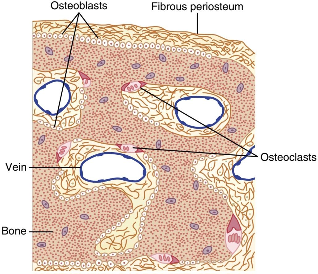
Figure 80-5. Osteoblastic and osteoclastic activity in the same bone.
شکل 80-5. فعالیت استئوبلاستیک و استئوکلاستیک در یک استخوان.
Resorption of Bone-Function of the Osteoclasts. Bone is also being continually resorbed in the presence of osteoclasts, which are large, phagocytic, multinucleated cells (containing as many as 50 nuclei) that are derivatives of monocytes or monocyte-like cells formed in the bone marrow. The osteoclasts are normally active on less than 1% of the bone surfaces of an adult and, as discussed later, PTH controls the bone resorptive activity of osteoclasts.
تحلیل عملکرد استخوانی استئوکلاست ها. استخوان همچنین در حضور استئوکلاستها که سلولهای فاگوسیتی بزرگ و چند هستهای هستند (حاوی 50 هسته) که مشتقاتی از مونوسیتها یا سلولهای مونوسیتمانند تشکیلشده در مغز استخوان هستند، پیوسته جذب میشود. استئوکلاست ها معمولاً در کمتر از 1 درصد از سطوح استخوانی یک فرد بالغ فعال هستند و همانطور که بعداً بحث شد، PTH فعالیت جذب استخوان استئوکلاست ها را کنترل می کند.
Histologically, bone absorption occurs immediately adjacent to the osteoclasts. The mechanism of this resorption is believed to be the following: The osteoclasts send out villus-like projections toward the bone, form- ing a ruffled border adjacent to the bone (Figure 80-6). The villi secrete two types of substances: (1) proteolytic enzymes, released from the lysosomes of the osteoclasts, and (2) several acids, including citric acid and lactic acid, released from the mitochondria and secretory vesicles. The enzymes digest or dissolve the organic matrix of the bone, and the acids cause dissolution of the bone salts. The osteoclastic cells also imbibe minute particles of bone matrix and crystals by phagocytosis, eventually also dis-soluting these particles and releasing the products into the blood.
از نظر بافت شناسی، جذب استخوان بلافاصله در مجاورت استئوکلاست ها اتفاق می افتد. اعتقاد بر این است که مکانیسم این تحلیل به شرح زیر است: استئوکلاستها برآمدگیهای پرز مانندی را به سمت استخوان میفرستند و یک مرز ناهموار در مجاورت استخوان تشکیل میدهند (شکل 80-6). پرزها دو نوع ماده ترشح می کنند: (1) آنزیم های پروتئولیتیک که از لیزوزوم های استئوکلاست ها آزاد می شوند و (2) چندین اسید از جمله اسید سیتریک و اسید لاکتیک که از میتوکندری و وزیکول های ترشحی آزاد می شوند. آنزیم ها ماتریکس آلی استخوان را هضم یا حل می کنند و اسیدها باعث انحلال نمک های استخوان می شوند. سلولهای استئوکلاستیک همچنین ذرات ریز ماتریکس استخوان و کریستالها را با فاگوسیتوز جذب میکنند و در نهایت این ذرات را نیز حل کرده و محصولات را در خون آزاد میکنند.
As discussed later, PTH stimulates osteoclast activity and bone resorption, but this process occurs through an indirect mechanism. The bone-resorbing osteoclast cells do not have PTH receptors. Instead, the osteoblasts signal osteoclast precursors to form mature osteoclasts. Two osteoblast proteins responsible for this signaling are receptor activator for nuclear factor KB ligand (RANKL) and macrophage colony-stimulating factor, which are both necessary for formation of mature osteoclasts.
همانطور که بعداً بحث شد، PTH فعالیت استئوکلاست ها و تحلیل استخوان را تحریک می کند، اما این فرآیند از طریق یک مکانیسم غیرمستقیم اتفاق می افتد. سلول های استئوکلاست جذب کننده استخوان گیرنده های PTH ندارند. در عوض، استئوبلاست ها به پیش سازهای استئوکلاست سیگنال می دهند تا استئوکلاست های بالغ را تشکیل دهند. دو پروتئین استئوبلاست مسئول این سیگنالدهی، فعالکننده گیرنده برای لیگاند فاکتور هستهای KB (RANKL) و فاکتور محرک کلنی ماکروفاژ هستند که هر دو برای تشکیل استئوکلاستهای بالغ ضروری هستند.
PTH binds to receptors on the adjacent osteoblasts, stimulating synthesis of RANKL, which is also called osteoprotegerin ligand (OPGL). RANKL binds to its receptors (RANK) on preosteoclast cells, causing them to differentiate into mature multinucleated osteoclasts. The mature osteoclasts then develop a ruffled border and release enzymes and acids that promote bone resorption.
PTH به گیرنده های استئوبلاست های مجاور متصل می شود و سنتز RANKL را تحریک می کند که به آن لیگاند استئوپروتجرین (OPGL) نیز می گویند. RANKL به گیرنده های خود (RANK) روی سلول های پرئوستئوکلاست متصل می شود و باعث تمایز آنها به استئوکلاست های چند هسته ای بالغ می شود. سپس استئوکلاستهای بالغ یک مرز ناهموار ایجاد میکنند و آنزیمها و اسیدهایی را آزاد میکنند که باعث جذب استخوان میشوند.
Osteoblasts also produce osteoprotegerin (OPG), sometimes called osteoclastogenesis inhibitory factor, a cytokine that inhibits bone resorption. OPG acts as a “decoy,” binding to RANKL and preventing it from inter- acting with its receptor, thereby inhibiting differentiation of preosteoclasts into mature osteoclasts that resorb bone. OPG opposes the bone resorptive activity of PTH, and mice with a genetic deficiency of OPG have severe decreases in bone mass compared with mice that have normal OPG formation.
استئوبلاست ها همچنین استئوپروتجرین (OPG) تولید می کنند که گاهی به آن فاکتور بازدارنده استئوکلاستوژنز می گویند، سیتوکینی که از جذب استخوان جلوگیری می کند. OPG به عنوان یک “فریب” عمل می کند، به RANKL متصل می شود و از تعامل آن با گیرنده آن جلوگیری می کند، در نتیجه از تمایز پیش استئوکلاست ها به استئوکلاست های بالغ که استخوان را جذب می کنند، جلوگیری می کند. OPG با فعالیت جذب استخوان PTH مخالف است و موش هایی با کمبود ژنتیکی OPG در مقایسه با موش هایی که تشکیل OPG طبیعی دارند کاهش شدیدی در توده استخوانی دارند.
Although the factors regulating OPG are not well understood, vitamin D and PTH appear to stimulate production of mature osteoclasts through the dual action of inhibiting OPG production and stimulating RANKL formation. Glucocorticoids also promote osteoclast activity and bone resorption by increasing RANKL production and decreasing formation of OPG. On the other hand, the hormone estrogen stimulates OPG production. The balance of OPG and RANKL produced by osteoblasts there- fore plays a major role in determining osteoclast activity and bone resorption.
اگرچه عوامل تنظیم کننده OPG به خوبی شناخته نشده اند، اما به نظر می رسد ویتامین D و PTH تولید استئوکلاست های بالغ را از طریق عملکرد دوگانه مهار تولید OPG و تحریک تشکیل RANKL تحریک می کنند. گلوکوکورتیکوئیدها همچنین با افزایش تولید RANKL و کاهش تشکیل OPG، فعالیت استئوکلاست ها و تحلیل استخوان را افزایش می دهند. از سوی دیگر، هورمون استروژن تولید OPG را تحریک می کند. بنابراین تعادل OPG و RANKL تولید شده توسط استئوبلاست ها نقش عمده ای در تعیین فعالیت استئوکلاست ها و تحلیل استخوان ایفا می کند.
The therapeutic importance of the OPG-RANKL path- way is currently being exploited. Novel drugs that mimic the action of OPG by blocking the interaction of RANKL with its receptor appear to be useful for treating bone loss in postmenopausal women and in some patients with bone cancer.
اهمیت درمانی مسیر OPG-RANKL در حال حاضر در حال بهره برداری است. به نظر می رسد داروهای جدیدی که با مسدود کردن تعامل RANKL با گیرنده آن، عملکرد OPG را تقلید می کنند، برای درمان از دست دادن استخوان در زنان یائسه و در برخی از بیماران مبتلا به سرطان استخوان مفید باشند.

Figure 80-6. Bone resorption by osteoclasts. Parathyroid hormone (PTH) binds to receptors on osteoblasts, causing them to form receptor activator for nuclear factor κB ligand (RANKL) and to release macrophage-colony stimulating factor (M-CSF). RANKL binds to RANK and M-CSF binds to its receptors on preosteoclast cells, causing them to differentiate into mature osteoclasts. PTH also decreases production of osteoprotegerin (OPG), which inhibits differentiation of preosteoclasts into mature osteoclasts by binding to RANKL and preventing it from interacting with its receptor on preosteoclasts. The mature osteoclasts develop a ruffled border and release enzymes from lysosomes, as well as acids that promote bone resorption. Osteocytes are osteoblasts that have become encased in bone matrix during bone tissue production; the osteocytes form a system of inter- connected cells that spreads throughout the bone.
شکل 80-6. تحلیل استخوان توسط استئوکلاست ها هورمون پاراتیروئید (PTH) به گیرندههای روی استئوبلاستها متصل میشود و باعث میشود آنها فعالکننده گیرنده لیگاند kB فاکتور هستهای (RANKL) و آزادسازی فاکتور محرک کلونی ماکروفاژ (M-CSF) شوند. RANKL به RANK متصل می شود و M-CSF به گیرنده های آن روی سلول های پرئوستئوکلاست متصل می شود و باعث تمایز آنها به استئوکلاست های بالغ می شود. PTH همچنین تولید استئوپروتجرین (OPG) را کاهش می دهد، که با اتصال به RANKL و جلوگیری از تعامل آن با گیرنده خود در پرئوستئوکلاست ها، از تمایز پره استئوکلاست ها به استئوکلاست های بالغ جلوگیری می کند. استئوکلاستهای بالغ یک مرز ناهموار ایجاد میکنند و آنزیمهایی را از لیزوزومها آزاد میکنند و همچنین اسیدهایی را که باعث جذب استخوان میشوند، آزاد میکنند. استئوسیت ها استئوبلاست هایی هستند که در طول تولید بافت استخوان در ماتریکس استخوان محصور شده اند. استئوسیت ها سیستمی از سلول های متصل به هم را تشکیل می دهند که در سرتاسر استخوان پخش می شوند.
Bone Deposition and Resorption Are Normally in Equilibrium. Except in growing bones, the rates of bone deposition and resorption are normally equal, so the total mass of bone remains constant. Osteoclasts usually exist in small but concentrated masses, and once a mass of osteoclasts begins to develop, it usually eats away at the bone for about 3 weeks, creating a tunnel that ranges in diameter from 0.2 to 1 millimeter and is several millimeters long. At the end of this time, the osteoclasts disappear, the tunnel is invaded by osteoblasts, and new bone begins to develop. Bone deposition continues for several months, with the new bone being laid down in successive layers of concentric circles (lamellae) on the inner surfaces of the cavity until the tunnel is filled. Deposition of new bone ceases when the bone begins to encroach on the blood vessels supplying the area. The canal through which these vessels run, called the haversian canal, is all that remains of the original cavity. Each new area of bone deposited in this way is called an osteon, as shown in Figure 80-7.
رسوب و جذب استخوان به طور معمول در تعادل هستند. به جز در استخوان های در حال رشد، سرعت رسوب و جذب استخوان معمولاً برابر است، بنابراین جرم کل استخوان ثابت می ماند. استئوکلاستها معمولاً در تودههای کوچک اما متمرکز وجود دارند و هنگامی که تودهای از استئوکلاستها شروع به رشد میکنند، معمولاً برای حدود 3 هفته استخوان را میخورند و تونلی ایجاد میکنند که قطر آن بین 0.2 تا 1 میلیمتر است و چندین میلیمتر طول دارد. در پایان این زمان، استئوکلاست ها ناپدید می شوند، تونل توسط استئوبلاست ها مورد هجوم قرار می گیرد و استخوان جدید شروع به رشد می کند. رسوب استخوان برای چندین ماه ادامه می یابد و استخوان جدید در لایه های متوالی از دایره های متحدالمرکز (لاملا) روی سطوح داخلی حفره قرار می گیرد تا اینکه تونل پر شود. رسوب استخوان جدید زمانی متوقف می شود که استخوان شروع به تجاوز به رگ های خونی تامین کننده ناحیه می کند. کانالی که این رگ ها از طریق آن عبور می کنند، به نام کانال هارسین، تنها چیزی است که از حفره اصلی باقی مانده است. همانطور که در شکل 80-7 نشان داده شده است، هر ناحیه جدیدی از استخوان که به این روش رسوب می کند، استئون نامیده می شود.

Figure 80-7. Structure of cortical bone.
شکل 80-7. ساختار استخوان قشر مغز.
Value of Continual Bone Remodeling. The continual deposition and resorption of bone have several important functions. First, bone ordinarily adjusts its strength in proportion to the degree of bone stress. Consequently, bones thicken when subjected to heavy loads. Second, even the shape of the bone can be rearranged for proper support of mechanical forces by deposition and resorption of bone in accordance with stress patterns. Third, because old bone becomes relatively brittle and weak, new organic matrix is needed as the old organic matrix degenerates. In this manner, the normal toughness of bone is maintained. Indeed, the bones of children, in whom the rates of deposition and absorption are rapid, show little brittleness in comparison with the bones of the elderly, in whom the rates of deposition and resorption are slow.
ارزش بازسازی مداوم استخوان. رسوب و جذب مداوم استخوان چندین عملکرد مهم دارد. ابتدا، استخوان معمولاً استحکام خود را متناسب با میزان استرس استخوان تنظیم می کند. در نتیجه، استخوان ها وقتی تحت بارهای سنگین قرار می گیرند، ضخیم می شوند. ثانیاً، حتی شکل استخوان را می توان برای حمایت مناسب از نیروهای مکانیکی با رسوب و جذب استخوان مطابق با الگوهای تنش بازآرایی کرد. سوم، از آنجایی که استخوان قدیمی نسبتا شکننده و ضعیف می شود، ماتریکس آلی جدید با تحلیل رفتن ماتریکس آلی قدیمی مورد نیاز است. به این ترتیب استحکام طبیعی استخوان حفظ می شود. در واقع، استخوانهای کودکانی که سرعت رسوب و جذب در آنها سریع است، در مقایسه با استخوانهای افراد مسن که سرعت رسوب و جذب در آنها کند است، شکنندگی کمی نشان میدهند.
Control of the Rate of Bone Deposition by Bone “Stress” Bone is deposited in proportion to the compressional load that the bone must carry. For example, the bones of athletes become considerably heavier than those of nonathletes. Also, if a person has one leg in a cast but continues to walk on the opposite leg, the bone of the leg in the cast becomes thin and as much as 30% decalcified within a few weeks, whereas the opposite bone remains thick and normally calcified. Therefore, continual physical stress stimulates osteoblastic deposition and calcification of bone.
کنترل میزان رسوب استخوان توسط استخوان “استرس”. استخوان متناسب با بار فشاری که استخوان باید تحمل کند رسوب می کند. به عنوان مثال، استخوان های ورزشکاران به طور قابل توجهی سنگین تر از غیر ورزشکاران می شود. همچنین، اگر فردی یک پا را در گچ داشته باشد اما به راه رفتن روی پای مخالف ادامه دهد، استخوان پای گچگیری در عرض چند هفته نازک میشود و تا 30 درصد کلسیمزدایی میشود، در حالی که استخوان مقابل ضخیم و معمولاً کلسیفیه میشود. بنابراین استرس فیزیکی مداوم رسوب استئوبلاست و کلسیفیکاسیون استخوان را تحریک می کند.
Bone stress also determines the shape of bones under certain circumstances. For example, if a long bone of the leg breaks in its center and then heals at an angle, the compression stress on the inside of the angle causes increased deposition of bone. Increased resorption occurs on the outer side of the angle where the bone is not compressed. After many years of increased deposition on the inner side of the angulated bone and resorption on the outer side, the bone can become almost straight, especially in children because of the rapid remodeling of bone at younger ages.
استرس استخوان نیز در شرایط خاصی شکل استخوان ها را تعیین می کند. به عنوان مثال، اگر یک استخوان بلند ساق در مرکز خود بشکند و سپس در یک زاویه بهبود یابد، فشار فشاری در داخل زاویه باعث افزایش رسوب استخوان می شود. افزایش جذب در سمت بیرونی زاویه ای که استخوان فشرده نشده است رخ می دهد. پس از سال ها افزایش رسوب در سمت داخلی استخوان زاویه دار و تحلیل در سمت خارجی، استخوان می تواند تقریباً صاف شود، به خصوص در کودکان به دلیل بازسازی سریع استخوان در سنین پایین تر.
Repair of a Fracture Activates Osteoblasts. Fracture of a bone in some way maximally activates all the periosteal and intraosseous osteoblasts involved in the break. Also, immense numbers of new osteoblasts are formed almost immediately from osteoprogenitor cells, which are bone stem cells in the surface tissue lining bone, called the “bone membrane.” Therefore, within a short time, a large bulge of osteoblastic tissue and new organic bone matrix, followed shortly by the deposition of calcium salts, develops be- tween the two broken ends of the bone. This area is called a callus.
ترمیم شکستگی استئوبلاست ها را فعال می کند. شکستگی استخوان به نحوی تمام استئوبلاست های پریوستال و داخل استخوانی درگیر در شکستگی را حداکثر فعال می کند. همچنین، تعداد بسیار زیادی از استئوبلاستهای جدید تقریباً بلافاصله از سلولهای استئوپروژنیتور، که سلولهای بنیادی استخوانی در استخوان پوششی بافت سطحی به نام «غشای استخوان» هستند، تشکیل میشوند. بنابراین، در مدت کوتاهی، برآمدگی بزرگی از بافت استئوبلاستیک و ماتریکس استخوان آلی جدید، به دنبال آن رسوب نمکهای کلسیم، بین دو انتهای شکسته استخوان ایجاد میشود. این ناحیه کالوس نامیده می شود.
Many orthopedic surgeons use the phenomenon of bone stress to accelerate fracture healing. This acceleration is achieved through use of special mechanical fixation apparatuses for holding the ends of the broken bone together so that the patient can continue to use the bone immediately. This use causes stress on the opposed ends of the broken bones, which accelerates osteoblastic activity at the break and often shortens convalescence.
بسیاری از جراحان ارتوپد از پدیده استرس استخوان برای تسریع در بهبود شکستگی استفاده می کنند. این تسریع با استفاده از دستگاههای فیکسکننده مکانیکی ویژه برای نگهداشتن انتهای استخوان شکسته در کنار هم بهدست میآید تا بیمار بتواند بلافاصله به استفاده از استخوان ادامه دهد. این استفاده باعث ایجاد استرس در انتهای مخالف استخوانهای شکسته میشود که باعث تسریع فعالیت استئوبلاستی در هنگام شکستگی و اغلب کوتاه شدن دوره نقاهت میشود.
VITAMIN D
Vitamin D has a potent effect to increase calcium absorption from the intestinal tract; it also has important effects on bone deposition and bone resorption, as discussed later. However, vitamin D itself is not the active substance that actually causes these effects. Instead, vitamin D must first be converted through a succession of reactions in the liver and the kidneys to the final active product, 1,25-dihydroxycholecalciferol, also called 1,25(OH)2D3. Figure 80-8 shows the succession of steps that lead to formation of this substance from vitamin D.
ویتامین D
ویتامین D اثر قوی برای افزایش جذب کلسیم از دستگاه روده دارد. همچنین اثرات مهمی بر رسوب استخوان و تحلیل استخوان دارد، همانطور که بعداً بحث شد. با این حال، ویتامین D خود ماده فعالی نیست که در واقع این اثرات را ایجاد می کند. در عوض، ویتامین D باید ابتدا از طریق واکنشهای متوالی در کبد و کلیهها به محصول فعال نهایی، 1,25-دی هیدروکسی کوله کلسیفرول، که 1،25(OH)2D3 نیز نامیده میشود، تبدیل شود. شکل 80-8 توالی مراحلی را نشان می دهد که منجر به تشکیل این ماده از ویتامین D می شود.

Figure 80-8. Activation of vitamin D3 to form 1,25- dihydroxycholecalciferol and the role of vitamin D in controlling the plasma calcium concentration.
شکل 80-8. فعال سازی ویتامین D3 برای تشکیل 1،25- دی هیدروکسی کوله کلسیفرول و نقش ویتامین D در کنترل غلظت کلسیم پلاسما.
Cholecalciferol (Vitamin D3) Is Formed in the Skin. Several compounds derived from sterols belong to the vitamin D family, and they all perform similar functions. Vitamin D3 (also called cholecalciferol) is the most important of these compounds and is formed in the skin as a result of irradiation of 7-dehydrocholesterol, a substance normally in the skin, by ultraviolet rays from the sun. Consequently, appropriate exposure to the sun prevents vitamin D deficiency. The additional vitamin D compounds that we ingest in food are identical to the cholecalciferol formed in the skin, except for the substitution of one or more atoms that do not affect their function.
کوله کلسیفرول (ویتامین D3) در پوست تشکیل می شود. چندین ترکیب مشتق شده از استرول ها به خانواده ویتامین D تعلق دارند و همگی عملکردهای مشابهی دارند. ویتامین D3 (همچنین کوله کلسیفرول نامیده می شود) مهمترین این ترکیبات است و در نتیجه تابش 7-دهیدروکلسترول، ماده ای که معمولاً در پوست وجود دارد، توسط اشعه ماوراء بنفش خورشید در پوست ایجاد می شود. در نتیجه، قرار گرفتن مناسب در معرض نور خورشید از کمبود ویتامین D جلوگیری می کند. ترکیبات اضافی ویتامین D که در غذا می خوریم با کل کلسیفرول تشکیل شده در پوست یکسان است، به جز جایگزینی یک یا چند اتم که بر عملکرد آنها تأثیر نمی گذارد.
Cholecalciferol Is Converted to 25-Hydroxycholecalciferol in the Liver. The first step in the activation of cholecalciferol is to convert it to 25-hydroxycholecalciferol, which occurs in the liver. The process is limited because the 25-hydroxycholecalciferol has a feedback inhibitory effect on the conversion reactions. This feedback effect is extremely important for two reasons.
کوله کلسیفرول در کبد به 25-هیدروکسی کوله کلسیفرول تبدیل می شود. اولین گام در فعال سازی کوله کلسیفرول تبدیل آن به 25-هیدروکسی کوله کلسیفرول است که در کبد ایجاد می شود. این فرآیند محدود است زیرا 25-هیدروکسی کوله کلسیفرول یک اثر بازدارنده بازخوردی بر واکنش های تبدیل دارد. این اثر بازخورد به دو دلیل بسیار مهم است.
First, the feedback mechanism precisely regulates the concentration of 25-hydroxycholecalciferol in the plasma, an effect that is shown in Figure 80-9. Note that intake of vitamin D3 can increase many times, and yet the concentration of 25-hydroxycholecalciferol remains nearly normal. This high degree of feedback control prevents excessive action of vitamin D when intake of vitamin D3 is altered over a wide range.
اول، مکانیسم بازخورد دقیقاً غلظت 25-هیدروکسی کوله کلسیفرول را در پلاسما تنظیم می کند، اثری که در شکل 80-9 نشان داده شده است. توجه داشته باشید که مصرف ویتامین D3 می تواند چندین برابر افزایش یابد، اما غلظت 25-هیدروکسی کوله کلسیفرول تقریباً طبیعی است. این درجه بالایی از کنترل بازخورد از عملکرد بیش از حد ویتامین D در زمانی که مصرف ویتامین D3 در محدوده وسیعی تغییر میکند، جلوگیری میکند.
Second, this controlled conversion of vitamin D3 to 25-hydroxycholecalciferol conserves the vitamin D stored in the liver for future use. Once vitamin D3 is converted, the 25-hydroxycholecalciferol persists in the body for only a few weeks, whereas in the vitamin D form, it can be stored in the liver for many months.
دوم، این تبدیل کنترل شده ویتامین D3 به 25-هیدروکسی کوله کلسیفرول، ویتامین D ذخیره شده در کبد را برای استفاده در آینده حفظ می کند. هنگامی که ویتامین D3 تبدیل می شود، 25-هیدروکسی کوله کلسیفرول تنها برای چند هفته در بدن باقی می ماند، در حالی که در فرم ویتامین D، می تواند ماه ها در کبد ذخیره شود.

Figure 80-9. Effect of increasing vitamin D3 intake on the plasma concentration of 25-hydroxycholecalciferol. This figure shows that increases in vitamin D intake, up to 2.5 times normal, have little effect on the final quantity of activated vitamin D that is formed. Deficiency of activated vitamin D occurs only at very low levels of vitamin D intake.
شکل 80-9. تأثیر افزایش دریافت ویتامین D3 بر غلظت 25-هیدروکسی کوله کلسیفرول پلاسما. این شکل نشان می دهد که افزایش دریافت ویتامین D، تا 2.5 برابر نرمال، تأثیر کمی بر مقدار نهایی ویتامین D فعال تشکیل شده دارد. کمبود ویتامین D فعال فقط در سطوح بسیار کم ویتامین D دریافت می شود.
Formation of 1,25-Dihydroxycholecalciferol in the Kidneys and Its Control by Parathyroid Hormone. Figure 80-8 also shows the conversion in the proximal tubules of the kidneys of 25-hydroxycholecalciferol to 1,25-dihydroxycholecalciferol. This latter substance is by far the most active form of vitamin D because the previous products in the scheme of Figure 80-8 have less than 1/1000 of the vitamin D effect. Therefore, in the absence of the kidneys, vitamin D loses almost all its effectiveness.
تشکیل 1،25-دی هیدروکسی کوله کلسیفرول در کلیه ها و کنترل آن توسط هورمون پاراتیروئید. شکل 80-8 نیز تبدیل 25-هیدروکسی کوله کلسیفرول در لوله های پروگزیمال کلیه ها به 1,25-دی هیدروکسی کوله کلسیفرول را نشان می دهد. این ماده اخیر تا حد زیادی فعال ترین شکل ویتامین D است زیرا محصولات قبلی در طرح شکل 80-8 کمتر از 1/1000 اثر ویتامین D را دارند. بنابراین، در غیاب کلیه ها، ویتامین D تقریباً تمام اثر خود را از دست می دهد.
Note also in Figure 80-8 that the conversion of 25-hydroxycholecalciferol to 1,25-dihydroxycholecalciferol requires PTH. In the absence of PTH, almost none of the 1,25-dihydroxycholecalciferol is formed. Therefore, PTH exerts a potent influence in determining the functional effects of vitamin D in the body.
همچنین در شکل 80-8 توجه داشته باشید که تبدیل 25-هیدروکسی کوله کلسیفرول به 1,25-دی هیدروکسی کوله کلسیفرول نیاز به PTH دارد. در غیاب PTH، تقریبا هیچ یک از 1،25-دی هیدروکسی کوله کلسیفرول تشکیل نمی شود. بنابراین، PTH تأثیر قوی در تعیین اثرات عملکردی ویتامین D در بدن دارد.
Calcium Ion Concentration Controls the Formation of 1,25 Dihydroxycholecalciferol. Figure 80-10 demonstrates that plasma concentration of 1,25-dihydroxycholecalciferol is inversely affected by the concentration of calcium in the plasma. There are two reasons for this effect. First, the calcium ion has a slight effect in preventing conversion of 25-hydroxycholecalciferol to 1,25-dihydroxycholecalciferol. Second, and even more important, as we discuss later in the chapter, the rate of PTH secretion is greatly suppressed when plasma calcium ion concentration rises above 9 to 10 mg/100 ml. Therefore, at calcium concentrations below this level, PTH promotes conversion of 25-hydroxycholecalciferol to 1,25-dihydroxycholecalciferol in the kidneys. At high- er calcium concentrations, when PTH is suppressed, 25-hydroxycholecalciferol is converted to a different compound-24,25-dihydroxycholecalciferol-that has al- most no vitamin D effect.
غلظت یون کلسیم تشکیل 1.25 دی هیدروکسی کوله کلسیفرول را کنترل می کند. شکل 80-10 نشان می دهد که غلظت 1،25-دی هیدروکسی کوله کلسیفرول پلاسما تحت تأثیر معکوس غلظت کلسیم در پلاسما قرار می گیرد. دو دلیل برای این اثر وجود دارد. اولاً، یون کلسیم در جلوگیری از تبدیل 25-هیدروکسی کوله کلسیفرول به 1،25-دی هیدروکسی کوله کلسیفرول تأثیر خفیفی دارد. دوم، و حتی مهمتر، همانطور که در ادامه این فصل بحث میکنیم، سرعت ترشح PTH زمانی که غلظت یون کلسیم پلاسما از 9 تا 10 میلیگرم در 100 میلیلیتر افزایش مییابد، تا حد زیادی سرکوب میشود. بنابراین، در غلظت های کلسیم زیر این سطح، PTH تبدیل 25-هیدروکسی کوله کلسیفرول به 1،25-دی هیدروکسی کوله کلسیفرول را در کلیه ها ترویج می کند. در غلظت های بالاتر کلسیم، وقتی PTH سرکوب می شود، 25-هیدروکسی کوله کلسیفرول به یک ترکیب متفاوت-24،25-dihydroxycholecalciferol تبدیل می شود که تقریباً هیچ اثر ویتامین D ندارد.
When plasma calcium concentration is already too high, formation of 1,25-dihydroxycholecalciferol is greatly depressed. Lack of 1,25-dihydroxycholecalciferol, in turn, decreases absorption of calcium from the intestines, bones, and renal tubules, thus causing the calcium ion concentration to fall back toward its normal level.
هنگامی که غلظت کلسیم پلاسما در حال حاضر بیش از حد بالا باشد، تشکیل 1،25-دی هیدروکسی کوله کلسیفرول به شدت کاهش می یابد. کمبود 1,25-دی هیدروکسی کوله کلسیفرول به نوبه خود جذب کلسیم را از روده ها، استخوان ها و لوله های کلیوی کاهش می دهد و در نتیجه باعث می شود که غلظت یون کلسیم به سطح طبیعی خود بازگردد.
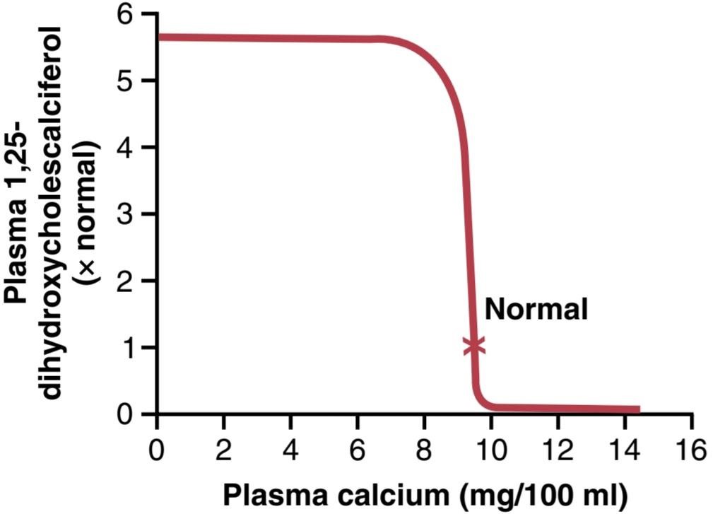
Figure 80-10. Effect of plasma calcium concentration on the plasma concentration of 1,25-dihydroxycholecalciferol. This figure shows that a slight decrease in calcium concentration below normal causes increased formation of activated vitamin D, which in turn leads to greatly increased absorption of calcium from the intestine.
شکل 80-10. اثر غلظت کلسیم پلاسما بر غلظت 1،25-دی هیدروکسی کوله کلسیفرول پلاسما. این شکل نشان می دهد که کاهش جزئی غلظت کلسیم کمتر از حد طبیعی باعث افزایش تشکیل ویتامین D فعال می شود که به نوبه خود منجر به افزایش شدید جذب کلسیم از روده می شود.
ACTIONS OF VITAMIN D
The active form of vitamin D, 1,25-dihydroxycholecalciferol, has several effects on the intestines, kidneys, and bones that increase absorption of calcium and phosphate into the extracellular fluid and contribute to feedback regulation of these substances.
اعمال ویتامین D
شکل فعال ویتامین D، 1،25-دی هیدروکسی کوله کلسیفرول، اثرات متعددی بر روده ها، کلیه ها و استخوان ها دارد که جذب کلسیم و فسفات را در مایع خارج سلولی افزایش می دهد و به تنظیم بازخورد این مواد کمک می کند.
Vitamin D receptors are present in most cells in the body and are located mainly in the nuclei of target cells. Similar to receptors for steroids and thyroid hormone, the vitamin D receptor has hormone-binding and DNA- binding domains. The vitamin D receptor forms a complex with another intracellular receptor, the retinoid-X receptor, and this complex binds to DNA and activates transcription in most cases. In some cases, however, vita- min D suppresses transcription. Although the vitamin D receptor binds several forms of cholecalciferol, its affinity for 1,25 dihydroxycholecalciferol is roughly 1000 times that for 25-hydroxycholecalciferol, which explains their relative biological potencies.
گیرنده های ویتامین D در اکثر سلول های بدن وجود دارند و عمدتاً در هسته سلول های هدف قرار دارند. مانند گیرنده های استروئیدها و هورمون تیروئید، گیرنده ویتامین D دارای حوزه های اتصال به هورمون و اتصال به DNA است. گیرنده ویتامین D با یک گیرنده درون سلولی دیگر، گیرنده رتینوئید-X، کمپلکس تشکیل می دهد و این کمپلکس به DNA متصل می شود و رونویسی را در بیشتر موارد فعال می کند. اما در برخی موارد، ویتامین D رونویسی را سرکوب می کند. اگرچه گیرنده ویتامین D به چندین شکل از کوله کلسیفرول متصل می شود، تمایل آن به 125 دی هیدروکسی کوله کلسیفرول تقریباً 1000 برابر 25-هیدروکسی کوله کلسیفرول است، که قدرت بیولوژیکی نسبی آنها را توضیح می دهد.
“Hormonal” Effect of Vitamin D to Promote Intestinal Calcium Absorption. 1,25-Dihydroxycholecalciferol functions as a type of “hormone” to promote intestinal absorption of calcium mainly by increasing, over a period of about 2 days, formation of calbindin, a calcium- binding protein, in intestinal epithelial cells. This protein functions in the brush border of these cells to transport calcium into the cell cytoplasm. The calcium then moves through the basolateral membrane of the cell by facilitated diffusion. The rate of calcium absorption is directly proportional to the quantity of this calcium-binding protein. Furthermore, this protein remains in the cells for several weeks after the 1,25-dihydroxycholecalciferol has been removed from the body, thus causing a prolonged effect on calcium absorption.
اثر “هورمونی” ویتامین D برای افزایش جذب کلسیم روده ای. 1،25-دی هیدروکسی کوله کلسیفرول به عنوان یک نوع “هورمون” عمل می کند تا جذب روده ای کلسیم را به طور عمده با افزایش در طی یک دوره زمانی حدود 2 روزه، تشکیل calbindin، یک پروتئین متصل کننده به کلسیم، در سلول های اپیتلیال روده افزایش دهد. این پروتئین در مرز برس این سلول ها برای انتقال کلسیم به سیتوپلاسم سلول عمل می کند. سپس کلسیم از طریق غشای قاعده جانبی سلول با انتشار تسهیل شده حرکت می کند. سرعت جذب کلسیم به طور مستقیم با مقدار این پروتئین متصل به کلسیم متناسب است. علاوه بر این، این پروتئین برای چندین هفته پس از حذف 1,25-دی هیدروکسی کوله کلسیفرول از بدن در سلول ها باقی می ماند و در نتیجه باعث ایجاد اثر طولانی مدت در جذب کلسیم می شود.
Other effects of 1,25-dihydroxycholecalciferol that might play a role in promoting calcium absorption are the formation of (1) a calcium-stimulated adenosine triphosphatase in the brush border of the epithelial cells and (2) an alkaline phosphatase in the epithelial cells. The precise details of all these effects are unclear.
سایر اثرات 1،25-دی هیدروکسی کوله کلسیفرول که ممکن است در افزایش جذب کلسیم نقش داشته باشد، تشکیل (1) یک آدنوزین تری فسفاتاز تحریک شده با کلسیم در مرز برس سلول های اپیتلیال و (2) یک آلکالین فسفاتاز در سلول های اپیتلیال است. جزئیات دقیق همه این اثرات نامشخص است.
Vitamin D Promotes Phosphate Absorption by the Intestines. Although phosphate is usually absorbed easily, phosphate flux through the gastrointestinal epithelium is enhanced by vitamin D. This enhancement is thought to result from a direct effect of 1,25-dihydroxycholecalciferol, but it is possible that it occurs secondarily from this hormone’s action on calcium absorption, with the calcium in turn acting as a transport mediator for the phosphate.
ویتامین D باعث افزایش جذب فسفات توسط روده می شود. اگرچه فسفات معمولاً به راحتی جذب میشود، شار فسفات از طریق اپیتلیوم گوارشی توسط ویتامین D افزایش مییابد. تصور میشود که این افزایش از اثر مستقیم 1,25-دی هیدروکسی کوله کلسیفرول ناشی میشود، اما ممکن است به طور ثانویه از اثر این هورمون بر روی عامل جذب کلسیم به عنوان عامل انتقال کلسیم رخ دهد. فسفات
Vitamin D Decreases Renal Calcium and Phosphate Excretion. Vitamin D also increases calcium and phosphate reabsorption by the epithelial cells of renal tubules, thereby tending to decrease excretion of these substances in the urine. However, this effect is weak and probably not of major importance in regulating extracellular fluid concentration of these substances.
ویتامین D باعث کاهش دفع کلسیم و فسفات از کلیه می شود. ویتامین D همچنین جذب مجدد کلسیم و فسفات توسط سلول های اپیتلیال لوله های کلیوی را افزایش می دهد و در نتیجه باعث کاهش دفع این مواد در ادرار می شود. با این حال، این اثر ضعیف است و احتمالاً در تنظیم غلظت مایع خارج سلولی این مواد اهمیت زیادی ندارد.
Effect of Vitamin D on Bone and Its Relation to Parathyroid Hormone Activity. Vitamin D plays important roles in bone resorption and bone deposition. Administration of extreme quantities of vitamin D causes resorption of bone. In the absence of vitamin D, the effect of PTH in causing bone resorption (discussed in the next section) is greatly reduced or even prevented. The mechanism of this action of vitamin D is not fully understood but is believed to result from the effect of 1,25-dihydroxycholecalciferol to increase calcium transport through cellular membranes.
تأثیر ویتامین D بر استخوان و ارتباط آن با فعالیت هورمون پاراتیروئید. ویتامین D نقش مهمی در تحلیل استخوان و رسوب استخوان دارد. مصرف مقادیر زیاد ویتامین D باعث تحلیل استخوان می شود. در غیاب ویتامین D، اثر PTH در ایجاد تحلیل استخوان (که در بخش بعدی به آن پرداخته شد) تا حد زیادی کاهش می یابد یا حتی از آن جلوگیری می شود. مکانیسم این عمل ویتامین D به طور کامل شناخته نشده است، اما اعتقاد بر این است که از اثر 1،25-دی هیدروکسی کوله کلسیفرول برای افزایش انتقال کلسیم از طریق غشای سلولی ناشی می شود.
Vitamin D in smaller quantities promotes bone calcification. One of the ways it promotes this calcification is to increase calcium and phosphate absorption from the intestines. However, even in the absence of such an increase, it enhances the mineralization of bone. Here again, the mechanism of the effect is unclear, but it probably also results from the ability of 1,25-dihydroxycholecalciferol to cause transport of calcium ions through cell membranes but in this case, perhaps in the opposite direction through the osteoblastic or osteocytic cell membranes.
ویتامین D در مقادیر کمتر باعث افزایش کلسیفیکاسیون استخوان می شود. یکی از راه هایی که این کلسیفیکاسیون را تقویت می کند، افزایش جذب کلسیم و فسفات از روده است. با این حال، حتی در صورت عدم وجود چنین افزایشی، معدنی شدن استخوان را افزایش می دهد. اینجا دوباره، مکانیسم اثر نامشخص است، اما احتمالاً ناشی از توانایی 1،25-دی هیدروکسی کوله کلسیفرول برای انتقال یون های کلسیم از طریق غشای سلولی است، اما در این مورد، شاید در جهت مخالف از طریق غشای سلولی استئوبلاست یا استئوسیتی باشد.
PARATHYROID HORMONE
PTH provides a powerful mechanism for controlling extracellular calcium and phosphate concentrations by regulating intestinal reabsorption, renal excretion, and exchange between the extracellular fluid and bone of these ions. Excess activity of the parathyroid gland causes rapid release of calcium salts from the bones, with resultant hypercalcemia in the extracellular fluid; conversely, hypofunction of the parathyroid glands causes hypocalcemia, often with resultant tetany.
هورمون پاراتیروئید
PTH یک مکانیسم قدرتمند برای کنترل غلظت کلسیم و فسفات خارج سلولی از طریق تنظیم بازجذب روده ای، دفع کلیوی و تبادل بین مایع خارج سلولی و استخوان این یون ها فراهم می کند. فعالیت بیش از حد غده پاراتیروئید باعث آزاد شدن سریع نمک های کلسیم از استخوان ها و در نتیجه هیپرکلسمی در مایع خارج سلولی می شود. برعکس، کم کاری غدد پاراتیروئید باعث هیپوکلسمی، اغلب با کزاز می شود.
Physiological Anatomy of the Parathyroid Glands.
Normally humans have four parathyroid glands, which are located immediately behind the thyroid gland-one behind each of the upper and each of the lower poles of the thyroid. Each parathyroid gland is about 6 millimeters long, 3 millimeters wide, and 2 millimeters thick and has a macroscopic appearance of dark brown fat. The parathyroid glands are difficult to locate during thyroid operations because they often look like just another lobule of the thyroid gland. For this reason, before the importance of these glands was generally recognized, total or subtotal thyroidectomy frequently resulted in removal of the parathyroid glands as well.
آناتومی فیزیولوژیکی غدد پاراتیروئید.
به طور معمول انسان ها دارای چهار غده پاراتیروئید هستند که بلافاصله در پشت غده تیروئید قرار دارند، یکی در پشت هر یک از قطب های فوقانی و هر یک از قطب های تحتانی تیروئید. هر غده پاراتیروئید حدود 6 میلی متر طول، 3 میلی متر عرض و 2 میلی متر ضخامت دارد و ظاهری ماکروسکوپی از چربی قهوه ای تیره دارد. مکان یابی غدد پاراتیروئید در حین عمل تیروئید دشوار است زیرا اغلب شبیه لوبول دیگری از غده تیروئید هستند. به همین دلیل، قبل از اینکه اهمیت این غدد به طور کلی شناخته شود، تیروئیدکتومی کامل یا ساب کل اغلب منجر به برداشتن غدد پاراتیروئید نیز می شد.
Removal of half the parathyroid glands usually causes no major physiological abnormalities. Removal of three of the four normal glands causes transient hypoparathyroidism, but even a small quantity of remaining parathyroid tissue is usually capable of hypertrophying to satisfactorily perform the function of all the glands.
برداشتن نیمی از غدد پاراتیروئید معمولاً هیچ ناهنجاری فیزیولوژیکی عمده ای ایجاد نمی کند. برداشتن سه غده از چهار غده طبیعی باعث هیپوپاراتیروئیدیسم گذرا می شود، اما حتی مقدار کمی از بافت پاراتیروئید باقیمانده معمولاً قادر به هیپرتروفی شدن است تا عملکرد همه غدد را به طور رضایت بخشی انجام دهد.
The parathyroid gland of the adult human being, shown in Figure 80-11, contains mainly chief cells and a small to moderate number of oxyphil cells, but oxyphil cells are absent in many animals and in young humans. The chief cells are believed to secrete most, if not all, of the PTH. The function of the oxyphil cells is not certain, but the cells are believed to be modified or depleted chief cells that no longer secrete hormone.
غده پاراتیروئید انسان بالغ، که در شکل 80-11 نشان داده شده است، عمدتاً حاوی سلول های اصلی و تعداد کمی تا متوسط سلول های اکسیفیل است، اما سلول های اکسیفیل در بسیاری از حیوانات و در انسان های جوان وجود ندارند. اعتقاد بر این است که سلول های اصلی بیشتر، اگر نه همه، PTH را ترشح می کنند. عملکرد سلولهای اکسیفیل مشخص نیست، اما اعتقاد بر این است که این سلولها سلولهای اصلی تغییر یافته یا ضعیف شدهاند که دیگر هورمون ترشح نمیکنند.
Chemistry of Parathyroid Hormone. PTH is first synthesized on the ribosomes in the form of a preprohormone, a polypeptide chain of 110 amino acids. The endoplasmic reticulum and Golgi apparatus first cleave this preprohormone to a prohormone with 90 amino acids and then to the hormone itself with 84 amino acids, and it is finally pack- aged in secretory granules in the cytoplasm of the cells. The final hormone has a molecular weight of about 9500. Smaller compounds with as few as 34 amino acids adjacent to the N terminus of the molecule have also been isolated from the parathyroid glands that exhibit full PTH activity. In fact, because the kidneys rapidly remove the whole 84-amino acid hormone within minutes but fail to remove many of the fragments for hours, a large share of the hormonal activity is caused by the fragments.
شیمی هورمون پاراتیروئید. PTH ابتدا بر روی ریبوزوم ها به شکل یک پیش هورمون، یک زنجیره پلی پپتیدی متشکل از 110 اسید آمینه، سنتز می شود. شبکه آندوپلاسمی و دستگاه گلژی ابتدا این پیش پروهورمون را به یک پروهورمون با 90 اسید آمینه و سپس به خود هورمون با 84 اسید آمینه می شکنند و در نهایت در گرانول های ترشحی در سیتوپلاسم سلول ها بسته بندی می شود. هورمون نهایی دارای وزن مولکولی حدود 9500 است. ترکیبات کوچکتر با 34 اسید آمینه در مجاورت انتهای N مولکول نیز از غدد پاراتیروئید که فعالیت کامل PTH را نشان می دهند جدا شده اند. در واقع، از آنجایی که کلیه ها به سرعت کل هورمون 84 آمینو اسیدی را در عرض چند دقیقه حذف می کنند، اما نمی توانند بسیاری از قطعات را برای ساعت ها از بین ببرند، سهم زیادی از فعالیت هورمونی توسط قطعات ایجاد می شود.

Figure 80-11. The four parathyroid glands lie immediately behind the thyroid gland. Almost all of the parathyroid hormone (PTH) is synthesized and secreted by the chief cells. The function of the oxyphil cells is uncertain, but they may be modified or depleted chief cells that no longer secrete PTH.
شکل 80-11. چهار غده پاراتیروئید بلافاصله در پشت غده تیروئید قرار دارند. تقریباً تمام هورمون پاراتیروئید (PTH) توسط سلولهای اصلی سنتز و ترشح میشود. عملکرد سلولهای اکسیفیل نامشخص است، اما ممکن است سلولهای اصلی تغییر یافته یا تخلیه شده باشند که دیگر PTH ترشح نمیکنند.
PARATHYROID HORMONE EFFECTS ON EXTRACELLULAR FLUID CALCIUM AND PHOSPHATE CONCENTRATIONS
Figure 80-12 shows the approximate effects on the blood calcium and phosphate concentrations caused by suddenly infusing PTH into an animal and continuing this infusion for several hours. Note that at the onset of infusion the calcium ion concentration begins to rise and reaches a plateau in about 4 hours. However, the phosphate concentration falls more rapidly than the calcium rises and reaches a depressed level within 1 or 2 hours. The rise in calcium concentration is caused mainly by two effects: (1) an effect of PTH to increase calcium and phosphate absorption from the bone, and (2) a rapid effect of 1000 PTH to decrease excretion of calcium by the kidneys. The decline in phosphate concentration is caused by a strong effect of PTH to increase renal phosphate excretion, an effect that is usually great enough to override increased phosphate absorption from the bone.
اثرات هورمون پاراتیروئید بر غلظت کلسیم و فسفات مایع خارج سلولی
شکل 80-12 اثرات تقریبی را بر غلظت کلسیم و فسفات خون نشان می دهد که ناشی از تزریق ناگهانی PTH به حیوان و ادامه این انفوزیون برای چند ساعت است. توجه داشته باشید که در شروع انفوزیون، غلظت یون کلسیم شروع به افزایش می کند و در عرض حدود 4 ساعت به فلات می رسد. با این حال، غلظت فسفات سریعتر از افزایش کلسیم کاهش می یابد و در عرض 1 یا 2 ساعت به سطح کاهش می یابد. افزایش غلظت کلسیم عمدتاً ناشی از دو اثر است: (1) اثر PTH برای افزایش جذب کلسیم و فسفات از استخوان و (2) اثر سریع 1000 PTH برای کاهش دفع کلسیم توسط کلیه ها. کاهش غلظت فسفات ناشی از اثر قوی PTH برای افزایش دفع فسفات کلیوی است، اثری که معمولاً به اندازهای عالی است که بر افزایش جذب فسفات از استخوان غلبه کند.

Figure 80-12. Approximate changes in calcium and phosphate concentrations during the first 5 hours of parathyroid hormone infusion at a moderate rate.
شکل 80-12. تغییرات تقریبی در غلظت کلسیم و فسفات در طول 5 ساعت اول انفوزیون هورمون پاراتیروئید با سرعت متوسط.
Parathyroid Hormone Mobilizes Calcium and Phosphate From Bone
PTH has two effects to mobilize calcium and phosphate from bone. One is a rapid phase that begins in minutes and increases progressively for several hours. This phase results from activation of the already existing bone cells (mainly the osteocytes) to promote calcium and phosphate release. The second phase is a much slower one, requiring several days or even weeks to become fully developed; it results from proliferation of the osteoclasts, followed by greatly increased osteoclastic resorption of bone, not merely release of the calcium phosphate salts from the bone.
هورمون پاراتیروئید کلسیم و فسفات را از استخوان به حرکت در می آورد
PTH دو اثر برای به حرکت درآوردن کلسیم و فسفات از استخوان دارد. یکی یک فاز سریع است که در عرض چند دقیقه شروع می شود و به تدریج برای چندین ساعت افزایش می یابد. این مرحله از فعال شدن سلول های استخوانی موجود (عمدتاً استئوسیت ها) برای ترویج آزادسازی کلسیم و فسفات حاصل می شود. فاز دوم بسیار کندتر است و به چندین روز یا حتی هفته نیاز دارد تا به طور کامل توسعه یابد. این ناشی از تکثیر استئوکلاستها و به دنبال آن افزایش بسیار زیاد تحلیل استئوکلاستیک استخوان است، نه صرفاً آزادسازی نمکهای فسفات کلسیم از استخوان.
Rapid Phase of Calcium and Phosphate Mobilization From Bone-Osteolysis. When large quantities of PTH are injected, calcium ion concentration in the blood begins to rise within minutes, long before any new bone cells can be developed. Histological and physiological studies have shown that PTH causes removal of bone salts from two areas in bone: (1) from the bone matrix in the vicinity of osteocytes lying within the bone and (2) in the vicinity of osteoblasts along the bone surface.
مرحله سریع بسیج کلسیم و فسفات از استئولیز استخوان. هنگامی که مقادیر زیادی PTH تزریق می شود، غلظت یون کلسیم در خون در عرض چند دقیقه شروع به افزایش می کند، مدت ها قبل از اینکه سلول های استخوانی جدید ایجاد شوند. مطالعات بافتشناسی و فیزیولوژیکی نشان دادهاند که PTH باعث حذف نمکهای استخوان از دو ناحیه در استخوان میشود: (1) از ماتریکس استخوان در مجاورت استئوسیتهای قرار گرفته در داخل استخوان و (2) در مجاورت استئوبلاستها در امتداد سطح استخوان.
One does not usually think of either osteoblasts or osteocytes functioning to mobilize bone salt, because both these types of cells are osteoblastic in nature and are normally associated with bone deposition and its calcification. However, the osteoblasts and osteocytes form a system of interconnected cells that spreads all through the bone and over all the bone surfaces except the small surface areas adjacent to the osteoclasts (see Figure 80-6). In fact, long, filmy processes extend from osteocyte to osteocyte throughout the bone structure and also connect with the surface osteocytes and osteoblasts. This extensive system is called the osteocytic membrane system, and it is believed to provide a membrane that separates the bone from the extracellular fluid.
معمولاً فکر نمیکنید استئوبلاستها یا استئوسیتها برای به حرکت درآوردن نمک استخوان عمل کنند، زیرا هر دوی این نوع سلولها ماهیتی استئوبلاستیک دارند و معمولاً با رسوب استخوان و کلسیفیکاسیون آن مرتبط هستند. با این حال، استئوبلاستها و استئوسیتها سیستمی از سلولهای به هم پیوسته را تشکیل میدهند که در سراسر استخوان و روی تمام سطوح استخوانی به جز نواحی سطح کوچک مجاور استئوکلاستها پخش میشوند (شکل 80-6 را ببینید). در واقع، فرآیندهای طولانی و لایهای از استئوسیت به استئوسیت در سراسر ساختار استخوان گسترش مییابد و همچنین با استئوسیتها و استئوبلاستهای سطحی ارتباط برقرار میکند. این سیستم گسترده سیستم غشای استخوانی نامیده می شود و اعتقاد بر این است که غشایی را فراهم می کند که استخوان را از مایع خارج سلولی جدا می کند.
Between the osteocytic membrane and the bone is a small amount of bone fluid. Experiments suggest that the osteocytic membrane pumps calcium ions from the bone fluid into the extracellular fluid, creating a calcium ion concentration in the bone fluid only one-third that in the extracellular fluid. When the osteocytic pump becomes excessively activated, the bone fluid calcium concentration falls even lower, and calcium phosphate salts are then released from the bone. This effect is called osteolysis, and it occurs without resorption of the bone’s fibrous and gel matrix. When the pump is inactivated, the bone fluid calcium concentration rises to a higher level and calcium phosphate salts are redeposited in the matrix.
بین غشای استئوسیتیک و استخوان مقدار کمی مایع استخوانی وجود دارد. آزمایشها نشان میدهند که غشای استئوسیتی، یونهای کلسیم را از مایع استخوانی به مایع خارج سلولی پمپ میکند و غلظت یون کلسیم را در مایع استخوان تنها یک سوم از مایع خارج سلولی ایجاد میکند. هنگامی که پمپ استئوسیتیک بیش از حد فعال می شود، غلظت کلسیم مایع استخوان حتی کمتر می شود و نمک های فسفات کلسیم از استخوان آزاد می شوند. این اثر استئولیز نامیده می شود و بدون تحلیل ماتریکس فیبری و ژل استخوان رخ می دهد. هنگامی که پمپ غیرفعال می شود، غلظت کلسیم مایع استخوان به سطح بالاتری می رسد و نمک های فسفات کلسیم دوباره در ماتریکس رسوب می کنند.
Where does PTH fit into this picture? First, the cell membranes of both the osteoblasts and the osteocytes have PTH receptors. PTH activates the calcium pump strongly, thereby causing rapid removal of calcium phosphate salts from the amorphous bone crystals that lie near the cells. PTH is believed to stimulate this pump by increasing the calcium permeability of the bone fluid side of the osteocytic membrane, thus allowing calcium ions to diffuse into the membrane cells from the bone fluid. Then the calcium pump on the other side of the cell mem- brane transfers the calcium ions the rest of the way into the extracellular fluid.
PTH در کجای این تصویر قرار می گیرد؟ اول، غشای سلولی هر دو استئوبلاست و استئوسیت دارای گیرنده های PTH هستند. PTH پمپ کلسیم را به شدت فعال می کند و در نتیجه باعث حذف سریع نمک های فسفات کلسیم از کریستال های استخوان آمورف که در نزدیکی سلول ها قرار دارند، می شود. اعتقاد بر این است که PTH این پمپ را با افزایش نفوذپذیری کلسیم در سمت مایع استخوانی غشای استئوسیتیک تحریک می کند، بنابراین به یون های کلسیم اجازه می دهد تا از مایع استخوان به سلول های غشایی نفوذ کنند. سپس پمپ کلسیم در سمت دیگر غشای سلولی، یون های کلسیم را بقیه راه به مایع خارج سلولی منتقل می کند.
Slow Phase of Bone Resorption and Calcium Phosphate Release-Activation of the Osteoclasts. A much better known effect of PTH and one for which the evidence is much clearer is its activation of the osteoclasts. Yet, the osteoclasts do not themselves have membrane receptors for PTH. Instead, the activated osteoblasts and osteocytes send secondary “signals” to the osteoclasts. As discussed previously, a major secondary signal is RANKL, which activates receptors on preosteoclast cells and transforms them into mature osteoclasts that set about their usual task of gobbling up the bone over a period of weeks or months.
فاز آهسته جذب استخوان و آزادسازی فسفات کلسیم-فعال شدن استئوکلاست ها. یک اثر بسیار بهتر شناخته شده PTH و یکی از مواردی که شواهد برای آن بسیار واضح تر است، فعال شدن آن در استئوکلاست ها است. با این حال، استئوکلاست ها خودشان گیرنده های غشایی برای PTH ندارند. در عوض، استئوبلاستها و استئوسیتهای فعال شده «سیگنالهای» ثانویه را به استئوکلاستها ارسال میکنند. همانطور که قبلاً گفته شد، یک سیگنال ثانویه اصلی RANKL است که گیرندههای سلولهای پرئوستئوکلاست را فعال میکند و آنها را به استئوکلاستهای بالغ تبدیل میکند که وظیفه معمول خود را برای بلعیدن استخوان در طی چند هفته یا چند ماه انجام میدهند.
Activation of the osteoclastic system occurs in two stages: (1) immediate activation of the osteoclasts that are already formed and (2) formation of new osteoclasts. Several days of excess PTH usually cause the osteoclastic system to become well developed, but it can continue to grow for months under the influence of strong PTH stimulation.
فعال سازی سیستم استئوکلاستیک در دو مرحله انجام می شود: (1) فعال شدن فوری استئوکلاست هایی که قبلاً تشکیل شده اند و (2) تشکیل استئوکلاست های جدید. چندین روز PTH بیش از حد معمولاً باعث می شود که سیستم استئوکلاستیک به خوبی توسعه یابد، اما می تواند ماه ها تحت تأثیر تحریک قوی PTH به رشد خود ادامه دهد.
After a few months of excess PTH, osteoclastic resorption of bone can lead to weakened bones and secondary stimulation of the osteoblasts that attempt to correct the weakened state. Therefore, the late effect is actually to enhance both osteoblastic and osteoclastic activity. Still, even in the late stages, there is more bone resorption than bone deposition in the presence of continued excess PTH.
پس از چند ماه PTH بیش از حد، تحلیل استئوکلاستیک استخوان میتواند منجر به ضعیف شدن استخوانها و تحریک ثانویه استئوبلاستها شود که سعی در اصلاح حالت ضعیف دارند. بنابراین، اثر دیرهنگام در واقع افزایش فعالیت استئوبلاستیک و استئوکلاستیک است. با این حال، حتی در مراحل پایانی، در حضور مداوم PTH اضافی، تحلیل استخوان بیشتر از رسوب استخوان وجود دارد.
Bone contains such great amounts of calcium in com- parison with the total amount in all the extracellular fluids (~1000 times as much) that even when PTH causes a great rise in calcium concentration in the fluids, it is impossible to discern any immediate effect on the bones. Prolonged administration or excess secretion of PTH-over a period of many months or years-finally results in very evident resorption in all the bones and even development of large cavities filled with large, multinucleated osteoclasts.
استخوان حاوی مقادیر زیادی کلسیم در مقایسه با مقدار کل در تمام مایعات خارج سلولی است (تقریبا 1000 برابر بیشتر) که حتی زمانی که PTH باعث افزایش شدید غلظت کلسیم در مایعات می شود، تشخیص هیچ اثر فوری روی استخوان ها غیرممکن است. تجویز طولانیمدت یا ترشح بیش از حد PTH- طی یک دوره چند ماهه یا چند ساله، در نهایت منجر به تحلیل بسیار واضح در تمام استخوانها و حتی ایجاد حفرههای بزرگ پر از استئوکلاستهای بزرگ و چند هستهای میشود.
Parathyroid Hormone Decreases Calcium Excretion and Increases Phosphate Excretion by the Kidneys
Administration of PTH causes rapid loss of phosphate in the urine as a result of the effect of the hormone to diminish proximal tubular reabsorption of phosphate ions.
هورمون پاراتیروئید باعث کاهش دفع کلسیم و افزایش دفع فسفات توسط کلیه ها می شود.
تجویز PTH باعث از دست دادن سریع فسفات در ادرار در نتیجه اثر هورمون برای کاهش بازجذب لوله پروگزیمال یونهای فسفات میشود.
PTH also increases renal tubular reabsorption of calcium at the same time that it diminishes phosphate reabsorption (see Chapter 30). Moreover, it increases reabsorption of magnesium ions and hydrogen ions while it decreases reabsorption of sodium, potassium, and amino acid ions in much the same way that it affects phosphate. The increased calcium reabsorption occurs mainly in the thick ascending loop of Henle and distal tubules.
PTH همچنین همزمان با کاهش بازجذب فسفات، بازجذب لولهای کلیوی کلسیم را افزایش میدهد (به فصل 30 مراجعه کنید). علاوه بر این، بازجذب یونهای منیزیم و یونهای هیدروژن را افزایش میدهد در حالی که جذب مجدد یونهای سدیم، پتاسیم و اسید آمینه را به همان روشی که بر فسفات تأثیر میگذارد کاهش میدهد. افزایش بازجذب کلسیم عمدتاً در حلقه صعودی ضخیم هنله و لولههای دیستال رخ میدهد.
Were it not for the effect of PTH on the kidneys to increase calcium reabsorption, continual loss of calcium into the urine would eventually deplete both the extracellular fluid and the bones of this mineral.
اگر اثر PTH بر کلیه ها برای افزایش بازجذب کلسیم نبود، از دست دادن مداوم کلسیم در ادرار در نهایت باعث تخلیه مایع خارج سلولی و استخوان های این ماده معدنی می شد.
Parathyroid Hormone Increases Intestinal Absorption of Calcium and Phosphate
At this point, we should be reminded again that PTH greatly enhances both calcium and phosphate absorption from the intestines by increasing formation in the kidneys of 1,25-dihydroxycholecalciferol from vitamin D, as discussed earlier in the chapter.
هورمون پاراتیروئید باعث افزایش جذب روده ای کلسیم و فسفات می شود
در این مرحله، باید دوباره یادآوری کنیم که PTH با افزایش تشکیل 1،25-دی هیدروکسی کوله کلسیفرول در کلیه ها، همانطور که قبلا در فصل بحث شد، جذب کلسیم و فسفات از روده ها را به شدت افزایش می دهد.
Cyclic Adenosine Monophosphate Mediates the Effects of Parathyroid Hormone. A large share of the effect of PTH on its target organs is mediated by the cyclic adenosine monophosphate (cAMP) second messenger mechanism. Within a few minutes after PTH administration, the concentration of cAMP increases in the osteocytes, osteoclasts, and other target cells. This cAMP in turn is probably responsible for such functions as osteo- clastic secretion of enzymes and acids to cause bone resorption and formation of 1,25-dihydroxycholecalciferol in the kidneys. Other direct effects of PTH may function independently of the second messenger mechanism.
آدنوزین مونوفسفات حلقوی تأثیرات هورمون پاراتیروئید را واسطه می کند. سهم زیادی از اثر PTH بر اندام های هدف آن توسط مکانیسم پیام رسان دوم آدنوزین مونوفسفات حلقوی (cAMP) واسطه می شود. در عرض چند دقیقه پس از تجویز PTH، غلظت cAMP در استئوسیتها، استئوکلاستها و سایر سلولهای هدف افزایش مییابد. این cAMP به نوبه خود احتمالاً مسئول عملکردهایی مانند ترشح استئوکلاستیک آنزیم ها و اسیدها برای ایجاد جذب استخوان و تشکیل 1,25-دی هیدروکسی کوله کلسیفرول در کلیه ها است. سایر اثرات مستقیم PTH ممکن است مستقل از مکانیسم پیام رسان دوم عمل کنند.
CONTROL OF PARATHYROID SECRETION BY CALCIUM ION CONCENTRATION
Even the slightest decrease in calcium ion concentration in the extracellular fluid causes the parathyroid glands to increase PTH secretion within minutes; if the decreased calcium concentration persists, the glands will hypertrophy, sometimes fivefold or more. For example, the para- thyroid glands become greatly enlarged in persons with rickets, in whom the calcium level is usually depressed only a small amount. These glands also become greatly enlarged during pregnancy, even though the decrease in calcium ion concentration in the mother’s extracellular fluid is hardly measurable, and they are greatly enlarged during lactation because calcium is used for the formation of milk.
کنترل ترشح پاراتیروئید با غلظت یون کلسیم
حتی کوچکترین کاهش غلظت یون کلسیم در مایع خارج سلولی باعث می شود که غدد پاراتیروئید در عرض چند دقیقه ترشح PTH را افزایش دهند. اگر کاهش غلظت کلسیم ادامه پیدا کند، غدد هیپرتروفی می کنند، گاهی اوقات 5 برابر یا بیشتر. به عنوان مثال، غدد پاراتیروئید در افراد مبتلا به راشیتیسم که سطح کلسیم در آنها معمولاً فقط مقدار کمی کاهش می یابد، به شدت بزرگ می شود. این غدد همچنین در دوران بارداری بسیار بزرگ می شوند، حتی اگر کاهش غلظت یون کلسیم در مایع خارج سلولی مادر به سختی قابل اندازه گیری باشد، و در دوران شیردهی به شدت بزرگ می شوند زیرا کلسیم برای تشکیل شیر استفاده می شود.
Conversely, conditions that increase the calcium ion concentration above normal cause decreased activity and reduced size of the parathyroid glands. Such conditions include (1) excess quantities of calcium in the diet, (2) increased vitamin D in the diet, and (3) bone resorption caused by factors other than PTH (e.g., disuse of the bones).
برعکس، شرایطی که غلظت یون کلسیم را بالاتر از حد طبیعی افزایش میدهند، باعث کاهش فعالیت و کاهش اندازه غدد پاراتیروئید میشوند. چنین شرایطی شامل (1) مقادیر اضافی کلسیم در رژیم غذایی، (2) افزایش ویتامین D در رژیم غذایی، و (3) تحلیل استخوان ناشی از عواملی غیر از PTH (مانند عدم استفاده از استخوان ها) است.
Changes in extracellular fluid calcium ion concentration are detected by a calcium-sensing receptor (CSR) in parathyroid cell membranes. The calcium-sensing receptor is a G protein-coupled receptor that, when stimulated by calcium ions, activates phospholipase C and increases intracellular inositol 1,4,5-triphosphate and diacylglycerol formation. This activity stimulates release of calcium from intracellular stores, which, in turn, decreases PTH secretion. Conversely, decreased extracellular fluid calcium ion concentration inhibits these pathways and stimulates PTH secretion. This process contrasts with that in many endocrine tissues in which hormone secretion is stimulated when these pathways are activated.
تغییرات در غلظت یون کلسیم مایع خارج سلولی توسط گیرنده حسگر کلسیم (CSR) در غشای سلولی پاراتیروئید تشخیص داده می شود. گیرنده حسگر کلسیم یک گیرنده جفت شده با پروتئین G است که وقتی توسط یون های کلسیم تحریک می شود، فسفولیپاز C را فعال می کند و تشکیل اینوزیتول 1،4،5-تری فسفات و دی اسیل گلیسرول داخل سلولی را افزایش می دهد. این فعالیت باعث آزاد شدن کلسیم از ذخایر درون سلولی می شود که به نوبه خود باعث کاهش ترشح PTH می شود. برعکس، کاهش غلظت یون کلسیم مایع خارج سلولی این مسیرها را مهار کرده و ترشح PTH را تحریک می کند. این فرآیند در تضاد با بسیاری از بافت های غدد درون ریز است که در آن ترشح هورمون با فعال شدن این مسیرها تحریک می شود.
Figure 80-13 shows the approximate relation between plasma calcium concentration and plasma PTH concentration. The solid red curve shows the acute effect when the calcium concentration is changed over a period of a few hours and illustrates that even small decreases in calcium concentration from the normal value can double or triple the plasma PTH. The approximate chronic effect when calcium ion concentration changes over a period of many weeks, thus allowing time for the glands to hypertrophy greatly, is shown by the dashed red line, which demonstrates that a decrease of only a fraction of a milligram per deciliter in plasma calcium concentration can double PTH secretion. This is the basis of the body’s extremely potent feedback system for long-term control of plasma calcium ion concentration.
شکل 80-13 رابطه تقریبی بین غلظت کلسیم پلاسما و غلظت PTH پلاسما را نشان می دهد. منحنی قرمز جامد اثر حاد را هنگامی که غلظت کلسیم در طی چند ساعت تغییر میکند نشان میدهد و نشان میدهد که حتی کاهش کوچک غلظت کلسیم از مقدار طبیعی میتواند PTH پلاسما را دو یا سه برابر کند. اثر مزمن تقریبی زمانی که غلظت یون کلسیم در طی یک دوره چند هفته تغییر می کند، بنابراین زمان زیادی را برای هیپرتروفی شدن غدد فراهم می کند، با خط قرمز شکسته نشان داده می شود، که نشان می دهد کاهش تنها کسری از میلی گرم در دسی لیتر در غلظت کلسیم پلاسما می تواند ترشح PTH را دو برابر کند. این اساس سیستم بازخورد بسیار قوی بدن برای کنترل طولانی مدت غلظت یون کلسیم پلاسما است.
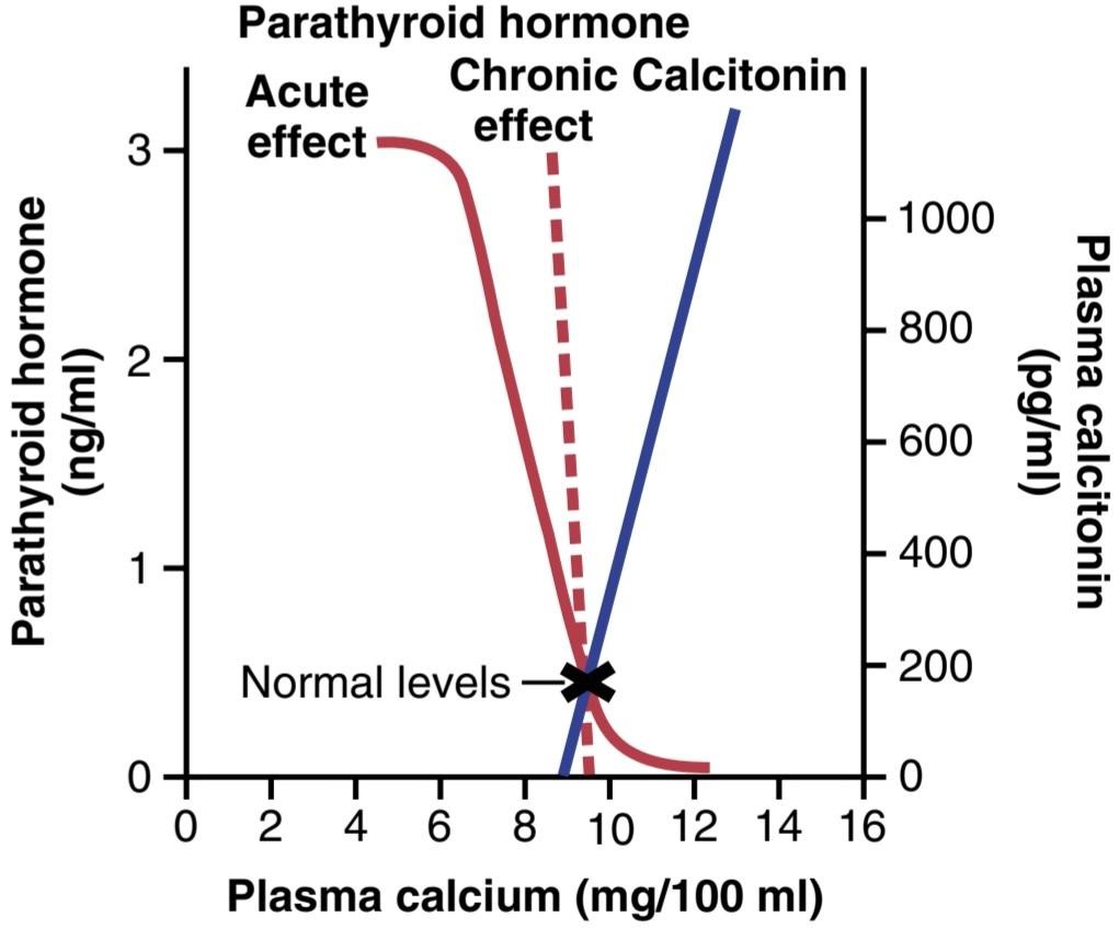
Figure 80-13. The approximate effect of plasma calcium concentration on the plasma concentrations of parathyroid hormone and calcitonin. Note especially that long-term changes in calcium concentration of only a few percentage points can cause as much as 100% change in parathyroid hormone concentration.
شکل 80-13. اثر تقریبی غلظت کلسیم پلاسما بر غلظت پلاسمایی هورمون پاراتیروئید و کلسی تونین. به خصوص توجه داشته باشید که تغییرات طولانی مدت در غلظت کلسیم تنها چند درصد می تواند باعث تغییر 100 درصدی در غلظت هورمون پاراتیروئید شود.
SUMMARY OF EFFECTS OF PARATHYROID HORMONE
Figure 80-14 summarizes the main effects of increased PTH secretion in response to decreased extracellular fluid calcium ion concentration: (1) PTH stimulates bone resorption, causing release of calcium into the extracellular fluid; (2) PTH increases calcium reabsorption and decreases phosphate reabsorption by the renal tubules, leading to decreased excretion of calcium and increased excretion of phosphate; and (3) PTH is necessary for conversion of 25-hydroxycholecalciferol to 1,25-dihydroxycholecalciferol, which, in turn, increases calcium absorption by the intestines. These actions together provide a powerful means of regulating extracellular fluid calcium concentration.
خلاصه ای از اثرات هورمون پاراتیروئید
شکل 80-14 اثرات اصلی افزایش ترشح PTH در پاسخ به کاهش غلظت یون کلسیم مایع خارج سلولی را خلاصه می کند: (1) PTH باعث تحریک تحلیل استخوان می شود و باعث آزاد شدن کلسیم در مایع خارج سلولی می شود. (2) PTH باعث افزایش بازجذب کلسیم و کاهش بازجذب فسفات توسط لوله های کلیوی می شود که منجر به کاهش دفع کلسیم و افزایش دفع فسفات می شود. و (3) PTH برای تبدیل 25-hydroxycholecalciferol به 1,25-dihydroxycholecalciferol ضروری است که به نوبه خود جذب کلسیم توسط روده ها را افزایش می دهد. این اقدامات با هم ابزار قدرتمندی برای تنظیم غلظت کلسیم مایع خارج سلولی فراهم می کند.

Figure 80-14. Summary of effects of parathyroid hormone (PTH) on bone, the kidneys, and the intestine in response to decreased extracellular fluid calcium ion concentration. CaSR, Calcium-sensing receptor.
شکل 80-14. خلاصه ای از اثرات هورمون پاراتیروئید (PTH) بر استخوان، کلیه ها و روده در پاسخ به کاهش غلظت یون کلسیم مایع خارج سلولی. CaSR، گیرنده حسگر کلسیم.
CALCITONIN
Calcitonin, a peptide hormone secreted by the thyroid gland, tends to decrease plasma calcium concentration and, in general, has effects opposite to those of PTH. However, the quantitative role of calcitonin in humans is far less than that of PTH in regulating calcium ion concentration.
کلسیتونین
کلسی تونین، یک هورمون پپتیدی که از غده تیروئید ترشح می شود، تمایل به کاهش غلظت کلسیم پلاسما دارد و به طور کلی، اثراتی برخلاف PTH دارد. با این حال، نقش کمی کلسی تونین در انسان به مراتب کمتر از PTH در تنظیم غلظت یون کلسیم است.
Synthesis and secretion of calcitonin occur in the parafollicular cells, or C cells, lying in the interstitial fluid between the follicles of the thyroid gland. These cells constitute only about 0.1% of the human thyroid gland and are the remnants of the ultimobranchial glands of fish, amphibians, reptiles, and birds. Calcitonin is a 32-amino acid peptide with a molecular weight of about 3400.
سنتز و ترشح کلسی تونین در سلول های پارافولیکولار یا سلول های C که در مایع بینابینی بین فولیکول های غده تیروئید قرار دارند، رخ می دهد. این سلول ها تنها حدود 0.1 درصد از غده تیروئید انسان را تشکیل می دهند و بقایای غدد غدد نشاطی ماهی، دوزیستان، خزندگان و پرندگان هستند. کلسی تونین یک پپتید 32 اسید آمینه با وزن مولکولی حدود 3400 است.
Increased Plasma Calcium Concentration Stimulates Calcitonin Secretion. The primary stimulus for calcitonin secretion is increased extracellular fluid calcium ion concentration. In contrast, PTH secretion is stimulated by decreased calcium concentration.
افزایش غلظت کلسیم پلاسما باعث تحریک ترشح کلسی تونین می شود. محرک اولیه برای ترشح کلسی تونین افزایش غلظت یون کلسیم مایع خارج سلولی است. در مقابل، ترشح PTH با کاهش غلظت کلسیم تحریک می شود.
In young animals, but much less so in older animals and in humans, an increase in plasma calcium concentration of about 10% causes an immediate twofold or more increase in calcitonin secretion rate, which is shown by the blue line in Figure 80-13. This increase provides a second hormonal feedback mechanism for controlling plasma calcium ion concentration, but one that is relatively weak and works in a way opposite that of the PTH system.
در حیوانات جوان، اما در حیوانات مسن تر و در انسان بسیار کمتر، افزایش غلظت کلسیم پلاسما حدود 10٪ باعث افزایش فوری دو برابر یا بیشتر در سرعت ترشح کلسی تونین می شود که با خط آبی در شکل 80-13 نشان داده شده است. این افزایش دومین مکانیسم بازخورد هورمونی را برای کنترل غلظت یون کلسیم پلاسما فراهم می کند، اما مکانیزم نسبتاً ضعیف است و برخلاف سیستم PTH عمل می کند.
Calcitonin Decreases Plasma Calcium Concentration. In some young animals, calcitonin decreases blood calcium ion concentration rapidly, beginning within minutes after injection of the calcitonin, in at least two ways.
کلسی تونین غلظت کلسیم پلاسما را کاهش می دهد. در برخی از حیوانات جوان، کلسی تونین غلظت یون کلسیم خون را به سرعت کاهش می دهد، که در عرض چند دقیقه پس از تزریق کلسی تونین، حداقل به دو روش شروع می شود.
1. The immediate effect is to decrease the absorptive activities of the osteoclasts and possibly the osteo- lytic effect of the osteocytic membrane throughout the bone, thus shifting the balance in favor of deposition of calcium in the exchangeable bone calcium salts. This effect is especially significant in young animals because of the rapid interchange of absorbed and deposited calcium.
1. اثر فوری کاهش فعالیت های جذبی استئوکلاست ها و احتمالاً اثر استئولیتیک غشای استئوسیتیک در سرتاسر استخوان است، در نتیجه تعادل به نفع رسوب کلسیم در نمک های کلسیم استخوانی قابل تعویض تغییر می کند. این اثر به ویژه در حیوانات جوان به دلیل تبادل سریع کلسیم جذب شده و رسوب شده قابل توجه است.
2. The second and more prolonged effect of calcitonin is to decrease formation of new osteoclasts. Also, because osteoclastic resorption of bone leads secondarily to osteoblastic activity, decreased numbers of osteoclasts are followed by decreased numbers of osteoblasts. Therefore, over a long period, the net result is reduced osteoclastic and osteoblastic activity and, consequently, little prolonged effect on plasma calcium ion concentration. That is, the effect on plasma calcium is mainly a transient one, lasting for a few hours to a few days at most.
2. اثر دوم و طولانی تر کلسی تونین کاهش تشکیل استئوکلاست های جدید است. همچنین، از آنجا که تحلیل استئوکلاستیک استخوان به طور ثانویه منجر به فعالیت استئوبلاستی میشود، کاهش تعداد استئوکلاستها با کاهش تعداد استئوبلاستها همراه است. بنابراین، در یک دوره طولانی، نتیجه خالص کاهش فعالیت استئوکلاستیک و استئوبلاستیک و در نتیجه اثر طولانی مدت کمی بر غلظت یون کلسیم پلاسما است. یعنی اثر روی کلسیم پلاسما عمدتاً گذرا است و از چند ساعت تا حداکثر چند روز ادامه دارد.
Calcitonin also has minor effects on calcium handling in the kidney tubules and the intestines. Again, the effects are opposite those of PTH, but they appear to be of such little importance that they are seldom considered.
کلسی تونین همچنین تأثیرات جزئی بر مدیریت کلسیم در لوله های کلیه و روده دارد. باز هم، اثرات مخالف PTH هستند، اما به نظر میرسد آنقدر اهمیت کمی دارند که به ندرت مورد توجه قرار میگیرند.
Calcitonin Has a Weak Effect on Plasma Calcium Concentration in Adult Humans. The reason for the weak effect of calcitonin on plasma calcium is twofold. First, any initial reduction of the calcium ion concentration caused by calcitonin leads within hours to a powerful stimulation of PTH secretion, which almost overrides the calcitonin effect. When the thyroid gland is removed, and calcitonin is no longer secreted, the long-term blood calcium ion concentration is not measurably altered, which again demonstrates the overriding effect of the PTH system of control.
کلسی تونین اثر ضعیفی بر غلظت کلسیم پلاسما در انسان بالغ دارد. دلیل اثر ضعیف کلسی تونین بر پلاسماکلسیم دو برابر است. اول، هر گونه کاهش اولیه غلظت یون کلسیم ناشی از کلسی تونین در عرض چند ساعت منجر به تحریک قوی ترشح PTH می شود که تقریباً اثر کلسی تونین را نادیده می گیرد. وقتی غده تیروئید برداشته میشود و کلسیتونین دیگر ترشح نمیشود، غلظت یون کلسیم خون در درازمدت بهطور قابل اندازهگیری تغییر نمیکند، که دوباره تأثیر مهم سیستم کنترل PTH را نشان میدهد.
Second, in the adult human, the daily rates of absorption and deposition of calcium are small, and even after the rate of absorption is slowed by calcitonin, this still has only a small effect on plasma calcium ion concentration. The effect of calcitonin in children is much greater because bone remodeling occurs rapidly in children, with absorption and deposition of calcium as great as 5 grams or more per day-equal to 5 to 10 times the total calcium in all the extracellular fluid. Also, in certain bone diseases, such as Paget’s disease, in which osteoclastic activity is greatly accelerated, calcitonin has a much more potent effect of reducing the calcium absorption.
ثانیاً، در انسان بالغ، نرخ روزانه جذب و رسوب کلسیم اندک است و حتی پس از کاهش سرعت جذب توسط کلسی تونین، این باز هم تأثیر کمی بر غلظت یون کلسیم پلاسما دارد. اثر کلسی تونین در کودکان بسیار بیشتر است زیرا بازسازی استخوان در کودکان به سرعت اتفاق می افتد، با جذب و رسوب کلسیم به اندازه 5 گرم یا بیشتر در روز – معادل 5 تا 10 برابر کلسیم کل در تمام مایع خارج سلولی. همچنین، در برخی بیماری های استخوانی، مانند بیماری پاژه، که در آن فعالیت استئوکلاستیک بسیار تسریع می شود، کلسی تونین اثر بسیار قوی تری در کاهش جذب کلسیم دارد.
SUMMARY OF CONTROL OF CALCIUM ION CONCENTRATION
At times, the amount of calcium absorbed into or lost from the body fluids is as much as 0.3 gram in 1 hour. For example, in cases of diarrhea, several grams of calcium can be secreted in the intestinal juices, passed into the intestinal tract, and lost into the feces each day.
خلاصه کنترل غلظت یون کلسیم
در برخی مواقع، میزان کلسیم جذب شده یا از دست رفته در مایعات بدن به 0.3 گرم در 1 ساعت می رسد. به عنوان مثال، در موارد اسهال، روزانه چندین گرم کلسیم در شیره روده ترشح می شود، به مجرای روده می رود و در مدفوع دفع می شود.
Conversely, after ingestion of large quantities of calcium, particularly when there is also excess vitamin D activity, a person may absorb as much as 0.3 gram in 1 hour. This figure compares with a total quantity of calcium in all the extracellular fluid of about 1 gram. The addition or subtraction of 0.3 gram to or from such a small amount of calcium in the extracellular fluid would cause serious hypercalcemia or hypocalcemia. However, there is a first line of defense to prevent this from occurring even before the parathyroid and calcitonin hormonal feedback systems have a chance to act.
برعکس، پس از مصرف مقادیر زیادی کلسیم، به ویژه زمانی که فعالیت ویتامین D بیش از حد وجود داشته باشد، فرد ممکن است تا 0.3 گرم را در یک ساعت جذب کند. این رقم با مقدار کل کلسیم موجود در تمام مایع خارج سلولی در حدود 1 گرم مقایسه می شود. افزودن یا تفریق 0.3 گرم به یا از چنین مقدار کمی کلسیم در مایع خارج سلولی باعث هیپرکلسمی یا هیپوکلسمی جدی می شود. با این حال، اولین خط دفاعی برای جلوگیری از این اتفاق وجود دارد، حتی قبل از اینکه سیستمهای بازخورد هورمونی پاراتیروئید و کلسیتونین فرصتی برای عمل داشته باشند.
Buffer Function of the Exchangeable Calcium in Bones-The First Line of Defense. The exchangeable calcium salts in the bones, discussed earlier in this chapter, are amorphous calcium phosphate compounds, prob- ably mainly CaHPO4 or some similar compound loosely bound in the bone and in reversible equilibrium with the calcium and phosphate ions in the extracellular fluid.
عملکرد بافر کلسیم قابل تعویض در استخوان ها – اولین خط دفاعی. نمکهای کلسیم قابل تعویض در استخوانها، که قبلاً در این فصل مورد بحث قرار گرفت، ترکیبات کلسیم فسفات آمورف هستند، احتمالاً عمدتاً CaHPO4 یا برخی از ترکیبات مشابه بهطور آزاد در استخوان و در تعادل برگشتپذیر با یونهای کلسیم و فسفات در مایع خارج سلولی.
The quantity of these salts that is available for exchange is about 0.5% to 1% of the total calcium salts of the bone, which is a total of 5 to 10 grams of calcium. Because of the ease of deposition of these exchangeable salts and their ease of resolubility, an increase in the concentrations of extracellular fluid calcium and phosphate ions above normal causes immediate deposition of exchange- able salt. Conversely, a decrease in these concentrations causes immediate absorption of exchangeable salt. This reaction is rapid because the amorphous bone crystals are extremely small and their total surface area exposed to the fluids of the bone is large-perhaps 1 acre or more.
مقدار این املاح که برای تبادل در دسترس است حدود 5/0 تا 1 درصد کل املاح کلسیم استخوان است که در مجموع 5 تا 10 گرم کلسیم است. به دلیل سهولت رسوب این نمکهای تبادلی و سهولت حلپذیری آنها، افزایش غلظت یونهای کلسیم و فسفات مایع خارج سلولی بیش از حد طبیعی باعث رسوب فوری نمک قابل تعویض میشود. برعکس، کاهش این غلظت ها باعث جذب فوری نمک قابل تعویض می شود. این واکنش سریع است زیرا کریستال های استخوان آمورف بسیار کوچک هستند و سطح کل آنها که در معرض مایعات استخوان قرار می گیرد، شاید 1 هکتار یا بیشتر باشد.
In addition, about 5% of all the blood flows through the bones each minute-that is, about 1% of all the extracellular fluid each minute. Therefore, about one-half of any excess calcium that appears in the extracellular fluid is removed by this buffer function of the bones in about 70 minutes.
علاوه بر این، حدود 5 درصد از کل خون در هر دقیقه از طریق استخوان ها جریان می یابد، یعنی حدود 1 درصد از کل مایع خارج سلولی در هر دقیقه. بنابراین، تقریباً نیمی از کلسیم اضافی که در مایع خارج سلولی ظاهر میشود، توسط این عملکرد بافر استخوانها در حدود 70 دقیقه حذف میشود.
In addition to the buffer function of the bones, the mitochondria of many of the tissues of the body, especially of the liver and intestine, contain a significant amount of exchangeable calcium (~10 grams in the whole body) that provides an additional buffer system to help maintain constancy of the extracellular fluid calcium ion concentration.
علاوه بر عملکرد بافر استخوان ها، میتوکندری بسیاری از بافت های بدن، به ویژه کبد و روده، حاوی مقدار قابل توجهی کلسیم قابل تعویض (حدود 10 گرم در کل بدن) است که یک سیستم بافر اضافی برای کمک به حفظ ثبات غلظت یون کلسیم مایع خارج سلولی فراهم می کند.
Hormonal Control of Calcium Ion Concentration-The Second Line of Defense. At the same time that the exchangeable calcium mechanism in the bones is “buffering” the calcium in the extracellular fluid, both the parathyroid and the calcitonin hormonal systems are beginning to act. Within 3 to 5 minutes after an acute increase in the calcium ion concentration, the rate of PTH secretion de- creases. As already explained, this sets into play multiple mechanisms for reducing the calcium ion concentration back toward normal.
کنترل هورمونی غلظت یون کلسیم – دومین خط دفاعی. در همان زمان که مکانیسم کلسیم قابل تعویض در استخوانها کلسیم مایع خارج سلولی را بافر میکند، سیستم هورمونی پاراتیروئید و کلسیتونین شروع به کار میکنند. در عرض 3 تا 5 دقیقه پس از افزایش شدید غلظت یون کلسیم، میزان ترشح PTH کاهش می یابد. همانطور که قبلاً توضیح داده شد، این مکانیسم های متعددی را برای کاهش غلظت یون کلسیم به حالت عادی ایجاد می کند.
At the same time that PTH decreases, calcitonin increases. In young animals and possibly in young children (but probably to a smaller extent in adults), the calcitonin causes rapid deposition of calcium in the bones, and perhaps in some cells of other tissues. Therefore, in very young animals, excess calcitonin can cause a high calcium ion concentration to return to normal perhaps consider- ably more rapidly than can be achieved by the exchangeable calcium-buffering mechanism alone.
همزمان با کاهش PTH، کلسی تونین افزایش می یابد. در حیوانات جوان و احتمالاً در کودکان خردسال (اما احتمالاً به میزان کمتری در بزرگسالان)، کلسی تونین باعث رسوب سریع کلسیم در استخوان ها و شاید در برخی از سلول های بافت های دیگر می شود. بنابراین، در حیوانات بسیار جوان، کلسی تونین بیش از حد می تواند باعث شود غلظت یون کلسیم زیاد به حالت عادی بازگردد، شاید خیلی سریعتر از آن چیزی که تنها با مکانیسم بافر کلسیم قابل تعویض به دست آید.
In prolonged calcium excess or prolonged calcium deficiency, only the PTH mechanism seems to be really important in maintaining a normal plasma calcium ion concentration. When a person has a continuing deficiency of calcium in the diet, PTH can often stimulate enough calcium absorption from the bones to maintain a normal plasma calcium ion concentration for 1 year or more, but eventually, even the bones will run out of calcium. Thus, in effect, the bones are a large buffer-reservoir of calcium that can be manipulated by PTH. Yet, when the bone reservoir either runs out of calcium or, alternately, becomes saturated with calcium, the long-term control of extracellular calcium ion concentration resides almost entirely in the roles of PTH and vitamin D in controlling calcium absorption from the gut and calcium excretion in the urine.
در کمبود کلسیم اضافی یا طولانی مدت، تنها مکانیسم PTH به نظر می رسد در حفظ غلظت طبیعی یون کلسیم پلاسما واقعاً مهم باشد. هنگامی که فردی در رژیم غذایی دچار کمبود مداوم کلسیم است، PTH اغلب میتواند جذب کلسیم کافی از استخوانها را تحریک کند تا غلظت یون کلسیم پلاسما در پلاسما به مدت یک سال یا بیشتر حفظ شود، اما در نهایت حتی استخوانها نیز فاقد کلسیم میشوند. بنابراین، در واقع، استخوان ها یک بافر-مخزن بزرگ کلسیم هستند که می تواند توسط PTH دستکاری شود. با این حال، هنگامی که مخزن استخوان یا کلسیم تمام میشود یا متناوباً با کلسیم اشباع میشود، کنترل طولانیمدت غلظت یون کلسیم خارج سلولی تقریباً به طور کامل در نقش PTH و ویتامین D در کنترل جذب کلسیم از روده و دفع کلسیم در ادرار است.
Pathophysiology of Parathyroid Hormone, Vitamin D, and Bone Disease
Hypoparathyroidism
When the parathyroid glands do not secrete sufficient PTH, the osteocytic resorption of exchangeable calcium decreases, and the osteoclasts become almost totally inactive. As a result, calcium release from the bones is so depressed that the level of calcium in the body fluids decreases. Yet, because calcium and phosphates are not being released from the bone, the bone usually remains strong.
پاتوفیزیولوژی هورمون پاراتیروئید، ویتامین D و بیماری استخوان
کم کاری پاراتیروئید
هنگامی که غدد پاراتیروئید PTH کافی ترشح نمی کنند، تحلیل استئوسیتی کلسیم قابل تعویض کاهش می یابد و استئوکلاست ها تقریباً کاملاً غیرفعال می شوند. در نتیجه، ترشح کلسیم از استخوان ها به حدی کاهش می یابد که سطح کلسیم در مایعات بدن کاهش می یابد. با این حال، به دلیل اینکه کلسیم و فسفات از استخوان آزاد نمی شوند، استخوان معمولاً قوی باقی می ماند.
When the parathyroid glands are suddenly removed, the calcium level in the blood falls from the normal of 9.4 mg/dl to 6 to 7 mg/dl within 2 to 3 days and the blood phosphate concentration may double. When this low calcium level is reached, the usual signs of tetany develop. Among the muscles of the body especially sensitive to tetanic spasm are the laryngeal muscles. Spasm of these muscles obstructs respiration, which is the usual cause of death in persons with tetany unless appropriate treatment is provided.
هنگامی که غدد پاراتیروئید به طور ناگهانی برداشته می شوند، سطح کلسیم خون از حد طبیعی 9.4 میلی گرم در دسی لیتر به 6 تا 7 میلی گرم در دسی لیتر در عرض 2 تا 3 روز کاهش می یابد و غلظت فسفات خون ممکن است دو برابر شود. وقتی به این سطح پایین کلسیم رسید، علائم معمول کزاز ایجاد می شود. از جمله عضلات بدن که به ویژه به اسپاسم کزاز حساس هستند، عضلات حنجره هستند. اسپاسم این ماهیچه ها باعث انسداد تنفس می شود که علت معمول مرگ در افراد مبتلا به کزاز است مگر اینکه درمان مناسب ارائه شود.
Treatment of Hypoparathyroidism With PTH and Vitamin D. PTH is occasionally used to treat hypoparathyroidism. However, hypoparathyroidism is usually not treated with PTH because this hormone is expensive, its effect lasts for a few hours at most, and the tendency of the body to develop antibodies against it makes it progressively less and less effective.
درمان کم کاری پاراتیروئید با PTH و ویتامین D. پی تی اچ گاهی اوقات برای درمان کم کاری پاراتیروئید استفاده می شود. با این حال، هیپوپاراتیروئیدیسم معمولاً با PTH درمان نمیشود، زیرا این هورمون گران است، اثر آن حداکثر چند ساعت طول میکشد و تمایل بدن به ایجاد آنتیبادیهایی علیه آن باعث میشود که به تدریج اثر آن کمتر و کمتر شود.
In most patients with hypoparathyroidism, administration of extremely large quantities of vitamin D, along with intake of 1 to 2 grams of calcium, keeps the calcium ion concentration in a normal range. At times, it might be necessary to administer 1,25-dihydroxycholecalciferol instead of the nonactivated form of vitamin D because of its much more potent and much more rapid action. However, administration of 1,25-dihydroxycholecalciferol can also cause unwanted effects because it is sometimes difficult to prevent overactivity by this activated form of vitamin D.
در اکثر بیماران مبتلا به کم کاری پاراتیروئید، تجویز مقادیر بسیار زیاد ویتامین D، همراه با مصرف 1 تا 2 گرم کلسیم، غلظت یون کلسیم را در محدوده طبیعی نگه می دارد. گاهی ممکن است لازم باشد 1،25-دی هیدروکسی کوله کلسیفرول به جای فرم غیرفعال ویتامین D تجویز شود، زیرا عملکرد بسیار قوی تر و بسیار سریعتر آن دارد. با این حال، تجویز 1،25-دی هیدروکسی کوله کلسیفرول نیز می تواند عوارض ناخواسته ای ایجاد کند زیرا گاهی اوقات جلوگیری از فعالیت بیش از حد توسط این شکل فعال شده ویتامین D دشوار است.
Primary Hyperparathyroidism
In primary hyperparathyroidism, an abnormality of the parathyroid glands causes inappropriate, excess PTH secretion. The cause of primary hyperparathyroidism ordinarily is a tumor of one of the parathyroid glands; such tumors occur much more frequently in women than in men or children, mainly because pregnancy and lactation stimulate the parathyroid glands and therefore predispose to the development of such a tumor.
هیپرپاراتیروئیدیسم اولیه
در هیپرپاراتیروئیدیسم اولیه، ناهنجاری غدد پاراتیروئید باعث ترشح نامناسب و بیش از حد PTH می شود. علت هیپرپاراتیروئیدیسم اولیه معمولاً تومور یکی از غدد پاراتیروئید است. چنین تومورهایی در زنان بسیار بیشتر از مردان یا کودکان رخ می دهد، عمدتاً به این دلیل که بارداری و شیردهی غدد پاراتیروئید را تحریک می کند و بنابراین مستعد ایجاد چنین توموری است.
Hyperparathyroidism causes extreme osteoclastic activity in the bones, which elevates calcium ion concentration in the extracellular fluid while usually depressing the concentration of phosphate ions because of increased renal excretion of phosphate.
هیپرپاراتیروئیدیسم باعث فعالیت شدید استئوکلاستیک در استخوان ها می شود که غلظت یون کلسیم را در مایع خارج سلولی افزایش می دهد در حالی که معمولاً به دلیل افزایش دفع کلیوی فسفات، غلظت یون های فسفات را کاهش می دهد.
Bone Disease in Hyperparathyroidism. In persons with mild hyperparathyroidism new bone can be deposited rapidly enough to compensate for the increased osteoclastic resorption of bone. However, in severe hyperparathyroidism, the osteoclastic absorption soon far outstrips osteoblastic deposition, and the bone may be eaten away almost entirely. Indeed, a broken bone is often the reason a person with hyperparathyroidism seeks medical attention. Radiographs of the bone typically show extensive decalcification and, occasionally, large punched-out cystic areas of the bone that are filled with osteoclasts in the form of so-called giant cell osteoclast “tumors.” Multiple fractures of the weakened bones can result from only slight trauma, especially where cysts develop. The cystic bone disease of hyperparathyroidism is called osteitis fibrosa cystica.
بیماری استخوان در هیپرپاراتیروئیدیسم. در افراد مبتلا به هیپرپاراتیروئیدیسم خفیف، استخوان جدید میتواند با سرعت کافی برای جبران افزایش تحلیل استئوکلاستیک استخوان رسوب کند. با این حال، در هیپرپاراتیروئیدیسم شدید، جذب استئوکلاستیک به زودی بسیار بیشتر از رسوب استئوبلاستیک است و ممکن است استخوان تقریباً به طور کامل خورده شود. در واقع، شکستگی استخوان اغلب دلیلی است که فرد مبتلا به هیپرپاراتیروئیدیسم به دنبال مراقبت پزشکی است. رادیوگرافی استخوان به طور معمول دکلسیفیکاسیون گسترده و گاهی اوقات، نواحی کیستیک بزرگ استخوان را نشان می دهد که با استئوکلاست ها به شکل تومورهای به اصطلاح سلول های غول پیکر استئوکلاست پر شده است. شکستگیهای متعدد استخوانهای ضعیف شده تنها میتواند ناشی از ضربههای خفیف باشد، مخصوصاً در جایی که کیستها ایجاد میشوند. بیماری استخوان کیستیک هیپرپاراتیروئیدیسم استیت فیبروزا سیستیکا نامیده می شود.
Osteoblastic activity in the bones also increases greatly in a vain attempt to form enough new bone to make up for the old bone absorbed by the osteoclastic activity. When the osteoblasts become active, they secrete large quantities of alkaline phosphatase. Therefore, one of the important diagnostic findings in hyperparathyroidism is a high level of plasma alkaline phosphatase.
فعالیت استئوبلاستیک در استخوان ها نیز در تلاشی بیهوده برای تشکیل استخوان جدید کافی برای جبران استخوان قدیمی جذب شده توسط فعالیت استئوکلاستیک، به شدت افزایش می یابد. هنگامی که استئوبلاست ها فعال می شوند، مقادیر زیادی آلکالین فسفاتاز ترشح می کنند. بنابراین یکی از یافته های مهم تشخیصی در پرکاری پاراتیروئید، سطح بالای آلکالین فسفاتاز پلاسما است.
Effects of Hypercalcemia in Hyperparathyroidism. Hyperparathyroidism can at times cause plasma calcium level to rise to 12 to 15 mg/dl and, rarely, even higher. The effects of such elevated calcium levels, as detailed earlier in the chapter, are depression of the central and peripheral nervous systems, muscle weakness, constipation, abdominal pain, peptic ulcer, lack of appetite, and depressed relaxation of the heart during diastole.
اثرات هیپرکلسمی در پرکاری پاراتیروئید. هیپرپاراتیروئیدیسم می تواند گاهی باعث افزایش سطح کلسیم پلاسما به 12 تا 15 میلی گرم در دسی لیتر و به ندرت حتی بیشتر شود. اثرات چنین سطوح بالای کلسیم، همانطور که قبلاً در این فصل توضیح داده شد، عبارتند از: افسردگی سیستم عصبی مرکزی و محیطی، ضعف عضلانی، یبوست، درد شکم، زخم معده، بی اشتهایی، و آرام سازی افسرده قلب در طول دیاستول.
Parathyroid Poisoning and Metastatic Calcification. When, on rare occasions, extreme quantities of PTH are secreted, the level of calcium in the body fluids rises rapidly to high values. Even the extracellular fluid phosphate concentration often rises markedly instead of falling, as is usually the case, probably because the kidneys cannot excrete all the phosphate being absorbed from the bone rapidly enough. Therefore, the calcium and phosphate in the body fluids become greatly supersaturated, and calcium phosphate (CaHPO4) crystals begin to deposit in the alveoli of the lungs, the tubules of the kidneys, the thyroid gland, the acid-producing area of the stomach mucosa, and the walls of the arteries throughout the body. This extensive metastatic deposition of calcium phosphate can develop within a few days.
مسمومیت پاراتیروئید و کلسیفیکاسیون متاستاتیک. هنگامی که در موارد نادر، مقادیر بسیار زیاد PTH ترشح می شود، سطح کلسیم در مایعات بدن به سرعت به مقادیر بالایی افزایش می یابد. حتی غلظت فسفات مایع خارج سلولی اغلب به جای کاهش به طور قابل توجهی افزایش می یابد، همانطور که معمولاً اتفاق می افتد، احتمالاً به این دلیل که کلیه ها نمی توانند تمام فسفات جذب شده از استخوان را با سرعت کافی دفع کنند. بنابراین، کلسیم و فسفات موجود در مایعات بدن به شدت فوق اشباع میشوند و کریستالهای فسفات کلسیم (CaHPO4) در آلوئولهای ریهها، لولههای کلیه، غده تیروئید، ناحیه تولیدکننده اسید مخاط معده و دیوارههای بدن شروع به رسوب میکنند. این رسوب متاستاتیک گسترده فسفات کلسیم می تواند در عرض چند روز ایجاد شود.
Ordinarily, the level of calcium in the blood must rise above 17 mg/dl before there is danger of parathyroid poisoning, but once such elevation develops along with con- current elevation of phosphate, death can occur in only a few days.
به طور معمول، قبل از خطر مسمومیت با پاراتیروئید، سطح کلسیم در خون باید بیش از 17 میلی گرم در دسی لیتر باشد، اما هنگامی که چنین افزایشی همراه با افزایش همزمان فسفات ایجاد شود، مرگ ممکن است تنها در چند روز رخ دهد.
Formation of Kidney Stones in Hyperparathyroidism. Most patients with mild hyperparathyroidism show few signs of bone disease and few general abnormalities as a result of elevated levels of calcium, but they do have an extreme tendency to form kidney stones. The reason for this tendency is that the excess calcium and phosphate absorbed from the intestines or mobilized from the bones in hyperparathyroidism must eventually be excreted by the kidneys, causing a proportionate increase in the concentrations of these substances in the urine. As a result, crystals of calcium phosphate tend to precipitate in the kidney, forming calcium phosphate stones. Also, calcium oxalate stones develop because even normal levels of oxalate cause calcium precipitation at high calcium levels.
تشکیل سنگ کلیه در پرکاری پاراتیروئید. اکثر بیماران مبتلا به هیپرپاراتیروئیدیسم خفیف علائم کمی از بیماری استخوانی و ناهنجاری های عمومی کمی را در نتیجه افزایش سطح کلسیم نشان می دهند، اما تمایل شدیدی به تشکیل سنگ کلیه دارند. دلیل این تمایل این است که کلسیم و فسفات اضافی جذب شده از روده ها یا حرکت از استخوان ها در هیپرپاراتیروئیدیسم باید در نهایت توسط کلیه ها دفع شود و باعث افزایش متناسب در غلظت این مواد در ادرار شود. در نتیجه، کریستالهای فسفات کلسیم تمایل دارند در کلیه رسوب کنند و سنگهای فسفات کلسیمی را تشکیل دهند. همچنین، سنگهای اگزالات کلسیمی ایجاد میشوند زیرا حتی سطوح طبیعی اگزالات باعث رسوب کلسیم در سطوح بالای کلسیم میشود.
Because the solubility of most renal stones is slight in alkaline media, the tendency for formation of renal calculi is considerably greater in alkaline urine than in acid urine. For this reason, acidotic diets and acidic drugs are frequently used to treat renal calculi.
از آنجایی که حلالیت بیشتر سنگ های کلیوی در محیط های قلیایی اندک است، تمایل به تشکیل سنگ های کلیوی در ادرار قلیایی به طور قابل توجهی بیشتر از ادرار اسیدی است. به همین دلیل، رژیم های غذایی اسیدوتیک و داروهای اسیدی اغلب برای درمان سنگ های کلیوی استفاده می شود.
Secondary Hyperparathyroidism
In secondary hyperparathyroidism, high levels of PTH occur as a compensation for hypocalcemia rather than as a primary abnormality of the parathyroid glands. In contrast, primary hyperparathyroidism is associated with hypercalcemia.
هیپرپاراتیروئیدیسم ثانویه
در هیپرپاراتیروئیدیسم ثانویه، سطوح بالای PTH به عنوان جبران هیپوکلسمی به جای ناهنجاری اولیه غدد پاراتیروئید رخ می دهد. در مقابل، هیپرپاراتیروئیدیسم اولیه با هیپرکلسمی همراه است.
Secondary hyperparathyroidism can be caused by vitamin D deficiency or chronic renal disease in which the damaged kidneys are unable to produce sufficient amounts of the active form of vitamin D, 1,25 dihydroxycholecalciferol. As discussed in more detail in the next section, the vitamin D deficiency leads to osteomalacia (inadequate mineralization of the bones), and high levels of PTH cause absorption of the bones.
هیپرپاراتیروئیدیسم ثانویه می تواند ناشی از کمبود ویتامین D یا بیماری مزمن کلیوی باشد که در آن کلیه های آسیب دیده قادر به تولید مقادیر کافی از فرم فعال ویتامین D، 1,25 دی هیدروکسی کوله کلسیفرول نیستند. همانطور که در بخش بعدی با جزئیات بیشتر بحث شد، کمبود ویتامین D منجر به استئومالاسی (معدنی سازی ناکافی استخوان ها) می شود و سطوح بالای PTH باعث جذب استخوان ها می شود.
Rickets Caused by Vitamin D Deficiency
Rickets occurs mainly in children. It results from calcium or phosphate deficiency in the extracellular fluid, usually caused by lack of vitamin D. If the child is adequately ex- posed to sunlight, the 7-dehydrocholesterol in the skin be- comes activated by the ultraviolet rays and forms vitamin D3, which prevents rickets by promoting calcium and phosphate absorption from the intestines, as discussed earlier in this chapter.
راشیتیسم ناشی از کمبود ویتامین D
راشیتیسم عمدتا در کودکان رخ می دهد. این ناشی از کمبود کلسیم یا فسفات در مایع خارج سلولی است که معمولاً ناشی از کمبود ویتامین D است. اگر کودک به اندازه کافی در معرض نور خورشید قرار گیرد، 7-دهیدروکلسترول موجود در پوست در اثر اشعه ماوراء بنفش فعال می شود و ویتامین D3 را تشکیل می دهد که با تحریک زودرس کلسیم و افزایش کلسیم از راشیتیسم جلوگیری می کند. در این فصل
Children who remain indoors through the winter in general do not receive adequate quantities of vitamin D without some supplementation in the diet. Rickets tends to occur especially in the spring months because vitamin D formed during the preceding summer is stored in the liver and is available for use during the early winter months. In addition, calcium and phosphate mobilization from the bones can prevent clinical signs of rickets for the first few months of vitamin D deficiency.
کودکانی که در طول زمستان در خانه می مانند، به طور کلی، بدون مقداری مکمل در رژیم غذایی، مقادیر کافی ویتامین D دریافت نمی کنند. راشیتیسم معمولاً در ماههای بهار اتفاق میافتد، زیرا ویتامین D تشکیل شده در تابستان قبل در کبد ذخیره میشود و برای استفاده در ماههای اولیه زمستان در دسترس است. علاوه بر این، انتقال کلسیم و فسفات از استخوان ها می تواند از علائم بالینی راشیتیسم در چند ماه اول کمبود ویتامین D جلوگیری کند.
Plasma Concentrations of Calcium and Phosphate Decrease in Rickets. The plasma calcium concentration in rickets is only slightly depressed, but the level of phosphate is greatly depressed. This phenomenon occurs because the parathyroid glands prevent the calcium level from falling by promoting bone resorption whenever the calcium level be- gins to fall. However, no good regulatory system exists for preventing a falling level of phosphate, and the increased parathyroid activity actually increases excretion of phosphates in the urine.
غلظت کلسیم و فسفات پلاسما در راشیتیسم کاهش می یابد. غلظت کلسیم پلاسما در راشیتیسم فقط اندکی کاهش می یابد، اما سطح فسفات به شدت کاهش می یابد. این پدیده به این دلیل اتفاق میافتد که غدد پاراتیروئید با ترویج تحلیل استخوان در زمانی که سطح کلسیم شروع به کاهش میکند، از کاهش سطح کلسیم جلوگیری میکند. با این حال، هیچ سیستم تنظیم کننده خوبی برای جلوگیری از کاهش سطح فسفات وجود ندارد و افزایش فعالیت پاراتیروئید در واقع باعث افزایش دفع فسفات در ادرار می شود.
Rickets Weakens the Bones. During prolonged cases of rickets, the marked compensatory increase in PTH secretion causes extreme osteoclastic bone resorption. This in turn causes the bone to become progressively weaker and imposes marked physical stress on the bone, resulting in rapid osteoblastic activity as well. The osteoblasts lay down large quantities of osteoid, which does not become calcified because of insufficient calcium and phosphate ions. Consequently, the newly formed, uncalcified, and weak osteoid gradually takes the place of the older bone that is being resorbed.
راشیتیسم استخوان ها را ضعیف می کند. در موارد طولانی راشیتیسم، افزایش جبرانی قابل توجهی در ترشح PTH باعث تحلیل شدید استخوان استئوکلاستیک می شود. این به نوبه خود باعث می شود که استخوان به تدریج ضعیف شود و فشار فیزیکی قابل توجهی بر استخوان تحمیل کند و در نتیجه فعالیت سریع استئوبلاستی نیز انجام شود. استئوبلاستها مقادیر زیادی استوئید را میگذارند که به دلیل ناکافی بودن یونهای کلسیم و فسفات کلسیفیه نمیشوند. در نتیجه، استئویید تازه تشکیل شده، کلسیف نشده و ضعیف به تدریج جای استخوان مسن تری را می گیرد که در حال جذب است.
Tetany in Rickets. In the early stages of rickets, tetany almost never occurs because the parathyroid glands continually stimulate osteoclastic resorption of bone and, therefore, maintain an almost normal level of calcium in the extracellular fluid. However, when the bones finally become exhausted of calcium, calcium concentration may fall rapidly. As the blood level of calcium falls below 7 mg/dl, the usual signs of tetany develop and the child may die of tetanic respiratory spasm unless calcium is administered intravenously, which relieves the tetany immediately.
تتانی در راشیتیسم. در مراحل اولیه راشیتیسم، کزاز تقریباً هرگز رخ نمی دهد زیرا غدد پاراتیروئید به طور مداوم تحلیل استئوکلاستیک استخوان را تحریک می کنند و بنابراین سطح تقریباً طبیعی کلسیم را در مایع خارج سلولی حفظ می کنند. با این حال، هنگامی که استخوان ها در نهایت از کلسیم خسته می شوند، غلظت کلسیم ممکن است به سرعت کاهش یابد. با کاهش سطح کلسیم خون به زیر 7 میلی گرم در دسی لیتر، علائم معمول کزاز ایجاد می شود و کودک ممکن است بر اثر اسپاسم تنفسی کزاز بمیرد، مگر اینکه کلسیم به صورت داخل وریدی تجویز شود که بلافاصله کزاز را تسکین می دهد.
Treatment of Rickets. Treatment of rickets entails sup- plying adequate calcium and phosphate in the diet and, equally important, administering large amounts of vitamin D. If vitamin D is not administered, little calcium and phosphate are absorbed from the gut.
درمان راشیتیسم. درمان راشیتیسم مستلزم تامین کلسیم و فسفات کافی در رژیم غذایی و به همان اندازه مهم، تجویز مقادیر زیادی ویتامین D است. اگر ویتامین D تجویز نشود، کلسیم و فسفات کمی از روده جذب میشود.
Osteomalacia “Adult Rickets.” Adults seldom have a serious dietary deficiency of vitamin D or calcium because large quantities of calcium are not needed for bone growth, as is the case in children. However, serious deficiencies of both vitamin D and calcium occasionally occur as a result of steatorrhea (failure to absorb fat), because vitamin D is fat-soluble and calcium tends to form insoluble soaps with fat;consequently, in steatorrhea, vitamin D and calcium tend to pass into the feces. Under these conditions, an adult occasionally has such poor calcium and phosphate absorption that rickets can occur. Rickets in adults almost never proceeds to the stage of tetany but often is a cause of severe bone disability.
استئومالاسی “راشیتیسم بزرگسالان”. بزرگسالان به ندرت دچار کمبود شدید ویتامین D یا کلسیم در رژیم غذایی میشوند، زیرا مقادیر زیادی کلسیم برای رشد استخوانها مورد نیاز نیست، همانطور که در کودکان وجود دارد. با این حال، کمبودهای جدی ویتامین D و کلسیم گهگاه در نتیجه استئاتوره (عدم جذب چربی) رخ می دهد، زیرا ویتامین D محلول در چربی است و کلسیم تمایل به تشکیل صابون های نامحلول با چربی دارد؛ در نتیجه در استئاتوره، ویتامین D و کلسیم تمایل به عبور از مدفوع دارند. تحت این شرایط، یک فرد بالغ گاهی اوقات آنقدر جذب کلسیم و فسفات ضعیفی دارد که راشیتیسم ممکن است رخ دهد. راشیتیسم در بزرگسالان تقریباً هرگز به مرحله تتانی نمی رسد، اما اغلب علت ناتوانی شدید استخوانی است.
Osteomalacia and Rickets Caused by Renal Disease. “Renal rickets” is a type of osteomalacia that results from prolonged kidney damage. The cause of this condition is mainly failure of the damaged kidneys to form 1,25 dihydroxycholecalciferol, the active form of vitamin D. In patients whose kidneys have been removed or destroyed and who are being treated by hemodialysis, the problem of renal rickets may be severe.
استئومالاسی و راشیتیسم ناشی از بیماری کلیوی. “راشیتیسم کلیه” نوعی استئومالاسی است که در نتیجه آسیب طولانی مدت کلیه ایجاد می شود. علت این عارضه عمدتاً نارسایی کلیه های آسیب دیده در تشکیل دی هیدروکسی کوله کلسیفرول 25/1 است که شکل فعال ویتامین D است. در بیمارانی که کلیه های آنها برداشته شده یا از بین رفته و تحت درمان همودیالیز هستند، ممکن است مشکل راشیتیسم کلیوی شدید باشد.
Another type of renal disease that leads to rickets and osteomalacia is congenital hypophosphatemia, resulting from congenitally reduced reabsorption of phosphates by the renal tubules. This type of rickets must be treated with phosphate compounds instead of calcium and vitamin D, and it is called vitamin D-resistant rickets.
نوع دیگری از بیماری کلیوی که منجر به راشیتیسم و استئومالاسی می شود، هیپوفسفاتمی مادرزادی است که در نتیجه کاهش مادرزادی بازجذب فسفات ها توسط لوله های کلیوی ایجاد می شود. این نوع راشیتیسم به جای کلسیم و ویتامین D باید با ترکیبات فسفاته درمان شود و به آن راشیتیسم مقاوم به ویتامین D می گویند.
Osteoporosis-Decreased Bone Matrix
Osteoporosis is the most common of all bone diseases in adults, especially in old age. It is different from osteomalacia and rickets because it results from diminished organic bone matrix rather than from poor bone calcification. In persons with osteoporosis the osteoblastic activity in the bone is usually less than normal, and consequently the rate of bone osteoid deposition is depressed. Occasionally, however, as in hyperparathyroidism, the cause of the di- minished bone is excess osteoclastic activity.
پوکی استخوان-کاهش ماتریکس استخوان
پوکی استخوان شایع ترین بیماری استخوان در بزرگسالان به ویژه در سنین بالا است. این بیماری با استئومالاسی و راشیتیسم متفاوت است زیرا ناشی از کاهش ماتریکس استخوان آلی است نه از کلسیفیکاسیون ضعیف استخوان. در افراد مبتلا به پوکی استخوان معمولاً فعالیت استئوبلاستی در استخوان کمتر از حد طبیعی است و در نتیجه میزان رسوب استئویید استخوان کاهش می یابد. با این حال، گاهی اوقات، مانند هیپرپاراتیروئیدیسم، علت کاهش استخوان، فعالیت بیش از حد استئوکلاستیک است.
The many common causes of osteoporosis are the following: (1) lack of physical stress on the bones because of inactivity; (2) malnutrition to the extent that sufficient protein matrix cannot be formed; (3) lack of vita- min C, which is necessary for secretion of intercellular substances by all cells, including formation of osteoid by the osteoblasts; (4) postmenopausal lack of estrogen secretion because estrogens decrease the number and activity of osteoclasts; (5) old age, in which growth hormone and other growth factors diminish greatly, plus the fact that many of the protein anabolic functions also deteriorate with age, so bone matrix cannot be deposited adequately; and (6) Cushing syndrome, because massive quantities of glucocorticoids secreted in this disease cause decreased deposition of protein throughout the body and increased catabolism of protein and have the specific effect of depressing osteoblastic activity. Thus, many diseases of deficiency of protein metabolism can cause osteoporosis.
بسیاری از علل رایج پوکی استخوان عبارتند از: (1) عدم فشار فیزیکی بر روی استخوان ها به دلیل عدم تحرک. (2) سوء تغذیه تا حدی که ماتریکس پروتئین کافی تشکیل نشود. (3) کمبود ویتامین C، که برای ترشح مواد بین سلولی توسط تمام سلول ها، از جمله تشکیل استئوئید توسط استئوبلاست ها ضروری است. (4) عدم ترشح استروژن پس از یائسگی، زیرا استروژن ها تعداد و فعالیت استئوکلاست ها را کاهش می دهند. (5) سن، که در آن هورمون رشد و سایر عوامل رشد به شدت کاهش می یابد، به علاوه این واقعیت که بسیاری از عملکردهای آنابولیک پروتئین نیز با افزایش سن بدتر می شوند، بنابراین ماتریکس استخوان نمی تواند به اندازه کافی رسوب کند. و (6) سندرم کوشینگ، زیرا مقادیر انبوه گلوکوکورتیکوئیدهای ترشح شده در این بیماری باعث کاهش رسوب پروتئین در سراسر بدن و افزایش کاتابولیسم پروتئین می شود و اثر ویژه ای بر کاهش فعالیت استئوبلاستی دارد. بنابراین، بسیاری از بیماری های ناشی از کمبود متابولیسم پروتئین می تواند باعث پوکی استخوان شود.
PHYSIOLOGY OF THE TEETH
The teeth cut, grind, and mix the food that is eaten. To perform these functions, the jaws have powerful muscles capable of providing an occlusive force between the front teeth of 50 to 100 pounds and for the jaw teeth, 150 to 200 pounds. In addition, the upper and lower teeth are provided with projections and facets that interdigitate, so the upper set of teeth fits with the lower. This fitting is called occlusion, and it allows even small particles of food to be caught and ground between the tooth surfaces.
فیزیولوژی دندان
دندان ها غذای خورده شده را می برند، آسیاب می کنند و مخلوط می کنند. برای انجام این عملکردها، فک ها دارای عضلات قدرتمندی هستند که می توانند نیروی انسدادی بین دندان های جلویی بین 50 تا 100 پوند و برای دندان های فک بین 150 تا 200 پوند ایجاد کنند. علاوه بر این، دندانهای بالا و پایین دارای برجستگیها و وجههایی هستند که در هم قرار میگیرند، بنابراین مجموعه دندانهای بالایی با دندانهای پایین تناسب دارند. این اتصال را اکلوژن می نامند و اجازه می دهد تا حتی ذرات کوچک غذا بین سطوح دندان گیر کرده و آسیاب شوند.
FUNCTION OF THE DIFFERENT PARTS OF THE TEETH
Figure 80-15 shows a sagittal section of a tooth, demonstrating its major functional parts-the enamel, dentin, cementum, and pulp. The tooth can also be divided into the crown, which is the portion that protrudes out from the gum into the mouth, and the root, which is the portion within the bony socket of the jaw. The collar between the crown and the root where the tooth is surrounded by the gum is called the neck.
عملکرد قسمت های مختلف دندان
شکل 80-15 یک بخش ساژیتال دندان را نشان می دهد که بخش های عملکردی اصلی آن مانند مینا، عاج، سمنتوم و پالپ را نشان می دهد. دندان را همچنین می توان به تاج، که قسمتی است که از لثه به داخل دهان بیرون می زند، و ریشه، که بخشی در حفره استخوانی فک است، تقسیم کرد. یقه بین تاج و ریشه جایی که دندان توسط لثه احاطه شده است گردن نامیده می شود.

Figure 80-15. The functional parts of a tooth.
شکل 80-15. قسمت های عملکردی دندان
Enamel. The outer surface of the tooth is covered by a layer of enamel that is formed before eruption of the tooth by special epithelial cells called ameloblasts. Once the tooth has erupted, no more enamel is formed. Enamel is composed of large and dense crystals of hydroxyapatite with adsorbed carbonate, magnesium, sodium, potassium, and other ions embedded in a fine meshwork of strong and almost insoluble protein fibers that are similar in physical characteristics (but not chemically identical) to the keratin of hair.
مینا. سطح خارجی دندان توسط لایه ای از مینا پوشانده شده است که قبل از رویش دندان توسط سلول های اپیتلیال خاصی به نام آملوبلاست تشکیل می شود. پس از رویش دندان، دیگر مینای دندان تشکیل نمی شود. مینا از کریستال های بزرگ و متراکم هیدروکسی آپاتیت با کربنات جذب شده، منیزیم، سدیم، پتاسیم و سایر یون ها تشکیل شده است که در شبکه ای ظریف از الیاف پروتئینی قوی و تقریبا نامحلول تعبیه شده اند که از نظر خصوصیات فیزیکی (اما نه از نظر شیمیایی یکسان) با کراتین مو هستند.
The crystalline structure of the salts makes the enamel extremely hard, much harder than the dentin. Also, the special protein fiber meshwork, although constituting only about 1% of the enamel mass, makes enamel resistant to acids, enzymes, and other corrosive agents because this protein is one of the most insoluble and resistant proteins known.
ساختار کریستالی نمک ها مینای دندان را به شدت سخت می کند، بسیار سخت تر از عاج. همچنین، شبکه فیبر پروتئینی ویژه، اگرچه تنها حدود 1 درصد از جرم مینای دندان را تشکیل می دهد، مینا را در برابر اسیدها، آنزیم ها و سایر عوامل خورنده مقاوم می کند زیرا این پروتئین یکی از نامحلول ترین و مقاوم ترین پروتئین های شناخته شده است.
Dentin. The main body of the tooth is composed of den- tin, which has a strong bony structure. Dentin is made up principally of hydroxyapatite crystals similar to those in bone but much denser. These crystals are embedded in a strong meshwork of collagen fibers. Thus, the principal constituents of dentin are much the same as those of bone. The major difference is its histological organization because dentin does not contain any osteoblasts, osteocytes, osteoclasts, or spaces for blood vessels or nerves. Instead, it is deposited and nourished by a layer of cells called odontoblasts, which line its inner surface along the wall of the pulp cavity.
عاج. بدنه اصلی دندان از عاج تشکیل شده است که ساختار استخوانی قوی دارد. عاج عمدتاً از کریستال های هیدروکسی آپاتیت شبیه به کریستال های موجود در استخوان اما بسیار متراکم تر ساخته شده است. این کریستال ها در شبکه ای قوی از الیاف کلاژن تعبیه شده اند. بنابراین، اجزای اصلی عاج تقریباً مشابه استخوان هستند. تفاوت عمده در سازمان بافتی آن است زیرا عاج حاوی هیچ گونه استئوبلاست، استئوسیت، استئوکلاست یا فضایی برای عروق خونی یا اعصاب نیست. در عوض، توسط لایهای از سلولها به نام ادونتوبلاست که سطح داخلی آن را در امتداد دیواره حفره پالپ میپوشاند، رسوب میکند و تغذیه میشود.
The calcium salts in dentin make it extremely resistant to compressional forces, and the collagen fibers make it tough and resistant to tensional forces that might result when the teeth are struck by solid objects.
نمکهای کلسیم موجود در عاج آن را در برابر نیروهای فشاری بسیار مقاوم میکند و فیبرهای کلاژن آن را در برابر نیروهای کششی که ممکن است در اثر برخورد اجسام جامد به دندانها ایجاد شود، سخت و مقاوم میکنند.
Cementum. Cementum is a bony substance secreted by cells of the periodontal membrane, which lines the tooth socket. Many collagen fibers pass directly from the bone of the jaw, through the periodontal membrane, and then into the cementum. These collagen fibers and the cementum hold the tooth in place. When the teeth are exposed to excessive strain, the layer of cementum becomes thicker and stronger. Also, it increases in thickness and strength with age, causing the teeth to become more firmly seated in the jaws by adulthood and later.
سیمان. سمنتوم یک ماده استخوانی است که از سلول های غشای پریودنتال ترشح می شود که حفره دندان را می پوشاند. بسیاری از فیبرهای کلاژن مستقیماً از استخوان فک، از غشای پریودنتال و سپس به سمنتوم عبور می کنند. این رشته های کلاژن و سیمان دندان را در جای خود نگه می دارند. هنگامی که دندان ها در معرض فشار بیش از حد قرار می گیرند، لایه سیمان ضخیم تر و قوی تر می شود. همچنین با افزایش سن ضخامت و استحکام آن افزایش مییابد و باعث میشود تا در بزرگسالی و بعد از آن دندانها محکمتر در فکها قرار بگیرند.
Pulp. The pulp cavity of each tooth is filled with pulp, which is composed of connective tissue with an abundant supply of nerve fibers, blood vessels, and lymphatics. The cells lining the surface of the pulp cavity are the odontoblasts, which, during the formative years of the tooth, lay down the dentin but at the same time encroach more and more on the pulp cavity, making it smaller. In later life, the dentin stops growing and the pulp cavity remains essentially constant in size. However, the odontoblasts are still viable and send projections into small dentinal tubules that penetrate all the way through the dentin; they are of importance for exchange of calcium, phosphate, and other minerals with the dentin.
پالپ. حفره پالپ هر دندان با پالپ پر شده است که از بافت همبند با منبع فراوانی از رشته های عصبی، رگ های خونی و لنفاوی تشکیل شده است. سلولهای پوشاننده سطح حفره پالپ، ادونتوبلاستها هستند که در طول سالهای شکلگیری دندان، عاج را میخوابانند اما در عین حال بیشتر و بیشتر به حفره پالپ نفوذ کرده و آن را کوچکتر میکنند. در زندگی بعدی، عاج رشد نمی کند و حفره پالپ اساساً از نظر اندازه ثابت می ماند. با این حال، ادنتوبلاست ها هنوز زنده هستند و برجستگی ها را به داخل لوله های کوچک عاجی می فرستند که تا آخر عاج نفوذ می کنند. آنها برای تبادل کلسیم، فسفات و سایر مواد معدنی با عاج مهم هستند.
Dentition. Humans and most other mammals develop two sets of teeth during a lifetime. The first teeth are called deciduous teeth, or milk teeth, and they number 20 in humans. They erupt between the seventh month and the second year of life, and they last until the sixth to the 13th year. After each deciduous tooth is lost, a permanent tooth replaces it, and an additional 8 to 12 molars appear posteriorly in the jaws, making the total number of permanent teeth 28 to 32, depending on whether the four wisdom teeth finally appear, which does not occur in everyone.
دندانی. انسان و اکثر پستانداران دیگر در طول زندگی دو دسته دندان در می آورند. اولین دندانها را دندانهای شیری یا شیری میگویند و در انسان به ۲۰ عدد میرسد. آنها بین ماه هفتم تا سال دوم زندگی فوران می کنند و تا سال ششم تا سیزدهم ادامه می یابند. پس از از دست دادن هر دندان شیری، یک دندان دائمی جایگزین آن می شود و 8 تا 12 دندان آسیاب اضافی در عقب در فک ها ظاهر می شود که تعداد کل دندان های دائمی را 28 تا 32 می کند، بسته به اینکه آیا چهار دندان عقل در نهایت ظاهر می شوند که در همه افراد دیده نمی شود.
Formation of the Teeth. Figure 80-16 shows the formation and eruption of teeth. Figure 80-16A shows invagination of the oral epithelium into the dental lamina, which is followed by the development of a toothproducing organ. The epithelial cells above form ameloblasts, which form the enamel on the outside of the tooth. The epithelial cells below invaginate upward into the middle of the tooth to form the pulp cavity and the odontoblasts that secrete dentin. Thus, enamel is formed on the outside of the tooth and dentin is formed on the inside, giving rise to an early tooth, as shown in Figure 80-16B.
شکل گیری دندان ها. شکل 80-16 شکل گیری و رویش دندان ها را نشان می دهد. شکل 80-16A هجوم اپیتلیوم دهان به لامینای دندانی را نشان می دهد که به دنبال آن یک اندام تولید کننده دندان ایجاد می شود. سلول های اپیتلیال بالا آملوبلاست ها را تشکیل می دهند که مینای بیرونی دندان را تشکیل می دهند. سلول های اپیتلیال زیر به سمت بالا به وسط دندان فرو می روند تا حفره پالپ و ادنتوبلاست هایی که عاج ترشح می کنند را تشکیل دهند. بنابراین، مینای دندان در قسمت بیرونی دندان و عاج در داخل تشکیل می شود که باعث ایجاد یک دندان زودرس می شود، همانطور که در شکل 80-16B نشان داده شده است.
Eruption of Teeth. During early childhood, the teeth begin to protrude outward from the bone through the oral epithelium into the mouth. The cause of “eruption” is unknown, although the most likely explanation is that growth of the tooth root and the bone underneath the tooth progressively shoves the tooth forward.
رویش دندان. در اوایل دوران کودکی، دندان ها شروع به بیرون زدگی از استخوان از طریق اپیتلیوم دهان به داخل دهان می کنند. علت “رویش” ناشناخته است، اگرچه محتمل ترین توضیح این است که رشد ریشه دندان و استخوان زیر دندان به تدریج دندان را به جلو می راند.

Figure 80-16. A, Primordial tooth organ. B, Developing tooth. C, Erupting tooth.
شکل 80-16. الف، اندام دندان اولیه. ب، در حال رشد دندان. ج، رویش دندان.
Development of the Permanent Teeth. During embryonic life, a tooth-forming organ also develops in the deeper dental lamina for each permanent tooth that will form after the deciduous teeth are gone. These toothproducing organs slowly form the permanent teeth throughout the first 6 to 20 years of life. When each permanent tooth becomes fully formed, it, like the deciduous tooth, pushes outward through the bone. In so doing, it erodes the root of the deciduous tooth and eventually causes it to loosen and fall out. Soon thereafter, the permanent tooth erupts to take the place of the original one.
رشد دندان های دائمی. در طول زندگی جنینی، برای هر دندان دائمی که پس از از بین رفتن دندان های شیری تشکیل می شود، یک عضو تشکیل دهنده دندان در لایه عمیق تر دندان ایجاد می شود. این اندام های مولد دندان در طی 6 تا 20 سال اول زندگی به آرامی دندان های دائمی را تشکیل می دهند. هنگامی که هر دندان دائمی به طور کامل تشکیل می شود، مانند دندان شیری، از طریق استخوان به بیرون فشار می آورد. با این کار ریشه دندان شیری را فرسایش می دهد و در نهایت باعث لق شدن و افتادن آن می شود. به زودی پس از آن، دندان دائمی رویش می کند تا جای دندان اصلی را بگیرد.
Metabolic Factors Influence Development of the Teeth. The rate of development and the speed of eruption of teeth can be accelerated by thyroid and growth hormones. Also, the deposition of salts in the early-forming teeth is affected considerably by various factors of metabolism, such as the availability of calcium and phosphate in the diet, the amount of vitamin D present, and the rate of PTH secretion. When all these factors are normal, the dentin and enamel will be correspondingly healthy, but when they are deficient, calcification of the teeth also may be defective and the teeth will be abnormal throughout life.
عوامل متابولیک بر رشد دندان ها تأثیر می گذارد. سرعت رشد و رویش دندان ها را می توان توسط هورمون های تیروئید و رشد تسریع کرد. همچنین رسوب املاح در دندانهای زودرس تحت تأثیر عوامل مختلف متابولیسم مانند در دسترس بودن کلسیم و فسفات در رژیم غذایی، میزان ویتامین D موجود و میزان ترشح PTH قرار میگیرد. وقتی همه این فاکتورها طبیعی باشند، عاج و مینای دندان به نسبت سالم خواهند بود، اما در صورت کمبود، کلسیفیکاسیون دندان ها نیز ممکن است معیوب باشد و دندان ها در طول زندگی غیرطبیعی باشند.
Mineral Exchange in Teeth. The salts of teeth, like those of bone, are composed of hydroxyapatite with adsorbed carbonates and various cations bound together in a hard crystalline substance. Also, new salts are constantly being deposited while old salts are being absorbed from the teeth, as occurs in bone. Deposition and absorption occur mainly in the dentin and cementum and to a limited ex- tent in the enamel. In the enamel, these processes occur mostly by diffusional exchange of minerals with the saliva instead of with the fluids of the pulp cavity.
تبادل مواد معدنی در دندان. نمک های دندان، مانند نمک های استخوان، از هیدروکسی آپاتیت با کربنات های جذب شده و کاتیون های مختلف تشکیل شده است که در یک ماده کریستالی سخت به یکدیگر متصل شده اند. همچنین، نمک های جدید به طور مداوم رسوب می کنند در حالی که نمک های قدیمی از دندان ها جذب می شوند، همانطور که در استخوان اتفاق می افتد. رسوب و جذب عمدتاً در عاج و سمنتوم و به میزان محدودی در مینای دندان رخ می دهد. در مینای دندان، این فرآیندها بیشتر از طریق تبادل انتشاری مواد معدنی با بزاق به جای مایعات حفره پالپ اتفاق میافتد.
The rate of absorption and deposition of minerals in the cementum is about equal to that in the surrounding bone of the jaw, whereas the rate of deposition and absorption of minerals in the dentin is only one third that of bone. The cementum has characteristics almost identical to those of usual bone, including the presence of osteoblasts and osteoclasts, whereas dentin does not have these characteristics, as explained earlier. This difference undoubtedly explains the different rates of mineral exchange.
سرعت جذب و رسوب مواد معدنی در سیمان تقریباً برابر با استخوان اطراف فک است، در حالی که سرعت رسوب و جذب مواد معدنی در عاج فقط یک سوم استخوان است. سیمان دارای ویژگی هایی تقریباً مشابه با استخوان های معمولی است، از جمله وجود استئوبلاست ها و استئوکلاست ها، در حالی که عاج این ویژگی ها را ندارد، همانطور که قبلا توضیح داده شد. این تفاوت بدون شک نرخ های مختلف تبادل مواد معدنی را توضیح می دهد.
In summary, continual mineral exchange occurs in the dentin and cementum of teeth, although the mechanism of this exchange in dentin is unclear. However, enamel exhibits extremely slow mineral exchange, so it maintains most of its original mineral complement throughout life.
به طور خلاصه، تبادل مداوم مواد معدنی در عاج و سمان دندان اتفاق می افتد، اگرچه مکانیسم این تبادل در عاج نامشخص است. با این حال، مینا تبادل مواد معدنی بسیار کندی را نشان می دهد، بنابراین بیشتر مواد معدنی اصلی خود را در طول زندگی حفظ می کند.
Dental Abnormalities
The two most common dental abnormalities are caries and malocclusion. Caries refers to erosion of the teeth, whereas malocclusion is failure of the projections of the upper and lower teeth to interdigitate properly.
ناهنجاری های دندانی
دو شایع ترین ناهنجاری های دندانی پوسیدگی و مال اکلوژن هستند. پوسیدگی به فرسایش دندان ها اشاره دارد، در حالی که مال اکلوژن عبارت است از عدم برجستگی دندان های بالا و پایین به درستی بین انگشتی.
Caries and the Role of Bacteria and Ingested Carbohydrates.
Caries result from the action of bacteria on the teeth, the most common of which is Streptococcus mutans. The first event in the development of caries is deposit of plaque, a film of precipitated products of saliva and food, on the teeth. Large numbers of bacteria inhabit this plaque and are readily available to cause caries. These bacteria depend to a great extent on carbohydrates for their food. When carbohydrates are available, their metabolic systems are strongly activated and they multiply. In addition, they form acids (particularly lactic acid) and proteolytic enzymes. The acids are the major culprit in causing caries because the calcium salts of teeth are slowly dissolved in a highly acidic medium. Once the salts have become absorbed, the remaining organic matrix is rapidly digested by the proteolytic enzymes.
پوسیدگی و نقش باکتری ها و کربوهیدرات های بلعیده شده
پوسیدگی ناشی از اثر باکتری بر روی دندان است که شایع ترین آنها استرپتوکوک موتانس است. اولین اتفاق در ایجاد پوسیدگی، رسوب پلاک، فیلمی از محصولات رسوب شده از بزاق و غذا، بر روی دندان است. تعداد زیادی باکتری در این پلاک ساکن هستند و به راحتی در دسترس هستند تا باعث پوسیدگی شوند. این باکتری ها برای غذای خود تا حد زیادی به کربوهیدرات ها وابسته هستند. هنگامی که کربوهیدرات ها در دسترس هستند، سیستم های متابولیک آنها به شدت فعال می شود و آنها تکثیر می شوند. علاوه بر این، آنها اسیدها (به ویژه اسید لاکتیک) و آنزیم های پروتئولیتیک را تشکیل می دهند. اسیدها مقصر اصلی ایجاد پوسیدگی هستند زیرا نمک های کلسیم دندان به آرامی در یک محیط بسیار اسیدی حل می شوند. هنگامی که نمک ها جذب شدند، ماتریکس آلی باقی مانده به سرعت توسط آنزیم های پروتئولیتیک هضم می شود.
The enamel of the tooth is the primary barrier to development of caries. Enamel is far more resistant to demineralization by acids than is dentin, primarily because the crystals of enamel are dense, but also because each enamel crystal is about 200 times as large in volume as each dentin crystal. Once the carious process has penetrated through the enamel to the dentin, it proceeds many times as rapidly because of the high degree of solubility of the dentin salts.
مینای دندان مانع اولیه برای ایجاد پوسیدگی است. مینای دندان نسبت به عاج در برابر املاحزدایی توسط اسیدها بسیار مقاومتر است، عمدتاً به این دلیل که کریستالهای مینا متراکم هستند، اما همچنین به این دلیل که حجم هر کریستال مینا حدود 200 برابر هر کریستال عاج است. هنگامی که فرآیند پوسیدگی از طریق مینای دندان به عاج نفوذ کرد، به دلیل حلالیت بالای نمک های عاج، چندین برابر با سرعت پیش می رود.
Because of the dependence of the caries bacteria on carbohydrates for their nutrition, it has frequently been taught that eating a diet high in carbohydrate content will lead to excessive development of caries. However, it is not the quantity of carbohydrate ingested but the frequency with which it is eaten that is important. If carbohydrates are eaten in many small parcels throughout the day, such as in the form of candy, the bacteria are supplied with their preferential metabolic substrate for many hours of the day and the development of caries is greatly increased.
به دلیل وابستگی باکتریهای پوسیدگی به کربوهیدراتها برای تغذیهشان، اغلب آموزش داده شده است که خوردن یک رژیم غذایی با محتوای کربوهیدرات بالا منجر به ایجاد بیش از حد پوسیدگی میشود. با این حال، مقدار کربوهیدرات مصرفی نیست بلکه دفعات مصرف آن مهم است. اگر کربوهیدرات ها در بسته های کوچک زیادی در طول روز خورده شوند، مانند آب نبات، باکتری ها با سوبسترای متابولیک ترجیحی خود در ساعات زیادی از روز تامین می شوند و ایجاد پوسیدگی تا حد زیادی افزایش می یابد.
Role of Fluorine in Preventing Caries. Teeth formed in children who drink water that contains small amounts of fluorine develop enamel that is more resistant to caries than the enamel in children who drink water that does not contain fluorine. Fluorine does not make the enamel harder than usual, but fluorine ions replace many of the hydroxyl ions in the hydroxyapatite crystals, which in turn makes the enamel several times less soluble. Fluorine may also be toxic to the bacteria. Finally, when small pits do develop in the enamel, fluorine is believed to promote deposition of calcium phosphate to “heal” the enamel surface.
نقش فلوئور در پیشگیری از پوسیدگی. دندانهایی که در کودکانی که آب حاوی مقدار کمی فلوئور مینوشند تشکیل میشوند، مینای دندانهایی که در برابر پوسیدگی مقاومتر از مینای کودکانی هستند که آبهای فاقد فلوئور مینوشند، ایجاد میکنند. فلوئور مینای دندان را سختتر از حد معمول نمیکند، اما یونهای فلوئور جایگزین بسیاری از یونهای هیدروکسیل در کریستالهای هیدروکسی آپاتیت میشوند که به نوبه خود باعث میشود مینای دندان چندین برابر کمتر حل شود. فلوئور همچنین ممکن است برای باکتری ها سمی باشد. در نهایت، زمانی که حفرههای کوچکی در مینای دندان ایجاد میشوند، اعتقاد بر این است که فلوئور رسوب فسفات کلسیم را برای “درمان” سطح مینا ترویج میکند.
Regardless of the precise means by which fluorine protects the teeth, it is known that small amounts of fluorine deposited in enamel make teeth about three times as resistant to caries as teeth without fluorine.
صرف نظر از ابزار دقیق محافظت فلوئور از دندان ها، مشخص شده است که مقادیر کمی فلوئور رسوب شده در مینای دندان، دندان ها را سه برابر دندان های بدون فلوئور در برابر پوسیدگی مقاوم می کند.
Malocclusion. Malocclusion is usually caused by a hereditary abnormality that causes the teeth of one jaw to grow to abnormal positions. In malocclusion, the teeth do not interdigitate properly and therefore cannot perform their normal grinding or cutting action adequately. Malocclusion occasionally also results in abnormal displacement of the lower jaw in relation to the upper jaw, causing such undesirable effects as pain in the mandibular joint and deterioration of the teeth.
مال اکلوژن. مال اکلوژن معمولاً به دلیل یک ناهنجاری ارثی ایجاد می شود که باعث می شود دندان های یک فک به موقعیت های غیر طبیعی رشد کنند. در مال اکلوژن، دندان ها به درستی به هم متصل نمی شوند و بنابراین نمی توانند عمل ساییدن یا برش طبیعی خود را به اندازه کافی انجام دهند. مال اکلوژن گاهی اوقات منجر به جابجایی غیرطبیعی فک پایین نسبت به فک بالا می شود که باعث عوارض نامطلوب مانند درد در مفصل فک پایین و خراب شدن دندان ها می شود.
The orthodontist can usually correct malocclusion by applying prolonged gentle pressure against the teeth with appropriate braces. The gentle pressure causes absorption of alveolar jaw bone on the compressed side of the tooth and deposition of new bone on the tensional side of the tooth. In this way, the tooth gradually moves to a new position as directed by the applied pressure.
متخصص ارتودنسی معمولاً می تواند با اعمال فشار ملایم طولانی مدت روی دندان ها با بریس های مناسب، مال اکلوژن را اصلاح کند. فشار ملایم باعث جذب استخوان فک آلوئولی در سمت فشرده دندان و رسوب استخوان جدید در سمت کششی دندان می شود. به این ترتیب، دندان به تدریج بر اساس فشار وارده به موقعیت جدیدی حرکت می کند.
کلیک کنید «Bibliography: فهرست کتب مربوطه»
Bilezikian JP, Bandeira L, Khan A, Cusano NE: Hyperparathyroidism. Lancet 391:168, 2018.
Chande S, Bergwitz C: Role of phosphate sensing in bone and mineral metabolism. Nat Rev Endocrinol 14:637, 2018.
Christakos S, Dhawan P, Verstuyf A, et al: Vitamin D: metabolism, molecular mechanism of action, and pleiotropic effects. Physiol Rev 96:365, 2016.
Compston JE, McClung MR, Leslie WD: Osteoporosis. Lancet 393:364, 2019.
Elefteriou F: Impact of the autonomic nervous system on the skel- eton. Physiol Rev 98:1083, 2018.
Gafni RI, Collins MT: Hypoparathyroidism. N Engl J Med 380:1738, 2019.
Hannan FM, Kallay E, Chang W, et al: The calcium-sensing receptor in physiology and in calcitropic and noncalcitropic diseases. Nat Rev Endocrinol 15:33, 2018.
Hernando N, Wagner CA: Mechanisms and regulation of intestinal phosphate absorption. Compr Physiol 8:1065, 2018.
Imai Y, Youn MY, Inoue K, et al: Nuclear receptors in bone physiology and diseases. Physiol Rev 93:481, 2013.
Insogna KL: Primary hyperparathyroidism. N Engl J Med 379:1050, 2018
Khairallah P, Nickolas TL: Management of osteoporosis in CKD. Clin J Am Soc Nephrol 13:962, 2018.
Khosla S, Farr JN, Kirkland JL: Inhibiting cellular senescence: a new therapeutic paradigm for age-related osteoporosis. J Clin Endo- crinol Metab 103:1282, 2018.
Khundmiri SJ, Murray RD, Lederer E: PTH and vitamin D. Compr Physiol 6:561, 2016.
Lacruz RS, Habelitz S, Wright JT, Paine ML: Dental enamel formation and implications for oral health and disease. Physiol Rev 97:939, 2017.
Levi M, Gratton E, Forster IC, et al: Mechanisms of phosphate transport. Nat Rev Nephrol 15:482, 2019.
Moe SM: Calcium homeostasis in health and in kidney disease. Compr Physiol 6:1781, 2016.
Naot D, Musson DS, Cornish J: The activity of peptides of the calci- tonin family in bone. Physiol Rev 99:781, 2019.
Pagnotti GM, Styner M, Uzer G, et al: Combating osteoporosis and obesity with exercise: leveraging cell mechanosensitivity. Nat Rev Endocrinol 15:339, 2019.
Walker MD, Silverberg SJ: Primary hyperparathyroidism. Nat Rev En- docrinol 14:115, 2018.
Zaidi M, Yuen T, Sun L, Rosen CJ: Regulation of skeletal homeostasis. Endocr Rev 39:701, 2018.
🚀 با ما همراه شوید!
تازهترین مطالب و آموزشهای مغز و اعصاب را از دست ندهید. با فالو کردن کانال تلگرام آیندهنگاران مغز، از ما حمایت کنید!



