
فیزیولوژی پزشکی گایتون و هال؛ انسولین، گلوکاگون و دیابت ملیتوس


دعای مطالعه [ نمایش ]
بِسْمِ الله الرَّحْمنِ الرَّحیمِ
اَللّهُمَّ اَخْرِجْنى مِنْ ظُلُماتِ الْوَهْمِ
خدايا مرا بيرون آور از تاريكىهاى وهم،
وَ اَكْرِمْنى بِنُورِ الْفَهْمِ
و به نور فهم گرامى ام بدار،
اَللّهُمَّ افْتَحْ عَلَيْنا اَبْوابَ رَحْمَتِكَ
خدايا درهاى رحمتت را به روى ما بگشا،
وَانْشُرْ عَلَيْنا خَزائِنَ عُلُومِكَ بِرَحْمَتِكَ يا اَرْحَمَ الرّاحِمينَ
و خزانههاى علومت را بر ما باز كن به امید رحمتت اى مهربانترين مهربانان.
کتاب «فیزیولوژی پزشکی گایتون و هال» بهعنوان یکی از جامعترین و معتبرترین منابع در حوزه علوم پزشکی، همچنان مرجع کلیدی برای درک عملکرد پیچیده بدن انسان است. این اثر با تکیه بر تازهترین پژوهشها و توضیحات دقیق از سازوکارهای فیزیولوژیک، پلی میان علوم پایه پزشکی و کاربردهای بالینی ایجاد میکند و نقشی بیبدیل در آموزش، پژوهش و ارتقای دانش سلامت ایفا مینماید.
ترجمه دقیق و علمی این شاهکار توسط برند علمی آیندهنگاران مغز به مدیریت داریوش طاهری، دسترسی فارسیزبانان به مرزهای نوین دانش فیزیولوژی را ممکن ساخته و رسالتی علمی برای ارتقای آموزش پزشکی، فهم عمیقتر سازوکارهای بدن و توسعه روشهای نوین در حوزه سلامت فراهم آورده است.
» Guyton and Hall Textbook of Medical Physiology
»» CHAPTER 79: Insulin, Glucagon, and Diabetes Mellitus

The pancreas, in addition to its digestive functions, secretes two major hormones, insulin and glucagon, that are crucial for normal regulation of glucose, lipid, and protein metabolism. Although the pancreas secretes other hormones, such as amylin, somatostatin, and pancreatic polypeptide, their functions are not as well established. The main purpose of this chapter is to discuss the physiological roles of insulin and glucagon and the pathophysiology of diseases, especially diabetes mellitus, caused by abnormal secretion or activity of these hormones.
لوزالمعده علاوه بر عملکرد گوارشی خود، دو هورمون اصلی به نامهای انسولین و گلوکاگون ترشح میکند که برای تنظیم طبیعی متابولیسم گلوکز، چربی و پروتئین بسیار مهم هستند. اگرچه لوزالمعده هورمون های دیگری مانند آمیلین، سوماتوستاتین و پلی پپتید پانکراس ترشح می کند، اما عملکرد آنها به خوبی ثابت نشده است. هدف اصلی این فصل بحث در مورد نقش فیزیولوژیکی انسولین و گلوکاگون و پاتوفیزیولوژی بیماری ها به ویژه دیابت شیرین است که در اثر ترشح یا فعالیت غیر طبیعی این هورمون ها ایجاد می شود.
Physiological Anatomy of the Pancreas
The pancreas is composed of two major types of tissues, as shown in Figure 79-1: (1) the acini, which secrete digestive juices into the duodenum, and (2) the islets of Langerhans, which secrete insulin and glucagon directly into the blood. The digestive secretions of the pancreas are discussed in Chapter 65.
آناتومی فیزیولوژیکی پانکراس
لوزالمعده از دو نوع بافت اصلی تشکیل شده است، همانطور که در شکل 79-1 نشان داده شده است: (1) آسین، که شیره گوارشی را به دوازدهه ترشح می کند، و (2) جزایر لانگرهانس، که انسولین و گلوکاگون را مستقیماً در خون ترشح می کند. ترشحات گوارشی پانکراس در فصل 65 مورد بحث قرار گرفته است.
The human pancreas has 1 to 2 million islets of Langerhans. Each islet is only about 0.3 millimeter in diameter and is organized around small capillaries, into which its cells secrete their hormones. The islets contain three major types of cells- alpha, beta, and delta cells-that are distinguished from one another by their morphological and staining characteristics.
پانکراس انسان دارای 1 تا 2 میلیون جزیره لانگرهانس است. قطر هر جزیره تنها حدود 0.3 میلی متر است و در اطراف مویرگ های کوچکی سازماندهی شده است که سلول های آن هورمون های خود را در آنها ترشح می کنند. این جزایر شامل سه نوع سلول اصلی – سلولهای آلفا، بتا و دلتا هستند که با ویژگیهای مورفولوژیکی و رنگآمیزی از یکدیگر متمایز میشوند.
The beta cells, constituting about 60% of all the cells of the islets, lie mainly in the middle of each islet and secrete insulin and amylin, a hormone that is often secreted in parallel with insulin, although its function is not well understood. The alpha cells, about 25% of the total, secrete glucagon, and the delta cells, about 10% of the total, secrete somatostatin. In addition, at least one other type of cell, the PP cell, is present in small numbers in the islets and secretes a hormone called pancreatic polypeptide.
سلول های بتا، که حدود 60 درصد از کل سلول های جزایر را تشکیل می دهند، عمدتاً در وسط هر جزایر قرار دارند و انسولین و آمیلین ترشح می کنند، هورمونی که اغلب به موازات انسولین ترشح می شود، اگرچه عملکرد آن به خوبی شناخته نشده است. سلول های آلفا، حدود 25 درصد از کل، گلوکاگون و سلول های دلتا، حدود 10 درصد از کل، سوماتوستاتین ترشح می کنند. علاوه بر این، حداقل یک نوع دیگر از سلول ها، سلول PP، به تعداد کمی در جزایر وجود دارد و هورمونی به نام پلی پپتید پانکراس ترشح می کند.
The close interrelations among these cell types in the islets of Langerhans allow cell-to-cell communication and direct control of secretion of some of the hormones by the other hormones. For example, insulin inhibits glucagon secretion, amylin inhibits insulin secretion, and somatostatin inhibits the secretion of both insulin and glucagon.
روابط نزدیک بین این نوع سلول ها در جزایر لانگرهانس امکان ارتباط سلول به سلول و کنترل مستقیم ترشح برخی از هورمون ها توسط هورمون های دیگر را فراهم می کند. به عنوان مثال، انسولین ترشح گلوکاگون را مهار می کند، آمیلین ترشح انسولین را مهار می کند و سوماتوستاتین ترشح هر دو انسولین و گلوکاگون را مهار می کند.
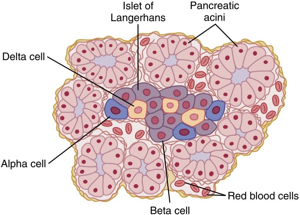
Figure 79-1. Physiological anatomy of an islet of Langerhans in the pancreas.
شکل 79-1. آناتومی فیزیولوژیکی جزایر لانگرهانس در پانکراس.
INSULIN AND ITS METABOLIC EFFECTS
Insulin was first isolated from the pancreas in 1922 by Banting and Best, and almost overnight rescued patients with severe cases of diabetes mellitus from a rapid decline in health and early death. Historically, insulin has been associated with “blood sugar,” and true enough, insulin has profound effects on carbohydrate metabolism. However, abnormalities of fat metabolism that cause conditions such as acidosis and arteriosclerosis are also important causes of morbidity and death in patients with diabetes mellitus. Patients with prolonged, untreated diabetes have diminished ability to synthesize proteins which leads to wasting of the tissues and many cellular functional disorders. Therefore, it is clear that insulin affects fat and protein metabolism almost as much as it affects carbohydrate metabolism.
انسولین و اثرات متابولیکی آن
انسولین برای اولین بار در سال 1922 توسط Banting و Best از پانکراس جدا شد و تقریباً یک شبه بیماران مبتلا به موارد شدید دیابت را از کاهش سریع سلامت و مرگ زودرس نجات داد. از نظر تاریخی، انسولین با “قند خون” مرتبط بوده است و به اندازه کافی درست است، انسولین اثرات عمیقی بر متابولیسم کربوهیدرات دارد. با این حال، ناهنجاریهای متابولیسم چربی که باعث بیماریهایی مانند اسیدوز و تصلب شرایین میشوند نیز از علل مهم مرگ و میر در بیماران مبتلا به دیابت هستند. بیماران مبتلا به دیابت طولانی مدت و درمان نشده توانایی سنتز پروتئین ها را کاهش می دهند که منجر به هدر رفتن بافت ها و بسیاری از اختلالات عملکردی سلولی می شود. بنابراین واضح است که انسولین تقریباً به همان اندازه که بر متابولیسم کربوهیدرات ها تأثیر می گذارد بر متابولیسم چربی و پروتئین تأثیر می گذارد.
INSULIN IS A HORMONE ASSOCIATED WITH ENERGY ABUNDANCE
As we discuss insulin in the next few pages, it will become apparent that insulin secretion is associated with energy abundance. That is, when a person’s diet includes a great abundance of foods that provide energy, especially excess amounts of carbohydrates, insulin secretion increases. In turn, the insulin plays an important role in storing the excess energy. In the case of excess carbohydrates, it causes them to be stored as glycogen, mainly in the liver and muscles. Furthermore, all the excess carbohydrates that cannot be stored as glycogen are converted under the stimulus of insulin into fats and stored in adipose tissue. In the case of proteins, insulin has a direct effect in promoting amino acid uptake by cells and conversion of these amino acids into protein. In addition, it inhibits breakdown of proteins that are already in the cells.
انسولین هورمونی است که با فراوانی انرژی مرتبط است
همانطور که در چند صفحه آینده به انسولین می پردازیم، آشکار خواهد شد که ترشح انسولین با فراوانی انرژی مرتبط است. به این معنا که وقتی رژیم غذایی فرد حاوی مقدار زیادی از مواد غذایی است که انرژی را تامین می کنند، به ویژه مقادیر اضافی کربوهیدرات، ترشح انسولین افزایش می یابد. به نوبه خود، انسولین نقش مهمی در ذخیره انرژی اضافی دارد. در مورد کربوهیدرات های اضافی، باعث می شود که آنها به عنوان گلیکوژن، عمدتاً در کبد و ماهیچه ها ذخیره شوند. علاوه بر این، تمام کربوهیدرات های اضافی که نمی توانند به عنوان گلیکوژن ذخیره شوند، تحت تحریک انسولین به چربی تبدیل شده و در بافت چربی ذخیره می شوند. در مورد پروتئین ها، انسولین تأثیر مستقیمی در ارتقاء جذب آمینو اسید توسط سلول ها و تبدیل این اسیدهای آمینه به پروتئین دارد. علاوه بر این، از تجزیه پروتئین هایی که از قبل در سلول ها هستند، جلوگیری می کند.
INSULIN CHEMISTRY AND SYNTHESIS
Human insulin, which has a molecular weight of 5808, is composed of two amino acid chains, shown in Figure 79-2, that are connected to each other by disulfide linkages. When the two amino acid chains are split apart, insulin’s functional activity is lost.
شیمی و سنتز انسولین
انسولین انسانی که وزن مولکولی آن 5808 است، از دو زنجیره آمینو اسید، که در شکل 79-2 نشان داده شده است، تشکیل شده است که توسط پیوندهای دی سولفیدی به یکدیگر متصل شده اند. هنگامی که دو زنجیره اسید آمینه از هم جدا می شوند، فعالیت عملکردی انسولین از بین می رود.
Insulin is synthesized in beta cells by the usual cell machinery for protein synthesis, as explained in Chapter 3, beginning with translation of the insulin RNA by ribosomes attached to the endoplasmic reticulum to form preproinsulin. This initial preproinsulin has a molecular weight of about 11,500, but it is then cleaved in the endo- plasmic reticulum to form a proinsulin with a molecular weight of about 9000 and consisting of three chains of peptides, A, B, and C. Most of the proinsulin is further cleaved in the Golgi apparatus to form insulin, which is composed of the A and B chains connected by disulfide linkages, and the C chain peptide, called connecting peptide (C peptide). The insulin and C peptide are pack- aged in secretory granules and secreted in equimolar amounts. About 5% to 10% of the final secreted product is still in the form of proinsulin.
همانطور که در فصل 3 توضیح داده شد، انسولین در سلول های بتا توسط ماشین سلولی معمول برای سنتز پروتئین سنتز می شود، که با ترجمه RNA انسولین توسط ریبوزوم های متصل به شبکه آندوپلاسمی برای تشکیل پیش پروانسولین شروع می شود. این پیش پروانسولین اولیه دارای وزن مولکولی حدود 11500 است، اما سپس در شبکه آندوپلاسمی شکاف داده می شود تا یک پروانسولین با وزن مولکولی حدود 9000 و متشکل از سه زنجیره پپتید A، B و C تشکیل شود. زنجیره های B با پیوندهای دی سولفید به هم متصل می شوند و پپتید زنجیره C به نام پپتید اتصال (پپتید C). انسولین و پپتید C در گرانول های ترشحی بسته بندی شده و در مقادیر هم مولار ترشح می شوند. حدود 5 تا 10 درصد از محصول نهایی ترشح شده هنوز به شکل پروانسولین است.
The proinsulin and C peptide have virtually no insulin activity. However, C peptide binds to a membrane structure, most likely a G protein-coupled membrane receptor, and elicits activation of at least two enzyme systems, sodium-potassium adenosine triphosphatase and endothelial nitric oxide synthase. Although both of these enzymes have multiple physiological functions, the importance of C peptide in regulating these enzymes is still uncertain.
پروانسولین و پپتید C عملاً هیچ فعالیت انسولینی ندارند. با این حال، پپتید C به یک ساختار غشایی، به احتمال زیاد یک گیرنده غشایی جفت شده با پروتئین G، متصل می شود و باعث فعال شدن حداقل دو سیستم آنزیمی، سدیم-پتاسیم آدنوزین تری فسفاتاز و اکسید نیتریک اندوتلیال سنتاز می شود. اگرچه هر دوی این آنزیم ها عملکردهای فیزیولوژیکی متعددی دارند، اهمیت پپتید C در تنظیم این آنزیم ها هنوز نامشخص است.
C peptide levels can be measured by radioimmunoassay in insulin-treated diabetic patients to determine how much of their own natural insulin they are still producing. Patients with type 1 diabetes who are unable to produce insulin will usually also have greatly decreased levels of C peptide.
سطح پپتید C را می توان با روش رادیو ایمونواسی در بیماران دیابتی تحت درمان با انسولین اندازه گیری کرد تا مشخص شود که هنوز چه مقدار از انسولین طبیعی خود را تولید می کنند. بیماران مبتلا به دیابت نوع 1 که قادر به تولید انسولین نیستند، معمولاً سطح پپتید C را به شدت کاهش می دهند.
When insulin is secreted into the blood, it circulates almost entirely in an unbound form. Because it has a plasma half-life that averages only about 6 minutes, it is mainly cleared from the circulation within 10 to 15 minutes. Except for the portion of the insulin that combines with receptors in the target cells, the insulin is degraded by the enzyme insulinase mainly in the liver, to a lesser extent in the kidneys and muscles, and slightly in most other tissues. This rapid removal from the plasma is important because, at times, it is as important to rapidly turn off the control functions of insulin as it is to turn them on.
هنگامی که انسولین در خون ترشح می شود، تقریباً به طور کامل به شکل غیر محدود در گردش است. از آنجایی که نیمه عمر پلاسمایی آن به طور متوسط تنها حدود 6 دقیقه است، عمدتاً در عرض 10 تا 15 دقیقه از گردش خون پاک می شود. به جز بخشی از انسولین که با گیرندههای سلولهای هدف ترکیب میشود، انسولین توسط آنزیم انسولیناز عمدتاً در کبد، به میزان کمتری در کلیهها و ماهیچهها و کمی در بیشتر بافتهای دیگر تجزیه میشود. این حذف سریع از پلاسما مهم است زیرا، گاهی اوقات، خاموش کردن سریع عملکردهای کنترل انسولین به همان اندازه که روشن کردن آنها مهم است.

Figure 79-2. A schematic of the human proinsulin molecule, which is cleaved in the Golgi apparatus of the pancreatic beta cells to form connecting peptide (C peptide), and insulin, which is composed of the A and B chains connected by disulfide bonds. The C peptide and insulin are packaged in granules and secreted in equimolar amounts, along with a small amount of proinsulin.
شکل 79-2. شماتیکی از مولکول پروانسولین انسانی، که در دستگاه گلژی سلولهای بتای پانکراس برای تشکیل پپتید اتصال (پپتید C) و انسولین که از زنجیرههای A و B تشکیل شده است که توسط پیوندهای دی سولفید به هم متصل شدهاند، شکافته میشود. پپتید C و انسولین به صورت گرانول بسته بندی شده و در مقادیر هممولار همراه با مقدار کمی پروانسولین ترشح می شوند.
ACTIVATION OF TARGET CELL RECEPTORS BY INSULIN AND THE RESULTING CELLULAR EFFECTS
To initiate its effects on target cells, insulin must first bind with and activate a membrane receptor protein that has a molecular weight of about 300,000 (Figure 79-3).
فعال سازی گیرنده های سلولی هدف توسط انسولین و اثرات سلولی ناشی از آن
انسولین برای شروع اثرات آن بر سلول های هدف، ابتدا باید با پروتئین گیرنده غشایی که وزن مولکولی آن حدود 300000 است، متصل شده و آن را فعال کند (شکل 79-3).
The insulin receptor is a combination of four subunits held together by disulfide linkages: two alpha subunits that lie entirely outside the cell membrane and two beta subunits that penetrate through the membrane, protruding into the cell cytoplasm. Insulin binds with the alpha subunits on the outside of the cell, but because of the linkages with the beta subunits, portions of the beta subunits protruding into the cell become auto phosphorylated. Thus, the insulin receptor is an example of an enzyme-linked receptor, discussed in Chapter 75. Autophosphorylation of the beta subunits of the receptor activates a local tyro- sine kinase, which in turn causes phosphorylation of multiple other intracellular enzymes, including a group called insulin-receptor substrates (IRS). Different types of IRS (e.g., IRS-1, IRS-2, and IRS-3) are expressed in different tissues. The net effect is to activate some of these enzymes while inactivating others. In this way, insulin directs the intracellular metabolic machinery to produce the desired effects on carbohydrate, fat, and protein metabolism. The following are the main end effects of insulin stimulation:
گیرنده انسولین ترکیبی از چهار زیر واحد است که توسط پیوندهای دی سولفیدی به هم متصل می شوند: دو زیر واحد آلفا که کاملاً خارج از غشای سلولی قرار دارند و دو زیر واحد بتا که از طریق غشاء نفوذ می کنند و به داخل سیتوپلاسم سلولی بیرون زده اند. انسولین به زیر واحدهای آلفا در خارج سلول متصل می شود، اما به دلیل پیوند با زیر واحدهای بتا، بخش هایی از زیر واحدهای بتا که به داخل سلول بیرون زده اند، خودفسفریله می شوند. بنابراین، گیرنده انسولین نمونهای از گیرندههای مرتبط با آنزیم است که در فصل 75 مورد بحث قرار گرفت. اتوفسفوریلاسیون زیر واحدهای بتا گیرنده، تیروزین کیناز موضعی را فعال میکند، که به نوبه خود باعث فسفوریلاسیون چندین آنزیم داخل سلولی، از جمله گروهی به نام انسولینهای فرعی (Substrase-IR) میشود. انواع مختلف IRS (به عنوان مثال IRS-1، IRS-2 و IRS-3) در بافت های مختلف بیان می شوند. اثر خالص این است که برخی از این آنزیم ها را فعال می کند در حالی که برخی دیگر را غیرفعال می کند. به این ترتیب، انسولین ماشینهای متابولیک داخل سلولی را هدایت میکند تا اثرات مطلوبی بر متابولیسم کربوهیدرات، چربی و پروتئین ایجاد کند. اثرات نهایی اصلی تحریک انسولین به شرح زیر است:
1. Within seconds after insulin binds with its membrane receptors, the membranes of about 80% of the body’s cells markedly increase their uptake of glucose. This action is especially true of muscle cells and adipose cells, but it is not true of most neurons in the brain. The increased glucose transported into the cells is immediately phosphorylated and becomes a substrate for all the usual carbohydrate metabolic functions. The increased glucose transport is believed to result from translocation of multiple intracellular vesicles to the cell membranes; these vesicles carry multiple molecules of glucose transport proteins, which bind with the cell membrane and facilitate glucose uptake into the cells. When insulin is no longer available, these vesicles separate from the cell membrane within about 3 to 5 minutes and move back to the cell interior to be used again and again, as needed.
1. در عرض چند ثانیه پس از اتصال انسولین به گیرنده های غشایی خود، غشای حدود 80 درصد سلول های بدن به طور قابل توجهی جذب گلوکز را افزایش می دهد. این عمل به ویژه در مورد سلول های ماهیچه ای و سلول های چربی صادق است، اما در مورد اکثر نورون های مغز صادق نیست. افزایش گلوکز منتقل شده به سلول ها بلافاصله فسفریله می شود و به بستری برای تمام عملکردهای متابولیک معمول کربوهیدرات تبدیل می شود. اعتقاد بر این است که افزایش انتقال گلوکز ناشی از جابجایی وزیکولهای درون سلولی متعدد به غشای سلولی است. این وزیکولها مولکولهای متعددی از پروتئینهای انتقال گلوکز را حمل میکنند که با غشای سلولی متصل میشوند و جذب گلوکز را به سلولها تسهیل میکنند. زمانی که انسولین دیگر در دسترس نباشد، این وزیکولها در عرض 3 تا 5 دقیقه از غشای سلولی جدا میشوند و به داخل سلول برمیگردند تا در صورت لزوم بارها و بارها مورد استفاده قرار گیرند.
2. The cell membrane becomes more permeable to many of the amino acids, potassium ions, and phosphate ions, causing increased transport of these substances into the cell.
2. غشای سلولی نسبت به بسیاری از اسیدهای آمینه، یون های پتاسیم و یون های فسفات نفوذ پذیرتر می شود و باعث افزایش انتقال این مواد به داخل سلول می شود.
3. Slower effects occur during the next 10 to 15 minutes to change the activity levels of many more intracellular metabolic enzymes. These effects result mainly from the changed states of phosphorylation of the enzymes.
3. اثرات آهسته تری در طی 10 تا 15 دقیقه آینده رخ می دهد تا سطح فعالیت بسیاری از آنزیم های متابولیک داخل سلولی را تغییر دهد. این اثرات عمدتاً از تغییر حالت های فسفوریلاسیون آنزیم ها ناشی می شود.
4. Much slower effects continue to occur for hours and even several days. These result from changed rates of translation of messenger RNAs at the ribosomes to form new proteins and still slower effects from changed rates of transcription of DNA in the cell nucleus. In this way, insulin remolds much of the cellular enzymatic machinery to achieve some of its metabolic effects.
4. اثرات بسیار کندتر برای ساعت ها و حتی چندین روز ادامه می یابد. اینها ناشی از تغییر نرخ ترجمه RNA های پیام رسان در ریبوزوم ها برای تشکیل پروتئین های جدید و تأثیرات کندتر از تغییر نرخ رونویسی DNA در هسته سلول است. به این ترتیب، انسولین بسیاری از ماشین آلات آنزیمی سلولی را برای دستیابی به برخی از اثرات متابولیکی آن بازسازی می کند.

Figure 79-3. A schematic of the insulin receptor. Insulin binds to the a subunit of its receptor, which causes autophosphorylation of the ẞ-subunit receptor, which in turn induces tyrosine kinase activity. The receptor tyrosine kinase activity begins a cascade of cell phosphorylation that increases or decreases the activity of enzymes, including insulin receptor substrates, that mediate the effects on glucose, fat, and protein metabolism. For example, glucose transporters are moved to the cell membrane to assist glucose entry into the cell.
شکل 79-3. شماتیکی از گیرنده انسولین. انسولین به زیر واحد گیرنده خود متصل می شود که باعث اتوفسفوریلاسیون گیرنده زیرواحد ẞ می شود که به نوبه خود باعث ایجاد فعالیت تیروزین کیناز می شود. فعالیت گیرنده تیروزین کیناز یک آبشار از فسفوریلاسیون سلولی را آغاز می کند که باعث افزایش یا کاهش فعالیت آنزیم ها، از جمله بسترهای گیرنده انسولین می شود، که واسطه اثرات بر متابولیسم گلوکز، چربی و پروتئین است. به عنوان مثال، انتقال دهنده های گلوکز به غشای سلولی منتقل می شوند تا به ورود گلوکز به داخل سلول کمک کنند.
EFFECT OF INSULIN ON CARBOHYDRATE METABOLISM
Immediately after a high-carbohydrate meal is consumed, glucose that is absorbed into the blood causes rapid secretion of insulin, which is discussed in detail later in the chapter. The insulin in turn causes rapid uptake, storage, and use of glucose by almost all tissues of the body but especially by the muscles, adipose tissue, and liver.
اثر انسولین بر متابولیسم کربوهیدرات
بلافاصله پس از مصرف یک وعده غذایی پر کربوهیدرات، گلوکزی که در خون جذب می شود باعث ترشح سریع انسولین می شود که در ادامه این فصل به تفصیل مورد بحث قرار می گیرد. انسولین به نوبه خود باعث جذب، ذخیره و استفاده سریع گلوکز توسط تقریباً تمام بافت های بدن، به ویژه توسط عضلات، بافت چربی و کبد می شود.
Insulin Promotes Muscle Glucose Uptake and Metabolism
During much of the day, muscle tissue depends not on glucose but on fatty acids for its energy. The principal reason for this dependence on fatty acids is that the normal resting muscle membrane is only slightly permeable to glucose, except when the muscle fiber is stimulated by insulin; between meals, the amount of insulin that is secreted is too small to promote significant amounts of glucose entry into the muscle cells.
انسولین جذب گلوکز و متابولیسم عضلانی را تقویت می کند
در بیشتر ساعات روز، بافت ماهیچه ای برای انرژی خود به گلوکز وابسته نیست، بلکه به اسیدهای چرب وابسته است. دلیل اصلی این وابستگی به اسیدهای چرب این است که غشای عضلانی در حالت استراحت فقط اندکی نسبت به گلوکز نفوذپذیر است، مگر زمانی که فیبر عضلانی توسط انسولین تحریک شود. بین وعدههای غذایی، مقدار انسولین ترشح شده بسیار کم است و نمیتواند مقدار قابل توجهی گلوکز را به سلولهای ماهیچهای وارد کند.
However, under two conditions the muscles do use large amounts of glucose. One of these is during moderate or heavy exercise. This usage of glucose does not require large amounts of insulin because muscle contraction increases translocation of glucose transporter 4 (GLUT 4) from intracellular storage depots to the cell membrane, which, in turn, facilitates diffusion of glucose into the cell.
با این حال، تحت دو شرایط، عضلات از مقادیر زیادی گلوکز استفاده می کنند. یکی از این موارد در حین ورزش متوسط یا سنگین است. این استفاده از گلوکز به مقادیر زیادی انسولین نیاز ندارد زیرا انقباض عضلانی باعث افزایش جابجایی ناقل گلوکز 4 (GLUT 4) از انبارهای ذخیره سازی درون سلولی به غشای سلولی می شود که به نوبه خود انتشار گلوکز به داخل سلول را تسهیل می کند.
The second condition for usage of large amounts of glucose by muscles is during the few hours after a meal. At this time the blood glucose concentration is high and the pancreas is secreting large quantities of insulin. The extra insulin causes rapid transport of glucose into the muscle cells, which causes the muscle cell to use glucose preferentially over fatty acids during this period, as will be discussed later.
شرط دوم استفاده از مقادیر زیاد گلوکز توسط عضلات، چند ساعت بعد از غذا است. در این زمان غلظت گلوکز خون بالا است و پانکراس مقادیر زیادی انسولین ترشح می کند. انسولین اضافی باعث انتقال سریع گلوکز به داخل سلولهای ماهیچهای میشود، که باعث میشود سلول عضلانی در این دوره از گلوکز ترجیحاً نسبت به اسیدهای چرب استفاده کند، همانطور که بعداً بحث خواهد شد.
Storage of Glycogen in Muscle. If the muscles are not exercised after a meal and yet glucose is transported into the muscle cells in abundance, instead of being used for energy, most of the glucose is stored in the form of muscle glycogen, up to a limit of 2% to 3% concentration. The glycogen can be used by the muscle later for energy. Glycogen is especially useful for short periods of extreme energy usage by the muscles and even to provide spurts of anaerobic energy for a few minutes at a time via glycolytic breakdown of the glycogen to lactic acid, which can occur even in the absence of oxygen.
ذخیره گلیکوژن در عضلات. اگر عضلات بعد از غذا ورزش نشوند و در عین حال گلوکز به وفور به سلولهای عضلانی منتقل شود، به جای استفاده از انرژی، بیشتر گلوکز به شکل گلیکوژن ماهیچهای تا حد غلظت 2 تا 3 درصد ذخیره میشود. گلیکوژن می تواند بعداً توسط عضله برای انرژی استفاده شود. گلیکوژن مخصوصاً برای دورههای کوتاه مصرف انرژی شدید توسط عضلات و حتی برای تأمین جهشهای انرژی بیهوازی برای چند دقیقه در یک زمان از طریق تجزیه گلیکولیتیک گلیکوژن به اسید لاکتیک، که میتواند حتی در غیاب اکسیژن رخ دهد، مفید است.
Quantitative Effect of Insulin to Facilitate Glucose Transport Through the Muscle Cell Membrane
The quantitative effect of insulin to facilitate glucose transport through the muscle cell membrane is demonstrated by the experimental results shown in Figure 79-4. The lower curve labeled “control” shows the concentration of free glucose measured inside the cell, demonstrating that the glucose concentration remained almost zero despite increased extracellular glucose concentration up to as high as 750 mg/100 ml. In contrast, the curve labeled “insulin” demonstrates that the intracellular glucose concentration rose to as high as 400 mg/100 ml when insulin was added. Thus, it is clear that insulin can increase the rate of transport of glucose into the resting muscle cell by at least 15-fold.
اثر کمی انسولین برای تسهیل انتقال گلوکز از طریق غشای سلول عضلانی
اثر کمی انسولین برای تسهیل انتقال گلوکز از طریق غشای سلول عضلانی با نتایج تجربی نشان داده شده در شکل 79-4 نشان داده شده است. منحنی پایینی با برچسب “کنترل” غلظت گلوکز آزاد اندازه گیری شده در داخل سلول را نشان می دهد و نشان می دهد که غلظت گلوکز با وجود افزایش غلظت گلوکز خارج سلولی تا 750 میلی گرم در 100 میلی لیتر تقریباً صفر باقی مانده است. در مقابل، منحنی با برچسب “انسولین” نشان می دهد که غلظت گلوکز درون سلولی با اضافه شدن انسولین به 400 میلی گرم در 100 میلی لیتر افزایش یافت. بنابراین، واضح است که انسولین می تواند سرعت انتقال گلوکز به سلول عضلانی در حال استراحت را حداقل 15 برابر افزایش دهد.

Figure 79-4. The effect of insulin in enhancing the concentration of glucose inside muscle cells. Note that in the absence of insulin (control), the intracellular glucose concentration remains near zero, despite high extracellular glucose concentrations. (Data from Eisenstein AB: The Biochemical Aspects of Hormone Action. Boston: Little, Brown, 1964.)
شکل 79-4. تاثیر انسولین در افزایش غلظت گلوکز درون سلول های عضلانی. توجه داشته باشید که در غیاب انسولین (کنترل)، غلظت گلوکز درون سلولی با وجود غلظت بالای گلوکز خارج سلولی، نزدیک به صفر باقی می ماند. (داده ها از آیزنشتاین AB: جنبه های بیوشیمیایی عملکرد هورمون. بوستون: لیتل، براون، 1964.)
Insulin Promotes Liver Uptake, Storage, and Use of Glucose
One of the most important effects of insulin is to cause most of the glucose absorbed after a meal to be rapidly stored in the liver in the form of glycogen. Then, between meals, when food is not available and the blood glucose concentration begins to fall, insulin secretion decreases rapidly and the liver glycogen is split back into glucose, which is released back into the blood to keep the glucose concentration from falling too low.
انسولین جذب کبد، ذخیره سازی و استفاده از گلوکز را تقویت می کند
یکی از مهمترین اثرات انسولین این است که باعث می شود بیشتر گلوکز جذب شده بعد از غذا به سرعت در کبد به شکل گلیکوژن ذخیره شود. سپس، بین وعدههای غذایی، زمانی که غذا در دسترس نیست و غلظت گلوکز خون شروع به کاهش میکند، ترشح انسولین به سرعت کاهش مییابد و گلیکوژن کبد دوباره به گلوکز تقسیم میشود، که برای جلوگیری از کاهش بیش از حد غلظت گلوکز به خون آزاد میشود.
The mechanism by which insulin causes glucose uptake and storage in the liver includes several almost simultaneous steps:
مکانیسمی که توسط آن انسولین باعث جذب و ذخیره گلوکز در کبد می شود شامل چندین مرحله تقریباً همزمان است:
1. Insulin inactivates liver phosphorylase, the principal enzyme that causes liver glycogen to split into glucose. This inactivation prevents breakdown of the glycogen that has been stored in liver cells.
1. انسولین فسفوریلاز کبد را غیرفعال می کند، آنزیم اصلی که باعث تجزیه گلیکوژن کبد به گلوکز می شود. این غیر فعال سازی از تجزیه گلیکوژن ذخیره شده در سلول های کبد جلوگیری می کند.
2. Insulin enhances uptake of glucose from the blood by the liver cells by increasing the activity of the enzyme glucokinase, which is one of the enzymes that causes the initial phosphorylation of glucose after it diffuses into the liver cells. Once phosphorylated, the glucose is temporarily trapped inside the liver cells because phosphorylated glucose cannot diffuse back through the cell membrane.
2. انسولین با افزایش فعالیت آنزیم گلوکوکیناز که یکی از آنزیم هایی است که باعث فسفوریلاسیون اولیه گلوکز پس از انتشار در سلول های کبدی می شود، جذب گلوکز از خون توسط سلول های کبدی را افزایش می دهد. پس از فسفریله شدن، گلوکز به طور موقت در داخل سلول های کبد به دام می افتد زیرا گلوکز فسفریله نمی تواند از طریق غشای سلولی پخش شود.
3. Insulin increases the activities of enzymes that pro- mote glycogen synthesis, including especially glycogen synthase. This is responsible for polymerization of the monosaccharide units to form glycogen molecules.
3. انسولین فعالیت آنزیم هایی را افزایش می دهد که سنتز گلیکوژن را تقویت می کنند، به ویژه گلیکوژن سنتاز. این مسئول پلیمریزاسیون واحدهای مونوساکارید برای تشکیل مولکول های گلیکوژن است.
The net effect of all these actions is to increase the amount of glycogen in the liver. The glycogen can increase to a total of about 5% to 6% of the liver mass, which is equivalent to almost 100 grams of stored glycogen in the entire liver.
اثر خالص همه این اقدامات افزایش میزان گلیکوژن در کبد است. گلیکوژن می تواند در مجموع به حدود 5 تا 6 درصد توده کبد افزایش یابد که معادل تقریباً 100 گرم گلیکوژن ذخیره شده در کل کبد است.
Glucose Is Released From the Liver Between Meals. When the blood glucose level begins to fall to a low level between meals, several events transpire that cause the liver to release glucose back into the circulating blood:
گلوکز بین وعده های غذایی از کبد آزاد می شود. هنگامی که سطح گلوکز خون بین وعده های غذایی شروع به کاهش به سطح پایین می کند، چندین اتفاق رخ می دهد که باعث می شود کبد گلوکز را دوباره به خون در گردش آزاد کند:
1. The decreasing blood glucose causes the pancreas to decrease its insulin secretion.
1. کاهش گلوکز خون باعث می شود که پانکراس ترشح انسولین خود را کاهش دهد.
2. The lack of insulin then reverses all the effects listed earlier for glycogen storage, essentially stopping further synthesis of glycogen in the liver and preventing further uptake of glucose by the liver from the blood.
2. فقدان انسولین سپس تمام اثرات ذکر شده قبلی برای ذخیره گلیکوژن را معکوس می کند، اساساً سنتز بیشتر گلیکوژن در کبد را متوقف می کند و از جذب بیشتر گلوکز توسط کبد از خون جلوگیری می کند.
3. The lack of insulin (along with increased glucagon, which is discussed later) activates the enzyme phosphorylase, which causes the splitting of glycogen into glucose phosphate.
3. کمبود انسولین (همراه با افزایش گلوکاگون که بعداً مورد بحث قرار می گیرد) آنزیم فسفوریلاز را فعال می کند که باعث تجزیه گلیکوژن به گلوکز فسفات می شود.
4. The enzyme glucose phosphatase, which had been inhibited by insulin, now becomes activated by the lack of insulin and causes the phosphate radical to split away from the glucose, allowing the free glucose to diffuse back into the blood.
4. آنزیم گلوکز فسفاتاز که توسط انسولین مهار شده بود، اکنون در اثر کمبود انسولین فعال می شود و باعث می شود رادیکال فسفات از گلوکز جدا شود و به گلوکز آزاد اجازه انتشار مجدد به خون را می دهد.
Thus, the liver removes glucose from the blood when it is present in excess after a meal and returns it to the blood when the blood glucose concentration falls between meals. Ordinarily, about 60% of the glucose in the meal is stored in this way in the liver and then returned later.
بنابراین، کبد وقتی گلوکز را بعد از غذا بیش از حد وجود دارد از خون خارج می کند و زمانی که غلظت گلوکز خون بین وعده های غذایی کاهش می یابد، آن را به خون باز می گرداند. به طور معمول، حدود 60 درصد از گلوکز موجود در وعده غذایی به این ترتیب در کبد ذخیره می شود و بعداً برمی گردد.
Insulin Promotes Conversion of Excess Glucose Into Fatty Acids and Inhibits Gluconeogenesis in the Liver. When the quantity of glucose entering the liver cells is more than can be stored as glycogen or can be used for local hepatocyte metabolism, insulin promotes the conversion of all this excess glucose into fatty acids. These fatty acids are subsequently packaged as triglycerides in very low density lipoproteins, which are transported in the blood to adipose tissue, and deposited as fat.
انسولین باعث تبدیل گلوکز اضافی به اسیدهای چرب و مهار گلوکونئوژنز در کبد می شود. هنگامی که مقدار گلوکز وارد شده به سلول های کبدی بیش از آن است که بتوان آن را به عنوان گلیکوژن ذخیره کرد یا برای متابولیسم موضعی سلول های کبدی استفاده کرد، انسولین تبدیل تمام این گلوکز اضافی به اسیدهای چرب را افزایش می دهد. این اسیدهای چرب متعاقباً به صورت تری گلیسیرید در لیپوپروتئین های با چگالی بسیار کم بسته بندی می شوند که در خون به بافت چربی منتقل می شوند و به صورت چربی رسوب می کنند.
Insulin also inhibits gluconeogenesis mainly by decreasing the quantities and activities of the liver enzymes required for gluconeogenesis. However, part of the effect is caused by an action of insulin that decreases release of amino acids from muscle and other extrahepatic tissues and in turn the availability of these necessary precursors required for gluconeogenesis. This phenomenon is dis- cussed further in relation to the effect of insulin on protein metabolism.
انسولین همچنین گلوکونئوژنز را عمدتاً با کاهش مقدار و فعالیت آنزیم های کبدی مورد نیاز برای گلوکونئوژنز مهار می کند. با این حال، بخشی از این اثر ناشی از عمل انسولین است که باعث کاهش آزاد شدن اسیدهای آمینه از ماهیچه ها و سایر بافت های خارج کبدی می شود و به نوبه خود دسترسی به این پیش سازهای لازم برای گلوکونئوژنز را کاهش می دهد. این پدیده بیشتر در رابطه با اثر انسولین بر متابولیسم پروتئین مورد بحث قرار می گیرد.
Lack of Effect of Insulin on Glucose Uptake and Usage by the Brain
The brain is quite different from most other tissues of the body in that insulin has little effect on uptake or use of glucose. Instead, most of the brain cells are permeable to glucose and can use glucose without the intermediation of insulin.
عدم تأثیر انسولین بر جذب و مصرف گلوکز توسط مغز
مغز کاملاً با سایر بافت های بدن متفاوت است زیرا انسولین تأثیر کمی بر جذب یا استفاده از گلوکز دارد. درعوض، بیشتر سلول های مغز نسبت به گلوکز نفوذپذیر هستند و می توانند بدون واسطه انسولین از گلوکز استفاده کنند.
The brain cells are also quite different from most other cells of the body in that they normally use only glucose for energy and can use other energy substrates, such as fats, only with difficulty. Therefore, it is essential that the blood glucose level always be maintained above a critical level, which is one of the most important functions of the blood glucose control system. When the blood glucose level falls too low, into the range of 20 to 50 mg/100 ml, symptoms of hypoglycemic shock develop, characterized by progressive nervous irritability that leads to fainting, seizures, and even coma.
سلولهای مغز نیز کاملاً متفاوت از سایر سلولهای بدن هستند، زیرا معمولاً فقط از گلوکز برای انرژی استفاده میکنند و میتوانند به سختی از سایر بسترهای انرژی مانند چربیها استفاده کنند. بنابراین ضروری است که سطح گلوکز خون همیشه بالاتر از حد بحرانی باشد که یکی از مهمترین وظایف سیستم کنترل قند خون است. هنگامی که سطح گلوکز خون بسیار پایین میآید، در محدوده 20 تا 50 میلیگرم در 100 میلیلیتر، علائم شوک هیپوگلیسمی ایجاد میشود که با تحریکپذیری عصبی پیشرونده که منجر به غش، تشنج و حتی کما میشود، مشخص میشود.
Effect of Insulin on Carbohydrate Metabolism in Other Cells
Insulin increases glucose transport into and glucose usage by most other cells of the body (with the exception of most brain cells, as noted) in the same way that it affects glucose transport and usage in muscle cells. The transport of glucose into adipose cells mainly provides substrate for the glycerol portion of the fat molecule. Therefore, in this indirect way, insulin promotes deposition of fat in these cells.
تأثیر انسولین بر متابولیسم کربوهیدرات در سایر سلولها
انسولین انتقال گلوکز را به داخل و مصرف گلوکز توسط اکثر سلولهای دیگر بدن (به استثنای اکثر سلولهای مغزی، همانطور که اشاره شد) افزایش میدهد به همان روشی که بر انتقال و استفاده گلوکز در سلولهای عضلانی تأثیر میگذارد. انتقال گلوکز به سلول های چربی عمدتاً بستری را برای بخش گلیسرول مولکول چربی فراهم می کند. بنابراین، انسولین به این روش غیرمستقیم باعث رسوب چربی در این سلول ها می شود.
EFFECT OF INSULIN ON FAT METABOLISM
Although not quite as visible as the acute effects of insulin on carbohydrate metabolism, the effects of insulin on fat metabolism are, in the long run, equally important. Especially dramatic is the long-term effect of insulin deficiency in causing extreme atherosclerosis, often leading to heart attacks, cerebral strokes, and other vascular accidents. First, however, let us discuss the acute effects of insulin on fat metabolism.
اثر انسولین بر متابولیسم چربی
اگرچه به اندازه اثرات حاد انسولین بر متابولیسم کربوهیدرات قابل مشاهده نیست، تأثیرات انسولین بر متابولیسم چربی، در دراز مدت، به همان اندازه مهم است. به ویژه تأثیر دراز مدت کمبود انسولین در ایجاد تصلب شرایین شدید، که اغلب منجر به حملات قلبی، سکته مغزی، و سایر حوادث عروقی می شود، چشمگیر است. با این حال، ابتدا اجازه دهید در مورد اثرات حاد انسولین بر متابولیسم چربی صحبت کنیم.
Insulin Promotes Fat Synthesis and Storage
Insulin has several effects that lead to fat storage in adipose tissue. First, insulin increases glucose utilization by most of the body’s tissues, which automatically decreases fat utilization, thus functioning as a fat sparer. However, insulin also promotes fatty acid synthesis, especially when more carbohydrates are ingested than can be used for immediate energy, thus providing the substrate for fat synthesis. Almost all this synthesis occurs in the liver cells, and the fatty acids are then transported from the liver by way of the blood lipoproteins to the adipose cells to be stored. The following factors lead to increased fatty acid synthesis in the liver:
انسولین سنتز و ذخیره چربی را تقویت می کند
انسولین اثرات متعددی دارد که منجر به ذخیره چربی در بافت چربی می شود. اول، انسولین استفاده از گلوکز را در بیشتر بافتهای بدن افزایش میدهد، که به طور خودکار مصرف چربی را کاهش میدهد، بنابراین به عنوان یک چربیدهی عمل میکند. با این حال، انسولین همچنین باعث تقویت سنتز اسیدهای چرب می شود، به ویژه زمانی که کربوهیدرات های بیشتری مصرف می شود که می توان برای انرژی فوری استفاده کرد، بنابراین بستری برای سنتز چربی فراهم می کند. تقریباً تمام این سنتز در سلولهای کبد اتفاق میافتد و سپس اسیدهای چرب از طریق لیپوپروتئینهای خون از کبد به سلولهای چربی منتقل میشوند تا ذخیره شوند. عوامل زیر منجر به افزایش سنتز اسیدهای چرب در کبد می شود:
1. Insulin increases glucose transport into the liver cells. After the liver glycogen concentration reaches 5% to 6%, further glycogen synthesis is inhibited. All the additional glucose entering the liver cells then be- comes available to form fat. The glucose is first split to pyruvate in the glycolytic pathway, and the pyruvate subsequently is converted to acetyl coenzyme A (acetyl-CoA), the substrate from which fatty acids are synthesized.
1. انسولین انتقال گلوکز به سلول های کبد را افزایش می دهد. پس از رسیدن غلظت گلیکوژن کبدی به 5 تا 6 درصد، سنتز گلیکوژن بیشتر مهار می شود. تمام گلوکز اضافی که وارد سلول های کبدی می شود سپس برای تشکیل چربی در دسترس می شود. گلوکز ابتدا در مسیر گلیکولیتیک به پیرووات تقسیم می شود و پیرووات متعاقبا به استیل کوآنزیم A (استیل-CoA) تبدیل می شود، بستری که اسیدهای چرب از آن سنتز می شوند.
2. An excess of citrate and isocitrate ions is formed by the citric acid cycle when excess amounts of glucose are used for energy. These ions then have a direct effect to activate acetyl-CoA carboxylase, the enzyme required to carboxylate acetyl-CoA to form malonyl-CoA, the first stage of fatty acid synthesis.
2. یون سیترات و ایزوسیترات اضافی در چرخه اسید سیتریک زمانی که مقادیر اضافی گلوکز برای انرژی استفاده می شود تشکیل می شود. سپس این یونها اثر مستقیمی برای فعال کردن استیل کوآ کربوکسیلاز دارند، آنزیمی که برای کربوکسیله کردن استیل کوآ برای تشکیل مالونیل کوآ، اولین مرحله از سنتز اسیدهای چرب لازم است.
3. Most of the fatty acids are then synthesized within the liver and used to form triglycerides, the usual form of storage fat. They are released from the liver cells to the blood in the lipoproteins. Insulin activates lipoprotein lipase in the capillary walls of the adipose tissue, which splits the triglycerides again into fatty acids, a requirement for them to be absorbed into adipose cells, where they are again converted to triglycerides and stored.
3. بیشتر اسیدهای چرب سپس در داخل کبد سنتز می شوند و برای تشکیل تری گلیسیرید، شکل معمول چربی ذخیره شده، استفاده می شوند. آنها از سلول های کبد به خون در لیپوپروتئین ها آزاد می شوند. انسولین لیپوپروتئین لیپاز را در دیواره های مویرگی بافت چربی فعال می کند، که تری گلیسیریدها را دوباره به اسیدهای چرب تقسیم می کند، که لازمه جذب آنها در سلول های چربی است، جایی که دوباره به تری گلیسیرید تبدیل شده و ذخیره می شوند.
Role of Insulin in Storage of Fat in the Adipose Cells. Insulin has two other essential effects that are required for fat storage in adipose cells:
نقش انسولین در ذخیره سازی چربی در سلول های چربی. انسولین دو اثر اساسی دیگر دارد که برای ذخیره چربی در سلول های چربی لازم است:
1. Insulin inhibits the action of hormone-sensitive lipase. Lipase is the enzyme that causes hydrolysis of triglycerides already stored in fat cells. Therefore, release of fatty acids from adipose tissue into the circulating blood is inhibited.
1. انسولین از عمل لیپاز حساس به هورمون جلوگیری می کند. لیپاز آنزیمی است که باعث هیدرولیز تری گلیسیریدهای ذخیره شده در سلول های چربی می شود. بنابراین آزاد شدن اسیدهای چرب از بافت چربی به داخل خون در گردش مهار می شود.
2. Insulin promotes glucose transport through cell membranes into fat cells in the same way that it promotes glucose transport into muscle cells. Some of this glucose is then used to synthesize minute amounts of fatty acids, but more important, it also forms large quantities of a-glycerol phosphate. This substance supplies the glycerol that combines with fatty acids to form triglycerides, the storage form of fat in adipose cells. Therefore, when insulin is not available, even storage of the large amounts of fatty acids transported from the liver in lipoproteins is almost blocked.
2. انسولین انتقال گلوکز را از طریق غشای سلولی به سلول های چربی به همان روشی که انتقال گلوکز را به سلول های ماهیچه ای ترویج می کند، افزایش می دهد. سپس مقداری از این گلوکز برای سنتز مقادیر بسیار اندک اسیدهای چرب استفاده می شود، اما مهمتر از آن، مقادیر زیادی از گلیسرول فسفات را نیز تشکیل می دهد. این ماده گلیسرول را تامین می کند که با اسیدهای چرب ترکیب می شود و تری گلیسیرید، شکل ذخیره چربی در سلول های چربی را تشکیل می دهد. بنابراین، هنگامی که انسولین در دسترس نباشد، حتی ذخیره مقادیر زیادی اسیدهای چرب منتقل شده از کبد در لیپوپروتئین ها تقریباً مسدود می شود.
Insulin Deficiency Increases Use of Fat for Energy
All aspects of fat breakdown and its use for providing energy are greatly enhanced in the absence of insulin. This enhancement occurs even normally between meals when secretion of insulin is minimal, but it becomes extreme in persons with diabetes mellitus when secretion of insulin is almost zero.
کمبود انسولین باعث افزایش استفاده از چربی برای انرژی می شود
تمام جنبه های تجزیه چربی و استفاده از آن برای تامین انرژی در غیاب انسولین بسیار افزایش می یابد. این افزایش حتی به طور معمول بین وعده های غذایی زمانی که ترشح انسولین حداقل است اتفاق می افتد، اما در افراد مبتلا به دیابت زمانی که ترشح انسولین تقریباً صفر است، شدید می شود.
Insulin Deficiency Causes Lipolysis of Storage Fat and Release of Free Fatty Acids. In the absence of insulin, all the effects of insulin noted earlier that cause storage of fat are reversed. The most important effect is that the enzyme hormone-sensitive lipase in the fat cells becomes strongly activated. This activation causes hydrolysis of stored triglycerides, releasing large quantities of fatty acids and glycerol into the circulating blood. Consequently, plasma concentration of free fatty acids begins to rise within minutes. These free fatty acids then become the main energy substrate used by essentially all tissues of the body except the brain.
کمبود انسولین باعث لیپولیز چربی ذخیره شده و آزاد شدن اسیدهای چرب آزاد می شود. در غیاب انسولین، تمام اثرات انسولین که قبلا ذکر شد و باعث ذخیره چربی می شود معکوس می شود. مهم ترین اثر این است که آنزیم لیپاز حساس به هورمون در سلول های چربی به شدت فعال می شود. این فعال سازی باعث هیدرولیز تری گلیسیریدهای ذخیره شده و آزاد شدن مقادیر زیادی اسیدهای چرب و گلیسرول در خون در گردش می شود. در نتیجه، غلظت اسیدهای چرب آزاد در پلاسما در عرض چند دقیقه شروع به افزایش می کند. سپس این اسیدهای چرب آزاد به زیرلایه اصلی انرژی تبدیل می شوند که اساساً توسط تمام بافت های بدن به جز مغز استفاده می شود.
Figure 79-5 shows the effect of a lack of insulin on the plasma concentrations of free fatty acids, glucose, and acetoacetic acid. Note that almost immediately after removal of the pancreas, the free fatty acid concentration in the plasma begins to rise, more rapidly even than the concentration of glucose.
شکل 79-5 اثر کمبود انسولین را بر غلظت پلاسمایی اسیدهای چرب آزاد، گلوکز و اسید استواستیک نشان می دهد. توجه داشته باشید که تقریباً بلافاصله پس از برداشتن پانکراس، غلظت اسیدهای چرب آزاد در پلاسما شروع به افزایش میکند، حتی سریعتر از غلظت گلوکز.
Insulin Deficiency Increases Plasma Cholesterol and Phospholipid Concentrations. The excess of fatty acids in the plasma associated with insulin deficiency also promotes liver conversion of some of the fatty acids into phospholipids and cholesterol, two of the major products of fat metabolism. These two substances, along with excess triglycerides formed at the same time in the liver, are then discharged into the blood in the lipoproteins. Occasionally the plasma lipoproteins increase as much as threefold in the absence of insulin, giving a total concentration of plasma lipids of several percent rather than the normal 0.6%. This high lipid concentration-especially the high concentration of cholesterol-promotes development of atherosclerosis in people with severe diabetes.
کمبود انسولین باعث افزایش غلظت کلسترول و فسفولیپید پلاسما می شود. اسیدهای چرب بیش از حد در پلاسما مرتبط با کمبود انسولین همچنین باعث تبدیل کبد برخی از اسیدهای چرب به فسفولیپیدها و کلسترول، دو محصول اصلی متابولیسم چربی می شود. این دو ماده به همراه تری گلیسیریدهای اضافی که به طور همزمان در کبد تشکیل می شوند، سپس در لیپوپروتئین ها وارد خون می شوند. گاهی اوقات در غیاب انسولین، لیپوپروتئینهای پلاسما تا سه برابر افزایش مییابند و غلظت کل لیپیدهای پلاسما چندین درصد به جای 0.6 درصد طبیعی است. این غلظت بالای چربی – به ویژه غلظت بالای کلسترول – باعث ایجاد آترواسکلروز در افراد مبتلا به دیابت شدید می شود.
Excess Usage of Fats During Insulin Deficiency Causes Ketosis and Acidosis. Insulin deficiency also causes excessive amounts of acetoacetic acid to be formed in liver cells. In the absence of insulin but in the presence of excess fatty acids in the liver cells, the carnitine transport mechanism for trans- porting fatty acids into the mitochondria becomes increasingly activated. In the mitochondria, beta oxidation of the fatty acids then proceeds rapidly, releasing extreme amounts of acetyl-CoA. A large part of this excess acetyl-CoA is then condensed to form acetoacetic acid, which is then released into the circulating blood. Most of this acetoacetic acid passes to the peripheral cells, where it is again converted into acetyl-CoA and used for energy in the usual manner.
مصرف بیش از حد چربی ها در هنگام کمبود انسولین باعث کتوز و اسیدوز می شود. کمبود انسولین همچنین باعث تشکیل مقادیر بیش از حد اسید استواستیک در سلول های کبد می شود. در غیاب انسولین اما در حضور اسیدهای چرب اضافی در سلول های کبد، مکانیسم انتقال کارنیتین برای انتقال اسیدهای چرب به داخل میتوکندری به طور فزاینده ای فعال می شود. در میتوکندری، اکسیداسیون بتا اسیدهای چرب به سرعت ادامه مییابد و مقادیر بسیار زیاد استیل کوآ آزاد میکند. سپس بخش بزرگی از این استیل-CoA اضافی متراکم میشود و اسید استواستیک تشکیل میدهد که سپس در خون در گردش آزاد میشود. بیشتر این اسید استواستیک به سلولهای محیطی میرود و در آنجا دوباره به استیل کوآ تبدیل میشود و به روش معمول برای انرژی استفاده میشود.
At the same time, the absence of insulin also depresses utilization of acetoacetic acid in peripheral tissues. Thus, so much acetoacetic acid is released from the liver that it cannot all be metabolized by the tissues. As shown in Figure 79-5, the concentration of acetoacetic acid rises during the days after cessation of insulin secretion, some- times reaching concentrations of 10 mEq/L or more, which is a severe state of body fluid acidosis.
در عین حال، فقدان انسولین نیز استفاده از اسید استواستیک را در بافتهای محیطی کاهش میدهد. بنابراین، اسید استواستیک به قدری از کبد آزاد می شود که نمی تواند تمام آن توسط بافت ها متابولیزه شود. همانطور که در شکل 79-5 نشان داده شده است، غلظت اسید استواستیک در طی روزهای پس از توقف ترشح انسولین افزایش مییابد و گاهی به غلظت mEq/L 10 یا بیشتر میرسد که حالت شدید اسیدوز مایع بدن است.
As explained in Chapter 69, some of the acetoacetic acid is also converted into ẞ-hydroxybutyric acid and acetone. These two substances, along with the acetoacetic acid, are called ketone bodies, and their presence in large quantities in the body fluids is called ketosis. We will see later that in severe diabetes, the acetoacetic acid and the B-hydroxybutyric acid can cause severe acidosis and coma, which may lead to death.
همانطور که در فصل 69 توضیح داده شد، مقداری از اسید استواستیک نیز به اسید هیدروکسی بوتیریک و استون تبدیل می شود. این دو ماده همراه با اسید استواستیک اجسام کتون نامیده می شوند و وجود آنها به مقدار زیاد در مایعات بدن کتوز نامیده می شود. بعداً خواهیم دید که در دیابت شدید، اسید استواستیک و اسید B-هیدروکسی بوتیریک می توانند باعث اسیدوز شدید و کما شوند که ممکن است منجر به مرگ شود.

Figure 79-5. The effect of removing the pancreas on the approximate concentrations of blood glucose, plasma free fatty acids, and acetoacetic acid.
شکل 79-5. تأثیر حذف پانکراس بر غلظت تقریبی گلوکز خون، اسیدهای چرب آزاد پلاسما و اسید استواستیک.
EFFECT OF INSULIN ON PROTEIN METABOLISM AND GROWTH
Insulin Promotes Protein Synthesis and Storage
Proteins, carbohydrates, and fats are stored in the tissues during the few hours after a meal when excess quantities of nutrients are available in the circulating blood; insulin is required for this storage to occur. The manner in which insulin causes protein storage is not as well understood as the mechanisms for both glucose and fat storage. Here are some of the facts:
اثر انسولین بر متابولیسم و رشد پروتئین
انسولین سنتز و ذخیره پروتئین را تقویت می کند
پروتئینها، کربوهیدراتها و چربیها در چند ساعت پس از غذا در بافتها ذخیره میشوند، زمانی که مقادیر اضافی مواد مغذی در خون در گردش وجود دارد. انسولین برای این ذخیره سازی لازم است. روشی که در آن انسولین باعث ذخیره پروتئین می شود به خوبی مکانیسم ذخیره سازی گلوکز و چربی شناخته نشده است. در اینجا برخی از حقایق آمده است:
1. Insulin stimulates transport of many of the amino acids into the cells. Among the amino acids most strongly transported are valine, leucine, isoleucine, tyrosine, and phenylalanine. Thus, insulin shares with growth hormone the capability of increasing uptake of amino acids into cells. However, the amino acids affected are not necessarily the same ones.
1. انسولین انتقال بسیاری از اسیدهای آمینه را به داخل سلول ها تحریک می کند. از جمله آمینو اسیدهایی که به شدت منتقل می شوند والین، لوسین، ایزولوسین، تیروزین و فنیل آلانین هستند. بنابراین، انسولین با هورمون رشد توانایی افزایش جذب اسیدهای آمینه به سلول ها را دارد. با این حال، اسیدهای آمینه تحت تأثیر لزوماً یکسان نیستند.
2. Insulin increases translation of messenger RNA, thus forming new proteins. Insulin “turns on” the ribosomal machinery and, in the absence of insulin, the ribosomes stop working, almost as if insulin operates by an “on-off” mechanism.
2. انسولین ترجمه RNA پیام رسان را افزایش می دهد و در نتیجه پروتئین های جدیدی تشکیل می دهد. انسولین ماشین ریبوزومی را «روشن» میکند و در غیاب انسولین، ریبوزومها از کار میافتند، تقریباً انگار انسولین با مکانیسم «روشن-خاموش» عمل میکند.
3. Over a longer period, insulin also increases the rate of transcription of selected DNA genetic sequences in the cell nuclei, thus forming increased quantities of RNA and still more protein synthesis-especially promoting a vast array of enzymes for storage of carbohydrates, fats, and proteins.
3. انسولین در یک دوره طولانیتر همچنین سرعت رونویسی توالیهای ژنتیکی انتخاب شده DNA را در هستههای سلولی افزایش میدهد، بنابراین مقادیر بیشتری از RNA و سنتز پروتئین بیشتری را تشکیل میدهد – بهویژه باعث ترویج مجموعه وسیعی از آنزیمها برای ذخیره کربوهیدراتها، چربیها و پروتئینها میشود.
4. Insulin inhibits catabolism of proteins, thus decreasing the rate of amino acid release from the cells, especially from muscle cells. Presumably this results from the ability of insulin to diminish the normal degradation of proteins by cellular lysosomes.
4. انسولین کاتابولیسم پروتئین ها را مهار می کند، بنابراین سرعت آزاد شدن اسید آمینه از سلول ها، به ویژه از سلول های ماهیچه ای را کاهش می دهد. احتمالاً این نتیجه از توانایی انسولین برای کاهش تخریب طبیعی پروتئین ها توسط لیزوزوم های سلولی است.
5. In the liver, insulin depresses the rate of gluconeogenesis by decreasing activity of the enzymes that promote gluconeogenesis. Because the substrates used most for synthesis of glucose by gluconeogenesis are plasma amino acids, this suppression of gluconeogenesis conserves amino acids in the protein stores of the body.
5. در کبد، انسولین با کاهش فعالیت آنزیم هایی که گلوکونئوژنز را تقویت می کنند، سرعت گلوکونئوژنز را کاهش می دهد. از آنجایی که سوبستراهای مورد استفاده برای سنتز گلوکز توسط گلوکونئوژنز اسیدهای آمینه پلاسما هستند، این سرکوب گلوکونئوژنز باعث حفظ اسیدهای آمینه در ذخایر پروتئینی بدن می شود.
In summary, insulin promotes formation of protein and prevents degradation of proteins.
به طور خلاصه، انسولین باعث تشکیل پروتئین می شود و از تجزیه پروتئین ها جلوگیری می کند.
Insulin Deficiency Causes Protein Depletion and Increased Plasma Amino Acids
Virtually all protein storage comes to a halt when insulin is not available. Catabolism of proteins increases, protein synthesis stops, and large quantities of amino acids are dumped into the plasma. Amino acid concentration in the plasma rises considerably, and most of the excess amino acids are used either directly for energy or as substrates for gluconeogenesis. This degradation of amino acids also leads to enhanced urea excretion in the urine. The resulting protein wasting is one of the most serious of all the effects of severe diabetes mellitus. It can lead to extreme weakness and many deranged functions of the organs.
کمبود انسولین باعث کاهش پروتئین و افزایش آمینو اسیدهای پلاسما می شود
تقریباً تمام ذخیره سازی پروتئین زمانی که انسولین در دسترس نباشد متوقف می شود. کاتابولیسم پروتئین ها افزایش می یابد، سنتز پروتئین متوقف می شود و مقادیر زیادی اسیدهای آمینه به پلاسما ریخته می شود. غلظت اسید آمینه در پلاسما به طور قابل توجهی افزایش می یابد و بیشتر اسیدهای آمینه اضافی یا مستقیماً برای انرژی یا به عنوان سوبسترا برای گلوکونئوژنز استفاده می شود. این تخریب اسیدهای آمینه همچنین منجر به افزایش دفع اوره در ادرار می شود. هدر رفتن پروتئین در نتیجه یکی از جدی ترین عوارض دیابت شیرین است. این می تواند منجر به ضعف شدید و بسیاری از عملکردهای اختلال در اندام ها شود.
Insulin and Growth Hormone Interact Synergistically to Promote Growth
Because insulin is required for synthesis of proteins, it is as essential as growth hormone for the growth of an animal. As demonstrated in Figure 79-6, a depan-creatized, hypophysectomized rat without replacement therapy hardly grows at all. Furthermore, administration of either growth hormone or insulin one at a time causes almost no growth. However, a combination of these hormones causes dramatic growth. Thus, it appears that the two hormones function synergistically to promote growth, with each performing a specific function separate from that of the other. Perhaps a small part of the necessity for both hormones results from the fact that each hormone promotes cellular uptake of a different selection of amino acids, all of which are required for growth.
انسولین و هورمون رشد به صورت سینرژیک برای تقویت رشد با هم تعامل دارند
از آنجایی که انسولین برای سنتز پروتئین ها لازم است، به اندازه هورمون رشد برای رشد یک حیوان ضروری است. همانطور که در شکل 79-6 نشان داده شده است، موش صحرایی هیپوفیزیکتومی شده با دپان کراتیزاسیون بدون درمان جایگزین به سختی رشد می کند. علاوه بر این، تجویز هورمون رشد یا انسولین در یک زمان تقریباً هیچ رشدی ایجاد نمی کند. با این حال، ترکیبی از این هورمون ها باعث رشد چشمگیر می شود. بنابراین، به نظر می رسد که این دو هورمون به طور هم افزایی برای تقویت رشد عمل می کنند و هر کدام عملکرد خاصی را جدا از دیگری انجام می دهند. شاید بخش کوچکی از نیاز به هر دو هورمون ناشی از این واقعیت است که هر هورمون جذب سلولی مجموعهای از اسیدهای آمینه متفاوت را که همگی برای رشد لازم هستند، افزایش میدهد.

Figure 79-6. The effect of growth hormone, insulin, and growth hormone plus insulin on growth in a depancreatized and hypophy- sectomized rat.
شکل 79-6. تأثیر هورمون رشد، انسولین و هورمون رشد به اضافه انسولین بر رشد موش صحرایی بدون پانکرات و هیپوفیکتومی شده.
MECHANISMS OF INSULIN SECRETION
Figure 79-7 shows the basic cellular mechanisms for insulin secretion by the pancreatic beta cells in response to increased blood glucose concentration, which is the primary controller of insulin secretion. The beta cells have a large number of glucose transporters that permit a rate of glucose influx that is proportional to the blood concentration in the physiological range. Once inside the cells, glucose is phosphorylated to glucose-6-phosphate by glucokinase. This phosphorylation appears to be the rate-limiting step for glucose metabolism in the beta cell and is considered the major mechanism for glucose sensing and adjustment of the amount of secreted insulin to the blood glucose levels.
مکانیسم های ترشح انسولین
شکل 79-7 مکانیسم های سلولی اساسی برای ترشح انسولین توسط سلول های بتای پانکراس را در پاسخ به افزایش غلظت گلوکز خون، که کنترل کننده اولیه ترشح انسولین است، نشان می دهد. سلولهای بتا دارای تعداد زیادی ناقل گلوکز هستند که به سرعت ورود گلوکز متناسب با غلظت خون در محدوده فیزیولوژیکی اجازه می دهد. هنگامی که گلوکز وارد سلول ها می شود، توسط گلوکوکیناز به گلوکز-6-فسفات فسفریله می شود. به نظر می رسد این فسفوریلاسیون مرحله محدود کننده سرعت برای متابولیسم گلوکز در سلول بتا است و مکانیسم اصلی برای سنجش گلوکز و تنظیم مقدار انسولین ترشح شده با سطح گلوکز خون در نظر گرفته می شود.
The glucose-6-phosphate is subsequently oxidized to form adenosine triphosphate (ATP), which inhibits the ATP-sensitive potassium channels of the cell. Closure of the potassium channels depolarizes the cell membrane, thereby opening voltage-gated calcium channels, which are sensitive to changes in membrane voltage. This effect produces an influx of calcium that stimulates fusion of the docked insulin-containing vesicles with the cell membrane and secretion of insulin into the extracellular fluid by exocytosis.
گلوکز-6-فسفات متعاقباً اکسید می شود و آدنوزین تری فسفات (ATP) را تشکیل می دهد که کانال های پتاسیم حساس به ATP سلول را مهار می کند. بسته شدن کانالهای پتاسیم غشای سلولی را دپولاریزه میکند و در نتیجه کانالهای کلسیمی با ولتاژ را باز میکند که به تغییرات ولتاژ غشا حساس هستند. این اثر هجوم کلسیم را ایجاد می کند که ادغام وزیکول های حاوی انسولین متصل به غشای سلولی و ترشح انسولین را در مایع خارج سلولی توسط اگزوسیتوز تحریک می کند.
Other nutrients, such as certain amino acids, can also be metabolized by the beta cells to increase intracellular ATP levels and stimulate insulin secretion. Some hormones, such as glucagon, glucagon-like peptide-1 (GLP- 1), glucose-dependent insulinotropic peptide (gastric inhibitory peptide), and acetylcholine, increase intracellular calcium levels through other signaling pathways and enhance the effect of glucose, although they do not have major effects on insulin secretion in the absence of glucose. Other hormones, including somatostatin and norepinephrine (by activating a-adrenergic receptors), inhibit exocytosis of insulin.
سایر مواد مغذی مانند اسیدهای آمینه خاص نیز می توانند توسط سلول های بتا متابولیزه شوند تا سطح ATP داخل سلولی را افزایش دهند و ترشح انسولین را تحریک کنند. برخی از هورمون ها مانند گلوکاگون، پپتید-1 شبه گلوکاگون (GLP-1)، پپتید انسولینوتروپیک وابسته به گلوکز (پپتید مهارکننده معده) و استیل کولین، سطح کلسیم داخل سلولی را از طریق مسیرهای سیگنال دهی دیگر افزایش می دهند و اثر گلوکز را افزایش می دهند، اگرچه در فقدان ترشح گلوئین اثر عمده ای ندارند. هورمون های دیگر، از جمله سوماتوستاتین و نوراپی نفرین (با فعال کردن گیرنده های آدرنرژیک)، از اگزوسیتوز انسولین جلوگیری می کنند.
Sulfonylurea drugs stimulate insulin secretion by binding to the ATP-sensitive potassium channels and blocking their activity. This mechanism results in a depolarizing effect that triggers insulin secretion, making these drugs useful in stimulating insulin secretion in patients with type 2 diabetes, as we will discuss later. Table 79-1 summarizes some of the factors that can increase or decrease secretion of insulin.
داروهای سولفونیل اوره با اتصال به کانال های پتاسیم حساس به ATP و مسدود کردن فعالیت آنها، ترشح انسولین را تحریک می کنند. این مکانیسم منجر به یک اثر دپلاریزاسیون می شود که باعث ترشح انسولین می شود و این داروها را در تحریک ترشح انسولین در بیماران مبتلا به دیابت نوع 2 مفید می کند، همانطور که در ادامه صحبت خواهیم کرد. جدول 79-1 برخی از عواملی را که می توانند ترشح انسولین را افزایش یا کاهش دهند، خلاصه می کند.

Figure 79-7. The basic mechanisms of glucose stimulation of insulin secretion by beta cells of the pancreas. GLUT, Glucose transporter.
شکل 79-7. مکانیسم های اصلی تحریک گلوکز ترشح انسولین توسط سلول های بتا پانکراس GLUT، انتقال دهنده گلوکز.
CONTROL OF INSULIN SECRETION
At one time it was believed that insulin secretion was controlled almost entirely by the concentration of glucose in the blood. However, as more has been learned about the metabolic functions of insulin for protein and fat metabolism, it has become apparent that blood amino acids and other factors also play important roles in controlling insulin secretion (see Table 79-1).
کنترل ترشح انسولین
زمانی اعتقاد بر این بود که ترشح انسولین تقریباً به طور کامل توسط غلظت گلوکز در خون کنترل می شود. با این حال، همانطور که بیشتر در مورد عملکردهای متابولیک انسولین برای متابولیسم پروتئین و چربی آموخته شده است، آشکار شده است که اسیدهای آمینه خون و سایر عوامل نیز نقش مهمی در کنترل ترشح انسولین دارند (جدول 79-1 را ببینید).
TABLE 79-1 Factors and conditions That Increase or Decrease Insulin Secretion
جدول 79-1 عوامل و شرایطی که ترشح انسولین را افزایش یا کاهش می دهند.

Increased Blood Glucose Stimulates Insulin Secretion. At the normal fasting level of blood glucose of 80 to 90 mg/100 ml, the rate of insulin secretion is minimalon the order of 25 ng/min/kg of body weight, a level that has only slight physiological activity. If the blood glucose concentration is suddenly increased to a level two to three times normal and is kept at this high level thereafter, insulin secretion increases markedly in two stages, as shown by the changes in plasma insulin concentration in Figure 79-8.
افزایش گلوکز خون باعث تحریک ترشح انسولین می شود. در سطح طبیعی قند خون ناشتا 80 تا 90 میلیگرم در 100 میلیلیتر، میزان ترشح انسولین حداقل است – در حدود 25 نانوگرم در دقیقه بر کیلوگرم وزن بدن، سطحی که فقط فعالیت فیزیولوژیکی کمی دارد. اگر غلظت گلوکز خون به طور ناگهانی به سطح دو تا سه برابر طبیعی افزایش یابد و پس از آن در این سطح بالا نگه داشته شود، ترشح انسولین به طور قابل توجهی در دو مرحله افزایش می یابد، همانطور که با تغییرات غلظت انسولین پلاسما در شکل 79-8 نشان داده شده است.
1. The concentration of insulin in plasma increases almost 10-fold within 3 to 5 minutes after acute elevation of the blood glucose. This increase results from immediate dumping of preformed insulin from the beta cells of the islets of Langerhans. However, the initial high rate of secretion is not maintained; instead, the insulin concentration decreases about halfway back toward normal in another 5 to 10 minutes.
1. غلظت انسولین در پلاسما در عرض 3 تا 5 دقیقه پس از افزایش شدید گلوکز خون تقریباً 10 برابر می شود. این افزایش در نتیجه تخلیه فوری انسولین از پیش ساخته شده از سلول های بتا جزایر لانگرهانس است. با این حال، نرخ بالای ترشح اولیه حفظ نمی شود. در عوض، غلظت انسولین در 5 تا 10 دقیقه دیگر تقریباً تا نیمه به حالت عادی کاهش می یابد.
2. Beginning at about 15 minutes, insulin secretion rises a second time and reaches a new plateau in 2 to 3 hours, this time usually at a rate of secretion even greater than that in the initial phase. This secretion results from additional release of preformed insulin and from activation of the enzyme system that synthesizes and releases new insulin from the cells.
2. با شروع حدود 15 دقیقه، ترشح انسولین برای بار دوم افزایش می یابد و در عرض 2 تا 3 ساعت به یک فلات جدید می رسد، این بار معمولاً با سرعت ترشح حتی بیشتر از فاز اولیه. این ترشح از آزادسازی اضافی انسولین از پیش ساخته شده و از فعال شدن سیستم آنزیمی است که انسولین جدید را از سلول ها سنتز و آزاد می کند.

Figure 79-8. An increase in plasma insulin concentration after a sudden increase in blood glucose to two to three times the normal range. Note an initial rapid surge in insulin concentration and then a delayed but higher and continuing increase in concentration beginning 15 to 20 minutes later.
شکل 79-8. افزایش غلظت انسولین پلاسما پس از افزایش ناگهانی گلوکز خون به دو تا سه برابر محدوده طبیعی. به افزایش سریع اولیه غلظت انسولین و سپس افزایش تاخیری اما بیشتر و مداوم در غلظت 15 تا 20 دقیقه بعد توجه کنید.
Feedback Relation Between Blood Glucose Concentration and the Insulin Secretion Rate. As blood glucose concentration rises above 100 mg/100 ml of blood, secretion of insulin rises rapidly, reaching a peak some 10 to 25 times the basal level at blood glucose concentrations between 400 and 600 mg/100 ml, as shown in Figure 79-9. Thus, the increase in insulin secretion during a glucose stimulus is dramatic both in its rapidity and in the high level of secretion that is achieved. Furthermore, the turnoff of insulin secretion is almost equally as rapid, occurring within 3 to 5 minutes after a reduction in blood glucose concentration back to the fasting level.
رابطه بازخورد بین غلظت گلوکز خون و میزان ترشح انسولین. همانطور که در شکل 79-9 نشان داده شده است، همانطور که غلظت گلوکز خون از 100 میلی گرم در 100 میلی لیتر خون بالاتر می رود، ترشح انسولین به سرعت افزایش می یابد و در غلظت های گلوکز خون بین 400 تا 600 میلی گرم در 100 میلی لیتر به اوج خود حدود 10 تا 25 برابر سطح پایه می رسد. بنابراین، افزایش ترشح انسولین در طی یک محرک گلوکز، هم از نظر سرعت و هم در سطح بالایی از ترشح که به دست می آید، چشمگیر است. علاوه بر این، کاهش ترشح انسولین تقریباً به همان سرعت است و در عرض 3 تا 5 دقیقه پس از کاهش غلظت گلوکز خون به سطح ناشتا رخ می دهد.
This response of insulin secretion to an elevated blood glucose concentration provides an extremely important feedback mechanism for regulating blood glucose concentration. That is, any rise in blood glucose increases insulin secretion, and the insulin in turn increases the rate of transport of glucose into liver, muscle, and other cells, thereby reducing blood glucose concentration back toward the normal value.
این پاسخ ترشح انسولین به افزایش غلظت گلوکز خون یک مکانیسم بازخورد بسیار مهم برای تنظیم غلظت گلوکز خون فراهم می کند. یعنی هر افزایشی در گلوکز خون باعث افزایش ترشح انسولین می شود و انسولین نیز به نوبه خود سرعت انتقال گلوکز به کبد، ماهیچه و سایر سلول ها را افزایش می دهد و در نتیجه غلظت گلوکز خون را به مقدار طبیعی کاهش می دهد.

Figure 79-9. Approximate insulin secretion at different plasma glucose levels.
شکل 79-9. ترشح تقریبی انسولین در سطوح مختلف گلوکز پلاسما.
Other Factors That Stimulate Insulin Secretion
Amino Acids. Some of the amino acids have an effect similar to excess blood glucose in stimulating insulin secretion. The most potent of these amino acids are arginine and lysine. This effect differs from glucose stimulation of insulin secretion in the following way: Amino acids administered in the absence of a rise in blood glucose cause only a small increase in insulin secretion. However, when administered at the same time that the blood glucose concentration is elevated, the glucose-induced secretion of insulin may be as much as doubled in the presence of the excess amino acids. Thus, amino acids strongly potentiate the glucose stimulus for insulin secretion.
سایر عواملی که ترشح انسولین را تحریک می کنند
اسیدهای آمینه. برخی از اسیدهای آمینه اثری مشابه گلوکز اضافی خون در تحریک ترشح انسولین دارند. قوی ترین این آمینو اسیدها آرژنین و لیزین هستند. این اثر با تحریک گلوکز ترشح انسولین به روش زیر متفاوت است: اسیدهای آمینه تجویز شده در غیاب افزایش گلوکز خون تنها باعث افزایش اندکی در ترشح انسولین می شوند. با این حال، هنگامی که همزمان با افزایش غلظت گلوکز خون تجویز می شود، ترشح انسولین ناشی از گلوکز ممکن است در حضور اسیدهای آمینه اضافی دو برابر شود. بنابراین، اسیدهای آمینه به شدت محرک گلوکز برای ترشح انسولین را تقویت می کنند.
The stimulation of insulin secretion by amino acids is important because the insulin in turn promotes transport of amino acids into the tissue cells, as well as the intracellular formation of protein. That is, insulin is important for proper utilization of excess amino acids in the same way that it is important for utilization of carbohydrates.
تحریک ترشح انسولین توسط اسیدهای آمینه مهم است زیرا انسولین به نوبه خود باعث انتقال اسیدهای آمینه به سلول های بافتی و همچنین تشکیل پروتئین درون سلولی می شود. یعنی انسولین برای استفاده مناسب از اسیدهای آمینه اضافی به همان اندازه که برای استفاده از کربوهیدرات ها مهم است، مهم است.
Gastrointestinal Hormones. A mixture of several important gastrointestinal hormones-gastrin, secretin, cholecystokinin, glucagonlike peptide-1 (GLP-1), and glucose-dependent insulinotropic peptide (GIP)-can cause moderate increases in insulin secretion. Two of these hormones, GLP-1 and GIP, appear to be the most potent and are often called incretins because they enhance the rate of insulin release from the pancreatic beta cells in response to an increase in plasma glucose. They also inhibit glucagon secretion from the alpha cells of the islets of Langerhans.
هورمون های گوارشی. مخلوطی از چندین هورمون مهم گوارشی – گاسترین، سکرتین، کوله سیستوکینین، پپتید گلوکاگون مانند-1 (GLP-1) و پپتید انسولینوتروپیک وابسته به گلوکز (GIP) – می تواند باعث افزایش متوسط در ترشح انسولین شود. دو تا از این هورمونها، GLP-1 و GIP، قویترینها به نظر میرسند و اغلب اینکرتین نامیده میشوند، زیرا در پاسخ به افزایش گلوکز پلاسما، سرعت آزادسازی انسولین از سلولهای بتای پانکراس را افزایش میدهند. آنها همچنین ترشح گلوکاگون از سلول های آلفا جزایر لانگرهانس را مهار می کنند.
These hormones are released in the gastrointestinal tract after a person eats a meal. They then cause an “anticipatory” increase in blood insulin in preparation for the glucose and amino acids to be absorbed from the meal. These gastrointestinal hormones generally act the same way as amino acids to increase the sensitivity of insulin response to increased blood glucose, almost doubling the rate of insulin secretion as the blood glucose level rises. As discussed later in the chapter, several drugs have been developed to mimic or enhance the actions of incretins for treatment of diabetes mellitus.
این هورمون ها پس از خوردن غذا در دستگاه گوارش ترشح می شوند. آنها سپس باعث افزایش “پیش بینی کننده” انسولین خون در آماده سازی برای جذب گلوکز و اسیدهای آمینه از وعده غذایی می شوند. این هورمونهای گوارشی معمولاً مانند اسیدهای آمینه برای افزایش حساسیت پاسخ انسولین به افزایش گلوکز خون عمل میکنند و با افزایش سطح گلوکز خون، سرعت ترشح انسولین را تقریباً دو برابر میکنند. همانطور که بعداً در این فصل بحث شد، داروهای متعددی برای تقلید یا تقویت عملکرد اینکرتین ها برای درمان دیابت ایجاد شده است.
Other Hormones and the Autonomic Nervous System. Other hormones that either directly increase insulin secretion or potentiate the glucose stimulus for insulin secretion include glucagon, growth hormone, cortisol, and, to a lesser extent, progesterone and estrogen. The importance of the stimulatory effects of these hormones is that prolonged secretion of any one of them in large quantities can occasionally lead to exhaustion of the beta cells of the islets of Langerhans and thereby increase the risk for development of diabetes mellitus. Indeed, diabetes often occurs in people who receive high pharmacological maintenance doses of some of these hormones. Diabetes is particularly common in giants or in acromegalic people who have tumors that secrete growth hormone, as well as in people whose adrenal glands secrete excess glucocorticoids.
سایر هورمون ها و سیستم عصبی خودمختار. هورمونهای دیگری که مستقیماً ترشح انسولین را افزایش میدهند یا محرک گلوکز برای ترشح انسولین را تقویت میکنند عبارتند از گلوکاگون، هورمون رشد، کورتیزول، و تا حدی پروژسترون و استروژن. اهمیت اثرات تحریکی این هورمون ها در این است که ترشح طولانی مدت هر یک از آنها به مقدار زیاد می تواند گهگاه منجر به فرسودگی سلول های بتای جزایر لانگرهانس و در نتیجه افزایش خطر ابتلا به دیابت شود. در واقع، دیابت اغلب در افرادی رخ می دهد که دوزهای نگهدارنده دارویی بالایی از برخی از این هورمون ها دریافت می کنند. دیابت به ویژه در غولها یا افراد آکرومگالیک که تومورهایی دارند که هورمون رشد ترشح میکنند، و همچنین در افرادی که غدد فوق کلیوی آنها گلوکوکورتیکوئیدهای اضافی ترشح میکنند، شایع است.
The pancreas islets are richly innervated with sympathetic and parasympathetic nerves. Stimulation of the parasympathetic nerves to the pancreas can increase insulin secretion during hyperglycemic conditions, whereas sympathetic nerve stimulation may increase glucagon secretion and decrease insulin secretion during hypoglycemia. Glucose concentrations are believed to be detected by specialized neurons of the hypothalamus and brain stem, as well as by glucose-sensing cells in peripheral locations such as the liver.
جزایر پانکراس به شدت با اعصاب سمپاتیک و پاراسمپاتیک عصب دهی می شوند. تحریک اعصاب پاراسمپاتیک به لوزالمعده می تواند ترشح انسولین را در شرایط هیپرگلیسمی افزایش دهد، در حالی که تحریک عصب سمپاتیک ممکن است باعث افزایش ترشح گلوکاگون و کاهش ترشح انسولین در طول هیپوگلیسمی شود. اعتقاد بر این است که غلظت گلوکز توسط نورون های تخصصی هیپوتالاموس و ساقه مغز و همچنین توسط سلول های حساس به گلوکز در مکان های محیطی مانند کبد شناسایی می شود.
THE ROLE OF INSULIN (AND OTHER HORMONES) IN “SWITCHING” BETWEEN CARBOHYDRATE AND LIPID METABOLISM
From the preceding discussions, it should be clear that insulin promotes utilization of carbohydrates for energy and depresses utilization of fats. Conversely, lack of insulin causes fat utilization mainly to the exclusion of glucose utilization, except by brain tissue. Furthermore, the signal that controls this switching mechanism is principally the blood glucose concentration. When glucose concentration is low, insulin secretion is suppressed, and fat is used almost exclusively for energy everywhere except in the brain. When the glucose concentration is high, insulin secretion is stimulated, and carbohydrate is used instead of fat. The excess blood glucose is stored in the form of liver glycogen, liver fat, and muscle glycogen. Therefore, one of the most important functional roles of insulin in the body is to control which of these two foods will be used by the cells for energy from moment to moment.
نقش انسولین (و سایر هورمون ها) در “تغییر” بین متابولیسم کربوهیدرات و لیپید
از بحثهای قبلی، باید مشخص شود که انسولین استفاده از کربوهیدراتها را برای انرژی افزایش میدهد و استفاده از چربیها را کاهش میدهد. برعکس، کمبود انسولین باعث استفاده از چربی عمدتاً به استثنای استفاده از گلوکز میشود، مگر توسط بافت مغز. علاوه بر این، سیگنالی که این مکانیسم سوئیچینگ را کنترل می کند، عمدتاً غلظت گلوکز خون است. هنگامی که غلظت گلوکز پایین است، ترشح انسولین سرکوب می شود و چربی تقریباً منحصراً برای انرژی در همه جا به جز مغز استفاده می شود. هنگامی که غلظت گلوکز بالا باشد، ترشح انسولین تحریک می شود و به جای چربی از کربوهیدرات استفاده می شود. گلوکز اضافی خون به شکل گلیکوژن کبد، چربی کبد و گلیکوژن ماهیچه ذخیره می شود. بنابراین یکی از مهم ترین نقش های عملکردی انسولین در بدن این است که لحظه به لحظه سلول ها از کدام یک از این دو ماده غذایی برای انرژی استفاده کنند.
At least four other known hormones also play important roles in this switching mechanism-growth hormone from the anterior pituitary gland, cortisol from the adrenal cortex, epinephrine from the adrenal medulla, and glucagon from the alpha cells of the islets of Langerhans in the pancreas. Glucagon is discussed in the next section of this chapter. Both growth hormone and cortisol are secreted in response to hypoglycemia, and both inhibit cellular utilization of glucose while promoting fat utilization. How- ever, the effects of both of these hormones develop slowly, usually requiring many hours for maximal expression.
حداقل چهار هورمون شناخته شده دیگر نیز نقش مهمی در این مکانیسم تعویض دارند: هورمون رشد از غده هیپوفیز قدامی، کورتیزول از قشر آدرنال، اپی نفرین از بصل الکلیف، و گلوکاگون از سلول های آلفا جزایر لانگرهانس در پانکراس. گلوکاگون در بخش بعدی این فصل مورد بحث قرار گرفته است. هر دو هورمون رشد و کورتیزول در پاسخ به هیپوگلیسمی ترشح می شوند و هر دو استفاده سلولی از گلوکز را مهار می کنند و در عین حال باعث افزایش استفاده از چربی می شوند. با این حال، اثرات هر دوی این هورمونها به کندی ایجاد میشود و معمولاً به ساعتهای زیادی برای بیان حداکثر نیاز دارد.
Epinephrine is especially important in increasing plasma glucose concentration during periods of stress when the sympathetic nervous system is excited. How- ever, epinephrine acts differently from the other hormones in that it increases plasma fatty acid concentration at the same time. The reasons for these effects are as follows: (1) epinephrine has the potent effect of causing glycogenolysis in the liver, thus releasing large quantities of glucose into the blood within minutes, and (2) it also has a direct lipolytic effect on the adipose cells because it activates adipose tissue hormone-sensitive lipase, thus greatly enhancing blood concentration of fatty acids as well. Quantitatively, the enhancement of fatty acids is far greater than the enhancement of blood glucose. There- fore, epinephrine especially increases utilization of fat in such stressful states as exercise, circulatory shock, and anxiety.
اپی نفرین به ویژه در افزایش غلظت گلوکز پلاسما در دوره های استرس که سیستم عصبی سمپاتیک برانگیخته است، مهم است. با این حال، اپی نفرین متفاوت از سایر هورمون ها عمل می کند، زیرا غلظت اسیدهای چرب پلاسما را به طور همزمان افزایش می دهد. دلایل این اثرات به شرح زیر است: (1) اپی نفرین دارای اثر قوی ایجاد گلیکوژنولیز در کبد است، بنابراین در عرض چند دقیقه مقادیر زیادی گلوکز را در خون آزاد می کند، و (2) همچنین دارای اثر لیپولیتیک مستقیم بر روی سلول های چربی است زیرا باعث فعال شدن هورمون حساس به اسید در بافت چربی می شود. از نظر کمی، افزایش اسیدهای چرب به مراتب بیشتر از افزایش گلوکز خون است. بنابراین، اپی نفرین به ویژه استفاده از چربی را در حالت های استرس زا مانند ورزش، شوک گردش خون و اضطراب افزایش می دهد.
GLUCAGON AND ITS FUNCTIONS
Glucagon, a hormone secreted by the alpha cells of the islets of Langerhans when blood glucose concentration falls, has several functions that are diametrically opposed to those of insulin. The most important of these functions is to increase the blood glucose concentration, an effect that is opposite to that of insulin.
گلوکاگون و عملکردهای آن
گلوکاگون، هورمونی که با کاهش غلظت گلوکز خون توسط سلولهای آلفای جزایر لانگرهانس ترشح میشود، عملکردهای مختلفی دارد که کاملاً مخالف عملکرد انسولین است. مهمترین این عملکردها افزایش غلظت گلوکز خون است، اثری که برخلاف انسولین است.
Like insulin, glucagon is a large polypeptide. It has a molecular weight of 3485 and is composed of a chain of 29 amino acids. Upon injection of purified glucagon into an animal, a profound hyperglycemic effect occurs. Only 1 μg/kg of glucagon can elevate the blood glucose concentration approximately 20 mg/100 ml of blood (a 25% increase) in about 20 minutes. For this reason, glucagon is also called the hyperglycemic hormone.
گلوکاگون مانند انسولین یک پلی پپتید بزرگ است. وزن مولکولی آن 3485 است و از زنجیره ای از 29 اسید آمینه تشکیل شده است. با تزریق گلوکاگون خالص شده به حیوان، یک اثر هیپرگلیسمی عمیق رخ می دهد. تنها 1 میکروگرم بر کیلوگرم گلوکاگون می تواند غلظت گلوکز خون را تقریباً 20 میلی گرم در 100 میلی لیتر خون (افزایش 25 درصدی) در حدود 20 دقیقه افزایش دهد. به همین دلیل گلوکاگون را هورمون هیپرگلیسمی نیز می نامند.
EFFECTS ON GLUCOSE METABOLISM
The major effects of glucagon on glucose metabolism are (1) breakdown of liver glycogen (glycogenolysis) and (2) increased gluconeogenesis in the liver. Both of these effects greatly enhance the availability of glucose to the other organs of the body.
اثرات بر متابولیسم گلوکز
اثرات عمده گلوکاگون بر متابولیسم گلوکز عبارتند از (1) تجزیه گلیکوژن کبد (گلیکوژنولیز) و (2) افزایش گلوکونئوژنز در کبد. هر دوی این اثرات به میزان زیادی در دسترس بودن گلوکز به سایر اندام های بدن را افزایش می دهند.
Glucagon Causes Glycogenolysis and Increased Blood Glucose Concentration
The most dramatic effect of glucagon is its ability to cause glycogenolysis in the liver, which in turn increases the blood glucose concentration within minutes. It performs this function through the following complex cascade of events:
گلوکاگون باعث گلیکوژنولیز و افزایش غلظت گلوکز خون می شود
چشمگیرترین اثر گلوکاگون توانایی آن در ایجاد گلیکوژنولیز در کبد است که به نوبه خود غلظت گلوکز خون را در عرض چند دقیقه افزایش می دهد. این تابع را از طریق رویدادهای پیچیده زیر انجام می دهد :
1. Glucagon activates adenylyl cyclase in the hepatic cell membrane,
2. Which causes the formation of cyclic adenosine monophosphate,
3. Which activates protein kinase regulator protein,
4. Which activates protein kinase,
5. Which activates phosphorylase b kinase,
6. Which converts phosphorylase b into phosphorylase a,
7. Which promotes the degradation of glycogen into glucose-1-phosphate,
8. Which is then dephosphorylated, and the glucose is released from the liver cells.
1. گلوکاگون آدنیلیل سیکلاز را در غشای سلولی کبد فعال می کند.
2. که باعث تشکیل آدنوزین مونوفسفات حلقوی می شود.
3. که پروتئین تنظیم کننده پروتئین کیناز را فعال می کند،
4. که پروتئین کیناز را فعال می کند،
5. که فسفوریلاز ب کیناز را فعال می کند،
6. که فسفوریلاز b را به فسفوریلاز a تبدیل می کند،
7. که باعث تخریب گلیکوژن به گلوکز-1-فسفات می شود،
8. که سپس دفسفریله می شود و گلوکز از سلول های کبد آزاد می شود.
This sequence of events is exceedingly important for several reasons. First, it is one of the most thoroughly studied of all the second messenger functions of cyclic adenosine monophosphate (CAMP). Second, it demonstrates a cascade system in which each succeeding product is produced in greater quantity than the preceding product. Therefore, it represents a potent amplifying mechanism. This type of amplifying mechanism is widely used throughout the body for controlling many, if not most, cellular metabolic systems, often causing as much as a millionfold amplification in response. This mechanism explains how only a few micrograms of glucagon can cause the blood glucose level to double or increase even more within a few minutes.
این توالی وقایع به چند دلیل بسیار مهم است. اولاً، این یکی از تمام عملکردهای پیام رسان دوم آدنوزین مونوفسفات حلقوی (CAMP) است که به طور کامل مورد مطالعه قرار گرفته است. دوم، یک سیستم آبشاری را نشان می دهد که در آن هر محصول بعدی در مقدار بیشتری نسبت به محصول قبلی تولید می شود. بنابراین، نشان دهنده یک مکانیسم تقویت کننده قوی است. این نوع مکانیسم تقویت کننده به طور گسترده در سراسر بدن برای کنترل بسیاری از سیستم های متابولیک سلولی، اگر نه بیشتر، استفاده می شود، که اغلب باعث تقویت یک میلیون برابری در پاسخ می شود. این مکانیسم توضیح میدهد که چگونه تنها چند میکروگرم گلوکاگون میتواند باعث شود سطح گلوکز خون در عرض چند دقیقه دو برابر یا حتی بیشتر شود.
Infusion of glucagon for about 4 hours can cause such intensive liver glycogenolysis that all the liver stores of glycogen become depleted.
تزریق گلوکاگون به مدت حدود 4 ساعت می تواند باعث گلیکوژنولیز شدید کبدی شود که تمام ذخایر گلیکوژن کبد تخلیه می شود.
Glucagon Increases Gluconeogenesis
Even after all the glycogen in the liver has been exhausted under the influence of glucagon, continued infusion of this hormone still causes continued hyperglycemia. This hyperglycemia results from the effect of glucagon to increase the rate of amino acid uptake by the liver cells and then the conversion of many of the amino acids to glucose by gluconeogenesis. This effect is achieved by activating multiple enzymes that are required for amino acid transport and gluconeogenesis, especially activation of the enzyme system for converting pyruvate to phosphoenolpyruvate, a rate-limiting step in gluconeogenesis.
گلوکاگون گلوکونئوژنز را افزایش می دهد
حتی پس از اتمام تمام گلیکوژن در کبد تحت تأثیر گلوکاگون، ادامه تزریق این هورمون همچنان باعث افزایش قند خون می شود. این هیپرگلیسمی ناشی از اثر گلوکاگون برای افزایش سرعت جذب آمینو اسید توسط سلول های کبدی و سپس تبدیل بسیاری از اسیدهای آمینه به گلوکز توسط گلوکونئوژنز است. این اثر با فعال کردن آنزیمهای متعددی که برای انتقال آمینو اسید و گلوکونئوژنز مورد نیاز هستند، بهویژه فعالسازی سیستم آنزیمی برای تبدیل پیرووات به فسفوئنولپیروات، یک مرحله محدودکننده سرعت در گلوکونئوژنز، به دست میآید. دیگر
Other Effects of Glucagon
Most other effects of glucagon occur only when its concentration rises well above the maximum normally found in the blood. Perhaps the most important effect is that glucagon activates adipose cell lipase, making increased quantities of fatty acids available to the energy systems of the body. Glucagon also inhibits storage of triglycerides in the liver, which prevents the liver from removing fatty acids from the blood; this also helps make additional amounts of fatty acids available for the other tissues of the body.
سایر اثرات گلوکاگون
بیشتر اثرات دیگر گلوکاگون تنها زمانی اتفاق میافتد که غلظت آن بسیار بالاتر از حداکثر مقدار معمول موجود در خون باشد. شاید مهمترین اثر این باشد که گلوکاگون لیپاز سلول چربی را فعال میکند و مقادیر بیشتری از اسیدهای چرب را در اختیار سیستمهای انرژی بدن قرار میدهد. گلوکاگون همچنین از ذخیره تری گلیسیرید در کبد جلوگیری می کند که مانع از بین بردن اسیدهای چرب از خون توسط کبد می شود. این همچنین کمک می کند تا مقادیر بیشتری از اسیدهای چرب در دسترس سایر بافت های بدن باشد.
Glucagon in high concentrations also (1) enhances the strength of the heart; (2) increases blood flow in some tissues, especially the kidneys; (3) enhances bile secretion; and (4) inhibits gastric acid secretion. These effects of glucagon are probably of much less importance in the normal function of the body compared with its effects on glucose.
گلوکاگون در غلظت های بالا نیز (1) قدرت قلب را افزایش می دهد. (2) جریان خون را در برخی از بافت ها، به ویژه کلیه ها افزایش می دهد. (3) ترشح صفرا را افزایش می دهد. و (4) ترشح اسید معده را مهار می کند. این اثرات گلوکاگون احتمالاً در عملکرد طبیعی بدن در مقایسه با تأثیر آن بر گلوکز اهمیت بسیار کمتری دارد.
REGULATION OF GLUCAGON SECRETION
Increased Blood Glucose Inhibits Glucagon Secretion.
Blood glucose concentration is by far the most potent factor that controls glucagon secretion. Note specifically, however, that the effect of blood glucose concentration on glucagon secretion is in exactly the opposite direction from the effect of glucose on insulin secretion.
تنظیم ترشح گلوکاگون
افزایش گلوکز خون ترشح گلوکاگون را مهار می کند.
غلظت گلوکز خون تا حد زیادی قوی ترین عاملی است که ترشح گلوکاگون را کنترل می کند. البته توجه داشته باشید که اثر غلظت گلوکز خون بر ترشح گلوکاگون دقیقاً در جهت مخالف اثر گلوکز بر ترشح انسولین است.
This is demonstrated in Figure 79-10, which shows that a decrease in the blood glucose concentration from its normal fasting level of about 90 mg/100 ml of blood down to hypoglycemic levels can increase the plasma con- centration of glucagon severalfold. Conversely, increasing blood glucose to hyperglycemic levels decreases plasma glucagon concentration. Thus, in hypoglycemia, glucagon is secreted in large amounts; it then greatly increases the output of glucose from the liver and thereby serves the important function of correcting the hypoglycemia.
این در شکل 79-10 نشان داده شده است، که نشان می دهد که کاهش غلظت گلوکز خون از سطح ناشتا عادی آن در حدود 90 میلی گرم در 100 میلی لیتر خون به سطوح هیپوگلیسمی می تواند غلظت گلوکاگون پلاسما را چندین برابر افزایش دهد. برعکس، افزایش گلوکز خون به سطوح هیپرگلیسمی باعث کاهش غلظت گلوکاگون پلاسما می شود. بنابراین، در هیپوگلیسمی، گلوکاگون در مقادیر زیادی ترشح می شود. سپس خروجی گلوکز از کبد را تا حد زیادی افزایش می دهد و در نتیجه عملکرد مهم اصلاح هیپوگلیسمی را ایفا می کند.

Figure 79-10. The approximate plasma glucagon concentration at different blood glucose levels.
شکل 79-10. غلظت تقریبی گلوکاگون پلاسما در سطوح مختلف گلوکز خون.
Increased Blood Amino Acids Stimulate Secretion of Glucagon.
High concentrations of amino acids, such as those that occur in the blood after a meal containing protein (especially the amino acids alanine and arginine), stimulate secretion of glucagon. This is the same effect that amino acids have in stimulating insulin secretion. Thus, in this instance, the glucagon and insulin responses are not opposites. The importance of amino acid stimulation of glucagon secretion is that the glucagon then promotes rapid conversion of the amino acids to glucose, thus making even more glucose available to the tissues.
افزایش آمینو اسیدهای خون باعث تحریک ترشح گلوکاگون می شود.
غلظت بالای آمینو اسیدها، مانند آنهایی که بعد از غذای حاوی پروتئین (به ویژه آمینو اسیدهای آلانین و آرژینین) در خون ایجاد می شود، ترشح گلوکاگون را تحریک می کند. این همان اثری است که اسیدهای آمینه در تحریک ترشح انسولین دارند. بنابراین، در این مثال، پاسخ های گلوکاگون و انسولین متضاد نیستند. اهمیت تحریک آمینو اسید ترشح گلوکاگون در این است که گلوکاگون تبدیل سریع اسیدهای آمینه به گلوکز را افزایش می دهد، بنابراین گلوکز بیشتری را در دسترس بافت ها قرار می دهد.
Exercise Stimulates Secretion of Glucagon. During exhaustive exercise, blood glucagon concentration often increases fourfold to fivefold. The cause of this increase is not well understood because the blood glucose concentration does not necessarily fall. A beneficial effect of the glucagon is that it prevents a decrease in blood glucose.
ورزش باعث تحریک ترشح گلوکاگون می شود. در طول تمرینات خسته کننده، غلظت گلوکاگون خون اغلب چهار تا پنج برابر افزایش می یابد. علت این افزایش به خوبی شناخته نشده است زیرا غلظت گلوکز خون لزوما کاهش نمی یابد. اثر مفید گلوکاگون این است که از کاهش گلوکز خون جلوگیری می کند.
One of the factors that might increase glucagon secretion during exercise is increased circulating amino acids. Other factors, such as ẞ-adrenergic stimulation of the islets of Langerhans, may also play a role.
یکی از عواملی که ممکن است باعث افزایش ترشح گلوکاگون در حین ورزش شود، افزایش اسیدهای آمینه در گردش است. عوامل دیگری مانند تحریک آدرنرژیک جزایر لانگرهانس نیز ممکن است نقش داشته باشند.
Somatostatin Inhibits Glucagon and Insulin Secretion
The delta cells of the islets of Langerhans secrete the hormone somatostatin, a 14-amino acid polypeptide that has an extremely short half-life of only 3 minutes in the circulating blood. Almost all factors related to ingestion of food stimulate somatostatin secretion. These factors include (1) increased blood glucose, (2) increased amino acids, (3) increased fatty acids, and (4) increased concentrations of several of the gastrointestinal hormones released from the upper gastrointestinal tract in response to food intake.
سوماتواستاتین ترشح گلوکاگون و انسولین را مهار می کند
سلول های دلتای جزایر لانگرهانس هورمون سوماتوستاتین را ترشح می کنند که یک پلی پپتید 14 اسید آمینه است که نیمه عمر بسیار کوتاهی دارد و تنها 3 دقیقه در خون در گردش است. تقریباً تمام عوامل مرتبط با مصرف غذا باعث تحریک ترشح سوماتوستاتین می شوند. این عوامل عبارتند از (1) افزایش گلوکز خون، (2) افزایش اسیدهای آمینه، (3) افزایش اسیدهای چرب، و (4) افزایش غلظت چندین هورمون گوارشی آزاد شده از دستگاه گوارش فوقانی در پاسخ به دریافت غذا.
In turn, somatostatin has multiple inhibitory effects, as follows:
به نوبه خود، سوماتوستاتین دارای اثرات مهاری متعددی است که به شرح زیر است:
1. Somatostatin acts locally within the islets of Langerhans themselves to depress secretion of both insulin and glucagon.
2. Somatostatin decreases motility of the stomach, duodenum, and gallbladder.
3. Somatostatin decreases both secretion and absorption in the gastrointestinal tract.
1. سوماتوستاتین به صورت موضعی در جزایر لانگرهانس عمل می کند تا ترشح انسولین و گلوکاگون را کاهش دهد.
2. سوماتواستاتین تحرک معده، دوازدهه و کیسه صفرا را کاهش می دهد.
3. سوماتوستاتین هم ترشح و هم جذب را در دستگاه گوارش کاهش می دهد.
In putting all this information together, it has been suggested that the principal role of somatostatin is to extend the period over which the food nutrients are assimilated into the blood. At the same time, the effect of somatostatin in depressing insulin and glucagon secretion decreases utilization of the absorbed nutrients by the tissues, thus preventing rapid exhaustion of the food and therefore making it available over a longer period.
در کنار هم قرار دادن تمام این اطلاعات، پیشنهاد شده است که نقش اصلی سوماتواستاتین طولانی کردن دوره ای است که در طی آن مواد مغذی غذا در خون جذب می شوند. در عین حال، اثر سوماتواستاتین در کاهش ترشح انسولین و گلوکاگون، استفاده از مواد مغذی جذب شده توسط بافت ها را کاهش می دهد، بنابراین از فرسودگی سریع غذا جلوگیری می کند و در نتیجه آن را در مدت طولانی در دسترس قرار می دهد.
Somatostatin is also the same chemical substance as growth hormone inhibitory hormone, which is secreted in the hypothalamus and suppresses secretion of growth hormone by the anterior pituitary gland.
سوماتواستاتین نیز همان ماده شیمیایی هورمون بازدارنده هورمون رشد است که در هیپوتالاموس ترشح می شود و ترشح هورمون رشد توسط غده هیپوفیز قدامی را سرکوب می کند.
SUMMARY OF BLOOD GLUCOSE REGULATION
The blood glucose concentration is narrowly controlled normally, usually between 80 and 90 mg/100 ml of blood in the fasting person each morning before breakfast. This concentration increases to 120 to 140 mg/100 ml during the first hour or so after a meal, but the feedback systems for controlling blood glucose rapidly return glucose concentration back to the control level, usually within 2 hours after the last absorption of carbohydrates. Conversely, in a state of starvation, the gluconeogenesis function of the liver provides the glucose required to maintain the fasting blood glucose level.
خلاصه تنظیم گلوکز خون
غلظت گلوکز خون به طور معمول به طور محدود کنترل می شود، معمولا بین 80 تا 90 میلی گرم در 100 میلی لیتر خون در فرد ناشتا هر روز صبح قبل از صبحانه. این غلظت در یک ساعت اول یا بیشتر بعد از غذا به 120 تا 140 میلیگرم در 100 میلیلیتر افزایش مییابد، اما سیستمهای بازخوردی برای کنترل گلوکز خون، معمولاً در عرض 2 ساعت پس از آخرین جذب کربوهیدراتها، به سرعت غلظت گلوکز را به سطح کنترل برمیگردانند. برعکس، در حالت گرسنگی، عملکرد گلوکونئوژنز کبد، گلوکز مورد نیاز برای حفظ سطح گلوکز خون ناشتا را فراهم می کند.
The mechanisms for achieving this high degree of control have been presented in this chapter and may be summarized as follows:
مکانیسم های دستیابی به این درجه کنترل بالا در این فصل ارائه شده است و می توان به شرح زیر خلاصه کرد:
1. The liver functions as an important blood glucose buffer system. That is, when blood glucose rises to a high concentration after a meal and insulin secretion also increases, as much as two-thirds of the glucose absorbed from the gut is rapidly stored as glycogen in the liver. Then, during the succeeding hours, when blood glucose concentration and insulin secretion fall, the liver releases the glucose back into the blood. In this way, the liver decreases fluctuations in blood glucose concentration to about one-third of what they would be otherwise. In fact, in patients with severe liver disease, it becomes almost impossible to maintain a narrow range of blood glucose concentration.
1. کبد به عنوان یک سیستم مهم بافر گلوکز خون عمل می کند. یعنی وقتی گلوکز خون بعد از غذا به غلظت بالایی می رسد و ترشح انسولین نیز افزایش می یابد، به اندازه دو سوم گلوکز جذب شده از روده به سرعت به عنوان گلیکوژن در کبد ذخیره می شود. سپس در ساعات بعدی که غلظت گلوکز خون و ترشح انسولین کاهش مییابد، کبد گلوکز را دوباره وارد خون میکند. به این ترتیب، کبد نوسانات غلظت گلوکز خون را به حدود یک سوم مقداری که در غیر این صورت بود کاهش می دهد. در واقع، در بیماران مبتلا به بیماری شدید کبدی، حفظ محدوده باریکی از غلظت گلوکز خون تقریبا غیرممکن می شود.
2. Both insulin and glucagon function as important feedback control systems for maintaining a normal blood glucose concentration. When the glucose concentration rises too high, increased insulin secretion causes blood glucose concentration to decrease toward normal. Conversely, a decrease in blood glucose stimulates glucagon secretion; the glucagon then functions in the opposite direction to increase glucose toward normal. Under most normal conditions, the insulin feedback mechanism is more important than the glucagon mechanism, but in in- stances of starvation or excessive utilization of glucose during exercise and other stressful situations, the glucagon mechanism also becomes valuable.
2. هر دو انسولین و گلوکاگون به عنوان سیستم های کنترل بازخورد مهم برای حفظ غلظت طبیعی گلوکز خون عمل می کنند. هنگامی که غلظت گلوکز بیش از حد بالا می رود، افزایش ترشح انسولین باعث کاهش غلظت گلوکز خون به سمت نرمال می شود. برعکس، کاهش گلوکز خون باعث تحریک ترشح گلوکاگون می شود. سپس گلوکاگون در جهت مخالف عمل می کند تا گلوکز را به سمت نرمال افزایش دهد. در اکثر شرایط عادی، مکانیسم بازخورد انسولین مهمتر از مکانیسم گلوکاگون است، اما در موارد گرسنگی یا استفاده بیش از حد از گلوکز در طول ورزش و سایر موقعیت های استرس زا، مکانیسم گلوکاگون نیز ارزشمند می شود.
3. In severe hypoglycemia, a direct effect of low blood glucose on the hypothalamus also stimulates the sympathetic nervous system. The epinephrine secreted by the adrenal glands further increases re- lease of glucose from the liver, which also helps protect against severe hypoglycemia.
3. در هیپوگلیسمی شدید، اثر مستقیم پایین بودن گلوکز خون بر روی هیپوتالاموس، سیستم عصبی سمپاتیک را نیز تحریک می کند. اپی نفرین ترشح شده توسط غدد فوق کلیوی باعث افزایش بیشتر گلوکز از کبد می شود که همچنین به محافظت در برابر هیپوگلیسمی شدید کمک می کند.
4. Finally, over a period of hours and days, both growth hormone and cortisol are secreted in response to prolonged hypoglycemia. They both decrease the rate of glucose utilization by most cells of the body, converting instead to greater fat utilization. This process, too, helps return blood glucose concentration toward normal.
4. در نهایت، در طی چند ساعت و روز، هر دو هورمون رشد و کورتیزول در پاسخ به هیپوگلیسمی طولانی مدت ترشح می شوند. هر دوی آنها میزان مصرف گلوکز توسط اکثر سلول های بدن را کاهش می دهند و در عوض به استفاده بیشتر از چربی تبدیل می شوند. این فرآیند همچنین به بازگرداندن غلظت گلوکز خون به حالت طبیعی کمک می کند.
Importance of Blood Glucose Regulation. One might ask, “Why is it so important to maintain a constant blood glucose concentration, particularly because most tissues can shift to utilization of fats and proteins for energy in the absence of glucose?” The answer is that glucose is the only nutrient that normally can be used by the brain, retina, and germinal epithelium of the gonads in sufficient quantities to supply them optimally with their required energy. Therefore, it is important to maintain blood glucose concentration at a level sufficient to provide this necessary nutrition.
اهمیت تنظیم گلوکز خون. ممکن است بپرسید: “چرا حفظ غلظت گلوکز خون بسیار مهم است، به ویژه به این دلیل که بیشتر بافت ها می توانند در غیاب گلوکز به استفاده از چربی ها و پروتئین ها برای انرژی روی آورند؟” پاسخ این است که گلوکز تنها ماده مغذی است که به طور معمول می تواند توسط مغز، شبکیه و اپیتلیوم ژرمینال غدد جنسی به مقدار کافی برای تامین بهینه انرژی مورد نیاز آنها استفاده شود. بنابراین، حفظ غلظت گلوکز خون در سطح کافی برای تأمین این تغذیه ضروری مهم است.
Most of the glucose formed by gluconeogenesis during the inter digestive period is used for metabolism in the brain. Indeed, it is important that the pancreas not secrete insulin during this time; otherwise, the scant supplies of glucose that are available would all go into the muscles and other peripheral tissues, leaving the brain without a nutritive source.
بیشتر گلوکز تشکیل شده توسط گلوکونئوژنز در طول دوره بین گوارشی برای متابولیسم در مغز استفاده می شود. در واقع، مهم است که پانکراس در این مدت انسولین ترشح نکند. در غیر این صورت، ذخایر کمی از گلوکز موجود همگی وارد عضلات و سایر بافتهای محیطی میشوند و مغز را بدون منبع غذایی باقی میگذارند.
It is also important that blood glucose concentration not rise too high for several reasons:
همچنین مهم است که غلظت گلوکز خون به چند دلیل خیلی بالا نرود:
1. Glucose can exert a large amount of osmotic pressure in the extracellular fluid, and a rise in glucose concentration to excessive values can cause considerable cellular dehydration.
1. گلوکز می تواند مقدار زیادی فشار اسمزی را در مایع خارج سلولی وارد کند و افزایش غلظت گلوکز به مقادیر بیش از حد می تواند باعث کم آبی سلولی قابل توجهی شود.
2. An excessively high level of blood glucose concentration causes loss of glucose in the urine.
2. سطح بیش از حد بالای غلظت گلوکز خون باعث از دست رفتن گلوکز در ادرار می شود.
3. Loss of glucose in the urine also causes osmotic diuresis by the kidneys, which can deplete the body of its fluids and electrolytes.
3. از دست دادن گلوکز در ادرار نیز باعث دیورز اسمزی توسط کلیه ها می شود که می تواند مایعات و الکترولیت های بدن را تخلیه کند.
4. Long-term increases in blood glucose may cause damage to many tissues, especially to blood vessels. Vascular injury associated with uncontrolled diabetes mellitus leads to increased risk for heart attack, stroke, end-stage renal disease, and blindness.
4. افزایش طولانی مدت گلوکز خون ممکن است باعث آسیب به بسیاری از بافت ها، به ویژه رگ های خونی شود. آسیب عروقی مرتبط با دیابت قندی کنترل نشده منجر به افزایش خطر حمله قلبی، سکته مغزی، بیماری کلیوی در مرحله نهایی و نابینایی می شود.
Diabetes Mellitus
Diabetes mellitus is a syndrome of impaired carbohydrate, fat, and protein metabolism caused by either lack of insulin secretion or decreased sensitivity of the tissues to insulin. There are two general types of diabetes mellitus:
دیابت ملیتوس
دیابت قندی یک سندرم اختلال در متابولیسم کربوهیدرات، چربی و پروتئین است که در اثر کمبود ترشح انسولین یا کاهش حساسیت بافتها به انسولین ایجاد میشود. دو نوع کلی دیابت وجود دارد:
1. Type 1 diabetes, also called insulin-dependent diabetes mellitus, is caused by lack of insulin secretion.
2.Type 2 diabetes, also called non-insulin-dependent diabetes mellitus, is initially caused by decreased sensitivity of target tissues to the metabolic effect of insulin. This reduced sensitivity to insulin is often called insulin resistance.
1. دیابت نوع 1 که دیابت ملیتوس وابسته به انسولین نیز نامیده می شود، به دلیل کمبود ترشح انسولین ایجاد می شود.
2- دیابت نوع 2 که دیابت شیرین غیر وابسته به انسولین نیز نامیده می شود، در ابتدا به دلیل کاهش حساسیت بافت های هدف به اثر متابولیک انسولین ایجاد می شود. این کاهش حساسیت به انسولین اغلب مقاومت به انسولین نامیده می شود.
In both types of diabetes mellitus, metabolism of all the main foodstuffs is altered. The basic effect of insulin deficiency or insulin resistance on glucose metabolism is to prevent efficient uptake and utilization of glucose by most cells of the body, except those of the brain. As a result, blood glucose concentration increases, cell utilization of glucose falls increasingly lower, and utilization of fats and proteins increases.
در هر دو نوع دیابت، متابولیسم تمام مواد غذایی اصلی تغییر می کند. اثر اساسی کمبود انسولین یا مقاومت به انسولین بر متابولیسم گلوکز، جلوگیری از جذب و استفاده موثر گلوکز توسط اکثر سلولهای بدن به جز سلولهای مغز است. در نتیجه غلظت گلوکز خون افزایش می یابد، استفاده سلولی از گلوکز به طور فزاینده ای کاهش می یابد و استفاده از چربی ها و پروتئین ها افزایش می یابد.
Type 1 Diabetes-Deficiency of Insulin Production by Beta Cells of the Pancreas
Injury to the beta cells of the pancreas or diseases that impair insulin production can lead to type 1 diabetes. Viral infections or autoimmune disorders may be involved in the destruction of beta cells in many patients with type 1 diabetes, although heredity also plays a major role in deter- mining the susceptibility of the beta cells to destruction by these insults. In some cases, persons may have a hereditary tendency for beta cell degeneration even without viral infections or autoimmune disorders.
دیابت نوع 1- کمبود تولید انسولین توسط سلول های بتا پانکراس
آسیب به سلولهای بتا پانکراس یا بیماریهایی که تولید انسولین را مختل میکنند، میتواند منجر به دیابت نوع 1 شود. عفونتهای ویروسی یا اختلالات خودایمنی ممکن است در تخریب سلولهای بتا در بسیاری از بیماران مبتلا به دیابت نوع 1 دخیل باشند، اگرچه وراثت نیز نقش عمدهای در تعیین حساسیت سلولهای بتا به تخریب توسط این توهینها دارد. در برخی موارد، افراد ممکن است حتی بدون عفونت های ویروسی یا اختلالات خودایمنی، تمایل ارثی به تخریب سلول های بتا داشته باشند.
The usual onset of type 1 diabetes occurs at about 14 years of age in the United States, and for this reason it is often called juvenile diabetes mellitus. However, type 1 diabetes can occur at any age, including adulthood, following disorders that lead to the destruction of pancreatic beta cells. Type 1 diabetes may develop abruptly, over a period of a few days or weeks, with three principal sequelae: (1) increased blood glucose levels, (2) increased utilization of fats for energy and for formation of cholesterol by the liver, and (3) depletion of the body’s proteins. Approximately 5% to 10% of people with diabetes mellitus have the type 1 form of the disease.
شروع معمول دیابت نوع 1 در حدود 14 سالگی در ایالات متحده رخ می دهد و به همین دلیل اغلب به آن دیابت جوانی می گویند. با این حال، دیابت نوع 1 می تواند در هر سنی، از جمله بزرگسالی، به دنبال اختلالاتی که منجر به تخریب سلول های بتای پانکراس می شود، رخ دهد. دیابت نوع 1 ممکن است به طور ناگهانی، طی چند روز یا چند هفته، با سه عواقب اصلی ایجاد شود: (1) افزایش سطح گلوکز خون، (2) افزایش استفاده از چربی ها برای انرژی و برای تشکیل کلسترول توسط کبد، و (3) کاهش پروتئین های بدن. تقریباً 5 تا 10 درصد افراد مبتلا به دیابت نوع 1 این بیماری را دارند.
Blood Glucose concentration Rises to High Levels in Diabetes Mellitus. Lack of insulin decreases the efficiency of peripheral glucose utilization and augments glucose production, raising plasma glucose to 300 to 1200 mg/100 ml. The increased plasma glucose then has multiple adverse effects throughout the body.
غلظت گلوکز خون در دیابت شیرین به سطوح بالا افزایش می یابد. کمبود انسولین کارایی استفاده از گلوکز محیطی را کاهش می دهد و تولید گلوکز را افزایش می دهد و گلوکز پلاسما را به 300 تا 1200 میلی گرم در 100 میلی لیتر می رساند. افزایش گلوکز پلاسما اثرات نامطلوب متعددی در سراسر بدن دارد.
Increased Blood Glucose Causes Loss of Glucose in the Urine. High levels of blood glucose cause more glucose to filter into the renal tubules than can be reabsorbed, and the excess glucose spills into the urine as explained in Chapter 28. This spillage normally occurs when the blood glucose concentration rises above about 200 mg/100 ml, a level that is called the blood “threshold” for the appearance of glucose in the urine. When the blood glucose level rises to 300 to 500 mg/100 ml-common values in people with severe untreated diabetes-100 or more grams of glucose can be lost into the urine each day.
افزایش گلوکز خون باعث از دست رفتن گلوکز در ادرار می شود. سطوح بالای گلوکز خون باعث میشود که گلوکز بیشتری نسبت به آنچه که بتوان دوباره جذب کرد به داخل لولههای کلیوی فیلتر شود و گلوکز اضافی همانطور که در فصل 28 توضیح داده شد به داخل ادرار میریزد. این ریزش معمولاً زمانی اتفاق میافتد که غلظت گلوکز خون از حدود 200 میلیگرم در 100 میلیلیتر بالاتر برود، که به آن سطحی از گلوکز خون میگویند. ادرار هنگامی که سطح گلوکز خون به 300 تا 500 میلی گرم در 100 میلی لیتر افزایش می یابد – مقادیر معمول در افراد مبتلا به دیابت شدید درمان نشده – 100 گرم یا بیشتر گلوکز می تواند هر روز در ادرار از بین برود.
Increased Blood Glucose Causes Dehydration. Extremely high levels of blood glucose (sometimes as high as 8 to 10 times normal in severe untreated diabetes) can cause severe cell dehydration throughout the body. This dehydration occurs partly because glucose does not diffuse easily through the pores of the cell membrane, and the increased osmotic pressure in the extracellular fluids causes osmotic transfer of water out of the cells.
افزایش گلوکز خون باعث کم آبی بدن می شود. سطوح بسیار بالای گلوکز خون (گاهی اوقات به 8 تا 10 برابر طبیعی در دیابت شدید درمان نشده) می تواند باعث کم آبی شدید سلول در سراسر بدن شود. این کم آبی تا حدی به این دلیل رخ می دهد که گلوکز به راحتی از طریق منافذ غشای سلولی پخش نمی شود و افزایش فشار اسمزی در مایعات خارج سلولی باعث انتقال اسمزی آب به خارج از سلول می شود.
In addition to the direct cellular dehydrating effect of excessive glucose, loss of glucose in the urine causes osmotic diuresis that is, the osmotic effect of glucose in the renal tubules greatly decreases tubular reabsorption of fluid. The overall effect is massive loss of fluid in the urine, causing dehydration of the extracellular fluid, which in turn causes compensatory dehydration of the intracellular fluid. Thus, polyuria (excessive urine excretion), intracellular and extracellular dehydration, and increased thirst are classic symptoms of diabetes.
علاوه بر اثر کمآبی مستقیم سلولی گلوکز بیش از حد، از دست دادن گلوکز در ادرار باعث دیورز اسمزی میشود، یعنی اثر اسمزی گلوکز در لولههای کلیوی، بازجذب لولهای مایع را تا حد زیادی کاهش میدهد. اثر کلی آن از دست دادن شدید مایع در ادرار است که باعث کم آبی مایع خارج سلولی می شود که به نوبه خود باعث کم آبی جبرانی مایع داخل سلولی می شود. بنابراین، پلی اوری (دفع ادرار بیش از حد)، کم آبی درون سلولی و خارج سلولی و افزایش تشنگی از علائم کلاسیک دیابت هستند.
Chronic High Glucose concentration Causes Tissue Injury. When blood glucose is poorly controlled over long periods in diabetes mellitus, blood vessels in multiple tissues throughout the body begin to function abnormally and undergo structural changes that result in inadequate blood supply to the tissues. This situation in turn leads to increased risk for heart attack, stroke, end-stage kidney disease, retinopathy and blindness, and ischemia and gangrene of the limbs.
غلظت بالای گلوکز مزمن باعث آسیب بافتی می شود. هنگامی که قند خون در دورههای طولانی در دیابت قندی به خوبی کنترل نمیشود، عروق خونی در بافتهای متعدد در سراسر بدن شروع به عملکرد غیر طبیعی میکنند و دچار تغییرات ساختاری میشوند که منجر به خون رسانی ناکافی به بافتها میشود. این وضعیت به نوبه خود منجر به افزایش خطر حمله قلبی، سکته مغزی، بیماری کلیوی در مرحله نهایی، رتینوپاتی و کوری، و ایسکمی و قانقاریا در اندام ها می شود.
Chronic high glucose concentration also causes damage to many other tissues. For example, peripheral neuropathy, which is abnormal function of peripheral nerves, and autonomic nervous system dysfunction are frequent complications of chronic, uncontrolled diabetes mellitus. These abnormalities can result in impaired cardiovascular reflexes, impaired bladder control, decreased sensation in the extremities, and other symptoms of peripheral nerve damage.
غلظت بالای گلوکز مزمن همچنین باعث آسیب به بسیاری از بافت های دیگر می شود. به عنوان مثال، نوروپاتی محیطی، که عملکرد غیرطبیعی اعصاب محیطی است، و اختلال عملکرد سیستم عصبی خودکار از عوارض شایع دیابت قندی مزمن و کنترل نشده هستند. این ناهنجاری ها می تواند منجر به اختلال در رفلکس های قلبی عروقی، اختلال در کنترل مثانه، کاهش حس در اندام ها و سایر علائم آسیب عصبی محیطی شود.
The precise mechanisms that cause tissue injury in diabetes are not well understood but probably involve multiple effects of high glucose concentrations and other metabolic abnormalities on proteins of endothelial and vascular smooth muscle cells, as well as other tissues. In addition, hypertension, secondary to renal injury, and atherosclerosis, secondary to abnormal lipid metabolism, often develop in patients with diabetes and amplify the tissue damage caused by elevated glucose levels.
مکانیسمهای دقیقی که باعث آسیب بافتی در دیابت میشوند به خوبی شناخته نشدهاند، اما احتمالاً شامل اثرات متعدد غلظت بالای گلوکز و سایر ناهنجاریهای متابولیک روی پروتئینهای سلولهای عضله صاف اندوتلیال و عروق و همچنین سایر بافتها میشود. علاوه بر این، فشار خون بالا، ثانویه به آسیب کلیوی، و آترواسکلروز، ثانویه به متابولیسم غیر طبیعی لیپید، اغلب در بیماران مبتلا به دیابت ایجاد می شود و آسیب بافتی ناشی از افزایش سطح گلوکز را تشدید می کند.
Diabetes Mellitus Causes Increased Utilization of Fats and Metabolic Acidosis. The shift from carbohydrate to fat metabolism in diabetes increases the release of keto acids, such as acetoacetic acid and ẞ-hydroxybutyric acid, into the plasma more rapidly than they can be taken up and oxidized by the tissue cells. As a result, metabolic acidosis develops from the excess keto acids, which, in association with dehydration, can cause severe acidosis. This scenario leads rapidly to diabetic coma and death unless the patient is treated immediately with large amounts of insulin.
دیابت شیرین باعث افزایش استفاده از چربی ها و اسیدوز متابولیک می شود. تغییر متابولیسم کربوهیدرات به چربی در دیابت باعث افزایش ترشح کتواسیدها مانند اسید استواستیک و اسید هیدروکسی بوتیریک به داخل پلاسما می شود تا اینکه توسط سلول های بافتی جذب و اکسید شوند. در نتیجه اسیدوز متابولیک از کتو اسیدهای اضافی ایجاد می شود که در ارتباط با کم آبی بدن می تواند باعث اسیدوز شدید شود. این سناریو به سرعت منجر به کمای دیابتی و مرگ می شود مگر اینکه بیمار بلافاصله با مقادیر زیادی انسولین درمان شود.
All the usual physiological compensations that occur in metabolic acidosis take place in diabetic acidosis, as dis- cussed in Chapter 31. They include rapid and deep breathing, which causes increased expiration of carbon dioxide; this mechanism buffers the acidosis but also depletes extra- cellular fluid bicarbonate stores. The kidneys compensate by decreasing bicarbonate excretion and generating new bicarbonate that is added back to the extracellular fluid.
تمام جبرانهای فیزیولوژیکی معمولی که در اسیدوز متابولیک رخ میدهد، در اسیدوز دیابتی اتفاق میافتد، همانطور که در فصل 31 بحث شد. این مکانیسم اسیدوز را بافر می کند اما ذخایر بی کربنات مایع خارج سلولی را نیز تخلیه می کند. کلیه ها با کاهش دفع بی کربنات و تولید بی کربنات جدید که دوباره به مایع خارج سلولی اضافه می شود، جبران می کنند.
Although extreme acidosis occurs only in the most severe cases of uncontrolled diabetes, when the pH of the blood falls below about 7.0, acidotic coma and death can occur within hours. The overall changes in blood electrolytes as a result of severe diabetic acidosis are shown in Figure 79-11.
اگرچه اسیدوز شدید فقط در شدیدترین موارد دیابت کنترل نشده رخ می دهد، اما زمانی که PH خون به کمتر از 7.0 می رسد، کما اسیدوتیک و مرگ در عرض چند ساعت رخ می دهد. تغییرات کلی در الکترولیت های خون در نتیجه اسیدوز دیابتی شدید در شکل 79-11 نشان داده شده است.
Excess fat utilization in the liver over a long time causes large amounts of cholesterol in the circulating blood and increased deposition of cholesterol in the arterial walls. This situation leads to severe arteriosclerosis and other vascular lesions, as discussed earlier.
مصرف بیش از حد چربی در کبد در طولانی مدت باعث ایجاد مقادیر زیادی کلسترول در خون در گردش و افزایش رسوب کلسترول در دیواره شریان ها می شود. این وضعیت منجر به تصلب شرایین شدید و سایر ضایعات عروقی، همانطور که قبلاً بحث شد، می شود.
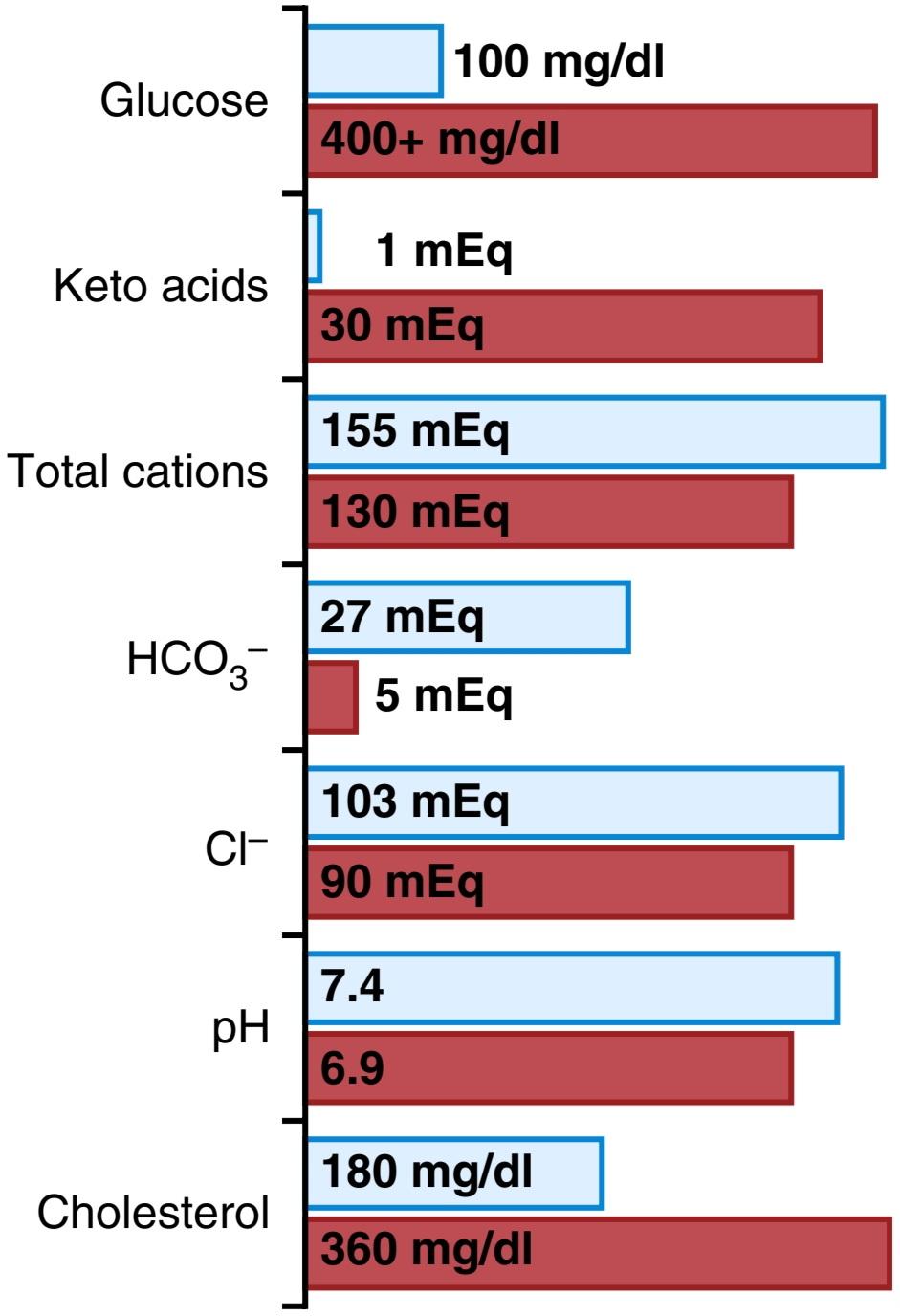
Figure 79-11. Changes in blood constituents in diabetic coma, showing normal values (blue bars) and diabetic coma values (red bars).
شکل 79-11. تغییرات در ترکیبات خون در کمای دیابتی، نشان دادن مقادیر طبیعی (نوارهای آبی) و مقادیر کمای دیابتی (نوارهای قرمز).
Diabetes Causes Depletion of the Body’s Proteins. Failure to use glucose for energy leads to increased utilization and decreased storage of proteins and fat. Therefore, a person with severe untreated diabetes mellitus experiences rapid weight loss and asthenia (lack of energy), despite eating large amounts of food (polyphagia). Without treatment, these metabolic abnormalities can cause severe wasting of the body tissues and death within a few weeks.
دیابت باعث کاهش پروتئین های بدن می شود. عدم استفاده از گلوکز برای انرژی منجر به افزایش استفاده و کاهش ذخیره پروتئین و چربی می شود. بنابراین، یک فرد مبتلا به دیابت شیرین درمان نشده شدید، علیرغم مصرف مقدار زیادی غذا (پلی فاژی) کاهش وزن سریع و آستنی (کمبود انرژی) را تجربه می کند. بدون درمان، این ناهنجاریهای متابولیک میتوانند باعث تحلیل رفتن شدید بافتهای بدن و مرگ در عرض چند هفته شوند.
Treatment of Type 1 Diabetes. Effective treatment of type 1 diabetes mellitus requires administration of enough insulin so that the patient will have carbohydrate, fat, and protein metabolism that is as normal as possible. Insulin is available in several forms. “Regular” insulin has a duration of action that lasts from 3 to 8 hours, whereas other forms of insulin (precipitated with zinc or with various protein derivatives) are absorbed slowly from the injection site and therefore have effects that last as long as 10 to 48 hours. Ordinarily, a patient with severe type 1 diabetes is given a single dose of one of the longer-acting insulins each day to increase overall carbohydrate metabolism throughout the day. Additional quantities of regular insulin are then given during the day at the times when the blood glucose level tends to rise too high, such as at mealtimes. Thus, each patient is provided with an individualized pattern of treatment.
درمان دیابت نوع 1. درمان موثر دیابت نوع 1 مستلزم تجویز انسولین کافی است تا بیمار متابولیسم کربوهیدرات، چربی و پروتئین را تا حد امکان طبیعی داشته باشد. انسولین به اشکال مختلف موجود است. انسولین منظم دارای مدت اثری است که از 3 تا 8 ساعت طول می کشد، در حالی که سایر اشکال انسولین (رسوب شده با روی یا مشتقات مختلف پروتئین) به آرامی از محل تزریق جذب می شوند و بنابراین اثراتی دارند که 10 تا 48 ساعت طول می کشد. به طور معمول، به بیمار مبتلا به دیابت نوع 1 شدید، هر روز یک دوز واحد از یکی از انسولینهای با اثر طولانیتر برای افزایش متابولیسم کلی کربوهیدرات در طول روز داده میشود. سپس مقادیر اضافی انسولین معمولی در طول روز و در مواقعی که سطح گلوکز خون بسیار بالا میرود، مانند زمان صرف غذا، داده میشود. بنابراین، هر بیمار با یک الگوی درمانی فردی ارائه می شود.
In the past, the insulin used for treatment was derived from animal pancreata. However, human insulin produced by the recombinant DNA process has become more widely used because immunity and sensitization against animal insulin develops in some patients, thus limiting its effectiveness.
در گذشته، انسولین مورد استفاده برای درمان از پانکراتای حیوانی گرفته می شد. با این حال، انسولین انسانی تولید شده توسط فرآیند DNA نوترکیب به طور گستردهتری مورد استفاده قرار گرفته است، زیرا ایمنی و حساسیت در برابر انسولین حیوانی در برخی از بیماران ایجاد میشود، بنابراین اثربخشی آن محدود میشود.
Type 2 Diabetes-Resistance to the Metabolic Effects of Insulin
Type 2 diabetes is far more common than type 1 diabetes, accounting for about 90% to 95% of all cases of diabetes mellitus. In most cases, the onset of type 2 diabetes occurs after age 30 years, often between the ages of 50 and 60 years, and the disease develops gradually. Therefore, this syndrome is often referred to as adult-onset diabetes. In recent years, however, there has been a steady increase in the number of younger individuals, some younger than 20 years old, with type 2 diabetes. This trend appears to be related mainly to the increasing prevalence of obesity, the most important risk factor for type 2 diabetes in children and adults.
دیابت نوع 2-مقاومت به اثرات متابولیکی انسولین
دیابت نوع 2 بسیار شایع تر از دیابت نوع 1 است و حدود 90 تا 95 درصد از کل موارد دیابت را تشکیل می دهد. در بیشتر موارد، شروع دیابت نوع 2 بعد از 30 سالگی و اغلب بین 50 تا 60 سالگی رخ می دهد و بیماری به تدریج ایجاد می شود. بنابراین، این سندرم اغلب به عنوان دیابت بزرگسالان شناخته می شود. با این حال، در سالهای اخیر، تعداد افراد جوانتر، برخی کمتر از 20 سال، به دیابت نوع 2 افزایش یافته است. به نظر می رسد این روند عمدتاً به افزایش شیوع چاقی، مهم ترین عامل خطر برای دیابت نوع 2 در کودکان و بزرگسالان مربوط می شود.
Obesity, Insulin Resistance, and “Metabolic Syndrome” Usually Precede Development of Type 2 Diabetes. Type 2 diabetes, in contrast to type 1 diabetes, is associated with increased plasma insulin concentration. The hyperinsulinemia occurs as a compensatory response by the pancreatic beta cells for insulin resistance, a diminished sensitivity of target tissues to the metabolic effects of insulin. The decrease in insulin sensitivity impairs carbohydrate utilization and storage, raising blood glucose and stimulating a compensatory increase in insulin secretion.
چاقی، مقاومت به انسولین و “سندرم متابولیک” معمولاً مقدم بر ابتلا به دیابت نوع 2 است. دیابت نوع 2، برخلاف دیابت نوع 1، با افزایش غلظت انسولین پلاسما همراه است. هیپرانسولینمی به عنوان یک پاسخ جبرانی توسط سلول های بتای پانکراس برای مقاومت به انسولین، کاهش حساسیت بافت های هدف به اثرات متابولیکی انسولین رخ می دهد. کاهش حساسیت به انسولین، مصرف و ذخیره کربوهیدرات را مختل می کند، گلوکز خون را افزایش می دهد و باعث افزایش جبرانی ترشح انسولین می شود.
The development of insulin resistance and impaired glucose metabolism is usually a gradual process, beginning with excess weight gain and obesity. Some studies suggest that obese subjects have fewer insulin receptors, especially in the skeletal muscle, liver, and adipose tissue, than do lean subjects. However, most of the insulin resistance appears to be caused by abnormalities of the signaling pathways that link receptor activation with multiple cellular effects. Impaired insulin signaling may be closely related to toxic effects of lipid accumulation in tissues such as skeletal muscle and liver as a result of excess weight gain.
ایجاد مقاومت به انسولین و اختلال در متابولیسم گلوکز معمولاً یک فرآیند تدریجی است که با افزایش وزن و چاقی شروع می شود. برخی از مطالعات نشان می دهد که افراد چاق نسبت به افراد لاغر گیرنده های انسولین کمتری دارند، به خصوص در ماهیچه های اسکلتی، کبد و بافت چربی. با این حال، به نظر می رسد که بیشتر مقاومت به انسولین ناشی از ناهنجاری های مسیرهای سیگنالینگ است که فعال شدن گیرنده را با اثرات سلولی متعدد مرتبط می کند. اختلال در سیگنال دهی انسولین ممکن است با اثرات سمی تجمع لیپید در بافت هایی مانند ماهیچه های اسکلتی و کبد در نتیجه افزایش وزن بیش از حد مرتبط باشد.
Insulin resistance is part of a cascade of disorders that is often called the “metabolic syndrome.” Some of the features of the metabolic syndrome include (1) obesity, especially accumulation of abdominal fat; (2) insulin resistance; (3) fasting hyperglycemia; (4) lipid abnormalities, such as increased blood triglycerides and decreased blood highdensity lipoprotein-cholesterol; and (5) hypertension. All of the features of the metabolic syndrome are closely related to accumulation of excess adipose tissue in the abdominal cavity around the visceral organs.
مقاومت به انسولین بخشی از مجموعه ای از اختلالات است که اغلب “سندرم متابولیک” نامیده می شود. برخی از ویژگی های سندرم متابولیک عبارتند از (1) چاقی، به ویژه تجمع چربی شکم. (2) مقاومت به انسولین؛ (3) هیپرگلیسمی ناشتا؛ (4) ناهنجاری های چربی، مانند افزایش تری گلیسیرید خون و کاهش لیپوپروتئین-کلسترول با چگالی بالا. و (5) فشار خون بالا. تمام ویژگی های سندرم متابولیک ارتباط نزدیکی با تجمع بافت چربی اضافی در حفره شکمی در اطراف اندام های احشایی دارد.
The role of insulin resistance in contributing to some of the components of the metabolic syndrome is uncertain, although it is clear that insulin resistance is the primary cause of increased blood glucose concentration. A major adverse consequence of the metabolic syndrome is cardiovascular disease, including atherosclerosis and in- jury to various organs throughout the body. Several of the metabolic abnormalities associated with the syndrome increase the risk for cardiovascular disease, and insulin resistance predisposes to the development of type 2 diabetes mellitus, which is also a major cause of cardiovascular disease.
نقش مقاومت به انسولین در کمک به برخی از اجزای سندرم متابولیک نامشخص است، اگرچه واضح است که مقاومت به انسولین علت اصلی افزایش غلظت گلوکز خون است. یکی از پیامدهای نامطلوب اصلی سندرم متابولیک، بیماری های قلبی عروقی، از جمله تصلب شرایین و آسیب به اندام های مختلف در سراسر بدن است. برخی از ناهنجاری های متابولیک مرتبط با این سندرم خطر ابتلا به بیماری های قلبی عروقی را افزایش می دهند و مقاومت به انسولین مستعد ابتلا به دیابت نوع 2 است که همچنین یکی از دلایل اصلی بیماری های قلبی عروقی است.
Other Factors That Can Cause Insulin Resistance and Type 2 Diabetes. Although most patients with type 2 diabetes are overweight or have substantial accumulation of visceral fat, severe insulin resistance and type 2 diabetes can also occur as a result of other acquired or genetic conditions that impair insulin signaling in peripheral tissues (Table 79-2).
عوامل دیگری که می توانند باعث مقاومت به انسولین و دیابت نوع 2 شوند. اگرچه اکثر بیماران مبتلا به دیابت نوع 2 اضافه وزن دارند یا تجمع قابل توجهی از چربی احشایی دارند، مقاومت شدید به انسولین و دیابت نوع 2 نیز می تواند در نتیجه سایر شرایط اکتسابی یا ژنتیکی که سیگنال دهی انسولین را در بافت های محیطی مختل می کند رخ دهد (جدول 79-2).
Polycystic ovary syndrome (PCOS), for example, is as- sociated with marked increases in ovarian androgen production and insulin resistance. PCOS is one of the most common endocrine disorders in women, affecting approximately 6% of all women during their reproductive life. Although the pathogenesis of PCOS remains uncertain, insulin resistance and hyperinsulinemia are found in approximately 80% of affected women. The long-term consequences include increased risk for diabetes mellitus, increased blood lipids, and cardiovascular disease.
به عنوان مثال، سندرم تخمدان پلی کیستیک (PCOS)، با افزایش قابل توجه تولید آندروژن تخمدان و مقاومت به انسولین مرتبط است. سندرم تخمدان پلی کیستیک یکی از شایع ترین اختلالات غدد درون ریز در زنان است که تقریباً 6 درصد از زنان را در طول زندگی باروری خود تحت تأثیر قرار می دهد. اگرچه پاتوژنز PCOS نامشخص است، مقاومت به انسولین و هیپرانسولینمی تقریباً در 80٪ از زنان مبتلا دیده می شود. پیامدهای درازمدت شامل افزایش خطر ابتلا به دیابت، افزایش چربی خون و بیماری های قلبی عروقی است.
Excess formation of glucocorticoids (Cushing’s syndrome) or excess formation of growth hormone (acromegaly) also decreases the sensitivity of various tissues to the metabolic effects of insulin and can lead to development of diabetes mellitus. Genetic causes of obesity and insulin resistance, if severe enough, also can lead to type 2 diabetes and many other features of the metabolic syndrome, including cardiovascular disease.
تشکیل بیش از حد گلوکوکورتیکوئیدها (سندرم کوشینگ) یا تشکیل بیش از حد هورمون رشد (آکرومگالی) نیز حساسیت بافت های مختلف به اثرات متابولیک انسولین را کاهش می دهد و می تواند منجر به ایجاد دیابت شود. علل ژنتیکی چاقی و مقاومت به انسولین، اگر به اندازه کافی شدید باشد، می تواند منجر به دیابت نوع 2 و بسیاری از ویژگی های دیگر سندرم متابولیک، از جمله بیماری های قلبی عروقی شود.
TABLE 79-2 Some Causes of Insulin Resistance
جدول 79-2 برخی از علل مقاومت به انسولین.
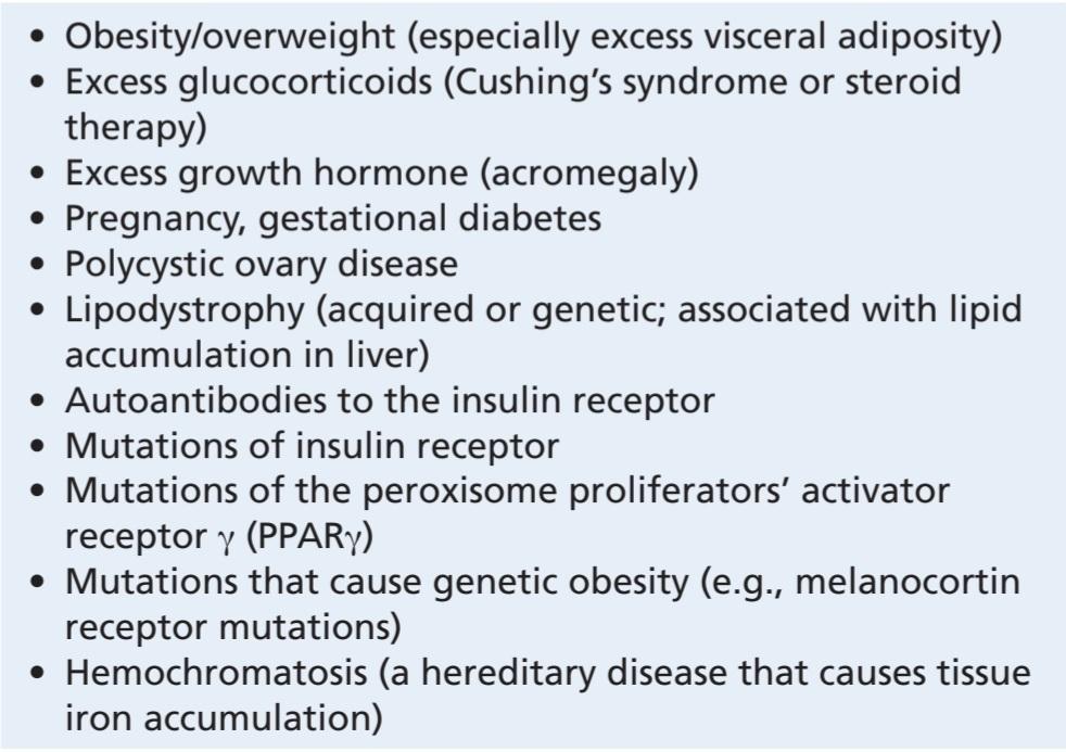
Development of Type 2 Diabetes During Prolonged Insulin Resistance. With prolonged and severe insulin resistance, even the increased levels of insulin are not sufficient to maintain normal glucose regulation. As a result, moderate hyperglycemia occurs after ingestion of carbohydrates in the early stages of the disease.
ایجاد دیابت نوع 2 در طول مقاومت طولانی مدت به انسولین. با مقاومت طولانی و شدید به انسولین، حتی افزایش سطح انسولین برای حفظ تنظیم طبیعی گلوکز کافی نیست. در نتیجه، هیپرگلیسمی متوسط پس از مصرف کربوهیدرات در مراحل اولیه بیماری رخ می دهد.
In the later stages of type 2 diabetes, the pancreatic beta cells become “exhausted” or damaged and are unable to produce enough insulin to prevent more severe hyperglycemia, especially after the person ingests a carbohydrate- rich meal.
در مراحل بعدی دیابت نوع 2، سلول های بتای پانکراس “فرسوده” یا آسیب دیده می شوند و قادر به تولید انسولین کافی برای جلوگیری از هیپرگلیسمی شدیدتر، به خصوص پس از مصرف یک وعده غذایی غنی از کربوهیدرات نیستند.
Clinically significant diabetes mellitus may not develop in some obese people, even though they have marked insulin resistance and greater than normal increases in blood glucose after a meal; apparently, the pancreas in these people produces enough insulin to prevent severe abnormalities of glucose metabolism. In other obese people, however, the pancreas gradually becomes exhausted from secreting large amounts of insulin or damaged by factors associated with lipid accumulation in the pancreas, and full-blown diabetes mellitus occurs. Some studies suggest that genetic factors play an important role in determining whether an individual’s pancreas can sustain the high output of insulin over many years that is necessary to avoid the severe abnormalities of glucose metabolism in type 2 diabetes.
دیابت ملیتوس قابل توجه بالینی ممکن است در برخی از افراد چاق ایجاد نشود، حتی اگر آنها مقاومت مشخصی به انسولین داشته باشند و قند خون بعد از غذا بیش از حد طبیعی افزایش یابد. ظاهراً پانکراس در این افراد انسولین کافی برای جلوگیری از ناهنجاری های شدید متابولیسم گلوکز تولید می کند. با این حال، در سایر افراد چاق، لوزالمعده به تدریج از ترشح مقادیر زیادی انسولین خسته می شود یا توسط عوامل مرتبط با تجمع چربی در پانکراس آسیب می بیند و دیابت شیرین کامل رخ می دهد. برخی از مطالعات نشان میدهند که عوامل ژنتیکی نقش مهمی در تعیین اینکه آیا لوزالمعده یک فرد میتواند تولید انسولین بالا را در طول سالیان متمادی حفظ کند که برای جلوگیری از ناهنجاریهای شدید متابولیسم گلوکز در دیابت نوع 2 ضروری است، بازی میکند.
Treatment of Type 2 Diabetes by Lifestyle Modifications, Increasing Insulin Sensitivity, and Enhancing Insulin Secretion. In many cases, type 2 diabetes can be effectively treated, at least in the early stages, with lifestyle modifications aimed at increasing physical activity, caloric restriction, and weight reduction, and no exogenous administration of insulin is required. Drugs that increase insulin sensitivity, such as thiazolidinediones, drugs that suppress liver glucose production, such as metformin, or drugs that cause additional release of insulin by the pancreas, such as sulfonylureas, may also be used. However, in the later stages of type 2 diabetes, insulin administration is usually required to control plasma glucose levels.
درمان دیابت نوع 2 با اصلاح سبک زندگی، افزایش حساسیت به انسولین و افزایش ترشح انسولین. در بسیاری از موارد، دیابت نوع 2 را می توان به طور موثر حداقل در مراحل اولیه، با اصلاح شیوه زندگی با هدف افزایش فعالیت بدنی، محدودیت کالری و کاهش وزن درمان کرد و نیازی به تجویز اگزوژن انسولین نیست. داروهایی که حساسیت به انسولین را افزایش می دهند، مانند تیازولیدین دیون ها، داروهایی که تولید گلوکز کبد را سرکوب می کنند، مانند متفورمین، یا داروهایی که باعث آزادسازی اضافی انسولین توسط پانکراس می شوند، مانند سولفونیل اوره نیز ممکن است استفاده شوند. با این حال، در مراحل بعدی دیابت نوع 2، معمولاً تجویز انسولین برای کنترل سطح گلوکز پلاسما لازم است.
Incretin drugs that mimic the actions of the GLP-1 have been developed for treatment of type 2 diabetes. These drugs enhance insulin secretion and are intended to be used in conjunction with other antidiabetic drugs. Another therapeutic approach is to inhibit the enzyme dipeptidyl peptidase 4 (DPP-4), which inactivates GLP-1 and GIP. By blocking the actions of DPP-4, the incretin effects of GLP-1 and GIP can be prolonged, leading to increased insulin secretion and improved control of blood glucose levels.
داروهای اینکرتین که عملکرد GLP-1 را تقلید می کنند برای درمان دیابت نوع 2 ساخته شده اند. این داروها ترشح انسولین را افزایش می دهند و برای استفاده همراه با سایر داروهای ضد دیابت در نظر گرفته شده اند. روش درمانی دیگر، مهار آنزیم دی پپتیدیل پپتیداز 4 (DPP-4) است که GLP-1 و GIP را غیرفعال می کند. با مسدود کردن فعالیتهای DPP-4، اثرات اینکرتین GLP-1 و GIP میتواند طولانیتر شود و منجر به افزایش ترشح انسولین و بهبود کنترل سطح گلوکز خون شود.
Treatment of Type 2 Diabetes by Inhibition of Sodium- Glucose Transporter 2 (SGLT2). As discussed in Chapter 28, approximately 90% of the glucose filtered by renal glomerular capillaries is reabsorbed from the proximal tubules by the sodium glucose co-transporter 2 (SGLT2). Several medications, called gliflozins, have been developed to treat type 2 diabetes by inhibiting SGLT2. These SGLT2 inhibitors greatly reduce renal glucose reabsorption, causing large amounts of glucose to be excreted in the urine and reducing the blood glucose concentration. SGLT2 inhibitors are often used in combination with other drugs that enhance insulin sensitivity or stimulate insulin secretion and have been shown in clinical trials to provide significant protection against cardiovascular and kidney disease in patients with diabetes.
درمان دیابت نوع 2 با مهار انتقال دهنده سدیم- گلوکز 2 (SGLT2). همانطور که در فصل 28 بحث شد، تقریباً 90 درصد از گلوکز فیلتر شده توسط مویرگ های گلومرولی کلیوی از لوله های پروگزیمال توسط هم انتقال دهنده گلوکز سدیم 2 (SGLT2) دوباره جذب می شود. چندین دارو به نام گلیفلوزین برای درمان دیابت نوع 2 با مهار SGLT2 ساخته شده است. این مهارکننده های SGLT2 بازجذب گلوکز کلیوی را تا حد زیادی کاهش می دهند و باعث می شوند که مقادیر زیادی گلوکز از طریق ادرار دفع شود و غلظت گلوکز خون کاهش یابد. مهارکنندههای SGLT2 اغلب در ترکیب با داروهای دیگری استفاده میشوند که حساسیت انسولین را افزایش میدهند یا ترشح انسولین را تحریک میکنند و در آزمایشهای بالینی نشان داده شدهاند که محافظت قابلتوجهی در برابر بیماریهای قلبی عروقی و کلیوی در بیماران مبتلا به دیابت دارند.
In addition to increasing glucose excretion, SGLT2 inhibitors also cause marked diuresis due to the osmotic effect of the glucose remaining in the renal tubules. The diuresis can be beneficial for causing small reductions in blood pressure in patients with type 2 diabetes who often suffer from hypertension, but may also increase the risk for dehydration and hypotension in patients who are already taking other diuretics and antihypertensive mediations.
مهارکنندههای SGLT2 علاوه بر افزایش دفع گلوکز، به دلیل اثر اسمزی گلوکز باقیمانده در لولههای کلیوی، باعث دیورز مشخصی نیز میشوند. دیورز می تواند برای ایجاد کاهش اندک فشار خون در بیماران مبتلا به دیابت نوع 2 که اغلب از فشار خون بالا رنج می برند مفید باشد، اما همچنین ممکن است خطر کم آبی و افت فشار خون را در بیمارانی که قبلاً سایر مدرها و واسطه های ضد فشار خون مصرف می کنند افزایش دهد.
Treatment of Type 2 Diabetes With Surgery. In many people who suffer from severe obesity and type 2 diabetes, treatment regimens focused on diet, exercise, and pharmacotherapy do not produce adequate reductions in adiposity and blood glucose. In these cases, various bariatric surgery procedures can be used to reduce fat mass and achieve improved control of blood glucose. The two most widely used procedures, gastric bypass surgery and vertical sleeve gastrectomy (discussed in Chapter 72), are often called “metabolic surgery” because many patients who undergo these operations experience complete remission of diabetes and no longer require antidiabetic drugs. Improvements of blood glucose, lipids and blood pressure often occur within a few days or weeks after surgery, suggesting that the mechanisms for these cardiovascular and metabolic benefits may extend beyond weight loss and reductions in adiposity. However, the physiological factors that contribute to the favorable metabolic effects of these surgical procedures are still unclear.
درمان دیابت نوع 2 با جراحی. در بسیاری از افرادی که از چاقی شدید و دیابت نوع 2 رنج میبرند، رژیمهای درمانی متمرکز بر رژیم غذایی، ورزش و دارودرمانی باعث کاهش کافی در چاقی و گلوکز خون نمیشوند. در این موارد می توان از روش های مختلف جراحی چاقی برای کاهش توده چربی و دستیابی به کنترل بهبود یافته گلوکز خون استفاده کرد. دو روش پرکاربرد، جراحی بای پس معده و گاسترکتومی عمودی اسلیو (در فصل 72 مورد بحث قرار گرفت)، اغلب “جراحی متابولیک” نامیده می شوند زیرا بسیاری از بیمارانی که تحت این عمل ها قرار می گیرند بهبودی کامل دیابت را تجربه می کنند و دیگر نیازی به داروهای ضد دیابت ندارند. بهبود قند خون، چربی ها و فشار خون اغلب در عرض چند روز یا چند هفته پس از جراحی رخ می دهد، که نشان می دهد مکانیسم های این فواید قلبی عروقی و متابولیک ممکن است فراتر از کاهش وزن و کاهش چاقی باشد. با این حال، عوامل فیزیولوژیکی که به اثرات متابولیکی مطلوب این روشهای جراحی کمک میکنند، هنوز نامشخص هستند.
Physiology of Diagnosis of Diabetes Mellitus
Table 79-3 compares some of the clinical features of type 1 and type 2 diabetes mellitus. The usual methods for diagnosing diabetes are based on various chemical tests of the urine and the blood.
فیزیولوژی تشخیص دیابت ملیتوس
جدول 79-3 برخی از ویژگی های بالینی دیابت نوع 1 و نوع 2 را مقایسه می کند. روش های معمول برای تشخیص دیابت بر اساس آزمایش های شیمیایی مختلف ادرار و خون است.
TABLE 79-3 Clinical Characteristics of Patients With Type 1 and Type 2 Diabetes Mellitus
جدول 79-3 ویژگی های بالینی بیماران مبتلا به دیابت نوع 1 و نوع 2.
 SGLT2, Sodium glucose co-transporter 2.
Urinary Glucose. Simple office tests or more complicated quantitative laboratory tests may be used to determine the quantity of glucose excreted in the urine. In general, a nondiabetic person excretes undetectable amounts of glucose, whereas a person with diabetes loses glucose in small to large amounts, in proportion to the severity of the dis- ease and the intake of carbohydrates.
گلوکز ادرار. ممکن است برای تعیین مقدار گلوکز دفع شده در ادرار از آزمایشهای اداری ساده یا آزمایشهای آزمایشگاهی کمی پیچیدهتر استفاده شود. به طور کلی، یک فرد غیر دیابتی مقادیر غیرقابل تشخیص گلوکز را دفع می کند، در حالی که فرد مبتلا به دیابت به نسبت شدت بیماری و دریافت کربوهیدرات، گلوکز را در مقادیر کم تا زیاد از دست می دهد.
Fasting Blood Glucose and Insulin Concentrations. Fasting blood glucose concentration in the early morning is normally 80 to 90 mg/100 ml, and 115 mg/100 ml is considered to be the upper limit of normal. A fasting blood glucose level above this value often indicates diabetes mellitus or at least marked insulin resistance and prediabetes.
غلظت گلوکز و انسولین خون ناشتا. غلظت گلوکز خون ناشتا در اوایل صبح معمولاً 80 تا 90 میلی گرم در 100 میلی لیتر است و 115 میلی گرم در 100 میلی لیتر به عنوان حد بالای طبیعی در نظر گرفته می شود. سطح گلوکز خون ناشتا بالاتر از این مقدار اغلب نشان دهنده دیابت یا حداقل مقاومت به انسولین مشخص و پیش دیابت است.
In persons with type 1 diabetes, plasma insulin levels are very low or undetectable during fasting and even after a meal. In persons with type 2 diabetes, plasma insulin concentration may be severalfold higher than normal and usually increases to a greater extent after ingestion of a standard glucose load during a glucose tolerance test (see the next section).
در افراد مبتلا به دیابت نوع 1، سطح انسولین پلاسما در طول روزه داری و حتی بعد از غذا بسیار کم یا غیرقابل تشخیص است. در افراد مبتلا به دیابت نوع 2، غلظت انسولین پلاسما ممکن است چندین برابر بیشتر از حد طبیعی باشد و معمولاً پس از مصرف یک بار استاندارد گلوکز در طول آزمایش تحمل گلوکز، به میزان بیشتری افزایش مییابد (به بخش بعدی مراجعه کنید).
Glycated Hemoglobin. When blood glucose levels are elevated for prolonged periods of time, glucose attaches to hemoglobin in red blood cells to form glycated hemoglobin, often called hemoglobin A1c (HbA1c). The longer hyperglycemia occurs, the more glucose binds to hemoglobin and once hemoglobin is glycated, it remains that way for the life of the cell. Therefore, buildup of HbA1c in a red blood cell reflects the average glucose concentration to which the cell has been exposed during its life-cycle. Because the average lifespan of red blood cells is about 120 days and individual cells have varying lifespans, the HbA1c test is used mainly to assess average blood glucose concentrations for the previous three months and can provide a diagnostic test for diabetes mellitus or an assessment test of glycemic control in people with diabetes.
هموگلوبین گلیکوزیله. هنگامی که سطح گلوکز خون برای مدت طولانی افزایش می یابد، گلوکز به هموگلوبین در گلبول های قرمز خون متصل می شود و هموگلوبین گلیکوزه شده را تشکیل می دهد که اغلب هموگلوبین A1c (HbA1c) نامیده می شود. هر چه هیپرگلیسمی طولانیتر باشد، گلوکز بیشتری به هموگلوبین متصل میشود و زمانی که هموگلوبین گلیکوزه شد، تا پایان عمر سلول به همین شکل باقی میماند. بنابراین، تجمع HbA1c در گلبول قرمز نشان دهنده میانگین غلظت گلوکزی است که سلول در طول چرخه زندگی خود در معرض آن قرار گرفته است. از آنجایی که میانگین طول عمر گلبول های قرمز خون حدود 120 روز است و سلول های فردی طول عمر متفاوتی دارند، آزمایش HbA1c عمدتاً برای ارزیابی میانگین غلظت گلوکز خون در سه ماه گذشته استفاده می شود و می تواند یک آزمایش تشخیصی برای دیابت شیرین یا یک آزمایش ارزیابی کنترل قند خون در افراد مبتلا به دیابت ارائه دهد.
Glucose Tolerance Test. As demonstrated by the bottom curve in Figure 79-12, called a “glucose tolerance curve,” when a normal, fasting person ingests 1 gram of glucose per kilogram of body weight, the blood glucose level rises from about 90 mg/100 ml to 120 to 140 mg/100 ml and falls back to below normal in about 2 hours.
تست تحمل گلوکز. همانطور که توسط منحنی پایینی در شکل 79-12 نشان داده شده است که “منحنی تحمل گلوکز” نامیده می شود، زمانی که یک فرد عادی و روزه دار 1 گرم گلوکز به ازای هر کیلوگرم وزن بدن خود را مصرف می کند، سطح گلوکز خون از حدود 90 میلی گرم در 100 میلی لیتر به 120 تا 140 میلی گرم در هر 100 میلی لیتر در حدود 2 ساعت به پایین تر از حد طبیعی می رسد.
In a person with diabetes, the fasting blood glucose concentration is almost always above 115 mg/100 ml and often is above 140 mg/100 ml. In addition, results of the glucose tolerance test are almost always abnormal. After ingestion of glucose, these people exhibit a much greater than normal rise in blood glucose level, as demonstrated by the up- per curve in Figure 79-12, and the glucose level falls back to the control value only after 4 to 6 hours; furthermore, it fails to fall below the control level. The slow fall of this curve and its failure to fall below the control level demonstrate that either (1) the normal increase in insulin secretion after glucose ingestion does not occur, or (2) the person has decreased sensitivity to insulin. A diagnosis of diabetes mellitus can usually be established on the basis of such a curve, and type 1 and type 2 diabetes can be distinguished from each other by measurements of plasma insulin, with plasma insulin being low or undetectable in type 1 diabetes and increased in type 2 diabetes.
در یک فرد مبتلا به دیابت، غلظت گلوکز خون ناشتا تقریباً همیشه بالای 115 میلی گرم در 100 میلی لیتر و اغلب بالای 140 میلی گرم در 100 میلی لیتر است. علاوه بر این، نتایج آزمایش تحمل گلوکز تقریباً همیشه غیر طبیعی است. پس از مصرف گلوکز، این افراد افزایش بسیار بیشتر از حد طبیعی در سطح گلوکز خون را نشان می دهند، همانطور که با منحنی بالا در شکل 79-12 نشان داده شده است، و سطح گلوکز تنها پس از 4 تا 6 ساعت به مقدار کنترل کاهش می یابد. علاوه بر این، نمی تواند به زیر سطح کنترل سقوط کند. کاهش آهسته این منحنی و پایین آمدن آن از سطح کنترل نشان می دهد که یا (1) افزایش طبیعی ترشح انسولین پس از مصرف گلوکز رخ نمی دهد، یا (2) حساسیت فرد به انسولین کاهش یافته است. تشخیص دیابت شیرین معمولاً بر اساس چنین منحنی مشخص می شود و دیابت نوع 1 و نوع 2 را می توان با اندازه گیری انسولین پلاسما از یکدیگر متمایز کرد، به طوری که انسولین پلاسما در دیابت نوع 1 کم یا غیرقابل تشخیص است و در دیابت نوع 2 افزایش می یابد.

Figure 79-12. Glucose tolerance curve in a normal person and in a person with diabetes.
شکل 79-12. منحنی تحمل گلوکز در یک فرد عادی و در یک فرد مبتلا به دیابت.
Acetone Breath. As pointed out in Chapter 69, small quantities of acetoacetic acid in the blood, which increase greatly in severe diabetes, are converted to acetone. Acetone is volatile and is vaporized into the expired air. Consequently, one can frequently make a diagnosis of type 1 diabetes mellitus simply by smelling acetone on the breath of a patient. Also, keto acids can be detected by chemical means in the urine, and their quantitation aids in determining the severity of the diabetes. In the early stages of type 2 diabetes, however, keto acids are usually not produced in excess amounts. However, when insulin resistance becomes severe and there is greatly increased utilization of fats for energy, keto acids are then produced in persons with type 2 diabetes.
تنفس استون. همانطور که در فصل 69 اشاره شد، مقادیر کمی اسید استواستیک در خون، که در دیابت شدید به شدت افزایش می یابد، به استون تبدیل می شود. استون فرار است و در هوای منقضی شده تبخیر می شود. در نتیجه، اغلب می توان دیابت نوع 1 را به سادگی با بوییدن استون در نفس بیمار تشخیص داد. همچنین، کتو اسیدها را می توان با روش های شیمیایی در ادرار تشخیص داد و کمیت آن ها به تعیین شدت دیابت کمک می کند. با این حال، در مراحل اولیه دیابت نوع 2، کتو اسیدها معمولاً در مقادیر بیش از حد تولید نمی شوند. با این حال، زمانی که مقاومت به انسولین شدید میشود و استفاده از چربیها برای انرژی افزایش مییابد، اسیدهای کتو در افراد مبتلا به دیابت نوع 2 تولید میشوند.
Relation of Treatment to Arteriosclerosis and Chronic Kidney Disease. Mainly because of the hypertension and high levels of circulating cholesterol and other lipids in diabetic patients, atherosclerosis, arteriosclerosis, severe coronary heart disease, chronic kidney disease, and multiple microcirculatory lesions develop far more easily than in nondiabetic people. Indeed, persons who have poorly controlled diabetes throughout childhood are likely to die of heart disease in early adulthood.
ارتباط درمان با تصلب شرایین و بیماری مزمن کلیه. عمدتاً به دلیل فشار خون بالا و سطوح بالای کلسترول در گردش و سایر چربی ها در بیماران دیابتی، آترواسکلروز، تصلب شرایین، بیماری شدید کرونری قلب، بیماری مزمن کلیوی و ضایعات متعدد میکروسیرکولاتوری بسیار راحت تر از افراد غیر دیابتی ایجاد می شود. در واقع، افرادی که دیابت در دوران کودکی به خوبی کنترل نمیشوند، احتمالاً در اوایل بزرگسالی بر اثر بیماری قلبی میمیرند.
In the early days of diabetes treatment, the tendency was to severely reduce carbohydrates in the diet to minimize insulin requirements. This approach kept the blood glucose from reaching too high a level and attenuated the loss of glucose in the urine, but it did not prevent many of the abnormalities of fat metabolism. Consequently, the current tendency is to permit the patient to consume an almost normal carbohydrate diet while administering enough insulin to metabolize the carbohydrates. This approach decreases the rate of fat metabolism and depresses the high level of cholesterol in the blood.
در روزهای اولیه درمان دیابت، تمایل به کاهش شدید کربوهیدرات در رژیم غذایی برای به حداقل رساندن نیاز به انسولین بود. این رویکرد باعث میشود که گلوکز خون به سطح بسیار بالایی نرسد و از دست رفتن گلوکز در ادرار را کاهش دهد، اما از بسیاری از ناهنجاریهای متابولیسم چربی جلوگیری نکرد. در نتیجه، تمایل فعلی این است که به بیمار اجازه میدهیم از یک رژیم غذایی تقریباً معمولی کربوهیدرات استفاده کند، در حالی که انسولین کافی برای متابولیسم کربوهیدراتها تجویز میشود. این رویکرد باعث کاهش سرعت متابولیسم چربی و کاهش سطح بالای کلسترول در خون می شود.
Because complications of diabetes such as atherosclerosis, increased susceptibility to infection, diabetic retinopathy, cataracts, hypertension, and chronic renal disease are closely associated with the levels of lipids and glucose in the blood, most physicians also prescribe lipid-lowering drugs to help prevent these disturbances.
از آنجایی که عوارض دیابت مانند تصلب شرایین، افزایش حساسیت به عفونت، رتینوپاتی دیابتی، آب مروارید، فشار خون بالا و بیماری مزمن کلیوی ارتباط تنگاتنگی با سطح لیپیدها و گلوکز در خون دارند، اکثر پزشکان داروهای کاهنده چربی را نیز برای جلوگیری از این اختلالات تجویز می کنند.
Insulinoma-Hyperinsulinism
Although excessive insulin production occurs much more rarely than does diabetes, it occasionally can be a consequence of an adenoma of an islet of Langerhans. About 10% to 15% of these adenomas are malignant, and occasionally metastases from the islets of Langerhans spread throughout the body, causing tremendous production of insulin by both the primary and metastatic cancers. Indeed, some of these patients have required more than 1000 grams of glucose every 24 hours to prevent hypoglycemia.
انسولینوما-هیپرانسولینیسم
اگرچه تولید بیش از حد انسولین بسیار نادرتر از دیابت اتفاق می افتد، اما گاهی اوقات می تواند نتیجه آدنوم جزایر لانگرهانس باشد. حدود 10 تا 15 درصد از این آدنوم ها بدخیم هستند و گاهی اوقات متاستاز از جزایر لانگرهانس در سراسر بدن پخش می شود و باعث تولید فوق العاده انسولین توسط سرطان های اولیه و متاستاتیک می شود. در واقع، برخی از این بیماران برای جلوگیری از هیپوگلیسمی به بیش از 1000 گرم گلوکز در هر 24 ساعت نیاز دارند.
Insulin Shock and Hypoglycemia. As already emphasized, the central nervous system normally derives essentially all its energy from glucose metabolism, and insulin is not necessary for this use of glucose. However, if high levels of insulin cause blood glucose to fall to low levels, the metabolism of the central nervous system becomes depressed. Consequently, in patients with insulin-secreting tumors or in patients with diabetes who administer too much insulin to themselves, the syndrome called insulin shock may occur as follows.
شوک انسولین و هیپوگلیسمی. همانطور که قبلاً تأکید شد، سیستم عصبی مرکزی به طور معمول اساساً تمام انرژی خود را از متابولیسم گلوکز می گیرد و انسولین برای این استفاده از گلوکز ضروری نیست. با این حال، اگر سطوح بالای انسولین باعث کاهش گلوکز خون به سطوح پایین شود، متابولیسم سیستم عصبی مرکزی افسرده می شود. در نتیجه، در بیماران مبتلا به تومورهای ترشح کننده انسولین یا در بیماران مبتلا به دیابت که انسولین بیش از حد برای خود تجویز می کنند، سندرمی به نام شوک انسولین ممکن است به شرح زیر رخ دهد.
As blood glucose level falls into the range of 50 to 70 mg/100 ml, the central nervous system usually becomes excitable because this degree of hypoglycemia sensitizes neuronal activity. Sometimes various forms of hallucinations result, but more often the patient simply experiences extreme nervousness, trembles all over, and breaks out in a sweat. As blood glucose level falls to 20 to 50 mg/100 ml, clonic seizures and loss of consciousness are likely to occur. As the glucose level falls still lower, the seizures cease and only a state of coma remains. Indeed, when using simple clinical observation, it is sometimes difficult to distinguish between diabetic coma as a result of acidosis caused by a lack of insulin and coma due to hypoglycemia caused by excess insulin. The acetone breath and the rapid, deep breathing of persons in a diabetic coma are not present in persons in a hypoglycemic coma.
با کاهش سطح گلوکز خون در محدوده 50 تا 70 میلی گرم در 100 میلی لیتر، سیستم عصبی مرکزی معمولاً تحریک پذیر می شود زیرا این درجه از هیپوگلیسمی فعالیت عصبی را حساس می کند. گاهی اوقات اشکال مختلفی از توهم ایجاد می شود، اما بیشتر اوقات بیمار به سادگی عصبی می شود، همه جا می لرزد و عرق می کند. با کاهش سطح گلوکز خون به 20 تا 50 میلی گرم در 100 میلی لیتر، تشنج های کلونیک و از دست دادن هوشیاری به احتمال زیاد رخ می دهد. همانطور که سطح گلوکز همچنان پایین تر است، تشنج متوقف می شود و تنها حالت کما باقی می ماند. در واقع، هنگام استفاده از مشاهدات بالینی ساده، گاهی اوقات تشخیص کمای دیابتی در نتیجه اسیدوز ناشی از کمبود انسولین و کما ناشی از هیپوگلیسمی ناشی از انسولین اضافی دشوار است. تنفس استون و تنفس سریع و عمیق افراد در کمای دیابتی در افرادی که در کمای هیپوگلیسمی هستند وجود ندارد.
Proper treatment for a patient who has hypoglycemic shock or coma is immediate intravenous administration of large quantities of glucose. This treatment usually brings the patient out of shock within a minute or more. Also, ad- ministration of glucagon (or, less effectively, epinephrine) can cause glycogenolysis in the liver and thereby increase the blood glucose level extremely rapidly. If treatment is not administered immediately, permanent damage to the neuronal cells of the central nervous system often occurs.
درمان مناسب برای بیمار مبتلا به شوک هیپوگلیسمی یا کما، تجویز فوری داخل وریدی مقادیر زیاد گلوکز است. این درمان معمولاً بیمار را در عرض یک دقیقه یا بیشتر از شوک خارج می کند. همچنین، تجویز گلوکاگون (یا به طور مؤثرتر اپی نفرین) می تواند باعث گلیکوژنولیز در کبد شود و در نتیجه سطح گلوکز خون را به سرعت افزایش دهد. اگر درمان بلافاصله انجام نشود، آسیب دائمی به سلول های عصبی سیستم عصبی مرکزی اغلب رخ می دهد.
کلیک کنید «Bibliography: فهرست کتب مربوطه»
Alicic RZ, Neumiller JJ, Johnson EJ, et al: Sodium-glucose cotransporter 2 inhibition and diabetic kidney disease. Diabetes 68:248, 2019.
Andersen A, Lund A, Knop FK, Vilsbøll T: Glucagon-like peptide 1 in health and disease. Nat Rev Endocrinol 14:390, 2018.
Bentsen MA, Mirzadeh Z, Schwartz MW: Revisiting how the brain senses glucoseand why. Cell Metab 29:11, 2018.
Capozzi ME, DiMarchi RD, Tschöp MH, et al: Targeting the incretin/glucagon system with triagonists to treat diabetes. Endocr Rev 39:719, 2018.
Clemmensen C, Finan B, Müller TD, et al: Emerging hormonal-based combination pharmacotherapies for the treatment of metabolic diseases. Nat Rev Endocrinol 15:90, 2019.
DiMeglio LA, Evans-Molina C, Oram RA: Type 1 diabetes. Lancet 391:2449, 2018.
Gancheva S, Jelenik T, Álvarez-Hernández E, Roden M: Interorgan metabolic crosstalk in human insulin resistance. Physiol Rev 98:1371, 2018.
Haeusler RA, McGraw TE, Accili D: Biochemical and cellular properties of insulin receptor signalling. Nat Rev Mol Cell Biol 19:31, 2018.
Kahn CR, Wang G, Lee KY: Altered adipose tissue and adipocyte function in the pathogenesis of metabolic syndrome. J Clin Invest 129:3990, 2019.
Klip A, McGraw TE, James DE: Thirty sweet years of GLUT4. J Biol Chem 294:11369, 2019.
Lee YS, Wollam J, Olefsky JM: An integrated view of immunometabolism. Cell 172(1-2):22, 2018.
Mann JP, Savage DB: What lipodystrophies teach us about the metabolic syndrome. J Clin Invest 130:4009, 2019.
Müller TD, Finan B, Clemmensen C, et al: The new biology and pharmacology of glucagon. Physiol Rev 97:721, 2017.
Oram RA, Sims EK, Evans-Molina C: Beta cells in type 1 diabetes: mass and function; sleeping or dead? Diabetologia 62:567, 2019.
Pareek M, Schauer PR, Kaplan LM, et al: Metabolic surgery: weight loss, diabetes, and beyond. J Am Coll Cardiol 71:670, 2018.
Petersen MC, Shulman Gl: Mechanisms of insulin action and insulin resistance. Physiol Rev 98:2133, 2018.
Rorsman P, Ashcroft FM: Pancreatic B-cell electrical activity and insulin secretion: of mice and men. Physiol Rev 98:117, 2018.
Ruegsegger GN, Creo AL, Cortes TM, Dasari S, Nair KS: Altered mitochondrial function in insulin-deficient and insulin-resistant states. J Clin Invest 128:3671, 2018.
Taylor R, Al-Mrabeh A, Sattar N: Understanding the mechanisms of reversal of type 2 diabetes. Lancet Diabetes Endocrinol 7:726, 2019.
Viner R, White B, Christie D: Type 2 diabetes in adolescents: a severe phenotype posing major clinical challenges and public health bur- den. Lancet 389:2252, 2017.
Wright EM, Loo DD, Hirayama BA: Biology of human sodium glucose transporters. Physiol Rev 91:733, 2011.
Yang Q, Vijayakumar A, Kahn BB: Metabolites as regulators of insulin sensitivity and metabolism. Nat Rev Mol Cell Biol 19:654, 2018.
🚀 با ما همراه شوید!
تازهترین مطالب و آموزشهای مغز و اعصاب را از دست ندهید. با فالو کردن کانال تلگرام آیندهنگاران مغز، از ما حمایت کنید!



