
فیزیولوژی پزشکی گایتون و هال؛ هورمون های قشر آدرنال


دعای مطالعه [ نمایش ]
بِسْمِ الله الرَّحْمنِ الرَّحیمِ
اَللّهُمَّ اَخْرِجْنى مِنْ ظُلُماتِ الْوَهْمِ
خدايا مرا بيرون آور از تاريكىهاى وهم،
وَ اَكْرِمْنى بِنُورِ الْفَهْمِ
و به نور فهم گرامى ام بدار،
اَللّهُمَّ افْتَحْ عَلَيْنا اَبْوابَ رَحْمَتِكَ
خدايا درهاى رحمتت را به روى ما بگشا،
وَانْشُرْ عَلَيْنا خَزائِنَ عُلُومِكَ بِرَحْمَتِكَ يا اَرْحَمَ الرّاحِمينَ
و خزانههاى علومت را بر ما باز كن به امید رحمتت اى مهربانترين مهربانان.
کتاب «فیزیولوژی پزشکی گایتون و هال» بهعنوان یکی از جامعترین و معتبرترین منابع در حوزه علوم پزشکی، همچنان مرجع کلیدی برای درک عملکرد پیچیده بدن انسان است. این اثر با تکیه بر تازهترین پژوهشها و توضیحات دقیق از سازوکارهای فیزیولوژیک، پلی میان علوم پایه پزشکی و کاربردهای بالینی ایجاد میکند و نقشی بیبدیل در آموزش، پژوهش و ارتقای دانش سلامت ایفا مینماید.
ترجمه دقیق و علمی این شاهکار توسط برند علمی آیندهنگاران مغز به مدیریت داریوش طاهری، دسترسی فارسیزبانان به مرزهای نوین دانش فیزیولوژی را ممکن ساخته و رسالتی علمی برای ارتقای آموزش پزشکی، فهم عمیقتر سازوکارهای بدن و توسعه روشهای نوین در حوزه سلامت فراهم آورده است.
» Guyton and Hall Textbook of Medical Physiology
»» CHAPTER 78: Adrenocortical Hormones
The two adrenal glands, each of which weighs about 4 grams, lie at the superior poles of the two kidneys. As shown in Figure 78-1, each gland is composed of two major parts, the adrenal medulla and the adrenal cortex. The adrenal medulla, the central 20% of the gland, is functionally related to the sympathetic nervous system; it secretes the hormones epinephrine and norepinephrine in response to sympathetic stimulation. In turn, these hormones cause almost the same effects as direct stimulation of the sympathetic nerves in all parts of the body. These hormones and their effects are discussed in detail in Chapter 61 in relation to the sympathetic nervous system.
دو غده فوق کلیوی که وزن هر کدام حدود 4 گرم است در قطب فوقانی دو کلیه قرار دارند. همانطور که در شکل 78-1 نشان داده شده است، هر غده از دو قسمت اصلی، بصل الکلی و قشر آدرنال تشکیل شده است. مدولای آدرنال، 20 درصد مرکزی غده، از نظر عملکردی با سیستم عصبی سمپاتیک مرتبط است. در پاسخ به تحریک سمپاتیک، هورمون های اپی نفرین و نوراپی نفرین را ترشح می کند. به نوبه خود، این هورمون ها تقریباً اثراتی مشابه با تحریک مستقیم اعصاب سمپاتیک در تمام قسمت های بدن ایجاد می کنند. این هورمون ها و اثرات آنها در فصل 61 در رابطه با سیستم عصبی سمپاتیک به تفصیل مورد بحث قرار گرفته است.
The adrenal cortex secretes an entirely different group of hormones, called corticosteroids. These hormones are all synthesized from the steroid cholesterol, and they all have similar chemical formulas. However, slight differences in their molecular structures give them several different but very important functions.
قشر آدرنال گروه کاملا متفاوتی از هورمونها به نام کورتیکواستروئیدها را ترشح میکند. این هورمون ها همگی از کلسترول استروئیدی سنتز می شوند و همگی فرمول های شیمیایی مشابهی دارند. با این حال، تفاوت های جزئی در ساختار مولکولی آنها چندین عملکرد متفاوت اما بسیار مهم را به آنها می دهد.

Figure 78-1. Secretion of adrenocortical hormones by the different zones of the adrenal cortex and secretion of catecholamines by the adrenal medulla.
شکل 78-1. ترشح هورمونهای قشر آدرنال توسط نواحی مختلف قشر آدرنال و ترشح کاتکول آمینها توسط مدولای آدرنال.
CORTICOSTEROIDS: MINERALOCORTICOIDS, GLUCOCORTICOIDS, AND ANDROGENS
Two major types of adrenocortical hormones, the mineralocorticoids and the glucocorticoids, are secreted by the adrenal cortex. In addition to these hormones, small amounts of sex hormones are secreted, especially androgenic hormones, which exhibit about the same effects in the body as the male sex hormone testosterone. They are normally of only slight importance, although in certain abnormalities of the adrenal cortices, extreme quantities can be secreted (which is discussed later in the chapter) and can result in masculinizing effects.
کورتیکواستروئیدها: مینرالوکورتیکوئیدها، گلوکوکورتیکوئیدها و آندروژن ها
دو نوع اصلی از هورمونهای قشر آدرنال، مینرالوکورتیکوئیدها و گلوکوکورتیکوئیدها، توسط قشر آدرنال ترشح میشوند. علاوه بر این هورمونها، مقادیر کمی از هورمونهای جنسی ترشح میشوند، بهویژه هورمونهای آندروژنی که تقریباً همان اثرات هورمون جنسی مردانه تستوسترون را در بدن نشان میدهند. آنها معمولاً اهمیت کمی دارند، اگرچه در برخی ناهنجاریهای قشر آدرنال، مقادیر بسیار زیاد میتواند ترشح شود (که در ادامه این فصل مورد بحث قرار میگیرد) و میتواند منجر به اثرات مردانه شود.
The mineralocorticoids gained this name because they especially affect the electrolytes (the “minerals”) of the extracellular fluids, especially sodium and potassium. The glucocorticoids gained their name because they exhibit important effects that increase blood glucose concentration. They have additional effects on protein and fat metabolism that are equally as important to body function as their effects on carbohydrate metabolism.
مینرالوکورتیکوئیدها این نام را به خود اختصاص دادند زیرا آنها به ویژه بر الکترولیت ها (“مواد معدنی”) مایعات خارج سلولی، به ویژه سدیم و پتاسیم تأثیر می گذارند. گلوکوکورتیکوئیدها به این دلیل نام خود را به دست آوردند که اثرات مهمی را نشان می دهند که غلظت گلوکز خون را افزایش می دهد. آنها اثرات اضافی بر متابولیسم پروتئین و چربی دارند که به همان اندازه برای عملکرد بدن مهم است که تأثیر آنها بر متابولیسم کربوهیدرات ها.
More than 30 steroids have been isolated from the adrenal cortex, but two are of exceptional importance to the normal endocrine function of the human body: aldosterone, which is the principal mineralocorticoid, and cortisol, which is the principal glucocorticoid.
بیش از 30 استروئید از قشر آدرنال جدا شده است، اما دو استروئید برای عملکرد طبیعی غدد درون ریز بدن انسان اهمیت استثنایی دارند: آلدوسترون، که مینرالوکورتیکوئید اصلی است، و کورتیزول، که گلوکوکورتیکوئید اصلی است.
SYNTHESIS AND SECRETION OF ADRENOCORTICAL HORMONES
THE ADRENAL CORTEX HAS THREE DIS- TINCT LAYERS
Figure 78-1 shows that the adrenal cortex is composed of three relatively distinct layers:
سنتز و ترشح هورمون های آدرنوکورتیکال
قشر آدرنال دارای سه لایه متمایز است
شکل 78-1 نشان می دهد که قشر آدرنال از سه لایه نسبتاً متمایز تشکیل شده است:
1. The zona glomerulosa, a thin layer of cells that lies just underneath the capsule, constitutes about 15% of the adrenal cortex. These cells are the only ones in the adrenal gland capable of secreting significant amounts of aldosterone because they contain the enzyme aldosterone synthase, which is necessary for synthesis of aldosterone. The secretion of these cells is controlled mainly by the extracellular fluid concentrations of angiotensin II and potassium, both of which stimulate aldosterone secretion.
1. زونا گلومرولوزا، یک لایه نازک از سلول ها که درست در زیر کپسول قرار دارد، حدود 15٪ از قشر آدرنال را تشکیل می دهد. این سلول ها تنها سلول هایی در غده فوق کلیوی هستند که قادر به ترشح مقادیر قابل توجهی آلدوسترون هستند زیرا حاوی آنزیم آلدوسترون سنتاز هستند که برای سنتز آلدوسترون ضروری است. ترشح این سلول ها عمدتاً توسط غلظت مایع خارج سلولی آنژیوتانسین II و پتاسیم کنترل می شود که هر دو باعث تحریک ترشح آلدوسترون می شوند.
2. The zona fasciculata, the middle and widest zone, constitutes about 75% of the adrenal cortex and secretes the glucocorticoids cortisol and corticosterone, as well as small amounts of adrenal androgens and estrogens. The secretion of these cells is con- trolled in large part by the hypothalamic-pituitary axis via adrenocorticotropic hormone (ACTH).
2. Zona fasciculata، میانی و وسیع ترین ناحیه، حدود 75 درصد از قشر آدرنال را تشکیل می دهد و گلوکوکورتیکوئیدهای کورتیزول و کورتیکوسترون و همچنین مقدار کمی آندروژن و استروژن آدرنال را ترشح می کند. ترشح این سلول ها تا حد زیادی توسط محور هیپوتالاموس-هیپوفیز از طریق هورمون آدرنوکورتیکوتروپیک (ACTH) کنترل می شود.
3. The zona reticularis, the inner zone of the cortex, secretes the adrenal androgens dehydroepiandrosterone and androstenedione, as well as small amounts of estrogens and some glucocorticoids. ACTH also regulates secretion of these cells, although other factors such as cortical androgen-stimulating hormone, released from the pituitary, may also be involved. The mechanisms for controlling adrenal androgen pro- duction, however, are not nearly as well understood as those for glucocorticoids and mineralocorticoids.
3. زونا رتیکولاریس، ناحیه داخلی قشر، آندروژن های آدرنال دهیدرواپی آندروسترون و آندروستندیون و همچنین مقدار کمی استروژن و برخی گلوکوکورتیکوئیدها را ترشح می کند. ACTH همچنین ترشح این سلولها را تنظیم میکند، اگرچه عوامل دیگری مانند هورمون تحریککننده آندروژن قشری که از هیپوفیز آزاد میشود نیز ممکن است دخیل باشند. با این حال، مکانیسمهای کنترل تولید آندروژن آدرنال به اندازه گلوکوکورتیکوئیدها و مینرالوکورتیکوئیدها شناخته نشده است.
Aldosterone and cortisol secretion are regulated by independent mechanisms. Factors such as angiotensin II that specifically increase the output of aldosterone and cause hypertrophy of the zona glomerulosa have no effect on the other two zones. Similarly, factors such as ACTH that increase secretion of cortisol and adrenal androgens and cause hypertrophy of the zona fasciculata and zona reticularis have little effect on the zona glomerulosa.
ترشح آلدوسترون و کورتیزول توسط مکانیسم های مستقل تنظیم می شود. عواملی مانند آنژیوتانسین II که به طور خاص برون ده آلدوسترون را افزایش می دهند و باعث هیپرتروفی زونا گلومرولوزا می شوند، بر دو ناحیه دیگر تأثیری ندارند. به طور مشابه، عواملی مانند ACTH که ترشح کورتیزول و آندروژن های آدرنال را افزایش می دهند و باعث هیپرتروفی زونا فاسیکولاتا و زونا رتیکولاریس می شوند، تأثیر کمی بر زونا گلومرولوزا دارند.
Adrenocortical Hormones Are Steroids Derived From Cholesterol. All human steroid hormones, including those produced by the adrenal cortex, are synthesized from cholesterol. Although the cells of the adrenal cortex can synthesize de novo small amounts of cholesterol from acetate, approximately 80% of the cholesterol used for steroid synthesis is provided by low-density lipoproteins (LDLs) in the circulating plasma. The LDLs, which have high concentrations of cholesterol, diffuse from the plasma into the interstitial fluid and attach to specific receptors contained in structures called coated pits on the adrenocortical cell membranes. The coated pits are then internalized by endocytosis, forming vesicles that eventually fuse with cell lysosomes and release cholesterol that can be used to synthesize adrenal steroid hormones.
هورمون های قشر آدرنال، استروئیدهایی هستند که از کلسترول مشتق می شوند. تمام هورمون های استروئیدی انسانی، از جمله آنهایی که توسط قشر آدرنال تولید می شوند، از کلسترول سنتز می شوند. اگرچه سلولهای قشر آدرنال میتوانند مقادیر کمی کلسترول را از استات سنتز کنند، اما تقریباً 80 درصد کلسترول مورد استفاده برای سنتز استروئیدها توسط لیپوپروتئینهای با چگالی کم (LDL) در پلاسمای در گردش تأمین میشود. LDLها که دارای غلظت بالایی از کلسترول هستند، از پلاسما به داخل مایع بینابینی منتشر می شوند و به گیرنده های خاصی که در ساختارهایی به نام چاله های پوشیده شده بر روی غشای سلولی قشر آدرنال وجود دارند، متصل می شوند. سپس حفره های پوشیده شده توسط اندوسیتوز درونی می شوند و وزیکول هایی تشکیل می دهند که در نهایت با لیزوزوم های سلولی ترکیب می شوند و کلسترول آزاد می کنند که می تواند برای سنتز هورمون های استروئیدی آدرنال استفاده شود.
Transport of cholesterol into the adrenal cells is regulated by feedback mechanisms that can markedly alter the amount available for steroid synthesis. For example, ACTH, which stimulates adrenal steroid synthesis, increases the number of adrenocortical cell receptors for LDL, as well as the activity of enzymes that liberate cholesterol from LDL.
انتقال کلسترول به داخل سلولهای آدرنال توسط مکانیسمهای بازخوردی تنظیم میشود که میتواند به طور قابل توجهی مقدار موجود برای سنتز استروئید را تغییر دهد. به عنوان مثال، ACTH که سنتز استروئید آدرنال را تحریک می کند، تعداد گیرنده های سلولی قشر آدرنال را برای LDL و همچنین فعالیت آنزیم هایی که کلسترول را از LDL آزاد می کنند، افزایش می دهد.
Once the cholesterol enters the cell, it is delivered to the mitochondria, where it is cleaved by the enzyme cholesterol desmolase to form pregnenolone; this is the rate-limiting step in the eventual formation of adrenal steroids (Figure 78-2). In all three zones of the adrenal cortex, this initial step in steroid synthesis is stimulated by the different factors that control secretion of the major hormone products aldosterone and cortisol. For example, both ACTH, which stimulates cortisol secretion, and angiotensin II, which stimulates aldosterone secretion, increase conversion of cholesterol to pregnenolone.
هنگامی که کلسترول وارد سلول می شود، به میتوکندری می رسد، جایی که توسط آنزیم کلسترول دسمولاز جدا می شود و پرگننولون را تشکیل می دهد. این مرحله محدود کننده سرعت در شکل گیری نهایی استروئیدهای آدرنال است (شکل 78-2). در هر سه ناحیه قشر آدرنال، این مرحله اولیه در سنتز استروئید توسط عوامل مختلفی تحریک می شود که ترشح محصولات اصلی هورمون آلدوسترون و کورتیزول را کنترل می کنند. برای مثال، هم ACTH که ترشح کورتیزول را تحریک می کند و هم آنژیوتانسین II که ترشح آلدوسترون را تحریک می کند، تبدیل کلسترول به پرگننولون را افزایش می دهند.
Synthetic Pathways for Adrenal Steroids. Figure 78-2 gives the principal steps in the formation of the important steroid products of the adrenal cortex: aldosterone, cortisol, and the androgens. Essentially all these steps occur in two of the organelles of the cell, the mitochondria and the endo- plasmic reticulum, with some steps occurring in one of these organelles and some in the other. Each step is catalyzed by a specific enzyme system. A change in even a single enzyme in the schema can cause vastly different types and relative pro- portions of hormones to be formed. For example, very large quantities of masculinizing sex hormones or other steroid compounds not normally present in the blood can occur with altered activity of only one of the enzymes in this pathway.
مسیرهای مصنوعی برای استروئیدهای آدرنال. شکل 78-2 مراحل اصلی تشکیل محصولات استروئیدی مهم قشر آدرنال را نشان می دهد: آلدوسترون، کورتیزول و آندروژن ها. اساساً تمام این مراحل در دو اندامک سلول به نامهای میتوکندری و شبکه آندوپلاسمی رخ میدهند که برخی از مراحل در یکی از این اندامکها و برخی در دیگری رخ میدهند. هر مرحله توسط یک سیستم آنزیمی خاص کاتالیز می شود. تغییر حتی در یک آنزیم منفرد در طرحواره می تواند باعث شکل گیری انواع بسیار متفاوت و نسبت نسبی هورمون ها شود. به عنوان مثال، مقادیر بسیار زیادی از هورمونهای جنسی مردانه یا سایر ترکیبات استروئیدی که به طور معمول در خون وجود ندارند، میتوانند با تغییر فعالیت تنها یکی از آنزیمها در این مسیر رخ دهند.
The chemical formulas of aldosterone and cortisol, which are the major mineralocorticoid and glucocorticoid hormones, respectively, are shown in Figure 78-2. Cortisol has a keto oxygen on carbon number 3 and is hydroxylated at carbon numbers 11 and 21. The mineralocorticoid aldosterone has an oxygen atom bound at the number 18 carbon.
فرمول های شیمیایی آلدوسترون و کورتیزول که به ترتیب هورمون های اصلی مینرالوکورتیکوئید و گلوکوکورتیکوئید هستند، در شکل 78-2 نشان داده شده است. کورتیزول دارای اکسیژن کتو روی کربن شماره 3 است و در کربن شماره 11 و 21 هیدروکسیله می شود. آلدوسترون مینرالوکورتیکوئید دارای یک اتم اکسیژن است که به کربن شماره 18 متصل است.
In addition to aldosterone and cortisol, other steroids having glucocorticoid or mineralocorticoid activities, or both, are normally secreted in small amounts by the adrenal cortex. Furthermore, several additional potent steroid hormones not normally formed in the adrenal glands have been synthesized and are used in various forms of therapy. Some of the more important of the corticosteroid hormones, including the synthetic ones, are the following, as summarized in Table 78-1.
علاوه بر آلدوسترون و کورتیزول، استروئیدهای دیگری که دارای فعالیت گلوکوکورتیکوئیدی یا مینرالوکورتیکوئیدی یا هر دو هستند، معمولاً در مقادیر کم توسط قشر آدرنال ترشح می شوند. علاوه بر این، چندین هورمون استروئیدی قوی اضافی که به طور معمول در غدد فوق کلیوی تشکیل نمی شوند، سنتز شده اند و در اشکال مختلف درمانی استفاده می شوند. برخی از مهمترین هورمونهای کورتیکواستروئیدی، از جمله هورمونهای مصنوعی، به شرح زیر هستند که در جدول 78-1 خلاصه شده است.
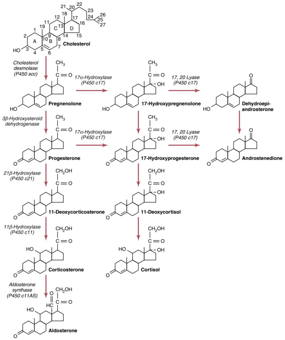
Figure 78-2. Pathways for synthesis of steroid hormones by the adrenal cortex. The enzymes are shown in italics.
شکل 78-2. مسیرهای سنتز هورمون های استروئیدی توسط قشر آدرنال آنزیم ها به صورت مورب نشان داده شده اند.
Table 78-1 Adrenal Steroid Hormones in Adults; Synthetic Steroids and Their Relative Glucocorticoid and Mineralocorticoid Activities.
جدول 78-1 هورمون های استروئیدی آدرنال در بزرگسالان. استروئیدهای مصنوعی و فعالیت های گلوکوکورتیکوئیدی و مینرالوکورتیکوئیدی نسبی آنها.

Glucocorticoid and mineralocorticoid activities of the steroids are relative to cortisol, with cortisol being 1.0.
فعالیت گلوکوکورتیکوئیدی و مینرالوکورتیکوئیدی استروئیدها نسبت به کورتیزول است و کورتیزول 1.0 است.
Mineralocorticoids
• Aldosterone (very potent; accounts for ~90% of all mineralocorticoid activity)
• Deoxycorticosterone (1/30 as potent as aldosterone, but very small quantities are secreted)
• Corticosterone (slight mineralocorticoid activity)
• 9a-Fluorocortisol (synthetic; slightly more potent than aldosterone)
• Cortisol (slight mineralocorticoid activity, but a large quantity is secreted)
• Cortisone (slight mineralocorticoid activity) Glucocorticoids
مینرالوکورتیکوئیدها
• آلدوسترون (بسیار قوی؛ 90٪ از کل فعالیت مینرالوکورتیکوئیدی را تشکیل می دهد)
• دئوکسی کورتیکوسترون (1/30 به اندازه آلدوسترون قوی است، اما مقادیر بسیار کمی ترشح می شود)
• کورتیکوسترون (فعالیت جزئی مینرالوکورتیکوئیدی)
• 9a- فلوروکورتیزول (مصنوعی؛ کمی قوی تر از آلدوسترون)
• کورتیزول (فعالیت مینرالوکورتیکوئیدی خفیف، اما مقدار زیادی ترشح می شود)
• کورتیزون (فعالیت جزئی مینرالوکورتیکوئید) گلوکوکورتیکوئیدها
Glucocorticoids
• Cortisol (very potent; accounts for ~95% of all glucocorticoid activity)
• Corticosterone (provides ≈4% of total glucocorticoid activity, but is much less potent than cortisol)
• Cortisone (almost as potent as cortisol)
• Prednisone (synthetic; four times as potent as cortisol)
• Methyl prednisone (synthetic; five times as potent as cortisol)
• Dexamethasone (synthetic; 30 times as potent as cortisol)
گلوکوکورتیکوئیدها
• کورتیزول (بسیار قوی؛ 95 درصد از کل فعالیت گلوکوکورتیکوئیدی را تشکیل می دهد)
• کورتیکوسترون (≈4٪ از کل فعالیت گلوکوکورتیکوئیدی را فراهم می کند، اما بسیار کمتر از کورتیزول است)
• کورتیزون (تقریبا به اندازه کورتیزول قوی)
• پردنیزون (مصنوعی؛ چهار برابر قوی تر از کورتیزول)
• متیل پردنیزون (مصنوعی؛ پنج برابر قوی تر از کورتیزول)
• دگزامتازون (مصنوعی؛ 30 برابر قوی تر از کورتیزول)
It is clear from this list that some of these hormones and synthetic steroids have both glucocorticoid and mineralocorticoid activities. It is especially significant that cortisol normally has some mineralocorticoid activity, because some syndromes of excess cortisol secretion can cause significant mineralocorticoid effects, along with its much more potent glucocorticoid effects.
از این فهرست مشخص است که برخی از این هورمون ها و استروئیدهای مصنوعی هم فعالیت گلوکوکورتیکوئیدی و هم مینرالوکورتیکوئیدی دارند. به ویژه مهم است که کورتیزول به طور معمول دارای فعالیت معدنی کورتیکوئیدی است، زیرا برخی از سندرم های ترشح بیش از حد کورتیزول می توانند اثرات معدنی کورتیکوئیدی قابل توجهی را همراه با اثرات گلوکوکورتیکوئیدی بسیار قوی تر ایجاد کنند.
The intense glucocorticoid activity of the synthetic hormone dexamethasone, which has almost zero mineralocorticoid activity, makes it an especially important drug for stimulating specific glucocorticoid activity.
فعالیت شدید گلوکوکورتیکوئیدی هورمون مصنوعی دگزامتازون، که فعالیت مینرالوکورتیکوئیدی تقریباً صفر دارد، آن را به داروی مهمی برای تحریک فعالیت گلوکوکورتیکوئیدی خاص تبدیل می کند.
Adrenocortical Hormones Are Bound to Plasma Proteins. Approximately 90% to 95% of the cortisol in plasma binds to plasma proteins, especially a globulin called cortisol-binding globulin or transcortin and, to a lesser extent, to albumin. This high degree of binding to plasma proteins slows the elimination of cortisol from the plasma; therefore, cortisol has a relatively long half-life of 60 to 90 minutes. Only about 60% of circulating aldosterone com- bines with the plasma proteins, and about 40% is in the free form; as a result, aldosterone has a relatively short half- life of about 20 minutes. These hormones are transported throughout the extracellular fluid compartment in both the combined and free forms.
هورمون های قشر آدرنال به پروتئین های پلاسما متصل می شوند. تقریباً 90 تا 95 درصد از کورتیزول موجود در پلاسما به پروتئین های پلاسما، به ویژه گلوبولینی به نام گلوبولین متصل شونده به کورتیزول یا ترانس کورتین و تا حدی به آلبومین متصل می شود. این درجه بالایی از اتصال به پروتئین های پلاسما، حذف کورتیزول از پلاسما را کند می کند. بنابراین، کورتیزول نیمه عمر نسبتا طولانی 60 تا 90 دقیقه دارد. تنها حدود 60 درصد از آلدوسترون در گردش با پروتئین های پلاسما ترکیب می شود و حدود 40 درصد به شکل آزاد است. در نتیجه، آلدوسترون نیمه عمر نسبتاً کوتاهی در حدود 20 دقیقه دارد. این هورمون ها به دو صورت ترکیبی و آزاد در سراسر محفظه مایع خارج سلولی منتقل می شوند.
Binding of adrenal steroids to the plasma proteins may serve as a reservoir to lessen rapid fluctuations in free hormone concentrations, as would occur, for example, with cortisol during brief periods of stress and episodic secretion of ACTH. This reservoir function may also help ensure a relatively uniform distribution of the adrenal hormones to the tissues.
اتصال استروئیدهای آدرنال به پروتئین های پلاسما ممکن است به عنوان مخزنی برای کاهش نوسانات سریع در غلظت هورمون آزاد عمل کند، همانطور که برای مثال، کورتیزول در دوره های کوتاه استرس و ترشح اپیزودیک ACTH اتفاق می افتد. این عملکرد مخزن همچنین ممکن است به اطمینان از توزیع نسبتاً یکنواخت هورمونهای آدرنال در بافتها کمک کند.
Adrenocortical Hormones Are Metabolized in the Liver. The adrenal steroids are degraded mainly in the liver and are conjugated especially to glucuronic acid and, to a lesser extent, to sulfates. These substances are inactive and do not have mineralocorticoid or glucocorticoid activity. About 25% of these conjugates are excreted in the bile and then in the feces. The remaining conjugates formed by the liver enter the circulation but are not bound to plasma proteins, are highly soluble in the plasma, and are therefore filtered readily by the kidneys and excreted in the urine. Diseases of the liver markedly depress the rate of inactivation of adrenocortical hormones, and kidney diseases reduce the excretion of the inactive conjugates.
هورمون های قشر آدرنال در کبد متابولیزه می شوند. استروئیدهای آدرنال عمدتاً در کبد تجزیه می شوند و به ویژه به اسید گلوکورونیک و به میزان کمتری به سولفات ها کونژوگه می شوند. این مواد غیر فعال هستند و فعالیت مینرالوکورتیکوئیدی یا گلوکوکورتیکوئیدی ندارند. حدود 25 درصد از این مزدوج ها از طریق صفرا و سپس از طریق مدفوع دفع می شوند. کونژوگه های باقی مانده که توسط کبد تشکیل می شوند وارد گردش خون می شوند اما به پروتئین های پلاسما متصل نمی شوند، در پلاسما بسیار محلول هستند و بنابراین به راحتی توسط کلیه ها فیلتر شده و از طریق ادرار دفع می شوند. بیماری های کبدی به طور قابل توجهی سرعت غیرفعال شدن هورمون های قشر آدرنال را کاهش می دهند و بیماری های کلیوی دفع مزدوج های غیر فعال را کاهش می دهند.
The normal concentration of aldosterone in blood is about 6 nanograms (6 billionths of a gram) per 100 milliliters, and the average secretory rate is approximately 150 μg/ day (0.15 mg/day). The blood concentration of aldosterone, however, depends greatly on several factors, including dietary intake of sodium and potassium.
غلظت طبیعی آلدوسترون در خون حدود 6 نانوگرم (6 میلیاردم گرم) در 100 میلی لیتر است و متوسط میزان ترشح آن تقریباً 150 میکروگرم در روز (0.15 میلی گرم در روز) است. با این حال، غلظت آلدوسترون در خون به عوامل متعددی از جمله دریافت سدیم و پتاسیم در رژیم غذایی بستگی دارد.
The concentration of cortisol in the blood averages 12 μg/100 ml, and the secretory rate averages 15 to 20 mg/day. However, blood concentration and secretion rate of cortisol fluctuate throughout the day, rising in the early morning and declining in the evening, as discussed later.
غلظت کورتیزول در خون به طور متوسط 12 میکروگرم در 100 میلی لیتر و میزان ترشح آن 15 تا 20 میلی گرم در روز است. با این حال، غلظت خون و میزان ترشح کورتیزول در طول روز در نوسان است، در اوایل صبح افزایش می یابد و در عصر کاهش می یابد، همانطور که بعداً بحث شد.
FUNCTIONS OF MINERALOCORTICOIDS— ALDOSTERONE
Mineralocorticoid Deficiency Causes Severe Renal Sodium Chloride Wasting and Hyperkalemia. Total loss of adrenocortical secretion may cause death within 3 to 14 days unless the person receives extensive salt therapy or injection of mineralocorticoids.
عملکرد مینرالوکورتیکوئیدها – آلدوسترون
کمبود مینرالوکورتیکوئید باعث هدر رفتن شدید کلرید سدیم کلیه و هیپرکالمی می شود. از دست دادن کامل ترشح قشر آدرنال ممکن است در عرض 3 تا 14 روز منجر به مرگ شود مگر اینکه فرد نمک درمانی گسترده یا تزریق مینرالوکورتیکوئیدها را دریافت کند.
Without mineralocorticoids, potassium ion concentration of the extracellular fluid rises markedly, sodium and chloride are rapidly lost from the body, and the total extracellular fluid volume and blood volume become greatly reduced. Diminished cardiac output soon develops, which progresses to a shock-like state, followed by death. This entire sequence can be prevented by administration of aldosterone or some other mineralocorticoid. Therefore, the mineralocorticoids are said to be the acute “lifesaving” portion of the adrenocortical hormones. The glucocorticoids are equally necessary, however, because they allow the person to resist the destructive effects of life’s intermittent physical and mental “stresses,” as discussed later in the chapter.
بدون مینرالوکورتیکوئیدها، غلظت یون پتاسیم مایع خارج سلولی به طور قابل توجهی افزایش می یابد، سدیم و کلرید به سرعت از بدن از بین می رود و حجم کل مایع خارج سلولی و حجم خون به شدت کاهش می یابد. کاهش برون ده قلبی به زودی ایجاد می شود که به حالت شوک مانند و به دنبال آن مرگ پیشرفت می کند. کل این توالی را می توان با تجویز آلدوسترون یا برخی مینرالوکورتیکوئیدهای دیگر جلوگیری کرد. بنابراین، گفته می شود که مینرالوکورتیکوئیدها بخش حاد “نجات دهنده” هورمون های آدرنوکورتیکال هستند. با این حال، گلوکوکورتیکوئیدها به همان اندازه ضروری هستند، زیرا به فرد اجازه میدهند تا در برابر تأثیرات مخرب «استرسهای» متناوب جسمی و ذهنی زندگی مقاومت کند، همانطور که بعداً در این فصل بحث شد.
Aldosterone Is the Major Mineralocorticoid Secreted by the Adrenals. In humans, aldosterone exerts nearly 90% of the mineralocorticoid activity of the adrenocortical secretions, but cortisol, the major glucocorticoid secreted by the adrenal cortex, also provides a significant amount of mineralocorticoid activity. The mineralocorticoid activity of aldosterone is about 3000 times greater than that of cortisol, but the plasma concentration of cortisol is nearly 2000 times that of aldosterone.
آلدوسترون اصلی ترین مینرالوکورتیکوئید ترشح شده توسط آدرنال است. در انسان، آلدوسترون تقریباً 90 درصد از فعالیت مینرالوکورتیکوئیدی ترشحات قشر آدرنال را اعمال می کند، اما کورتیزول، گلوکوکورتیکوئید اصلی ترشح شده از قشر آدرنال، مقدار قابل توجهی از فعالیت مینرالوکورتیکوئیدی را نیز فراهم می کند. فعالیت مینرالوکورتیکوئیدی آلدوسترون حدود 3000 برابر بیشتر از کورتیزول است، اما غلظت کورتیزول پلاسما تقریباً 2000 برابر آلدوسترون است.
Cortisol can also bind to mineralocorticoid receptors with high affinity. However, the renal epithelial cells express the enzyme 11ẞ-hydroxysteroid dehydrogenase type 2 (11ẞ-HSD2), which has actions that prevent cortisol from activating mineralocorticoid receptors. One action of 11ẞ-HSD2 is to convert cortisol to cortisone, which does not avidly bind mineralocorticoid receptors. There is also evidence that 11ẞ-HSD2 may have effects on the intracellular redox (reduction and oxidation) state that prevent cortisol from activating the mineralocorticoid receptors. In patients with genetic deficiency of 11ẞ-HSD2 activity, cortisol may have substantial mineralocorticoid effects. This condition is called apparent mineralocorticoid excess syndrome (AME) because the patient has essentially the same pathophysiological changes as a patient with excess aldosterone secretion, except that plasma aldosterone levels are very low in the patient with AME. Ingestion of large amounts of licorice, which contains glycyrrhetinic acid, may also cause AME because of its ability to block 11ẞ-HSD2 enzyme activity.
کورتیزول همچنین می تواند به گیرنده های معدنی کورتیکوئید با میل ترکیبی بالا متصل شود. با این حال، سلول های اپیتلیال کلیه، آنزیم 11-هیدروکسی استروئید دهیدروژناز نوع 2 (11ẞ-HSD2) را بیان می کنند، که دارای اقداماتی است که از فعال کردن گیرنده های معدنی کورتیکوئید کورتیزول جلوگیری می کند. یکی از اقدامات 11ẞ-HSD2 تبدیل کورتیزول به کورتیزون است که مشتاقانه به گیرنده های معدنی کورتیکوئید متصل نمی شود. همچنین شواهدی وجود دارد مبنی بر اینکه 11ẞ-HSD2 ممکن است اثراتی بر روی حالت ردوکس داخل سلولی (کاهش و اکسیداسیون) داشته باشد که از فعال شدن گیرنده های معدنی کورتیکوئید کورتیزول جلوگیری می کند. در بیماران مبتلا به کمبود ژنتیکی فعالیت 11⺞-HSD2، کورتیزول ممکن است اثرات معدنی کورتیکوئیدی قابل توجهی داشته باشد. این وضعیت سندرم اضافی معدنی کورتیکوئید (AME) نامیده می شود، زیرا بیمار اساساً تغییرات پاتوفیزیولوژیکی مشابهی با یک بیمار با ترشح بیش از حد آلدوسترون دارد، با این تفاوت که سطح آلدوسترون پلاسما در بیمار مبتلا به AME بسیار پایین است. مصرف مقادیر زیادی شیرین بیان که حاوی اسید گلیسیرتینیک است نیز به دلیل توانایی آن در مسدود کردن فعالیت آنزیم 11ẞ-HSD2 ممکن است باعث ایجاد AME شود.
RENAL AND CIRCULATORY EFFECTS OF ALDOSTERONE
Aldosterone Increases Renal Tubular Reabsorption of Sodium and Secretion of Potassium. As discussed in Chapter 28, aldosterone increases reabsorption of sodium and simultaneously increases secretion of potassium by the renal tubular epithelial cells, especially in the principal cells of the collecting tubules and, to a lesser extent, in the distal tubules and collecting ducts. Therefore, aldosterone causes sodium to be conserved in the extracellular fluid while increasing potassium excretion in the urine.
اثرات کلیوی و گردش خون آلدوسترون
آلدوسترون بازجذب سدیم لولهای کلیوی و ترشح پتاسیم را افزایش میدهد. همانطور که در فصل 28 بحث شد، آلدوسترون بازجذب سدیم را افزایش می دهد و به طور همزمان ترشح پتاسیم را توسط سلول های اپیتلیال لوله کلیوی، به ویژه در سلول های اصلی توبول های جمع کننده و به میزان کمتر، در لوله های انتهایی و مجاری جمع کننده افزایش می دهد. بنابراین، آلدوسترون باعث حفظ سدیم در مایع خارج سلولی و افزایش دفع پتاسیم در ادرار می شود.
A high concentration of aldosterone in the plasma can transiently decrease the sodium loss into the urine to as little as a few milliequivalents per day. At the same time, potassium loss into the urine transiently increases severalfold. Therefore, the net effect of excess aldosterone in the plasma is to increase the total quantity of sodium in the extracellular fluid while decreasing the potassium.
غلظت بالای آلدوسترون در پلاسما می تواند به طور موقت از دست دادن سدیم در ادرار را تا چند میلی اکی والان در روز کاهش دهد. در همان زمان، از دست دادن پتاسیم در ادرار به طور موقت چندین برابر افزایش می یابد. بنابراین، اثر خالص آلدوسترون اضافی در پلاسما افزایش مقدار کل سدیم در مایع خارج سلولی و در عین حال کاهش پتاسیم است.
Conversely, total lack of aldosterone secretion can cause transient loss of 10 to 20 grams of sodium in the urine a day, an amount equal to one tenth to one fifth of all the sodium in the body. At the same time, potassium is conserved tenaciously in the extracellular fluid.
برعکس، فقدان کامل ترشح آلدوسترون می تواند باعث از دست دادن گذرا بین 10 تا 20 گرم سدیم در ادرار در روز شود که این مقدار برابر با یک دهم تا یک پنجم کل سدیم بدن است. در عین حال، پتاسیم به شدت در مایع خارج سلولی حفظ می شود.
Excess Aldosterone Increases Extracellular Fluid Volume and Arterial Pressure But Has Only a Small Effect on Plasma Sodium Concentration; Aldosterone Deficiency Causes Hyponatremia. Although aldosterone has a potent effect to decrease the rate of sodium excretion by the kidneys, the concentration of sodium in the extracellular fluid often rises only a few milliequivalents. The reason for this is that when sodium is reabsorbed by the tubules, simultaneous osmotic absorption of almost equivalent amounts of water occurs. Also, small increases in extracellular fluid sodium concentration stimulate thirst and increased water intake, if water is available, and increase secretion of antidiuretic hormone, which enhances water reabsorption by the distal and collecting tubules of the kidneys. Therefore, the extracellular fluid volume increases almost as much as the retained sodium, but without much change in sodium concentration.
آلدوسترون اضافی حجم مایع خارج سلولی و فشار شریانی را افزایش میدهد اما تأثیر کمی بر غلظت سدیم پلاسما دارد. کمبود آلدوسترون باعث هیپوناترمی می شود. اگرچه آلدوسترون اثر قوی در کاهش سرعت دفع سدیم توسط کلیه ها دارد، غلظت سدیم در مایع خارج سلولی اغلب تنها چند میلی اکی والان افزایش می یابد. دلیل این امر این است که وقتی سدیم توسط لولهها بازجذب میشود، جذب اسمزی همزمان تقریباً مقادیر معادل آب رخ میدهد. همچنین افزایش اندک غلظت سدیم مایع خارج سلولی باعث تحریک تشنگی و افزایش مصرف آب در صورت وجود آب می شود و ترشح هورمون آنتی دیورتیک را افزایش می دهد که باعث افزایش بازجذب آب توسط لوله های دیستال و جمع کننده کلیه ها می شود. بنابراین، حجم مایع خارج سلولی تقریباً به اندازه سدیم باقی مانده افزایش می یابد، اما بدون تغییر زیادی در غلظت سدیم.
Even though aldosterone is one of the body’s most powerful sodium-retaining hormones, only transient sodium retention occurs when excess amounts are secreted. An aldosterone-mediated increase in extracellular fluid volume lasting more than 1 to 2 days also leads to an increase in arterial pressure, as explained in Chapter 19. The rise in arterial pressure then increases kidney excretion of both sodium and water, called pressure natriuresis and pressure diuresis, respectively. Thus, after the extracellular fluid volume increases 5% to 15% above normal, arterial pressure also increases 15 to 25 mm Hg, and this elevated blood pressure returns the renal output of sodium and water to normal despite excess aldosterone (Figure 78-3).
اگرچه آلدوسترون یکی از قویترین هورمونهای نگهدارنده سدیم در بدن است، تنها احتباس گذرا سدیم زمانی اتفاق میافتد که مقادیر اضافی ترشح شود. افزایش با واسطه آلدوسترون در حجم مایع خارج سلولی که بیش از 1 تا 2 روز طول می کشد همچنین منجر به افزایش فشار شریانی می شود، همانطور که در فصل 19 توضیح داده شد. سپس افزایش فشار شریانی باعث افزایش دفع کلیه سدیم و آب می شود که به ترتیب ناتریورز فشاری و دیورز فشاری نامیده می شوند. بنابراین، پس از افزایش 5 تا 15 درصدی حجم مایع خارج سلولی بالاتر از حد طبیعی، فشار شریانی نیز 15 تا 25 میلیمتر جیوه افزایش مییابد و این فشار خون بالا خروجی کلیوی سدیم و آب را با وجود آلدوسترون اضافی به حالت عادی برمیگرداند (شکل 78-3).
This return to normal sodium and water excretion by the kidneys as a result of pressure natriuresis and diuresis is called aldosterone escape. Thereafter, the rate of gain of sodium and water by the body is zero, and balance is maintained between sodium and water intake and output by the kidneys, despite continued excess aldosterone. In the meantime, however, hypertension has developed, which lasts as long as the person remains exposed to high levels of aldosterone.
این بازگشت به دفع طبیعی سدیم و آب توسط کلیه ها در نتیجه ناتریورز فشاری و دیورز، فرار آلدوسترون نامیده می شود. پس از آن، میزان دریافت سدیم و آب توسط بدن صفر است و با وجود ادامه آلدوسترون اضافی، تعادل بین دریافت و خروج سدیم و آب توسط کلیه ها حفظ می شود. با این حال، در این بین، فشار خون بالا ایجاد شده است، که تا زمانی که فرد در معرض سطوح بالای آلدوسترون باقی بماند، ادامه دارد.
In contrast, severe aldosterone deficiency may cause substantial reductions in plasma sodium concentration (hyponatremia) due to reduced renal sodium reabsorption and increased sodium excretion. The renal sodium wasting causes reductions in extracellular fluid volume, arterial pressure, and cardiac output, which stimulate secretion of antidiuretic hormone (ADH). Increased levels of ADH attenuate renal water excretion and contribute to hyponatremia, along with increases in thirst and water intake that are also stimulated by hypovolemia and hypotension.
در مقابل، کمبود شدید آلدوسترون ممکن است باعث کاهش قابل توجهی در غلظت سدیم پلاسما (هیپوناترمی) به دلیل کاهش بازجذب کلیوی سدیم و افزایش دفع سدیم شود. هدر رفتن سدیم کلیه باعث کاهش حجم مایع خارج سلولی، فشار شریانی و برون ده قلبی می شود که باعث تحریک ترشح هورمون آنتی دیورتیک (ADH) می شود. افزایش سطح ADH دفع آب کلیوی را کاهش می دهد و به هیپوناترمی کمک می کند، همراه با افزایش تشنگی و مصرف آب که با هیپوولمی و افت فشار خون نیز تحریک می شود.
When aldosterone secretion becomes zero, large amounts of sodium are lost in the urine, not only diminishing the amount of sodium chloride in the extracellular fluid but also decreasing the extracellular fluid volume. The result is severe extracellular fluid dehydration and low blood volume, leading to circulatory shock. Without therapy, this usually causes death within a few days after the adrenal glands suddenly stop secreting aldosterone.
وقتی ترشح آلدوسترون به صفر می رسد، مقادیر زیادی سدیم در ادرار از بین می رود و نه تنها مقدار کلرید سدیم در مایع خارج سلولی را کاهش می دهد، بلکه حجم مایع خارج سلولی را نیز کاهش می دهد. نتیجه کم آبی شدید مایع خارج سلولی و حجم کم خون است که منجر به شوک گردش خون می شود. بدون درمان، این معمولا باعث مرگ در عرض چند روز پس از توقف ناگهانی ترشح آلدوسترون توسط غدد فوق کلیوی می شود.

Figure 78-3. Effect of aldosterone infusion on arterial pressure, extracellular fluid volume, and sodium excretion in dogs. Although aldosterone was infused at a rate that raised plasma concentrations to about 20 times normal, note the “escape” from sodium retention on the second day of infusion as arterial pressure increased and urinary sodium excretion returned to normal. (Data from Hall JE, Granger JP, Smith MJ Jr, et al: Role of hemodynamics and arterial pressure in aldosterone “escape.” Hypertension 6[suppl 1):1183-1192, 1984.)
شکل 78-3. تأثیر انفوزیون آلدوسترون بر فشار شریانی، حجم مایع خارج سلولی و دفع سدیم در سگ ها. اگرچه آلدوسترون با سرعتی تزریق شد که غلظت پلاسما را تا حدود 20 برابر طبیعی افزایش داد، اما به “فرار” از احتباس سدیم در روز دوم انفوزیون با افزایش فشار شریانی و بازگشت سدیم ادراری به حالت عادی توجه کنید. (داده ها از هال JE، گرنجر جی پی، اسمیت ام جی جونیور، و همکاران: نقش همودینامیک و فشار شریانی در “فرار” آلدوسترون.
Excess Aldosterone Causes Hypokalemia and Muscle Weakness; Aldosterone Deficiency Causes Hyperkalemia and Cardiac Toxicity. Excess aldosterone not only causes loss of potassium ions from the extracellular fluid into the urine but also stimulates transport of potassium from the extracellular fluid into most cells of the body. Therefore, excessive secretion of aldosterone, as occurs with some types of adrenal tumors, may cause a serious decrease in the plasma potassium concentration (hypokalemia), sometimes from the normal value of 4.5 mEq/L to as low as 2 mEq/L. When plasma potassium ion concentration falls below about one-half normal, severe muscle weakness often develops. This muscle weakness is caused by alteration of the electrical excitability of the nerve and muscle fiber membranes (see Chapter 5), which prevents transmission of normal action potentials.
آلدوسترون اضافی باعث هیپوکالمی و ضعف عضلانی می شود. کمبود آلدوسترون باعث هیپرکالمی و مسمومیت قلبی می شود. آلدوسترون اضافی نه تنها باعث از بین رفتن یون های پتاسیم از مایع خارج سلولی به داخل ادرار می شود، بلکه باعث تحریک انتقال پتاسیم از مایع خارج سلولی به اکثر سلول های بدن می شود. بنابراین، ترشح بیش از حد آلدوسترون، همانطور که در مورد برخی از انواع تومورهای آدرنال رخ می دهد، ممکن است باعث کاهش جدی غلظت پتاسیم پلاسما (هیپوکالمی) شود، که گاهی اوقات از مقدار طبیعی 4.5 میلی اکی والان در لیتر به کمتر از 2 میلی اکی والان در لیتر می رسد. هنگامی که غلظت یون پتاسیم پلاسما به کمتر از نصف نرمال می رسد، اغلب ضعف شدید عضلانی ایجاد می شود. این ضعف عضلانی ناشی از تغییر در تحریک پذیری الکتریکی غشاهای عصبی و فیبر عضلانی است (به فصل 5 مراجعه کنید)، که از انتقال پتانسیل های عمل طبیعی جلوگیری می کند.
Conversely, when aldosterone is deficient, the extracellular fluid potassium ion concentration can rise far above normal. When it rises to 60% to 100% above normal, serious cardiac toxicity, including weakness of heart contraction and development of arrhythmia, becomes evident, and progressively higher concentrations of potassium lead inevitably to heart failure.
برعکس، زمانی که آلدوسترون کمبود داشته باشد، غلظت یون پتاسیم مایع خارج سلولی می تواند بسیار بالاتر از حد طبیعی باشد. هنگامی که به 60% تا 100% بالاتر از حد طبیعی می رسد، مسمومیت قلبی جدی، از جمله ضعف انقباض قلب و ایجاد آریتمی، آشکار می شود، و به تدریج غلظت های بالاتر پتاسیم به ناچار منجر به نارسایی قلبی می شود.
Excess Aldosterone Increases Tubular Hydrogen lon Secretion and Causes Alkalosis. Aldosterone not only causes potassium to be secreted into the tubules in exchange for sodium reabsorption in the principal cells of the renal collecting tubules but also causes secretion of hydrogen ions in exchange for potassium in the intercalated cells of the cortical collecting tubules, as discussed in Chapters 28 and 31. This decreases the hydrogen ion concentration in the extracellular fluid, causing metabolic alkalosis.
آلدوسترون اضافی ترشح هیدروژن لون لوله ای را افزایش می دهد و باعث آلکالوز می شود. آلدوسترون نه تنها باعث ترشح پتاسیم به داخل لولهها میشود تا برای جذب مجدد سدیم در سلولهای اصلی لولههای جمعکننده کلیوی، یونهای هیدروژن در ازای پتاسیم ترشح شود، همانطور که در فصلهای 28 و 31 بحث شد. آلکالوز متابولیک.
ALDOSTERONE STIMULATES SODIUM AND POTASSIUM TRANSPORT IN SWEAT GLANDS, SALIVARY GLANDS, AND INTESTINAL EPITHELIAL CELLS
Aldosterone has almost the same effects on sweat glands and salivary glands as it has on the renal tubules. Both these glands form a primary secretion that contains large quantities of sodium chloride, but much of the sodium chloride, upon passing through the excretory ducts, is reabsorbed, whereas potassium and bicarbonate ions are secreted. Aldosterone greatly increases the reabsorption of sodium chloride and the secretion of potassium by the ducts. The effect on the sweat glands is important to conserve body salt in hot environments (see Chapter 74), and the effect on the salivary glands is necessary to conserve salt when excessive quantities of saliva are lost.
آلدوسترون باعث تحریک انتقال سدیم و پتاسیم در غدد عرق، غدد بزاقی و سلول های اپیتلیال روده می شود.
آلدوسترون تقریباً همان تأثیری را که روی لوله های کلیوی دارد، روی غدد عرق و غدد بزاقی دارد. هر دو این غده یک ترشح اولیه تشکیل می دهند که حاوی مقادیر زیادی کلرید سدیم است، اما بیشتر کلرید سدیم با عبور از مجاری دفعی، دوباره جذب می شود، در حالی که یون های پتاسیم و بی کربنات ترشح می شوند. آلدوسترون بازجذب کلرید سدیم و ترشح پتاسیم توسط مجاری را به شدت افزایش می دهد. تأثیر بر غدد عرق برای حفظ نمک بدن در محیط های گرم مهم است (به فصل 74 مراجعه کنید)، و تأثیر آن بر غدد بزاقی برای حفظ نمک زمانی که مقادیر زیادی بزاق از بین می رود ضروری است.
Aldosterone also greatly enhances sodium absorption by the intestines, especially in the colon, which prevents loss of sodium in the stools. Conversely, in the absence of aldosterone, sodium absorption can be poor, leading to failure to absorb chloride and other anions and water as well. The unabsorbed sodium chloride and water then lead to diarrhea, with further loss of salt from the body.
آلدوسترون همچنین جذب سدیم توسط رودهها را بهویژه در روده بزرگ افزایش میدهد که از از دست رفتن سدیم در مدفوع جلوگیری میکند. برعکس، در غیاب آلدوسترون، جذب سدیم می تواند ضعیف باشد که منجر به عدم جذب کلرید و سایر آنیون ها و آب نیز می شود. کلرید سدیم و آب جذب نشده منجر به اسهال و از دست دادن بیشتر نمک از بدن می شود.
CELLULAR MECHANISM OF ALDOSTERONE ACTION
Although for many years we have known the overall effects of mineralocorticoids on the body, the molecular mechanisms of the actions of aldosterone on the tubular cells to increase transport of sodium are still not fully understood. However, the cellular sequence of events that leads to increased sodium reabsorption seems to unfold as follows.
مکانیسم سلولی عمل آلدوسترون
اگرچه سالهاست که ما اثرات کلی مینرالوکورتیکوئیدها را بر بدن میدانیم، مکانیسمهای مولکولی اعمال آلدوسترون بر روی سلولهای لولهای برای افزایش انتقال سدیم هنوز به طور کامل شناخته نشده است. با این حال، به نظر می رسد توالی سلولی رویدادهایی که منجر به افزایش بازجذب سدیم می شود، به شرح زیر آشکار می شود.
First, because of its lipid solubility in the cellular mem- branes, aldosterone diffuses readily to the interior of the tubular epithelial cells.
اول، به دلیل حلالیت لیپیدی آن در غشای سلولی، آلدوسترون به راحتی به داخل سلول های اپیتلیال لوله ای منتشر می شود.
Second, in the cytoplasm of the tubular cells, aldosterone combines with a highly specific cytoplasmic mineralocorticoid receptor (MR) protein (Figure 78-4), which has a stereoolecular configuration that allows only aldosterone or similar compounds to combine with it. Although renal tubular epithelial cell MR receptors also have a high affinity for cortisol, the enzyme 11ẞ-HSD2 normally converts most of the cortisol to cortisone, which does not readily bind to MR receptors, as discussed previously.
دوم، در سیتوپلاسم سلولهای لولهای، آلدوسترون با پروتئین گیرنده مینرالوکورتیکوئید سیتوپلاسمی بسیار اختصاصی (MR) ترکیب میشود (شکل 78-4)، که دارای یک پیکربندی استریولکولی است که فقط به آلدوسترون یا ترکیبات مشابه اجازه میدهد با آن ترکیب شوند. اگرچه گیرندههای MR سلولهای اپیتلیال لولههای کلیوی نیز تمایل بالایی به کورتیزول دارند، آنزیم 11ẞ-HSD2 معمولاً بیشتر کورتیزول را به کورتیزون تبدیل میکند، که به آسانی به گیرندههای MR متصل نمیشود، همانطور که قبلاً بحث شد.
Third, the aldosterone-receptor complex or a product of this complex diffuses into the nucleus, where it may undergo further alterations, finally inducing one or more specific portions of the DNA to form one or more types of messenger RNA (mRNA) related to the process of sodium and potassium transport.
سوم، کمپلکس گیرنده آلدوسترون یا محصول این کمپلکس به درون هسته منتشر میشود، جایی که ممکن است دچار تغییرات بیشتری شود، در نهایت یک یا چند بخش خاص از DNA را برای تشکیل یک یا چند نوع RNA پیامرسان (mRNA) مرتبط با فرآیند انتقال سدیم و پتاسیم وادار میکند.
Fourth, the mRNA diffuses back into the cytoplasm where, operating in conjunction with the ribosomes, it causes protein formation. The proteins formed are a mixture of (1) one or more enzymes and (2) membrane transport proteins that, all acting together, are required for sodium, potassium, and hydrogen transport through the cell membrane (see Figure 78-4). One of the enzymes especially increased is sodium-potassium adenosine tri- phosphatase (Na+-K+ ATPase), which serves as the principal part of the pump for sodium and potassium exchange at the basolateral membranes of the renal tubular cells. Additional proteins, perhaps equally important, are epithelial sodium channels and potassium channels inserted into the luminal membrane of the same tubular cells; these channels allow rapid diffusion of sodium ions from the tubular lumen into the cell and diffusion of potassium from the cell interior to the tubular lumen. (See Chapters 28 and 30 for further discussion of the effects of aldosterone on sodium, potassium, and hydrogen transport by renal tubular epithelial cells.)
چهارم، mRNA دوباره به داخل سیتوپلاسم منتشر می شود، جایی که، در ارتباط با ریبوزوم ها، باعث تشکیل پروتئین می شود. پروتئین های تشکیل شده مخلوطی از (1) یک یا چند آنزیم و (2) پروتئین های ناقل غشایی هستند که همگی با هم برای انتقال سدیم، پتاسیم و هیدروژن از طریق غشای سلولی مورد نیاز هستند (شکل 78-4 را ببینید). یکی از آنزیم هایی که به ویژه افزایش یافته است، سدیم پتاسیم آدنوزین تری فسفاتاز (Na+-K+ ATPase) است که به عنوان بخش اصلی پمپ برای تبادل سدیم و پتاسیم در غشای قاعده جانبی سلول های لوله کلیوی عمل می کند. پروتئین های اضافی، شاید به همان اندازه مهم باشند، کانال های سدیم اپیتلیال و کانال های پتاسیم هستند که در غشای مجرای همان سلول های لوله ای قرار می گیرند. این کانالها اجازه انتشار سریع یونهای سدیم از لومن لولهای به داخل سلول و انتشار پتاسیم از داخل سلول به لومن لولهای را میدهند. (برای بحث بیشتر در مورد اثرات آلدوسترون بر انتقال سدیم، پتاسیم و هیدروژن توسط سلول های اپیتلیال لوله های کلیوی، به فصل های 28 و 30 مراجعه کنید.)
Thus, aldosterone does not have a major immediate effect on sodium transport; rather, this effect must await the sequence of events that leads to the formation of the specific intracellular substances required for sodium transport. About 30 minutes is required before new RNA appears in the cells, and about 45 minutes is required before the rates of sodium and potassium transport begin to increase; these effects reach a maximum only after several hours.
بنابراین، آلدوسترون اثر فوری عمده ای بر انتقال سدیم ندارد. بلکه این اثر باید منتظر توالی وقایعی باشد که منجر به تشکیل مواد درون سلولی خاص مورد نیاز برای انتقال سدیم شود. حدود 30 دقیقه قبل از ظاهر شدن RNA جدید در سلولها و حدود 45 دقیقه قبل از شروع افزایش سرعت انتقال سدیم و پتاسیم لازم است. این اثرات تنها پس از چند ساعت به حداکثر می رسد.
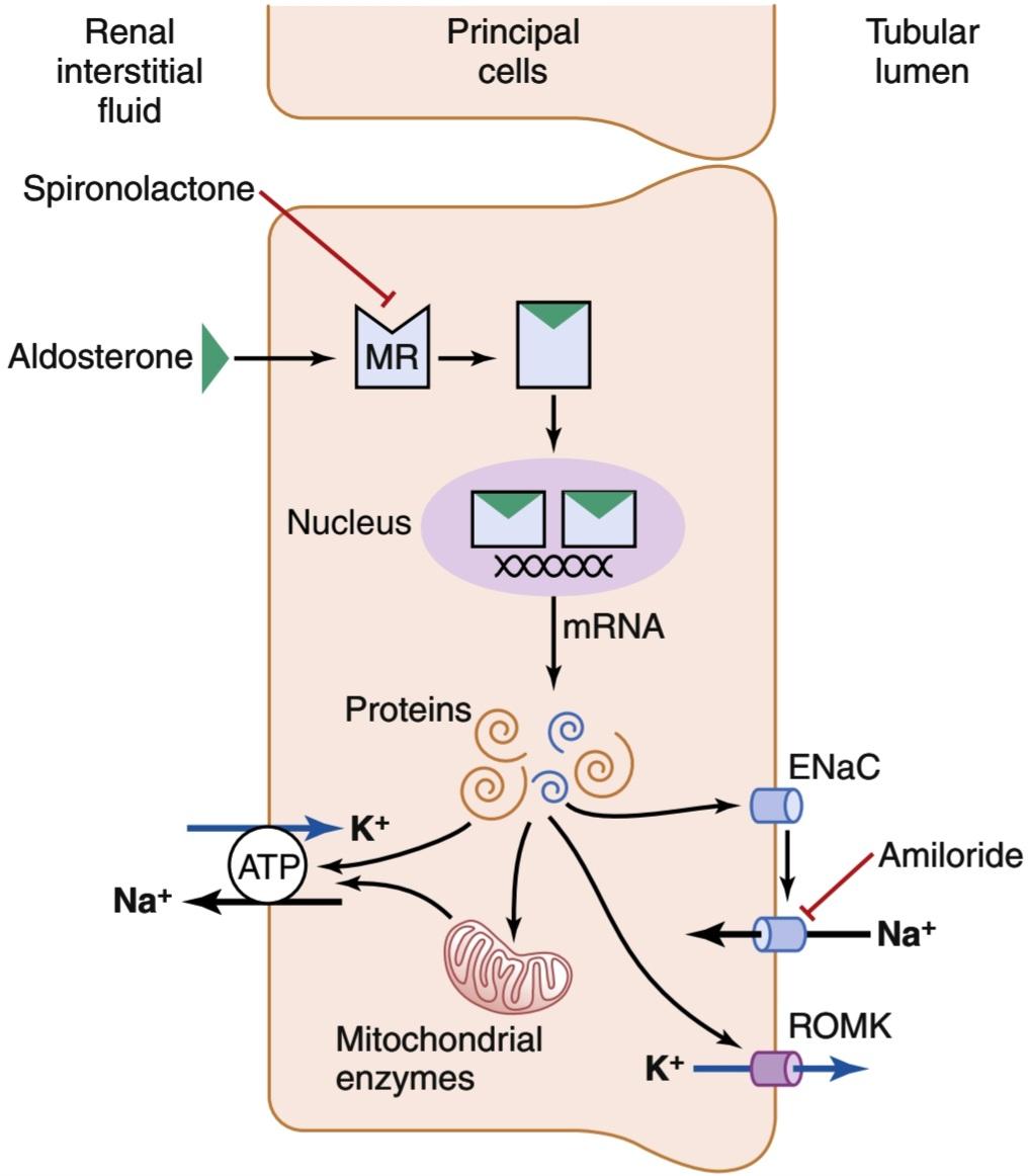
Figure 78-4. Aldosterone-responsive epithelial cell signaling path- ways. Activation of the mineralocorticoid receptor (MR) by aldosterone can be antagonized with spironolactone. Amiloride is a drug that can be used to block epithelial sodium channel (ENaC) proteins. ATP, ROMK, Renal outer medullary potassium channel.
شکل 78-4. مسیرهای سیگنالینگ سلول های اپیتلیال پاسخگو به آلدوسترون. فعال شدن گیرنده مینرالوکورتیکوئید (MR) توسط آلدوسترون می تواند با اسپیرونولاکتون مخالفت کند. آمیلوراید دارویی است که می تواند برای مسدود کردن پروتئین های کانال سدیم اپیتلیال (ENaC) استفاده شود. ATP، ROMK، کانال پتاسیم مدولاری بیرونی کلیه.
POSSIBLE NONGENOMIC ACTIONS OF ALDOSTERONE AND OTHER STEROID HORMONES
Some studies suggest that many steroids, including aldosterone, elicit not only slowly developing genomic effects that have a latency of 45 to 60 minutes and require gene transcription and synthesis of new proteins but also more rapid nongenomic effects that take place in a few seconds or minutes.
اعمال غیر ژنومیک احتمالی آلدوسترون و سایر هورمون های استروئیدی
برخی از مطالعات نشان میدهند که بسیاری از استروئیدها، از جمله آلدوسترون، نه تنها اثرات ژنومی آهستهای را ایجاد میکنند که تاخیری بین 45 تا 60 دقیقه دارند و نیاز به رونویسی ژن و سنتز پروتئینهای جدید دارند، بلکه اثرات غیر ژنومیک سریعتری را نیز ایجاد میکنند که در چند ثانیه یا دقیقه اتفاق میافتد.
These nongenomic actions are believed to be mediated by binding of steroids to cell membrane receptors that are coupled to second messenger systems, similar to those used for peptide hormone signal transduction. For example, aldosterone has been shown to increase formation of cyclic adenosine monophosphate (cAMP) in vascular smooth muscle cells and in epithelial cells of the renal collecting tubules in less than 2 minutes, a period that is far too short for gene transcription and synthesis of new proteins. In other cell types, aldosterone has been shown to rapidly stimulate the phosphatidylinositol second messenger system. However, the precise structure of receptors responsible for the rapid effects of aldosterone has not been determined, nor is the physiological significance of these nongenomic actions of steroids well understood.
اعتقاد بر این است که این اعمال غیر ژنومیک با اتصال استروئیدها به گیرنده های غشای سلولی که به سیستم های پیام رسان دوم جفت شده اند، انجام می شود، شبیه به آنچه برای انتقال سیگنال هورمون پپتیدی استفاده می شود. به عنوان مثال، نشان داده شده است که آلدوسترون تشکیل آدنوزین مونوفسفات حلقوی (cAMP) را در سلول های ماهیچه صاف عروق و در سلول های اپیتلیال لوله های جمع کننده کلیه در کمتر از 2 دقیقه افزایش می دهد، دوره ای که برای رونویسی ژن و سنتز پروتئین های جدید بسیار کوتاه است. در سایر انواع سلول، نشان داده شده است که آلدوسترون به سرعت سیستم پیام رسان دوم فسفاتیدیلینوزیتول را تحریک می کند. با این حال، ساختار دقیق گیرنده های مسئول اثرات سریع آلدوسترون مشخص نشده است، و همچنین اهمیت فیزیولوژیکی این اعمال غیر ژنومیک استروئیدها به خوبی شناخته نشده است.
REGULATION OF ALDOSTERONE SECRETION
Regulation of aldosterone secretion is so deeply inter- twined with regulation of extracellular fluid electrolyte concentrations, extracellular fluid volume, blood volume, arterial pressure, and many special aspects of renal function that it is difficult to discuss control of aldosterone secretion independently of all these other factors. This subject is presented in more detail in Chapters 28 and 30, to which the reader is referred. However, it is important to list here some of the more important points of aldosterone secretion control.
تنظیم ترشح آلدوسترون
تنظیم ترشح آلدوسترون چنان عمیقاً با تنظیم غلظت الکترولیت مایع خارج سلولی، حجم مایع خارج سلولی، حجم خون، فشار شریانی و بسیاری از جنبههای خاص عملکرد کلیه در هم تنیده است که بحث کنترل ترشح آلدوسترون مستقل از همه این عوامل دشوار است. این موضوع در فصل های 28 و 30 به تفصیل ارائه شده است که خواننده به آن اشاره می کند. با این حال، ذکر برخی از نکات مهم در کنترل ترشح آلدوسترون در اینجا مهم است.
Regulation of aldosterone secretion by the zona glomerulosa cells is almost entirely independent of regulation of cortisol and androgens by the zona fasciculata and zona reticularis.
تنظیم ترشح آلدوسترون توسط سلول های زونا گلومرولوزا تقریباً به طور کامل مستقل از تنظیم کورتیزول و آندروژن ها توسط زونا فاسیکولاتا و زونا رتیکولاریس است.
The following factors are known to play roles in regulation of aldosterone:
عوامل زیر در تنظیم آلدوسترون نقش دارند:
1. Increased potassium ion concentration in the extra- cellular fluid greatly increases aldosterone secretion.
1. افزایش غلظت یون پتاسیم در مایع خارج سلولی ترشح آلدوسترون را بسیار افزایش می دهد.
2. Increased angiotensin II concentration in the extracellular fluid also greatly increases aldosterone secretion.
2. افزایش غلظت آنژیوتانسین II در مایع خارج سلولی نیز ترشح آلدوسترون را بسیار افزایش می دهد.
3. Increased sodium ion concentration in the extracellular fluid slightly decreases aldosterone secretion.
3. افزایش غلظت یون سدیم در مایع خارج سلولی ترشح آلدوسترون را اندکی کاهش می دهد.
4. Increased atrial natriuretic peptide (ANP), a hormone secreted by the heart when specific cells of the cardiac atria are stretched (see Chapter 28), decreases aldosterone secretion.
4. افزایش پپتید ناتریورتیک دهلیزی (ANP)، هورمونی که از قلب ترشح می شود زمانی که سلول های خاص دهلیزهای قلب کشیده می شوند (به فصل 28 مراجعه کنید)، ترشح آلدوسترون را کاهش می دهد.
5. ACTH from the anterior pituitary gland is necessary for aldosterone secretion but has little effect in controlling the rate of secretion in most physiological conditions.
5. ACTH از غده هیپوفیز قدامی برای ترشح آلدوسترون ضروری است اما در اکثر شرایط فیزیولوژیکی تأثیر کمی در کنترل میزان ترشح دارد.
Of these factors, potassium ion concentration and the angiotensin II are by far the most potent in regulating aldosterone secretion. A small percentage increase in potassium concentration can cause a severalfold increase in aldosterone secretion. Likewise, increased angiotensin II,, usually in response to diminished blood flow to the kidneys or to sodium loss, can increase aldosterone secretion severalfold. In turn, the aldosterone acts on the kidneys (1) to help them excrete the excess potassium ions and (2) to increase the blood volume and arterial pressure, thus returning the renin-angiotensin system toward its normal level of activity. These feedback control mechanisms are essential for maintaining life, and the reader is referred again to Chapters 28 and 30 for a more complete description of their functions.
از بین این عوامل، غلظت یون پتاسیم و آنژیوتانسین II تا حد زیادی قوی ترین در تنظیم ترشح آلدوسترون هستند. افزایش درصد کمی در غلظت پتاسیم می تواند باعث افزایش چند برابری ترشح آلدوسترون شود. به همین ترتیب، افزایش آنژیوتانسین II، معمولاً در پاسخ به کاهش جریان خون به کلیه ها یا از دست دادن سدیم، می تواند ترشح آلدوسترون را چندین برابر افزایش دهد. به نوبه خود، آلدوسترون روی کلیه ها (1) برای کمک به دفع یون های پتاسیم اضافی و (2) برای افزایش حجم خون و فشار شریانی عمل می کند، بنابراین سیستم رنین-آنژیوتانسین را به سطح فعالیت طبیعی خود باز می گرداند. این مکانیسمهای کنترل بازخورد برای حفظ زندگی ضروری هستند و خواننده برای شرح کاملتر عملکرد آنها دوباره به فصلهای 28 و 30 ارجاع داده میشود.
Figure 78-5 shows the effects on plasma aldosterone concentration caused by blocking formation of angiotensin II with an angiotensin-converting enzyme inhibitor after several weeks of a low-sodium diet that increases plasma aldosterone concentration. Note that blockade of angiotensin II formation markedly decreased plasma aldosterone concentration without significantly changing cortisol concentration, which indicates the important role of angiotensin II in stimulating aldosterone secretion when sodium intake and extracellular fluid volume are reduced.
شکل 78-5 اثرات روی غلظت آلدوسترون پلاسما ناشی از مسدود کردن تشکیل آنژیوتانسین II با یک مهار کننده آنزیم تبدیل کننده آنژیوتانسین را پس از چندین هفته رژیم کم سدیم نشان می دهد که غلظت آلدوسترون پلاسما را افزایش می دهد. توجه داشته باشید که انسداد تشکیل آنژیوتانسین II به طور قابل توجهی غلظت آلدوسترون پلاسما را بدون تغییر قابل توجهی در غلظت کورتیزول کاهش می دهد، که نشان دهنده نقش مهم آنژیوتانسین II در تحریک ترشح آلدوسترون در هنگام کاهش مصرف سدیم و حجم مایع خارج سلولی است.
By contrast, the effects of ANP, sodium ion concentration per se, and ACTH in controlling aldosterone secretion are usually minor. Nevertheless, a 10% to 20% decrease in extracellular fluid sodium ion con- centration, which occurs on rare occasions, can perhaps increase aldosterone secretion by about 50%. An increase in ANP concentration, secondary to plasma volume expansion and stretch of the cardiac atria, may induce natriuresis, in part by inhibiting aldosterone secretion. In the case of ACTH, if even a small amount is secreted by the anterior pituitary gland, it is usually enough to permit the adrenal glands to secrete whatever amount of aldosterone is required, but total absence of ACTH can significantly reduce aldosterone secretion. Therefore, ACTH appears to play a “permissive” role in regulation of aldosterone secretion.
در مقابل، اثرات ANP، غلظت یون سدیم به خودی خود، و ACTH در کنترل ترشح آلدوسترون معمولا جزئی هستند. با این وجود، کاهش 10 تا 20 درصدی غلظت یون سدیم مایع خارج سلولی، که در موارد نادر رخ می دهد، احتمالاً می تواند ترشح آلدوسترون را تا حدود 50 درصد افزایش دهد. افزایش غلظت ANP، ثانویه به افزایش حجم پلاسما و کشش دهلیزهای قلب، ممکن است تا حدی با مهار ترشح آلدوسترون باعث ناتریورز شود. در مورد ACTH، اگر حتی مقدار کمی از غده هیپوفیز قدامی ترشح شود، معمولاً کافی است که به غدد فوق کلیوی اجازه دهید هر مقدار آلدوسترون مورد نیاز را ترشح کنند، اما فقدان کامل ACTH می تواند به طور قابل توجهی ترشح آلدوسترون را کاهش دهد. بنابراین، به نظر می رسد ACTH نقش “مجاز” را در تنظیم ترشح آلدوسترون بازی می کند.
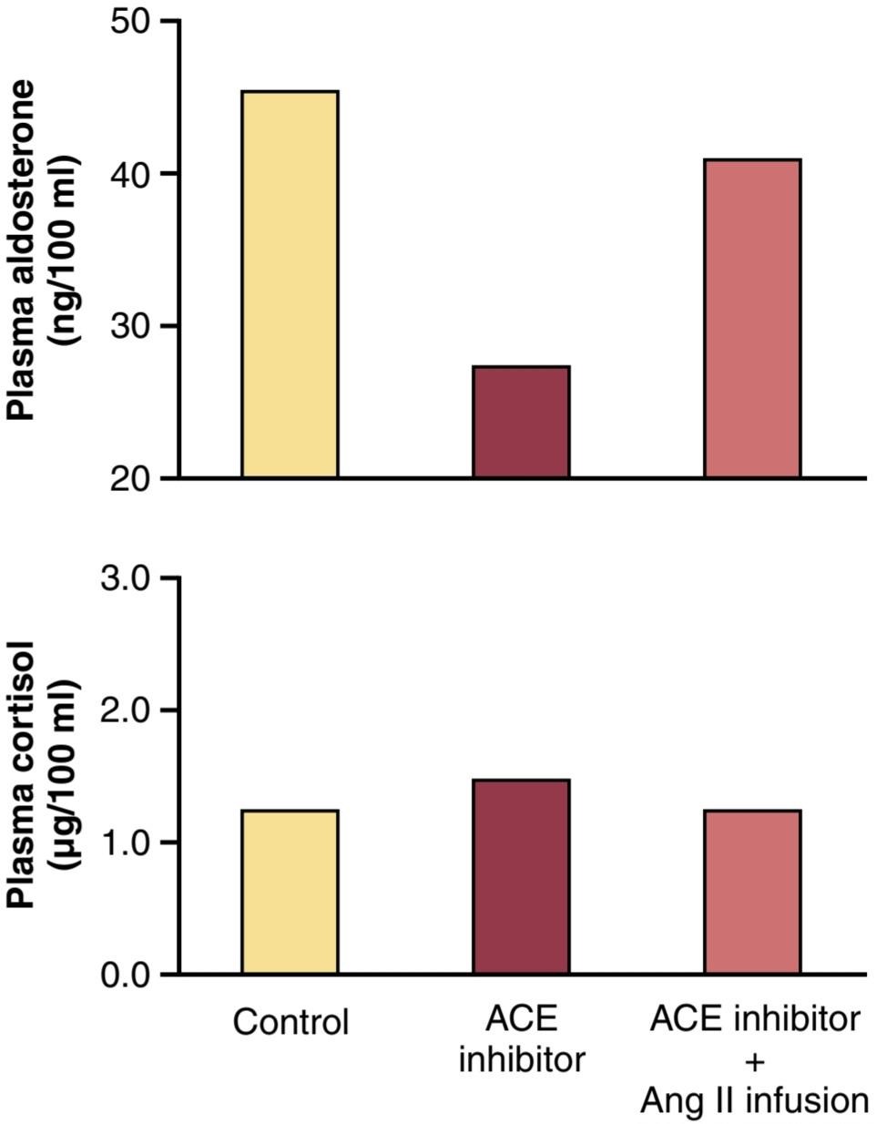
Figure 78-5. Effects of treating sodium-depleted dogs with an angiotensin-converting enzyme (ACE) inhibitor for 7 days to block formation of angiotensin II (Ang II) and of infusing exogenous Ang II to restore plasma Ang II levels after ACE inhibition. Note that block- ing Ang II formation reduced plasma aldosterone concentration with little effect on cortisol, demonstrating the important role of Ang II in stimulating aldosterone secretion during sodium depletion. (Data from Hall JE, Guyton AC, Smith MJ Jr, et al: Chronic blockade of angiotensin II formation during sodium deprivation. Am J Physiol 237:F424, 1979.)
شکل 78-5. اثرات درمان سگهای فاقد سدیم با یک مهارکننده آنزیم مبدل آنژیوتانسین (ACE) به مدت 7 روز برای جلوگیری از تشکیل آنژیوتانسین II (Ang II) و تزریق Ang II اگزوژن برای بازگرداندن سطوح Ang II پلاسما پس از مهار ACE. توجه داشته باشید که مسدود کردن تشکیل Ang II غلظت آلدوسترون پلاسما را با تأثیر کمی بر کورتیزول کاهش می دهد و نقش مهم Ang II را در تحریک ترشح آلدوسترون در طول کاهش سدیم نشان می دهد. (داده ها از هال JE، گایتون AC، اسمیت ام جی جونیور، و همکاران: محاصره مزمن تشکیل آنژیوتانسین II در طول محرومیت از سدیم. Am J Physiol 237:F424، 1979.)
FUNCTIONS OF GLUCOCORTICOIDS
Even though mineralocorticoids can save the life of an acutely adrenalectomized animal, the animal still is far from normal. Instead, the animal’s metabolic systems for utilization of proteins, carbohydrates, and fats remain considerably deranged. Furthermore, the animal cannot resist different types of physical or even mental stress, and minor illnesses such as respiratory tract infections can lead to death. Therefore, the glucocorticoids have functions just as important to the long-continued life of the animal as those of the mineralocorticoids. These functions are explained in the following sections.
عملکرد گلوکوکورتیکوئیدها
حتی اگر مینرالوکورتیکوئیدها می توانند جان یک حیوان حاد آدرنالکتومی شده را نجات دهند، حیوان هنوز از حالت طبیعی فاصله دارد. در عوض، سیستم متابولیک حیوان برای استفاده از پروتئین ها، کربوهیدرات ها و چربی ها به طور قابل توجهی مختل می شود. علاوه بر این، حیوان نمی تواند در برابر انواع مختلف استرس فیزیکی یا حتی روحی مقاومت کند و بیماری های جزئی مانند عفونت های دستگاه تنفسی می تواند منجر به مرگ شود. بنابراین، عملکرد گلوکوکورتیکوئیدها به همان اندازه برای ادامه زندگی طولانی حیوان مهم است که عملکرد مینرالوکورتیکوئیدها. این توابع در بخش های زیر توضیح داده شده است.
At least 95% of the glucocorticoid activity of the adrenocortical secretions results from the secretion of cortisol, known also as hydrocortisone. In addition, a small but significant amount of glucocorticoid activity is provided by corticosterone.
حداقل 95 درصد از فعالیت گلوکوکورتیکوئیدی ترشحات قشر آدرنال ناشی از ترشح کورتیزول است که به نام هیدروکورتیزون نیز شناخته می شود. علاوه بر این، مقدار کمی از فعالیت گلوکوکورتیکوئیدی توسط کورتیکوسترون تامین می شود.
EFFECTS OF CORTISOL ON CARBOHYDRATE METABOLISM
Stimulation of Gluconeogenesis. The best-known metabolic effect of cortisol and other glucocorticoids on metabolism is the ability to stimulate gluconeogenesis (i.e., the formation of carbohydrate from proteins and some other substances) by the liver, often increasing the rate of gluconeogenesis as much as 6- to 10-fold. This in- creased rate of gluconeogenesis results mainly from direct effects of cortisol on the liver, as well as by antagonizing the effects of insulin.
اثرات کورتیزول بر متابولیسم کربوهیدرات
تحریک گلوکونئوژنز. شناخته شده ترین اثر متابولیک کورتیزول و سایر گلوکوکورتیکوئیدها بر متابولیسم، توانایی تحریک گلوکونئوژنز (یعنی تشکیل کربوهیدرات از پروتئین ها و برخی مواد دیگر) توسط کبد است که اغلب سرعت گلوکونئوژنز را تا 6 تا 10 برابر افزایش می دهد. این افزایش سرعت گلوکونئوژنز عمدتاً ناشی از اثرات مستقیم کورتیزول بر روی کبد و همچنین از طریق تضاد با اثرات انسولین است.
1. Cortisol increases the enzymes required to convert amino acids into glucose in liver cells. Glucocorticoids activate DNA transcription in the liver cell nuclei in the same way that aldosterone functions in renal tubular cells, with formation of mRNAs that in turn lead to the array of enzymes required for gluconeogenesis.
1. کورتیزول آنزیم های مورد نیاز برای تبدیل اسیدهای آمینه به گلوکز در سلول های کبد را افزایش می دهد. گلوکوکورتیکوئیدها رونویسی DNA را در هسته سلول های کبدی به همان روشی فعال می کنند که آلدوسترون در سلول های لوله کلیوی عمل می کند، با تشکیل mRNA هایی که به نوبه خود منجر به آرایه ای از آنزیم های مورد نیاز برای گلوکونئوژنز می شود.
2. Cortisol causes mobilization of amino acids from extrahepatic tissues, mainly from muscle. As a result, more amino acids become available in the plasma to enter into the gluconeogenesis process of the liver and thereby to promote formation of glucose.
2. کورتیزول باعث تحرک اسیدهای آمینه از بافت های خارج کبدی، عمدتاً از ماهیچه ها می شود. در نتیجه، اسیدهای آمینه بیشتری در پلاسما برای ورود به فرآیند گلوکونئوژنز کبد و در نتیجه تشکیل گلوکز در دسترس میشوند.
3. Cortisol antagonizes insulin’s effects to inhibit gluconeogenesis in the liver. As discussed in Chapter 79, insulin stimulates glycogen synthesis in the liver and inhibits enzymes involved in glucose production by the liver. The net effect of cortisol is to increase glucose production by the liver.
3. کورتیزول با اثرات انسولین برای مهار گلوکونئوژنز در کبد مخالفت می کند. همانطور که در فصل 79 بحث شد، انسولین سنتز گلیکوژن را در کبد تحریک می کند و آنزیم های دخیل در تولید گلوکز توسط کبد را مهار می کند. اثر خالص کورتیزول افزایش تولید گلوکز توسط کبد است.
The marked increase in glycogen storage in liver cells that accompanies increased gluconeogenesis potentiates the effects of other glycolytic hormones, such as epinephrine and glucagon, to mobilize glucose in times of need, such as between meals.
افزایش قابل توجه ذخیره گلیکوژن در سلول های کبد که همراه با افزایش گلوکونئوژنز است، اثرات سایر هورمون های گلیکولیتیک مانند اپی نفرین و گلوکاگون را برای بسیج گلوکز در مواقع ضروری، مانند بین وعده های غذایی، تقویت می کند.
Decreased Glucose Utilization by Cells. Cortisol also causes a moderate decrease in glucose utilization by most cells in the body. Although the precise cause of this de- crease is unclear, one important effect of cortisol is to de- crease translocation of the glucose transporter GLUT 4 to the cell membrane, especially in skeletal muscle cells, leading to insulin resistance. Glucocorticoids may also depress the expression and phosphorylation of other signaling cascades that influence glucose utilization directly or indirectly by affecting protein and lipid metabolism. For example, glucocorticoids reduce the expression of insulin receptor substrate-1 and phosphatidylinositol 3 kinase, both of which are involved in mediating the actions of insulin, as well as oxidation of nicotinamide-adenine dinucleotide (NADH) to form NAD+. Because NADH must be oxidized to allow glycolysis, this effect could also contribute to diminished utilization of glucose by the cells.
کاهش استفاده از گلوکز توسط سلول ها. کورتیزول همچنین باعث کاهش متوسط در استفاده از گلوکز توسط اکثر سلول های بدن می شود. اگرچه علت دقیق این کاهش نامشخص است، اما یکی از تأثیرات مهم کورتیزول، کاهش انتقال ناقل گلوکز GLUT 4 به غشای سلولی، به ویژه در سلولهای عضلانی اسکلتی است که منجر به مقاومت به انسولین میشود. گلوکوکورتیکوئیدها همچنین ممکن است بیان و فسفوریلاسیون سایر آبشارهای سیگنالینگ را که به طور مستقیم یا غیرمستقیم با تأثیر بر متابولیسم پروتئین و لیپید بر مصرف گلوکز تأثیر میگذارند، کاهش دهند. به عنوان مثال، گلوکوکورتیکوئیدها بیان سوبسترای گیرنده انسولین-1 و فسفاتیدیلنوزیتول 3 کیناز را کاهش میدهند، که هر دو در واسطهسازی اعمال انسولین و همچنین اکسیداسیون نیکوتین آمید-آدنین دی نوکلئوتید (NADH) برای تشکیل +NAD نقش دارند. از آنجا که NADH باید اکسید شود تا گلیکولیز انجام شود، این اثر همچنین میتواند به کاهش استفاده از گلوکز توسط سلولها کمک کند.
Elevated Blood Glucose Concentration and “Adrenal Diabetes.” Both the increased gluconeogenesis and moderate reduction in glucose utilization by the cells cause the blood glucose concentrations to rise. The rise in blood glucose in turn stimulates insulin secretion. The increased plasma levels of insulin, however, are not as effective in maintaining plasma glucose as they are under normal conditions. For reasons that were discussed previously, high levels of glucocorticoid reduce the sensitivity of many tissues, especially skeletal muscle and adipose tissue, to the stimulatory effects of insulin on glucose uptake and utilization. Besides potential direct effects of cortisol on expression of glucose transporters and enzymes involved in glucose regulation, the high levels of fatty acids, caused by the effect of glucocorticoids to mobilize lipids from fat depots, may impair the actions of insulin on the tissues. In this way, excess secretion of glucocorticoids produce disturbances of carbohydrate metabolism similar to those found in patients with excess levels of growth hormone.
افزایش غلظت گلوکز خون و “دیابت آدرنال”. هم افزایش گلوکونئوژنز و هم کاهش متوسط در استفاده از گلوکز توسط سلول ها باعث افزایش غلظت گلوکز خون می شود. افزایش گلوکز خون به نوبه خود باعث تحریک ترشح انسولین می شود. با این حال، افزایش سطح انسولین پلاسما به اندازه شرایط عادی در حفظ گلوکز پلاسما مؤثر نیست. به دلایلی که قبلاً مورد بحث قرار گرفت، سطوح بالای گلوکوکورتیکوئید حساسیت بسیاری از بافت ها، به ویژه ماهیچه های اسکلتی و بافت چربی، را به اثرات تحریکی انسولین بر جذب و استفاده گلوکز کاهش می دهد. علاوه بر اثرات مستقیم بالقوه کورتیزول بر بیان ناقلهای گلوکز و آنزیمهای دخیل در تنظیم گلوکز، سطوح بالای اسیدهای چرب، ناشی از اثر گلوکوکورتیکوئیدها برای بسیج لیپیدها از انبارهای چربی، ممکن است عملکرد انسولین را بر روی بافتها مختل کند. به این ترتیب، ترشح بیش از حد گلوکوکورتیکوئیدها باعث ایجاد اختلالاتی در متابولیسم کربوهیدرات می شود که مشابه مواردی است که در بیماران با سطوح بیش از حد هورمون رشد مشاهده می شود.
The increase in blood glucose concentration is occasionally great enough (≥50% normal) that the condition is called adrenal diabetes. Administration of insulin lowers the blood glucose concentration only a moderate amount in adrenal diabetes-not nearly as much as it does in pancreatic diabetes-because the tissues are resistant to the effects of insulin.
افزایش غلظت گلوکز خون گاهی به اندازه کافی زیاد است (≥50٪ طبیعی) که به این بیماری دیابت آدرنال می گویند. تجویز انسولین غلظت گلوکز خون را در دیابت آدرنال به میزان متوسطی کاهش می دهد – نه تقریباً به اندازه دیابت پانکراس – زیرا بافت ها در برابر اثرات انسولین مقاوم هستند.
EFFECTS OF CORTISOL ON PROTEIN METABOLISM
Reduction in Cellular Protein. One of the principal effects of cortisol on the metabolic systems of the body is reduction of protein stores in essentially all cells of the body, except those of the liver. This reduction is caused by both decreased protein synthesis and increased catabolism of protein already in the cells. Both these effects may result partly from decreased amino acid transport into extrahepatic tissues, as discussed later, but this is probably not the major cause because cortisol also depresses formation of RNA and subsequent protein synthesis in many extrahepatic tissues, especially in muscle and lymphoid tissue.
اثرات کورتیزول بر متابولیسم پروتئین
کاهش پروتئین سلولی . یکی از اثرات اصلی کورتیزول بر سیستم های متابولیک بدن کاهش ذخایر پروتئین در اساساً در تمام سلول های بدن به جز سلول های کبد است. این کاهش به دلیل کاهش سنتز پروتئین و افزایش کاتابولیسم پروتئین موجود در سلول ها ایجاد می شود. هر دوی این اثرات ممکن است تا حدی ناشی از کاهش انتقال اسید آمینه به بافتهای خارج کبدی باشد، همانطور که بعداً بحث شد، اما احتمالاً این دلیل اصلی نیست زیرا کورتیزول همچنین تشکیل RNA و سنتز پروتئین بعدی را در بسیاری از بافتهای خارج کبدی، به ویژه در بافت عضلانی و لنفوئیدی کاهش میدهد.
In the presence of great excesses of cortisol, the muscles can become so weak that the person cannot rise from the squatting position. In addition, the immunity functions of the lymphoid tissue can be decreased to a small fraction of normal.
در حضور بیش از حد کورتیزول، ماهیچه ها می توانند آنقدر ضعیف شوند که فرد نتواند از حالت چمباتمه بلند شود. علاوه بر این، عملکردهای ایمنی بافت لنفاوی را می توان به کسری کوچک از حد طبیعی کاهش داد.
Cortisol Increases Liver and Plasma Proteins. Coincidentally with the effect of glucocorticoids to reduce proteins elsewhere in the body, the liver proteins are increased. Furthermore, the plasma proteins (which are produced by the liver and then released into the blood) are also increased. These increases are exceptions to the protein depletion that occurs elsewhere in the body. It is believed that this difference results from a possible effect of cortisol to enhance amino acid transport into liver cells-but not into most other cells-and to enhance the liver enzymes required for protein synthesis.
کورتیزول پروتئین های کبد و پلاسما را افزایش می دهد. همزمان با اثر گلوکوکورتیکوئیدها برای کاهش پروتئین در سایر نقاط بدن، پروتئین های کبد افزایش می یابد. علاوه بر این، پروتئین های پلاسما (که توسط کبد تولید شده و سپس در خون آزاد می شوند) نیز افزایش می یابد. این افزایش ها استثنایی برای کاهش پروتئین است که در سایر نقاط بدن رخ می دهد. اعتقاد بر این است که این تفاوت ناشی از اثر احتمالی کورتیزول برای افزایش انتقال آمینو اسید به سلولهای کبد – اما نه به بیشتر سلولهای دیگر – و افزایش آنزیمهای کبدی مورد نیاز برای سنتز پروتئین است.
Increased Blood Amino Acids, Diminished Transport of Amino Acids Into Extrahepatic Cells, and Enhanced Transport Into Hepatic Cells. Studies in isolated tissues have demonstrated that cortisol depresses amino acid transport into muscle cells and perhaps into other extra- hepatic cells.
افزایش آمینو اسیدهای خون، کاهش انتقال آمینو اسیدها به سلول های خارج کبدی و افزایش انتقال به سلول های کبدی. مطالعات در بافت های جدا شده نشان داده است که کورتیزول انتقال اسید آمینه به سلول های ماهیچه ای و شاید به سایر سلول های خارج کبدی را کاهش می دهد.
The decreased transport of amino acids into extrahepatic cells decreases their intracellular amino acid concentrations and consequently decreases the synthesis of protein. Yet, cell catabolism of proteins continues to release amino acids that diffuse out of the cells to increase the plasma amino acid concentration. Therefore, cortisol mobilizes amino acids from nonhepatic tissues and in doing so diminishes tissue stores of protein.
کاهش انتقال آمینو اسیدها به داخل سلول های خارج کبدی باعث کاهش غلظت اسید آمینه درون سلولی آنها و در نتیجه کاهش سنتز پروتئین می شود. با این حال، کاتابولیسم سلولی پروتئین ها به آزادسازی اسیدهای آمینه ادامه می دهد که برای افزایش غلظت اسید آمینه پلاسما از سلول ها پخش می شوند. بنابراین، کورتیزول اسیدهای آمینه را از بافتهای غیر کبدی به حرکت در میآورد و با انجام این کار، ذخایر پروتئینی بافت را کاهش میدهد.
The increased plasma concentration of amino acids and enhanced amino acid transport into the hepatic cells by cortisol could also account for enhanced utilization of amino acids by the liver to cause such effects as (1) increased rate of deamination of amino acids by the liver, (2) increased protein synthesis in the liver, (3) increased formation of plasma proteins by the liver, and (4) increased conversion of amino acids to glucose-that is, enhanced gluconeogenesis. Thus, it is possible that many of the effects of cortisol on the metabolic systems of the body result mainly from this ability of cortisol to mobilize amino acids from the peripheral tissues while at the same time increasing the liver enzymes required for the hepatic effects.
افزایش غلظت آمینو اسیدها در پلاسما و افزایش انتقال اسید آمینه به سلولهای کبدی توسط کورتیزول همچنین میتواند باعث افزایش استفاده از اسیدهای آمینه توسط کبد برای ایجاد اثراتی مانند (1) افزایش سرعت دآمیناسیون اسیدهای آمینه توسط کبد، (2) افزایش سنتز پروتئین در کبد، (3) افزایش تشکیل پلاسما، پروتئینهای آمینه و پروتئینهای پلاسما (4) شود. گلوکز – یعنی افزایش گلوکونئوژنز. بنابراین، ممکن است بسیاری از اثرات کورتیزول بر سیستم های متابولیک بدن عمدتاً ناشی از این توانایی کورتیزول در به حرکت درآوردن اسیدهای آمینه از بافت های محیطی و در عین حال افزایش آنزیم های کبدی مورد نیاز برای اثرات کبدی باشد.
EFFECTS OF CORTISOL ON FAT METABOLISM
Mobilization of Fatty Acids. In much the same manner that cortisol promotes amino acid mobilization from muscle, it also promotes mobilization of fatty acids from adipose tissue. This mobilization increases the concentration of free fatty acids in the plasma, which also increases their utilization for energy. Cortisol also seems to have a direct effect to enhance the oxidation of fatty acids in the cells.
اثرات کورتیزول بر متابولیسم چربی
بسیج اسیدهای چرب. به همان روشی که کورتیزول حرکت آمینو اسید را از ماهیچهها تقویت میکند، همچنین باعث حرکت اسیدهای چرب از بافت چربی میشود. این حرکت باعث افزایش غلظت اسیدهای چرب آزاد در پلاسما می شود که همچنین استفاده از آنها برای انرژی را افزایش می دهد. همچنین به نظر می رسد که کورتیزول تأثیر مستقیمی در افزایش اکسیداسیون اسیدهای چرب در سلول ها دارد.
The mechanism by which cortisol promotes fatty acid mobilization is not completely understood. However, part of the effect probably results from diminished transport of glucose into the fat cells. Recall that aglycerophosphate, which is derived from glucose, is required for both deposition and maintenance of triglycerides in these cells. In its absence, the fat cells begin to release fatty acids.
مکانیسمی که توسط آن کورتیزول به حرکت درآوردن اسیدهای چرب کمک می کند به طور کامل شناخته نشده است. با این حال، بخشی از تأثیر احتمالاً ناشی از کاهش انتقال گلوکز به سلولهای چربی است. به یاد بیاورید که a-glycerophosphate که از گلوکز به دست می آید، هم برای رسوب و هم برای حفظ تری گلیسیرید در این سلول ها مورد نیاز است. در غیاب آن، سلول های چربی شروع به ترشح اسیدهای چرب می کنند.
The increased mobilization of fats by cortisol, combined with increased oxidation of fatty acids in the cells, helps shift the metabolic systems of the cells from utilization of glucose for energy to utilization of fatty acids in times of starvation or other stresses. This cortisol mechanism, however, requires several hours to become fully developed-not nearly so rapid or so powerful an effect as a similar shift elicited by a decrease in insulin, as we discuss in Chapter 79. Nevertheless, the increased use of fatty acids for metabolic energy is an important factor for long-term conservation of body glucose and glycogen.
افزایش تحرک چربی ها توسط کورتیزول، همراه با افزایش اکسیداسیون اسیدهای چرب در سلول ها، به تغییر سیستم های متابولیک سلول ها از استفاده از گلوکز برای انرژی به استفاده از اسیدهای چرب در زمان گرسنگی یا استرس های دیگر کمک می کند. با این حال، این مکانیسم کورتیزول به چندین ساعت زمان نیاز دارد تا کاملاً توسعه یابد – تأثیری نه تقریباً سریع یا آنقدر قوی که تغییری مشابه با کاهش انسولین ایجاد می شود، همانطور که در فصل 79 بحث می کنیم.
Excess Cortisol Causes Obesity. Although cortisol can cause a moderate degree of fatty acid mobilization from adipose tissue, a peculiar type of obesity develops in many people with excess cortisol secretion, with excess deposition of fat in the chest and head regions of the body, giving a buffalo-like torso and a rounded “moon face.” Although the cause is unclear, it has been suggested that this obesity results from excess stimulation of food intake, with fat being generated in some tissues of the body more rapidly than it is mobilized and oxidized.
کورتیزول اضافی باعث چاقی می شود. اگرچه کورتیزول میتواند باعث حرکت اسیدهای چرب در حد متوسطی از بافت چربی شود، اما نوع خاصی از چاقی در بسیاری از افراد با ترشح بیش از حد کورتیزول، همراه با رسوب بیش از حد چربی در نواحی سینه و سر بدن، ایجاد میشود که نیم تنهای شبیه به گاومیش و چهرهای گرد ایجاد میکند. اگرچه علت آن نامشخص است، پیشنهاد شده است که این چاقی ناشی از تحریک بیش از حد دریافت غذا است، زیرا چربی در برخی از بافتهای بدن سریعتر از متحرک شدن و اکسیده شدن آن تولید میشود.
CORTISOL IS IMPORTANT IN RESISTING STRESS AND INFLAMMATION
Almost any type of stress, whether physical or neurogenic, causes an immediate and marked increase in ACTH secretion by the anterior pituitary gland, followed within minutes by greatly increased adrenocortical secretion of cortisol. This effect is demonstrated dramatically by the experiment shown in Figure 78-6, in which corticosteroid formation and secretion increased sixfold in a rat within 4 to 20 minutes after fracture of two leg bones.
کورتیزول در مقاومت در برابر استرس و التهاب مهم است
تقریباً هر نوع استرس، چه فیزیکی و چه عصبی، باعث افزایش فوری و قابل توجه ترشح ACTH توسط غده هیپوفیز قدامی می شود و در عرض چند دقیقه باعث افزایش شدید ترشح کورتیزول از قشر آدرنال می شود. این اثر به طور چشمگیری با آزمایش نشان داده شده در شکل 78-6 نشان داده شده است، که در آن تشکیل و ترشح کورتیکواستروئید در موش صحرایی طی 4 تا 20 دقیقه پس از شکستگی دو استخوان پا شش برابر شد.
The following list details some of the different types of stress that increase cortisol release:
فهرست زیر برخی از انواع مختلف استرس را که باعث افزایش ترشح کورتیزول می شود، توضیح می دهد:
1. Trauma
2. Infection
3. Intense heat or cold
4. Injection of norepinephrine and other sympathomimetic drugs
5. Surgery
6. Injection of necrotizing substances beneath the skin
7. Restraining an animal so it cannot move
8. Debilitating diseases
1. تروما
2. عفونت
3. گرما یا سرمای شدید
4. تزریق نوراپی نفرین و سایر داروهای سمپاتومیمتیک
5. جراحی
6. تزریق مواد نکروز کننده به زیر پوست
7. مهار حیوان به طوری که نتواند حرکت کند
8. بیماری های ناتوان کننده
Even though cortisol secretion often increases greatly in stressful situations, we are not sure why this is of significant benefit to the animal. One possibility is that the glucocorticoids cause rapid mobilization of amino acids and fats from their cellular stores, making them immediately available both for energy and for synthesis of other compounds, including glucose, needed by different tissues of the body. Indeed, it has been shown in a few instances that damaged tissues that are momentarily depleted of proteins can use the newly available amino acids to form new proteins that are essential to the lives of the cells. Also, the amino acids are perhaps used to synthesize other essential intracellular substances, such as purines, pyrimidines, and creatine phosphate, which are necessary for maintenance of cellular life and reproduction of new cells.
حتی اگر ترشح کورتیزول اغلب در موقعیتهای استرسزا به شدت افزایش مییابد، ما مطمئن نیستیم که چرا این امر برای حیوان مفید است. یک احتمال این است که گلوکوکورتیکوئیدها باعث حرکت سریع آمینو اسیدها و چربی ها از ذخایر سلولی خود می شوند و آنها را هم برای انرژی و هم برای سنتز سایر ترکیبات، از جمله گلوکز، مورد نیاز بافت های مختلف بدن، فوراً در دسترس قرار می دهند. در واقع، در موارد معدودی نشان داده شده است که بافتهای آسیبدیده که بهطور لحظهای از پروتئینها تهی میشوند، میتوانند از اسیدهای آمینه تازه موجود برای تشکیل پروتئینهای جدید استفاده کنند که برای زندگی سلولها ضروری هستند. همچنین اسیدهای آمینه ممکن است برای سنتز سایر مواد ضروری درون سلولی مانند پورین ها، پیریمیدین ها و کراتین فسفات که برای حفظ حیات سلولی و تولید مثل سلول های جدید ضروری هستند، استفاده شود.
All this is mainly supposition and is supported only by the fact that cortisol usually does not mobilize the basic functional proteins of the cells, such as the muscle contractile proteins and the proteins of neurons, until almost all other proteins have been released. This preferential effect of cortisol in mobilizing labile proteins could make amino acids available to needy cells to synthesize sub- stances essential to life.
همه اینها عمدتاً یک فرضیه است و تنها با این واقعیت پشتیبانی می شود که کورتیزول معمولاً پروتئین های عملکردی اساسی سلول ها مانند پروتئین های انقباض ماهیچه ها و پروتئین های نورون ها را تا زمانی که تقریباً همه پروتئین های دیگر آزاد نشده اند، بسیج نمی کند. این اثر ترجیحی کورتیزول در بسیج پروتئین های حساس می تواند اسیدهای آمینه را در دسترس سلول های نیازمند قرار دهد تا مواد ضروری برای زندگی را سنتز کنند.

Figure 78-6. Rapid reaction of the adrenal cortex of a rat to stress caused by fracture of the tibia and fibula at time zero. (In the rat, corticosterone is secreted in place of cortisol.)
شکل 78-6. واکنش سریع قشر آدرنال موش صحرایی به استرس ناشی از شکستگی استخوان درشت نی و نازک نی در زمان صفر. (در موش، کورتیکوسترون به جای کورتیزول ترشح می شود.)
Anti-inflammatory Effects of High Levels of Cortisol
When tissues are damaged by trauma, by infection with bacteria, or in other ways, they almost always become “inflamed.” In some conditions, such as in rheumatoid arthritis, the inflammation is more damaging than the trauma or disease itself. Administration of large amounts of cortisol can usually block this inflammation or even reverse many of its effects once it has begun. Before attempting to explain the way in which cortisol functions to block inflammation, let us review the basic steps in the inflammation process, which are discussed in more detail in Chapter 34.
اثرات ضد التهابی سطوح بالای کورتیزول
وقتی بافتها در اثر ضربه، عفونت با باکتری یا به روشهای دیگر آسیب میبینند، تقریباً همیشه «التهاب» میشوند. در برخی شرایط، مانند آرتریت روماتوئید، التهاب بیشتر از خود آسیب یا بیماری است. تجویز مقادیر زیادی کورتیزول معمولاً می تواند این التهاب را مسدود کند یا حتی بسیاری از اثرات آن را پس از شروع آن معکوس کند. قبل از تلاش برای توضیح نحوه عملکرد کورتیزول برای جلوگیری از التهاب، اجازه دهید مراحل اساسی در فرآیند التهاب را که با جزئیات بیشتر در فصل 34 مورد بحث قرار گرفته است، مرور کنیم.
Five main stages of inflammation occur: (1) release from the damaged tissue cells of chemicals such as histamine, bradykinin, proteolytic enzymes, prostaglandins, and leukotrienes that activate the inflammation process; (2) an increase in blood flow in the inflamed area caused by some of the released products from the tissues, an effect called erythema; (3) leakage of large quantities of almost pure plasma out of the capillaries into the damaged areas because of increased capillary permeability, followed by clotting of the tissue fluid, thus causing a nonpitting type of edema; (4) infiltration of the area by leukocytes; and (5) after days or weeks, ingrowth of fibrous tissue that often helps in the healing process.
پنج مرحله اصلی التهاب رخ می دهد: (1) آزاد شدن از سلول های بافت آسیب دیده مواد شیمیایی مانند هیستامین، برادی کینین، آنزیم های پروتئولیتیک، پروستاگلاندین ها و لکوترین ها که فرآیند التهاب را فعال می کنند. (2) افزایش جریان خون در ناحیه ملتهب ناشی از برخی از محصولات آزاد شده از بافت ها، اثری به نام اریتم. (3) نشت مقادیر زیادی پلاسمای تقریباً خالص از مویرگها به مناطق آسیب دیده به دلیل افزایش نفوذپذیری مویرگی، به دنبال آن لخته شدن مایع بافت، در نتیجه باعث ایجاد یک نوع ادم بدون حفره می شود. (4) نفوذ لکوسیت ها به منطقه. و (5) پس از روزها یا هفته ها، رشد درونی بافت فیبری که اغلب به روند بهبودی کمک می کند.
When large amounts of cortisol are secreted or injected into a person, the glucocorticoid has two basic anti- inflammatory effects: (1) it can block the early stages of the inflammation process before noticeable inflammation even begins, or (2) if inflammation has already begun, it causes rapid resolution of the inflammation and increased rapidity of healing. These effects are explained further in the following sections.
هنگامی که مقادیر زیادی کورتیزول ترشح یا به فرد تزریق می شود، گلوکوکورتیکوئید دو اثر ضد التهابی اساسی دارد: (1) می تواند مراحل اولیه فرآیند التهاب را قبل از شروع التهاب قابل توجه مسدود کند، یا (2) اگر التهاب از قبل شروع شده باشد، باعث رفع سریع التهاب و افزایش سرعت بهبود می شود. این اثرات در بخش های بعدی بیشتر توضیح داده شده است.
Cortisol Prevents the Development of Inflammation by Stabilizing Lysosomes and by Other Effects. Cortisol has the following effects in preventing inflammation:
کورتیزول با تثبیت لیزوزوم ها و سایر اثرات از ایجاد التهاب جلوگیری می کند. کورتیزول اثرات زیر را در جلوگیری از التهاب دارد:
1. Cortisol stabilizes lysosomal membranes. This stabilization is one of its most important anti- inflammatory effects because it is much more difficult than normal for the membranes of the intracellular lysosomes to rupture. Therefore, the proteolytic enzymes that are stored in lysosomes and are released by damaged cells to cause inflammation are released in greatly decreased quantities.
1. کورتیزول غشاهای لیزوزومی را تثبیت می کند. این تثبیت یکی از مهمترین اثرات ضدالتهابی آن است زیرا پاره شدن غشای لیزوزوم های داخل سلولی بسیار دشوارتر از حد معمول است. بنابراین، آنزیمهای پروتئولیتیکی که در لیزوزومها ذخیره میشوند و توسط سلولهای آسیبدیده آزاد میشوند و باعث التهاب میشوند، در مقادیر بسیار کاهش یافته آزاد میشوند.
2. Cortisol decreases permeability of the capillaries, probably as a secondary effect of the reduced release of proteolytic enzymes. This decrease in permeability prevents loss of plasma into the tissues.
2. کورتیزول نفوذپذیری مویرگ ها را کاهش می دهد، احتمالاً به عنوان یک اثر ثانویه کاهش آزاد شدن آنزیم های پروتئولیتیک. این کاهش نفوذپذیری مانع از دست رفتن پلاسما در بافت ها می شود.
3. Cortisol decreases migration of white blood cells into the inflamed area and phagocytosis of the damaged cells. These effects probably result from the fact that cortisol diminishes formation of prostaglandins and leukotrienes that otherwise would increase vasodilation, capillary permeability, and mobility of white blood cells.
3. کورتیزول مهاجرت گلبول های سفید خون به ناحیه ملتهب و فاگوسیتوز سلول های آسیب دیده را کاهش می دهد. این اثرات احتمالاً ناشی از این واقعیت است که کورتیزول تشکیل پروستاگلاندین ها و لکوترین ها را کاهش می دهد که در غیر این صورت باعث افزایش اتساع عروق، نفوذپذیری مویرگی و تحرک گلبول های سفید می شود.
4. Cortisol suppresses the immune system, causing lymphocyte reproduction to decrease markedly. The T lymphocytes are especially suppressed. In turn, reduced amounts of T cells and antibodies in the inflamed area lessen tissue reactions that would otherwise promote inflammation.
4. کورتیزول سیستم ایمنی را سرکوب می کند و باعث می شود تولید مثل لنفوسیت ها به طور قابل توجهی کاهش یابد. لنفوسیت های T به ویژه سرکوب می شوند. به نوبه خود، کاهش مقدار سلول های T و آنتی بادی ها در ناحیه ملتهب واکنش های بافتی را کاهش می دهد که در غیر این صورت باعث افزایش التهاب می شود.
5. Cortisol attenuates fever mainly because it reduces release of interleukin-1 from white blood cells, which is one of the principal excitants to the hypothalamic temperature control system. The decreased temperature in turn reduces the degree of vasodilation.
5. کورتیزول عمدتاً تب را کاهش میدهد زیرا آزاد شدن اینترلوکین 1 را از گلبولهای سفید که یکی از محرکهای اصلی سیستم کنترل دمای هیپوتالاموس است، کاهش میدهد. کاهش دما به نوبه خود درجه اتساع عروق را کاهش می دهد.
Thus, cortisol has an almost global effect in reducing all aspects of the inflammatory process. It is unclear how much of this reduction results from the simple effect of cortisol in stabilizing lysosomal and cell membranes versus its effect in reducing the formation of prostaglandins and leukotrienes from arachidonic acid in damaged cell membranes and other effects of cortisol.
بنابراین، کورتیزول تأثیر تقریباً جهانی در کاهش تمام جنبه های فرآیند التهابی دارد. مشخص نیست که چه مقدار از این کاهش ناشی از اثر ساده کورتیزول در تثبیت غشای لیزوزومی و سلولی در مقابل اثر آن در کاهش تشکیل پروستاگلاندین ها و لکوترین ها از اسید آراشیدونیک در غشای سلولی آسیب دیده و سایر اثرات کورتیزول است.
Cortisol Causes Resolution of Inflammation. Even after inflammation has become well established, administration of cortisol can often reduce inflammation within hours to a few days. The immediate effect is to block most of the factors that promote inflammation. In addition, however, the rate of healing is enhanced. This prob- ably results from the same, mainly undefined, factors that allow the body to resist many other types of physical stress when large quantities of cortisol are secreted. Perhaps this results from (1) mobilization of amino acids and use of these acids to repair the damaged tissues; (2) increased glucogenesis that makes extra glucose available in critical metabolic systems; (3) increased amounts of fatty acids available for cellular energy; or (4) some effect of cortisol for inactivating or removing inflammatory products.
کورتیزول باعث رفع التهاب می شود. حتی پس از اینکه التهاب به خوبی ایجاد شد، تجویز کورتیزول اغلب می تواند التهاب را در عرض چند ساعت تا چند روز کاهش دهد. اثر فوری این است که بسیاری از عواملی که باعث التهاب می شوند را مسدود می کند. علاوه بر این، با این حال، سرعت بهبودی افزایش می یابد. این احتمالاً ناشی از همان عواملی است که عمدتاً تعریف نشده است که به بدن اجازه میدهد در مقابل بسیاری از انواع دیگر استرس فیزیکی در هنگام ترشح مقادیر زیادی کورتیزول مقاومت کند. شاید این نتیجه (1) بسیج اسیدهای آمینه و استفاده از این اسیدها برای ترمیم بافت های آسیب دیده باشد. (2) افزایش گلوکوژنز که باعث می شود گلوکز اضافی در سیستم های متابولیک حیاتی موجود باشد. (3) افزایش مقادیر اسیدهای چرب موجود برای انرژی سلولی. یا (4) برخی از اثرات کورتیزول برای غیر فعال کردن یا حذف محصولات التهابی.
Regardless of the precise mechanisms by which the antiinflammatory effect occurs, this effect of cortisol plays a major role in combating certain types of diseases, such as rheumatoid arthritis, rheumatic fever, and acute glomerulonephritis. All these diseases are characterized by severe local inflammation, and the harmful effects on the body are caused mainly by the inflammation associated with the disease.
صرف نظر از مکانیسم های دقیق اثر ضد التهابی، این اثر کورتیزول در مبارزه با انواع خاصی از بیماری ها مانند آرتریت روماتوئید، تب روماتیسمی و گلومرولونفریت حاد نقش مهمی ایفا می کند. همه این بیماری ها با التهاب موضعی شدید مشخص می شوند و اثرات مضر آن بر بدن عمدتاً ناشی از التهاب مرتبط با بیماری است.
When cortisol or other glucocorticoids are administered to patients with these diseases, almost invariably the inflammation begins to subside within 24 hours. Even though the cortisol does not correct the basic disease condition, preventing the damaging effects of the inflammatory response can often be a lifesaving measure.
هنگامی که کورتیزول یا سایر گلوکوکورتیکوئیدها برای بیماران مبتلا به این بیماری ها تجویز می شود، تقریباً همیشه التهاب در عرض 24 ساعت شروع به فروکش می کند. حتی اگر کورتیزول شرایط اصلی بیماری را اصلاح نمی کند، جلوگیری از اثرات مخرب پاسخ التهابی اغلب می تواند یک اقدام نجات دهنده باشد.
Other Effects of Cortisol
Cortisol Blocks the Inflammatory Response to Allergic Reactions. The basic allergic reaction between antigen and antibody is not affected by cortisol, and even some of the secondary effects of the allergic reaction still occur. How- ever, because the inflammatory response is responsible for many of the serious and sometimes lethal effects of allergic reactions, administration of cortisol, followed by its effect in reducing inflammation and release of inflammatory products, can be lifesaving. For instance, cortisol effectively prevents shock or death as a result of anaphylaxis, a condition that otherwise kills many people, as explained in Chapter 35.
سایر اثرات کورتیزول
کورتیزول پاسخ التهابی به واکنش های آلرژیک را مسدود می کند. واکنش آلرژیک پایه بین آنتی ژن و آنتی بادی تحت تأثیر کورتیزول قرار نمی گیرد و حتی برخی از اثرات ثانویه واکنش آلرژیک هنوز رخ می دهد. با این حال، از آنجایی که پاسخ التهابی مسئول بسیاری از اثرات جدی و گاه کشنده واکنش های آلرژیک است، تجویز کورتیزول و به دنبال آن اثر آن در کاهش التهاب و انتشار محصولات التهابی می تواند نجات دهنده باشد. به عنوان مثال، کورتیزول به طور موثر از شوک یا مرگ در نتیجه آنافیلاکسی جلوگیری می کند، وضعیتی که در غیر این صورت بسیاری از افراد را می کشد، همانطور که در فصل 35 توضیح داده شد.
Effect on Blood Cells and on Immunity in Infectious Diseases.
Cortisol decreases the number of eosinophils and lymphocytes in the blood; this effect begins within a few minutes after injection of cortisol and becomes marked within a few hours. Indeed, a finding of lymphocytopenia or eosinopenia is an important diagnostic criterion for overproduction of cortisol by the adrenal gland.
اثر بر سلول های خونی و ایمنی در بیماری های عفونی.
کورتیزول تعداد ائوزینوفیل ها و لنفوسیت ها را در خون کاهش می دهد. این اثر در عرض چند دقیقه پس از تزریق کورتیزول شروع می شود و در عرض چند ساعت مشخص می شود. در واقع، یافتن لنفوسیتوپنی یا ائوزینوپنی یک معیار تشخیصی مهم برای تولید بیش از حد کورتیزول توسط غده فوق کلیوی است.
Likewise, administration of large doses of cortisol causes significant atrophy of lymphoid tissue throughout the body, which in turn decreases output of T cells and antibodies from the lymphoid tissue. As a result, the level of immunity for almost all foreign invaders of the body is decreased. This decrease occasionally can lead to fulminating infection and death from diseases that would otherwise not be lethal, such as fulminating tuberculosis in a person whose disease had previously been arrested. However, this ability of cortisol and other glucocorticoids to suppress immunity makes them useful drugs in preventing immunological rejection of transplanted hearts, kidneys, and other tissues.
به همین ترتیب، تجویز دوزهای زیاد کورتیزول باعث آتروفی قابل توجهی در بافت لنفوئیدی در سراسر بدن می شود که به نوبه خود باعث کاهش خروجی سلول های T و آنتی بادی از بافت لنفاوی می شود. در نتیجه، سطح ایمنی تقریباً برای همه مهاجمان خارجی بدن کاهش می یابد. این کاهش گهگاه میتواند منجر به عفونت و مرگ ناشی از بیماریهایی شود که در غیر این صورت کشنده نیستند، مانند بیماری سل در فردی که قبلاً بیماری او دستگیر شده است. با این حال، این توانایی کورتیزول و سایر گلوکوکورتیکوئیدها در سرکوب ایمنی، آنها را به داروهای مفیدی در جلوگیری از رد ایمنی قلب، کلیه ها و سایر بافت های پیوندی تبدیل می کند.
Cortisol increases production of red blood cells by mechanisms that are unclear. When excess cortisol is secreted by the adrenal glands, polycythemia often results, and conversely, when the adrenal glands secrete no cortisol, anemia often results.
کورتیزول تولید گلبول های قرمز را با مکانیسم هایی که نامشخص است افزایش می دهد. هنگامی که کورتیزول اضافی توسط غدد فوق کلیوی ترشح می شود، اغلب پلی سیتمی ایجاد می شود و برعکس، زمانی که غدد فوق کلیوی کورتیزول ترشح نمی کنند، اغلب کم خونی ایجاد می شود.
Cellular Mechanism of Cortisol Action
Cortisol, like other steroid hormones, exerts its effects by first interacting with intracellular receptors in target cells. Because cortisol is lipid soluble, it can easily diffuse through the cell membrane. Once inside the cell, cortisol binds with its protein receptor in the cytoplasm, and the hormone-receptor complex then interacts with specific regulatory DNA sequences, called glucocorticoid response elements, to induce or repress gene transcription. Other proteins in the cell, called transcription factors, are also necessary for the hormone-receptor complex to interact appropriately with the glucocorticoid response elements.
مکانیسم سلولی عمل کورتیزول
کورتیزول، مانند سایر هورمونهای استروئیدی، اثرات خود را با تعامل با گیرندههای داخل سلولی در سلولهای هدف اعمال میکند. از آنجایی که کورتیزول محلول در چربی است، می تواند به راحتی از طریق غشای سلولی پخش شود. هنگامی که کورتیزول وارد سلول می شود، با گیرنده پروتئینی خود در سیتوپلاسم متصل می شود و مجموعه هورمون-گیرنده سپس با توالی های تنظیم کننده DNA خاص، به نام عناصر پاسخ گلوکوکورتیکوئید، برای القا یا سرکوب رونویسی ژن، تعامل می کند. سایر پروتئینهای موجود در سلول، به نام فاکتورهای رونویسی، نیز برای تعامل مناسب با عناصر پاسخ گلوکوکورتیکوئیدی مجتمع هورمون-گیرنده ضروری هستند.
Glucocorticoids increase or decrease transcription of many genes to alter synthesis of mRNA for the proteins that mediate their multiple physiological effects. Thus, most of the metabolic effects of cortisol are not immediate but require 45 to 60 minutes for proteins to be synthesized, and up to several hours or days to fully develop. Some evidence suggests that glucocorticoids, especially at high concentrations, may also have some rapid nongenomic effects on cell membrane ion transport that may contribute to their therapeutic benefits.
گلوکوکورتیکوئیدها رونویسی بسیاری از ژن ها را افزایش یا کاهش می دهند تا سنتز mRNA را برای پروتئین هایی که واسطه اثرات فیزیولوژیکی متعدد آنها هستند، تغییر دهند. بنابراین، بیشتر اثرات متابولیکی کورتیزول فوری نیست، بلکه به 45 تا 60 دقیقه برای سنتز پروتئین ها و تا چندین ساعت یا چند روز برای توسعه کامل نیاز دارد. برخی شواهد نشان میدهند که گلوکوکورتیکوئیدها، بهویژه در غلظتهای بالا، ممکن است برخی اثرات غیر ژنومیک سریع بر انتقال یون غشای سلولی داشته باشند که ممکن است به مزایای درمانی آنها کمک کند.
Modulation of Glucocorticoid Effects by 118- Hydroxysteroid Dehydrogenase
An important mechanism for modulating the physiological effects of cortisol is local tissue expression of isoforms of the enzyme 11ẞ-hydroxysteroid dehydrogenase (11ẞ-HSD). As discussed previously, one isoform, 11ẞ-HSD2, metabolizes cortisol to inactive cortisone at the pre-receptor level in the renal tubules and therefore protects the mineralocorticoid receptor from activation by cortisol. This enzyme is also present in other tissues such as the colon, sweat glands, salivary glands, and the placenta (Figure 78-7). When 11ẞ-HSD2 is deficient, as in apparent mineralocorticoid excess syndrome due to genetic mutations or excessive licorice ingestion, or when circulating cortisol concentrations are extremely high, as in Cushing’s syndrome, this mechanism for cortisol metabolism may be overwhelmed. As a result, the high levels of cortisol strongly activate the mineralocorticoid receptor and cause sodium retention, hypertension, and hypokalemia.
تعدیل اثرات گلوکوکورتیکوئید توسط 118- هیدروکسی استروئید دهیدروژناز
یک مکانیسم مهم برای تعدیل اثرات فیزیولوژیکی کورتیزول، بیان بافتی موضعی ایزوفرم های آنزیم 11-هیدروکسی استروئید دهیدروژناز (11ẞ-HSD) است. همانطور که قبلاً بحث شد، یک ایزوفرم، 11ẞ-HSD2، کورتیزول را به کورتیزون غیرفعال در سطح پیش گیرنده در لوله های کلیوی متابولیزه می کند و بنابراین گیرنده مینرالوکورتیکوئید را از فعال شدن توسط کورتیزول محافظت می کند. این آنزیم در بافت های دیگر مانند روده بزرگ، غدد عرق، غدد بزاقی و جفت نیز وجود دارد (شکل 78-7). هنگامی که 11ẞ-HSD2 کمبود دارد، مانند سندرم اضافی معدنی کورتیکوئید به دلیل جهش های ژنتیکی یا مصرف بیش از حد شیرین بیان، یا زمانی که غلظت کورتیزول در گردش خون بسیار بالا است، مانند سندرم کوشینگ، این مکانیسم برای متابولیسم کورتیزول ممکن است تحت تأثیر قرار گیرد. در نتیجه، سطوح بالای کورتیزول به شدت گیرنده مینرالوکورتیکوئید را فعال می کند و باعث احتباس سدیم، فشار خون بالا و هیپوکالمی می شود.
Conversely, tissues such as the liver, brain, adipose tissue, skeletal muscle, lung, and skin express another iso- form, 11ẞ-HSD1, which converts inactive cortisone to active cortisol (see Figure 78-7). Thus, 11ẞ-HSD1 expression amplifies the physiological effects of glucocorticoids in tissues whereas 11ẞ-HSD2 has the opposite effect. For this reason the isoforms of 11ẞ-HSD2 may serve as intracellular “gate-keepers” of tissue glucocorticoid action.
برعکس، بافت هایی مانند کبد، مغز، بافت چربی، ماهیچه های اسکلتی، ریه و پوست، ایزوفرم دیگری به نام 11ẞ-HSD1 را بیان می کنند که کورتیزون غیرفعال را به کورتیزول فعال تبدیل می کند (شکل 78-7 را ببینید). بنابراین، بیان 11ẞ-HSD1 اثرات فیزیولوژیکی گلوکوکورتیکوئیدها را در بافت ها تقویت می کند در حالی که 11ẞ-HSD2 اثر معکوس دارد. به همین دلیل، ایزوفرم های 11ẞ-HSD2 ممکن است به عنوان “دروازه نگهدارنده” درون سلولی عملکرد گلوکوکورتیکوئید بافتی عمل کنند.
Some studies suggest that increased adipose tissue expression of 11ẞ-HSD1 and excessive glucocorticoid activity may contribute to the metabolic abnormalities, including insulin resistance and diabetes mellitus, associated with obesity. Elevated brain 11ẞ-HSD1 has also been associated with cognitive decline in aging. However, the physiological factors that regulate these isoforms of 11ẞ-HSD and their role in common diseases such as obesity and dementia are still poorly understood.
برخی از مطالعات نشان می دهد که افزایش بیان 11-HSD1 در بافت چربی و فعالیت بیش از حد گلوکوکورتیکوئید ممکن است به ناهنجاری های متابولیک، از جمله مقاومت به انسولین و دیابت، مرتبط با چاقی کمک کند. افزایش 11-HSD1 مغز نیز با کاهش شناختی در پیری مرتبط است. با این حال، عوامل فیزیولوژیکی تنظیم کننده این ایزوفرم های 11ẞ-HSD و نقش آنها در بیماری های رایج مانند چاقی و زوال عقل هنوز به خوبی شناخته نشده است.

Figure 78-7. Interconversion of active cortisol and inactive cortisone by the two 11ẞ-hydroxysteroid dehydrogenase isoforms (11ẞ-HSD1 and 11ẞ-HSD2) in various tissues.
شکل 78-7. تبدیل کورتیزول فعال و کورتیزون غیر فعال توسط دو ایزوفرم 11-هیدروکسی استروئید دهیدروژناز (11ẞ-HSD1 و 11ẞ-HSD2) در بافت های مختلف.
REGULATION OF CORTISOL SECRETION BY ADRENOCORTICOTROPIC HORMONE FROM THE PITUITARY GLAND
ACTH Stimulates Cortisol Secretion. Unlike aldosterone secretion by the zona glomerulosa, which is controlled mainly by potassium and angiotensin II acting directly on the adrenocortical cells, secretion of cortisol is controlled almost entirely by ACTH that is secreted by the anterior pituitary gland. This hormone, also called corticotropin or adrenocorticotropin, also enhances the production of ad- renal androgens.
تنظیم ترشح کورتیزول توسط هورمون آدرنوکورتیکوتروپیک از غده هیپوفیز
ACTH ترشح کورتیزول را تحریک می کند. بر خلاف ترشح آلدوسترون توسط زونا گلومرولوزا، که عمدتاً توسط پتاسیم و آنژیوتانسین II که مستقیماً روی سلولهای قشر آدرنال اثر میکنند، کنترل میشود، ترشح کورتیزول تقریباً به طور کامل توسط ACTH که توسط غده هیپوفیز قدامی ترشح میشود، کنترل میشود. این هورمون که کورتیکوتروپین یا آدرنوکورتیکوتروپین نیز نامیده می شود، تولید آندروژن آدرنال را نیز افزایش می دهد.
Chemistry of ACTH. ACTH has been isolated in pure form from the anterior pituitary. It is a large polypeptide, having a chain length of 39 amino acids. A smaller polypeptide, a digested product of ACTH having a chain length of 24 amino acids, has all the effects of the total molecule.
شیمی ACTH . ACTH به شکل خالص از هیپوفیز قدامی جدا شده است. این یک پلی پپتید بزرگ است که طول زنجیره ای از 39 اسید آمینه دارد. یک پلی پپتید کوچکتر، یک محصول هضم شده از ACTH با طول زنجیره ای 24 اسید آمینه، تمام اثرات کل مولکول را دارد.
ACTH secretion Is Controlled by Corticotropin- Releasing Factor From the Hypothalamus. In the same way that other pituitary hormones are controlled by releasing factors from the hypothalamus, an important releasing factor also controls ACTH secretion. This factor is called corticotropin-releasing factor (CRF). It is secreted into the primary capillary plexus of the hypophysial portal system in the median eminence of the hypothalamus and then carried to the anterior pituitary gland, where it induces ACTH secretion. CRF is a peptide composed of 41 amino acids. The cell bodies of the neurons that secrete CRF are located mainly in the paraventricular nucleus of the hypothalamus. This nucleus in turn receives many nervous connections from the limbic system and lower brain stem.
ترشح ACTH توسط فاکتور آزاد کننده کورتیکوتروپین از هیپوتالاموس کنترل می شود. همانطور که سایر هورمون های هیپوفیز توسط عوامل آزاد کننده از هیپوتالاموس کنترل می شوند، یک عامل آزاد کننده مهم نیز ترشح ACTH را کنترل می کند. این فاکتور فاکتور آزاد کننده کورتیکوتروپین (CRF) نامیده می شود. به شبکه مویرگی اولیه سیستم پورتال هیپوفیزیال در برجستگی میانی هیپوتالاموس ترشح می شود و سپس به غده هیپوفیز قدامی منتقل می شود و در آنجا ترشح ACTH را القا می کند. CRF یک پپتید است که از 41 اسید آمینه تشکیل شده است. اجسام سلولی نورون هایی که CRF ترشح می کنند عمدتاً در هسته پارابطنی هیپوتالاموس قرار دارند. این هسته به نوبه خود اتصالات عصبی زیادی را از سیستم لیمبیک و ساقه تحتانی مغز دریافت می کند.
The anterior pituitary gland can secrete only minute quantities of ACTH in the absence of CRF. Instead, most conditions that cause high ACTH secretory rates initiate this secretion by signals that begin in the basal regions of the brain, including the hypothalamus, and are then transmitted by CRF to the anterior pituitary gland.
در غیاب CRF، غده هیپوفیز قدامی می تواند تنها مقادیر اندکی از ACTH ترشح کند. در عوض، بیشتر شرایطی که باعث افزایش ترشح ACTH میشوند، این ترشح را با سیگنالهایی آغاز میکنند که از نواحی پایه مغز از جمله هیپوتالاموس شروع میشوند و سپس توسط CRF به غده هیپوفیز قدامی منتقل میشوند.
ACTH Activates Adrenocortical Cells to Produce Steroids by Increasing cAMP. The principal effect of ACTH on the adrenocortical cells is to activate adenylyl cyclase in the cell membrane. This activation then induces formation of cAMP in the cell cytoplasm, reaching its maximal effect in about 3 minutes. The cAMP in turn activates the intracellular enzymes that cause formation of the adreno- cortical hormones, which is another example of cAMP as a second messenger signal system.
ACTH سلول های قشر آدرنال را برای تولید استروئید با افزایش cAMP فعال می کند. اثر اصلی ACTH بر روی سلول های قشر آدرنال، فعال کردن آدنیلیل سیکلاز در غشای سلولی است. این فعال سازی سپس باعث تشکیل cAMP در سیتوپلاسم سلولی می شود و در حدود 3 دقیقه به حداکثر اثر خود می رسد. cAMP به نوبه خود آنزیم های داخل سلولی را فعال می کند که باعث تشکیل هورمون های قشر آدرنال می شود که نمونه دیگری از cAMP به عنوان یک سیستم سیگنال پیام رسان دوم است.
The most important of all the ACTH-stimulated steps for controlling adrenocortical secretion is activation of the enzyme protein kinase A, which causes initial conversion of cholesterol to pregnenolone. This initial conversion is the “rate-limiting” step for all the adrenocortical hormones, which explains why ACTH is normally necessary for any adrenocortical hormones to be formed. Long-term stimulation of the adrenal cortex by ACTH not only increases secretory activity but also causes hypertrophy and proliferation of the adrenocortical cells, especially in the zona fasciculata and zona reticularis, where cortisol and the androgens are secreted.
مهمترین مرحله از تمام مراحل تحریک شده توسط ACTH برای کنترل ترشح قشر آدرنال، فعال شدن آنزیم پروتئین کیناز A است که باعث تبدیل اولیه کلسترول به پرگننولون می شود. این تبدیل اولیه مرحله “محدود کننده سرعت” برای همه هورمون های قشر آدرنال است، که توضیح می دهد که چرا ACTH به طور معمول برای تشکیل هر هورمون قشر آدرنال ضروری است. تحریک طولانی مدت قشر آدرنال توسط ACTH نه تنها فعالیت ترشحی را افزایش می دهد، بلکه باعث هیپرتروفی و تکثیر سلول های قشر آدرنال، به ویژه در زونا فاسیکولاتا و زونا رتیکولاریس می شود، جایی که کورتیزول و آندروژن ها ترشح می شوند.
Physiological Stress Increases ACTH and Adrenocortical Secretion. As pointed out earlier in the chapter, almost any type of physical or mental stress can lead within minutes to greatly enhanced secretion of ACTH and consequently cortisol as well, often increasing cortisol secretion as much as 20-fold. This effect was demonstrated by the rapid and strong adrenocortical secretory responses after trauma shown in Figure 78-6.
استرس فیزیولوژیکی باعث افزایش ACTH و ترشح قشر آدرنال می شود. همانطور که قبلاً در این فصل اشاره شد، تقریباً هر نوع استرس جسمی یا ذهنی می تواند در عرض چند دقیقه منجر به افزایش شدید ترشح ACTH و در نتیجه کورتیزول شود، که اغلب ترشح کورتیزول را تا 20 برابر افزایش می دهد. این اثر با پاسخ های ترشحی سریع و قوی قشر آدرنال پس از تروما نشان داده شده در شکل 78-6 نشان داده شد.
Pain stimuli caused by physical stress or tissue dam- age are transmitted first upward through the brain stem to neurons of the paraventricular nucleus, and eventually to the median eminence of the hypothalamus, as shown in Figure 78-8. Here CRF is secreted into the hypophysial portal system. Within minutes the entire control sequence leads to large quantities of cortisol in the blood.
همانطور که در شکل 78-8 نشان داده شده است، محرک های درد ناشی از استرس فیزیکی یا آسیب بافت، ابتدا از طریق ساقه مغز به نورون های هسته پارا بطنی و در نهایت به برجستگی میانه هیپوتالاموس منتقل می شوند. در اینجا CRF به سیستم پورتال هیپوفیزیال ترشح می شود. در عرض چند دقیقه کل توالی کنترل منجر به مقادیر زیادی کورتیزول در خون می شود.
Mental stress can cause an equally rapid increase in ACTH secretion. This increase is believed to result from increased activity in the limbic system, especially in the region of the amygdala and hippocampus, both of which then transmit signals to the posterior medial hypothalamus.
استرس روانی می تواند باعث افزایش به همان اندازه سریع در ترشح ACTH شود. اعتقاد بر این است که این افزایش ناشی از افزایش فعالیت در سیستم لیمبیک، به ویژه در ناحیه آمیگدال و هیپوکامپ است، که هر دو سیگنالها را به هیپوتالاموس داخلی خلفی منتقل میکنند.
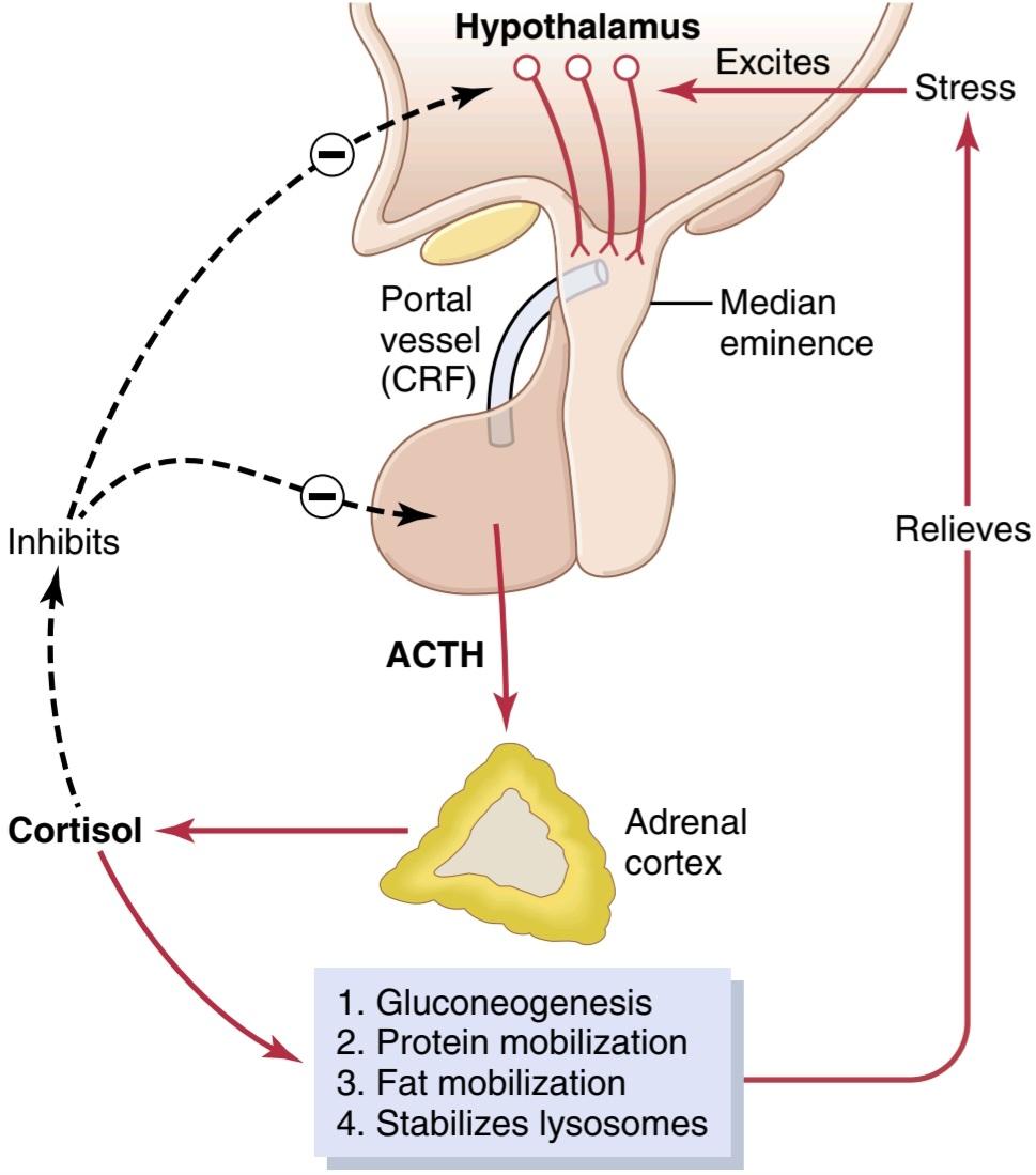
Figure 78-8. Mechanism for regulation of glucocorticoid secretion. ACTH, Adrenocorticotropic hormone; CRF, corticotropin-releasing factor.
شکل 78-8. مکانیسم تنظیم ترشح گلوکوکورتیکوئید ACTH، هورمون آدرنوکورتیکوتروپیک؛ CRF، فاکتور آزاد کننده کورتیکوتروپین.
Inhibitory Effect of Cortisol on the Hypothalamus and Anterior Pituitary to Decrease ACTH Secretion. Cortisol has direct negative feedback effects on (1) the hypothalamus to decrease formation of CRF and (2) the anterior pituitary gland to decrease formation of ACTH. Both of these feedbacks help regulate the plasma concentration of cortisol. That is, whenever the cortisol concentration becomes too great, the feedbacks automatically reduce the ACTH toward a normal control level.
اثر مهاری کورتیزول بر هیپوتالاموس و هیپوفیز قدامی برای کاهش ترشح ACTH. کورتیزول دارای اثرات بازخورد منفی مستقیم بر روی (1) هیپوتالاموس برای کاهش تشکیل CRF و (2) غده هیپوفیز قدامی برای کاهش تشکیل ACTH است. هر دوی این بازخوردها به تنظیم غلظت کورتیزول پلاسما کمک می کنند. یعنی هر زمان که غلظت کورتیزول خیلی زیاد شود، بازخوردها به طور خودکار ACTH را به سمت یک سطح کنترل طبیعی کاهش می دهند.
Summary of the Cortisol Control System
Figure 78-8 shows the overall system for control of cortisol secretion. The key to this control is the excitation of the hypothalamus by different types of stress. Stress stimuli activate the entire system to cause rapid release of cortisol, and cortisol in turn initiates a series of metabolic effects directed toward relieving the damaging nature of the stressful state.
خلاصه ای از سیستم کنترل کورتیزول
شکل 78-8 سیستم کلی برای کنترل ترشح کورتیزول را نشان می دهد. کلید این کنترل تحریک هیپوتالاموس توسط انواع مختلف استرس است. محرک های استرس کل سیستم را فعال می کنند تا باعث آزاد شدن سریع کورتیزول شود و کورتیزول به نوبه خود یک سری از اثرات متابولیکی را آغاز می کند که به سمت تسکین ماهیت مضر حالت استرس زا هدایت می شوند.
Direct feedback of cortisol to both the hypothalamus and the anterior pituitary gland also occurs to decrease the concentration of cortisol in the plasma at times when the body is not experiencing stress. How- ever, the stress stimuli are the most potent ones; they can always break through this direct inhibitory feed- back of cortisol, causing either periodic exacerbations of cortisol secretion at multiple times during the day (Figure 78-9) or prolonged cortisol secretion in times of chronic stress.
بازخورد مستقیم کورتیزول به هیپوتالاموس و غده هیپوفیز قدامی نیز برای کاهش غلظت کورتیزول در پلاسما در مواقعی که بدن استرس را تجربه نمی کند، رخ می دهد. با این حال، محرک های استرس قوی ترین محرک ها هستند. آنها همیشه می توانند این بازخورد مهاری مستقیم کورتیزول را بشکنند و یا باعث تشدید دوره ای ترشح کورتیزول در چند نوبت در طول روز (شکل 9-78) یا ترشح طولانی مدت کورتیزول در زمان استرس مزمن شوند.
Circadian Rhythm of Glucocorticoid Secretion. The secretory rates of CRF, ACTH, and cortisol are high in the early morning but low in the late evening, as shown in Figure 78-9; plasma cortisol level ranges between a high of about 20 μg/dl an hour before arising in the morning and a low of about 5 ug/dl around midnight. This effect results from a 24-hour cyclical alteration in the signals from the hypothalamus that cause cortisol secretion. When a person changes his or her daily sleeping habits, the cycle changes correspondingly. Therefore, measurements of blood cortisol levels are meaningful only when expressed in terms of the time in the cycle at which the measurements are made.
ریتم شبانه روزی ترشح گلوکوکورتیکوئید. همانطور که در شکل 78-9 نشان داده شده است، میزان ترشح CRF، ACTH و کورتیزول در اوایل صبح زیاد اما در اواخر عصر کم است. سطح کورتیزول پلاسما بین حداکثر 20 میکروگرم در دسی لیتر یک ساعت قبل از ظهور در صبح و حدود 5 میکروگرم در دسی لیتر در حوالی نیمه شب متغیر است. این اثر ناشی از یک تغییر چرخه ای 24 ساعته در سیگنال های هیپوتالاموس است که باعث ترشح کورتیزول می شود. زمانی که فرد عادات خواب روزانه خود را تغییر می دهد، چرخه نیز به همان نسبت تغییر می کند. بنابراین، اندازه گیری سطح کورتیزول خون تنها زمانی معنادار است که بر حسب زمان در چرخه ای که اندازه گیری ها انجام می شود بیان شود.

Figure 78-9. Typical pattern of cortisol concentration during the day. Note the oscillations in secretion, as well as a daily secretory surge an hour or so after waking in the morning.
شکل 78-9. الگوی معمول غلظت کورتیزول در طول روز. به نوسانات ترشح و همچنین افزایش ترشح روزانه یک ساعت یا بیشتر پس از بیدار شدن در صبح توجه کنید.
Synthesis and Secretion of ACTH in Association With Melanocyte-Stimulating Hormone, Lipotropin, and Endorphin
When ACTH is secreted by the anterior pituitary gland, several other hormones that have similar chemical structures are secreted simultaneously. The gene that is transcribed to form the RNA molecule that causes ACTH synthesis initially causes formation of a considerably larger protein, a preprohormone called pro-opiomelanocortin (POMC), which is the precursor of ACTH and several other peptides, including melanocyte-stimulating hormone (MSH), B-lipotropin, B-endorphin, and a few others (Figure 78-10). Under normal conditions, most of these hormones are not secreted in enough quantity by the pituitary to have a major effect on the human body, but when the rate of secretion of ACTH is high, as may occur in persons with Addison’s disease, formation of some of the other POMC-derived hormones may also be increased.
سنتز و ترشح ACTH در ارتباط با هورمون محرک ملانوسیت، لیپوتروپین و اندورفین
هنگامی که ACTH توسط غده هیپوفیز قدامی ترشح می شود، چندین هورمون دیگر که ساختار شیمیایی مشابهی دارند به طور همزمان ترشح می شوند. ژنی که برای تشکیل مولکول RNA رونویسی می شود که باعث سنتز ACTH می شود، در ابتدا باعث تشکیل یک پروتئین بسیار بزرگتر می شود، یک پیش هورمون به نام pro-opiomelanocortin (POMC) که پیش ساز ACTH و چندین پپتید دیگر، از جمله هورمون محرک ملانوسیت (MSH)، B-لیپوتروفین، B-lipotrophin، و سایرین است.(شکل 78-10). در شرایط عادی، بیشتر این هورمونها به اندازه کافی توسط هیپوفیز ترشح نمیشوند تا تأثیر عمدهای بر بدن انسان داشته باشند، اما زمانی که میزان ترشح ACTH بالا باشد، همانطور که ممکن است در افراد مبتلا به بیماری آدیسون رخ دهد، ممکن است تشکیل برخی دیگر از هورمونهای مشتق شده از POMC نیز افزایش یابد.
The POMC gene is actively transcribed in several tissues, including the corticotroph cells of the anterior pituitary, POMC neurons in the arcuate nucleus of the hypothalamus, cells of the dermis, and lymphoid tissue. In all of these cell types, POMC is processed to form a series of smaller peptides. The precise type of POMC- derived products from a particular tissue depends on the type of processing enzymes present in the tissue. Thus, pituitary corticotroph cells express prohormone convertase 1 (PC1), resulting in the production of N-terminal peptide, joining peptide, ACTH, and ẞ-lipotropin. In the hypothalamus, the expression of PC2 leads to production of a-MSH, B-MSH, y-MSH, and ẞ-endorphin, but not ACTH. As discussed in Chapter 72, a-MSH formed by neurons of the hypothalamus plays a major role in appetite regulation.
ژن POMC به طور فعال در چندین بافت از جمله سلول های کورتیکوتروف هیپوفیز قدامی، نورون های POMC در هسته کمانی هیپوتالاموس، سلول های درم و بافت لنفاوی رونویسی می شود. در همه این انواع سلول، POMC برای تشکیل یک سری پپتیدهای کوچکتر پردازش می شود. نوع دقیق محصولات مشتق شده از POMC از یک بافت خاص به نوع آنزیم های پردازش موجود در بافت بستگی دارد. بنابراین، سلول های کورتیکوتروف هیپوفیز پروهورمون کانورتاز 1 (PC1) را بیان می کنند که منجر به تولید پپتید N ترمینال، پیوستن پپتید، ACTH و ẞ-lipotropin می شود. در هیپوتالاموس، بیان PC2 منجر به تولید a-MSH، B-MSH، y-MSH و ẞ-اندورفین می شود، اما نه ACTH. همانطور که در فصل 72 بحث شد، a-MSH که توسط نورون های هیپوتالاموس تشکیل می شود، نقش عمده ای در تنظیم اشتها دارد.
In melanocytes located in abundance between the der- mis and epidermis of the skin, MSH stimulates formation of the black pigment melanin and disperses it to the epi- dermis. Injection of MSH into a person over 8 to 10 days can greatly increase darkening of the skin. The effect is much greater in people who have genetically dark skins than in light-skinned people.
در ملانوسیت هایی که به وفور بین درم و اپیدرم پوست قرار دارند، MSH تشکیل رنگدانه سیاه ملانین را تحریک می کند و آن را به اپیدرم پراکنده می کند. تزریق MSH به فرد در طی 8 تا 10 روز می تواند تا حد زیادی تیره شدن پوست را افزایش دهد. این تاثیر در افرادی که دارای پوست ژنتیکی تیره هستند بسیار بیشتر از افراد با پوست روشن است.
In some animals, an intermediate “lobe” of the pituitary gland, called the pars intermedia, is highly developed, lying between the anterior and posterior pituitary lobes. This lobe secretes an especially large amount of MSH. Furthermore, this secretion is independently controlled by the hypothalamus in response to the amount of light to which the animal is exposed or in response to other environmental factors. For instance, some arctic animals develop darkened fur in the summer and yet have entirely white fur in the winter.
در برخی از حیوانات، یک “لوب” میانی غده هیپوفیز به نام pars intermedia بسیار توسعه یافته است که بین لوب های قدامی و خلفی هیپوفیز قرار دارد. این لوب مقدار بسیار زیادی MSH ترشح می کند. علاوه بر این، این ترشح به طور مستقل توسط هیپوتالاموس در پاسخ به میزان نوری که حیوان در معرض آن قرار می گیرد یا در پاسخ به سایر عوامل محیطی کنترل می شود. به عنوان مثال، برخی از حیوانات قطب شمال در تابستان خز تیره میکنند و در زمستان خز کاملاً سفید دارند.
ACTH, because it contains an MSH sequence, has about 30 as much melanocyte-stimulating effect as MSH. Furthermore, because the quantities of pure MSH secreted in humans are extremely small, whereas those of ACTH are large, it is likely that ACTH is normally more important than MSH in determining the amount of melanin in the skin.
ACTH، به دلیل اینکه حاوی یک توالی MSH است، حدود 30 به اندازه MSH اثر محرک ملانوسیت دارد. علاوه بر این، از آنجایی که مقادیر MSH خالص ترشح شده در انسان بسیار کم است، در حالی که مقادیر ACTH زیاد است، این احتمال وجود دارد که ACTH به طور معمول در تعیین میزان ملانین در پوست مهمتر از MSH باشد.

Figure 78-10. Pro-opiomelanocortin processing by prohormone convertase 1 (PC1; red arrows) and PC2 (blue arrows). Tissue-specific expression of these two enzymes results in different peptides produced in various tissues. ACTH, Adrenocorticotropic hormone; CLIP, corticotropin-like intermediate peptide; MSH, melanocyte-stimulating hormone.
شکل 78-10. پردازش Pro-opiomelanocortin توسط پروهورمون کانورتاز 1 (PC1؛ فلش های قرمز) و PC2 (فلش های آبی). بیان بافتی این دو آنزیم منجر به تولید پپتیدهای مختلف در بافت های مختلف می شود. ACTH، هورمون آدرنوکورتیکوتروپیک؛ CLIP، پپتید واسط شبه کورتیکوتروپین. MSH، هورمون محرک ملانوسیت.
Adrenal Androgens
Several moderately active male sex hormones called adrenal androgens (the most important of which is dehydroepiandrosterone) are continually secreted by the adrenal cortex, especially during fetal life, as discussed in Chapter 84. Also, progesterone and estrogens, which are female sex hormones, are secreted in minute quantities.
آندروژن های آدرنال
چندین هورمون جنسی مردانه نسبتاً فعال به نام آندروژن های آدرنال (که مهمترین آنها دهیدرواپی آندروسترون است) به طور مداوم توسط قشر آدرنال، به ویژه در طول زندگی جنینی، همانطور که در فصل 84 مورد بحث قرار گرفت، ترشح می شود. همچنین، پروژسترون و استروژن ها که هورمون های جنسی زنانه هستند، به مقدار کمی ترشح می شوند.
Normally, the adrenal androgens have only weak effects in humans. It is possible that part of the early development of the male sex organs results from childhood secretion of adrenal androgens. The adrenal androgens also exert mild effects in the female, not only before puberty but also throughout life. Much of the growth of the pubic and axillary hair in the female results from the action of these hormones.
به طور معمول، آندروژن های آدرنال فقط اثرات ضعیفی در انسان دارند. ممکن است بخشی از رشد اولیه اندام های جنسی مردانه ناشی از ترشح آندروژن های آدرنال در دوران کودکی باشد. آندروژن های آدرنال نه تنها قبل از بلوغ بلکه در طول زندگی نیز اثرات خفیفی در زنان دارند. بیشتر رشد موهای ناحیه تناسلی و زیر بغل در زنان ناشی از عملکرد این هورمون ها است.
In extra-adrenal tissues, some of the adrenal androgens are converted to testosterone, the primary male sex hormone, which probably accounts for much of their androgenic activity. The physiological effects of androgens are discussed in Chapter 81 in relation to male sexual function.
در بافتهای خارج آدرنال، برخی از آندروژنهای آدرنال به تستوسترون، هورمون جنسی اولیه مردانه، تبدیل میشوند که احتمالاً بیشتر فعالیت آندروژنی آنها را تشکیل میدهد. اثرات فیزیولوژیکی آندروژن ها در فصل 81 در رابطه با عملکرد جنسی مردان مورد بحث قرار گرفته است.
Abnormalities of Adrenocortical Secretion Hypoadrenalism (Adrenal Insufficiency)— Addison’s Disease
Addison’s disease results from an inability of the adrenal cortices to produce sufficient adrenocortical hormones, and this in turn is most frequently caused by primary atrophy or injury of the adrenal cortices. In about 80% of cases, the atrophy is caused by autoimmunity against the cortices. Adrenal gland hypofunction may also be caused by tuberculous destruction of the adrenal glands or invasion of the adrenal cortices by cancer.
اختلالات ترشح قشر آدرنالیسم هیپوآدرنالیسم (نارسایی آدرنال) – بیماری آدیسون
بیماری آدیسون ناشی از ناتوانی قشر آدرنال در تولید هورمون های قشر آدرنال کافی است و این به نوبه خود اغلب در اثر آتروفی اولیه یا آسیب قشر آدرنال ایجاد می شود. در حدود 80 درصد موارد، آتروفی ناشی از خودایمنی در برابر قشر مغز است. کم کاری غده آدرنال نیز ممکن است در اثر تخریب سل غدد فوق کلیوی یا حمله سرطان به قشر آدرنال ایجاد شود.
In some cases, adrenal insufficiency is secondary to im- paired function of the pituitary gland, which fails to pro- duce sufficient ACTH. When ACTH output is too low, cortisol and aldosterone production decrease, and eventually the adrenal glands may atrophy because of a lack of ACTH stimulation. Secondary adrenal insufficiency is much more common than Addison’s disease, which is sometimes called primary adrenal insufficiency. Disturbances in severe ad- renal insufficiency are described in the following sections.
در برخی موارد، نارسایی آدرنال ثانویه به اختلال عملکرد غده هیپوفیز است که قادر به تولید ACTH کافی نیست. هنگامی که برون ده ACTH خیلی کم باشد، تولید کورتیزول و آلدوسترون کاهش می یابد و در نهایت غدد فوق کلیوی ممکن است به دلیل عدم تحریک ACTH آتروفی شوند. نارسایی ثانویه آدرنال بسیار شایع تر از بیماری آدیسون است که گاهی اوقات به آن نارسایی اولیه آدرنال می گویند. اختلالات در نارسایی شدید آدرنال در بخش های زیر توضیح داده شده است.
Mineralocorticoid Deficiency. Lack of aldosterone secretion greatly decreases renal tubular sodium reabsorption and consequently allows sodium ions, chloride ions, and water to be lost into urine in great profusion. The net result is a greatly decreased extracellular fluid volume and hyponatremia. Furthermore, hyperkalemia and mild acidosis develop because of failure of potassium and hydrogen ions to be secreted in exchange for sodium reabsorption.
کمبود مینرالوکورتیکوئید. کمبود ترشح آلدوسترون به میزان زیادی بازجذب سدیم لولهای کلیوی را کاهش میدهد و در نتیجه باعث میشود یونهای سدیم، یونهای کلرید و آب به میزان زیادی در ادرار از بین بروند. نتیجه خالص کاهش شدید حجم مایع خارج سلولی و هیپوناترمی است. علاوه بر این، هیپرکالمی و اسیدوز خفیف به دلیل عدم ترشح یون های پتاسیم و هیدروژن در ازای بازجذب سدیم ایجاد می شود.
As the extracellular fluid becomes depleted, plasma volume falls, red blood cell concentration rises markedly, cardiac output and blood pressure decrease, and the patient may in shock, with death usually occurring in the untreated patient 4 days to 2 weeks after complete cessation of mineralocorticoid secretion.
همانطور که مایع خارج سلولی تخلیه می شود، حجم پلاسما کاهش می یابد، غلظت گلبول های قرمز به طور قابل توجهی افزایش می یابد، برون ده قلبی و فشار خون کاهش می یابد، و بیمار ممکن است دچار شوک شود و معمولاً در بیمار درمان نشده 4 روز تا 2 هفته پس از توقف کامل ترشح مینرالوکورتیکوئید، مرگ رخ می دهد.
Glucocorticoid Deficiency. Loss of cortisol secretion makes it impossible for a person with Addison’s disease to maintain normal blood glucose concentration between meals because he or she cannot synthesize significant quantities of glucose by gluconeogenesis. Furthermore, lack of cortisol reduces the mobilization of proteins and fats from the tissues, thereby depressing many other metabolic functions of the body. This sluggishness of energy mobilization when cortisol is not available is one of the major detrimental effects of glucocorticoid deficiency. Even when excess quantities of glucose and other nutrients are available, the person’s muscles are weak, indicating that glucocorticoids are necessary to maintain other metabolic functions of the tissues in addition to energy metabolism.
کمبود گلوکوکورتیکوئید. از دست دادن ترشح کورتیزول باعث میشود که فرد مبتلا به بیماری آدیسون نتواند غلظت طبیعی گلوکز خون را بین وعدههای غذایی حفظ کند، زیرا نمیتواند مقادیر قابل توجهی گلوکز را با گلوکونئوژنز سنتز کند. علاوه بر این، کمبود کورتیزول باعث کاهش تحرک پروتئین ها و چربی ها از بافت ها می شود و در نتیجه بسیاری از عملکردهای متابولیک دیگر بدن را کاهش می دهد. این کندی بسیج انرژی زمانی که کورتیزول در دسترس نیست یکی از اثرات مخرب عمده کمبود گلوکوکورتیکوئید است. حتی زمانی که مقادیر بیش از حد گلوکز و سایر مواد مغذی در دسترس باشد، ماهیچههای فرد ضعیف میشوند و این نشان میدهد که گلوکوکورتیکوئیدها برای حفظ سایر عملکردهای متابولیک بافتها علاوه بر متابولیسم انرژی ضروری هستند.
Lack of adequate glucocorticoid secretion also makes a person with Addison’s disease highly susceptible to the deteriorating effects of different types of stress, and even a mild respiratory infection can cause death.
عدم ترشح کافی گلوکوکورتیکوئید همچنین باعث می شود که فرد مبتلا به بیماری آدیسون به شدت در معرض اثرات بدتر انواع مختلف استرس قرار گیرد و حتی یک عفونت خفیف تنفسی می تواند باعث مرگ شود.
Melanin Pigmentation. Another characteristic of most people with Addison’s disease is melanin pigmentation of the mucous membranes and skin. This melanin is not always deposited evenly but occasionally is deposited in blotches, and it is deposited especially in the thin skin areas, such as the mucous membranes of the lips and the thin skin of the nipples.
رنگدانه ملانین. یکی دیگر از ویژگی های بیشتر افراد مبتلا به بیماری آدیسون، رنگدانه های ملانین در غشاهای مخاطی و پوست است. این ملانین همیشه به طور یکنواخت رسوب نمیکند، اما گاهی اوقات به صورت لکههایی رسوب میکند و بهویژه در نواحی نازک پوست، مانند غشای مخاطی لبها و پوست نازک نوک سینهها رسوب میکند.
When cortisol secretion is depressed, the normal negative feedback to the hypothalamus and anterior pituitary gland is also depressed, therefore allowing tremendous rates of ACTH secretion, as well as simultaneous secretion of increased amounts of MSH. The large amounts of ACTH probably cause most of the pigmenting effect because they can stimulate formation of melanin by the melanocytes in the same way that MSH does.
هنگامی که ترشح کورتیزول کاهش می یابد، بازخورد منفی طبیعی به هیپوتالاموس و غده هیپوفیز قدامی نیز کاهش می یابد، بنابراین نرخ فوق العاده ای از ترشح ACTH و همچنین ترشح همزمان مقادیر افزایش یافته MSH را امکان پذیر می کند. مقادیر زیاد ACTH احتمالاً باعث بیشتر اثر رنگدانه می شود زیرا می توانند تشکیل ملانین توسط ملانوسیت ها را به همان روشی که MSH انجام می دهد تحریک کنند.
Treatment of People With Addison’s Disease. An un- treated person with total adrenal destruction dies within a few days to a few weeks because of weakness and, usually, circulatory shock. Yet, such a person can live for years if small quantities of mineralocorticoids and glucocorticoids are administered daily.
درمان افراد مبتلا به بیماری آدیسون. یک فرد درمان نشده با تخریب کامل آدرنال در عرض چند روز تا چند هفته به دلیل ضعف و معمولاً شوک گردش خون می میرد. با این حال، اگر روزانه مقادیر کمی از مینرالوکورتیکوئیدها و گلوکوکورتیکوئیدها تجویز شود، چنین فردی می تواند سال ها زندگی کند.
Adrenal Crisis. As noted earlier in the chapter, large quantities of glucocorticoids are occasionally secreted in response to different types of physical or mental stress. In a person with Addison’s disease, the output of glucocorticoids does not increase during stress. Yet, during different types of trauma, disease, or other stresses, such as surgical operations, a person is likely to have an acute need for increased amounts of glucocorticoids and often must be given 10 or more times the normal quantities of glucocorticoids to prevent death.
بحران آدرنال. همانطور که قبلاً در این فصل ذکر شد، مقادیر زیادی گلوکوکورتیکوئیدها گهگاه در پاسخ به انواع مختلف استرس فیزیکی یا روانی ترشح میشوند. در فرد مبتلا به بیماری آدیسون، برون ده گلوکوکورتیکوئیدها در هنگام استرس افزایش نمی یابد. با این حال، در طول انواع مختلف تروما، بیماری، یا استرسهای دیگر، مانند عملهای جراحی، فرد احتمالاً نیاز شدیدی به مقادیر زیاد گلوکوکورتیکوئیدها دارد و اغلب باید ۱۰ برابر یا بیشتر از مقادیر طبیعی گلوکوکورتیکوئیدها برای جلوگیری از مرگ تجویز شود.
This critical need for extra glucocorticoids and the as- sociated severe debility in times of acute stress is called an adrenal (or Addisonian) crisis.
این نیاز حیاتی به گلوکوکورتیکوئیدهای اضافی و ناتوانی شدید مرتبط با آن در مواقع استرس حاد، بحران آدرنال (یا آدیسون) نامیده می شود.
Hyperadrenalism-Cushing’s Syndrome
Hypersecretion by the adrenal cortex causes a complex cascade of hormone effects called Cushing’s syndrome. Many of the abnormalities of Cushing’s syndrome can be ascribed to abnormal amounts of cortisol, but excess secretion of androgens may also cause important effects. Hypercortisolism can occur from multiple causes, including (1) adenomas of the anterior pituitary that secrete large amounts of ACTH, which then causes adrenal hyperplasia and excess cortisol secretion; (2) abnormal function of the hypothalamus that causes high levels of corticotropin-releasing hormone, which stimulates excess ACTH release; (3) “ectopic secretion” of ACTH by a tumor elsewhere in the body, such as an abdominal carcinoma; and (4) adenomas of the adrenal cortex. When Cushing’s syndrome is secondary to excess secretion of ACTH by the anterior pituitary, this condition is referred to as Cushing’s disease.
هایپرآدرنالیسم-سندرم کوشینگ
ترشح بیش از حد توسط قشر آدرنال باعث ایجاد آبشار پیچیده ای از اثرات هورمونی به نام سندرم کوشینگ می شود. بسیاری از ناهنجاریهای سندرم کوشینگ را میتوان به مقادیر غیرطبیعی کورتیزول نسبت داد، اما ترشح بیش از حد آندروژنها نیز ممکن است اثرات مهمی ایجاد کند. هیپرکورتیزولیسم می تواند به دلایل متعددی رخ دهد، از جمله (1) آدنوم هیپوفیز قدامی که مقادیر زیادی ACTH ترشح می کند، که سپس باعث هیپرپلازی آدرنال و ترشح اضافی کورتیزول می شود. (2) عملکرد غیر طبیعی هیپوتالاموس که باعث سطوح بالایی از هورمون آزاد کننده کورتیکوتروپین می شود که باعث تحریک ترشح بیش از حد ACTH می شود. (3) “ترشح نابجا” ACTH توسط یک تومور در جای دیگر بدن، مانند کارسینوم شکم. و (4) آدنوم قشر آدرنال. هنگامی که سندرم کوشینگ ثانویه به ترشح بیش از حد ACTH توسط هیپوفیز قدامی باشد، این وضعیت به عنوان بیماری کوشینگ شناخته می شود.
Excess ACTH secretion is the most common cause of Cushing’s syndrome and is characterized by high plasma levels of ACTH and cortisol. Primary overproduction of cortisol by the adrenal glands accounts for about 20% to 25% of clinical cases of Cushing’s syndrome and is usually associated with reduced ACTH levels due to cortisol feedback inhibition of ACTH secretion by the anterior pituitary gland.
ترشح بیش از حد ACTH شایع ترین علت سندرم کوشینگ است و با سطوح بالای ACTH و کورتیزول پلاسما مشخص می شود. تولید بیش از حد اولیه کورتیزول توسط غدد فوق کلیوی حدود 20 تا 25 درصد از موارد بالینی سندرم کوشینگ را تشکیل می دهد و معمولاً با کاهش سطح ACTH به دلیل مهار بازخورد کورتیزول از ترشح ACTH توسط غده هیپوفیز قدامی همراه است.
Administration of large doses of dexamethasone, a synthetic glucocorticoid, can be used to distinguish be- tween ACTH-dependent and ACTH-independent Cushing’s syndrome. In patients who have overproduction of ACTH due to an ACTH-secreting pituitary adenoma or to hypothalamic-pituitary dysfunction, low doses of dexamethasone usually do not suppress ACTH secretion normally. By increasing the dose of dexamethasone to very high levels, ACTH eventually can be suppressed in most patients with Cushing’s disease. In contrast, patients with primary adrenal overproduction of cortisol (ACTH- independent Cushing’s syndrome) usually have low or un- detectable levels of ACTH.
تجویز دوزهای زیاد دگزامتازون، یک گلوکوکورتیکوئید مصنوعی، می تواند برای تشخیص سندرم کوشینگ وابسته به ACTH و مستقل از ACTH استفاده شود. در بیمارانی که تولید بیش از حد ACTH به دلیل آدنوم هیپوفیز ترشح کننده ACTH یا اختلال عملکرد هیپوتالاموس-هیپوفیز دارند، دوزهای پایین دگزامتازون معمولاً ترشح ACTH را سرکوب نمی کند. با افزایش دوز دگزامتازون به سطوح بسیار بالا، در نهایت می توان ACTH را در اکثر بیماران مبتلا به بیماری کوشینگ سرکوب کرد. در مقابل، بیماران مبتلا به تولید بیش از حد کورتیزول اولیه آدرنال (سندرم کوشینگ مستقل از ACTH) معمولاً سطوح پایین یا غیرقابل تشخیص ACTH دارند.
The dexamethasone test, although widely used, can sometimes result in an incorrect diagnosis because some ACTH-secreting pituitary tumors do respond to dexamethasone with suppressed ACTH secretion. Also, non- pituitary malignant tumors that produce ACTH ectopically, such as some lung carcinomas, are not responsive to glucocorticoid-negative feedback. Therefore, the dexamethasone test is usually considered to be a first step in the differential diagnosis of Cushing’s syndrome.
آزمایش دگزامتازون، اگرچه به طور گسترده مورد استفاده قرار می گیرد، گاهی اوقات می تواند منجر به تشخیص نادرست شود زیرا برخی از تومورهای هیپوفیز ترشح کننده ACTH با ترشح سرکوب شده ACTH به دگزامتازون پاسخ می دهند. همچنین، تومورهای بدخیم غیر هیپوفیز که ACTH را به صورت نابجا تولید می کنند، مانند برخی از کارسینوم های ریه، به بازخورد منفی گلوکوکورتیکوئید پاسخ نمی دهند. بنابراین، آزمایش دگزامتازون معمولاً اولین قدم در تشخیص افتراقی سندرم کوشینگ در نظر گرفته می شود.
Cushing’s syndrome can also occur when large amounts of glucocorticoids are administered over prolonged periods for therapeutic purposes. For example, patients with chronic inflammation associated with diseases such as rheumatoid arthritis are often treated with glucocorticoids and may experience some of the clinical symptoms of Cushing syndrome.
سندرم کوشینگ همچنین می تواند زمانی رخ دهد که مقادیر زیادی گلوکوکورتیکوئیدها در دوره های طولانی برای اهداف درمانی تجویز شوند. به عنوان مثال، بیماران مبتلا به التهاب مزمن مرتبط با بیماری هایی مانند آرتریت روماتوئید اغلب با گلوکوکورتیکوئیدها درمان می شوند و ممکن است برخی از علائم بالینی سندرم کوشینگ را تجربه کنند.
A special characteristic of Cushing’s syndrome is mobilization of fat from the lower part of the body, with concomitant extra deposition of fat in the thoracic and upper abdominal regions, giving rise to a buffalo-like torso. The excess secretion of steroids also leads to an edematous appearance of the face, and the androgenic potency of some of the hormones sometimes causes acne and hirsutism (excess growth of facial hair). The appearance of the face is frequently described as a “moon face,” as demonstrated in the untreated patient with Cushing’s syndrome to the left in Figure 78-11. About 80% of patients have hypertension, presumably because of the mineralocorticoid effects of cortisol.
ویژگی خاص سندرم کوشینگ، حرکت چربی از قسمت پایین بدن، همراه با رسوب چربی اضافی در نواحی قفسه سینه و بالای شکم است که باعث ایجاد نیم تنه گاومیش مانند می شود. ترشح بیش از حد استروئیدها همچنین منجر به ادم ظاهری صورت می شود و قدرت آندروژنیک برخی از هورمون ها گاهی باعث آکنه و هیرسوتیسم (رشد بیش از حد موهای صورت) می شود. ظاهر صورت اغلب به عنوان “صورت ماه” توصیف می شود، همانطور که در بیمار درمان نشده مبتلا به سندرم کوشینگ در سمت چپ در شکل 78-11 نشان داده شده است. حدود 80 درصد از بیماران فشار خون بالا دارند، احتمالاً به دلیل اثرات معدنی کورتیکوئید کورتیزول.
Effects of Cushing’s Syndrome on Carbohydrate and Protein Metabolism. The abundance of cortisol secreted in Cushing’s syndrome can increase blood glucose concentration, sometimes to values as high as 200 mg/dl after meals as much as twice normal. This increase results mainly from enhanced gluconeogenesis and decreased glucose utilization by the tissues.
اثرات سندرم کوشینگ بر متابولیسم کربوهیدرات و پروتئین. فراوانی کورتیزول ترشح شده در سندرم کوشینگ می تواند غلظت گلوکز خون را افزایش دهد، گاهی اوقات تا مقادیر 200 میلی گرم در دسی لیتر بعد از غذا تا دو برابر طبیعی. این افزایش عمدتاً ناشی از افزایش گلوکونئوژنز و کاهش استفاده از گلوکز توسط بافت ها است.
The effects of glucocorticoids on protein catabolism are of- ten profound in Cushing’s syndrome, causing greatly decreased tissue proteins almost everywhere in the body with the exception of the liver; the plasma proteins also remain unaffected. The loss of protein from the muscles in particular causes severe weakness. The loss of protein synthesis in the lymphoid tissues leads to a suppressed immune system, and thus these patients are highly susceptible to infections. Even the protein collagen fibers in the subcutaneous tissue are diminished so that the subcutaneous tissues tear easily, resulting in development of large purplish striae where they have torn apart. In addition, severely diminished protein deposition in the bones often causes severe osteoporosis with consequent weakness of the bones.
اثرات گلوکوکورتیکوئیدها بر کاتابولیسم پروتئین اغلب در سندرم کوشینگ عمیق است و باعث کاهش شدید پروتئین های بافتی تقریباً در همه جای بدن به استثنای کبد می شود. پروتئین های پلاسما نیز بدون تأثیر باقی می مانند. از دست دادن پروتئین از عضلات به ویژه باعث ضعف شدید می شود. از دست دادن سنتز پروتئین در بافت های لنفاوی منجر به سرکوب سیستم ایمنی می شود و بنابراین این بیماران به شدت مستعد ابتلا به عفونت هستند. حتی الیاف پروتئینی کلاژن در بافت زیر جلدی کاهش مییابد به طوری که بافتهای زیر جلدی به راحتی پاره میشوند و در نتیجه خطوط بزرگ ارغوانی در جایی که از هم جدا شدهاند ایجاد میشود. علاوه بر این، کاهش شدید رسوب پروتئین در استخوان ها اغلب باعث پوکی استخوان شدید و در نتیجه ضعف استخوان ها می شود.
Treatment of People With Cushing’s Syndrome. Treatment of people with Cushing’s syndrome consists of removing an adrenal tumor if this is the cause or decreasing the secretion of ACTH, if possible. Hypertrophied pituitary glands or even small tumors in the pituitary that oversecrete ACTH can sometimes be surgically removed or destroyed by radiation. Drugs that block steroidogenesis, such as metyrapone, ketoconazole, and aminoglutethimide, or that inhibit ACTH secretion, such as serotonin antagonists and GABA- transaminase inhibitors, can also be used when surgery is not feasible. If ACTH secretion cannot easily be decreased, the only satisfactory treatment is usually bilateral partial (or even total) adrenalectomy, followed by administration of adrenal steroids to make up for any insufficiency that develops.
درمان افراد مبتلا به سندرم کوشینگ. درمان افراد مبتلا به سندرم کوشینگ شامل برداشتن تومور آدرنال در صورت وجود علت یا کاهش ترشح ACTH در صورت امکان است. غدد هیپوفیز هیپرتروفی شده یا حتی تومورهای کوچک در هیپوفیز که ACTH را بیش از حد ترشح می کنند، گاهی اوقات می توانند با عمل جراحی برداشته یا توسط اشعه از بین بروند. داروهایی که استروئیدزایی را مسدود میکنند، مانند متیراپون، کتوکونازول، و آمینوگلوتیمید، یا مهارکننده ترشح ACTH، مانند آنتاگونیستهای سروتونین و مهارکنندههای گابا-ترانس آمیناز نیز میتوانند در مواردی که جراحی امکانپذیر نیست، استفاده شوند. اگر ترشح ACTH را نتوان به راحتی کاهش داد، تنها درمان رضایت بخش معمولاً آدرنالکتومی جزئی (یا حتی کل) دو طرفه و به دنبال آن تجویز استروئیدهای آدرنال برای جبران هر گونه نارسایی ایجاد شده است.

Figure 78-11. A person with Cushing’s syndrome before (left) and after (right) a subtotal adrenalectomy. (Courtesy Dr. Leonard Posey.)
شکل 78-11. یک فرد مبتلا به سندرم کوشینگ قبل ا (چپ) و بعد از (راست) آدرنالکتومی ساب توتال. (با احترام از دکتر لئونارد پوزی.)
Primary Aldosteronism (Conn’s Syndrome)
Occasionally a small tumor of the zona glomerulosa cells occurs and secretes large amounts of aldosterone; the resulting condition is called primary aldosteronism or Conn’s syndrome. Also, in a few instances, hyperplastic adrenal cortices secrete aldosterone rather than cortisol. The effects of the excess aldosterone are discussed in detail earlier in the chapter. The most important effects are hypokalemia, mild metabolic alkalosis, a slight increase in extracellular fluid volume and blood volume, a modest increase in plasma sodium concentration (usually <4-6-mEq/L increase) and, almost always, hypertension. Especially interesting in persons with primary aldosteronism are occasional periods of muscle paralysis caused by the hypokalemia. The paralysis is caused by a depressant effect of low extracellular potassium concentration on action potential transmission by the nerve fibers, as explained in Chapter 5.
آلدوسترونیسم اولیه (سندرم کان)
گاهی اوقات تومور کوچکی از سلول های زونا گلومرولوزا ایجاد می شود و مقادیر زیادی آلدوسترون ترشح می کند. شرایط ایجاد شده آلدوسترونیسم اولیه یا سندرم کان نامیده می شود. همچنین، در موارد معدودی، قشر آدرنال هیپرپلاستیک به جای کورتیزول، آلدوسترون ترشح می کند. اثرات آلدوسترون اضافی قبلاً در فصل به تفصیل مورد بحث قرار گرفته است. مهم ترین اثرات عبارتند از هیپوکالمی، آلکالوز متابولیک خفیف، افزایش جزئی در حجم مایع خارج سلولی و حجم خون، افزایش ملایم غلظت سدیم پلاسما (معمولاً کمتر از 6-4 میلی اکی والان در لیتر افزایش می یابد) و تقریباً همیشه فشار خون بالا. به خصوص در افراد مبتلا به آلدوسترونیسم اولیه، دوره های گاه به گاه فلج عضلانی ناشی از هیپوکالمی جالب توجه است. همانطور که در فصل 5 توضیح داده شد، فلج ناشی از اثر کاهش دهنده غلظت کم پتاسیم خارج سلولی بر انتقال پتانسیل عمل توسط رشته های عصبی است.
One of the diagnostic criteria of primary aldosteronism is a decreased plasma renin concentration. This de- crease results from feedback suppression of renin secretion caused by the excess aldosterone or by the excess extracellular fluid volume and arterial pressure resulting from the aldosteronism. Treatment of primary aldosteronism may include surgical removal of the tumor or of most of the adrenal tissue when hyperplasia is the cause. Another option for treatment is pharmacological antagonism of the mineralocorticoid receptor with spironolactone or eplerenone.
یکی از معیارهای تشخیصی آلدوسترونیسم اولیه کاهش غلظت رنین پلاسما است. این کاهش ناشی از سرکوب فیدبک ترشح رنین ناشی از آلدوسترون اضافی یا حجم اضافی مایع خارج سلولی و فشار شریانی ناشی از آلدوسترونیسم است. درمان آلدوسترونیسم اولیه ممکن است شامل برداشتن تومور یا بیشتر بافت آدرنال با جراحی باشد که علت آن هیپرپلازی است. گزینه دیگر برای درمان تضاد فارماکولوژیک گیرنده مینرالوکورتیکوئید با اسپیرونولاکتون یا اپلرنون است.
Adrenogenital Syndrome
Occasionally an adrenocortical tumor secretes excessive quantities of androgens that cause intense masculinizing effects throughout the body. If this phenomenon occurs in a female, virile characteristics develop, including growth of a beard, a much deeper voice, occasionally baldness if she also has the genetic trait for baldness, masculine distribution of hair on the body and the pubis, growth of the clitoris to resemble a penis, and deposition of proteins in the skin and especially in the muscles to give typical masculine characteristics.
سندرم آدرنوژنیتال
گاهی اوقات یک تومور قشر آدرنال مقادیر زیادی آندروژن ترشح می کند که باعث ایجاد اثرات مردانه شدید در سراسر بدن می شود. اگر این پدیده در یک زن رخ دهد، ویژگیهای مردانه ایجاد میشود، از جمله رشد ریش، صدای بسیار عمیقتر، گاهی اوقات طاسی در صورتی که ویژگی ژنتیکی طاسی، توزیع مردانه مو در بدن و ناحیه تناسلی، رشد کلیتوریس به شکل آلت تناسلی و رسوب پروتئین در پوست و بهویژه در ماهیچهها برای ایجاد ویژگیهای مردانه معمولی باشد.
In the prepubertal male, a virilizing adrenal tumor causes the same characteristics as in the female plus rapid development of the male sexual organs, as shown in Figure 78-12, which depicts a 4-year-old boy with adrenogenital syndrome. In the adult male, the virilizing characteristics of adrenogenital syndrome are usually obscured by the nor- mal virilizing characteristics of the testosterone secreted by the testes. It is often difficult to make a diagnosis of adreno- genital syndrome in the adult male. In adrenogenital syndrome, the excretion of 17-ketosteroids (which are derived from androgens) in the urine may be 10 to 15 times normal. This finding can be used in diagnosing the disease.
در مرد قبل از بلوغ، تومور آدرنال ویریل کننده همان ویژگی هایی را ایجاد می کند که در زنان به علاوه رشد سریع اندام های جنسی مردانه، همانطور که در شکل 78-12 نشان داده شده است، که پسر 4 ساله ای را با سندرم آدرنوژنیتال نشان می دهد. در مرد بالغ، ویژگیهای ویریلسازی سندرم آدرنوژنیتال معمولاً با ویژگیهای طبیعی زننده تستوسترون ترشح شده از بیضهها پنهان میشود. تشخیص سندرم آدرنوژنیتال در مردان بالغ اغلب دشوار است. در سندرم آدرنوژنیتال، دفع 17-کتوستروئیدها (که از آندروژن ها مشتق شده اند) در ادرار ممکن است 10 تا 15 برابر طبیعی باشد. از این یافته می توان در تشخیص بیماری استفاده کرد.

Figure 78-12. Adrenogenital syndrome in a 4-year-old boy. (Courtesy Dr. Leonard Posey.)
شکل 78-12. سندرم آدرنوژنیتال در یک پسر 4 ساله. (با احترام از دکتر لئونارد پوزی.)
کلیک کنید «Bibliography: فهرست کتب مربوطه»
Berger I, Werdermann M, Bornstein SR, Steenblock C: The adrenal gland in stress adaptation on a cellular level. J Steroid Biochem Mol Biol 190:198, 2019.
Bornstein SR: Predisposing factors for adrenal insufficiency. N Engl J Med 360:2328, 2009.
Chapman K, Holmes M, Seckl J: 11ẞ-hydroxysteroid dehydrogenases: intracellular gate-keepers of tissue glucocorticoid action. Physiol Rev 93:1139, 2013.
Dineen R, Stewart PM, Sherlock M: Factors impacting on the action of glucocorticoids in patients receiving glucocorticoid therapy. Clin Endocrinol (Oxf) 90:3, 2019.
Feelders RA, Hofland UJ: Medical treatment of Cushing disease. J Clin Endocrinol Metab 98:425, 2013.
Funder JW: Primary aldosteronism. Hypertension 74:458, 2019.
Hall JE, Granger JP, Smith MJ Jr, Premen AJ: Role of renal hemody- namics and arterial pressure in aldosterone “escape.” Hyperten- sion 6:1183, 1984.
Hardy RS, Zhou H, Seibel MJ, Cooper MS: Glucocorticoids and bone: Consequences of endogenous and exogenous excess and replace- ment therapy. Endocr Rev 39:519, 2018.
Loriaux DL: Diagnosis and differential diagnosis of Cushing’s syn- drome. N Engl J Med 376:1451, 2017.
Raff H, Carroll T: Cushing’s syndrome: from physiological principles to diagnosis and clinical care. J Physiol 593:493, 2015.
Raff H, Sharma ST, Nieman LK: Physiological basis for the etiology, di- agnosis, and treatment of adrenal disorders: Cushing’s syndrome, adrenal insufficiency, and congenital adrenal hyperplasia. Compr Physiol 4:739, 2014.
Rushworth RL, Torpy DJ, Falhammar H: Adrenal crisis. N Engl J Med 381:852, 2019.
Seccia TM, Caroccia B, Gomez-Sanchez EP, et al: Endocr Rev 39:1029, 2018.
Scaroni C, Zilio M, Foti M, Boscaro M: Glucose metabolism abnor- malities in Cushing syndrome: from molecular basis to clinical man- agement. Endocr Rev 38:189, 2017.
Spat A, Hunyady L: Control of aldosterone secretion: a model for con- vergence in cellular signaling pathways. Physiol Rev 84:489, 2004.
Stowasser M, Gordon RD: Primary aldosteronism: changing defini- tions and new concepts of physiology and pathophysiology both inside and outside the kidney. Physiol Rev 96:1327, 2016.
Tritos NA, Biller BMK: Medical therapy for Cushing’s syndrome in the twenty-first century. Endocrinol Metab Clin North Am 47:427, 2018.
Vaidya A, Mulatero P, Baudrand R, Adler GK: The expanding spec- trum of primary aldosteronism: implications for diagnosis, patho- genesis, and treatment. Endocr Rev 39:1057, 2018.
Wehling M: Rapid actions of aldosterone revisited: receptors in the limelight. J Steroid Biochem Mol Biol 176:94, 2018.
Weikum ER, Knuesel MT, Ortlund EA, Yamamoto KR: Glucocorticoid receptor control of transcription: precision and plasticity via allos- tery. Nat Rev Mol Cell Biol 18:159, 2017.
🚀 با ما همراه شوید!
تازهترین مطالب و آموزشهای مغز و اعصاب را از دست ندهید. با فالو کردن کانال تلگرام آیندهنگاران مغز، از ما حمایت کنید!



