
فیزیولوژی پزشکی گایتون و هال؛ هورمون های متابولیک تیروئید


دعای مطالعه [ نمایش ]
بِسْمِ الله الرَّحْمنِ الرَّحیمِ
اَللّهُمَّ اَخْرِجْنى مِنْ ظُلُماتِ الْوَهْمِ
خدايا مرا بيرون آور از تاريكىهاى وهم،
وَ اَكْرِمْنى بِنُورِ الْفَهْمِ
و به نور فهم گرامى ام بدار،
اَللّهُمَّ افْتَحْ عَلَيْنا اَبْوابَ رَحْمَتِكَ
خدايا درهاى رحمتت را به روى ما بگشا،
وَانْشُرْ عَلَيْنا خَزائِنَ عُلُومِكَ بِرَحْمَتِكَ يا اَرْحَمَ الرّاحِمينَ
و خزانههاى علومت را بر ما باز كن به امید رحمتت اى مهربانترين مهربانان.
کتاب «فیزیولوژی پزشکی گایتون و هال» بهعنوان یکی از جامعترین و معتبرترین منابع در حوزه علوم پزشکی، همچنان مرجع کلیدی برای درک عملکرد پیچیده بدن انسان است. این اثر با تکیه بر تازهترین پژوهشها و توضیحات دقیق از سازوکارهای فیزیولوژیک، پلی میان علوم پایه پزشکی و کاربردهای بالینی ایجاد میکند و نقشی بیبدیل در آموزش، پژوهش و ارتقای دانش سلامت ایفا مینماید.
ترجمه دقیق و علمی این شاهکار توسط برند علمی آیندهنگاران مغز به مدیریت داریوش طاهری، دسترسی فارسیزبانان به مرزهای نوین دانش فیزیولوژی را ممکن ساخته و رسالتی علمی برای ارتقای آموزش پزشکی، فهم عمیقتر سازوکارهای بدن و توسعه روشهای نوین در حوزه سلامت فراهم آورده است.
» Guyton and Hall Textbook of Medical Physiology
»» CHAPTER 77: Thyroid Metabolic Hormones
The thyroid gland, located immediately below the larynx on each side of and anterior to the trachea, is one of the largest of the endocrine glands, normally weighing 15 to 20 grams in adults. The thyroid secretes two major metabolic hormones, thyroxine and triiodothyronine, commonly called T4 and T3, respectively. Both of these hormones profoundly increase the metabolic rate of the body. Complete lack of thyroid secretion usually causes the basal metabolic rate to fall 40% to 50% below normal, and extreme excesses of thyroid secretion can increase the basal metabolic rate to 60% to 100% above normal. Thyroid secretion is controlled primarily by thyroid- stimulating hormone (TSH) secreted by the anterior pituitary gland.
غده تیروئید که بلافاصله در زیر حنجره در هر طرف و جلوی نای قرار دارد، یکی از بزرگترین غدد درون ریز است که در بزرگسالان معمولاً 15 تا 20 گرم وزن دارد. تیروئید دو هورمون متابولیک اصلی به نام های تیروکسین و تری یدوتیرونین ترشح می کند که معمولاً به ترتیب T4 و T3 نامیده می شوند. هر دوی این هورمون ها سرعت متابولیسم بدن را به شدت افزایش می دهند. فقدان کامل ترشح تیروئید معمولاً باعث میشود که میزان متابولیسم پایه 40 تا 50 درصد کمتر از حد طبیعی باشد و ترشح بیش از حد تیروئید میتواند میزان متابولیسم پایه را تا 60 تا 100 درصد بالاتر از حد طبیعی افزایش دهد. ترشح تیروئید در درجه اول توسط هورمون محرک تیروئید (TSH) که توسط غده هیپوفیز قدامی ترشح می شود، کنترل می شود.
The thyroid gland also secretes calcitonin, a hormone involved in calcium metabolism that is discussed in Chapter 80.
غده تیروئید همچنین کلسی تونین ترشح می کند، هورمونی که در متابولیسم کلسیم نقش دارد و در فصل 80 مورد بحث قرار گرفته است.
The purpose of this chapter is to discuss the formation and secretion of the thyroid hormones, their metabolic functions, and regulation of their secretion.
هدف این فصل بحث در مورد تشکیل و ترشح هورمون های تیروئید، عملکرد متابولیک آنها و تنظیم ترشح آنها است.
SYNTHESIS AND SECRETION OF THE THYROID METABOLIC HORMONES
About 93% of the metabolically active hormones secreted by the thyroid gland is thyroxine and 7% is triiodothyronine. However, almost all the thyroxine is eventually converted to triiodothyronine in the tissues, so both are functionally important. The functions of these two hormones are qualitatively the same, but they differ in rapidity and intensity of action. Triiodothyronine is about four times as potent as thyroxine, but it is present in the blood in much smaller quantities and persists for a much shorter time compared with thyroxine.
سنتز و ترشح هورمون های متابولیک تیروئید
حدود 93 درصد از هورمون های فعال متابولیکی که از غده تیروئید ترشح می شود تیروکسین و 7 درصد تری یدوتیرونین است. با این حال، تقریباً تمام تیروکسین در نهایت در بافت ها به تری یدوتیرونین تبدیل می شود، بنابراین هر دو از نظر عملکردی مهم هستند. عملکرد این دو هورمون از نظر کیفی یکسان است، اما از نظر سرعت و شدت عمل متفاوت هستند. تری یدوتیرونین تقریباً چهار برابر تیروکسین قوی تر است، اما در مقادیر بسیار کمتر در خون وجود دارد و در مقایسه با تیروکسین برای مدت زمان بسیار کوتاه تری باقی می ماند.
PHYSIOLOGIC ANATOMY OF THE THYROID GLAND
As shown in Figure 77-1, the thyroid gland is composed of large numbers of closed follicles (100-300 micrometers in diameter) that are filled with a secretory substance called colloid and lined with cuboidal epithelial cells that secrete into the interior of the follicles. The major constituent of colloid is the large glycoprotein thyroglobulin, which contains the thyroid hormones. Once the secretion has entered the follicles, it must be absorbed back through the follicular epithelium into the blood before it can function in the body. The thyroid gland has a blood flow about five times the weight of the gland each minute, which is a blood supply as great as that of any other area of the body, with the possible exception of the adrenal cortex.
آناتومی فیزیولوژیکی غده تیروئید
همانطور که در شکل 77-1 نشان داده شده است، غده تیروئید از تعداد زیادی فولیکول بسته (قطر 100-300 میکرومتر) تشکیل شده است که با یک ماده ترشحی به نام کلوئید پر شده و با سلول های اپیتلیال مکعبی پوشانده شده است که به داخل فولیکول ها ترشح می کنند. ماده اصلی تشکیل دهنده کلوئید، گلیکوپروتئین بزرگ تیروگلوبولین است که حاوی هورمون های تیروئید است. هنگامی که ترشح وارد فولیکول ها شد، قبل از اینکه بتواند در بدن عمل کند، باید از طریق اپیتلیوم فولیکولی دوباره جذب خون شود. غده تیروئید تقریباً پنج برابر وزن غده در هر دقیقه جریان خون دارد، که به اندازه خون هر ناحیه دیگری از بدن، به استثنای قشر آدرنال، جریان خون دارد.
The thyroid gland also contains C cells that secrete calcitonin, a hormone that contributes to regulation of plasma calcium ion concentration, as discussed in Chapter 80.
غده تیروئید همچنین حاوی سلول های C است که کلسی تونین ترشح می کنند، هورمونی که به تنظیم غلظت یون کلسیم پلاسما کمک می کند، همانطور که در فصل 80 بحث شد.

Figure 77-1 Anatomy and microscopic appearance of the thyroid gland, showing secretion of thyroglobulin into the follicles.
شکل 77-1 آناتومی و ظاهر میکروسکوپی غده تیروئید، ترشح تیروگلوبولین را در فولیکول ها نشان می دهد.
IODINE IS REQUIRED FOR THYROXINE FORMATION
To form normal quantities of thyroxine, about 50 milligrams of ingested iodine in the form of iodides are required each year, or about 1 mg/week. To prevent iodine deficiency, common table salt is iodized with about 1 part sodium iodide to every 100,000 parts sodium chloride.
ید برای تشکیل تیروکسین مورد نیاز است
برای تشکیل مقادیر طبیعی تیروکسین، حدود 50 میلی گرم ید مصرفی به شکل یدید در سال یا حدود 1 میلی گرم در هفته مورد نیاز است. برای جلوگیری از کمبود ید، نمک خوراکی معمولی با حدود 1 قسمت یدید سدیم به هر 100000 قسمت کلرید سدیم یددار می شود.
Fate of Ingested lodides. Iodides ingested orally are absorbed from the gastrointestinal tract into the blood in about the same manner as chlorides. Normally, most of the iodides are rapidly excreted by the kidneys, but only after about one fifth are selectively removed from the circulating blood by the cells of the thyroid gland and used for synthesis of the thyroid hormones.
سرنوشت لودیدهای بلعیده شده. یدیدهایی که به صورت خوراکی خورده می شوند تقریباً مانند کلریدها از دستگاه گوارش به خون جذب می شوند. به طور معمول، بیشتر یدیدها به سرعت توسط کلیه ها دفع می شوند، اما تنها پس از حدود یک پنجم به طور انتخابی توسط سلول های غده تیروئید از خون در گردش خارج می شوند و برای سنتز هورمون های تیروئید استفاده می شوند.
IODIDE PUMP-THE SODIUM-IODIDE SYMPORTER (IODIDE TRAPPING)
The first stage in formation of thyroid hormones, shown in Figure 77-2, is transport of iodides from the blood into the thyroid glandular cells and follicles. The basal membrane of the thyroid cell has the specific ability to pump the iodide actively to the interior of the cell. This pumping is achieved by the action of a sodium-iodide symporter, which co-transports one iodide ion along with two sodium ions across the basolateral (plasma) membrane into the cell. The energy for transporting iodide against a concentration gradient comes from the sodium-potassium adenosine triphosphatase (Na+-K+ ATPase) pump, which pumps sodium out of the cell, thereby establishing a low intracellular sodium concentration and a gradient for facilitated diffusion of sodium into the cell.
پمپ یدید-سیمپورتر سدیم-یدید (به دام انداختن ید)
اولین مرحله در تشکیل هورمون های تیروئید، که در شکل 77-2 نشان داده شده است، انتقال یدیدها از خون به سلول ها و فولیکول های غده تیروئید است. غشای پایه سلول تیروئید این توانایی خاص را دارد که یدید را به طور فعال به داخل سلول پمپ کند. این پمپاژ با عمل یک سمپورتر یدید سدیم به دست می آید که یک یون یدید را همراه با دو یون سدیم از غشای قاعده جانبی (پلاسما) به داخل سلول منتقل می کند. انرژی برای انتقال یدید در برابر گرادیان غلظت از پمپ سدیم پتاسیم آدنوزین تری فسفاتاز (Na+-K+ ATPase) تامین میشود که سدیم را از سلول پمپ میکند و در نتیجه غلظت سدیم درون سلولی کم و گرادیان برای انتشار تسهیل شده سدیم در سلول ایجاد میشود.
This process of concentrating the iodide in the cell is called iodide trapping. In a normal gland, the iodide pump concentrates the iodide to about 30 times its concentration in the blood. When the thyroid gland becomes maximally active, this concentration ratio can rise to as high as 250 times. The rate of iodide trapping by the thyroid is influenced by several factors, the most important being the concentration of TSH; TSH stimulates and hypophysectomy greatly diminishes the activity of the iodide pump in thyroid cells.
این فرآیند تمرکز یدید در سلول را به دام انداختن یدید می گویند. در یک غده طبیعی، پمپ یدید یدید را تا حدود 30 برابر غلظت آن در خون متمرکز می کند. هنگامی که غده تیروئید به حداکثر فعالیت می رسد، این نسبت غلظت می تواند تا 250 برابر افزایش یابد. میزان به دام افتادن یدید توسط تیروئید تحت تأثیر عوامل متعددی است که مهمترین آنها غلظت TSH است. TSH تحریک می شود و هیپوفیسکتومی تا حد زیادی فعالیت پمپ یدید را در سلول های تیروئید کاهش می دهد.
Iodide is transported out of the thyroid cells across the apical membrane into the follicle by a chlorideiodide ion counter-transporter molecule called pendrin. The thyroid epithelial cells also secrete into the follicle thyroglobulin that contains tyrosine amino acids to which the iodine will bind, as discussed in the next section.
یدید توسط یک مولکول ضد انتقال دهنده یون کلرید یدید به نام پندرین از سلول های تیروئید از طریق غشای آپیکال به داخل فولیکول منتقل می شود. سلول های اپیتلیال تیروئید همچنین به داخل فولیکول تیروگلوبولین ترشح می کنند که حاوی اسیدهای آمینه تیروزین است که ید به آن متصل می شود، همانطور که در بخش بعدی بحث شد.

Figure 77-2 Thyroid cellular mechanisms for iodine transport, thyroxine and triiodothyronine formation, and thyroxine and triiodothyronine release into the blood. DIT, Diiodotyrosine; ER, endoplasmic reticulum; I-, iodide ion; 12, iodine; MIT, monoiodotyrosine; NIS, sodium-iodide symporter; RT3, reverse triiodothyronine; T3, triiodothyronine; T4, thyroxine; TG, thyroglobulin.
شکل 77-2 مکانیسم های سلولی تیروئید برای انتقال ید، تشکیل تیروکسین و تری یدوتیرونین و آزاد شدن تیروکسین و تری یدوتیرونین در خون. DIT، دیودوتیروزین؛ ER، شبکه آندوپلاسمی. I-، یون یدید؛ 12، ید؛ MIT، مونوئیدوتیروزین؛ NIS، سمپورتر یدید سدیم؛ RT3، تری یدوتیرونین معکوس. T3، تری یدوتیرونین؛ T4، تیروکسین؛ TG، تیروگلوبولین.
THYROGLOBULIN AND FORMATION OF THYROXINE AND TRIIODOTHYRONINE
Formation and Secretion of Thyroglobulin by the Thyroid Cells. The thyroid cells are typical protein- secreting glandular cells, as shown in Figure 77-2. The endoplasmic reticulum and Golgi apparatus synthesize and secrete into the follicles a large glycoprotein molecule called thyroglobulin, with a molecular weight of about 335,000.
تیروگلوبولین و تشکیل تیروکسین و تری یدوتیرونین
تشکیل و ترشح تیروگلوبولین توسط سلول های تیروئید. همانطور که در شکل 77-2 نشان داده شده است، سلول های تیروئید سلول های غده ای معمولی ترشح کننده پروتئین هستند. شبکه آندوپلاسمی و دستگاه گلژی یک مولکول گلیکوپروتئین بزرگ به نام تیروگلوبولین را با وزن مولکولی حدود 335000 سنتز کرده و در فولیکول ها ترشح می کنند.
Each molecule of thyroglobulin contains about 70 tyrosine amino acids, and they are the major substrates that combine with iodine to form the thyroid hormones. Thus, the thyroid hormones form within the thyroglobulin molecule. That is, the thyroxine and triiodothyronine hormones formed from the tyrosine amino acids remain part of the thyroglobulin molecule during synthesis of the thyroid hormones and even afterward as stored hormones in the follicular colloid.
هر مولکول تیروگلوبولین حاوی حدود 70 اسید آمینه تیروزین است، و آنها زیرلایه های اصلی هستند که با ید ترکیب می شوند و هورمون های تیروئید را تشکیل می دهند. بنابراین، هورمون های تیروئید در مولکول تیروگلوبولین تشکیل می شوند. یعنی هورمون های تیروکسین و تری یدوتیرونین که از اسیدهای آمینه تیروزین تشکیل می شوند، در طول سنتز هورمون های تیروئید و حتی پس از آن به عنوان هورمون های ذخیره شده در کلوئید فولیکولی، بخشی از مولکول تیروگلوبولین باقی می مانند.
Oxidation of the lodide lon. The first essential step in thyroid hormone formation is conversion of iodide ions to an oxidized form of iodine, either nascent iodine (1o) or I3, which is then capable of combining directly with the amino acid tyrosine. This oxidation of iodine is promoted by the enzyme peroxidase and its accompanying hydrogen peroxide, which provide a potent system capable of oxidizing iodides. The peroxidase is either located in the apical membrane of the cell or attached to it, thus pro- viding the oxidized iodine at exactly the point in the cell where the thyroglobulin molecule issues forth from the Golgi apparatus and through the cell membrane into the stored thyroid gland colloid. When the peroxidase system is blocked or when it is hereditarily absent from the cells, the rate of formation of thyroid hormones falls to zero.
اکسیداسیون لودید lon. اولین مرحله ضروری در تشکیل هورمون تیروئید، تبدیل یون های یدید به شکل اکسید شده ید، ید نوپا (1o) یا I3 است، که سپس می تواند مستقیما با اسید آمینه تیروزین ترکیب شود. این اکسیداسیون ید توسط آنزیم پراکسیداز و پراکسید هیدروژن همراه آن، که یک سیستم قوی با قابلیت اکسید کردن یدیدها را فراهم می کند، تقویت می شود. پراکسیداز یا در غشای آپیکال سلول قرار دارد یا به آن متصل است، بنابراین ید اکسیده شده را دقیقاً در نقطه ای از سلول که مولکول تیروگلوبولین از دستگاه گلژی خارج می شود و از طریق غشای سلولی به کلوئید ذخیره شده غده تیروئید می رسد، فراهم می کند. هنگامی که سیستم پراکسیداز مسدود می شود یا به طور ارثی در سلول ها وجود ندارد، سرعت تشکیل هورمون های تیروئید به صفر می رسد.
lodination of Tyrosine and Thyroid Hormone Formation-“Organification” of Thyroglobulin. The binding of iodine with the thyroglobulin molecule is called organification of the thyroglobulin. Oxidized iodine even in the molecular form will bind directly but slowly with the amino acid tyrosine. In thyroid cells, however, the oxidized iodine is associated with thyroid peroxidase enzyme (see Figure 77-2) that causes the process to occur within seconds or minutes. Therefore, almost as rapidly as thyroglobulin is released from the Golgi apparatus or as it is secreted through the apical cell membrane into the follicle, iodine binds with about one sixth of the tyrosine amino acids within the thyroglobulin molecule.
لودیناسیون تیروزین و تشکیل هورمون تیروئید – “سازماندهی” تیروگلوبولین. اتصال ید با مولکول تیروگلوبولین را سازماندهی تیروگلوبولین می نامند. ید اکسیده شده حتی در شکل مولکولی به طور مستقیم اما به آرامی با اسید آمینه تیروزین متصل می شود. با این حال، در سلول های تیروئید، ید اکسید شده با آنزیم پراکسیداز تیروئید همراه است (شکل 77-2 را ببینید) که باعث می شود این فرآیند در عرض چند ثانیه یا چند دقیقه اتفاق بیفتد. بنابراین، تقریباً به همان سرعتی که تیروگلوبولین از دستگاه گلژی آزاد می شود یا از طریق غشای سلولی آپیکال به فولیکول ترشح می شود، ید با حدود یک ششم اسیدهای آمینه تیروزین در مولکول تیروگلوبولین متصل می شود.
Figure 77-3 shows the successive stages of iodination of tyrosine and final formation of thyroxine and triiodo- thyronine. Tyrosine is first iodized to monoiodotyrosine and then to diiodotyrosine. Then, during the next few minutes, hours, and even days, more and more of the iodotyrosine residues become coupled with one another.
شکل 77-3 مراحل متوالی ید زایی تیروزین و تشکیل نهایی تیروکسین و تری یدوتیرونین را نشان می دهد. تیروزین ابتدا به مونو یدوتیروزین و سپس به دی یدوتیروزین ید می شود. سپس، در طی چند دقیقه، ساعت و حتی روزهای بعدی، تعداد بیشتری از باقی مانده های یدوتیروزین با یکدیگر جفت می شوند.
The major hormonal product of the coupling reaction is the molecule thyroxine (T4), which is formed when two molecules of diiodotyrosine are joined together; the thyroxine then remains part of the thyroglobulin molecule. Or one molecule of monoiodotyrosine couples with one molecule of diiodotyrosine to form triiodothyronine (T3), which represents about one-fifteenth of the final hormones. Small amounts of reverse T3 (RT) are formed by coupling of diiodotyrosine with monoiodotyrosine, but RT3 does not appear to be of functional significance in humans.
محصول هورمونی اصلی واکنش جفت شدن مولکول تیروکسین (T4) است که از اتصال دو مولکول دی یدوتیروزین به یکدیگر تشکیل می شود. سپس تیروکسین بخشی از مولکول تیروگلوبولین باقی می ماند. یا یک مولکول مونو یدوتیروزین با یک مولکول دی یدوتیروزین جفت می شود و تری یدوتیرونین (T3) را تشکیل می دهد که حدود یک پانزدهم هورمون های نهایی را نشان می دهد. مقادیر کمی از T3 معکوس (RT) با جفت شدن دی یدوتیروزین با مونویدوتیروزین تشکیل می شود، اما به نظر نمی رسد که RT3 در انسان از اهمیت عملکردی برخوردار باشد.
Storage of Thyroglobulin. The thyroid gland is unusual among the endocrine glands in its ability to store large amounts of hormone. After synthesis of the thyroid hormones has run its course, each thyroglobulin molecule contains up to 30 thyroxine molecules and a few triiodo- thyronine molecules. In this form, the thyroid hormones are stored in the follicles in an amount sufficient to supply the body with its normal requirements of thyroid hormones for 2 to 3 months. Therefore, when synthesis of thyroid hormone ceases, the physiological effects of deficiency are not observed for several months.
ذخیره سازی تیروگلوبولین. غده تیروئید در بین غدد درون ریز در توانایی خود برای ذخیره مقادیر زیادی هورمون غیر معمول است. پس از اینکه سنتز هورمونهای تیروئید به پایان رسید، هر مولکول تیروگلوبولین حاوی 30 مولکول تیروکسین و چند مولکول تری یدوتیرونین است. در این شکل، هورمونهای تیروئید به مقدار کافی در فولیکولها ذخیره میشوند تا نیاز طبیعی بدن به هورمونهای تیروئید را برای ۲ تا ۳ ماه تامین کند. بنابراین، هنگامی که سنتز هورمون تیروئید متوقف می شود، اثرات فیزیولوژیکی کمبود تا چند ماه مشاهده نمی شود.
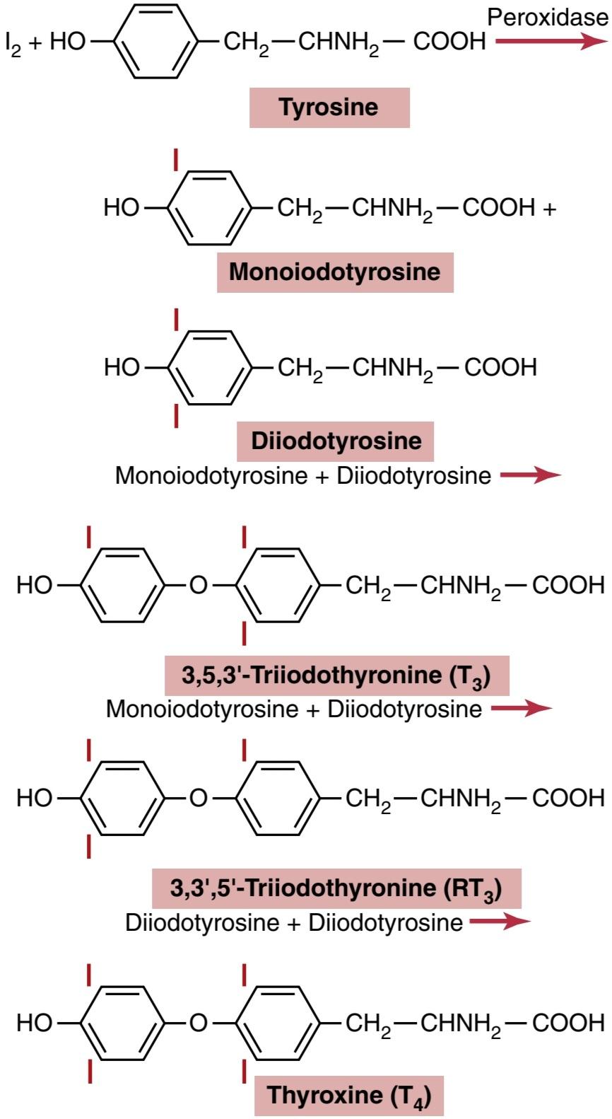
Figure 77-3 Chemistry of thyroxine and triiodothyronine formation.
شکل 77-3 شیمی تشکیل تیروکسین و تری یدوتیرونین.
RELEASE OF THYROXINE AND TRIIODOTHYRONINE FROM THE THYROID GLAND
Most of the thyroglobulin is not released into the circulating blood; instead, thyroxine and triiodothyronine are cleaved from the thyroglobulin molecule, and then these free hormones are released. This process occurs as follows: The apical surface of thyroid cells sends out pseudopod extensions that close around small portions of the colloid to form pinocytic vesicles that enter the apex of the thyroid cell. Then lysosomes in the cell cytoplasm immediately fuse with these vesicles to form digestive vesicles containing digestive enzymes from the lysosomes mixed with the colloid. Multiple proteases among the enzymes digest the thyroglobulin molecules and release thyroxine and triiodothyronine in free form, which then diffuse through the base of the thyroid cell into the surrounding capillaries. Thus, the thyroid hormones are released into the blood.
آزادسازی تیروکسین و تری یدوتیرونین از غده تیروئید
بیشتر تیروگلوبولین در خون در گردش آزاد نمی شود. در عوض، تیروکسین و تری یدوتیرونین از مولکول تیروگلوبولین جدا می شوند و سپس این هورمون های آزاد آزاد می شوند. این فرآیند به صورت زیر اتفاق میافتد: سطح آپیکال سلولهای تیروئید اکستنشنهای شبهپایی را میفرستد که در اطراف بخشهای کوچکی از کلوئید بسته میشوند تا وزیکولهای پینوسیتی را تشکیل دهند که وارد راس سلول تیروئید میشوند. سپس لیزوزومهای موجود در سیتوپلاسم سلولی بلافاصله با این وزیکولها ترکیب میشوند و از لیزوزومهای مخلوط شده با کلوئید، وزیکولهای گوارشی حاوی آنزیمهای گوارشی تشکیل میدهند. پروتئازهای متعدد در بین آنزیم ها، مولکول های تیروگلوبولین را هضم می کنند و تیروکسین و تری یدوتیرونین را به شکل آزاد آزاد می کنند، که سپس از طریق پایه سلول تیروئید به مویرگ های اطراف پخش می شود. بنابراین، هورمون های تیروئید در خون ترشح می شوند.
Some of the thyroglobulin in the colloid enters the thyroid cell by endocytosis after binding to megalin, a protein located on the lumen membrane of the cells. The megalin-thyroglobulin complex is then carried across the cell by transcytosis to the basolateral membrane, where a portion of the megalin remains bound to thyroglobulin and is released into the capillary blood.
مقداری از تیروگلوبولین موجود در کلوئید پس از اتصال به مگالین، پروتئینی که روی غشای لومن سلول ها قرار دارد، از طریق اندوسیتوز وارد سلول تیروئید می شود. سپس کمپلکس مگالین-تیروگلوبولین از طریق ترانس سیتوز در سراسر سلول به غشای قاعده ای جانبی منتقل می شود، جایی که بخشی از مگالین به تیروگلوبولین متصل می شود و در خون مویرگی آزاد می شود.
About three-quarters of the iodinated tyrosine in the thyroglobulin never become thyroid hormones but remain monoiodotyrosine and diiodotyrosine. During the digestion of the thyroglobulin molecule to cause release of thyroxine and triiodothyronine, these iodinated tyro- sines also are freed from the thyroglobulin molecules. However, they are not secreted into the blood. Instead, their iodine is cleaved from them by a deiodinase enzyme that makes virtually all this iodine available again for recycling within the gland for forming additional thyroid hormones. Congenital absence of this deiodinase enzyme may cause iodine deficiency because of failure of this recycling process.
حدود سه چهارم تیروزین ید دار موجود در تیروگلوبولین هرگز به هورمون تیروئید تبدیل نمی شود، اما مونودوتیروزین و دی یدوتیروزین باقی می ماند. در طی هضم مولکول تیروگلوبولین برای آزادسازی تیروکسین و تری یدوتیرونین، این تیروزین های یددار نیز از مولکول های تیروگلوبولین آزاد می شوند. با این حال، آنها در خون ترشح نمی شوند. در عوض، ید آنها توسط یک آنزیم دیودیناز از آنها جدا می شود که تقریباً تمام این ید را دوباره برای بازیافت در غده برای تشکیل هورمون های تیروئید اضافی در دسترس قرار می دهد. فقدان مادرزادی این آنزیم دیودیناز ممکن است به دلیل شکست این فرآیند بازیافت باعث کمبود ید شود.
Daily Rate of Secretion of Thyroxine and Triiodo- thyronine. About 93% of the thyroid hormone released from the thyroid gland is normally thyroxine and only 7% is triiodothyronine. However, during the ensuing few days, about one-half of the thyroxine is slowly deiodinated to form additional triiodothyronine. Therefore, the hormone finally delivered to and used by the tissues is mainly triiodothyronine-a total of about 35 μg of triiodothyronine per day.
میزان ترشح روزانه تیروکسین و تریودوتیرونین. حدود 93 درصد از هورمون تیروئید آزاد شده از غده تیروئید به طور معمول تیروکسین و تنها 7 درصد آن تری یدوتیرونین است. با این حال، در طی چند روز بعد، حدود نیمی از تیروکسین به آرامی دیوده می شود تا تری یدوتیرونین اضافی تشکیل شود. بنابراین، هورمونی که در نهایت به بافت ها تحویل داده می شود و توسط آن استفاده می شود، عمدتاً تری یدوتیرونین است – در مجموع حدود 35 میکروگرم تری یدوتیرونین در روز.
TRANSPORT OF THYROXINE AND TRIIODOTHYRONINE TO TISSUES
Thyroxine and Triiodothyronine Are Bound to Plasma Proteins. Upon entering the blood, more than 99% of the thyroxine and triiodothyronine combines immediately with several of the plasma proteins, all of which are synthesized by the liver. They combine mainly with thyroxine- binding globulin and much less so with thyroxine-binding prealbumin and albumin.
انتقال تیروکسین و تری یدوتیرونین به بافت
تیروکسین و تری یدوتیرونین به پروتئین های پلاسما متصل می شوند. پس از ورود به خون، بیش از 99 درصد از تیروکسین و تری یدوتیرونین بلافاصله با چندین پروتئین پلاسما ترکیب می شود که همه آنها توسط کبد سنتز می شوند. آنها عمدتاً با گلوبولین متصل شونده به تیروکسین و بسیار کمتر با پرآلبومین و آلبومین متصل شونده به تیروکسین ترکیب می شوند.
Thyroxine and Triiodothyronine Are Released Slowly to Tissue Cells. Because of high affinity of the plasma- binding proteins for the thyroid hormones, these sub- stances in particular, thyroxine-are released to the tissue cells slowly. Half the thyroxine in the blood is released to the tissue cells about every 6 days, whereas half the triiodothyronine because of its lower affinity-is released to the cells in about 1 day.
تیروکسین و تری یدوتیرونین به آرامی در سلول های بافتی آزاد می شوند. به دلیل میل ترکیبی زیاد پروتئین های متصل به پلاسما به هورمون های تیروئید، این مواد به ویژه تیروکسین به آرامی به سلول های بافتی آزاد می شوند. نیمی از تیروکسین موجود در خون تقریباً هر 6 روز یک بار به سلول های بافتی آزاد می شود، در حالی که نیمی از تری یدوتیرونین به دلیل میل ترکیبی پایین تر در حدود 1 روز به سلول ها آزاد می شود.
Upon entering the tissue cells, both thyroxine and triiodothyronine again bind with intracellular proteins, with the thyroxine binding more strongly than the triiodothyronine. Therefore, they are again stored, but this time in the target cells, and they are used slowly over a period of days or weeks.
پس از ورود به سلول های بافت، هر دو تیروکسین و تری یدوتیرونین دوباره با پروتئین های داخل سلولی متصل می شوند و تیروکسین قوی تر از تری یدوتیرونین است. بنابراین، آنها دوباره اما این بار در سلول های هدف ذخیره می شوند و به آرامی در مدت چند روز یا چند هفته استفاده می شوند.
Thyroid Hormones Have Slow Onset and Long Duration of Action. After injection of a large quantity of thyroxine into a human being, essentially no effect on the metabolic rate can be discerned for 2 to 3 days, thereby demonstrating that there is a long latent period before thyroxine activity begins. Once activity does begin, it increases progressively and reaches a maximum in 10 to 12 days, as shown in Figure 77-4. Thereafter, it decreases with a half-life of about 15 days. Some of the activity persists for as long as 6 weeks to 2 months.
هورمون های تیروئید شروع آهسته و مدت اثر طولانی دارند. پس از تزریق مقدار زیادی تیروکسین به انسان، اساساً هیچ تأثیری بر میزان متابولیسم برای 2 تا 3 روز نمی توان تشخیص داد، در نتیجه نشان می دهد که یک دوره نهفته طولانی قبل از شروع فعالیت تیروکسین وجود دارد. همانطور که در شکل 77-4 نشان داده شده است، هنگامی که فعالیت شروع می شود، به تدریج افزایش می یابد و در 10 تا 12 روز به حداکثر می رسد. پس از آن، با نیمه عمر حدود 15 روز کاهش می یابد. برخی از فعالیت ها به مدت 6 هفته تا 2 ماه ادامه می یابد.
The actions of triiodothyronine occur about four times as rapidly as those of thyroxine, with a latent period as short as 6 to 12 hours and maximal cellular activity occur- ring within 2 to 3 days.
اقدامات تری یدوتیرونین تقریباً چهار برابر سریعتر از تیروکسین اتفاق می افتد، با دوره نهفته کوتاه 6 تا 12 ساعت و حداکثر فعالیت سلولی در عرض 2 تا 3 روز رخ می دهد.
Most of the latency and the prolonged period of action of these hormones are probably caused by their binding with proteins both in the plasma and in the tissue cells, followed by their slow release. However, we shall see in subsequent discussions that part of the latent period also results from the manner in which these hormones per- form their functions in the cells.
بیشتر تأخیر و دوره طولانی اثر این هورمون ها احتمالاً به دلیل اتصال آنها با پروتئین ها در پلاسما و سلول های بافتی و به دنبال آن آزاد شدن آهسته آنها است. با این حال، در بحث های بعدی خواهیم دید که بخشی از دوره نهفته نیز ناشی از روشی است که این هورمون ها عملکرد خود را در سلول ها انجام می دهند.

Figure 77-4 Approximate prolonged effect on the basal metabolic rate caused by administering a single large dose of thyroxine.
شکل 77-4 اثر طولانی مدت تقریبی بر سرعت متابولیسم پایه ناشی از تجویز یک دوز بزرگ تیروکسین.
PHYSIOLOGICAL FUNCTIONS OF THE THYROID HORMONES
THYROID HORMONES INCREASE TRANSCRIPTION OF MANY GENES
The general effect of thyroid hormone is to activate nuclear transcription of many genes (Figure 77-5). Therefore, in virtually all cells of the body, great numbers of protein enzymes, structural proteins, transport proteins, and other substances are synthesized. The net result is a generalized increase in functional activity throughout the body.
عملکردهای فیزیولوژیکی هورمون های تیروئید
هورمون های تیروئیدی رونویسی بسیاری از ژن ها را افزایش می دهند
اثر کلی هورمون تیروئید فعال کردن رونویسی هسته ای بسیاری از ژن ها است (شکل 77-5). بنابراین، تقریباً در تمام سلولهای بدن، تعداد زیادی آنزیم پروتئینی، پروتئینهای ساختاری، پروتئینهای حملونقل و سایر مواد سنتز میشوند. نتیجه خالص افزایش عمومی فعالیت عملکردی در سراسر بدن است.
Most of the Thyroxine Secreted by the Thyroid Is Converted to Triiodothyronine. Before acting on the genes to increase genetic transcription, one iodide is removed from almost all the thyroxine, thus forming triiodothyronine. Intracellular thyroid hormone receptors have a high affinity for triiodothyronine. Consequently, more than 90% of the thyroid hormone that binds with the receptors is triiodothyronine.
بیشتر تیروکسین ترشح شده توسط تیروئید به تری یدوتیرونین تبدیل می شود. قبل از اقدام بر روی ژن ها برای افزایش رونویسی ژنتیکی، یک یدید تقریباً از تمام تیروکسین حذف می شود و در نتیجه تری یدوتیرونین تشکیل می شود. گیرنده های هورمون تیروئید درون سلولی تمایل زیادی به تری یدوتیرونین دارند. در نتیجه، بیش از 90 درصد هورمون تیروئیدی که با گیرنده ها متصل می شود، تری یدوتیرونین است.
Thyroid Hormones Activate Nuclear Receptors. Thyroid hormone receptors are either attached to the DNA genetic strands or located in proximity to them. The thyroid hormone receptor usually forms a heterodimer with retinoid X receptor (RXR) at specific thyroid hormone response elements on the DNA. After binding with thyroid hormone, the receptors become activated and initiate the transcription process. Large numbers of different types of messenger RNA are then formed, followed within another few minutes or hours by RNA translation on the cytoplasmic ribosomes to form hundreds of new intracellular proteins. However, not all the proteins are increased by similar percentages-some are increased only slightly, and others at least as much as sixfold. Most of the actions of thyroid hormone result from the subsequent enzymatic and other functions of these new proteins.
هورمون های تیروئید گیرنده های هسته ای را فعال می کنند. گیرنده های هورمون تیروئید یا به رشته های ژنتیکی DNA متصل هستند یا در مجاورت آنها قرار دارند. گیرنده هورمون تیروئید معمولاً یک هترودایمر با گیرنده رتینوئید X (RXR) در عناصر واکنش هورمون تیروئید خاص روی DNA تشکیل می دهد. پس از اتصال به هورمون تیروئید، گیرنده ها فعال می شوند و فرآیند رونویسی را آغاز می کنند. سپس تعداد زیادی از انواع مختلف RNA پیام رسان تشکیل می شود و در عرض چند دقیقه یا چند ساعت دیگر RNA بر روی ریبوزوم های سیتوپلاسمی ترجمه می شود تا صدها پروتئین درون سلولی جدید تشکیل شود. با این حال، همه پروتئینها با درصدهای مشابه افزایش نمییابند – برخی فقط اندکی و برخی دیگر حداقل تا شش برابر افزایش مییابند. بیشتر اعمال هورمون تیروئید ناشی از عملکردهای آنزیمی بعدی و سایر عملکردهای این پروتئین های جدید است.
Thyroid hormones also appear to have nongenomic cellular effects that are independent of their effects on gene transcription. For example, some effects of thyroid hormones occur within minutes, too rapidly to be explained by changes in protein synthesis, and are not affected by inhibitors of gene transcription and translation. Such actions have been described in several tissues, including the heart and pituitary, as well as adipose tissue. The site of nongenomic thyroid hormone action appears to be the plasma membrane, cytoplasm, and perhaps some cell organelles such as mitochondria. Nongenomic actions of thyroid hormone include regulation of ion channels and oxidative phosphorylation and appear to involve activation of intracellular secondary messengers such as cyclic adenosine monophosphate (cAMP) or protein kinase signaling cascades.
به نظر می رسد هورمون های تیروئید دارای اثرات سلولی غیر ژنومیک هستند که مستقل از اثرات آنها بر رونویسی ژن است. به عنوان مثال، برخی از اثرات هورمونهای تیروئید در عرض چند دقیقه اتفاق میافتند، خیلی سریع که با تغییرات در سنتز پروتئین توضیح داده نمیشوند، و تحت تأثیر مهارکنندههای رونویسی و ترجمه ژن قرار نمیگیرند. چنین اعمالی در چندین بافت از جمله قلب و هیپوفیز و همچنین بافت چربی توصیف شده است. به نظر می رسد محل عملکرد غیر ژنومیک هورمون تیروئید غشای پلاسمایی، سیتوپلاسم و شاید برخی از اندامک های سلولی مانند میتوکندری باشد. اعمال غیر ژنومیک هورمون تیروئید شامل تنظیم کانالهای یونی و فسفوریلاسیون اکسیداتیو است و به نظر میرسد که شامل فعالسازی پیامرسانهای ثانویه درون سلولی مانند آدنوزین مونوفسفات حلقوی (cAMP) یا آبشارهای سیگنالینگ پروتئین کیناز باشد.
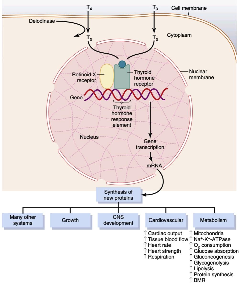
Figure 77-5 Thyroid hormone activation of target cells. Thyroxine (T4) and triiodothyronine (T3) enter the cell membrane by a carrier-mediated adenosine triphosphate-dependent transport process. Much of the T4 is deiodinated to form T3, which interacts with the thyroid hormone receptor, bound as a heterodimer with a retinoid X receptor, of the thyroid hormone response element of the gene. This action causes either increases or decreases in transcription of genes that lead to the formation of proteins, thus producing the thyroid hormone response of the cell. The actions of thyroid hormone on cells of several different systems are shown. BMR, Basal metabolic rate; CNS, central nervous system; mRNA, messenger ribonucleic acid; Na+-K+-ATPase, sodium-potassium-adenosine triphosphatase.
شکل 77-5 فعال شدن هورمون تیروئید سلول های هدف. تیروکسین (T4) و تری یدوتیرونین (T3) توسط یک فرآیند انتقال وابسته به آدنوزین تری فسفات با واسطه به غشای سلول وارد می شوند. بخش اعظم T4 دی یدین می شود و T3 را تشکیل می دهد که با گیرنده هورمون تیروئید که به عنوان یک هترودایمر با گیرنده رتینوئید X از عنصر پاسخ هورمون تیروئید ژن مرتبط است، تعامل دارد. این عمل باعث افزایش یا کاهش رونویسی ژنها میشود که منجر به تشکیل پروتئین میشود و در نتیجه پاسخ هورمون تیروئید سلول را تولید میکند. اعمال هورمون تیروئید بر روی سلول های چندین سیستم مختلف نشان داده شده است. BMR، میزان متابولیسم پایه؛ CNS، سیستم عصبی مرکزی؛ mRNA، اسید ریبونوکلئیک پیام رسان؛ Na+-K+-ATPase، سدیم-پتاسیم-آدنوزین تری فسفاتاز.
THYROID HORMONES INCREASE CELLULAR METABOLIC ACTIVITY
Thyroid hormones increase the metabolic activities of almost all the tissues of the body. The basal metabolic rate can increase to 60% to 100% above normal when large quantities of thyroid hormones are secreted. The rate of utilization of foods for energy is greatly accelerated. Although the rate of protein synthesis is increased, at the same time the rate of protein catabolism is also increased. The growth rate of young people is greatly accelerated. The mental processes are excited, and the activities of most of the other endocrine glands are increased.
هورمون های تیروئیدی فعالیت متابولیک سلولی را افزایش می دهند
هورمون های تیروئید فعالیت های متابولیک تقریباً تمام بافت های بدن را افزایش می دهند. هنگامی که مقادیر زیادی هورمون تیروئید ترشح می شود، میزان متابولیسم پایه می تواند به 60 تا 100 درصد بالاتر از حد طبیعی افزایش یابد. سرعت استفاده از غذاها برای انرژی بسیار تسریع شده است. اگرچه سرعت سنتز پروتئین افزایش می یابد، اما در عین حال سرعت کاتابولیسم پروتئین نیز افزایش می یابد. سرعت رشد جوانان بسیار تسریع شده است. فرآیندهای ذهنی هیجان زده می شوند و فعالیت بیشتر غدد درون ریز دیگر افزایش می یابد.
Thyroid Hormones Increase the Number and Activity of Mitochondria. When thyroxine or triiodothyronine is given to an animal, the mitochondria in most cells of the animal’s body increase in size and number. Furthermore, the total membrane surface area of the mitochondria increases almost directly in proportion to the increased metabolic rate of the whole animal. Therefore, one of the principal functions of thyroxine might be simply to increase the number and activity of mitochondria, which in turn increases the rate of formation of adenosine triphosphate to energize cellular function. However, increases in the number and activity of mitochondria could be the result of increased activity of the cells, as well as the cause of the increase.
هورمون های تیروئید تعداد و فعالیت میتوکندری ها را افزایش می دهند. هنگامی که تیروکسین یا تری یدوتیرونین به حیوان داده می شود، میتوکندری در اکثر سلول های بدن حیوان از نظر اندازه و تعداد افزایش می یابد. علاوه بر این، سطح کل غشای میتوکندری تقریباً مستقیماً متناسب با افزایش نرخ متابولیک کل حیوان افزایش می یابد. بنابراین، یکی از عملکردهای اصلی تیروکسین ممکن است صرفاً افزایش تعداد و فعالیت میتوکندری ها باشد که به نوبه خود سرعت تشکیل آدنوزین تری فسفات را برای انرژی بخشیدن به عملکرد سلولی افزایش می دهد. با این حال، افزایش تعداد و فعالیت میتوکندری ها می تواند نتیجه افزایش فعالیت سلول ها و همچنین علت افزایش باشد.
Thyroid Hormones Increase Active Transport of lons Through Cell Membranes. One of the enzymes that increases its activity in response to thyroid hormone is Na+-K+ ATPase. This increased activity in turn increases the rate of transport of sodium and potassium ions through the cell membranes of some tissues. Because this process uses energy and increases the amount of heat produced in the body, it has been suggested that this might be one of the mechanisms by which thyroid hormone increases the body’s metabolic rate. In fact, thyroid hormone also causes the cell membranes of most cells to become leaky to sodium ions, which further activates the sodium pump and further increases heat production.
هورمون های تیروئید انتقال فعال لون ها را از طریق غشای سلولی افزایش می دهند. یکی از آنزیم هایی که فعالیت آن را در پاسخ به هورمون تیروئید افزایش می دهد، Na+-K+ ATPase است. این افزایش فعالیت به نوبه خود سرعت انتقال یون های سدیم و پتاسیم را از طریق غشای سلولی برخی از بافت ها افزایش می دهد. از آنجایی که این فرآیند از انرژی استفاده می کند و میزان گرمای تولید شده در بدن را افزایش می دهد، پیشنهاد شده است که این ممکن است یکی از مکانیسم هایی باشد که توسط آن هورمون تیروئید سرعت متابولیسم بدن را افزایش می دهد. در واقع، هورمون تیروئید همچنین باعث میشود که غشای سلولی اکثر سلولها به یونهای سدیم نشت کند، که پمپ سدیم را بیشتر فعال میکند و تولید گرما را بیشتر میکند.
EFFECT OF THYROID HORMONE ON GROWTH
Thyroid hormone has both general and specific effects on growth. For example, it has long been known that thyroid hormone is essential for the metamorphic change of the tadpole into the frog.
تأثیر هورمون تیروئید بر رشد
هورمون تیروئید هم اثرات عمومی و هم خاص روی رشد دارد. به عنوان مثال، مدتهاست که مشخص شده است که هورمون تیروئید برای تغییر دگرگونی قورباغه به قورباغه ضروری است.
In humans, the effect of thyroid hormone on growth is manifest mainly in growing children. In children with hypothyroidism, the rate of growth is greatly retarded. In children with hyperthyroidism, excessive skeletal growth often occurs, causing the child to become considerably taller at an earlier age. However, the bones also mature more rapidly and the epiphyses close at an early age, so the duration of growth and the eventual height of the adult actually may be shortened.
در انسان، تأثیر هورمون تیروئید بر رشد عمدتاً در کودکان در حال رشد آشکار می شود. در کودکان مبتلا به کم کاری تیروئید، سرعت رشد به شدت عقب افتاده است. در کودکان مبتلا به پرکاری تیروئید، رشد بیش از حد اسکلتی اغلب رخ می دهد که باعث می شود کودک در سنین پایین تر به طور قابل توجهی بلندتر شود. با این حال، استخوانها نیز سریعتر بالغ میشوند و اپیفیزها در سنین پایین بسته میشوند، بنابراین مدت زمان رشد و قد نهایی بزرگسالان ممکن است در واقع کوتاه شود.
An important effect of thyroid hormone is to promote growth and development of the brain during fetal life and for the first few years of postnatal life. If the fetus does not secrete sufficient quantities of thyroid hormone, growth and maturation of the brain both before birth and after- ward are greatly retarded and the brain remains smaller than normal. Without specific thyroid therapy within days or weeks after birth, the child without a thyroid gland will remain mentally deficient throughout life. This condition is discussed later in the chapter.
اثر مهم هورمون تیروئید، تقویت رشد و تکامل مغز در طول زندگی جنینی و چند سال اول زندگی پس از زایمان است. اگر جنین مقادیر کافی هورمون تیروئید ترشح نکند، رشد و بلوغ مغز چه قبل از تولد و چه بعد از آن به شدت عقب افتاده و مغز کوچکتر از حد طبیعی باقی می ماند. بدون درمان خاص تیروئید در طی چند روز یا چند هفته پس از تولد، کودک بدون غده تیروئید در طول زندگی دچار کمبود ذهنی خواهد بود. این شرط بعداً در فصل مورد بحث قرار می گیرد.
EFFECTS OF THYROID HORMONE ON SPECIFIC BODY FUNCTIONS
Stimulation of Carbohydrate Metabolism. Thyroid hormone stimulates almost all aspects of carbohydrate metabolism, including rapid glucose uptake by cells, enhanced glycolysis, enhanced gluconeogenesis, increased rate of absorption from the gastrointestinal tract, and even increased insulin secretion with its resultant secondary effects on carbohydrate metabolism. All these effects probably result from the overall increase in cellular metabolic enzymes caused by thyroid hormone.
اثرات هورمون تیروئید بر عملکردهای خاص بدن
تحریک متابولیسم کربوهیدرات. هورمون تیروئید تقریباً تمام جنبههای متابولیسم کربوهیدرات را تحریک میکند، از جمله جذب سریع گلوکز توسط سلولها، افزایش گلیکولیز، افزایش گلوکونئوژنز، افزایش سرعت جذب از دستگاه گوارش و حتی افزایش ترشح انسولین با اثرات ثانویه متابولیسم کربوهیدرات بر روی خودرو. همه این اثرات احتمالاً ناشی از افزایش کلی آنزیم های متابولیک سلولی ناشی از هورمون تیروئید است.
Stimulation of Fat Metabolism. Essentially all aspects of fat metabolism are also enhanced by thyroid hormone. In particular, lipids are mobilized rapidly from the fat tissue, which decreases fat stores of the body to a greater extent than almost any other tissue element. Mobilization of lipids from fat tissue also increases free fatty acid con- centration in the plasma and greatly accelerates oxidation of free fatty acids by the cells.
تحریک متابولیسم چربی. اساساً تمام جنبه های متابولیسم چربی نیز توسط هورمون تیروئید تقویت می شود. به طور خاص، لیپیدها به سرعت از بافت چربی بسیج می شوند، که باعث کاهش ذخایر چربی بدن به میزانی بیشتر از هر عنصر بافتی دیگر می شود. حرکت لیپیدها از بافت چربی همچنین باعث افزایش غلظت اسیدهای چرب آزاد در پلاسما شده و اکسیداسیون اسیدهای چرب آزاد توسط سلول ها را تا حد زیادی تسریع می کند.
Effect on Plasma and Liver Fats. Increased thyroid hormone decreases the concentrations of cholesterol, phospholipids, and triglycerides in the plasma, even though it increases the free fatty acids. Conversely, decreased thyroid secretion greatly increases the plasma concentrations of cholesterol, phospholipids, and triglycerides and almost always causes excessive deposition of fat in the liver as well. The large increase in circulating plasma cholesterol in prolonged hypothyroidism is often associated with severe atherosclerosis, as discussed in Chapter 69.
تاثیر بر چربی های پلاسما و کبد. افزایش هورمون تیروئید باعث کاهش غلظت کلسترول، فسفولیپیدها و تری گلیسیرید در پلاسما می شود، حتی اگر باعث افزایش اسیدهای چرب آزاد شود. برعکس، کاهش ترشح تیروئید غلظت کلسترول، فسفولیپیدها و تری گلیسیریدهای پلاسمایی را تا حد زیادی افزایش می دهد و تقریباً همیشه باعث رسوب بیش از حد چربی در کبد می شود. همانطور که در فصل 69 بحث شد، افزایش زیاد کلسترول پلاسما در کم کاری طولانی مدت اغلب با آترواسکلروز شدید همراه است.
One of the mechanisms by which thyroid hormone decreases plasma cholesterol concentration is to increase significantly cholesterol secretion in the bile and consequent loss in the feces. A possible mechanism for the increased cholesterol secretion is that thyroid hormone induces increased numbers of low-density lipoprotein receptors on the liver cells, leading to rapid removal of low-density lipoproteins from the plasma by the liver and subsequent secretion of cholesterol in these lipoproteins by the liver cells.
یکی از مکانیسم هایی که هورمون تیروئید غلظت کلسترول پلاسما را کاهش می دهد، افزایش قابل توجه ترشح کلسترول در صفرا و در نتیجه کاهش در مدفوع است. یک مکانیسم احتمالی برای افزایش ترشح کلسترول این است که هورمون تیروئید باعث افزایش تعداد گیرنده های لیپوپروتئین با چگالی کم در سلول های کبدی می شود که منجر به حذف سریع لیپوپروتئین های با چگالی کم از پلاسما توسط کبد و ترشح کلسترول در این لیپوپروتئین ها توسط سلول های کبدی می شود.
Increased Requirement for Vitamins. Because thyroid hormone increases the quantities of many bodily enzymes and because vitamins are essential parts of some of the enzymes or coenzymes, thyroid hormone increases the need for vitamins. Therefore, a relative vitamin deficiency can occur when excess thyroid hormone is secreted, unless at the same time increased quantities of vitamins are made available.
افزایش نیاز به ویتامین. از آنجایی که هورمون تیروئید مقدار بسیاری از آنزیم های بدن را افزایش می دهد و از آنجا که ویتامین ها بخش های ضروری برخی از آنزیم ها یا کوآنزیم ها هستند، هورمون تیروئید نیاز به ویتامین ها را افزایش می دهد. بنابراین، کمبود نسبی ویتامین ممکن است زمانی رخ دهد که هورمون تیروئید اضافی ترشح شود، مگر اینکه همزمان مقادیر بیشتری از ویتامین ها در دسترس باشند.
Increased Basal Metabolic Rate. Because thyroid hormone increases metabolism in almost all cells of the body, excessive quantities of the hormone can occasionally in- crease the basal metabolic rate by 60% to 100% above normal. Conversely, when no thyroid hormone is produced, the basal metabolic rate falls to almost one-half of normal. Figure 77-6 shows the approximate relation between the daily supply of thyroid hormones and the basal metabolic rate. Extreme amounts of the hormones are required to cause high basal metabolic rates.
افزایش نرخ متابولیسم پایه. از آنجایی که هورمون تیروئید متابولیسم را تقریباً در تمام سلولهای بدن افزایش میدهد، مقادیر بیش از حد هورمون گاهی اوقات میتواند میزان متابولیسم پایه را 60 تا 100 درصد بالاتر از حد طبیعی افزایش دهد. برعکس، وقتی هیچ هورمون تیروئیدی تولید نمیشود، میزان متابولیسم پایه تقریباً به نصف مقدار طبیعی کاهش مییابد. شکل 77-6 رابطه تقریبی بین عرضه روزانه هورمون های تیروئید و میزان متابولیسم پایه را نشان می دهد. مقادیر زیادی از هورمون ها برای ایجاد متابولیسم پایه بالا مورد نیاز است.

Figure 77-6 Approximate relation of the daily rate of thyroid hormone (T3, triiodothyronine, and T4, thyroxine) secretion to percent change in basal metabolic rate, compared with normal.
شکل 77-6 رابطه تقریبی میزان روزانه ترشح هورمون تیروئید (T3، تری یدوتیرونین و T4، تیروکسین) با درصد تغییر در میزان متابولیسم پایه، در مقایسه با نرمال.
Decreased Body Weight. A greatly increased amount of thyroid hormone almost always decreases body weight, and a greatly decreased amount of thyroid hormone al- most always increases body weight. However, these effects do not always occur because thyroid hormone also increases the appetite, which may counterbalance the change in the metabolic rate.
کاهش وزن بدن. افزایش بسیار زیاد هورمون تیروئید تقریباً همیشه وزن بدن را کاهش می دهد و مقدار بسیار کاهش یافته هورمون تیروئید تقریباً همیشه وزن بدن را افزایش می دهد. با این حال، این اثرات همیشه رخ نمی دهد زیرا هورمون تیروئید نیز اشتها را افزایش می دهد، که ممکن است تغییر در نرخ متابولیسم را متعادل کند.
Increased Blood Flow and Cardiac Output. Increased metabolism in the tissues causes more rapid utilization of oxygen than normal and increased release of metabolic end products from the tissues. These effects cause vasodilation in most body tissues, thus increasing blood flow. The rate of blood flow in the skin especially increases because of the increased need for heat elimination from the body. As a consequence of the increased blood flow, cardiac output also increases, sometimes rising to 60% or more above normal when excessive thyroid hormone is present and falling to only 50% of normal in severe hypothyroidism.
افزایش جریان خون و برون ده قلبی. افزایش متابولیسم در بافت ها باعث استفاده سریع تر از اکسیژن نسبت به حالت عادی و افزایش آزادسازی محصولات نهایی متابولیک از بافت ها می شود. این اثرات باعث اتساع عروق در بیشتر بافت های بدن می شود و در نتیجه جریان خون را افزایش می دهد. سرعت جریان خون در پوست به ویژه به دلیل افزایش نیاز به دفع گرما از بدن افزایش می یابد. در نتیجه افزایش جریان خون، برون ده قلبی نیز افزایش مییابد، گاهی اوقات در صورت وجود هورمون تیروئید بیش از حد به 60 درصد یا بیشتر از حد طبیعی میرسد و در کمکاری شدید تیروئید تنها به 50 درصد طبیعی کاهش مییابد.
Increased Heart Rate. The heart rate increases considerably more under the influence of thyroid hormone than would be expected from the increase in cardiac output. Therefore, thyroid hormone seems to have a direct effect on the excitability of the heart, which in turn increases the heart rate. This effect is especially important because the heart rate is one of the sensitive physical signs that the clinician uses in determining whether a patient has excessive or diminished thyroid hormone production.
افزایش ضربان قلب. ضربان قلب تحت تأثیر هورمون تیروئید به طور قابل توجهی بیشتر از آنچه از افزایش برون ده قلبی انتظار می رود افزایش می یابد. بنابراین به نظر می رسد هورمون تیروئید تأثیر مستقیمی بر تحریک پذیری قلب دارد که به نوبه خود باعث افزایش ضربان قلب می شود. این تأثیر به ویژه مهم است زیرا ضربان قلب یکی از علائم فیزیکی حساسی است که پزشک در تعیین اینکه آیا بیمار تولید هورمون تیروئید بیش از حد یا کاهش یافته است استفاده می کند.
Increased Heart Strength. A slight excess of thyroid hormone can increase the strength of the heart. This effect is analogous to the increase in heart strength that occurs in mild fevers and during exercise. However, when thyroid hormone is increased markedly, heart muscle strength becomes depressed because of long-term excessive protein catabolism. Indeed, some severely thyrotoxic patients die of cardiac decompensation secondary to myocardial failure and increased cardiac workload imposed by the increase in cardiac output.
افزایش قدرت قلب. مقدار کمی بیش از حد هورمون تیروئید می تواند قدرت قلب را افزایش دهد. این اثر مشابه افزایش قدرت قلب است که در تب های خفیف و در حین ورزش رخ می دهد. با این حال، هنگامی که هورمون تیروئید به طور قابل توجهی افزایش می یابد، قدرت عضله قلب به دلیل کاتابولیسم پروتئین بیش از حد طولانی مدت افسرده می شود. در واقع، برخی از بیماران شدیداً تیروتوکسیک به دلیل جبران نارسایی قلبی ثانویه به نارسایی میوکارد و افزایش بار کاری قلبی ناشی از افزایش برون ده قلبی می میرند.
Normal Arterial Pressure. The mean arterial pressure usually remains about normal after administration of thyroid hormone. Because of increased blood flow through the tissues between heartbeats, the pulse pressure is often increased, with the systolic pressure elevated 10 to 15 mm Hg in hyperthyroidism and the diastolic pressure reduced a corresponding amount.
فشار شریانی طبیعی. فشار متوسط شریانی معمولاً پس از تجویز هورمون تیروئید تقریباً نرمال باقی می ماند. به دلیل افزایش جریان خون در بافت ها بین ضربان قلب، فشار نبض اغلب افزایش می یابد، با فشار سیستولیک 10 تا 15 میلی متر جیوه در پرکاری تیروئید و فشار دیاستولیک به میزان متناظر کاهش می یابد.
Increased Respiration. The increased rate of metabolism increases oxygen utilization and carbon dioxide formation; these effects activate all the mechanisms that in- crease the rate and depth of respiration.
افزایش تنفس. افزایش سرعت متابولیسم باعث افزایش استفاده از اکسیژن و تشکیل دی اکسید کربن می شود. این اثرات تمام مکانیسم هایی را فعال می کند که سرعت و عمق تنفس را افزایش می دهد.
Increased Gastrointestinal Motility. In addition to increased appetite and food intake, which has been dis- cussed, thyroid hormone increases secretion of digestive juices and motility of the gastrointestinal tract. Hyperthyroidism therefore often results in diarrhea, whereas lack of thyroid hormone can cause constipation.
افزایش تحرک دستگاه گوارش. هورمون تیروئید علاوه بر افزایش اشتها و مصرف غذا که در مورد آن صحبت شد، ترشح شیره های گوارشی و تحرک دستگاه گوارش را افزایش می دهد. بنابراین پرکاری تیروئید اغلب منجر به اسهال می شود، در حالی که کمبود هورمون تیروئید می تواند باعث یبوست شود.
Excitatory Effects on the Central Nervous System. In general, thyroid hormone increases the rapidity of cerebration, although thought processes may be dissociated; conversely, lack of thyroid hormone decreases rapidity of cerebration. A person with hyperthyroidism is likely to be extremely nervous and have many psychoneurotic tendencies, such as anxiety complexes, extreme worry, and paranoia.
اثرات تحریکی بر روی سیستم عصبی مرکزی. به طور کلی، هورمون تیروئید سرعت مغز را افزایش می دهد، اگرچه فرآیندهای فکری ممکن است جدا شوند. برعکس، کمبود هورمون تیروئید باعث کاهش سرعت مغز می شود. فرد مبتلا به پرکاری تیروئید به احتمال زیاد بسیار عصبی است و تمایلات روان عصبی بسیاری مانند عقده های اضطرابی، نگرانی شدید و پارانویا دارد.
Effect on the Function of the Muscles. A slight increase in thyroid hormone usually makes the muscles react with vigor but, with excessive thyroid hormone, the muscles become weakened because of excess protein catabolism. Conversely, lack of thyroid hormone causes the muscles to become sluggish, and they relax slowly after a contraction.
تاثیر بر عملکرد عضلات. افزایش خفیف هورمون تیروئید معمولاً عضلات را با قدرت واکنش نشان می دهد، اما با هورمون تیروئید بیش از حد، ماهیچه ها به دلیل کاتابولیسم پروتئین اضافی ضعیف می شوند. برعکس، کمبود هورمون تیروئید باعث سست شدن ماهیچه ها می شود و بعد از انقباض به آرامی شل می شوند.
Muscle Tremor. One of the most characteristic signs of hyperthyroidism is a fine muscle tremor. This symptom is not the coarse tremor that occurs in Parkinson’s disease or when a person shivers because it occurs at the rapid frequency of 10 to 15 times per second. The tremor can be observed easily by placing a sheet of paper on the extended fingers and noting the degree of vibration of the paper. This tremor is believed to be caused by increased reactivity of the neuronal synapses in the areas of the spinal cord that control muscle tone. The tremor is an important means for assessing the degree of thyroid hormone effect on the central nervous system.
لرزش عضلانی. یکی از بارزترین نشانه های پرکاری تیروئید، لرزش ریز عضلانی است. این علامت لرزش شدیدی نیست که در بیماری پارکینسون یا زمانی که فرد میلرزد، زیرا با فرکانس سریع 10 تا 15 بار در ثانیه رخ میدهد. لرزش را می توان به راحتی با قرار دادن یک ورق کاغذ روی انگشتان کشیده و توجه به درجه ارتعاش کاغذ مشاهده کرد. اعتقاد بر این است که این لرزش ناشی از افزایش واکنش سیناپس های عصبی در مناطقی از نخاع است که تون عضلانی را کنترل می کند. لرزش وسیله مهمی برای ارزیابی میزان تأثیر هورمون تیروئید بر سیستم عصبی مرکزی است.
Effect on Sleep. Because of the exhausting effect of thyroid hormone on the musculature and on the central nervous system, persons with hyperthyroidism often have a feeling of constant tiredness, but because of the excitable effects of thyroid hormone on the synapses, it is difficult to sleep. Conversely, extreme somnolence is characteristic of hypothyroidism, with sleep sometimes lasting 12 to 14 hours a day.
تاثیر بر خواب. به دلیل اثر طاقت فرسا هورمون تیروئید بر عضلات و سیستم عصبی مرکزی، افراد مبتلا به پرکاری تیروئید اغلب احساس خستگی دائمی دارند، اما به دلیل اثرات تحریکپذیر هورمون تیروئید بر سیناپسها، خوابیدن دشوار است. برعکس، خوابآلودگی شدید مشخصه کمکاری تیروئید است که گاهی اوقات خواب ۱۲ تا ۱۴ ساعت در روز طول میکشد.
Effect on Other Endocrine Glands. Increased thyroid hormone increases the secretion rates of several other endocrine glands, but it also increases the need of the tissues for the hormones. For example, increased thyroxine secretion increases the rate of glucose metabolism almost everywhere in the body and therefore causes a corresponding need for increased insulin secretion by the pancreas. Also, thyroid hormone increases many metabolic activities related to bone formation and, as a consequence, increases the need for parathyroid hormone. Thyroid hormone also increases the rate at which adrenal glucocorticoids are inactivated by the liver. This increased rate of inactivation leads to feedback increase in adreno- corticotropic hormone production by the anterior pituitary and, therefore, an increased rate of glucocorticoid secretion by the adrenal glands.
اثر بر سایر غدد درون ریز. افزایش هورمون تیروئید باعث افزایش میزان ترشح چندین غدد درون ریز دیگر می شود، اما نیاز بافت ها به هورمون ها را نیز افزایش می دهد. به عنوان مثال، افزایش ترشح تیروکسین سرعت متابولیسم گلوکز را تقریباً در همه جای بدن افزایش می دهد و بنابراین نیاز متناظر به افزایش ترشح انسولین توسط پانکراس را ایجاد می کند. همچنین هورمون تیروئید بسیاری از فعالیت های متابولیکی مربوط به تشکیل استخوان را افزایش می دهد و در نتیجه نیاز به هورمون پاراتیروئید را افزایش می دهد. هورمون تیروئید همچنین سرعت غیرفعال شدن گلوکوکورتیکوئیدهای آدرنال توسط کبد را افزایش می دهد. این افزایش نرخ غیرفعال منجر به افزایش بازخورد در تولید هورمون آدرنوکورتیکوتروپیک توسط هیپوفیز قدامی و در نتیجه افزایش میزان ترشح گلوکوکورتیکوئید توسط غدد فوق کلیوی می شود.
Effect of Thyroid Hormone on Sexual Function. For normal sexual function, thyroid secretion needs to be approximately normal. In men, lack of thyroid hormone is likely to cause loss of libido; a great excess of the hormone, however, sometimes causes impotence.
تاثیر هورمون تیروئید بر عملکرد جنسی. برای عملکرد جنسی طبیعی، ترشح تیروئید باید تقریباً طبیعی باشد. در مردان، کمبود هورمون تیروئید به احتمال زیاد باعث کاهش میل جنسی می شود. با این حال، افزایش زیاد هورمون گاهی اوقات باعث ناتوانی جنسی می شود.
In women, lack of thyroid hormone often causes menorrhagia and polymenorrhea that is, excessive and frequent menstrual bleeding, respectively. Yet, strangely enough, in other women a lack of thyroid hormone may cause irregular periods and occasionally even amenorrhea (absence of menstrual bleeding).
در زنان، کمبود هورمون تیروئید اغلب باعث منوراژی و پلی منوره می شود که به ترتیب خونریزی بیش از حد و مکرر قاعدگی است. با این حال، به طرز عجیبی، در زنان دیگر کمبود هورمون تیروئید ممکن است باعث پریودهای نامنظم و گاهی حتی آمنوره (عدم خونریزی قاعدگی) شود.
Hypothyroidism in women, as in men, is likely to result in a greatly decreased libido. To make the picture still more confusing, in women with hyperthyroidism, oligomenorrhea (greatly reduced bleeding) is common, and occasionally amenorrhea occurs.
کم کاری تیروئید در زنان، مانند مردان، احتمالاً منجر به کاهش شدید میل جنسی می شود. برای اینکه تصویر گیج کننده تر باشد، در زنان مبتلا به پرکاری تیروئید، الیگومنوره (خونریزی بسیار کاهش یافته) شایع است و گاهی اوقات آمنوره رخ می دهد.
The action of thyroid hormone on the gonads cannot be pinpointed to a specific function but probably results from a combination of direct metabolic effects on the gonads, as well as excitatory and inhibitory feedback effects operating through the anterior pituitary hormones that control sexual functions.
عملکرد هورمون تیروئید روی غدد جنسی را نمی توان به طور دقیق مشخص کرد، اما احتمالاً ناشی از ترکیبی از اثرات متابولیک مستقیم بر غدد جنسی، و همچنین اثرات بازخورد تحریکی و مهاری است که از طریق هورمون های هیپوفیز قدامی که عملکردهای جنسی را کنترل می کنند، ایجاد می شود.
REGULATION OF THYROID HORMONE SECRETION
To maintain normal levels of metabolic activity in the body, precisely the right amount of thyroid hormone must be secreted at all times; to achieve this ideal level of secretion, specific feedback mechanisms operate through the hypothalamus and anterior pituitary gland to control the rate of thyroid secretion. These mechanisms are described in the following sections.
تنظیم ترشح هورمون تیروئید
برای حفظ سطح طبیعی فعالیت متابولیک در بدن، دقیقاً مقدار مناسبی از هورمون تیروئید باید همیشه ترشح شود. برای دستیابی به این سطح ایده آل از ترشح، مکانیسم های بازخورد خاصی از طریق هیپوتالاموس و غده هیپوفیز قدامی برای کنترل میزان ترشح تیروئید عمل می کنند. این مکانیسم ها در بخش های زیر توضیح داده شده اند.
TSH (FROM THE ANTERIOR PITUITARY GLAND) INCREASES THYROID SECRETION
TSH, also known as thyrotropin, is an anterior pituitary hormone; it is a glycoprotein with a molecular weight of about 28,000. This hormone, also discussed in Chapter 75, increases secretion of thyroxine and triiodothyronine by the thyroid gland. It has the following specific effects on the thyroid gland:
TSH (از غده هیپوفیز قدامی) ترشح تیروئید را افزایش می دهد
TSH، همچنین به عنوان تیروتروپین شناخته می شود، یک هورمون هیپوفیز قدامی است. این یک گلیکوپروتئین با وزن مولکولی حدود 28000 است. این هورمون که در فصل 75 نیز مورد بحث قرار گرفت، باعث افزایش ترشح تیروکسین و تری یدوتیرونین توسط غده تیروئید می شود. این اثرات خاص زیر بر غده تیروئید دارد:
1. Increased proteolysis of thyroglobulin that has already been stored in the follicles, releasing the thyroid hormones into the circulating blood and diminishing the follicular substance
1. افزایش پروتئولیز تیروگلوبولین که قبلاً در فولیکولها ذخیره شده است، هورمونهای تیروئید را در خون در گردش آزاد میکند و ماده فولیکولی را کاهش میدهد.
2. Increased activity of the iodide pump, which increases the rate of “iodide trapping” in the glandular cells, sometimes increasing the ratio of intracellular to extracellular iodide concentration in the glandular substance to as much as eight times normal
2. افزایش فعالیت پمپ یدید، که سرعت “به دام انداختن یدید” را در سلول های غده ای افزایش می دهد، گاهی اوقات نسبت غلظت یدید درون سلولی به خارج سلولی در ماده غده را تا 8 برابر طبیعی افزایش می دهد.
3. Increased iodination of tyrosine to form the thyroid hormones
3. افزایش یدسازی تیروزین برای تشکیل هورمون های تیروئید.
4. Increased size and increased secretory activity of the thyroid cells
4. افزایش اندازه و افزایش فعالیت ترشحی سلول های تیروئید.
5. Increased number of thyroid cells plus a change from cuboidal to columnar cells and much infolding of the thyroid epithelium into the follicles In summary, TSH increases all the known secretory activities of the thyroid glandular cells.
5. افزایش تعداد سلول های تیروئید به علاوه تغییر از سلول های مکعبی به سلول های ستونی و انباشتگی زیاد اپیتلیوم تیروئید در فولیکول ها به طور خلاصه، TSH تمام فعالیت های ترشحی شناخته شده سلول های غده تیروئید را افزایش می دهد.
The most important early effect after administration of TSH is to initiate proteolysis of thyroglobulin, which causes release of thyroxine and triiodothyronine into the blood within 30 minutes. The other effects require hours or even days and weeks to develop fully.
مهمترین اثر اولیه پس از تجویز TSH، شروع پروتئولیز تیروگلوبولین است که باعث آزاد شدن تیروکسین و تری یدوتیرونین در خون در عرض 30 دقیقه می شود. سایر اثرات به ساعت ها یا حتی روزها و هفته ها نیاز دارند تا به طور کامل ایجاد شوند.
Cyclic Adenosine Monophosphate Mediates the Stimulatory Effect of TSH. Most of the varied effects of TSH on the thyroid cell result from activation of the “second messenger” CAMP system of the cell.
آدنوزین مونوفسفات حلقوی اثر تحریکی TSH را واسطه می کند. بیشتر اثرات متنوع TSH بر روی سلول تیروئید ناشی از فعال شدن سیستم CAMP “پیام رسان دوم” سلول است.
The first event in this activation is binding of TSH with specific TSH receptors on the basal membrane surfaces of the thyroid cell. This binding then activates adenylyl cyclase in the membrane, which increases formation of CAMP inside the cell. Finally, cAMP acts as a second messenger to activate protein kinase, which causes multiple phosphorylations throughout the cell. The result is both an immediate increase in secretion of thyroid hormones and prolonged growth of the thyroid glandular tissue.
اولین رویداد در این فعال سازی اتصال TSH با گیرنده های TSH خاص در سطوح غشای پایه سلول تیروئید است. این اتصال سپس آدنیلیل سیکلاز را در غشاء فعال می کند که باعث افزایش تشکیل CAMP در داخل سلول می شود. در نهایت، cAMP به عنوان پیام رسان دوم برای فعال کردن پروتئین کیناز عمل می کند که باعث فسفریلاسیون های متعدد در سراسر سلول می شود. نتیجه هم افزایش فوری ترشح هورمون های تیروئید و هم رشد طولانی مدت بافت غده تیروئید است.
This method for control of thyroid cell activity is similar to the function of cAMP as a “second messenger” in many other target tissues of the body, as discussed in Chapter 75.
این روش برای کنترل فعالیت سلول های تیروئید مشابه عملکرد cAMP به عنوان “پیام رسان دوم” در بسیاری دیگر از بافت های هدف بدن است، همانطور که در فصل 75 مورد بحث قرار گرفت.
ANTERIOR PITUITARY SECRETION OF TSH IS REGULATED BY THYROTROPIN- RELEASING HORMONE FROM THE HYPOTHALAMUS
Anterior pituitary secretion of TSH is controlled by a hypothalamic hormone, thyrotropin-releasing hormone (TRH), which is synthesized by neurons in the paraventricular nucleus (PVN) of the hypothalamus and secreted from their nerve endings in the median eminence of the hypothalamus. From the median eminence, TRH is then transported to the anterior pituitary by way of the hypothalamic-hypophysial portal blood, as explained in Chapter 75.
ترشح TSH هیپوفیز قدامی توسط هورمون آزاد کننده تیروتروپین از هیپوتالاموس تنظیم می شود.
ترشح هیپوفیز قدامی TSH توسط یک هورمون هیپوتالاموس به نام هورمون آزاد کننده تیروتروپین (TRH) کنترل می شود که توسط نورون های موجود در هسته پارا بطنی (PVN) هیپوتالاموس سنتز می شود و از انتهای عصبی آنها در برجستگی میانه هیپوتالاموس ترشح می شود. همانطور که در فصل 75 توضیح داده شد، TRH از برجستگی متوسط، سپس از طریق خون پورتال هیپوتالاموس-هیپوفیزیال به هیپوفیز قدامی منتقل می شود.
TRH is a tripeptide amide-pyroglutamyl-histidyl- proline-amide. TRH stimulates the anterior pituitary gland cells to increase their output of TSH. When the blood portal system from the hypothalamus to the anterior pituitary gland becomes blocked, the rate of secretion of TSH by the anterior pituitary decreases greatly but is not reduced to zero.
TRH یک آمید-پیروگلوتامیل-هیستیدیل-پرولین-آمید تری پپتیدی است. TRH سلول های غده هیپوفیز قدامی را تحریک می کند تا خروجی TSH را افزایش دهد. هنگامی که سیستم پورتال خونی از هیپوتالاموس به غده هیپوفیز قدامی مسدود می شود، میزان ترشح TSH توسط هیپوفیز قدامی به شدت کاهش می یابد اما به صفر نمی رسد.
The molecular mechanism by which TRH causes TSH- secreting cells of the anterior pituitary to produce TSH is first to bind with TRH receptors in the pituitary cell membrane. This binding in turn activates the phospholipase second messenger system inside the pituitary cells to produce large amounts of phospholipase C, followed by a cascade of other second messengers, including calcium ions and diacyl glycerol, which eventually leads to TSH release.
مکانیسم مولکولی که توسط آن TRH باعث می شود سلول های ترشح کننده TSH هیپوفیز قدامی TSH تولید کنند، ابتدا با گیرنده های TRH در غشای سلولی هیپوفیز متصل می شود. این اتصال به نوبه خود سیستم پیام رسان دوم فسفولیپاز را در داخل سلول های هیپوفیز فعال می کند تا مقادیر زیادی فسفولیپاز C تولید کند و به دنبال آن آبشاری از پیام رسان های ثانویه دیگر، از جمله یون های کلسیم و دی آسیل گلیسرول، که در نهایت منجر به آزاد شدن TSH می شود.
Effects of Cold and Other Neurogenic Stimuli on TRH and TSH Secretion. One of the best-known stimuli for increasing TRH secretion by the hypothalamus, and therefore TSH secretion by the anterior pituitary gland, is exposure of an animal to cold. This effect almost certainly results from excitation of the hypothalamic centers for body temperature control. Exposure of rats for several weeks to severe cold increases the output of thyroid hormones sometimes to more than 100% of normal and can increase the basal metabolic rate as much as 50%. Indeed, persons moving to arctic regions have been known to develop basal metabolic rates that are 15% to 20% above normal.
اثرات سرماخوردگی و سایر محرک های نوروژنیک بر ترشح TRH و TSH. یکی از شناخته شده ترین محرک ها برای افزایش ترشح TRH توسط هیپوتالاموس و در نتیجه ترشح TSH توسط غده هیپوفیز قدامی، قرار گرفتن حیوان در معرض سرما است. این اثر تقریباً به طور قطع ناشی از تحریک مراکز هیپوتالاموس برای کنترل دمای بدن است. قرار گرفتن موشها در معرض سرمای شدید به مدت چند هفته، برون ده هورمونهای تیروئید را گاهی تا بیش از 100 درصد طبیعی افزایش میدهد و میتواند میزان متابولیسم پایه را تا 50 درصد افزایش دهد. در واقع، افرادی که به مناطق قطبی می روند، میزان متابولیسم پایه ای را 15 تا 20 درصد بالاتر از حد طبیعی دارند.
TRH neurons in the PVN receive input from leptinresponsive neurons in the arcuate nucleus of the hypothalamus that regulate energy balance-the neuropeptide Y (NPY)/agouti-related protein (AGRP) and pro-opiomelanocortin (POMC) neurons which were discussed in Chapter 72. Prolonged fasting reduces plasma leptin levels which, in turn, decreases POMC activity and increases NPY/AGRP neuronal activity. Decreased levels of leptin may also directly inhibit TRH neurons. Together these effects reduce expression of TRH, TSH, and thyroid hormone secretion, contributing to reduced metabolic rate and conservation of energy when food supplies are scarce.
نورونهای TRH در PVN ورودیهایی را از نورونهای پاسخدهنده به لپتین در هسته کمانی هیپوتالاموس دریافت میکنند که تعادل انرژی را تنظیم میکنند – نوروپپتید Y (NPY)/پروتئین مرتبط با آگوتی (AGRP) و نورونهای پرو-اپیوملانوکورتین (POMC) که سطوح طولانیمدت را کاهش میدهند. فعالیت POMC را کاهش می دهد و فعالیت عصبی NPY/AGRP را افزایش می دهد. کاهش سطح لپتین همچنین ممکن است مستقیماً نورون های TRH را مهار کند. این اثرات با هم بیان TRH، TSH و ترشح هورمون تیروئید را کاهش میدهند و به کاهش سرعت متابولیک و حفظ انرژی در زمانی که منابع غذایی کمیاب هستند کمک میکنند.
Various emotional reactions can also affect the output of TRH and TSH and therefore indirectly affect the secretion of thyroid hormones. Excitement and anxiety- conditions that greatly stimulate the sympathetic nervous system-cause an acute decrease in secretion of TSH, perhaps because these states increase the metabolic rate and body heat and therefore exert an inverse effect on the heat control center.
واکنشهای احساسی مختلف نیز میتواند بر خروجی TRH و TSH تأثیر بگذارد و بنابراین به طور غیرمستقیم بر ترشح هورمونهای تیروئید تأثیر میگذارد. هیجان و اضطراب – شرایطی که به شدت سیستم عصبی سمپاتیک را تحریک می کند – باعث کاهش شدید ترشح TSH می شود، شاید به این دلیل که این حالت ها سرعت متابولیسم و گرمای بدن را افزایش می دهند و بنابراین تأثیر معکوس بر مرکز کنترل گرما می گذارند.
FEEDBACK EFFECT OF THYROID HORMONE TO DECREASE ANTERIOR PITUITARY SECRETION OF TSH
Increased thyroid hormone in the body fluids decreases TSH secretion by the anterior pituitary. When the rate of thyroid hormone secretion rises to about 1.75 times normal, the rate of TSH secretion falls essentially to zero. Almost all this feedback depressant effect occurs even when the anterior pituitary has been separated from the hypothalamus. Therefore, as shown in Figure 77-7, it is probable that increased thyroid hormone inhibits anterior pituitary secretion of TSH mainly by a direct effect on the anterior pituitary gland. However, there is also evidence for negative feedback effects of thyroid hormone to inhibit thyrotropin releasing hormone by the hypothalamus. Regardless of the mechanism, this feedback maintains an almost constant concentration of free thyroid hormones in the circulating body fluids.
اثر بازخوردی هورمون تیروئید برای کاهش ترشح TSH هیپوفیز قدامی
افزایش هورمون تیروئید در مایعات بدن باعث کاهش ترشح TSH توسط هیپوفیز قدامی می شود. هنگامی که میزان ترشح هورمون تیروئید به حدود 1.75 برابر نرمال می رسد، میزان ترشح TSH اساساً به صفر می رسد. تقریباً تمام این اثر کاهش دهنده بازخورد، حتی زمانی که هیپوفیز قدامی از هیپوتالاموس جدا شده باشد، رخ می دهد. بنابراین، همانطور که در شکل 7-77 نشان داده شده است، این احتمال وجود دارد که افزایش هورمون تیروئید ترشح هیپوفیز قدامی TSH را عمدتاً با تأثیر مستقیم بر غده هیپوفیز قدامی مهار کند. با این حال، همچنین شواهدی برای اثرات بازخورد منفی هورمون تیروئید برای مهار هورمون آزاد کننده تیروتروپین توسط هیپوتالاموس وجود دارد. صرف نظر از مکانیسم، این بازخورد غلظت تقریبا ثابتی از هورمون های تیروئید آزاد را در مایعات بدن در گردش حفظ می کند.

Figure 77-7 Regulation of thyroid secretion. T3, triiodothyronine; T4, thyroxine.
شکل 77-7 تنظیم ترشح تیروئید. T3، تری یدوتیرونین؛ T4، تیروکسین.
Antithyroid Substances Suppress Thyroid Secretion
The best known antithyroid drugs are thiocyanate, propylthiouracil, and high concentrations of inorganic iodides. The mechanism by which each of these drugs blocks thyroid secretion is different from the others and can be explained as follows.
مواد ضد تیروئید ترشح تیروئید را سرکوب می کنند
شناخته شده ترین داروهای ضد تیروئید تیوسیانات، پروپیل تیوراسیل و غلظت بالای یدیدهای معدنی هستند. مکانیسمی که هر یک از این داروها ترشح تیروئید را مسدود می کند با سایرین متفاوت است و می توان آن را به شرح زیر توضیح داد.
Thiocyanate lons Decrease lodide Trapping. The same active pump that transports iodide ions into the thyroid cells can also pump thiocyanate ions, perchlorate ions, and nitrate ions. Therefore, administration of thiocyanate (or one of the other ions as well) in a high enough concentration can cause competitive inhibition of iodide transport into the cell-that is, inhibition of the iodide-trapping mechanism.
تیوسیانات lons کاهش تله لودید. همان پمپ فعالی که یونهای یدید را به داخل سلولهای تیروئید منتقل میکند، میتواند یونهای تیوسیانات، یونهای پرکلرات و یونهای نیترات را نیز پمپاژ کند. بنابراین، تجویز تیوسیانات (یا یکی از یونهای دیگر) در غلظت کافی میتواند باعث مهار رقابتی انتقال یدید به داخل سلول شود، یعنی ممانعت از مکانیسم به دام انداختن یدید.
The decreased availability of iodide in the glandular cells does not stop the formation of thyroglobulin; it merely prevents the thyroglobulin that is formed from becoming iodinated and therefore from forming thyroid hormones. This deficiency of thyroid hormones in turn leads to increased secretion of TSH by the anterior pituitary gland, which causes overgrowth of the thyroid gland even though the gland still does not form adequate quantities of thyroid hormones. Therefore, the use of thiocyanates and some other ions to block thyroid secretion can lead to the development of a greatly enlarged thyroid gland, which is called a goiter.
کاهش در دسترس بودن یدید در سلول های غدد، تشکیل تیروگلوبولین را متوقف نمی کند. این فقط از یددار شدن تیروگلوبولین تشکیل شده و در نتیجه تشکیل هورمون های تیروئیدی جلوگیری می کند. این کمبود هورمون های تیروئید به نوبه خود منجر به افزایش ترشح TSH توسط غده هیپوفیز قدامی می شود که باعث رشد بیش از حد غده تیروئید می شود حتی اگر غده هنوز مقادیر کافی از هورمون های تیروئید را تشکیل نمی دهد. بنابراین، استفاده از تیوسیانات ها و برخی یون های دیگر برای جلوگیری از ترشح تیروئید می تواند منجر به بزرگ شدن غده تیروئید شود که به آن گواتر می گویند.
Propylthiouracil Decreases Thyroid Hormone Formation. Propylthiouracil (along with other similar com- pounds, such as methimazole and carbimazole) prevents formation of thyroid hormone from iodides and tyrosine. The mechanism of this action is partly to block the peroxidase enzyme that is required for iodination of tyrosine and partly to block the coupling of two iodinated tyrosines to form thyroxine or triiodothyronine.
پروپیل تیوراسیل تشکیل هورمون تیروئید را کاهش می دهد. پروپیل تیوراسیل (همراه با سایر ترکیبات مشابه مانند متیمازول و کاربیمازول) از تشکیل هورمون تیروئید از یدیدها و تیروزین جلوگیری می کند. مکانیسم این عمل تا حدی مسدود کردن آنزیم پراکسیداز است که برای ید زایی تیروزین لازم است و تا حدی مسدود کردن جفت شدن دو تیروزین یددار برای تشکیل تیروکسین یا تری یدوتیرونین است.
Propylthiouracil, like thiocyanate, does not prevent formation of thyroglobulin. The absence of thyroxine and triiodothyronine in the thyroglobulin can lead to tremendous feedback enhancement of TSH secretion by the anterior pituitary gland, thus promoting growth of the glandular tissue and forming a goiter.
پروپیل تیواوراسیل مانند تیوسیانات از تشکیل تیروگلوبولین جلوگیری نمی کند. فقدان تیروکسین و تری یدوتیرونین در تیروگلوبولین میتواند منجر به افزایش فیدبک فوقالعاده ترشح TSH توسط غده هیپوفیز قدامی شود، بنابراین رشد بافت غدهای و تشکیل گواتر را افزایش میدهد.
lodides in High Concentrations Decrease Thyroid Activity and Thyroid Gland Size. When iodides are present in the blood in a high concentration (100 times the normal plasma level), most activities of the thyroid gland are decreased, but often they remain decreased for only a few weeks. The effect is to reduce the rate of iodide trapping so that the rate of iodination of tyrosine to form thyroid hormones is also decreased. Even more important, the normal endocytosis of colloid from the follicles by the thyroid glandular cells is paralyzed by the high iodide concentrations. Because this is the first step in release of thyroid hormones from the storage colloid, there is almost immediate shutdown of thyroid hormone secretion into the blood.
لودیدها در غلظت بالا باعث کاهش فعالیت تیروئید و اندازه غده تیروئید می شوند. هنگامی که یدیدها در خون در غلظت بالا (100 برابر سطح طبیعی پلاسما) وجود داشته باشند، بیشتر فعالیت های غده تیروئید کاهش می یابد، اما اغلب آنها تنها برای چند هفته کاهش می یابند. اثر این است که سرعت به دام افتادن یدید را کاهش می دهد به طوری که میزان ید شدن تیروزین برای تشکیل هورمون های تیروئید نیز کاهش می یابد. حتی مهمتر از آن، اندوسیتوز طبیعی کلوئید از فولیکول ها توسط سلول های غده تیروئید به دلیل غلظت بالای یدید فلج می شود. از آنجایی که این اولین گام در آزادسازی هورمون های تیروئید از کلوئید ذخیره شده است، ترشح هورمون تیروئید در خون تقریباً بلافاصله متوقف می شود.
Because iodides in high concentrations decrease all phases of thyroid activity, they slightly decrease the size of the thyroid gland and especially decrease its blood supply, in contradistinction to the opposite effects caused by most of the other antithyroid agents. For this reason, iodides are frequently administered to patients for 2 to 3 weeks before surgical removal of the thyroid gland to decrease the necessary amount of surgery, and especially to decrease the amount of bleeding.
از آنجایی که یدیدها در غلظتهای بالا تمام مراحل فعالیت تیروئید را کاهش میدهند، اندازه غده تیروئید را اندکی کاهش میدهند و بهویژه جریان خون آن را کاهش میدهند، برخلاف اثرات معکوس ناشی از اکثر داروهای ضد تیروئید دیگر. به همین دلیل، یدیدها به طور مکرر به مدت 2 تا 3 هفته قبل از برداشتن غده تیروئید به بیماران تجویز می شود تا میزان لازم جراحی و به ویژه کاهش خونریزی کاهش یابد.
Diseases of the Thyroid
Hyperthyroidism
Most effects of hyperthyroidism are obvious from the preceding discussion of the various physiological effects of thyroid hormone. However, some specific effects should be mentioned, especially in connection with the development, diagnosis, and treatment of hyperthyroidism.
بیماری های تیروئید
پرکاری تیروئید
بیشتر اثرات پرکاری تیروئید از بحث قبلی در مورد اثرات فیزیولوژیکی مختلف هورمون تیروئید آشکار است. با این حال، باید به برخی از اثرات خاص به ویژه در ارتباط با ایجاد، تشخیص و درمان پرکاری تیروئید اشاره کرد.
Causes of Hyperthyroidism (Toxic Goiter, Thyrotoxicosis, Graves’ Disease). In most patients with hyperthyroid- ism, the thyroid gland is increased to two to three times its normal size, with tremendous hyperplasia and infolding of the follicular cell lining into the follicles, so the number of cells is increased greatly. Also, each cell increases its rate of secretion severalfold; radioactive iodine uptake studies indicate that some of these hyperplastic glands secrete thyroid hormone at rates 5 to 15 times normal.
علل پرکاری تیروئید (گواتر سمی، تیروتوکسیکوز، بیماری گریوز). در اکثر بیماران مبتلا به پرکاری تیروئید، غده تیروئید به دو تا سه برابر اندازه طبیعی خود افزایش می یابد، با هیپرپلازی شدید و تاخوردگی پوشش سلول های فولیکولی به داخل فولیکول ها، بنابراین تعداد سلول ها به شدت افزایش می یابد. همچنین، هر سلول سرعت ترشح خود را چندین برابر افزایش می دهد. مطالعات جذب ید رادیواکتیو نشان می دهد که برخی از این غدد هیپرپلاستیک هورمون تیروئید را با سرعت 5 تا 15 برابر طبیعی ترشح می کنند.
Graves’ disease, the most common form of hyperthyroidism, is an autoimmune disease in which antibodies called thyroid-stimulating immunoglobulins (TSIs) form against the TSH receptor in the thyroid gland. These anti- bodies bind with the same membrane receptors that bind TSH and induce continual activation of the cAMP system of the cells, with resultant development of hyperthyroid- ism. The TSI antibodies have a prolonged stimulating effect on the thyroid gland, lasting for as long as 12 hours, in contrast to a little over 1 hour for TSH. The high level of thyroid hormone secretion caused by TSI in turn suppresses anterior pituitary TSH formation. Therefore, TSH concentrations are less than normal (often essentially zero) rather than enhanced in almost all patients with Graves’ disease.
بیماری گریوز، شایع ترین شکل پرکاری تیروئید، یک بیماری خودایمنی است که در آن آنتی بادی هایی به نام ایمونوگلوبولین های محرک تیروئید (TSIs) علیه گیرنده TSH در غده تیروئید تشکیل می شود. این آنتیبادیها با همان گیرندههای غشایی که به TSH متصل میشوند، متصل میشوند و باعث فعالسازی مداوم سیستم cAMP سلولها میشوند و در نتیجه پرکاری تیروئید ایجاد میشود. آنتی بادی های TSI اثر تحریک کننده طولانی مدتی بر غده تیروئید دارند که تا 12 ساعت طول می کشد، در حالی که برای TSH کمی بیش از 1 ساعت است. سطح بالای ترشح هورمون تیروئید ناشی از TSI به نوبه خود تشکیل TSH هیپوفیز قدامی را سرکوب می کند. بنابراین، غلظت TSH کمتر از حد طبیعی است (اغلب اساساً صفر) به جای افزایش تقریباً در همه بیماران مبتلا به بیماری گریوز.
The antibodies that cause hyperthyroidism almost certainly occur as the result of autoimmunity that has developed against thyroid tissue. Presumably, at some time in the person’s history, an excess of thyroid cell antigens was released from the thyroid cells, resulting in formation of antibodies against the thyroid gland.
آنتی بادی هایی که باعث پرکاری تیروئید می شوند تقریباً به طور قطع در نتیجه خودایمنی ایجاد شده علیه بافت تیروئید ایجاد می شوند. احتمالاً در دورهای از تاریخچه فرد، مقدار زیادی آنتیژن سلولی تیروئید از سلولهای تیروئید آزاد میشود که منجر به تشکیل آنتیبادیهایی علیه غده تیروئید میشود.
Thyroid Adenoma. Hyperthyroidism occasionally results from a localized adenoma (a tumor) that develops in the thyroid tissue and secretes large quantities of thyroid hormone. This presentation is different from the more usual type of hyperthyroidism in that it is usually not associated with evidence of any autoimmune disease. As long as the adenoma continues to secrete large quantities of thyroid hormone, secretory function in the remainder of the thyroid gland is al- most totally inhibited because thyroid hormone from the adenoma depresses production of TSH by the pituitary gland.
آدنوم تیروئید. پرکاری تیروئید گاهی اوقات از یک آدنوم موضعی (تومور) ناشی می شود که در بافت تیروئید ایجاد می شود و مقادیر زیادی هورمون تیروئید ترشح می کند. این تظاهرات با نوع معمول پرکاری تیروئید متفاوت است زیرا معمولاً با شواهدی از بیماری خودایمنی همراه نیست. تا زمانی که آدنوم به ترشح مقادیر زیادی هورمون تیروئید ادامه دهد، عملکرد ترشحی در بقیه غده تیروئید تقریباً کاملاً مهار می شود زیرا هورمون تیروئید از آدنوم تولید TSH توسط غده هیپوفیز را کاهش می دهد.
Symptoms of Hyperthyroidism
The symptoms of hyperthyroidism are obvious from the preceding discussion of the physiology of the thyroid hormones: (1) a high state of excitability, (2) intolerance to heat, (3) increased sweating, (4) mild to extreme weight loss (sometimes as much as 100 pounds), (5) varying degrees of diarrhea, (6) muscle weakness, (7) nervousness or other psychic disorders, (8) extreme fatigue but inability to sleep, and (9) tremor of the hands.
علائم پرکاری تیروئید
علائم پرکاری تیروئید از بحث قبلی در مورد فیزیولوژی هورمون های تیروئید آشکار است: (1) حالت تحریک پذیری بالا، (2) عدم تحمل گرما، (3) افزایش تعریق، (4) کاهش وزن خفیف تا شدید (گاهی به 100 پوند)، (5) درجات مختلف (7) اختلالات روانی (6) اختلالات عصبی، اسهال دیگر، (8) خستگی شدید اما ناتوانی در خواب و (9) لرزش دستها.
Exophthalmos. Most people with hyperthyroidism exhibit some degree of protrusion of the eyeballs, as shown in Figure 77-8. This condition is called exophthalmos. A major degree of exophthalmos occurs in about one-third of patients with hyperthyroidism, with the condition sometimes becoming so severe that the eyeball protrusion stretches the optic nerve enough to damage vision. Much more often, the eyes are damaged because the eyelids do not close completely when the person blinks or is asleep. As a result, the epithelial surfaces of the eyes become dry and irritated and often infected, resulting in ulceration of the cornea.
اگزوفتالموس. همانطور که در شکل 77-8 نشان داده شده است، اکثر افراد مبتلا به پرکاری تیروئید درجاتی از بیرون زدگی کره چشم را نشان می دهند. این حالت اگزوفتالموس نامیده می شود. درجه عمده ای از اگزوفتالموس در حدود یک سوم بیماران مبتلا به پرکاری تیروئید رخ می دهد، که گاهی اوقات این وضعیت به حدی شدید می شود که بیرون زدگی کره چشم به اندازه ای عصب بینایی را کشیده که به بینایی آسیب برساند. در اغلب موارد، چشم ها آسیب می بینند، زیرا پلک ها در هنگام پلک زدن یا خواب به طور کامل بسته نمی شوند. در نتیجه سطوح اپیتلیال چشم خشک و تحریک شده و اغلب عفونی می شود و در نتیجه قرنیه زخم می شود.
The cause of the protruding eyes is edematous swelling of the retro-orbital tissues and degenerative changes in the extraocular muscles. In most patients, immunoglobulins that react with the eye muscles can be found in the blood. Furthermore, the concentration of these immunoglobulins is usually highest in patients who have high concentrations of TSIS. Therefore, exophthalmos, like hyperthyroidism, is likely an autoimmune process. The exophthalmos is usually greatly ameliorated with treatment of the hyperthyroidism.
علت بیرون زدگی چشم ها تورم ادماتیک بافت های رترو-اوربیتال و تغییرات دژنراتیو در عضلات خارج چشمی است. در بیشتر بیماران، ایمونوگلوبولین هایی که با عضلات چشم واکنش نشان می دهند را می توان در خون یافت. علاوه بر این، غلظت این ایمونوگلوبولین ها معمولاً در بیمارانی که غلظت بالایی از TSIS دارند، بالاتر است. بنابراین، اگزوفتالموس، مانند پرکاری تیروئید، احتمالاً یک فرآیند خودایمنی است. اگزوفتالموس معمولاً با درمان پرکاری تیروئید تا حد زیادی بهبود می یابد.
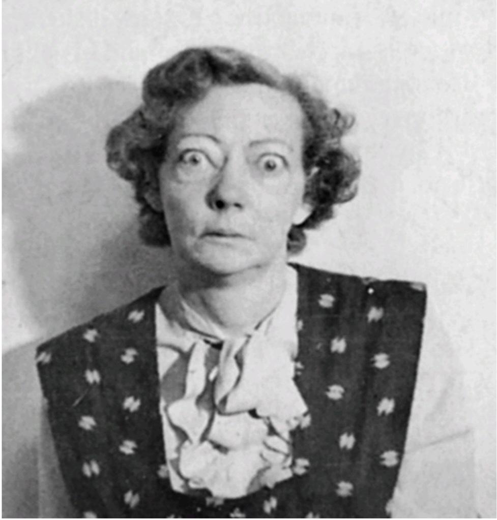
Figure 77-8 A patient with exophthalmic hyperthyroidism. Note protrusion of the eyes and retraction of the superior eyelids. Her basal metabolic rate was +40. (Courtesy Dr. Leonard Posey.)
شکل 77-8 بیمار مبتلا به پرکاری تیروئید اگزوفتالمیک. به بیرون زدگی چشم ها و جمع شدن پلک های فوقانی توجه کنید. میزان متابولیسم پایه او 40+ بود. (با احترام از دکتر لئونارد پوزی.)
Diagnostic Tests for Hyperthyroidism. For the usual case of hyperthyroidism, the most accurate diagnostic test is direct measurement of the concentration of “free” thyroxine (and sometimes triiodothyronine) in the plasma, using appropriate immunoassay procedures.
تست های تشخیصی پرکاری تیروئید. در مورد معمول پرکاری تیروئید، دقیق ترین تست تشخیصی اندازه گیری مستقیم غلظت تیروکسین «آزاد» (و گاهی اوقات تری یدوتیرونین) در پلاسما با استفاده از روش های ایمونواسی مناسب است.
The following tests also are sometimes used:
گاهی اوقات از آزمایشات زیر نیز استفاده می شود:
1. The basal metabolic rate is usually increased to +30 to +60 in severe hyperthyroidism.
1. میزان متابولیسم پایه معمولاً در پرکاری تیروئید شدید به 30+ تا 60+ افزایش می یابد.
2. The concentration of TSH in the plasma is measured by immunoassay. In the usual type of thyrotoxicosis, anterior pituitary secretion of TSH is so completely suppressed by the large amounts of circulating thyroxine and triiodothyronine that there is almost no plasma TSH.
2. غلظت TSH در پلاسما با روش ایمونواسی اندازه گیری می شود. در نوع معمول تیروتوکسیکوز، ترشح هیپوفیز قدامی TSH توسط مقادیر زیاد تیروکسین و تری یدوتیرونین در گردش کاملاً سرکوب می شود که تقریباً هیچ پلاسما TSH وجود ندارد.
3. The concentration of TSI is measured by immunoassay. This concentration is usually high in thyrotoxicosis but low in thyroid adenoma.
3. غلظت TSI با روش ایمونواسی اندازه گیری می شود. این غلظت معمولا در تیروتوکسیکوز زیاد ولی در آدنوم تیروئید کم است.
Treatment in Hyperthyroidism. The most direct treatment for hyperthyroidism is surgical removal of most of the thyroid gland. In general, it is desirable to prepare the patient for surgical removal of the gland before the operation by administering propylthiouracil, usually for several weeks, until the basal metabolic rate of the patient has returned to normal. Then, administration of high concentrations of iodides for 1 to 2 weeks immediately before operation causes the gland to recede in size and its blood supply to diminish. With use of these preoperative procedures, the operative mortality is less than 1 in 1000, whereas before the development of modern procedures, operative mortality was 1 in 25.
درمان پرکاری تیروئید. مستقیم ترین درمان پرکاری تیروئید، برداشتن بیشتر غده تیروئید با جراحی است. به طور کلی، آماده سازی بیمار برای برداشتن غده با جراحی قبل از عمل با تجویز پروپیل تیوراسیل، معمولاً به مدت چند هفته، تا زمانی که میزان متابولیسم پایه بیمار به حالت عادی برگردد، مطلوب است. سپس تجویز غلظت بالای یدید به مدت 1 تا 2 هفته بلافاصله قبل از عمل باعث کاهش اندازه غده و کاهش خون رسانی آن می شود. با استفاده از این روش های قبل از عمل، مرگ و میر ناشی از عمل کمتر از 1 در 1000 است، در حالی که قبل از توسعه روش های مدرن، مرگ و میر ناشی از عمل 1 در 25 بود.
Treatment of the Hyperplastic Thyroid Gland With Radioactive lodine. Eighty to 90% of an injected dose of iodide is absorbed by the hyperplastic, toxic thyroid gland within 1 day after injection. If this injected iodine is radioactive, it can destroy most of the secretory cells of the thyroid gland. Usually 5 millicuries of radioactive iodine is given to the patient, whose condition is reassessed several weeks later. If the patient is still in a hyperthyroid state, additional doses are administered until normal thyroid status is reached.
درمان غده تیروئید هیپرپلاستیک با لودین رادیواکتیو. 80 تا 90 درصد از دوز تزریقی یدید ظرف 1 روز پس از تزریق توسط غده تیروئید هیپرپلاستیک و سمی جذب می شود. اگر این ید تزریقی رادیواکتیو باشد، می تواند بیشتر سلول های ترشحی غده تیروئید را از بین ببرد. معمولاً 5 میلیکوری ید رادیواکتیو به بیمار داده میشود که وضعیت او چند هفته بعد مجدداً ارزیابی میشود. اگر بیمار هنوز در حالت پرکاری تیروئید باشد، دوزهای اضافی تا رسیدن به وضعیت تیروئید طبیعی تجویز می شود.
Hypothyroidism
The effects of hypothyroidism, in general, are opposite to those of hyperthyroidism, but a few physiological mechanisms are peculiar to hypothyroidism. Hypothyroidism, like hyperthyroidism, is often initiated by autoimmunity against the thyroid gland (Hashimoto’s disease), but in this case the autoimmunity destroys the gland rather than stimulates it. The thyroid glands of most of these patients first demonstrate autoimmune “thyroiditis,” which means thyroid inflammation. Thyroiditis causes progressive deterioration and finally fibrosis of the gland, with resultant diminished or absent secretion of thyroid hormone. Several other types of hypothyroidism also occur that are often associated with development of enlarged thyroid glands, called thyroid goiter, as described in the following sections.
کم کاری تیروئید
اثرات کم کاری تیروئید، به طور کلی، برخلاف پرکاری تیروئید است، اما مکانیسم های فیزیولوژیکی کمی برای کم کاری تیروئید خاص است. کم کاری تیروئید، مانند پرکاری تیروئید، اغلب با خودایمنی علیه غده تیروئید (بیماری هاشیموتو) شروع می شود، اما در این مورد، خودایمنی به جای تحریک غده، غده را از بین می برد. غدد تیروئید اکثر این بیماران ابتدا “تیروئیدیت” خود ایمنی را نشان می دهند که به معنای التهاب تیروئید است. تیروئیدیت باعث بدتر شدن تدریجی و در نهایت فیبروز غده می شود که در نتیجه ترشح هورمون تیروئید کاهش یافته یا وجود ندارد. چندین نوع دیگر از کم کاری تیروئید نیز رخ می دهد که اغلب با بزرگ شدن غدد تیروئید به نام گواتر تیروئید همراه است که در بخش های زیر توضیح داده شده است.
Endemic Colloid Goiter Caused by Dietary lodide Deficiency. The term “goiter” means a greatly enlarged thyroid gland. As pointed out in the discussion of iodine metabolism, about 50 milligrams of iodine are required each year for the formation of adequate quantities of thyroid hormone. In certain areas of the world, notably in the Swiss Alps, the Andes, and the Great Lakes region of the United States, insufficient iodine is present in the soil for the food- stuffs to contain even this minute quantity. Therefore, in the days before iodized table salt, many people who lived in these areas developed extremely large thyroid glands, called endemic goiters.
گواتر کلوئیدی بومی ناشی از کمبود لودید در رژیم غذایی است. اصطلاح گواتر به معنای بزرگ شدن غده تیروئید است. همانطور که در بحث متابولیسم ید اشاره شد، سالانه حدود 50 میلی گرم ید برای تشکیل مقادیر کافی هورمون تیروئید مورد نیاز است. در مناطق خاصی از جهان، به ویژه در کوههای آلپ سوئیس، رشته کوههای آند، و منطقه دریاچههای بزرگ ایالات متحده، ید کافی در خاک وجود ندارد تا مواد غذایی حتی به این مقدار کمی داشته باشند. بنابراین، در روزهای قبل از نمک خوراکی یددار، بسیاری از مردمی که در این مناطق زندگی میکردند غدد تیروئید بسیار بزرگی داشتند که گواتر آندمیک نامیده میشود.
The following mechanism results in the development of large endemic goiters: Lack of iodine prevents production of both thyroxine and triiodothyronine. As a result, no hormone is available to inhibit production of TSH by the anterior pituitary, which causes the pituitary to secrete excessively large quantities of TSH. The TSH then stimulates the thyroid cells to secrete large amounts of thyroglobulin colloid into the follicles, and the gland grows larger and larger. However, because of lack of iodine, thyroxine and triiodo- thyronine production does not occur in the thyroglobulin molecule and therefore does not cause the normal suppression of TSH production by the anterior pituitary. The follicles greatly enlarge, and the thyroid gland may increase to 10 to 20 times its normal size.
مکانیسم زیر منجر به ایجاد گواتر بومی بزرگ می شود: کمبود ید از تولید تیروکسین و تری یدوتیرونین جلوگیری می کند. در نتیجه، هیچ هورمونی برای مهار تولید TSH توسط هیپوفیز قدامی در دسترس نیست، که باعث می شود هیپوفیز مقادیر بیش از حد زیادی TSH ترشح کند. سپس TSH سلول های تیروئید را تحریک می کند تا مقادیر زیادی تیروگلوبولین کلوئید را در فولیکول ها ترشح کنند و غده بزرگتر و بزرگتر می شود. با این حال، به دلیل کمبود ید، تولید تیروکسین و تری یدوتیرونین در مولکول تیروگلوبولین رخ نمی دهد و بنابراین باعث سرکوب طبیعی تولید TSH توسط هیپوفیز قدامی نمی شود. فولیکول ها به شدت بزرگ می شوند و غده تیروئید ممکن است به 10 تا 20 برابر اندازه طبیعی خود افزایش یابد.
Idiopathic Nontoxic Colloid Goiter. Enlarged thyroid glands similar to those of endemic colloid goiter can also occur in people who do not have iodine deficiency. These goitrous glands may secrete normal quantities of thyroid hormones, but more frequently, the secretion of hormone is depressed, as in endemic colloid goiter.
گواتر کلوئیدی غیر سمی ایدیوپاتیک. بزرگ شدن غدد تیروئید شبیه به گواتر کلوئیدی بومی می تواند در افرادی که کمبود ید ندارند نیز رخ دهد. این غدد گواتر ممکن است مقادیر طبیعی هورمون های تیروئیدی ترشح کنند، اما بیشتر اوقات، مانند گواتر کلوئیدی بومی، ترشح هورمون کاهش می یابد.
The exact cause of the enlarged thyroid gland in patients with idiopathic colloid goiter is not known, but most of these patients show signs of mild thyroiditis; therefore, it has been suggested that the thyroiditis causes slight hypothyroidism, which then leads to increased TSH secretion and progressive growth of the noninflamed portions of the gland. This theory could explain why these glands are usually nodular, with some portions of the gland growing while other portions are being destroyed by thyroiditis.
علت دقیق بزرگ شدن غده تیروئید در بیماران مبتلا به گواتر کلوئیدی ایدیوپاتیک مشخص نیست، اما اغلب این بیماران علائم تیروئیدیت خفیف را نشان می دهند. بنابراین، پیشنهاد شده است که تیروئیدیت باعث کم کاری تیروئید خفیف می شود که سپس منجر به افزایش ترشح TSH و رشد پیشرونده بخش های غیر ملتهب غده می شود. این تئوری میتواند توضیح دهد که چرا این غدد معمولاً ندولار هستند و برخی از بخشهای غده رشد میکنند در حالی که بخشهای دیگر در اثر تیروئیدیت از بین میروند.
In some persons with colloid goiter, the thyroid gland has an abnormality of the enzyme system required for formation of thyroid hormones. The following abnormalities are often encountered:
در برخی از افراد مبتلا به گواتر کلوئیدی، غده تیروئید دارای اختلال در سیستم آنزیمی مورد نیاز برای تشکیل هورمون های تیروئید است. ناهنجاری های زیر اغلب مشاهده می شود:
1. A deficient iodide-trapping mechanism, in which iodine is not pumped adequately into the thyroid cells
1. مکانیسم کمبود ید به دام انداختن، که در آن ید به اندازه کافی به سلول های تیروئید پمپ نمی شود.
2. A deficient peroxidase system, in which the iodides are not oxidized to the iodine state
2. یک سیستم پراکسیداز کمبود، که در آن یدیدها به حالت ید اکسید نمی شوند.
3. Deficient coupling of iodinated tyrosines in the thyroglobulin molecule so that the final thyroid hormones cannot be formed
3. عدم جفت شدن تیروزین های یددار در مولکول تیروگلوبولین به طوری که هورمون های تیروئید نهایی نمی توانند تشکیل شوند.
4. Deficiency of the deiodinase enzyme, which prevents recovery of iodine from the iodinated tyrosines that are not coupled to form the thyroid hormones (this is about two-thirds of the iodine), thus leading to iodine deficiency
4. کمبود آنزیم دیودیناز، که از بازیابی ید از تیروزین های یددار که برای تشکیل هورمون های تیروئید جفت نشده اند (این حدود دو سوم ید است) جلوگیری می کند، بنابراین منجر به کمبود ید می شود.
Finally, some foods contain goitrogenic substances that have a propylthiouracil-type of antithyroid activity, thus also leading to TSH-stimulated enlargement of the thyroid gland. Such goitrogenic substances are found especially in some varieties of turnips and cabbages.
در نهایت، برخی از غذاها حاوی مواد گواتروژن هستند که دارای فعالیت ضد تیروئیدی از نوع پروپیل تیوراسیل هستند، بنابراین منجر به بزرگ شدن غده تیروئید توسط TSH می شود. چنین مواد گواتروژن به ویژه در برخی از انواع شلغم و کلم یافت می شود.
Physiological Characteristics of Hypothyroidism.
Whether hypothyroidism is due to thyroiditis, endemic col- loid goiter, idiopathic colloid goiter, destruction of the thyroid gland by irradiation, or surgical removal of the thyroid gland, the physiological effects are the same. They include fatigue and extreme somnolence, with persons sleeping up to 12 to 14 hours a day, extreme muscular sluggishness, a slowed heart rate, decreased cardiac output, decreased blood volume, sometimes increased body weight, constipation, mental sluggishness, failure of many trophic functions in the body as evidenced by depressed growth of hair and scaliness of the skin, development of a froglike, husky voice, and, in severe cases, development of an edematous appearance throughout the body called myxedema.
ویژگی های فیزیولوژیکی کم کاری تیروئید.
چه کم کاری تیروئید به دلیل تیروئیدیت، گواتر کلوئیدی بومی، گواتر کلوئیدی ایدیوپاتیک، تخریب غده تیروئید توسط تابش یا برداشتن غده تیروئید با جراحی باشد، اثرات فیزیولوژیکی یکسان است. اینها شامل خستگی و خواب آلودگی شدید، با خوابیدن افراد بین 12 تا 14 ساعت در روز، کندی شدید عضلانی، کند شدن ضربان قلب، کاهش برون ده قلبی، کاهش حجم خون، گاهی اوقات افزایش وزن بدن، یبوست، کندی ذهنی، نارسایی بسیاری از عملکردهای تغذیه ای در بدن است که با رشد مو و ریزش مو مشخص می شود. صدای دراز کشیده و در موارد شدید، ایجاد یک ظاهر ادماتیک در سراسر بدن به نام میکسدم.
Myxedema. Myxedema develops in persons who have almost total lack of thyroid hormone function. Figure 77-9 shows such a patient, demonstrating bagginess under the eyes and swelling of the face. In this condition, for reasons that are not fully explained, greatly increased quantities of hyaluronic acid and chondroitin sulfate bound with protein form excessive tissue gel in the interstitial spaces, which causes the total quantity of interstitial fluid to increase. Be- cause of the gel nature of the excess fluid, it is mainly im- mobile and the edema is the nonpitting type.
میکسدم. میکسدم در افرادی ایجاد می شود که تقریباً کمبود کامل عملکرد هورمون تیروئید دارند. شکل 77-9 چنین بیمار را نشان می دهد که گودی زیر چشم و تورم صورت را نشان می دهد. در این شرایط، به دلایلی که به طور کامل توضیح داده نشده است، مقادیر زیاد اسید هیالورونیک و کندرویتین سولفات متصل به پروتئین، ژل بافتی بیش از حد در فضاهای بینابینی ایجاد می کند که باعث افزایش مقدار کل مایع بینابینی می شود. به دلیل ماهیت ژلی مایع اضافی، عمدتاً بی حرکت است و ادم از نوع بدون حفره است.
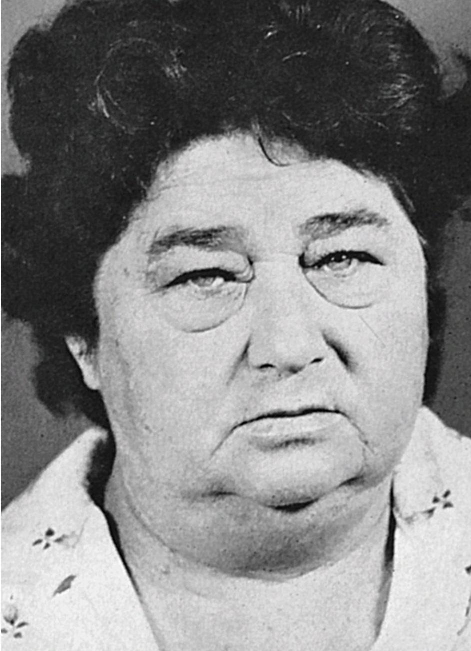
Figure 77-9 A patient with myxedema. (Courtesy Dr. Herbert Lang- ford.)
شکل 77-9 بیمار مبتلا به میکسدم. (با احترام از دکتر هربرت لنگفورد.)
Atherosclerosis in Hypothyroidism. As pointed out earlier, lack of thyroid hormone increases the quantity of blood cholesterol because of altered fat and cholesterol metabolism and diminished liver excretion of cholesterol in the bile. The increase in blood cholesterol is often associated with increased atherosclerosis. Therefore, many hypothyroid patients, particularly those with myxedema, develop atherosclerosis, which in turn results in peripheral vascular disease, deafness, and coronary artery disease with subsequent early death.
آترواسکلروز در کم کاری تیروئید. همانطور که قبلاً اشاره شد، کمبود هورمون تیروئید به دلیل تغییر متابولیسم چربی و کلسترول و کاهش دفع کبدی کلسترول در صفرا، باعث افزایش مقدار کلسترول خون می شود. افزایش کلسترول خون اغلب با افزایش آترواسکلروز همراه است. بنابراین، بسیاری از بیماران هیپوتیروئیدی، به ویژه آنهایی که مبتلا به میکسدم هستند، دچار تصلب شرایین می شوند که به نوبه خود منجر به بیماری عروق محیطی، ناشنوایی و بیماری عروق کرونر با مرگ زودهنگام بعدی می شود.
Diagnostic Tests for Hypothyroidism. The tests already described for diagnosis of hyperthyroidism give opposite results in hypothyroidism. The free thyroxine in the blood is low. The basal metabolic rate in myxedema is reduced by 30% to 50%. In addition, the secretion of TSH by the anterior pituitary when a test dose of TRH is administered is usually greatly increased (except in the rare cases of hypothyroidism caused by depressed response of the pituitary gland to TRH).
تست های تشخیصی کم کاری تیروئید. آزمایشاتی که قبلاً برای تشخیص پرکاری تیروئید توضیح داده شد، نتایج متضادی را در کم کاری تیروئید نشان می دهد. تیروکسین آزاد در خون کم است. میزان متابولیسم پایه در میکسدم 30 تا 50 درصد کاهش می یابد. علاوه بر این، ترشح TSH توسط هیپوفیز قدامی هنگام تجویز دوز آزمایشی TRH معمولاً به شدت افزایش می یابد (به جز در موارد نادر کم کاری تیروئید ناشی از پاسخ افسرده غده هیپوفیز به TRH).
Treatment of Hypothyroidism. Figure 77-4 shows the effect of thyroxine on basal metabolic rate, demonstrating that the hormone normally has a duration of action of more than 1 month. Consequently, a steady level of thyroid hormone activity is easily maintained in the body via daily oral ingestion of one or more tablets containing thyroxine. Furthermore, proper treatment of hypothyroidism results in such complete normality that formerly myxedematous patients have lived into their 90s after undergoing treatment for more than 50 years.
درمان کم کاری تیروئید. شکل 77-4 اثر تیروکسین را بر میزان متابولیسم پایه نشان می دهد و نشان می دهد که این هورمون به طور معمول بیش از 1 ماه طول می کشد. در نتیجه، سطح ثابتی از فعالیت هورمون تیروئید به راحتی از طریق مصرف خوراکی روزانه یک یا چند قرص حاوی تیروکسین در بدن حفظ می شود. علاوه بر این، درمان مناسب کم کاری تیروئید منجر به آنچنان طبیعی بودن کامل می شود که بیمارانی که قبلا میکسدماتوز داشتند پس از بیش از 50 سال تحت درمان تا 90 سالگی زندگی کرده اند.
Cretinism
Cretinism is caused by extreme hypothyroidism during fetal life, infancy, or childhood. This condition is characterized especially by failure of body growth and by mental retardation. It results from congenital lack of a thyroid gland (congenital cretinism), from failure of the thyroid gland to produce thy- roid hormone because of a genetic defect of the gland, or from a lack of iodine in the diet (endemic cretinism).
کرتینیسم
کرتینیسم در اثر کم کاری شدید تیروئید در دوران جنینی، نوزادی یا کودکی ایجاد می شود. این وضعیت به ویژه با نارسایی رشد بدن و عقب ماندگی ذهنی مشخص می شود. این بیماری ناشی از کمبود مادرزادی غده تیروئید (کرتینیسم مادرزادی)، از نارسایی غده تیروئید در تولید هورمون تیروئید به دلیل نقص ژنتیکی غده، یا از کمبود ید در رژیم غذایی (کرتینیسم بومی) است.
A neonate without a thyroid gland may have a normal appearance and function because she or he was supplied with some (but usually not enough) thyroid hormone by the mother while in utero. A few weeks after birth, how- ever, the neonate’s movements become sluggish and both physical and mental growth begin to be greatly retarded. Treatment of the neonate with cretinism at any time with adequate iodine or thyroxine usually causes normal return of physical growth, but unless the cretinism is treated with- in a few weeks after birth, mental growth remains permanently retarded. This state results from retardation of the growth, branching, and myelination of the neuronal cells of the central nervous system at this critical time in the nor- mal development of the mental powers.
نوزاد بدون غده تیروئید ممکن است ظاهر و عملکرد طبیعی داشته باشد زیرا در دوران بارداری مقداری (اما معمولاً کافی نبود) هورمون تیروئید توسط مادر به او داده شده است. با این حال، چند هفته پس از تولد، حرکات نوزاد کند می شود و رشد جسمی و ذهنی به شدت به تاخیر می افتد. درمان نوزاد مبتلا به کرتینیسم در هر زمان با ید یا تیروکسین کافی معمولاً باعث بازگشت طبیعی رشد فیزیکی می شود، اما مگر اینکه کرتینیسم طی چند هفته پس از تولد درمان شود، رشد ذهنی برای همیشه عقب مانده است. این حالت ناشی از تاخیر در رشد، انشعاب و میلین شدن سلول های عصبی سیستم عصبی مرکزی در این زمان حساس در رشد طبیعی قوای ذهنی است.
Skeletal growth in a child with cretinism is characteristically more inhibited than is soft tissue growth. As a result of this disproportionate rate of growth, the soft tissues are likely to enlarge excessively, giving the child with cretinism an obese, stocky, and short appearance. Occasionally the tongue becomes so large in relation to the skeletal growth that it obstructs swallowing and breathing, inducing a characteristic guttural breathing that sometimes chokes the child.
رشد اسکلتی در یک کودک مبتلا به کرتینیسم به طور مشخص بیشتر از رشد بافت نرم مهار می شود. در نتیجه این سرعت نامتناسب رشد، بافت های نرم احتمالاً بیش از حد بزرگ می شوند و ظاهری چاق، چاق و کوتاه به کودک مبتلا به کرتینیسم می دهند. گاهی اوقات زبان نسبت به رشد اسکلتی آنقدر بزرگ می شود که مانع بلع و تنفس می شود و باعث ایجاد یک تنفس روده ای مشخص می شود که گاهی اوقات کودک را خفه می کند.
کلیک کنید «Bibliography: فهرست کتب مربوطه»
Bianco AC, Dumitrescu A, Gereben B, et al: Paradigms of dynamic control of thyroid hormone signaling. Endocr Rev 40:1000, 2019.
Biondi B, Cappola AR, Cooper DS: Subclinical hypothyroidism: a review. JAMA 322:153, 2019.
Biondi B, Cooper DS: Subclinical hyperthyroidism N Engl J Med 378:2411, 2018.
Brent GA: Mechanisms of thyroid hormone action. J Clin Invest 122:3035, 2012.
Burch HB: Drug effects on the thyroid. N Engl J Med 381:749, 2019.
Citterio CE, Targovnik HM, Arvan P: The role of thyroglobulin in thyroid hormonogenesis. Nat Rev Endocrinol 15:323, 2019.
De La Vieja A, Dohan O, Levy O, Carrasco N: Molecular analysis of the sodium/iodide symporter: impact on thyroid and extrathyroid pathophysiology. Physiol Rev 80:1083, 2000.
Gerdes AM, Ojamaa K: Thyroid hormone and cardioprotection. Com- pr Physiol 6:1199, 2016.
Ikegami K, Refetoff S, Van Cauter E, Yoshimura T: Interconnection between circadian clocks and thyroid function. Nat Rev Endocrinol 15:590, 2019.
Lanni A, Moreno M, Goglia F: Mitochondrial actions of thyroid hormone. Compr Physiol 6:1591, 2016.
Lee S, Farwell AP: Euthyroid sick syndrome. Compr Physiol 6:1071, 2016.
Luongo C, Dentice M, Salvatore D: Deiodinases and their intricate role in thyroid hormone homeostasis. Nat Rev Endocrinol 15:479-, 2019.
Mullur R, Liu YY, Brent GA: Thyroid hormone regulation of metabo- lism. Physiol Rev 94:355, 2014.
Ortiga-Carvalho TM, Chiamolera MI, Pazos-Moura CC, Wondisford FE: Hypothalamus-pituitary-thyroid axis. Compr Physiol 6:1387, 2016.
Razvi S, Jabbar A, Pingitore A, et al: Thyroid hormones and cardiovascular function and diseases. J Am Coll Cardiol 71:1781, 2018.
Singh I, Hershman JM: Pathogenesis of hyperthyroidism. Compr Physiol 7:67, 2016.
🚀 با ما همراه شوید!
تازهترین مطالب و آموزشهای مغز و اعصاب را از دست ندهید. با فالو کردن کانال تلگرام آیندهنگاران مغز، از ما حمایت کنید!



