
فیزیولوژی پزشکی گایتون و هال؛ هورمونهای هیپوفیز و کنترل آنها توسط هیپوتالاموس


دعای مطالعه [ نمایش ]
بِسْمِ الله الرَّحْمنِ الرَّحیمِ
اَللّهُمَّ اَخْرِجْنى مِنْ ظُلُماتِ الْوَهْمِ
خدايا مرا بيرون آور از تاريكىهاى وهم،
وَ اَكْرِمْنى بِنُورِ الْفَهْمِ
و به نور فهم گرامى ام بدار،
اَللّهُمَّ افْتَحْ عَلَيْنا اَبْوابَ رَحْمَتِكَ
خدايا درهاى رحمتت را به روى ما بگشا،
وَانْشُرْ عَلَيْنا خَزائِنَ عُلُومِكَ بِرَحْمَتِكَ يا اَرْحَمَ الرّاحِمينَ
و خزانههاى علومت را بر ما باز كن به امید رحمتت اى مهربانترين مهربانان.
کتاب «فیزیولوژی پزشکی گایتون و هال» بهعنوان یکی از جامعترین و معتبرترین منابع در حوزه علوم پزشکی، همچنان مرجع کلیدی برای درک عملکرد پیچیده بدن انسان است. این اثر با تکیه بر تازهترین پژوهشها و توضیحات دقیق از سازوکارهای فیزیولوژیک، پلی میان علوم پایه پزشکی و کاربردهای بالینی ایجاد میکند و نقشی بیبدیل در آموزش، پژوهش و ارتقای دانش سلامت ایفا مینماید.
ترجمه دقیق و علمی این شاهکار توسط برند علمی آیندهنگاران مغز به مدیریت داریوش طاهری، دسترسی فارسیزبانان به مرزهای نوین دانش فیزیولوژی را ممکن ساخته و رسالتی علمی برای ارتقای آموزش پزشکی، فهم عمیقتر سازوکارهای بدن و توسعه روشهای نوین در حوزه سلامت فراهم آورده است.
» کتاب فیزیولوژی پزشکی گایتون و هال
» » فصل ۷۶: هورمون های هیپوفیز و کنترل آنها توسط هیپوتالاموس
در حال ویرایش
» Guyton and Hall Textbook of Medical Physiology
»» CHAPTER 76: Pituitary Hormones and Their Control by the Hypothalamus

PITUITARY GLAND AND ITS RELATION TO THE HYPOTHALAMUS
ANTERIOR AND POSTERIOR LOBES OF THE PITUITARY GLAND
The pituitary gland (Figure 76-1), also called the hypophysis, is a small gland-about 1 centimeter in diameter and 0.5 to 1 gram in weight-that lies in the sella turcica, a bony cavity at the base of the brain-and is connected to the hypothalamus by the pituitary (or hypophysial) stalk. Physiologically, the pituitary gland is divisible into two distinct portions: the anterior pituitary, also known as the adenohypophysis, and the posterior pituitary, also known as the neurohypophysis. Between these portions is a small, relatively avascular zone called the pars intermedia, which is much less developed in humans but is larger and much more functional in some animals.
غده هیپوفیز و ارتباط آن با هیپوتالاموس
لوب های قدامی و خلفی غده هیپوفیز
غده هیپوفیز (شکل 76-1) که هیپوفیز نیز نامیده می شود، غده کوچکی است – حدود 1 سانتی متر قطر و 0.5 تا 1 گرم وزن – که در sella turcica، یک حفره استخوانی در پایه مغز قرار دارد و توسط هیپوفیز (یا هیپوفیزیکال) به هیپوتالاموس متصل است. از نظر فیزیولوژیکی، غده هیپوفیز به دو بخش مجزا تقسیم می شود: هیپوفیز قدامی، که به عنوان آدنوهیپوفیز نیز شناخته می شود، و هیپوفیز خلفی، که به عنوان نوروهیپوفیز نیز شناخته می شود. بین این بخشها یک ناحیه کوچک و نسبتاً بدون عروق به نام pars intermedia وجود دارد که در انسان بسیار کمتر توسعه یافته است، اما در برخی از حیوانات بزرگتر و بسیار کاربردیتر است.
Embryologically, the two portions of the pituitary originate from different sources-the anterior pituitary from Rathke’s pouch, which is an embryonic invagination of the pharyngeal epithelium, and the posterior pituitary from a neural tissue outgrowth from the hypothalamus. The origin of the anterior pituitary from the pharyngeal epithelium explains the epithelioid nature of its cells, and the origin of the posterior pituitary from neural tissue explains the presence of large numbers of glial-type cells in this gland.
از نظر جنین شناسی، دو قسمت هیپوفیز از منابع مختلف منشأ می گیرند – هیپوفیز قدامی از کیسه Rathke که یک هجوم جنینی به اپیتلیوم حلق است و هیپوفیز خلفی از رشد بافت عصبی از هیپوتالاموس. منشأ هیپوفیز قدامی از اپیتلیوم حلقی ماهیت اپیتلیوئیدی سلول های آن را توضیح می دهد و منشاء هیپوفیز خلفی از بافت عصبی وجود تعداد زیادی سلول از نوع گلیال را در این غده توضیح می دهد.
Six major peptide hormones plus several other hormones of lesser known importance are secreted by the anterior pituitary, and two important peptide hormones are secreted by the posterior pituitary.
شش هورمون پپتیدی اصلی به اضافه چندین هورمون دیگر با اهمیت کمتر توسط هیپوفیز قدامی ترشح می شوند و دو هورمون پپتیدی مهم توسط هیپوفیز خلفی ترشح می شوند.
The hormones of the anterior pituitary play major roles in the control of metabolic functions throughout the body, as shown in Figure 76-2.
همانطور که در شکل 76-2 نشان داده شده است، هورمون های هیپوفیز قدامی نقش عمده ای در کنترل عملکردهای متابولیک در سراسر بدن دارند.
Growth hormone promotes growth of the entire body by affecting protein formation, cell multiplication, and cell differentiation.
هورمون رشد با تأثیر بر تشکیل پروتئین، تکثیر سلولی و تمایز سلولی باعث رشد کل بدن می شود.
Adrenocorticotropic hormone (corticotropin) controls the secretion of some of the adrenocortical hormones, which affect metabolism of glucose, proteins, and fats.
هورمون آدرنوکورتیکوتروپیک (کورتیکوتروپین) ترشح برخی از هورمون های قشر آدرنال را کنترل می کند که بر متابولیسم گلوکز، پروتئین ها و چربی ها تأثیر می گذارد.
Thyroid-stimulating hormone (thyrotropin) controls the secretion rate of thyroxine and triiodothyronine by the thyroid gland, and these hormones control the rates of most intracellular chemical reactions in the body.
هورمون محرک تیروئید (تیروتروپین) میزان ترشح تیروکسین و تری یدوتیرونین را توسط غده تیروئید کنترل می کند و این هورمون ها سرعت اکثر واکنش های شیمیایی درون سلولی را در بدن کنترل می کنند.
Prolactin promotes mammary gland development and milk production.
پرولاکتین باعث رشد غدد پستانی و تولید شیر می شود.
Two separate gonadotropic hormones, follicle- stimulating hormone and luteinizing hormone, control growth of the ovaries and testes, as well as their hormonal and reproductive activities.
دو هورمون گنادوتروپیک مجزا، هورمون محرک فولیکول و هورمون لوتئینه کننده، رشد تخمدان ها و بیضه ها و همچنین فعالیت های هورمونی و تولید مثلی آنها را کنترل می کنند.
The two hormones secreted by the posterior pituitary play other roles.
دو هورمون ترشح شده توسط هیپوفیز خلفی نقش دیگری را ایفا می کنند.
Antidiuretic hormone (also called vasopressin) controls the rate of water excretion into the urine, thus helping to control water concentration in the body fluids.
هورمون آنتی دیورتیک (همچنین به نام وازوپرسین) سرعت دفع آب در ادرار را کنترل می کند، بنابراین به کنترل غلظت آب در مایعات بدن کمک می کند.
Oxytocin helps express milk from the glands of the breast to the nipples during suckling and helps in delivery of the baby at the end of gestation.
اکسی توسین به بیرون ریختن شیر از غدد سینه به نوک سینه در هنگام شیردهی کمک می کند و به زایمان نوزاد در پایان بارداری کمک می کند.
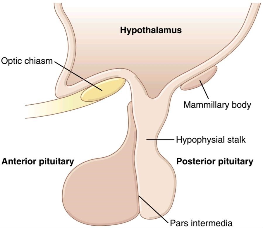
Figure 76-1 Pituitary gland.
شکل 76-1 غدده ی هیپوفیز.

Figure 76-2. Metabolic functions of the anterior pituitary hormones. ACH, Adrenal corticosteroid hormones.
شکل 76-2. عملکرد متابولیک هورمون های هیپوفیز قدامی. ACH، هورمون های کورتیکواستروئیدی آدرنال.
The Anterior Pituitary Gland Contains Several Different Cell Types That Synthesize and Secrete Hormones. Usually, there is one cell type for each major hormone formed in the anterior pituitary gland. With special stains attached to high-affinity antibodies that bind with the distinctive hormones, at least five cell types can be differentiated (Figure 76-3). Table 76-1 provides a summary of these cell types, the hormones they produce, and their physiological actions. These five cell types are as follows:
غده هیپوفیز قدامی حاوی چندین نوع سلول مختلف است که هورمون ها را سنتز و ترشح می کنند. معمولاً برای هر هورمون اصلی تشکیل شده در غده هیپوفیز قدامی یک نوع سلول وجود دارد. با لکههای مخصوص متصل به آنتیبادیهای با میل ترکیبی بالا که با هورمونهای متمایز متصل میشوند، حداقل پنج نوع سلول را میتوان تمایز داد (شکل 76-3). جدول 76-1 خلاصه ای از انواع این سلول ها، هورمون هایی که تولید می کنند و اعمال فیزیولوژیکی آنها را ارائه می دهد. این پنج نوع سلول به شرح زیر است:
1. Somatotropeshuman growth hormone (hGH)
2. Corticotropes-adrenocorticotropic hormone (ACTH)
3. Thyrotropes-thyroid-stimulating hormone (TSH)
4. Gonadotropes-gonadotropic hormones, which include both luteinizing hormone (LH) and folliclestimulating hormone (FSH)
5. Lactotropes-prolactin (PRL)
1. هورمون رشد سوماتوتروپشومن (hGH)
2. کورتیکوتروپ ها- هورمون آدرنوکورتیکوتروپیک (ACTH)
3. تیروتروپ ها-هورمون محرک تیروئید (TSH)
4. هورمون های گنادوتروپ-گنادوتروپیک، که شامل هر دو هورمون لوتئینه کننده (LH) و هورمون محرک فولیکول (FSH) می شود.
5. لاکتوتروپ – پرولاکتین (PRL)
About 30% to 40% of the anterior pituitary cells are somatotropes that secrete growth hormone (GH), and about 20% are corticotropes that secrete ACTH. Each of the other cell types accounts for only 3% to 5% of the total; nevertheless, they secrete powerful hormones for controlling thyroid function, sexual functions, and milk secretion by the breasts. Somatotropes stain strongly with acid dyes and are therefore called acidophils. Thus, pituitary tumors that secrete large quantities of hGH are called acidophilic tumors.
حدود 30 تا 40 درصد از سلول های هیپوفیز قدامی، سوماتوتروپ هایی هستند که هورمون رشد (GH) را ترشح می کنند و حدود 20 درصد نیز کورتیکوتروپ هایی هستند که ACTH ترشح می کنند. هر یک از انواع سلول های دیگر تنها 3 تا 5 درصد از کل را تشکیل می دهند. با این حال، هورمونهای قدرتمندی برای کنترل عملکرد تیروئید، عملکرد جنسی و ترشح شیر توسط سینهها ترشح میکنند. سوماتوتروپ ها به شدت با رنگ های اسیدی رنگ می شوند و بنابراین اسیدوفیل نامیده می شوند. بنابراین، تومورهای هیپوفیز که مقادیر زیادی hGH ترشح می کنند، تومورهای اسیدوفیل نامیده می شوند.
Posterior Pituitary Hormones Are Synthesized by Cell Bodies in the Hypothalamus. The bodies of the cells that secrete the posterior pituitary hormones are not located in the pituitary gland but are large neurons, called magnocellular neurons, located in the supraoptic and paraventricular nuclei of the hypothalamus. The hormones are then transported in the axoplasm of the neuron’s nerve fibers passing from the hypothalamus to the posterior pituitary gland. This mechanism is discussed later in the chapter.
هورمون های هیپوفیز خلفی توسط اجسام سلولی در هیپوتالاموس سنتز می شوند. بدن سلولهایی که هورمونهای هیپوفیز خلفی را ترشح میکنند در غده هیپوفیز قرار ندارند، بلکه نورونهای بزرگی هستند که نورونهای ماگنوسلولار نامیده میشوند و در هستههای فوقاپتیک و پارا بطنی هیپوتالاموس قرار دارند. سپس هورمون ها در آکسوپلاسم رشته های عصبی نورون که از هیپوتالاموس به غده هیپوفیز خلفی عبور می کنند، منتقل می شوند. این مکانیسم بعداً در فصل مورد بحث قرار می گیرد.

Figure 76-3. Cellular structure of the anterior pituitary gland. (Modified from Guyton AC: Physiology of the Human Body, 6th ed. Philadelphia: Saunders College Publishing, 1984.)
شکل 76-3. ساختار سلولی غده هیپوفیز قدامی. (اصلاح شده از Guyton AC: Physiology of the Human Body، ویرایش ششم فیلادلفیا: انتشارات کالج ساندرز، 1984.)
Table 76-1 Cells and Hormones of the Anterior Pituitary Gland and Their Physiological Functions
جدول 76-1 سلول ها و هورمون های غده هیپوفیز قدامی و عملکردهای فیزیولوژیکی آنها.

HYPOTHALAMUS CONTROLS PITUITARY SECRETION
Almost all pituitary secretion is controlled by hormonal or nervous signals from the hypothalamus. Indeed, when the pituitary gland is removed from its normal position beneath the hypothalamus and transplanted to some other part of the body, its secretion rates of the different hormones (except for prolactin) fall to very low levels.
هیپوتالاموس ترشح هیپوفیز را کنترل می کند
تقریباً تمام ترشح هیپوفیز توسط سیگنال های هورمونی یا عصبی از هیپوتالاموس کنترل می شود. در واقع، هنگامی که غده هیپوفیز از موقعیت طبیعی خود در زیر هیپوتالاموس برداشته می شود و به قسمت دیگری از بدن پیوند می زند، میزان ترشح هورمون های مختلف آن (به جز پرولاکتین) به سطوح بسیار پایین می رسد.
Secretion from the posterior pituitary is controlled by nerve signals that originate in the hypothalamus and terminate in the posterior pituitary. In contrast, secretion by the anterior pituitary is controlled by hormones called hypothalamic releasing and hypothalamic inhibitory hormones (or factors) secreted within the hypothalamus and then conducted, as shown in Figure 76-4, to the anterior pituitary through minute blood vessels called hypothalamic-hypophysial portal vessels. In the anterior pituitary, these releasing and inhibitory hormones act on the glandular cells to control their secretion. This system of control is discussed in the next section of this chapter.
ترشح از هیپوفیز خلفی توسط سیگنال های عصبی که از هیپوتالاموس منشا گرفته و به هیپوفیز خلفی ختم می شود، کنترل می شود. در مقابل، ترشح هیپوفیز قدامی توسط هورمونهایی به نام آزادکننده هیپوتالاموس و هورمونهای بازدارنده هیپوتالاموس (یا عوامل) که در هیپوتالاموس ترشح میشوند، کنترل میشود و سپس، همانطور که در شکل 76-4 نشان داده شده است، از طریق عروق خونی کوچک به نام رگهای پورتال هیپوتالاموس-هیپوفیزیال به هیپوفیز قدامی هدایت میشود. در هیپوفیز قدامی، این هورمون های آزاد کننده و بازدارنده بر روی سلول های غده ای عمل می کنند تا ترشح آنها را کنترل کنند. این سیستم کنترل در بخش بعدی این فصل مورد بحث قرار می گیرد.
The hypothalamus receives signals from many sources in the nervous system. Thus, when a person is exposed to pain, a portion of the pain signal is transmitted into the hypothalamus. Likewise, when a person experiences some powerful depressing or exciting thought, a portion of the signal is transmitted into the hypothalamus. Olfactory stimuli denoting pleasant or unpleasant smells transmit strong signals directly and through the amygdaloid nuclei into the hypothalamus. Even the concentrations of nutrients, electrolytes, water, and various hormones in the blood excite or inhibit various portions of the hypothalamus. Thus, the hypothalamus is a center for integrating information concerning the internal well-being of the body, and much of this information is used to control secretions of the many globally important pituitary hormones.
هیپوتالاموس سیگنال ها را از منابع بسیاری در سیستم عصبی دریافت می کند. بنابراین، هنگامی که فرد در معرض درد قرار می گیرد، بخشی از سیگنال درد به هیپوتالاموس منتقل می شود. به همین ترتیب، هنگامی که یک فرد افکار قوی افسرده یا هیجان انگیز را تجربه می کند، بخشی از سیگنال به هیپوتالاموس منتقل می شود. محرکهای بویایی که به بوی خوش یا نامطبوع اشاره میکنند، سیگنالهای قوی را مستقیماً و از طریق هستههای آمیگدال به هیپوتالاموس منتقل میکنند. حتی غلظت مواد مغذی، الکترولیت ها، آب و هورمون های مختلف در خون، بخش های مختلف هیپوتالاموس را تحریک یا مهار می کند. بنابراین، هیپوتالاموس مرکزی برای یکپارچه سازی اطلاعات مربوط به رفاه داخلی بدن است و بیشتر این اطلاعات برای کنترل ترشح بسیاری از هورمون های هیپوفیز مهم جهانی استفاده می شود.
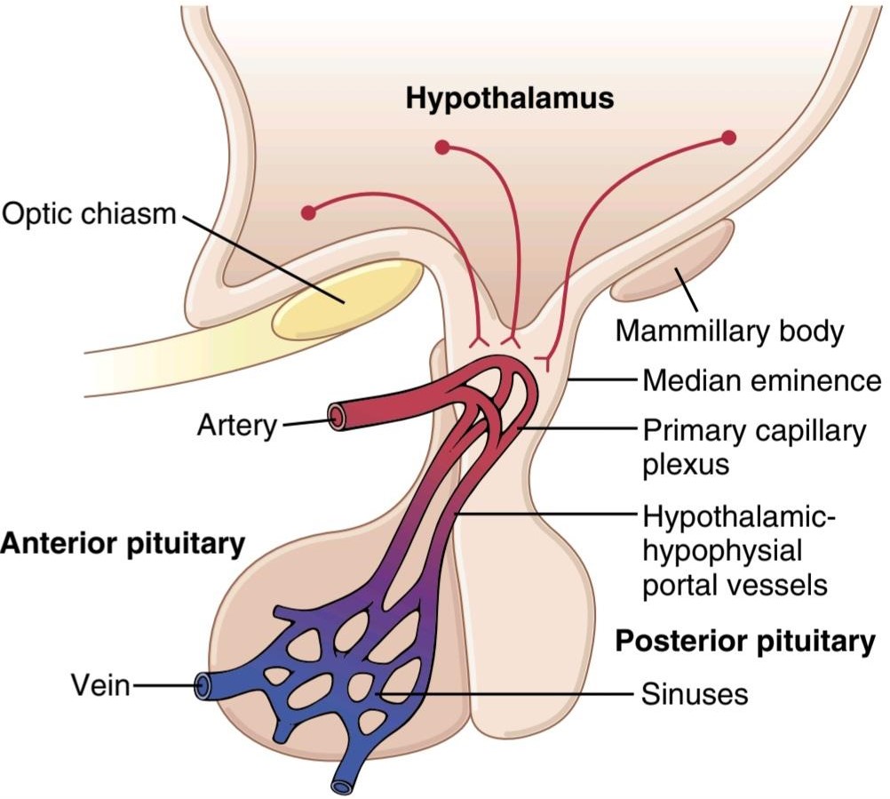
Figure 76-4. Hypothalamic-hypophysial portal system.
شکل 76-4. سیستم پورتال هیپوتالاموس-هیپوفیزیال.
HYPOTHALAMIC-HYPOPHYSIAL PORTAL BLOOD VESSELS OF THE ANTERIOR PITUITARY GLAND
The anterior pituitary is a highly vascular gland with extensive capillary sinuses among the glandular cells. Almost all the blood that enters these sinuses passes first through another capillary bed in the lower hypothalamus. The blood then flows through small hypothalamic-hypophysial portal blood vessels into the anterior pituitary sinuses. Figure 76-4 shows the lowermost portion of the hypothalamus, called the median eminence, which connects inferiorly with the pituitary stalk. The median eminence is the functional link between the hypothalamus and the anterior pituitary gland. Small arteries penetrate into the median eminence and then additional small vessels return to its surface, coalescing to form the hypothalamic- hypophysial portal blood vessels. These vessels pass downward along the pituitary stalk to supply blood to the anterior pituitary sinuses.
عروق خونی پورتال هیپوتالامیک-هیپوفیزیال غده هیپوفیتاری قدامی
هیپوفیز قدامی یک غده بسیار عروقی با سینوس های مویرگی گسترده در میان سلول های غده ای است. تقریباً تمام خونی که وارد این سینوس ها می شود ابتدا از بستر مویرگی دیگری در هیپوتالاموس تحتانی عبور می کند. سپس خون از طریق عروق خونی پورتال هیپوتالاموس-هیپوفیزیال کوچک به سینوس های هیپوفیز قدامی جریان می یابد. شکل 76-4 پایینترین قسمت هیپوتالاموس را نشان میدهد که برجستگی میانی نامیده میشود، که در قسمت پایینی با ساقه هیپوفیز متصل میشود. برجستگی میانی پیوند عملکردی بین هیپوتالاموس و غده هیپوفیز قدامی است. شریانهای کوچک به داخل برجستگی میانی نفوذ میکنند و سپس عروق کوچک اضافی به سطح آن بازمیگردند و به هم میپیوندند و رگهای خونی پورتال هیپوتالاموس- هیپوفیزیال را تشکیل میدهند. این عروق در امتداد ساقه هیپوفیز به سمت پایین حرکت می کنند تا خون را به سینوس های هیپوفیز قدامی برسانند.
Hypothalamic Releasing and Inhibitory Hormones Are Secreted Into the Median Eminence. Special neurons in the hypothalamus synthesize and secrete the hypothalamic releasing and inhibitory hormones that control secretion of the anterior pituitary hormones. These neurons originate in various parts of the hypothalamus and send their nerve fibers to the median eminence and tuber cinereum, an extension of hypothalamic tissue into the pituitary stalk.
هورمون های آزاد کننده و بازدارنده هیپوتالاموس در برجستگی میانه ترشح می شوند. نورون های خاصی در هیپوتالاموس، هورمون های آزاد کننده و بازدارنده هیپوتالاموس را که ترشح هورمون های هیپوفیز قدامی را کنترل می کنند، سنتز و ترشح می کنند. این نورونها از قسمتهای مختلف هیپوتالاموس منشأ میگیرند و رشتههای عصبی خود را به برجستگی میانی و غده سینرئوم، امتداد بافت هیپوتالاموس به ساقه هیپوفیز میفرستند.
The endings of these fibers are different from most endings in the central nervous system in that their function is not to transmit signals from one neuron to another but rather to secrete the hypothalamic releasing and inhibitory hormones into the tissue fluids. These hormones are immediately absorbed into the hypothalamic-hypophysial portal system and carried directly to the sinuses of the anterior pituitary gland.
انتهای این فیبرها با اکثر انتهای سیستم عصبی مرکزی متفاوت است، زیرا وظیفه آنها انتقال سیگنال از یک نورون به نورون دیگر نیست، بلکه ترشح هورمون های آزاد کننده و بازدارنده هیپوتالاموس در مایعات بافتی است. این هورمون ها بلافاصله به سیستم پورتال هیپوتالاموس-هیپوفیزیال جذب می شوند و مستقیماً به سینوس های غده هیپوفیز قدامی منتقل می شوند.
Hypothalamic Releasing and Inhibitory Hormones Control Anterior Pituitary Secretion. The function of the releasing and inhibitory hormones is to control secretion of the anterior pituitary hormones. For most of the anterior pituitary hormones, it is the releasing hormones that are important, but for prolactin, a hypo- thalamic inhibitory hormone probably exerts more control. The major hypothalamic releasing and inhibitory hormones, which are summarized in Table 76-2, are the following:
هورمون های آزاد کننده و بازدارنده هیپوتالاموس ترشح هیپوفیز قدامی را کنترل می کنند. عملکرد هورمون های آزاد کننده و بازدارنده، کنترل ترشح هورمون های هیپوفیز قدامی است. برای بسیاری از هورمون های هیپوفیز قدامی، این هورمون های آزاد کننده هستند که مهم هستند، اما برای پرولاکتین، یک هورمون مهار کننده هیپوتالاموس احتمالاً کنترل بیشتری را اعمال می کند. عمده ترین هورمون های آزاد کننده و بازدارنده هیپوتالاموس که در جدول 76-2 خلاصه شده است به شرح زیر است:
1. Thyrotropin-releasing hormone (TRH), which causes release of TSH
1. هورمون آزاد کننده تیروتروپین (TRH) که باعث ترشح TSH می شود.
2. Corticotropin-releasing hormone (CRH), which causes release of ACTH
2. هورمون آزاد کننده کورتیکوتروپین (CRH) که باعث آزاد شدن ACTH می شود.
3. Growth hormone-releasing hormone (GHRH), which causes release of GH, and growth hormone inhibitory hormone (GHIH), also called somatostatin, which inhibits release of GH
3. هورمون آزاد کننده هورمون رشد (GHRH) که باعث آزاد شدن GH می شود و هورمون بازدارنده هورمون رشد (GHIH) که سوماتوستاتین نیز نامیده می شود، که آزاد شدن GH را مهار می کند.
4. Gonadotropin-releasing hormone (GnRH), which causes release of the two gonadotropic hormones, LH and FSH
4. هورمون آزاد کننده گنادوتروپین (GnRH) که باعث ترشح دو هورمون گنادوتروپیک LH و FSH می شود.
5. Prolactin inhibitory hormone (PIH), also known as dopamine, which causes inhibition of prolactin secretion Additional hypothalamic hormones include one that stimulates prolactin secretion and perhaps others that inhibit release of the anterior pituitary hormones. Each of the more important hypothalamic hormones is discussed. in detail as the specific hormonal systems controlled by them are presented in this and subsequent chapters.
5. هورمون بازدارنده پرولاکتین (PIH)، که به نام دوپامین نیز شناخته میشود، که باعث مهار ترشح پرولاکتین میشود. هر یک از مهمترین هورمونهای هیپوتالاموس مورد بحث قرار میگیرد. به طور مفصل به عنوان سیستم های هورمونی خاص کنترل شده توسط آنها در این و فصل های بعدی ارائه شده است.
Specific Areas in the Hypothalamus Control Secretion of Specific Hypothalamic Releasing and Inhibitory Hormones. All or most of the hypothalamic hormones are secreted at nerve endings in the median eminence before being transported to the anterior pituitary gland. Electrical stimulation of this region excites these nerve endings and, therefore, causes release of essentially all the hypothalamic hormones. However, the neuronal cell bodies that give rise to these median eminence nerve endings are located in other discrete areas of the hypothalamus or in closely related areas of the basal brain.
نواحی خاص در هیپوتالاموس کنترل ترشح هورمون های آزاد کننده و مهارکننده خاص هیپوتالاموس. تمام یا بیشتر هورمون های هیپوتالاموس قبل از انتقال به غده هیپوفیز قدامی در انتهای عصب در برجستگی میانی ترشح می شوند. تحریک الکتریکی این ناحیه این انتهای عصبی را تحریک می کند و بنابراین باعث آزاد شدن اساساً تمام هورمون های هیپوتالاموس می شود. با این حال، اجسام سلول عصبی که باعث ایجاد این پایانههای عصبی برجستگی میانه میشوند، در سایر نواحی مجزای هیپوتالاموس یا در نواحی نزدیک به مغز پایه قرار دارند.
Table 76-2 Hypothalamic Releasing and Inhibitory Hormones That Control Secretion of the Anterior Pituitary Gland
جدول 76-2 هورمون های آزاد کننده و بازدارنده هیپوتالاموس که ترشح غده هیپوفیز قدامی را کنترل می کنند.
 ACTH, Adrenocorticotropic hormone, FSH, follicle-stimulating hormone; LH, luteinizing hormone; TSH, thyroid-stimulating hormone.
PHYSIOLOGICAL FUNCTIONS OF GROWTH HORMONE
All the major anterior pituitary hormones, except for GH, exert their principal effects mainly by stimulating target glands, including thyroid gland, adrenal cortex, ovaries, testicles, and mammary glands. The functions of each of these pituitary hormones are so intimately concerned with the functions of the respective target glands that, except for GH, their functions are discussed in subsequent chapters along with the target glands. GH, however, exerts its effects directly on all or almost all tissues of the body.
عملکردهای فیزیولوژیکی هورمون رشد
تمام هورمون های اصلی هیپوفیز قدامی، به جز GH، اثرات اصلی خود را عمدتاً با تحریک غدد هدف، از جمله غده تیروئید، قشر فوق کلیوی، تخمدان ها، بیضه ها و غدد پستانی اعمال می کنند. عملکرد هر یک از این هورمون های هیپوفیز به قدری با عملکرد غدد هدف مربوطه مرتبط است که به جز GH، عملکرد آنها در فصل های بعدی همراه با غدد هدف مورد بحث قرار می گیرد. با این حال، GH اثرات خود را مستقیماً روی تمام یا تقریباً تمام بافت های بدن اعمال می کند.
GROWTH HORMONE PROMOTES GROWTH OF MANY BODY TISSUES
GH, also called somatotropic hormone or somatotropin, is a small protein molecule that contains 191 amino acids in a single chain and has a molecular weight of 22,005. It causes growth of almost all tissues of the body that are capable of growing. It promotes increased sizes of the cells and increased mitosis, with development of greater numbers of cells and specific differentiation of certain types of cells such as bone growth cells and early muscle cells.
هورمون رشد باعث افزایش رشد بسیاری از بافت های بدن می شود
GH که هورمون سوماتوتروپیک یا سوماتوتروپین نیز نامیده می شود، یک مولکول پروتئینی کوچک است که حاوی 191 اسید آمینه در یک زنجیره است و وزن مولکولی آن 22005 است. باعث رشد تقریباً تمام بافت های بدن می شود که قابلیت رشد دارند. افزایش اندازه سلول ها و افزایش میتوز، با توسعه تعداد بیشتری از سلول ها و تمایز خاص انواع خاصی از سلول ها مانند سلول های رشد استخوان و سلول های عضلانی اولیه را افزایش می دهد.
Figure 76-5 shows typical weight charts of two growing littermate rats; one received daily injections of GH, and the other did not receive GH. This figure shows marked enhancement of growth in the rat given GH, in the early days of life and even after the two rats reached adulthood. In the early stages of development, all organs of the treated rat increased proportionately in size; after adulthood was reached, most of the bones stopped lengthening, but many of the soft tissues continued to grow. Once the epiphyses of the long bones have united with the shafts, further lengthening of bone cannot occur, even though many other tissues of the body can continue to grow throughout life.
شکل 76-5 نمودار وزن معمولی دو موش صحرایی در حال رشد را نشان می دهد. یکی تزریق روزانه GH دریافت کرد و دیگری GH دریافت نکرد. این شکل نشان دهنده افزایش قابل توجه رشد در موش هایی است که GH دریافت کرده اند، در روزهای اولیه زندگی و حتی پس از رسیدن دو موش به بلوغ. در مراحل اولیه رشد، تمام اندامهای موشهای تحت درمان به نسبت اندازه افزایش یافتند. پس از رسیدن به سن بلوغ، بیشتر استخوانها درازتر نشدند، اما بسیاری از بافتهای نرم به رشد خود ادامه دادند. هنگامی که اپی فیز استخوانهای بلند با شفتها متحد میشوند، طولانیتر شدن استخوان ممکن نیست، حتی اگر بسیاری از بافتهای دیگر بدن میتوانند در طول زندگی به رشد خود ادامه دهند.

Figure 76-5. Comparison of weight gain of a rat injected daily with growth hormone with that of a normal littermate.
شکل 76-5. مقایسه افزایش وزن موشهایی که روزانه هورمون رشد تزریق میشود با یک همزاد طبیعی.
GROWTH HORMONE HAS SEVERAL METABOLIC EFFECTS
Aside from its general effect in causing growth, GH has multiple specific metabolic effects (Figure 76-6), including the following: (1) increased rate of protein synthesis in most cells of the body; (2) increased mobilization of fatty acids from adipose tissue, increased free fatty acids in the blood, and increased use of fatty acids for energy; and (3) decreased rate of glucose utilization throughout the body. Thus, in effect, GH enhances body protein, decreases fat stores, and conserves carbohydrates.
هورمون رشد دارای چندین اثر متابولیک است
جدا از اثر کلی آن در ایجاد رشد، GH دارای اثرات متابولیکی خاص متعددی است (شکل 76-6)، از جمله موارد زیر: (1) افزایش سرعت سنتز پروتئین در اکثر سلول های بدن. (2) افزایش تحرک اسیدهای چرب از بافت چربی، افزایش اسیدهای چرب آزاد در خون و افزایش استفاده از اسیدهای چرب برای انرژی. و (3) کاهش میزان استفاده از گلوکز در سراسر بدن. بنابراین، در واقع، GH پروتئین بدن را افزایش می دهد، ذخایر چربی را کاهش می دهد و کربوهیدرات ها را حفظ می کند.
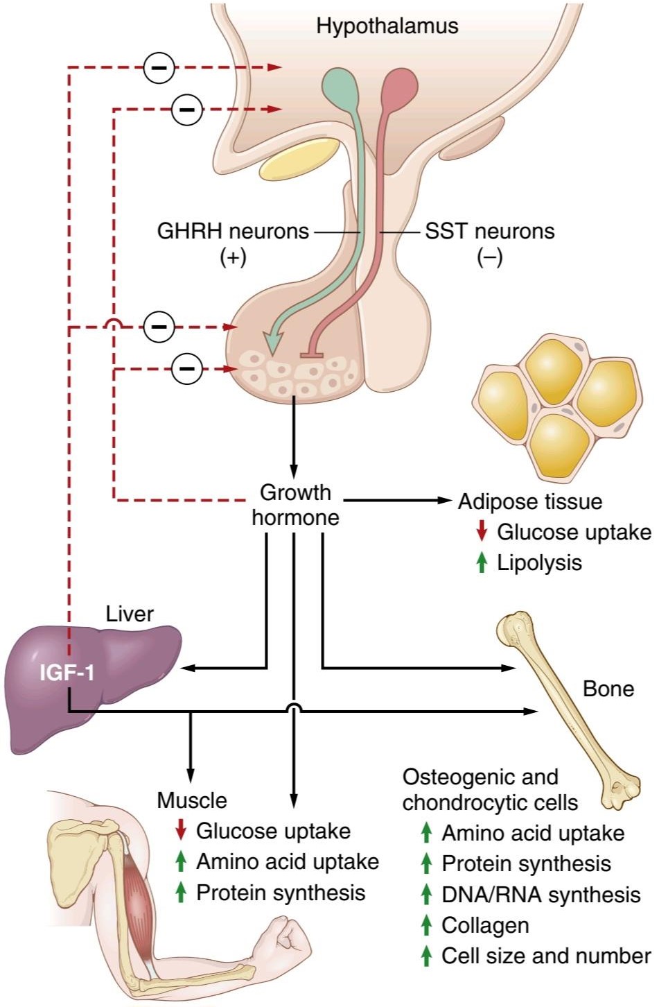
Figure 76-6. Effects of growth hormone and insulin-like growth factor-1 (IGF-1) on growth and metabolism. Growth hormone secretion is stimulated by growth hormone-releasing hormone (GHRH) and inhibited by somatostatin (SST), as well as negative feedback effects of growth hormone and IGF-1 on the anterior pituitary gland and the hypothalamic neurons. Other factors that control growth hormone secretion are described in Table 76-3.
شکل 76-6. اثرات هورمون رشد و فاکتور رشد شبه انسولین-1 (IGF-1) بر رشد و متابولیسم. ترشح هورمون رشد توسط هورمون آزاد کننده هورمون رشد (GHRH) تحریک می شود و توسط سوماتوستاتین (SST) مهار می شود، همچنین اثرات بازخورد منفی هورمون رشد و IGF-1 بر روی غده هیپوفیز قدامی و نورون های هیپوتالاموس انجام می شود. سایر عوامل کنترل کننده ترشح هورمون رشد در جدول 76-3 توضیح داده شده است.
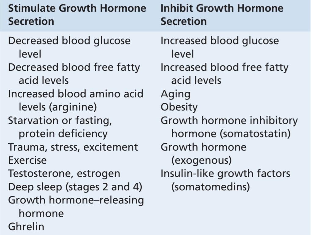
Table 76-3 Factors That Stimulate or Inhibit Secretion of Growth Hormone
جدول 76-3 عواملی که باعث تحریک یا مهار ترشح هورمون رشد می شوند.
Growth Hormone Promotes Protein Deposition in Tissues
Although the precise mechanisms by which growth hormone increases protein deposition are not fully understood, a series of different effects are known, all of which could lead to enhanced protein deposition.
هورمون رشد باعث افزایش رسوب پروتئین در بافت ها می شود
اگرچه مکانیسمهای دقیقی که توسط آن هورمون رشد رسوب پروتئین را افزایش میدهد به طور کامل شناخته نشده است، اما مجموعهای از اثرات مختلف شناخته شدهاند که همگی میتوانند منجر به افزایش رسوب پروتئین شوند.
Enhancement of Amino Acid Transport Through the Cell Membranes. GH directly enhances transport of most amino acids through cell membranes to the interior of the cells. This increases amino acid concentrations in the cells and is presumed to be at least partly responsible for the increased protein synthesis. This control of amino acid transport is similar to the effect of insulin in controlling glucose transport through the membrane, as dis- cussed in Chapters 68 and 79.
افزایش انتقال آمینو اسید از طریق غشای سلولی. GH مستقیماً انتقال بیشتر اسیدهای آمینه را از طریق غشای سلولی به داخل سلول ها افزایش می دهد. این باعث افزایش غلظت اسید آمینه در سلول ها می شود و فرض می شود که حداقل تا حدی مسئول افزایش سنتز پروتئین است. این کنترل انتقال اسید آمینه مشابه اثر انسولین در کنترل انتقال گلوکز از طریق غشاء است، همانطور که در فصل های 68 و 79 بحث شد.
Enhancement of RNA Translation to Cause Protein Synthesis by the Ribosomes. Even when the amino acid concentrations are not increased in the cells, GH still increases RNA translation, causing protein to be synthesized in greater amounts by the ribosomes in the cytoplasm.
افزایش ترجمه RNA برای ایجاد سنتز پروتئین توسط ریبوزوم ها. حتی زمانی که غلظت اسید آمینه در سلول ها افزایش نمی یابد، GH همچنان ترجمه RNA را افزایش می دهد و باعث می شود پروتئین در مقادیر بیشتری توسط ریبوزوم های سیتوپلاسم سنتز شود.
Increased Nuclear Transcription of DNA to Form RNA.
Over more prolonged periods (24-48 hours), GH also stimulates transcription of DNA in the nucleus, causing formation of increased quantities of RNA. This promotes more protein synthesis and growth if sufficient energy, amino acids, vitamins, and other requisites for growth are available. In the long run, this may be the most important function of GH.
افزایش رونویسی هسته ای DNA به شکل RNA.
در دوره های طولانی تر (24-48 ساعت)، GH همچنین رونویسی DNA در هسته را تحریک می کند و باعث تشکیل مقادیر بیشتری از RNA می شود. در صورتی که انرژی کافی، آمینو اسیدها، ویتامین ها و سایر نیازهای لازم برای رشد در دسترس باشد، این امر باعث افزایش سنتز و رشد پروتئین می شود. در دراز مدت، این ممکن است مهمترین عملکرد GH باشد.
Decreased Catabolism of Protein and Amino Acids. In addition to the increase in protein synthesis, GH decreases breakdown of cell protein. A probable reason for this decrease is that GH also mobilizes large quantities of free fatty acids from the adipose tissue, and these are used to supply most of the energy for the body’s cells, thus acting as a potent “protein sparer.”
کاهش کاتابولیسم پروتئین و اسیدهای آمینه. علاوه بر افزایش سنتز پروتئین، GH تجزیه پروتئین سلولی را کاهش می دهد. دلیل احتمالی این کاهش این است که GH همچنین مقادیر زیادی اسیدهای چرب آزاد را از بافت چربی به حرکت در می آورد و این اسیدهای چرب برای تامین بیشتر انرژی برای سلول های بدن استفاده می شوند، بنابراین به عنوان یک “پرهیز کننده پروتئین” قوی عمل می کنند.
Summary. GH enhances almost all facets of amino acid uptake and protein synthesis by cells, while at the same time reducing the breakdown of proteins.
خلاصه. GH تقریباً تمام جنبه های جذب آمینو اسید و سنتز پروتئین توسط سلول ها را افزایش می دهد و در عین حال تجزیه پروتئین ها را کاهش می دهد.
Growth Hormone Enhances Fat Utilization for Energy
GH has a specific effect to cause release of fatty acids from adipose tissue and, therefore, increases the concentration of fatty acids in body fluids. Also, in tissues throughout the body, GH enhances conversion of fatty acids to acetyl coenzyme A (acetyl-CoA) and its subsequent utilization for energy. Therefore, under the influence of GH, fat is used for energy in preference to use of carbohydrates and proteins.
هورمون رشد مصرف چربی برای انرژی را افزایش می دهد
GH اثر خاصی برای آزادسازی اسیدهای چرب از بافت چربی دارد و در نتیجه غلظت اسیدهای چرب را در مایعات بدن افزایش می دهد. همچنین، در بافتهای سراسر بدن، GH تبدیل اسیدهای چرب به استیل کوآنزیم A (استیل-CoA) و استفاده بعدی از آن برای انرژی را افزایش میدهد. بنابراین، تحت تأثیر GH، چربی برای انرژی به جای استفاده از کربوهیدرات ها و پروتئین ها استفاده می شود.
The ability of GH to promote fat utilization, together with its protein anabolic effect, causes an increase in lean body mass. However, mobilization of fat by GH requires several hours to occur, whereas enhancement of protein synthesis can begin in minutes under the influence of GH.
توانایی GH در ترویج استفاده از چربی، همراه با اثر آنابولیک پروتئین، باعث افزایش توده بدون چربی بدن می شود. با این حال، بسیج چربی توسط GH به چندین ساعت نیاز دارد تا اتفاق بیفتد، در حالی که افزایش سنتز پروتئین می تواند در عرض چند دقیقه تحت تأثیر GH آغاز شود.
“Ketogenic” Effect of Excessive Growth Hormone. Under the influence of excessive amounts of GH, fat mobilization from adipose tissue sometimes becomes so great that large quantities of acetoacetic acid are formed. by the liver and released into the body fluids, thus causing ketosis. This excessive mobilization of fat from the adipose tissue also frequently causes a fatty liver.
اثر “کتوژنیک” هورمون رشد بیش از حد. تحت تأثیر مقادیر بیش از حد GH، حرکت چربی از بافت چربی گاهی به حدی می شود که مقادیر زیادی اسید استواستیک تشکیل می شود. توسط کبد وارد مایعات بدن می شود و در نتیجه باعث کتوز می شود. این تحرک بیش از حد چربی از بافت چربی نیز اغلب باعث ایجاد کبد چرب می شود.
Growth Hormone Decreases Carbohydrate Utilization
GH has multiple effects that influence carbohydrate metabolism, including (1) decreased glucose uptake in tissues such as skeletal muscle and fat, (2) increased glucose production by the liver, and (3) increased insulin secretion.
هورمون رشد مصرف کربوهیدرات را کاهش می دهد
GH دارای اثرات متعددی است که بر متابولیسم کربوهیدرات ها تأثیر می گذارد، از جمله (1) کاهش جذب گلوکز در بافت هایی مانند ماهیچه های اسکلتی و چربی، (2) افزایش تولید گلوکز توسط کبد، و (3) افزایش ترشح انسولین.
Each of these changes results from GH-induced “insulin resistance,” which attenuates insulin’s actions to stimulate uptake and utilization of glucose in skeletal muscle and adipose tissue and to inhibit gluconeogenesis (glucose production) by the liver; this leads to increased blood glucose concentration and a compensatory increase in insulin secretion. For these reasons, GH’s effects are called diabetogenic, and excess secretion of GH can produce metabolic disturbances similar to those found in patients with type 2 (non-insulin-dependent) diabetes, who are also resistant to the metabolic effects of insulin. However, patients with acromegaly who have excess GH secretion are usually lean with little visceral fat whereas patients with type 2 diabetes are frequently overweight with excessive visceral fat which drives insulin resistance.
هر یک از این تغییرات ناشی از «مقاومت به انسولین» ناشی از GH است که فعالیت انسولین را برای تحریک جذب و استفاده از گلوکز در ماهیچههای اسکلتی و بافت چربی و مهار گلوکونئوژنز (تولید گلوکز) توسط کبد کاهش میدهد. این منجر به افزایش غلظت گلوکز خون و افزایش جبرانی در ترشح انسولین می شود. به این دلایل، اثرات GH دیابت زا نامیده می شود و ترشح بیش از حد GH می تواند باعث ایجاد اختلالات متابولیکی مشابه آنچه در بیماران مبتلا به دیابت نوع 2 (غیر وابسته به انسولین) مشاهده می شود، که به اثرات متابولیکی انسولین نیز مقاوم هستند، ایجاد کند. با این حال، بیماران مبتلا به آکرومگالی که ترشح GH بیش از حد دارند، معمولاً لاغر با چربی احشایی کمی هستند در حالی که بیماران مبتلا به دیابت نوع 2 اغلب اضافه وزن دارند و چربی احشایی بیش از حد دارند که باعث مقاومت به انسولین می شود.
We do not know the precise mechanism by which GH causes insulin resistance and decreased glucose utilization by the cells. However, GH-induced increases in lipolysis and blood concentrations of fatty acids likely contribute to impairment of insulin’s actions on tissue glucose utilization. Experimental studies indicate that raising blood levels of fatty acids above normal rapidly decreases the sensitivity of the liver and skeletal muscle to insulin’s effects on carbohydrate metabolism.
ما مکانیسم دقیقی که GH باعث مقاومت به انسولین و کاهش استفاده از گلوکز توسط سلول ها می شود را نمی دانیم. با این حال، افزایش های ناشی از GH در لیپولیز و غلظت اسیدهای چرب خون به احتمال زیاد باعث اختلال در عملکرد انسولین در استفاده از گلوکز بافت می شود. مطالعات تجربی نشان می دهد که بالا بردن سطح اسیدهای چرب خون بالاتر از حد طبیعی به سرعت حساسیت کبد و ماهیچه های اسکلتی را نسبت به اثرات انسولین بر متابولیسم کربوهیدرات کاهش می دهد.
Necessity of Insulin and Carbohydrate for the Growth-Promoting Action of Growth Hormone
GH fails to cause growth in animals that lack a pancreas; it also fails to cause growth if carbohydrates are excluded from the diet. Thus, adequate insulin activity and adequate availability of carbohydrates are necessary for GH to be effective. Part of this requirement for carbohydrates and insulin is to provide the energy needed for the metabolism of growth, but there seem to be other effects as well. Especially important is the ability of insulin to enhance transport of some amino acids into cells, in the same way that it stimulates glucose transport.
ضرورت انسولین و کربوهیدرات برای عملکرد رشد هورمون رشد
GH در حیواناتی که فاقد پانکراس هستند باعث رشد نمی شود. همچنین اگر کربوهیدرات ها از رژیم غذایی حذف شوند باعث رشد نمی شود. بنابراین، فعالیت کافی انسولین و در دسترس بودن کافی کربوهیدرات ها برای موثر بودن GH ضروری است. بخشی از این نیاز برای کربوهیدرات ها و انسولین تامین انرژی مورد نیاز برای متابولیسم رشد است، اما به نظر می رسد اثرات دیگری نیز وجود دارد. توانایی انسولین در افزایش انتقال برخی از اسیدهای آمینه به داخل سلول ها، به همان شیوه ای که انتقال گلوکز را تحریک می کند، بسیار مهم است.
GROWTH HORMONE STIMULATES CARTILAGE AND BONE GROWTH
Although GH stimulates increased deposition of protein and increased growth in almost all tissues of the body, its most obvious effect is to increase growth of the skeletal frame. This results from multiple effects of growth hormone on bone, including (1) increased deposition of protein by the chondrocytic and osteogenic cells that cause bone growth, (2) increased rate of reproduction of these cells, and (3) a specific effect of converting chondrocytes into osteogenic cells, thus causing deposition of new bone.
هورمون رشد غضروف و رشد استخوان را تحریک می کند
اگرچه GH باعث افزایش رسوب پروتئین و افزایش رشد تقریباً در تمام بافت های بدن می شود، بارزترین اثر آن افزایش رشد قاب اسکلتی است. این ناشی از اثرات متعدد هورمون رشد بر روی استخوان است، از جمله (1) افزایش رسوب پروتئین توسط سلولهای غضروفی و استخوانساز که باعث رشد استخوان میشوند، (2) افزایش سرعت تولید مثل این سلولها، و (3) اثر خاص تبدیل سلولهای غضروفی به سلولهای استخوانی، در نتیجه باعث رسوب استخوان جدید میشود.
There are two principal mechanisms of bone growth. First, in response to GH stimulation, the long bones grow in length at the epiphyseal cartilages, where the epiphyses at the ends of the bone are separated from the shaft. This growth first causes deposition of new cartilage, followed by its conversion into new bone, thus elongating the shaft and pushing the epiphyses farther and farther apart. At the same time, the epiphyseal cartilage is progressively used up so, by late adolescence, no additional epiphyseal cartilage remains to provide for further long bone growth. At this time, bony fusion occurs between the shaft and the epiphysis at each end, so no further lengthening of the long bone can occur.
دو مکانیسم اصلی رشد استخوان وجود دارد. اول، در پاسخ به تحریک GH، استخوانهای بلند در غضروفهای اپیفیز رشد میکنند، جایی که اپیفیزها در انتهای استخوان از شفت جدا میشوند. این رشد ابتدا باعث رسوب غضروف جدید و به دنبال آن تبدیل آن به استخوان جدید می شود و در نتیجه شفت کشیده می شود و اپی فیزها دورتر و دورتر می شوند. در همان زمان، غضروف اپی فیزیال به تدریج مصرف می شود، بنابراین، در اواخر نوجوانی، غضروف اپی فیزیال اضافی برای رشد بیشتر استخوان های طولانی باقی نمی ماند. در این زمان، همجوشی استخوانی بین شفت و اپی فیز در هر انتها اتفاق میافتد، بنابراین دیگر نمیتوان طول استخوان بلند را ایجاد کرد.
Second, osteoblasts in the bone periosteum and in some bone cavities deposit new bone on the surfaces of older bone. Simultaneously, osteoclasts in the bone (discussed in detail in Chapter 80) remove old bone. When the rate of deposition is greater than that of resorption, the thickness of the bone increases. Growth hormone strongly stimulates osteoblasts. Therefore, the bones can continue to become thicker throughout life under the influence of GH; this is especially true for the membranous bones. For example, the jaw bones can be stimulated to grow even after adolescence, causing forward protrusion of the chin and lower teeth. Likewise, the bones of the skull can grow in thickness and give rise to bony protrusions over the eyes.
دوم، استئوبلاستها در پریوستوم استخوان و در برخی حفرههای استخوانی، استخوان جدید را روی سطوح استخوان قدیمیتر رسوب میکنند. به طور همزمان، استئوکلاست ها در استخوان (که در فصل 80 به تفصیل مورد بحث قرار گرفته است) استخوان قدیمی را جدا می کنند. وقتی سرعت رسوب بیشتر از تحلیل باشد، ضخامت استخوان افزایش می یابد. هورمون رشد استئوبلاست ها را به شدت تحریک می کند. بنابراین، استخوان ها می توانند در طول زندگی تحت تأثیر GH به ضخیم تر شدن ادامه دهند. این به ویژه برای استخوان های غشایی صادق است. به عنوان مثال، استخوان های فک را می توان حتی پس از نوجوانی برای رشد تحریک کرد و باعث بیرون زدگی چانه و دندان های پایین به جلو می شود. به همین ترتیب، استخوان های جمجمه می توانند در ضخامت رشد کرده و برجستگی های استخوانی روی چشم ها ایجاد کنند.
GROWTH HORMONE EXERTS MUCH OF ITS EFFECT THROUGH INSULIN-LIKE GROWTH FACTORS (SOMATOMEDINS)
GH causes the liver (and, to a much less extent, other tissues) to form several small proteins called insulin- like growth factors (IGFs, also called somatomedins) that mediate some of the growth and metabolic effects of GH (Figure 76-6).
هورمون رشد بیشتر اثر خود را از طریق فاکتورهای رشد انسولین مانند (SOMATOMEDINS) اعمال می کند.
GH باعث می شود که کبد (و تا حد بسیار کمتری سایر بافت ها) چندین پروتئین کوچک به نام فاکتورهای رشد شبه انسولین (IGFs که سوماتومدین ها نیز نامیده می شوند) تشکیل دهند که برخی از اثرات رشد و متابولیک GH را واسطه می کند (شکل 76-6).
At least four IGFs have been isolated, but by far the most important of these is IGF-1 (somatomedin C). The molecular weight of IGF-1 is about 7500, and its concentration in the plasma closely follows the rate of GH secretion.
حداقل چهار IGF جدا شده است، اما مهم ترین آنها IGF-1 (سوماتومدین C) است. وزن مولکولی IGF-1 حدود 7500 است و غلظت آن در پلاسما از سرعت ترشح GH پیروی می کند.
Children with deficiency of IGF fail to grow normally even though they may have normal or elevated secretion of GH. The pygmy peoples of Africa, for example, have very small stature due to a congenital inability to synthesize significant amounts of IGF-1. Although their plasma concentration of GH is either normal or high, they have diminished amounts of IGF-1 in the plasma, which apparently accounts for the small stature of these people. Some other dwarfs (e.g., Laron syndrome) have a similar problem, generally caused by a mutation of the GH receptor and therefore failure of GH to stimulate formation of IGF-1.
کودکان مبتلا به کمبود IGF با وجود اینکه ممکن است ترشح GH طبیعی یا افزایش یافته داشته باشند، رشد طبیعی ندارند. برای مثال، مردم کوتوله آفریقا به دلیل ناتوانی مادرزادی در سنتز مقادیر قابل توجهی IGF-1، قد بسیار کوچکی دارند. اگرچه غلظت پلاسمایی GH آنها نرمال یا زیاد است، اما مقادیر IGF-1 در پلاسما آنها کاهش یافته است که ظاهراً دلیل کوچکی قد این افراد است. برخی از کوتوله های دیگر (مانند سندرم لارون) مشکل مشابهی دارند که عموماً به دلیل جهش گیرنده GH و در نتیجه شکست GH در تحریک تشکیل IGF-1 ایجاد می شود.
It has been postulated that most of the growth effects of GH result from IGF-1 and other IGFs, rather than from direct effects of GH on the bones and other peripheral tissues. Even so, experiments have demonstrated that injection of GH directly into the epiphyseal cartilages of bones of living animals causes growth of these cartilage areas, and the amount of GH required for this growth to occur is minute. One possible explanation for this finding is that GH can cause formation of enough IGF-1 in the local tissue to cause local growth. However, GH also has IGF-independent effects that stimulate growth in some tissues such as chondrocytes of cartilage.
فرض شده است که بیشتر اثرات رشد GH ناشی از IGF-1 و سایر IGF ها است، نه از اثرات مستقیم GH بر استخوان ها و سایر بافت های محیطی. با این حال، آزمایشها نشان دادهاند که تزریق GH به طور مستقیم به غضروفهای اپیفیز استخوانهای حیوانات زنده باعث رشد این نواحی غضروفی میشود و مقدار GH مورد نیاز برای این رشد بسیار اندک است. یک توضیح احتمالی برای این یافته این است که GH می تواند باعث تشکیل IGF-1 کافی در بافت محلی شود تا باعث رشد موضعی شود. با این حال، GH همچنین دارای اثرات مستقل از IGF است که باعث تحریک رشد در برخی از بافتها مانند سلولهای غضروفی میشود.
Short Duration of Action of Growth Hormone but Prolonged Action of IGF-1. GH attaches only weakly to the plasma proteins in blood.
مدت کوتاه اثر هورمون رشد اما اثر طولانی مدت IGF-1 . جی اچ فقط ضعیف به پروتئین های پلاسما در خون می چسبد.
Therefore, it is released. from the blood into the tissues rapidly, having a half-time in blood of less than 20 minutes. By contrast, IGF-1 attaches strongly to a carrier protein in the blood that, like IGF-1, is produced in response to GH. As a result, IGF-1 is released only slowly from the blood to the tissues, with a half-time of about 20 hours. This slow release greatly prolongs the growth-promoting effects of the bursts of GH secretion shown in Figure 76-7.
بنابراین منتشر می شود. از خون بهسرعت وارد بافتها میشود و نیمتایم در خون کمتر از 20 دقیقه است. در مقابل، IGF-1 به شدت به یک پروتئین حامل در خون می چسبد که مانند IGF-1 در پاسخ به GH تولید می شود. در نتیجه، IGF-1 تنها به آرامی از خون به بافتها آزاد میشود، با نیمهتایم حدود 20 ساعت. این آزادسازی آهسته تا حد زیادی اثرات محرک رشد انفجارهای ترشح GH نشان داده شده در شکل 76-7 را طولانی می کند.

Figure 76-7. Typical variations in growth hormone secretion throughout the day, demonstrating the especially powerful effect of strenuous exercise and also the high rate of growth hormone secretion that occurs during the first few hours of deep sleep.
شکل 76-7. تغییرات معمولی در ترشح هورمون رشد در طول روز، نشان دهنده تأثیر ویژه قوی ورزش شدید و همچنین میزان بالای ترشح هورمون رشد است که در چند ساعت اول خواب عمیق رخ می دهد.
REGULATION OF GROWTH HORMONE SECRETION
After adolescence, GH secretion decreases slowly with aging, finally falling to about 25% of the adolescent level in very old age.
تنظیم ترشح هورمون رشد
پس از نوجوانی، ترشح GH به آرامی با افزایش سن کاهش می یابد و در نهایت به حدود 25 درصد از سطح نوجوانان در سنین بسیار بالا می رسد.
GH is secreted in a pulsatile pattern, increasing and decreasing. The precise mechanisms that control secretion of GH are not fully understood, but several factors related to a person’s state of nutrition or stress are known to stimulate secretion: (1) starvation, especially with severe protein deficiency; (2) hypoglycemia or low concentration of fatty acids in the blood; (3) exercise; (4) excitement; (5) trauma; (6) ghrelin, a hormone secreted by the stomach before meals; and (7) some amino acids, including arginine. GH also characteristically increases during the first 2 hours of deep sleep, as shown in Figure 76-7. Table 76-3 summarizes some of the factors that are known to influence growth hormone secretion.
GH در یک الگوی ضربانی ترشح می شود، افزایش و کاهش می یابد. مکانیسم های دقیق کنترل ترشح GH به طور کامل شناخته نشده است، اما چندین عامل مرتبط با وضعیت تغذیه یا استرس فرد برای تحریک ترشح شناخته شده است: (1) گرسنگی، به ویژه با کمبود شدید پروتئین. (2) هیپوگلیسمی یا غلظت کم اسیدهای چرب در خون. (3) ورزش؛ (4) هیجان؛ (5) تروما؛ (6) گرلین، هورمونی که قبل از غذا از معده ترشح می شود. و (7) برخی از اسیدهای آمینه، از جمله آرژنین. همانطور که در شکل 76-7 نشان داده شده است GH همچنین در طول 2 ساعت اول خواب عمیق افزایش می یابد. جدول 76-3 برخی از عواملی را که بر ترشح هورمون رشد تأثیر می گذارند، خلاصه می کند.
The normal concentration of GH in the plasma of an adult is between 1.6 and 3 ng/ml; in a child or adolescent, it is about 6 ng/ml. These values may increase to as high as 50 ng/ml after depletion of the body stores of proteins or carbohydrates during prolonged starvation.
غلظت طبیعی GH در پلاسمای یک فرد بالغ بین 1.6 تا 3 نانوگرم در میلی لیتر است. در یک کودک یا نوجوان، حدود 6 نانوگرم در میلی لیتر است. این مقادیر ممکن است پس از خالی شدن ذخایر پروتئین یا کربوهیدرات بدن در طول گرسنگی طولانی مدت تا 50 نانوگرم در میلی لیتر افزایش یابد.
Under acute conditions, hypoglycemia is a far more potent stimulator of GH secretion than is an acute decrease in protein intake. Conversely, in chronic conditions, GH secretion seems to correlate more with the degree of cellular protein depletion than with the degree of glucose insufficiency. For example, the extremely high levels of GH that occur during starvation are closely related to the amount of protein depletion.
در شرایط حاد، هیپوگلیسمی محرک بسیار قویتری برای ترشح GH نسبت به کاهش شدید دریافت پروتئین است. برعکس، در شرایط مزمن، به نظر می رسد که ترشح GH بیشتر با میزان کاهش پروتئین سلولی ارتباط دارد تا با درجه نارسایی گلوکز. به عنوان مثال، سطوح بسیار بالای GH که در هنگام گرسنگی رخ می دهد، ارتباط نزدیکی با میزان کاهش پروتئین دارد.
Figure 76-8 demonstrates the effect of protein deficiency on plasma GH and then the effect of adding protein to the diet. The first column shows very high levels of GH in children with extreme protein deficiency during the protein malnutrition condition called kwashiorkor; the second column shows the levels in the same children after 3 days of treatment with more than adequate quantities of carbohydrates in their diets, demonstrating that the carbohydrates did not lower the plasma GH concentration.
شکل 76-8 اثر کمبود پروتئین بر GH پلاسما و سپس اثر افزودن پروتئین به جیره را نشان می دهد. ستون اول سطوح بسیار بالایی از GH را در کودکان مبتلا به کمبود شدید پروتئین در شرایط سوء تغذیه پروتئینی به نام کواشیورکور نشان می دهد. ستون دوم سطوح را در همان کودکان پس از 3 روز درمان با مقادیر بیش از مقدار کافی کربوهیدرات در رژیم غذایی آنها نشان می دهد، که نشان می دهد کربوهیدرات ها غلظت GH پلاسما را کاهش نداده اند.
The third and fourth columns show the levels after treatment with protein supplements for 3 and 25 days, respectively, with a concomitant decrease in the hormone.
ستون سوم و چهارم سطوح بعد از درمان با مکمل های پروتئینی را به ترتیب به مدت 3 و 25 روز با کاهش همزمان هورمون نشان می دهد.
These results demonstrate that under severe conditions of protein malnutrition, adequate calories alone are not sufficient to correct the excess production of GH. The protein deficiency must also be corrected before the GH concentration will return to normal.
این نتایج نشان می دهد که در شرایط شدید سوء تغذیه پروتئینی، کالری کافی به تنهایی برای اصلاح تولید اضافی GH کافی نیست. قبل از اینکه غلظت GH به حالت عادی برگردد، کمبود پروتئین نیز باید اصلاح شود.

Figure 76-8. Effect of extreme protein deficiency on the plasma concentration of growth hormone in the disease kwashiorkor. Also shown is the failure of carbohydrate treatment but the effectiveness of protein treatment in lowering growth hormone concentration. (Data from Pimstone BL, Barbezat G, Hansen JD, et al: Studies on growth hormone secretion in protein-calorie malnutrition. Am J Clin Nutr 21:482, 1968.)
شکل 76-8. تأثیر کمبود شدید پروتئین بر غلظت پلاسمایی هورمون رشد در بیماری کواشیورکور. همچنین شکست درمان کربوهیدرات اما اثربخشی درمان پروتئین در کاهش غلظت هورمون رشد نشان داده شده است. (دادههای Pimstone BL، Barbezat G، Hansen JD، و همکاران: مطالعات در مورد ترشح هورمون رشد در سوء تغذیه پروتئین-کالری. Am J Clin Nutr 21:482، 1968.)
Hypothalamic Growth Hormone- Releasing Hormone Stimulates, and Somatostatin Inhibits Growth Hormone Secretion
From the preceding description of the many factors that can affect GH secretion, one can readily understand the perplexity of physiologists as they attempted to unravel the mysteries of regulation of GH secretion. It is known that GH secretion is controlled by two factors secreted in the hypothalamus and then transported to the anterior pituitary gland through the hypothalamic-hypophysial portal vessels. They are growth hormone-releasing hormone (GHRH) and growth hormone inhibitory hormone (also called somatostatin). Both of these are polypeptides; GHRH is composed of 44 amino acids, and somatostatin is composed of 14 amino acids.
هورمون رشد هیپوتالاموس، هورمون آزاد کننده را تحریک می کند و سوماتوستاتین ترشح هورمون رشد را مهار می کند.
از توضیحات قبلی در مورد بسیاری از عواملی که میتوانند بر ترشح GH تأثیر بگذارند، میتوان به راحتی متوجه گیجی فیزیولوژیستها در تلاش برای کشف رمز و راز تنظیم ترشح GH شد. مشخص شده است که ترشح GH توسط دو عامل ترشح شده در هیپوتالاموس کنترل می شود و سپس از طریق عروق پورتال هیپوتالاموس-هیپوفیزیال به غده هیپوفیز قدامی منتقل می شود. آنها هورمون آزاد کننده هورمون رشد (GHRH) و هورمون بازدارنده هورمون رشد (همچنین به نام سوماتوستاتین) هستند. هر دوی اینها پلی پپتید هستند. GHRH از 44 اسید آمینه و سوماتوستاتین از 14 اسید آمینه تشکیل شده است.
Neurons in the arcuate and ventromedial nuclei of the hypothalamus secrete GHRH; this is the same area of the hypothalamus that is sensitive to blood glucose concentration, causing satiety in hyperglycemic states and hunger in hypoglycemic states. Secretion of somatostatin is controlled by the nearby periventricular neurons of the hypothalamus. Therefore, it is reasonable to expect that some of the same signals that modify a person’s behavioral feeding instincts also alter the rate of GH secretion.
نورون ها در هسته های کمانی و شکمی هیپوتالاموس GHRH ترشح می کنند. این همان ناحیه ای از هیپوتالاموس است که به غلظت گلوکز خون حساس است و در حالت هایپرگلیسمی باعث سیری و در حالت های هیپوگلیسمی گرسنگی می شود. ترشح سوماتوستاتین توسط نورون های اطراف بطنی هیپوتالاموس کنترل می شود. بنابراین، منطقی است که انتظار داشته باشیم برخی از سیگنالهای مشابهی که غرایز تغذیه رفتاری فرد را تغییر میدهند، سرعت ترشح GH را نیز تغییر دهند.
In a similar manner, hypothalamic signals depicting emotions, stress, and trauma can all affect hypothalamic control of GH secretion. In fact, experiments have shown that catecholamines, dopamine, and serotonin, each of which is released by a different neuronal system in the hypothalamus, all increase the rate of GH secretion.
به روشی مشابه، سیگنالهای هیپوتالاموس که احساسات، استرس و ضربه را نشان میدهند، همگی میتوانند بر کنترل هیپوتالاموس ترشح GH تأثیر بگذارند. در واقع، آزمایشات نشان داده است که کاتکول آمین ها، دوپامین و سروتونین که هر کدام توسط یک سیستم عصبی متفاوت در هیپوتالاموس آزاد می شوند، همگی سرعت ترشح GH را افزایش می دهند.
Most of the control of GH secretion is probably mediated through GHRH rather than through the inhibitory hormone somatostatin. GHRH stimulates GH secretion by attaching to specific cell membrane receptors on the outer surfaces of the GH cells in the pituitary gland. The receptors activate the adenylyl cyclase system inside the cell membrane, increasing the intracellular level of cyclic adenosine monophosphate (cAMP). This increase has both short-term and long-term effects. The short-term effect is to increase calcium ion transport into the cell; within minutes, this increase causes fusion of the GH secretory vesicles with the cell membrane and release of the hormone into the blood. The long-term effect is to increase transcription in the nucleus by the genes to stimulate synthesis of new GH.
بیشتر کنترل ترشح GH احتمالاً از طریق GHRH انجام می شود نه از طریق هورمون بازدارنده سوماتوستاتین. GHRH ترشح GH را با اتصال به گیرنده های غشای سلولی خاص در سطوح خارجی سلول های GH در غده هیپوفیز تحریک می کند. گیرنده ها سیستم آدنیلیل سیکلاز را در داخل غشای سلولی فعال می کنند و سطح داخل سلولی آدنوزین مونوفسفات حلقوی (cAMP) را افزایش می دهند. این افزایش اثرات کوتاه مدت و بلند مدت دارد. اثر کوتاه مدت افزایش انتقال یون کلسیم به داخل سلول است. این افزایش در عرض چند دقیقه باعث ادغام وزیکول های ترشحی GH با غشای سلولی و آزاد شدن هورمون در خون می شود. اثر بلندمدت افزایش رونویسی در هسته توسط ژن ها برای تحریک سنتز GH جدید است.
When GH is administered directly into the blood of an animal over several hours, the rate of endogenous GH secretion decreases. This decrease demonstrates that GH secretion is subject to typical negative feedback control, as is true for essentially all hormones. The nature of this feedback mechanism and whether it is mediated mainly through inhibition of GHRH or enhancement of somatostatin, which inhibits growth hormone secretion, are uncertain.
وقتی GH مستقیماً در طی چند ساعت در خون حیوان تجویز می شود، سرعت ترشح GH درون زا کاهش می یابد. این کاهش نشان می دهد که ترشح GH تحت کنترل بازخورد منفی معمولی است، همانطور که اساساً برای همه هورمون ها صادق است. ماهیت این مکانیسم بازخورد و اینکه آیا این مکانیسم عمدتاً از طریق مهار GHRH یا افزایش سوماتوستاتین که ترشح هورمون رشد را مهار می کند انجام می شود، نامشخص است.
In summary, our knowledge of the regulation of GH secretion is not sufficient to describe a composite picture. Yet, because of the extreme secretion of GH during starvation and its important long-term effect to promote protein synthesis and tissue growth, we can propose the following: the major long-term controller of GH secretion is the longterm state of nutrition of the tissues, especially their level of protein nutrition. That is, nutritional deficiency or excess tissue need for cellular proteins-for example, after a severe bout of exercise when the muscle’s nutritional status has been taxed-in some way increases the rate of GH secretion. GH, in turn, promotes synthesis of new proteins while at the same time conserving the proteins already present in the cells.
به طور خلاصه، دانش ما در مورد تنظیم ترشح GH برای توصیف یک تصویر ترکیبی کافی نیست. با این حال، به دلیل ترشح شدید GH در هنگام گرسنگی و تأثیر طولانی مدت مهم آن برای تقویت سنتز پروتئین و رشد بافت، میتوان موارد زیر را پیشنهاد کرد: کنترلکنندهی طولانیمدت ترشح GH، وضعیت تغذیه طولانیمدت بافتها، بهویژه سطح تغذیه پروتئین آنها است. یعنی کمبود مواد مغذی یا نیاز بافت اضافی به پروتئینهای سلولی – به عنوان مثال، پس از یک دوره تمرینی شدید که وضعیت تغذیهای ماهیچه کاهش یافته است – به نوعی باعث افزایش سرعت ترشح GH میشود. GH، به نوبه خود، سنتز پروتئین های جدید را ترویج می کند و در عین حال پروتئین های موجود در سلول ها را حفظ می کند.
Abnormalities of Growth Hormone Secretion
Panhypopituitarism-Decreased Secretion of All Anterior Pituitary Hormones. Decreased secretion of all anterior pituitary hormones in patients with panhypopituitarism may be congenital (present from birth), or it may occur suddenly or slowly at any time during life, most often resulting from a pituitary tumor that destroys the pituitary gland.
اختلالات ترشح هورمون رشد
پان هیپوفیتاریسم-کاهش ترشح تمام هورمون های هیپوفیز قدامی. کاهش ترشح تمام هورمون های هیپوفیز قدامی در بیماران مبتلا به پان هیپوفیز ممکن است مادرزادی باشد (از بدو تولد وجود داشته باشد)، یا ممکن است به طور ناگهانی یا آهسته در هر زمانی از زندگی رخ دهد، که اغلب ناشی از تومور هیپوفیز است که غده هیپوفیز را تخریب می کند.
Panhypopituitarism in the Adult. Panhypopituitarism first occurring in adulthood generally results from one of three abnormalities. Two tumorous conditions, craniopharyngiomas or chromophobe tumors, may compress the pituitary gland until the functioning anterior pituitary cells are totally or almost totally destroyed. The third cause is thrombosis of the pituitary blood vessels. This abnormality occasionally occurs when a new mother experiences circulatory shock after the birth of her baby.
پان هیپوفیتاریسم در بزرگسالان. پان هیپوفیتاریسم که برای اولین بار در بزرگسالی رخ می دهد، به طور کلی ناشی از یکی از سه ناهنجاری است. دو وضعیت تومور، کرانیوفارنژیوم یا تومورهای کروموفوب، ممکن است غده هیپوفیز را تا زمانی که سلول های هیپوفیز قدامی عملکرد به طور کامل یا تقریباً کامل از بین بروند، فشرده کنند. علت سوم ترومبوز عروق خونی هیپوفیز است. این ناهنجاری گاهی اوقات زمانی رخ می دهد که یک مادر جدید پس از تولد نوزادش شوک گردش خون را تجربه کند.
The general effects of adult panhypopituitarism are (1) hypothyroidism, (2) depressed production of glucocorticoids by the adrenal glands, and (3) suppressed secretion of the gonadotropic hormones so that sexual functions are lost. Thus, the picture is that of a lethargic person (from lack of thyroid hormones) who is gaining weight (because of lack of fat mobilization by growth, adrenocorticotropic, adrenocortical, and thyroid hormones) and has lost all sexual functions. Except for the abnormal sexual functions, the patient can usually be treated satisfactorily by administering adrenocortical and thyroid hormones.
اثرات عمومی پانهیپوفیتاریسم بزرگسالان عبارتند از: (1) کم کاری تیروئید، (2) کاهش تولید گلوکوکورتیکوئیدها توسط غدد فوق کلیوی، و (3) ترشح سرکوب شده هورمون های گنادوتروپیک به طوری که عملکردهای جنسی از بین می رود. بنابراین، تصویر مربوط به یک فرد بی حال (به دلیل کمبود هورمون های تیروئید) است که در حال افزایش وزن (به دلیل عدم تحرک چربی از طریق رشد، هورمون های آدرنوکورتیکوتروپیک، قشر آدرنال و تیروئید) است و تمام اعمال جنسی را از دست داده است. به جز عملکردهای جنسی غیرطبیعی، معمولاً می توان بیمار را با تجویز هورمون های غده آدرنوکورتیکال و تیروئید به طور رضایت بخشی درمان کرد.
Panhypopituitarism During Childhood and Dwarfism. Most cases of dwarfism result from generalized deficiency of anterior pituitary secretion (panhypopituitarism) during childhood. In general, all the physical parts of the body develop in appropriate proportion to one another, but the rate of development is greatly decreased. A child who has reached the age of 10 years may have the bodily development of a child aged 4 to 5 years, and the same person at age 20 years may have the bodily development of a child aged 7 to 10 years.
پانهیپوفیتاریسم در دوران کودکی و کوتولگی. اکثر موارد کوتولگی ناشی از کمبود عمومی ترشح قدامی هیپوفیز (پان هیپوپیتویتاریسم) در دوران کودکی است. به طور کلی، تمام اعضای بدن به نسبت مناسب رشد می کنند، اما سرعت رشد بسیار کاهش می یابد. کودکی که به سن 10 سالگی رسیده است ممکن است رشد بدنی یک کودک 4 تا 5 ساله داشته باشد و همان فرد در سن 20 سالگی ممکن است رشد بدنی یک کودک 7 تا 10 ساله داشته باشد.
A person with panhypopituitary dwarfism does not pass through puberty and never secretes sufficient quantities of gonadotropic hormones to develop adult sexual functions. In one-third of such dwarfs, however, only GH is deficient; these persons do mature sexually and occasionally reproduce. In Laron dwarfs and African pygmies, the rate of GH secretion is normal or high, but responsiveness to GH is impaired due to mutations of the GH receptor or a hereditary inability to form IGF-1, a key step for the promotion of growth by GH.
فرد مبتلا به کوتولگی پانهیپوفیتیتر از دوران بلوغ عبور نمی کند و هرگز مقادیر کافی هورمون گنادوتروپیک برای ایجاد عملکردهای جنسی بزرگسالان ترشح نمی کند. با این حال، در یک سوم از این کوتوله ها، تنها GH کمبود دارد. این افراد از نظر جنسی بالغ می شوند و گهگاه تولید مثل می کنند. در کوتوله های لارون و کوتوله های آفریقایی، میزان ترشح GH طبیعی یا زیاد است، اما پاسخگویی به GH به دلیل جهش گیرنده GH یا ناتوانی ارثی در تشکیل IGF-1، گامی کلیدی برای ارتقای رشد توسط GH، مختل می شود.
Treatment With Human Growth Hormone. Growth hormones from different species of animals are sufficiently different from one another that they will cause growth only in the one species or, at most, closely related species. For this reason, GH prepared from other animals (except, to some extent, from primates) is not effective in human beings. Therefore, the GH of the human being is called human growth hormone (hGH) to distinguish it from the others.
درمان با هورمون رشد انسانی. هورمون های رشد گونه های مختلف جانوران به اندازه کافی با یکدیگر متفاوت هستند که باعث رشد تنها در یک گونه یا حداکثر گونه های نزدیک به هم می شوند. به همین دلیل GH تهیه شده از حیوانات دیگر (به جز تا حدودی از پستانداران) در انسان مؤثر نیست. بنابراین GH انسان را هورمون رشد انسانی (hGH) می نامند تا آن را از سایرین متمایز کند.
In the past, because GH had to be prepared from human pituitary glands, it was difficult to obtain sufficient quantities to treat patients with GH deficiency, except on an experimental basis. However, hGH can now be synthesized by Escherichia coli bacteria as a result of successful application of recombinant DNA technology. Therefore, this hormone is now available in sufficient quantities for treatment purposes. Dwarfs who have pure GH deficiency can be completely cured if treated early in life.
در گذشته، چون GH باید از غدد هیپوفیز انسان تهیه میشد، به سختی میتوانست مقادیر کافی برای درمان بیماران مبتلا به کمبود GH را بهجز بهصورت تجربی بهدست آورد. با این حال، hGH اکنون می تواند توسط باکتری اشرشیاکلی در نتیجه کاربرد موفقیت آمیز فناوری DNA نوترکیب سنتز شود. بنابراین، این هورمون در حال حاضر به مقدار کافی برای اهداف درمانی در دسترس است. کوتوله هایی که کمبود GH خالص دارند اگر در اوایل زندگی درمان شوند می توانند به طور کامل درمان شوند.
Gigantism and Excess Growth Hormone Before Adolescence.
Occasionally, the acidophilic, GH-producing cells of the anterior pituitary gland become excessively active, and sometimes even acidophilic tumors occur in the gland. As a result, large quantities of GH are produced. All body tissues grow rapidly, including the bones. If the condition occurs. before adolescence, before the epiphyses of the long bones have become fused with the shafts, height increases so that the person becomes a giant-up to 8 feet tall.
غولپیکری و هورمون رشد بیش از حد قبل از نوجوانی.
گاهی اوقات، سلول های اسیدوفیل و تولید کننده GH غده هیپوفیز قدامی بیش از حد فعال می شوند و گاهی اوقات حتی تومورهای اسیدوفیل در غده ایجاد می شوند. در نتیجه، مقادیر زیادی GH تولید می شود. تمام بافت های بدن از جمله استخوان ها به سرعت رشد می کنند. اگر شرایط پیش بیاید. قبل از نوجوانی، قبل از اینکه اپیفیز استخوانهای دراز با شفتها ترکیب شوند، قد افزایش مییابد به طوری که فرد به یک غول بزرگ تا 8 فوت قد میرسد.
The giant ordinarily has hyperglycemia, and the beta cells of the islets of Langerhans in the pancreas are prone to degenerate because they become overactive owing to the hyperglycemia. Consequently, in about 10% of giants, fullblown diabetes mellitus eventually develops.
این غول معمولاً هیپرگلیسمی دارد و سلولهای بتای جزایر لانگرهانس در لوزالمعده مستعد انحطاط هستند زیرا به دلیل افزایش قند خون بیش از حد فعال میشوند. در نتیجه، در حدود 10٪ از غول ها، دیابت شیرین کامل در نهایت ایجاد می شود.
In most giants, panhypopituitarism eventually develops if they remain untreated because the gigantism is usually caused by a tumor of the pituitary gland that grows until the gland is destroyed. This eventual general deficiency of pituitary hormones usually causes death in early adulthood. However, once gigantism is diagnosed, further effects can often be blocked by microsurgical removal of the tumor or by irradiation of the pituitary gland.
در بیشتر غولها، پانهیپوفیتاریسم در صورت درمان نشدن در نهایت ایجاد میشود، زیرا غولپیکری معمولاً توسط تومور غده هیپوفیز ایجاد میشود که تا زمانی که غده از بین برود رشد میکند. این کمبود عمومی نهایی هورمون های هیپوفیز معمولاً باعث مرگ در اوایل بزرگسالی می شود. با این حال، هنگامی که غولپیکری تشخیص داده میشود، اغلب میتوان با برداشتن تومور با میکروسرجری یا تابش غده هیپوفیز جلوی اثرات بیشتر را گرفت.
Acromegaly and Excess Growth Hormone After Adolescence. If an acidophilic tumor occurs after adolescence- that is, after the epiphyses of the long bones have fused with the shafts-the person cannot grow taller, but the bones can become thicker and the soft tissues can continue to grow. This condition, shown in Figure 76-9, is known as acromegaly. Enlargement is especially marked in the bones of the hands and feet and in the membranous bones, including the cranium, nose, bosses on the forehead, supraorbital ridges, lower jawbone, and portions of the vertebrae, because their growth does not cease at adolescence. Consequently, the lower jaw protrudes forward, sometimes as much as half an inch, the forehead slants forward because of excess development of the supraorbital ridges, the nose increases to as much as twice normal size, the feet require size 14 or larger shoes, and the fingers become extremely thickened so that the hands are almost twice normal size. In addition to these effects, changes in the vertebrae ordinarily cause a hunched back, which is known clinically as kyphosis. Finally, many soft tissue organs, such as the tongue, the liver, and especially the kidneys, become greatly enlarged.
آکرومگالی و هورمون رشد بیش از حد بعد از نوجوانی. اگر یک تومور اسیدوفیل بعد از نوجوانی رخ دهد – یعنی بعد از اینکه اپی فیز استخوان های بلند با شفت ها ترکیب شد – فرد نمی تواند بلندتر شود، اما استخوان ها می توانند ضخیم تر شوند و بافت های نرم می توانند به رشد خود ادامه دهند. این وضعیت که در شکل 76-9 نشان داده شده است، به عنوان آکرومگالی شناخته می شود. بزرگ شدن به ویژه در استخوانهای دست و پا و استخوانهای غشایی، از جمله جمجمه، بینی، برجستگیهای روی پیشانی، برجستگیهای فوقاوربیتال، استخوان فک پایین و بخشهایی از مهرهها مشخص میشود، زیرا رشد آنها در نوجوانی متوقف نمیشود. در نتیجه، فک پایین به سمت جلو، گاهی به اندازه نیم اینچ بیرون زده، پیشانی به دلیل رشد بیش از حد برآمدگی های فوق اربیتال به سمت جلو متمایل می شود، بینی تا دو برابر اندازه طبیعی افزایش می یابد، پاها به کفش هایی با سایز 14 یا بزرگتر نیاز دارند و انگشتان به شدت ضخیم می شوند به طوری که دست ها تقریباً دو برابر اندازه طبیعی می شوند. علاوه بر این اثرات، تغییرات در مهره ها معمولاً باعث قوز پشت می شود که از نظر بالینی به عنوان کیفوز شناخته می شود. در نهایت بسیاری از اندام های بافت نرم مانند زبان، کبد و به خصوص کلیه ها به شدت بزرگ می شوند.

Figure 76-9. Patient with acromegaly. (Courtesy of Dr. Vishnu Garla.)
شکل 76-9 بیمار مبتلا به آکرومالی. (برگرفته از Dr. Vishnu Garla.)
Possible Role of Decreased Growth Hormone Secretion in Causing Changes Associated With Aging
In people who have lost the ability to secrete GH, some features of the aging process accelerate. For example, a 50-year-old person who has been without GH for many years may have the appearance of a person aged 65 years or older. The aged appearance seems to result mainly from decreased protein deposition in most tissues of the body and increased fat deposition in its place. The physical and physiological effects are increased wrinkling of the skin, diminished rates of function of some of the organs, and diminished muscle mass and strength.
نقش احتمالی کاهش ترشح هورمون رشد در ایجاد تغییرات مرتبط با افزایش سن
در افرادی که توانایی ترشح GH را از دست داده اند، برخی از ویژگی های روند پیری تسریع می شود. به عنوان مثال، یک فرد 50 ساله که سال ها بدون GH بوده است، ممکن است ظاهر فردی 65 ساله یا بالاتر را داشته باشد. ظاهر پیر عمدتاً ناشی از کاهش رسوب پروتئین در بیشتر بافت های بدن و افزایش رسوب چربی در جای خود است. اثرات فیزیکی و فیزیولوژیکی آن عبارتند از افزایش چین و چروک پوست، کاهش سرعت عملکرد برخی از اندام ها و کاهش توده عضلانی و قدرت.
As one ages, the average plasma concentration of growth hormone in an otherwise normal person changes approximately as follows:
با افزایش سن، میانگین غلظت پلاسمایی هورمون رشد در یک فرد عادی تقریباً به صورت زیر تغییر می کند:

Thus, it is possible that some of the normal aging effects result from diminished growth hormone secretion. In fact, some studies of growth hormone therapy in older people have demonstrated three important beneficial effects: (1) increased protein deposition in the body, especially in the muscles; (2) decreased fat deposits; and (3) a feeling of increased energy. Other studies, however, have shown that treatment of elderly patients with recombinant GH may produce several undesirable adverse effects including insulin resistance and diabetes, edema, carpal tunnel syndrome, and arthralgias (joint pain). Therefore, recombinant GH therapy is generally not recommended for use in healthy elderly patients with normal endocrine function.
POSTERIOR PITUITARY GLAND AND ITS RELATION TO THE HYPOTHALAMUS
The posterior pituitary gland, also called the neurohypophysis, is composed mainly of glial-like cells called pituicytes. The pituicytes do not secrete hormones; they act simply as a supporting structure for large numbers of terminal nerve fibers and terminal nerve endings from nerve tracts that originate in the supraoptic and paraventricular nuclei of the hypothalamus, as shown in Figure 76-10. These tracts pass to the neurohypophysis through the pituitary stalk (hypophysial stalk). The nerve endings are bulbous knobs that contain many secretory granules. These endings lie on the surfaces of capillaries, where they secrete two posterior pituitary hormones: (1) antidiuretic hormone (ADH), also called vasopressin, and (2) oxytocin.
غده هیپوفیز خلفی و ارتباط آن با هیپوتالاموس
غده هیپوفیز خلفی که نوروهیپوفیز نیز نامیده می شود، عمدتاً از سلول های گلیال مانند به نام هیپوفیز تشکیل شده است. هیپوسیت ها هورمون ترشح نمی کنند. همانطور که در شکل 76-10 نشان داده شده است، آنها به سادگی به عنوان یک ساختار حمایت کننده برای تعداد زیادی از رشته های عصبی انتهایی و پایانه های عصبی انتهایی از مجاری عصبی که از هسته های فوقاپتیک و پارا بطنی هیپوتالاموس منشا می گیرند، عمل می کنند. این مسیرها از طریق ساقه هیپوفیز (ساقه هیپوفیز) به نوروهیپوفیز می روند. انتهای عصب دستگیره های پیازی هستند که حاوی دانه های ترشحی زیادی هستند. این پایانه ها روی سطوح مویرگ ها قرار دارند، جایی که دو هورمون هیپوفیز خلفی ترشح می کنند: (1) هورمون ضد ادرار (ADH)، که وازوپرسین نیز نامیده می شود، و (2) اکسی توسین.
If the pituitary stalk is cut above the pituitary gland but the entire hypothalamus is left intact, the posterior pituitary hormones continue to be secreted normally, after a transient decrease for a few days; they are then secreted by the cut ends of the fibers within the hypothalamus and not by the nerve endings in the posterior pituitary. The reason for this is that the hormones are initially synthesized in the cell bodies of the supraoptic and paraventricular nuclei and are then transported in combination with “carrier” proteins called neurophysins down to the nerve endings in the posterior pituitary gland, requiring several days to reach the gland.
اگر ساقه هیپوفیز بالای غده هیپوفیز بریده شود اما کل هیپوتالاموس دست نخورده باقی بماند، هورمون های هیپوفیز خلفی پس از کاهش گذرا برای چند روز به طور طبیعی ترشح می شوند. آنها سپس توسط انتهای بریده شده الیاف در هیپوتالاموس ترشح می شوند و نه توسط پایانه های عصبی در هیپوفیز خلفی. دلیل این امر این است که هورمون ها ابتدا در بدنه سلولی هسته های فوق اپتیک و پارا بطنی سنتز می شوند و سپس در ترکیب با پروتئین های “حامل” به نام نوروفیزین ها به انتهای عصبی در غده هیپوفیز خلفی منتقل می شوند و رسیدن به غده به چندین روز نیاز دارد.
ADH is formed primarily in the supraoptic nuclei, whereas oxytocin is formed primarily in the paraventricular nuclei. Each of these nuclei can synthesize about one sixth as much of the second hormone as of its primary hormone.
ADH عمدتاً در هسته های فوق اپتیک تشکیل می شود، در حالی که اکسی توسین عمدتاً در هسته های پارابطنی تشکیل می شود. هر یک از این هسته ها می توانند حدود یک ششم هورمون دوم را نسبت به هورمون اولیه خود سنتز کنند.
When nerve impulses are transmitted downward along the fibers from the supraoptic or paraventricular nuclei, the hormone is immediately released from the secretory granules in the nerve endings by the usual secretory mechanism of exocytosis and is absorbed into adjacent capillaries. Both the neurophysin and the hormone are secreted together, but because they are only loosely bound to each other, the hormone separates almost immediately. The neurophysin has no known function after leaving the nerve terminals.
هنگامی که تکانههای عصبی از هستههای فوقاپتیک یا پارا بطنی به سمت پایین در امتداد رشتهها منتقل میشوند، هورمون بلافاصله با مکانیسم ترشحی معمول اگزوسیتوز از گرانولهای ترشحی در انتهای عصب آزاد میشود و به مویرگهای مجاور جذب میشود. هر دو نوروفیزین و هورمون با هم ترشح می شوند، اما از آنجایی که آنها فقط به طور سست به یکدیگر متصل هستند، هورمون تقریباً بلافاصله از هم جدا می شود. نوروفیزین پس از خروج از پایانه های عصبی هیچ عملکرد شناخته شده ای ندارد.

Figure 76-10. Hypothalamic control of the posterior pituitary.
شکل 76-10. کنترل هیپوتالاموس هیپوفیز خلفی.
Chemical Structures of Antidiuretic Hormone and Oxytocin
Both oxytocin and ADH (vasopressin) are polypeptides, each containing nine amino acids. Their amino acid sequences are the following:
ساختارهای شیمیایی هورمون آنتی دیورتیک و اکسی توسین
هر دو اکسی توسین و ADH (وازوپرسین) پلی پپتیدهایی هستند که هر کدام حاوی 9 اسید آمینه هستند. توالی اسید آمینه آنها به شرح زیر است:
Vasopressin: Cys-Tyr-Phe-Gln-Asn-Cys-Pro-Arg- GlyNH2
Oxytocin: Cys-Tyr-Ile-Gln-Asn-Cys-Pro-Leu-GlyNH2
وازوپرسین: Cys-Tyr-Phe-Gln-Asn-Cys-Pro-Arg-GlyNH2
اکسی توسین: Cys-Tyr-Ile-Gln-Asn-Cys-Pro-Leu-GlyNH2
Note that these two hormones are almost identical except that in vasopressin, phenylalanine and arginine replace isoleucine and leucine of the oxytocin molecule. The similarity of the molecules explains their partial functional similarities.
توجه داشته باشید که این دو هورمون تقریباً یکسان هستند با این تفاوت که در وازوپرسین، فنیل آلانین و آرژنین جایگزین ایزولوسین و لوسین مولکول اکسی توسین می شوند. شباهت مولکول ها شباهت های عملکردی جزئی آنها را توضیح می دهد.
PHYSIOLOGICAL FUNCTIONS OF ANTIDIURETIC HORMONE
Injection of extremely minute quantities of ADH-as small as 2 nanograms-can cause decreased excretion of water by the kidneys (antidiuresis). This antidiuretic effect is discussed in Chapters 28 and 29. Briefly, in the absence of ADH, the collecting tubules and ducts become almost impermeable to water, which prevents significant reabsorption of water and therefore allows extreme loss of water into the urine, also causing extreme dilution of the urine, a condition called central diabetes insipidus. Conversely, in the presence of high levels of ADH, the permeability of the collecting ducts and tubules to water increases greatly and allows most of the water to be reabsorbed as the tubular fluid passes through these ducts, thereby conserving water in the body and producing very concentrated urine.
عملکردهای فیزیولوژیکی هورمون آنتی دیورتیک
تزریق مقادیر بسیار کم ADH – به اندازه 2 نانوگرم – می تواند باعث کاهش دفع آب توسط کلیه ها شود (ضد ادرار). این اثر ضد دیورتیک در فصلهای 28 و 29 مورد بحث قرار گرفته است. به طور خلاصه، در غیاب ADH، لولهها و مجاری جمعآوری تقریباً به آب غیرقابل نفوذ میشوند، که از جذب مجدد قابل توجه آب جلوگیری میکند و بنابراین باعث از دست دادن شدید آب در ادرار میشود، همچنین باعث رقیق شدن شدید ادرار میشود، وضعیتی به نام دیابت بیمزه مرکزی. برعکس، در حضور سطوح بالای ADH، نفوذپذیری مجاری و لولههای جمعکننده به آب به شدت افزایش مییابد و اجازه میدهد تا بیشتر آب با عبور مایع لولهای از این مجاری دوباره جذب شود و در نتیجه آب در بدن حفظ شود و ادرار بسیار غلیظی تولید شود.
Without ADH, the luminal membranes of the tubular epithelial cells of the collecting ducts are almost impermeable to water. However, immediately inside the cell membrane are a large number of special vesicles that have highly water-permeable pores called aquaporins (see Figure 28-19). When ADH acts on the cell, it first com- bines with membrane receptors that activate adenylyl cyclase and cause the formation of cAMP inside the tubular cell cytoplasm. This formation causes phosphorylation of elements in the special vesicles, which then causes the vesicles to insert into the apical cell membranes, thus providing many areas of high water permeability. All this occurs within 5 to 10 minutes. Then, in the absence of ADH, the entire process reverses in another 5 to 10 minutes. Thus, this process temporarily provides many new pores that allow free diffusion of water from the tubular fluid through the tubular epithelial cells and into the renal interstitial fluid. Water is then absorbed from the collecting tubules and ducts by osmosis, as explained in Chapter 29 in relation to the urine-concentrating mechanism of the kidneys.
بدون ADH، غشاهای مجرای سلول های اپیتلیال لوله ای مجاری جمع کننده تقریباً به آب نفوذناپذیر هستند. با این حال، بلافاصله در داخل غشای سلولی تعداد زیادی وزیکول ویژه وجود دارد که دارای منافذ بسیار نفوذپذیر در آب به نام آکواپورین هستند (شکل 28-19 را ببینید). هنگامی که ADH روی سلول اثر می کند، ابتدا با گیرنده های غشایی ترکیب می شود که آدنیلیل سیکلاز را فعال می کند و باعث تشکیل cAMP در داخل سیتوپلاسم سلول لوله ای می شود. این تشکیل باعث فسفوریلاسیون عناصر در وزیکولهای خاص میشود، که سپس باعث میشود وزیکولها به غشای سلولی آپیکال وارد شوند و در نتیجه بسیاری از مناطق با نفوذپذیری آب بالا را فراهم میکنند. همه اینها در عرض 5 تا 10 دقیقه اتفاق می افتد. سپس، در غیاب ADH، کل فرآیند طی 5 تا 10 دقیقه دیگر معکوس می شود. بنابراین، این فرآیند به طور موقت بسیاری از منافذ جدید را ایجاد می کند که امکان انتشار آزاد آب از مایع لوله ای را از طریق سلول های اپیتلیال لوله ای و به داخل مایع بینابینی کلیوی فراهم می کند. همانطور که در فصل 29 در رابطه با مکانیسم غلیظ ادرار کلیه ها توضیح داده شد، سپس آب از لوله ها و مجاری جمع آوری توسط اسمز جذب می شود.
REGULATION OF ANTIDIURETIC HORMONE PRODUCTION
Increased Extracellular Fluid Osmolarity Stimulates ADH Secretion. When a concentrated electrolyte solution is injected into the artery that supplies the hypothalamus, the ADH neurons in the supraoptic and paraventricular nuclei immediately transmit impulses into the posterior pituitary to release large quantities of ADH into the circulating blood, sometimes increasing the ADH secretion to as high as 20 times normal. Conversely, injection of a dilute solution into this artery causes cessation of these impulses and therefore almost total cessation of ADH secretion. Thus, the concentration of ADH in the body fluids can change from small amounts to large amounts, or vice versa, in only a few minutes.
تنظیم تولید هورمون آنتی دیورتیک
افزایش اسمولاریته سیال خارج سلولی ترشح ADH را تحریک می کند. هنگامی که یک محلول الکترولیت غلیظ به سرخرگی که هیپوتالاموس را تامین میکند تزریق میشود، نورونهای ADH در هستههای فوقاپتیک و پارا بطنی بلافاصله تکانههایی را به هیپوفیز خلفی منتقل میکنند تا مقادیر زیادی ADH را در خون در گردش آزاد کنند و گاهی ترشح ADH را تا 20 برابر طبیعی افزایش میدهند. برعکس، تزریق یک محلول رقیق در این شریان باعث توقف این تکانه ها و در نتیجه تقریباً قطع کامل ترشح ADH می شود. بنابراین، غلظت ADH در مایعات بدن می تواند تنها در چند دقیقه از مقادیر کم به مقادیر زیاد یا برعکس تغییر کند.
In or near the hypothalamus are modified neuron receptors called osmoreceptors. When the extracellular fluid becomes too concentrated, fluid is pulled by osmosis out of the osmoreceptor cell, decreasing its size and initiating appropriate nerve signals in the hypothalamus to cause additional ADH secretion. Conversely, when the extracellular fluid becomes too dilute, water moves by osmosis in the opposite direction, into the cell, which decreases the signal for ADH secretion. Although some researchers place these osmoreceptors in the hypothalamus (in the supraoptic nuclei), others believe that they are located in the organum vasculosum, a highly vascular structure in the anteroventral wall of the third ventricle (AV3V region). As discussed in Chapter 29, lesions of the AV3V region greatly impair ADH secretion, whereas electrical stimulation or stimulation by angiotensin II increases ADH secretion.
در داخل یا نزدیک هیپوتالاموس گیرنده های عصبی تغییر یافته ای به نام osmoreceptors وجود دارد. هنگامی که مایع خارج سلولی بیش از حد متمرکز می شود، مایع توسط اسمز از سلول گیرنده اسمزی به بیرون کشیده می شود و اندازه آن کاهش می یابد و سیگنال های عصبی مناسب در هیپوتالاموس برای ایجاد ترشح اضافی ADH آغاز می شود. برعکس، وقتی مایع خارج سلولی خیلی رقیق می شود، آب با اسمز در جهت مخالف به داخل سلول حرکت می کند که سیگنال ترشح ADH را کاهش می دهد. اگرچه برخی از محققان این گیرندههای اسمزی را در هیپوتالاموس (در هستههای سوپراپتیک) قرار میدهند، برخی دیگر بر این باورند که آنها در اندام واسکولوزوم، یک ساختار بسیار عروقی در دیواره قدامی بطن سوم (ناحیه AV3V) قرار دارند. همانطور که در فصل 29 بحث شد، ضایعات ناحیه AV3V ترشح ADH را تا حد زیادی مختل می کند، در حالی که تحریک الکتریکی یا تحریک توسط آنژیوتانسین II باعث افزایش ترشح ADH می شود.
Regardless of the mechanism, concentrated body fluids stimulate these osmoreceptors and ADH secretion whereas dilute body fluids inhibit them, providing a powerful feedback control system for controlling the total osmotic pressure of the body fluids. Further details on the control of ADH secretion and the role of ADH in controlling renal function and body fluid osmolality are presented in Chapter 29.
صرف نظر از مکانیسم، مایعات غلیظ بدن این گیرنده های اسمزی و ترشح ADH را تحریک می کنند در حالی که مایعات بدن رقیق آنها را مهار می کنند و یک سیستم کنترل بازخورد قدرتمند برای کنترل فشار اسمزی کل مایعات بدن ارائه می دهند. جزئیات بیشتر در مورد کنترل ترشح ADH و نقش ADH در کنترل عملکرد کلیه و اسمولالیته مایع بدن در فصل 29 ارائه شده است.
Low Blood Volume and Low Blood Pressure Stimulate ADH Secretion-Vasoconstrictor Effects of ADH.
Whereas minute concentrations of ADH cause increased water conservation by the kidneys, higher concentrations of ADH have a potent effect of constricting the arterioles throughout the body and therefore increasing the arterial pressure. For this reason, ADH is also called vasopressin.
حجم خون پایین و فشار خون پایین، ترشح ADH را تحریک می کند – اثرات منقبض کننده عروق ADH.
در حالی که غلظتهای کوچک ADH باعث افزایش ذخیره آب توسط کلیهها میشود، غلظتهای بالاتر ADH اثر قوی در منقبض کردن شریانها در سراسر بدن و در نتیجه افزایش فشار شریانی دارد. به همین دلیل به ADH وازوپرسین نیز گفته می شود.
One of the stimuli for causing intense ADH secretion is decreased blood volume. This occurs strongly when the blood volume decreases 15% to 25% or more; the secretory rate then sometimes rises to as high as 50 times normal.
یکی از محرک های ایجاد ترشح شدید ADH کاهش حجم خون است. زمانی که حجم خون 15% تا 25% یا بیشتر کاهش می یابد، به شدت رخ می دهد. گاهی اوقات میزان ترشح تا 50 برابر نرمال افزایش می یابد.
The atria have stretch receptors that are excited by overfilling. When excited, they send signals to the brain to inhibit ADH secretion. Conversely, when the receptors are unexcited as a result of underfilling, the opposite occurs, with greatly increased ADH secretion. Decreased stretch of the baroreceptors of the carotid, aortic, and pulmonary regions also stimulates ADH secretion. For further details about this blood volume-pressure feedback mechanism, refer to Chapter 29.
دهلیزها دارای گیرنده های کششی هستند که با پر شدن بیش از حد برانگیخته می شوند. هنگامی که هیجان زده می شوند، سیگنال هایی را برای مهار ترشح ADH به مغز می فرستند. برعکس، هنگامی که گیرنده ها در نتیجه کم پر شدن تحریک نمی شوند، برعکس اتفاق می افتد و ترشح ADH به شدت افزایش می یابد. کاهش کشش بارورسپتورهای نواحی کاروتید، آئورت و ریوی نیز باعث تحریک ترشح ADH می شود. برای جزئیات بیشتر در مورد این مکانیسم بازخورد فشار خون، به فصل 29 مراجعه کنید.
PHYSIOLOGICAL FUNCTIONS OF OXYTOCIN
Oxytocin Causes Contraction of the Pregnant Uterus. The hormone oxytocin, in accordance with its name, powerfully stimulates contraction of the pregnant uterus, especially toward the end of gestation. Therefore, many obstetricians believe that this hormone is at least partially responsible for causing birth of the baby .This belief is supported by the following facts: (1) In a hypophysectomized animal, the duration of labor is prolonged, indicating a possible effect of oxytocin during delivery; (2) the amount of oxytocin in the plasma increases during labor, especially during the last stage; and (3) stimulation of the cervix in a pregnant animal elicits nervous signals that pass to the hypothalamus and cause increased secretion of oxytocin. These effects and this possible mechanism for aiding in the birth process are discussed in more detail in Chapter 83.
عملکردهای فیزیولوژیکی اکسی توسین
اکسی توسین باعث انقباض رحم باردار می شود. هورمون اکسی توسین، مطابق با نامش، انقباض رحم باردار را بهویژه در اواخر دوران بارداری به شدت تحریک میکند. بنابراین، بسیاری از متخصصان زنان و زایمان معتقدند که این هورمون حداقل تا حدی مسئول تولد نوزاد است. این باور با حقایق زیر تأیید می شود: (1) در حیوان هیپوفیسکتومی شده، مدت زمان زایمان طولانی می شود که نشان دهنده تأثیر احتمالی اکسی توسین در حین زایمان است. (2) مقدار اکسی توسین در پلاسما در طول زایمان، به ویژه در آخرین مرحله افزایش می یابد. و (3) تحریک دهانه رحم در یک حیوان باردار سیگنال های عصبی را برمی انگیزد که به هیپوتالاموس می رسد و باعث افزایش ترشح اکسی توسین می شود. این اثرات و این مکانیسم احتمالی برای کمک به فرآیند تولد با جزئیات بیشتر در فصل 83 مورد بحث قرار گرفته است.
Oxytocin Aids in Milk Ejection by the Breasts. Oxytocin also plays an especially important role in lactation- a role that is far better understood than its role in delivery. In lactation, oxytocin causes milk to be expressed from the alveoli into the ducts of the breast so that the baby can obtain it by suckling.
اکسی توسین به دفع شیر توسط سینه ها کمک می کند. اکسی توسین همچنین نقش مهمی در شیردهی ایفا می کند – نقشی که به مراتب بهتر از نقش آن در زایمان درک شده است. در شیردهی، اکسی توسین باعث می شود که شیر از آلوئول ها به مجاری پستان ریخته شود تا نوزاد بتواند آن را با شیر دادن به دست آورد.
This mechanism works as follows: The suckling stimulus on the nipple of the breast causes signals to be trans- mitted through sensory nerves to the oxytocin neurons in the paraventricular and supraoptic nuclei in the hypo- thalamus, which causes release of oxytocin by the posterior pituitary gland. The oxytocin is then carried by the blood to the breasts, where it causes contraction of myo- epithelial cells that lie outside of and form a latticework surrounding the alveoli of the mammary glands. In less than a minute after the beginning of suckling, milk begins to flow. This mechanism is called milk letdown or milk ejection. It is discussed further in Chapter 83 in relation to the physiology of lactation.
این مکانیسم به شرح زیر عمل می کند: محرک شیردهی روی نوک پستان باعث می شود سیگنال هایی از طریق اعصاب حسی به نورون های اکسی توسین در هسته های پارا بطنی و فوقاپتیک در هیپوتالاموس منتقل شود که باعث آزاد شدن اکسی توسین توسط غده هیپوفیز خلفی می شود. سپس اکسی توسین توسط خون به سینهها منتقل میشود، جایی که باعث انقباض سلولهای میوپیتلیال میشود که خارج از آن قرار دارند و شبکهای را در اطراف آلوئولهای غدد پستانی تشکیل میدهند. در کمتر از یک دقیقه پس از شروع شیردهی، شیر شروع به جاری شدن می کند. به این مکانیسم رها شدن شیر یا دفع شیر می گویند. بیشتر در فصل 83 در رابطه با فیزیولوژی شیردهی مورد بحث قرار گرفته است.
کلیک کنید «Bibliography: فهرست کتب مربوطه»
Aguiar-Oliveira MH, Bartke A: Growth hormone deficiency: health and longevity. Endocr Rev 40:575, 2019.
Allen DB, Cuttler L: Clinical practice. Short stature in childhoodchallenges and choices. N Engl J Med 368:1220, 2013.
Bartke A, Sun LY, Longo V: Somatotropic signaling: trade-offs between growth, reproductive development, and longevity. Physiol Rev 93:571, 2013.
Beckers A, Petrossians P, Hanson J, Daly AF: The causes and consequences of pituitary gigantism. Nat Rev Endocrinol 14:705, 2018.
Brown CH: Magnocellular neurons and posterior pituitary function. Compr Physiol 6:1701, 2016.
Cohen LE: Idiopathic short stature: a clinical review. JAMA 311:1787, 2014.
Deussing JM, Chen A: The corticotropin-releasing factor family: physiology of the stress response. Physiol Rev 98:2225, 2018.
Freeman ME, Kanyicska B, Lerant A, Nagy G: Prolactin: structure, function, and regulation of secretion. Physiol Rev 80:1523, 2000.
Gimpl G, Fahrenholz F: The oxytocin receptor system: structure, func- tion, and regulation. Physiol Rev 81:629, 2001.
Hannon AM, Thompson CJ, Sherlock M: Diabetes in patients with acromegaly. Curr Diab Rep 2017 Feb;17(2):8. doi: 10.1007/s11892- 017-0838-7
Jurek B, Neumann ID: The oxytocin receptor: from intracellular signaling to behavior. Physiol Rev 98:1805, 2018.
Juul KV, Bichet DG, Nielsen S, Nørgaard JP: The physiological and pathophysiological functions of renal and extrarenal vasopressin V2 receptors. Am J Physiol Renal Physiol 306:F931, 2014.
Knepper MA, Kwon TH, Nielsen S: Molecular physiology of water balance. N Engl J Med 372:1349, 2015.
Koshimizu TA, Nakamura K, Egashira N, et al: Vasopressin V1a and V1b receptors: from molecules to physiological systems. Physiol Rev 92:1813, 2012.
Melmed S: Pathogenesis and diagnosis of growth hormone deficiency in adults. N Engl J Med 380:2551, 2019.
Perez-Castro C, Renner U, Haedo MR, et al: Cellular and molecular specificity of pituitary gland physiology. Physiol Rev 92:1, 2012. Storr
HL, Chatterjee S, Metherell LA, et al: Nonclassical GH insensitivity: characterization of mild abnormalities of GH action. Endocr Rev 40:476, 2019.
Tudor RM, Thompson CJ: Posterior pituitary dysfunction following traumatic brain injury: review. Pituitary 22:296, 2019.
🚀 با ما همراه شوید!
تازهترین مطالب و آموزشهای مغز و اعصاب را از دست ندهید. با فالو کردن کانال تلگرام آیندهنگاران مغز، از ما حمایت کنید!



