
فیزیولوژی پزشکی گایتون و هال؛ مقدمه ای بر غدد درون ریز


دعای مطالعه [ نمایش ]
بِسْمِ الله الرَّحْمنِ الرَّحیمِ
اَللّهُمَّ اَخْرِجْنى مِنْ ظُلُماتِ الْوَهْمِ
خدايا مرا بيرون آور از تاريكىهاى وهم،
وَ اَكْرِمْنى بِنُورِ الْفَهْمِ
و به نور فهم گرامى ام بدار،
اَللّهُمَّ افْتَحْ عَلَيْنا اَبْوابَ رَحْمَتِكَ
خدايا درهاى رحمتت را به روى ما بگشا،
وَانْشُرْ عَلَيْنا خَزائِنَ عُلُومِكَ بِرَحْمَتِكَ يا اَرْحَمَ الرّاحِمينَ
و خزانههاى علومت را بر ما باز كن به امید رحمتت اى مهربانترين مهربانان.
کتاب «فیزیولوژی پزشکی گایتون و هال» بهعنوان یکی از جامعترین و معتبرترین منابع در حوزه علوم پزشکی، همچنان مرجع کلیدی برای درک عملکرد پیچیده بدن انسان است. این اثر با تکیه بر تازهترین پژوهشها و توضیحات دقیق از سازوکارهای فیزیولوژیک، پلی میان علوم پایه پزشکی و کاربردهای بالینی ایجاد میکند و نقشی بیبدیل در آموزش، پژوهش و ارتقای دانش سلامت ایفا مینماید.
ترجمه دقیق و علمی این شاهکار توسط برند علمی آیندهنگاران مغز به مدیریت داریوش طاهری، دسترسی فارسیزبانان به مرزهای نوین دانش فیزیولوژی را ممکن ساخته و رسالتی علمی برای ارتقای آموزش پزشکی، فهم عمیقتر سازوکارهای بدن و توسعه روشهای نوین در حوزه سلامت فراهم آورده است.
» Guyton and Hall Textbook of Medical Physiology
»» CHAPTER 75: Introduction to Endocrinology

COORDINATION OF BODY FUNCTIONS BY CHEMICAL MESSENGERS
The multiple activities of the cells, tissues, and organs of the body are coordinated by the interplay of several types of chemical messenger systems:
هماهنگی عملکردهای بدن توسط پیام رسان های شیمیایی
فعالیتهای چندگانه سلولها، بافتها و اندامهای بدن با تعامل چندین نوع سیستم پیامرسان شیمیایی هماهنگ میشوند:
1. Neurotransmitters are released by axon terminals of neurons into the synaptic junctions and act locally to control nerve cell functions.
1. انتقال دهنده های عصبی توسط پایانه های آکسون نورون ها در اتصالات سیناپسی آزاد می شوند و به صورت موضعی برای کنترل عملکرد سلول های عصبی عمل می کنند.
2. Endocrine hormones are released by glands or specialized cells into the circulating blood and influence the function of target cells at another location in the body.
2. هورمون های غدد درون ریز توسط غدد یا سلول های تخصصی در خون در گردش آزاد می شوند و بر عملکرد سلول های هدف در مکان دیگری از بدن تأثیر می گذارند.
3. Neuroendocrine hormones are secreted by neurons into the circulating blood and influence the function of target cells at another location in the body.
3. هورمون های عصبی غدد درون ریز توسط نورون ها در خون در گردش ترشح می شوند و بر عملکرد سلول های هدف در محل دیگری از بدن تأثیر می گذارند.
4. Paracrines are secreted by cells into the extracellular fluid and affect neighboring target cells of a different type.
4. پاراکرین ها توسط سلول ها به داخل مایع خارج سلولی ترشح می شوند و سلول های هدف مجاور از نوع متفاوت را تحت تاثیر قرار می دهند.
5. Autocrines are secreted by cells into the extracellular fluid and affect the function of the same cells that produced them.
5. اتوکرین ها توسط سلول ها به داخل مایع خارج سلولی ترشح می شوند و بر عملکرد همان سلول هایی که آنها را تولید کرده اند تأثیر می گذارند.
6. Cytokines are peptides secreted by cells into the extracellular fluid and can function as autocrines, paracrines, or endocrine hormones. Examples of cytokines include the interleukins and other lymphokines that are secreted by helper cells and act on other cells of the immune system (see Chapter 35). Cytokine hormones (e.g., leptin) produced by adipocytes are sometimes called adipokines.
6. سیتوکین ها پپتیدهایی هستند که توسط سلول ها در مایع خارج سلولی ترشح می شوند و می توانند به عنوان اتوکرین، پاراکرین یا هورمون های غدد درون ریز عمل کنند. نمونههایی از سیتوکینها شامل اینترلوکینها و سایر لنفوکینها هستند که توسط سلولهای کمکی ترشح میشوند و روی سلولهای دیگر سیستم ایمنی اثر میگذارند (به فصل 35 مراجعه کنید). هورمون های سیتوکین (به عنوان مثال، لپتین) تولید شده توسط سلول های چربی گاهی اوقات آدیپوکین نامیده می شوند.
In the next few chapters, we discuss mainly the endocrine and neuroendocrine hormone systems, keeping in mind that many of the body’s chemical messenger systems interact with one another to maintain homeostasis. For example, the adrenal medullae and the pituitary gland secrete their hormones primarily in response to neural stimuli. The neuroendocrine cells, located in the hypothalamus, have axons that terminate in the posterior pituitary gland and median eminence and secrete several neurohormones, including antidiuretic hormone, oxytocin, and hypophysiotropic hormones, which control the secretion of anterior pituitary hormones.
در چند فصل بعدی، ما عمدتاً در مورد سیستم های هورمونی غدد درون ریز و عصبی غدد بحث می کنیم، با در نظر گرفتن این که بسیاری از سیستم های پیام رسان شیمیایی بدن برای حفظ هموستاز با یکدیگر تعامل دارند. به عنوان مثال، مدولای آدرنال و غده هیپوفیز هورمون های خود را عمدتاً در پاسخ به محرک های عصبی ترشح می کنند. سلول های عصبی غدد درون ریز، واقع در هیپوتالاموس، دارای آکسون هایی هستند که به غده هیپوفیز خلفی و برجستگی میانی ختم می شوند و هورمون های عصبی متعددی از جمله هورمون ضد ادرار، اکسی توسین و هورمون های هیپوفیزیوتروپ ترشح می کنند که ترشح هورمون های هیپوفیز قدامی را کنترل می کنند.
The endocrine hormones are carried by the circulatory system to cells throughout the body, including the nervous system in some cases, where they bind with receptors and initiate many cell reactions. Some endocrine hormones affect many different types of cells of the body; for example, growth hormone from the anterior pituitary gland causes growth in most parts of the body, and thyroxine from the thyroid gland increases the rate of many chemical reactions in almost all the body’s cells.
هورمونهای غدد درون ریز توسط سیستم گردش خون به سلولهای سراسر بدن، از جمله سیستم عصبی در برخی موارد، منتقل میشوند، جایی که با گیرندهها متصل میشوند و بسیاری از واکنشهای سلولی را آغاز میکنند. برخی از هورمون های غدد درون ریز بر انواع مختلف سلول های بدن تأثیر می گذارند. به عنوان مثال، هورمون رشد از غده هیپوفیز قدامی باعث رشد در بیشتر قسمت های بدن می شود و تیروکسین از غده تیروئید سرعت بسیاری از واکنش های شیمیایی را تقریبا در تمام سلول های بدن افزایش می دهد.
Other hormones affect mainly specific target tissues because these tissues have abundant receptors for the hormone. For example, adrenocorticotropic hormone from the anterior pituitary gland specifically stimulates the adrenal cortex, causing it to secrete adrenocortical hormones, and the ovarian hormones have their main effects on the female sex organs and the secondary sexual characteristics of the female body.
هورمونهای دیگر عمدتاً بر بافتهای هدف خاص تأثیر میگذارند، زیرا این بافتها گیرندههای فراوانی برای هورمون دارند. به عنوان مثال، هورمون آدرنوکورتیکوتروپیک از غده هیپوفیز قدامی به طور خاص قشر آدرنال را تحریک می کند و باعث ترشح هورمون های قشر آدرنال می شود و هورمون های تخمدانی تأثیرات اصلی خود را بر اندام های جنسی زنانه و ویژگی های جنسی ثانویه بدن زنانه می گذارند.
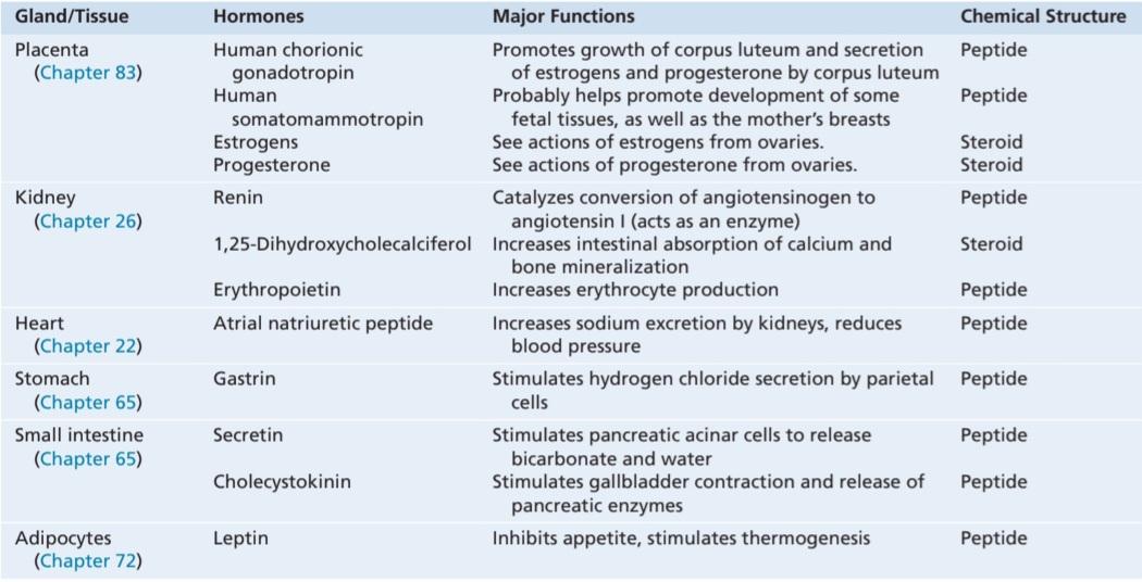
Figure 75-1 shows the anatomical loci of the major endocrine glands and endocrine tissues of the body, except for the placenta, which is an additional source of the sex hormones. Table 75-1 provides an overview of the different hormone systems and their major actions.
شکل 75-1 جایگاه های آناتومیکی غدد درون ریز اصلی و بافت های درون ریز بدن را نشان می دهد، به جز جفت که منبع اضافی هورمون های جنسی است. جدول 75-1 یک نمای کلی از سیستم های هورمونی مختلف و اقدامات اصلی آنها را ارائه می دهد.
The multiple hormone systems play a key role in regulating almost all body functions, including metabolism, growth and development, water and electrolyte balance, reproduction, and behavior. For example, without growth hormone, a person would have very short stature. Without thyroxine and triiodothyronine from the thyroid gland, almost all the chemical reactions of the body would become sluggish and the person would become sluggish as well. Without insulin from the pancreas, the body’s cells could use little of the food carbohydrates for energy. And without the sex hormones, sexual development and sexual functions would be absent.
سیستم های هورمونی متعدد نقش کلیدی در تنظیم تقریباً تمام عملکردهای بدن از جمله متابولیسم، رشد و تکامل، تعادل آب و الکترولیت، تولید مثل و رفتار دارند. به عنوان مثال، بدون هورمون رشد، یک فرد قد بسیار کوتاهی خواهد داشت. بدون تیروکسین و تری یدوتیرونین از غده تیروئید، تقریباً تمام واکنش های شیمیایی بدن کند می شود و فرد نیز تنبل می شود. بدون انسولین از پانکراس، سلولهای بدن میتوانند از کربوهیدراتهای غذایی کمی برای انرژی استفاده کنند. و بدون هورمون های جنسی، رشد جنسی و عملکرد جنسی وجود ندارد.
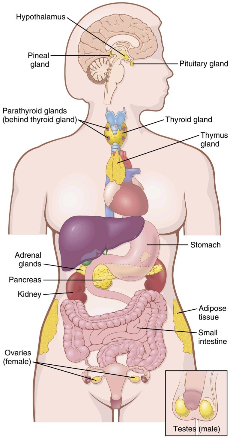
Figure 75-1. Anatomical loci of the principal endocrine glands and tissues of the body.
شکل 75-1. جایگاه های تشریحی غدد درون ریز اصلی و بافت های بدن.
Table 75-1 Endocrine Glands, Hormones, and Their Functions and Structure.
جدول 75-1 غدد درون ریز، هورمون ها، و عملکرد و ساختار آنها.

CHEMICAL STRUCTURE AND SYNTHESIS OF HORMONES
Three general classes of hormones exist:
ساختار شیمیایی و سنتز هورمون ها
سه دسته کلی از هورمون ها وجود دارد:
1. Proteins and polypeptides, including hormones secreted by the anterior and posterior pituitary gland, the pancreas (insulin and glucagon), the parathyroid gland (parathyroid hormone), and many others (see Table 75-1).
1. پروتئین ها و پلی پپتیدها، از جمله هورمون های ترشح شده توسط غده هیپوفیز قدامی و خلفی، پانکراس (انسولین و گلوکاگون)، غده پاراتیروئید (هورمون پاراتیروئید)، و بسیاری دیگر (به جدول 75-1 مراجعه کنید).
2. Steroids secreted by the adrenal cortex (cortisol and aldosterone), the ovaries (estrogen and progesterone), the testes (testosterone), and the placenta (estrogen and progesterone).
2. استروئیدهای ترشح شده از قشر آدرنال (کورتیزول و آلدوسترون)، تخمدان ها (استروژن و پروژسترون)، بیضه ها (تستوسترون) و جفت (استروژن و پروژسترون).
3. Derivatives of the amino acid tyrosine, secreted by the thyroid (thyroxine and triiodothyronine) and the adrenal medullae (epinephrine and norepinephrine). There are no known polysaccharides or nucleic acid hormones.
3. مشتقات آمینو اسید تیروزین که توسط تیروئید (تیروکسین و تری یدوتیرونین) و بصل الکلیه آدرنال (اپی نفرین و نوراپی نفرین) ترشح می شود. هیچ پلی ساکارید یا هورمون اسید نوکلئیک شناخته شده ای وجود ندارد.
Polypeptide and Protein Hormones Are Stored in Secretory Vesicles Until Needed. Most of the hormones in the body are polypeptides and proteins. These hormones range in size from small peptides with as few as three amino acids (e.g., thyrotropin-releasing hormone) to proteins with almost 200 amino acids (e.g., growth hormone and prolactin). In general, polypeptides with 100 or more amino acids are called proteins, and those with fewer than 100 amino acids are referred to as peptides.
هورمون های پلی پپتیدی و پروتئینی تا زمانی که لازم باشد در وزیکول های ترشحی ذخیره می شوند. بیشتر هورمون های بدن پلی پپتیدها و پروتئین ها هستند. اندازه این هورمون ها از پپتیدهای کوچک با حداقل سه اسید آمینه (مانند هورمون آزاد کننده تیروتروپین) تا پروتئین هایی با تقریبا 200 اسید آمینه (مانند هورمون رشد و پرولاکتین) متغیر است. به طور کلی، پلی پپتیدهایی که 100 اسید آمینه یا بیشتر دارند، پروتئین نامیده می شوند و به پلی پپتیدهایی که کمتر از 100 اسید آمینه دارند، پپتید می گویند.
Protein and peptide hormones are synthesized on the rough end of the endoplasmic reticulum of the different endocrine cells, in the same fashion as most other proteins (Figure 75-2). They are usually synthesized first as larger proteins that are not biologically active (preprohormones) and are cleaved to form smaller prohormones in the endoplasmic reticulum. These prohormones are then transferred to the Golgi apparatus for packaging into secretory vesicles. In this process, enzymes in the vesicles cleave the prohormones to produce smaller, biologically active hormones and inactive fragments. The vesicles are stored within the cytoplasm, and many are bound to the cell membrane until their secretion is needed. Secretion of the hormones (as well as the inactive fragments) occurs when the secretory vesicles fuse with the cell membrane and the granular contents are extruded into the interstitial fluid or directly into the blood stream by exocytosis.
پروتئین و هورمون های پپتیدی در انتهای ناهموار شبکه آندوپلاسمی سلول های غدد درون ریز مختلف، به همان روشی که اکثر پروتئین های دیگر سنتز می شوند (شکل 75-2). آنها معمولا ابتدا به عنوان پروتئین های بزرگتر سنتز می شوند که از نظر بیولوژیکی فعال نیستند (پیش پروهورمون ها) و برای تشکیل پروهورمون های کوچکتر در شبکه آندوپلاسمی شکافته می شوند. سپس این پروهورمون ها برای بسته بندی در وزیکول های ترشحی به دستگاه گلژی منتقل می شوند. در این فرآیند، آنزیمهای موجود در وزیکولها، پروهورمونها را میشکنند تا هورمونهای کوچکتر و از نظر بیولوژیکی فعال و قطعات غیرفعال تولید کنند. وزیکول ها در داخل سیتوپلاسم ذخیره می شوند و بسیاری از آنها به غشای سلولی متصل می شوند تا زمانی که ترشح آنها مورد نیاز باشد. ترشح هورمونها (و همچنین قطعات غیرفعال) زمانی اتفاق میافتد که وزیکولهای ترشحی با غشای سلولی ترکیب میشوند و محتویات دانهای به داخل مایع بینابینی یا مستقیماً توسط اگزوسیتوز وارد جریان خون میشوند.
In many cases, the stimulus for exocytosis is increased cytosolic calcium concentration caused by depolarization of the plasma membrane. In other cases, stimulation of an endocrine cell surface receptor causes increased cyclic adenosine monophosphate (cAMP) and subsequently activation of protein kinases that initiate secretion of the hormone. The peptide hormones are water soluble, allowing them to enter the circulatory system easily, where they are carried to their target tissues.
در بسیاری از موارد، محرک اگزوسیتوز افزایش غلظت کلسیم سیتوزولی ناشی از دپلاریزاسیون غشای پلاسمایی است. در موارد دیگر، تحریک گیرنده سطح سلولی غدد درون ریز باعث افزایش آدنوزین مونوفسفات حلقوی (cAMP) و متعاقبا فعال شدن پروتئین کینازهایی می شود که ترشح هورمون را آغاز می کنند. هورمونهای پپتیدی محلول در آب هستند و به آنها اجازه میدهند به راحتی وارد سیستم گردش خون شوند و در آنجا به بافتهای هدف منتقل میشوند.

Figure 75-2. Synthesis and secretion of peptide hormones. The stimulus for hormone secretion often involves changes in intracellular calcium or changes in cyclic adenosine monophosphate (CAMP) in the cell.
شکل 75-2. سنتز و ترشح هورمون های پپتیدی. محرک ترشح هورمون اغلب شامل تغییرات در کلسیم داخل سلولی یا تغییر در آدنوزین مونوفسفات حلقوی (CAMP) در سلول است.
Steroid Hormones Are Usually Synthesized From Cholesterol and Are Not Stored. Steroid hormones have a chemical structure that is similar to cholesterol, and in most cases are synthesized from cholesterol. They are lipid soluble and consist of three cyclohexyl rings and one cyclopentyl ring combined into a single structure (Figure 75-3).
هورمونهای استروئیدی معمولاً از کلسترول سنتز میشوند و ذخیره نمیشوند. هورمون های استروئیدی ساختار شیمیایی مشابهی با کلسترول دارند و در بیشتر موارد از کلسترول سنتز می شوند. آنها محلول در چربی هستند و شامل سه حلقه سیکلوهگزیل و یک حلقه سیکلوپنتیل هستند که در یک ساختار واحد ترکیب شده اند (شکل 75-3).
Although there is usually very little hormone storage in steroid-producing endocrine cells, large stores of cholesterol esters in cytoplasm vacuoles can be rapidly mobilized for steroid synthesis after a stimulus. Much of the cholesterol in steroid-producing cells comes from the plasma, but there is also de novo synthesis of cholesterol in steroid-producing cells. Because steroids are highly lipid soluble, once they are synthesized, they can simply diffuse across the cell membrane and enter the interstitial fluid and then the blood.
اگرچه معمولاً ذخیره هورمونی بسیار کمی در سلول های غدد درون ریز تولید کننده استروئید وجود دارد، اما ذخایر بزرگ استرهای کلسترول در واکوئل های سیتوپلاسم را می توان به سرعت پس از یک محرک برای سنتز استروئید بسیج کرد. بیشتر کلسترول در سلول های تولید کننده استروئید از پلاسما می آید، اما سنتز جدید کلسترول در سلول های تولید کننده استروئید نیز وجود دارد. از آنجایی که استروئیدها به شدت محلول در چربی هستند، پس از سنتز، می توانند به سادگی در غشای سلولی منتشر شده و وارد مایع بینابینی و سپس خون شوند.
Amine Hormones Are Derived From Tyrosine. The two groups of hormones derived from tyrosine, the thyroid and the adrenal medullary hormones, are formed by the actions of enzymes in the cytoplasmic compartments of glandular cells. The thyroid hormones are synthesized and stored in the thyroid gland and incorporated into macromolecules of the protein thyroglobulin, which is stored in large follicles within the thyroid gland. Hormone secretion occurs when the amines are split from thyroglobulin, and the free hormones are then released into the blood stream. After entering the blood, most of the thyroid hormones combine with plasma proteins, especially thyroxine-binding globulin, which slowly releases the hormones to the target tissues.
هورمون های آمینی از تیروزین مشتق می شوند. دو گروه از هورمونهای مشتق شده از تیروزین، هورمونهای تیروئید و مدولاری آدرنال، توسط فعالیت آنزیمها در بخشهای سیتوپلاسمی سلولهای غدد تشکیل میشوند. هورمونهای تیروئید در غده تیروئید سنتز و ذخیره میشوند و در ماکرومولکولهای پروتئین تیروگلوبولین که در فولیکولهای بزرگ درون غده تیروئید ذخیره میشود، ترکیب میشوند. ترشح هورمون زمانی اتفاق میافتد که آمینها از تیروگلوبولین جدا میشوند و هورمونهای آزاد در جریان خون آزاد میشوند. پس از ورود به خون، بیشتر هورمون های تیروئید با پروتئین های پلاسما، به ویژه گلوبولین متصل شونده به تیروکسین، ترکیب می شوند که به آرامی هورمون ها را به بافت های هدف آزاد می کند.
Epinephrine and norepinephrine are formed in the adrenal medulla, which normally secretes about four times more epinephrine than norepinephrine. Catechol- amines are taken up into preformed vesicles and stored until secreted. Similar to the protein hormones stored in secretory granules, catecholamines are also released from adrenal medullary cells by exocytosis. Once the catechol- amines enter the circulation, they can exist in the plasma in free form or in conjugation with other substances.
اپی نفرین و نوراپی نفرین در مدولای آدرنال تشکیل می شوند که به طور معمول حدود چهار برابر بیشتر از نوراپی نفرین اپی نفرین ترشح می کند. کاتکول آمین ها به داخل وزیکول های از پیش ساخته شده جذب می شوند و تا زمانی که ترشح شوند ذخیره می شوند. مشابه هورمون های پروتئینی ذخیره شده در گرانول های ترشحی، کاتکول آمین ها نیز از سلول های مدولاری آدرنال توسط اگزوسیتوز آزاد می شوند. هنگامی که کاتکول آمین ها وارد گردش خون می شوند، می توانند در پلاسما به صورت آزاد یا به صورت ترکیبی با مواد دیگر وجود داشته باشند.

Figure 75-3. Chemical structures of several steroid hormones.
شکل 75-3. ساختارهای شیمیایی چندین هورمون استروئیدی.
HORMONE SECRETION, TRANSPORT, AND CLEARANCE FROM THE BLOOD
Hormone Secretion After a Stimulus and Duration of Action of Different Hormones. Some hormones, such as norepinephrine and epinephrine, are secreted within seconds after the gland is stimulated and may develop full action within another few seconds to minutes; the actions of other hormones, such as thyroxine or growth hormone, may require months for full effect. Thus, each of the different hormones has its own characteristic onset and duration of action-each tailored to perform its specific control function.
ترشح هورمون، حمل و نقل و پاکسازی از خون
ترشح هورمون پس از یک محرک و مدت زمان اثر هورمون های مختلف. برخی از هورمونها، مانند نوراپی نفرین و اپی نفرین، در عرض چند ثانیه پس از تحریک غده ترشح میشوند و ممکن است در عرض چند ثانیه تا چند دقیقه دیگر عمل کنند. اعمال سایر هورمونها، مانند تیروکسین یا هورمون رشد، ممکن است ماهها طول بکشد تا اثر کامل داشته باشد. بنابراین، هر یک از هورمون های مختلف شروع مشخصه و مدت زمان عمل خود را دارد که هر کدام برای انجام عملکرد کنترلی خاص خود تنظیم شده است.
Concentrations of Hormones in the Circulating Blood and Hormonal Secretion Rates. The concentrations of hormones required to control most metabolic and endocrine functions are incredibly small. Their concentrations in the blood range from as little as 1 picogram (which is one millionth of one millionth of a gram) in each milliliter of blood up to at most a few micro- grams (a few millionths of a gram) per milliliter of blood. Similarly, the rates of secretion of the various hormones are extremely small, usually measured in micrograms or milligrams per day. We shall see later in this chapter that highly specialized mechanisms are available in the target tissues that allow even these minute quantities of hormones to exert powerful control over the physiological systems.
غلظت هورمون ها در خون در گردش و میزان ترشح هورمونی. غلظت هورمون های مورد نیاز برای کنترل بیشتر عملکردهای متابولیک و غدد درون ریز بسیار کم است. غلظت آنها در خون از 1 پیکو گرم (که یک میلیونم یک میلیونیم گرم است) در هر میلی لیتر خون تا حداکثر چند میکروگرم (چند میلیونم گرم) در هر میلی لیتر خون متغیر است. به طور مشابه، میزان ترشح هورمونهای مختلف بسیار کم است و معمولاً بر حسب میکروگرم یا میلیگرم در روز اندازهگیری میشود. بعداً در این فصل خواهیم دید که مکانیسمهای بسیار تخصصی در بافتهای هدف در دسترس هستند که به این مقادیر کوچک از هورمونها اجازه میدهند تا کنترل قدرتمندی بر سیستمهای فیزیولوژیکی اعمال کنند.
FEEDBACK CONTROL OF HORMONE SECRETION
Negative Feedback Prevents Overactivity of Hormone Systems. Although the plasma concentrations of many hormones fluctuate in response to various stimuli that occur throughout the day, all hormones studied thus far appear to be closely controlled. In most cases, this control is exerted through negative feedback mechanisms (described in Chapter 1) that ensure a proper level of hormone activity at the target tissue. After a stimulus causes release of the hormone, conditions or products resulting from the action of the hormone tend to suppress its further release. In other words, the hormone (or one of its products) has a negative feedback effect to prevent oversecretion of the hormone or overactivity at the target tissue. Feedback regulation of hormones can occur at all levels, including gene transcription and translation steps involved in the synthesis of hormones and steps involved in processing hormones or releasing stored hormones.
کنترل بازخورد ترشح هورمون
بازخورد منفی از بیش فعالی سیستم های هورم یک جلوگیری می کند. اگرچه غلظت پلاسمایی بسیاری از هورمونها در پاسخ به محرکهای مختلفی که در طول روز رخ میدهند در نوسان است، به نظر میرسد همه هورمونهایی که تاکنون مورد مطالعه قرار گرفتهاند به دقت کنترل میشوند. در بیشتر موارد، این کنترل از طریق مکانیسم های بازخورد منفی (شرح شده در فصل 1) اعمال می شود که سطح مناسبی از فعالیت هورمون را در بافت هدف تضمین می کند. پس از اینکه یک محرک باعث آزاد شدن هورمون می شود، شرایط یا محصولات ناشی از عمل هورمون تمایل به سرکوب ترشح بیشتر آن دارند. به عبارت دیگر، هورمون (یا یکی از محصولات آن) دارای اثر بازخورد منفی برای جلوگیری از ترشح بیش از حد هورمون یا فعالیت بیش از حد در بافت هدف است. تنظیم بازخورد هورمونها میتواند در همه سطوح اتفاق بیفتد، از جمله مراحل رونویسی و ترجمه ژن که در سنتز هورمونها و مراحل دخیل در پردازش هورمونها یا آزادسازی هورمونهای ذخیره شده نقش دارند.
The controlled variable is sometimes the degree of activity of the target tissue rather than the synthesis or secretory rates of the hormone. Therefore, only when the target tissue activity rises to an appropriate level will feedback signals to the endocrine gland become powerful enough to slow further synthesis and secretion of the hormone.
متغیر کنترل شده گاهی اوقات میزان فعالیت بافت هدف به جای سنتز یا میزان ترشح هورمون است. بنابراین، تنها زمانی که فعالیت بافت هدف به سطح مناسبی افزایش یابد، سیگنالهای بازخوردی به غده درون ریز به اندازه کافی قدرتمند میشوند تا سنتز و ترشح بیشتر هورمون را کند کنند.
Surges of Hormones Can Occur With Positive Feedback. In a few cases, positive feedback occurs when the biological action of the hormone causes additional secretion of the hormone. One example of positive feedback is the surge of luteinizing hormone (LH) that occurs as a result of the stimulatory effect of estrogen on the anterior pituitary before ovulation. The secreted LH then acts on the ovaries to stimulate additional secretion of estrogen, which in turn causes more secretion of LH. Eventually, LH reaches an appropriate concentration and typical negative feedback control of hormone secretion is then exerted.
جهش هورمون ها می تواند با بازخورد مثبت رخ دهد. در موارد معدودی، بازخورد مثبت زمانی رخ می دهد که عملکرد بیولوژیکی هورمون باعث ترشح اضافی هورمون شود. یکی از نمونه های بازخورد مثبت، افزایش هورمون لوتئینیزه کننده (LH) است که در نتیجه اثر تحریکی استروژن بر هیپوفیز قدامی قبل از تخمک گذاری رخ می دهد. سپس LH ترشح شده بر روی تخمدان ها برای تحریک ترشح اضافی استروژن عمل می کند که به نوبه خود باعث ترشح بیشتر LH می شود. در نهایت، LH به غلظت مناسب می رسد و کنترل بازخورد منفی معمول ترشح هورمون اعمال می شود.
Cyclical Variations Occur in Hormone Release. Superimposed on the negative and positive feedback control of hormone secretion are periodic variations in hormone release that are influenced by seasonal changes, various stages of development and aging, the diurnal (daily) cycle, and sleep. For example, the secretion of growth hormone is markedly increased during the early period of sleep but is reduced during the later stages of sleep. In many cases, these cyclical variations in hormone secretion are due to changes in activity of neural pathways involved in controlling hormone release.
تغییرات چرخه ای در ترشح هورمون رخ می دهد. بر کنترل بازخورد منفی و مثبت ترشح هورمون، تغییرات دورهای در ترشح هورمون قرار دارند که تحت تأثیر تغییرات فصلی، مراحل مختلف رشد و پیری، چرخه روزانه (روزانه) و خواب قرار دارند. به عنوان مثال، ترشح هورمون رشد در دوره اولیه خواب به طور قابل توجهی افزایش می یابد اما در مراحل بعدی خواب کاهش می یابد. در بسیاری از موارد، این تغییرات چرخهای در ترشح هورمون به دلیل تغییر در فعالیت مسیرهای عصبی درگیر در کنترل ترشح هورمون است.
Oscillations of endocrine signaling are driven, in part by circadian clocks. As discussed in Chapter 59 (see Figure 59-8), the suprachiasmatic nucleus (SCN) of the hypothalamus serves as a “master clock” that controls rhythmic patterns of biological clocks in many parts of the body, including neuroendocrine cells and endocrine glands. There is also evidence for local clock control in peripheral endocrine tissues, such as the adrenal gland and pancreas, which have cyclical changes in their sensitivity to various signals.
نوسانات سیگنالینگ غدد درون ریز، تا حدی توسط ساعت های شبانه روزی هدایت می شود. همانطور که در فصل 59 بحث شد (نگاه کنید به شکل 59-8)، هسته سوپراکیاسماتیک (SCN) هیپوتالاموس به عنوان یک “ساعت اصلی” عمل می کند که الگوهای ریتمیک ساعت های بیولوژیکی را در بسیاری از قسمت های بدن، از جمله سلول های عصبی غدد درون ریز و غدد درون ریز، کنترل می کند. همچنین شواهدی مبنی بر کنترل ساعت موضعی در بافتهای غدد درون ریز محیطی، مانند غده فوق کلیوی و پانکراس وجود دارد، که تغییرات چرخهای در حساسیت خود به سیگنالهای مختلف دارند.
Cyclical changes in tissue responsiveness and fluctuations in hormone concentrations provide important mechanisms for the body to anticipate and adapt to the dramatic changes in stresses and demands that normally occur over the course of a dayranging from restful sleep to eating meals and high levels of mental and physical activity. Rhythmic changes in female sex hormones that occur with an average 28-day cycle are also essential for reproduction, as discussed in Chapter 82.
تغییرات چرخهای در پاسخدهی بافت و نوسانات غلظت هورمون مکانیسمهای مهمی را برای بدن فراهم میکند تا تغییرات چشمگیر استرسها و نیازهایی را که معمولاً در طول یک روز رخ میدهد، از خواب آرام گرفته تا خوردن وعدههای غذایی و سطوح بالای فعالیت ذهنی و فیزیکی، پیشبینی کند و با آن سازگار شود. همانطور که در فصل 82 مورد بحث قرار گرفت، تغییرات ریتمیک در هورمون های جنسی زنانه که با یک چرخه متوسط 28 روزه رخ می دهد نیز برای تولید مثل ضروری است.
TRANSPORT OF HORMONES IN THE BLOOD
Water-soluble hormones (peptides and catecholamines) are dissolved in the plasma and transported from their sites of synthesis to target tissues, where they diffuse out of the capillaries, into the interstitial fluid, and ultimately to target cells.
انتقال هورمون ها در خون
هورمونهای محلول در آب (پپتیدها و کاتکول آمینها) در پلاسما حل میشوند و از محلهای سنتز خود به بافتهای هدف منتقل میشوند، جایی که از مویرگها به داخل مایع بینابینی و در نهایت به سلولهای هدف منتشر میشوند.
Steroid and thyroid hormones, in contrast, circulate in the blood while being mainly bound to plasma proteins. Usually less than 10% of steroid or thyroid hormones in the plasma exist free in solution. For example, more than 99% of the thyroxine in the blood is bound to plasma proteins. However, protein-bound hormones cannot easily diffuse across the capillaries and gain access to their target cells and are therefore biologically inactive until they dissociate from plasma proteins.
در مقابل، هورمون های استروئیدی و تیروئیدی در خون گردش می کنند در حالی که عمدتاً به پروتئین های پلاسما متصل می شوند. معمولاً کمتر از 10 درصد هورمون های استروئیدی یا تیروئیدی در پلاسما به صورت آزاد در محلول وجود دارد. به عنوان مثال، بیش از 99 درصد از تیروکسین موجود در خون به پروتئین های پلاسما متصل می شود. با این حال، هورمون های متصل به پروتئین نمی توانند به راحتی در مویرگ ها پخش شوند و به سلول های هدف خود دسترسی پیدا کنند و بنابراین تا زمانی که از پروتئین های پلاسما جدا نشوند، از نظر بیولوژیکی غیر فعال هستند.
The relatively large amounts of hormones bound to proteins serve as reservoirs, replenishing the concentration of free hormones when they are bound to target receptors or lost from the circulation. Binding of hormones to plasma proteins greatly slows their clearance from the plasma.
مقادیر نسبتاً زیادی از هورمونهای متصل به پروتئینها به عنوان مخزن عمل میکنند و غلظت هورمونهای آزاد را هنگامی که به گیرندههای هدف متصل میشوند یا از گردش خون گم میشوند، دوباره پر میکنند. اتصال هورمونها به پروتئینهای پلاسما، پاکسازی آنها از پلاسما را بسیار کند میکند.
“Clearance” of Hormones From the Blood. Two factors can increase or decrease the concentration of a hormone in the blood: (1) the rate of hormone secretion into the blood; (2) the rate of removal of the hormone from the blood, which is called the metabolic clearance rate and is usually expressed in terms of the number of milliliters of plasma cleared of the hormone per minute. To calculate this clearance rate, one measures (1) the rate of disappearance of the hormone from the plasma (e.g., nanograms per minute) and (2) the plasma concentration of the hormone (e.g., nanograms per milliliter of plasma). Then, the metabolic clearance rate is calculated with use of the following formula:
“پاکسازی” هورمون ها از خون. دو عامل می توانند غلظت یک هورمون در خون را افزایش یا کاهش دهند: (1) سرعت ترشح هورمون در خون. (2) سرعت حذف هورمون از خون که به آن نرخ کلیرانس متابولیک می گویند و معمولاً بر حسب تعداد میلی لیتر پلاسمای پاک شده از هورمون در دقیقه بیان می شود. برای محاسبه این میزان پاکسازی، (1) سرعت ناپدید شدن هورمون از پلاسما (به عنوان مثال، نانوگرم در دقیقه) و (2) غلظت پلاسمایی هورمون (به عنوان مثال، نانوگرم در هر میلی لیتر پلاسما) اندازه گیری می شود. سپس نرخ کلیرانس متابولیک با استفاده از فرمول زیر محاسبه می شود:

The usual procedure for making this measurement is as follows. A purified solution of the hormone to be measured is tagged with a radioactive substance. Then the radioactive hormone is infused at a constant rate into the blood stream until the radioactive concentration in the plasma becomes steady. At this time, the disappearance rate of the radioactive hormone from the plasma equals the rate at which it is infused, which gives one the rate of disappearance. At the same time, the plasma concentration of the radioactive hormone is measured using a standard radioactive counting procedure. Then, using the formula just cited, the metabolic clearance rate is calculated.
روش معمول برای انجام این اندازه گیری به شرح زیر است. محلول خالص شده هورمونی که باید اندازه گیری شود با یک ماده رادیواکتیو برچسب گذاری می شود. سپس هورمون رادیواکتیو با سرعت ثابتی به جریان خون تزریق می شود تا زمانی که غلظت رادیواکتیو در پلاسما ثابت شود. در این زمان، سرعت ناپدید شدن هورمون رادیواکتیو از پلاسما برابر با سرعت تزریق آن است که سرعت ناپدید شدن را به فرد می دهد. در همان زمان، غلظت پلاسمایی هورمون رادیواکتیو با استفاده از روش استاندارد شمارش رادیواکتیو اندازه گیری می شود. سپس با استفاده از فرمول ذکر شده، نرخ کلیرانس متابولیک محاسبه می شود.
Hormones are “cleared” from the plasma in several ways, including (1) metabolic destruction by the tissues, (2) binding with the tissues, (3) excretion by the liver into the bile, and (4) excretion by the kidneys into the urine. For certain hormones, a decreased metabolic clearance rate may cause an excessively high concentration of the hormone in the circulating body fluids. For example, excessive steroid hormones accumulate when the liver is diseased because these hormones are conjugated mainly in the liver and then “cleared” into the bile.
هورمون ها به روش های مختلفی از پلاسما پاک می شوند، از جمله (1) تخریب متابولیک توسط بافت ها، (2) اتصال به بافت ها، (3) دفع توسط کبد به صفرا، و (4) دفع توسط کلیه ها به ادرار. برای برخی از هورمون ها، کاهش نرخ کلیرانس متابولیک ممکن است باعث غلظت بیش از حد هورمون در مایعات در گردش بدن شود. به عنوان مثال، هورمونهای استروئیدی بیش از حد در هنگام بیماری کبد تجمع مییابند، زیرا این هورمونها عمدتاً در کبد کونژوگه شده و سپس در صفرا «پاک میشوند».
Hormones are sometimes degraded at their target cells by enzymatic processes that cause endocytosis of the cell membrane hormone-receptor complex; the hormone is then metabolized in the cell, and the receptors are usually recycled back to the cell membrane.
گاهی اوقات هورمون ها در سلول های هدف خود توسط فرآیندهای آنزیمی تجزیه می شوند که باعث اندوسیتوز مجموعه هورمون-گیرنده غشای سلولی می شود. سپس هورمون در سلول متابولیزه می شود و گیرنده ها معمولاً به غشای سلولی بازیافت می شوند.
Most of the peptide hormones and catecholamines are water soluble and circulate freely in the blood. They are usually degraded by enzymes in the blood and tissues and rapidly excreted by the kidneys and liver, thus remaining in the blood for only a short time. For example, the half- life of angiotensin II circulating in the blood is less than a minute.
بیشتر هورمون های پپتیدی و کاتکول آمین ها محلول در آب هستند و آزادانه در خون گردش می کنند. آنها معمولاً توسط آنزیم های موجود در خون و بافت ها تجزیه می شوند و به سرعت توسط کلیه ها و کبد دفع می شوند، بنابراین فقط برای مدت کوتاهی در خون باقی می مانند. به عنوان مثال، نیمه عمر آنژیوتانسین II در گردش خون کمتر از یک دقیقه است.
Hormones that are bound to plasma proteins are cleared from the blood at much slower rates and may remain in the circulation for several hours or even days. The half-life of adrenal steroids in the circulation, for example, ranges between 20 and 100 minutes, whereas the half-life of the protein-bound thyroid hormones may be as long as 1 to 6 days.
هورمون هایی که به پروتئین های پلاسما متصل می شوند با سرعت بسیار کمتری از خون پاک می شوند و ممکن است برای چندین ساعت یا حتی روزها در گردش خون باقی بمانند. به عنوان مثال، نیمه عمر استروئیدهای آدرنال در گردش خون بین 20 تا 100 دقیقه است، در حالی که نیمه عمر هورمون های تیروئیدی متصل به پروتئین ممکن است 1 تا 6 روز باشد.
MECHANISMS OF ACTION OF HORMONES
HORMONE RECEPTORS AND THEIR ACTIVATION
The first step of a hormone’s action is to bind to specific receptors at the target cell. Cells that lack receptors for the hormones do not respond. Receptors for some hormones are located on the target cell membrane, whereas other hormone receptors are located in the cytoplasm or the nucleus. When the hormone combines with its receptor, this action usually initiates a cascade of reactions in the cell, with each stage becoming more powerfully activated so that even small concentrations of the hormone can have a large effect.
مکانیسم های عمل هورمون ها
گیرنده های هورمونی و فعال سازی آنها
اولین مرحله از عملکرد هورمون، اتصال به گیرنده های خاص در سلول هدف است. سلول هایی که فاقد گیرنده های هورمونی هستند، پاسخ نمی دهند. گیرنده های برخی از هورمون ها روی غشای سلول هدف قرار دارند، در حالی که گیرنده های هورمونی دیگر در سیتوپلاسم یا هسته قرار دارند. وقتی هورمون با گیرندهاش ترکیب میشود، این عمل معمولاً آبشاری از واکنشها را در سلول آغاز میکند و هر مرحله با قدرت بیشتری فعال میشود، به طوری که حتی غلظتهای کوچک هورمون میتواند تأثیر زیادی داشته باشد.
Hormone receptors are large proteins, and each cell that is to be stimulated usually has some 2000 to 100,000 receptors. Also, each receptor is usually highly specific for a single hormone, which determines the type of hormone that will act on a particular tissue. The target tissues that are affected by a hormone are those that contain its specific receptors.
گیرنده های هورمونی پروتئین های بزرگی هستند و هر سلولی که قرار است تحریک شود معمولاً بین 2000 تا 100000 گیرنده دارد. همچنین، هر گیرنده معمولاً برای یک هورمون بسیار اختصاصی است، که نوع هورمونی را که بر روی یک بافت خاص عمل می کند، تعیین می کند. بافت های هدفی که تحت تاثیر یک هورمون قرار می گیرند، آنهایی هستند که گیرنده های خاص آن را دارند.
The locations for the different types of hormone receptors are generally the following:
مکان انواع مختلف گیرنده های هورمونی به طور کلی به شرح زیر است:
1. In or on the surface of the cell membrane. The membrane receptors are specific mostly for the protein, peptide, and catecholamine hormones.
1. داخل یا روی سطح غشای سلولی. گیرنده های غشایی بیشتر برای هورمون های پروتئین، پپتید و کاتکول آمین اختصاص دارند.
2. In the cell cytoplasm. The primary receptors for the different steroid hormones are found mainly in the cytoplasm.
2. در سیتوپلاسم سلولی. گیرنده های اولیه برای هورمون های استروئیدی مختلف عمدتاً در سیتوپلاسم یافت می شوند.
3. In the cell nucleus. The receptors for the thyroid hormones are found in the nucleus and are believed to be located in direct association with one or more of the chromosomes.
3. در هسته سلول. گیرنده های هورمون های تیروئید در هسته یافت می شوند و اعتقاد بر این است که در ارتباط مستقیم با یک یا چند کروموزوم قرار دارند.
The Number and Sensitivity of Hormone Receptors Are Regulated. The number of receptors in a target cell usually does not remain constant from day to day or even from minute to minute. Receptor proteins are often inactivated or destroyed during the course of their function, and at other times they are reactivated or new ones are manufactured by the cell. For example, increased hormone concentration and increased binding with its target cell receptors sometimes cause the number of active receptors to decrease. This down-regulation of the receptors can occur as a result of (1) inactivation of some of the receptor molecules; (2) inactivation of some of the intracellular protein signaling molecules; (3) temporary sequestration of the receptor to the inside of the cell, away from the site of action of hormones that interact with cell membrane receptors; (4) destruction of the receptors by lysosomes after they are internalized; or (5) decreased production of the receptors. In each case, receptor down- regulation decreases the target tissue’s responsiveness to the hormone.
تعداد و حساسیت گیرنده های هورمونی تنظیم شده است. تعداد گیرنده ها در یک سلول هدف معمولاً روز به روز یا حتی از دقیقه به دقیقه ثابت نمی ماند. پروتئینهای گیرنده اغلب در طول عملکرد خود غیرفعال یا از بین میروند و در زمانهای دیگر دوباره فعال میشوند یا پروتئینهای جدیدی توسط سلول تولید میشوند. به عنوان مثال، افزایش غلظت هورمون و افزایش اتصال با گیرنده های سلول هدف گاهی اوقات باعث کاهش تعداد گیرنده های فعال می شود. این تنظیم پایین گیرنده ها می تواند در نتیجه (1) غیرفعال شدن برخی از مولکول های گیرنده رخ دهد. (2) غیر فعال کردن برخی از مولکول های سیگنال دهنده پروتئین داخل سلولی. (3) جداسازی موقت گیرنده در داخل سلول، به دور از محل عمل هورمونهایی که با گیرندههای غشای سلولی تعامل دارند. (4) تخریب گیرنده ها توسط لیزوزوم ها پس از درونی شدن. یا (5) کاهش تولید گیرنده ها. در هر مورد، تنظیم پایین گیرنده، پاسخ بافت هدف به هورمون را کاهش می دهد.
Some hormones cause up-regulation of receptors and intracellular signaling proteins; that is, the stimulating hormone induces greater than normal formation of receptor or intracellular signaling molecules by the target cell or greater availability of the receptor for interaction with the hormone. When up-regulation occurs, the target tissue becomes progressively more sensitive to the stimulating effects of the hormone.
برخی از هورمون ها باعث افزایش تنظیم گیرنده ها و پروتئین های سیگنال دهی داخل سلولی می شوند. یعنی هورمون محرک بیش از حد طبیعی باعث ایجاد گیرنده یا مولکول های سیگنال دهی درون سلولی توسط سلول هدف یا در دسترس بودن بیشتر گیرنده برای تعامل با هورمون می شود. هنگامی که تنظیم بالا اتفاق می افتد، بافت هدف به تدریج نسبت به اثرات تحریک کننده هورمون حساس تر می شود.
INTRACELLULAR SIGNALING AFTER HORMONE RECEPTOR ACTIVATION
Almost without exception, a hormone affects its target tissues by first forming a hormone-receptor complex. Formation of this complex alters the function of the receptor, and the activated receptor initiates the hormonal effects. To explain this process, let us give a few examples of the different types of interactions.
سیگنال دهی داخل سلولی پس از فعال شدن گیرنده های هورمونی
تقریباً بدون استثنا، یک هورمون ابتدا با تشکیل یک مجموعه هورمون – گیرنده بر بافت های هدف خود تأثیر می گذارد. تشکیل این کمپلکس عملکرد گیرنده را تغییر می دهد و گیرنده فعال شده شروع کننده اثرات هورمونی است. برای توضیح این فرآیند، اجازه دهید چند نمونه از انواع مختلف تعاملات را بیان کنیم.
Ion Channel-Linked Receptors. Virtually all the neurotransmitter substances, such as acetylcholine and nor- epinephrine, combine with receptors in the postsynaptic membrane. This combination almost always causes a change in the structure of the receptor, usually opening or closing a channel for one or more ions. Some of these ion channel-linked receptors open (or close) channels for sodium ions, others for potassium ions, others for calcium ions, and so forth. The altered movement of these ions through the channels causes the subsequent effects on the postsynaptic cells. Although a few hormones may exert some of their actions through activation of ion channel receptors, most hormones that open or close ions channels do this indirectly by coupling with G protein-linked or enzyme-linked receptors, as discussed next.
گیرنده های مرتبط با کانال یونی. تقریباً تمام مواد انتقال دهنده عصبی، مانند استیل کولین و نوراپی نفرین، با گیرنده های غشای پس سیناپسی ترکیب می شوند. این ترکیب تقریباً همیشه باعث تغییر در ساختار گیرنده می شود، معمولاً کانالی را برای یک یا چند یون باز یا بسته می کند. برخی از این گیرندههای مرتبط با کانال یونی، کانالهایی را برای یونهای سدیم، برخی دیگر برای یونهای پتاسیم، برخی دیگر برای یونهای کلسیم و غیره باز میکنند (یا میبندند). حرکت تغییر یافته این یون ها از طریق کانال ها باعث اثرات بعدی بر سلول های پس سیناپسی می شود. اگرچه تعداد کمی از هورمونها ممکن است برخی از اعمال خود را از طریق فعالسازی گیرندههای کانال یونی انجام دهند، اکثر هورمونهایی که کانالهای یونی را باز یا بسته میکنند، این کار را بهطور غیرمستقیم از طریق جفت شدن با گیرندههای مرتبط با پروتئین G یا مرتبط با آنزیم انجام میدهند.
G Protein-Linked Hormone Receptors. Many hormones activate receptors that indirectly regulate the activity of target proteins (e.g., enzymes or ion channels) by coupling with groups of cell membrane proteins called heterotrimeric guanosine triphosphate (GTP)-binding proteins (G proteins) (Figure 75-4). All of the known G protein-coupled receptors (~1000) have seven trans- membrane segments that loop in and out of the cell mem- brane. Some parts of the receptor that protrude into the cell cytoplasm (especially the cytoplasmic tail of the receptor) are coupled to G proteins that include three (i.e., trimeric) parts—the a, ẞ, and y subunits. When the ligand (hormone) binds to the extracellular part of the receptor, a conformational change occurs in the receptor that activates the G proteins and induces intracellular signals that either (1) open or close cell membrane ion channels, (2) change the activity of an enzyme in the cytoplasm of the cell, or (3) activate gene transcription.
گیرنده های هورمونی مرتبط با پروتئین G. بسیاری از هورمونها گیرندههایی را فعال میکنند که به طور غیرمستقیم فعالیت پروتئینهای هدف (مانند آنزیمها یا کانالهای یونی) را با جفت شدن با گروههایی از پروتئینهای غشای سلولی به نام پروتئینهای متصل شونده گوانوزین تری فسفات هتروتریمریک (GTP) (پروتئینهای G) تنظیم میکنند (شکل 75-4). همه گیرنده های شناخته شده جفت شده با پروتئین G (~1000) دارای هفت بخش غشایی هستند که به داخل و خارج غشای سلولی حلقه می زنند. برخی از بخشهای گیرنده که به داخل سیتوپلاسم سلولی بیرون زدهاند (بهویژه دم سیتوپلاسمی گیرنده) با پروتئینهای G که شامل سه بخش (یعنی سهگانه) هستند – زیرواحدهای a، ẞ و y جفت میشوند. هنگامی که لیگاند (هورمون) به بخش خارج سلولی گیرنده متصل می شود، یک تغییر ساختاری در گیرنده رخ می دهد که پروتئین های G را فعال می کند و سیگنال های درون سلولی را القا می کند که (1) کانال های یونی غشای سلولی را باز یا بسته می کنند، (2) فعالیت آنزیم را در سیتوپلاسم سلول اسکریپت فعال تغییر می دهند، یا (3)
The trimeric G proteins are named for their ability to bind guanosine nucleotides. In their inactive state, the a, ẞ, and y subunits of G proteins form a complex that binds guanosine diphosphate (GDP) on the a subunit.
پروتئین های G trimeric به دلیل توانایی آنها در اتصال نوکلئوتیدهای گوانوزین نامگذاری شده اند. در حالت غیرفعال، زیرواحدهای a، ẞ و y پروتئین های G یک کمپلکس تشکیل می دهند که گوانوزین دی فسفات (GDP) را به زیر واحد a متصل می کند.
When the receptor is activated, it undergoes a conformational change that causes the GDP-bound trimeric G protein to associate with the cytoplasmic part of the receptor and to exchange GDP for GTP. Displacement of GDP by GTP causes the a subunit to dissociate from the trimeric complex and to associate with other intracellular signaling proteins; these proteins, in turn, alter the activity of ion channels or intracellular enzymes such as adenylyl cyclase or phospholipase C, which alter cell function.
هنگامی که گیرنده فعال می شود، تحت یک تغییر ساختاری قرار می گیرد که باعث می شود پروتئین G trimeric متصل به GDP با بخش سیتوپلاسمی گیرنده مرتبط شود و GDP را با GTP مبادله کند. جابجایی تولید ناخالص داخلی توسط GTP باعث می شود که یک زیر واحد از کمپلکس تریمریک جدا شود و با سایر پروتئین های سیگنال دهنده درون سلولی مرتبط شود. این پروتئین ها به نوبه خود فعالیت کانال های یونی یا آنزیم های درون سلولی مانند آدنیلیل سیکلاز یا فسفولیپاز C را تغییر می دهند که عملکرد سلول را تغییر می دهند.
The signaling event is terminated when the hormone is removed and the a subunit inactivates itself by converting its bound GTP to GDP; then the a subunit once again combines with the ẞ and y subunits to form an inactive, membrane-bound trimeric G protein. Additional details of G protein signaling are discussed in Chapter 46 and shown in Figure 46-7.
رویداد سیگنالینگ زمانی خاتمه می یابد که هورمون حذف شود و زیرواحد a با تبدیل GTP محدود به تولید ناخالص داخلی خود را غیرفعال می کند. سپس زیرواحد a یک بار دیگر با زیرواحدهای ẞ و y ترکیب می شود و یک پروتئین G trimeric غیرفعال و متصل به غشاء را تشکیل می دهد. جزئیات اضافی سیگنال دهی پروتئین G در فصل 46 مورد بحث قرار گرفته و در شکل 46-7 نشان داده شده است.
Some hormones are coupled to inhibitory G proteins (denoted as G proteins), whereas others are coupled to stimulatory G proteins (denoted as G, proteins). Thus, depending on the coupling of a hormone receptor to an inhibitory or stimulatory G protein, a hormone can either increase or decrease the activity of intracellular enzymes. This complex system of cell membrane G proteins provides a vast array of potential cell responses to different hormones in the various target tissues of the body.
برخی از هورمونها با پروتئینهای مهارکننده G (که با پروتئینهای G مشخص میشوند)، جفت میشوند، در حالی که برخی دیگر با پروتئینهای G تحریککننده (که با پروتئینهای G مشخص میشوند) جفت میشوند. بنابراین، بسته به جفت شدن یک گیرنده هورمونی به پروتئین G مهاری یا محرک، یک هورمون می تواند فعالیت آنزیم های داخل سلولی را افزایش یا کاهش دهد. این سیستم پیچیده از پروتئینهای G غشای سلولی، مجموعه وسیعی از پاسخهای بالقوه سلولی را به هورمونهای مختلف در بافتهای هدف مختلف بدن ارائه میکند.
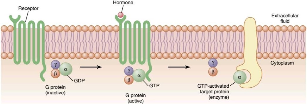
Figure 75-4. Mechanism of activation of a G protein-coupled receptor. When the hormone activates the receptor, the inactive a, ẞ, and y G protein complex associates with the receptor and is activated, with an exchange of guanosine triphosphate (GTP) for guanosine diphosphate (GDP). This process causes the a subunit (to which the GTP is bound) to dissociate from the ẞ and y subunits of the G protein and to interact with membrane-bound target proteins (enzymes) that initiate intracellular signals.
شکل 75-4. مکانیسم فعال شدن گیرنده جفت شده با پروتئین G. هنگامی که هورمون گیرنده را فعال می کند، مجتمع پروتئینی غیرفعال a، ẞ و y G با گیرنده مرتبط می شود و با تبادل گوانوزین تری فسفات (GTP) با گوانوزین دی فسفات (GDP) فعال می شود. این فرآیند باعث میشود که یک زیر واحد (که GTP به آن متصل است) از زیرواحدهای ẞ و y پروتئین G جدا شود و با پروتئینهای هدف (آنزیمهای) متصل به غشاء که سیگنالهای درون سلولی را آغاز میکنند، تعامل کند.
Enzyme-Linked Hormone Receptors. Some receptors, when activated, function directly as enzymes or are closely associated with enzymes that they activate. These enzyme- linked receptors are proteins that pass through the membrane only once, in contrast to the seven-transmembrane G protein-coupled receptors. Enzyme-linked receptors have their hormone-binding site on the outside of the cell membrane and their catalytic or enzyme-binding site on the inside. When the hormone binds to the extracellular part of the receptor, an enzyme immediately inside the cell membrane is activated (or occasionally inactivated). Although many enzyme-linked receptors have intrinsic enzyme activity, others rely on enzymes that are closely associated with the receptor to produce changes in cell function.
گیرنده های هورمونی مرتبط با آنزیم. برخی از گیرنده ها، هنگامی که فعال می شوند، مستقیماً به عنوان آنزیم عمل می کنند یا با آنزیم هایی که فعال می شوند، ارتباط نزدیکی دارند. این گیرندههای مرتبط با آنزیم پروتئینهایی هستند که تنها یک بار از غشاء عبور میکنند، برخلاف گیرندههای جفت شده با پروتئین G هفت غشایی. گیرنده های مرتبط با آنزیم محل اتصال هورمونی خود را در قسمت بیرونی غشای سلولی و محل اتصال کاتالیزوری یا اتصال آنزیمی خود را در داخل دارند. هنگامی که هورمون به بخش خارج سلولی گیرنده متصل می شود، آنزیمی بلافاصله در داخل غشای سلولی فعال می شود (یا گاهی اوقات غیرفعال می شود). اگرچه بسیاری از گیرندههای مرتبط با آنزیم دارای فعالیت آنزیمی ذاتی هستند، برخی دیگر برای ایجاد تغییرات در عملکرد سلول به آنزیمهایی وابسته هستند که ارتباط نزدیکی با گیرنده دارند.
Table 75-2 lists a few of the many peptide growth factors, cytokines, and hormones that use the enzyme-linked receptor tyrosine kinases for cell signaling. One example of an enzyme-linked receptor is the leptin receptor (Figure 75-5). Leptin is a hormone secreted by fat cells and has many physiological effects, but it is especially important in regulating appetite and energy balance, as discussed in Chapter 72. The leptin receptor is a member of a large family of cytokine receptors that do not themselves contain enzymatic activity but signal through associated enzymes. In the case of the leptin receptor, one of the signaling path- ways occurs through a tyrosine kinase of the janus kinase (JAK) family, JAK2. The leptin receptor exists as a dimer (i.e., in two parts), and binding of leptin to the extracellular part of the receptor alters its conformation, enabling phosphorylation and activation of the intracellular associated JAK2 molecules. The activated JAK2 molecules then phosphorylate other tyrosine residues within the leptin receptor-JAK2 complex to mediate intracellular signaling. The intracellular signals include phosphorylation of signal transducer and activator of transcription (STAT) proteins, which activates transcription by leptin target genes to initiate protein synthesis. Phosphorylation of JAK2 also leads to activation of other intracellular enzyme pathways such as mitogen-activated protein kinases (MAPK) and phosphatidylinositol 3-kinase (PI3K). Some of the effects of leptin occur rapidly as a result of activation of these intracellular enzymes, whereas other actions occur more slowly and require synthesis of new proteins.
جدول 75-2 تعدادی از فاکتورهای رشد پپتیدی، سیتوکینها و هورمونهایی را فهرست میکند که از تیروزین کینازهای گیرنده مرتبط با آنزیم برای سیگنالدهی سلولی استفاده میکنند. یک نمونه از گیرنده های مرتبط با آنزیم، گیرنده لپتین است (شکل 75-5). لپتین هورمونی است که توسط سلول های چربی ترشح می شود و اثرات فیزیولوژیکی زیادی دارد، اما به ویژه در تنظیم اشتها و تعادل انرژی، همانطور که در فصل 72 بحث شد، مهم است. گیرنده لپتین عضوی از خانواده بزرگی از گیرنده های سیتوکین است که خود حاوی فعالیت آنزیمی نیستند، اما از طریق آنزیم های مرتبط سیگنال می دهند. در مورد گیرنده لپتین، یکی از مسیرهای سیگنال دهی از طریق تیروزین کیناز از خانواده ژانوس کیناز (JAK)، JAK2 رخ می دهد. گیرنده لپتین به عنوان یک دایمر (یعنی در دو قسمت) وجود دارد و اتصال لپتین به بخش خارج سلولی گیرنده، ساختار آن را تغییر میدهد و فسفوریلاسیون و فعال شدن مولکولهای JAK2 مرتبط داخل سلولی را ممکن میسازد. سپس مولکولهای فعالشده JAK2 باقیماندههای تیروزین دیگر را در کمپلکس گیرنده لپتین-JAK2 فسفریله میکنند تا پیامرسانی درون سلولی را میانجیگری کنند. سیگنال های درون سلولی شامل فسفوریلاسیون مبدل سیگنال و فعال کننده پروتئین های رونویسی (STAT) است که رونویسی توسط ژن های هدف لپتین را برای شروع سنتز پروتئین فعال می کند. فسفوریلاسیون JAK2 همچنین منجر به فعال شدن سایر مسیرهای آنزیمی داخل سلولی مانند پروتئین کینازهای فعال شده با میتوژن (MAPK) و فسفاتیدیل 3-کیناز (PI3K) می شود. برخی از اثرات لپتین به سرعت در نتیجه فعال شدن این آنزیم های درون سلولی رخ می دهد، در حالی که سایر اعمال کندتر رخ می دهند و نیاز به سنتز پروتئین های جدید دارند.
Another example, one widely used in hormonal control of cell function, is for the hormone to bind with a special transmembrane receptor, which then becomes the activated enzyme adenylyl cyclase at the end that protrudes to the interior of the cell. This cyclase catalyzes the formation of CAMP, which has a multitude of effects inside the cell to control cell activity, as discussed later. CAMP is called a second messenger because it is not the hormone itself that directly institutes the intracellular changes; instead, the cAMP serves as a second messenger to cause these effects.
مثال دیگری که به طور گسترده در کنترل هورمونی عملکرد سلول مورد استفاده قرار می گیرد، اتصال هورمون با یک گیرنده گذر غشایی ویژه است که سپس به آنزیم فعال شده آدنیلیل سیکلاز در انتهای سلول تبدیل می شود که به داخل سلول بیرون زده است. این سیکلاز تشکیل CAMP را کاتالیز می کند، که اثرات زیادی در داخل سلول برای کنترل فعالیت سلول دارد، همانطور که بعداً بحث شد. CAMP پیام رسان دوم نامیده می شود زیرا این خود هورمون نیست که مستقیماً تغییرات درون سلولی را ایجاد می کند. در عوض، cAMP به عنوان پیام رسان دوم برای ایجاد این اثرات عمل می کند.
For a few peptide hormones, such as atrial natriuretic peptide, cyclic guanosine monophosphate, which is only slightly different from CAMP, serves in a similar manner as a second messenger.
برای چند هورمون پپتیدی، مانند پپتید ناتریورتیک دهلیزی، گوانوزین مونوفسفات حلقوی، که فقط کمی متفاوت از CAMP است، به روشی مشابه به عنوان پیام رسان دوم عمل می کند.
Table 75-2 Hormones That Use Receptor Tyrosine Kinase Signaling
جدول 75-2 هورمون هایی که از سیگنال دهی گیرنده تیروزین کیناز استفاده می کنند.
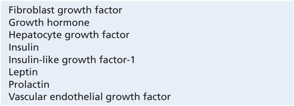
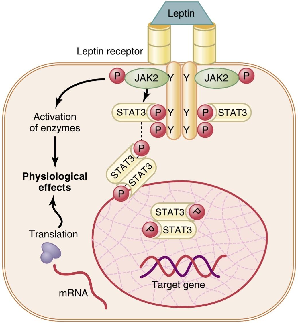
Figure 75-5. An enzyme-linked receptor-the leptin receptor. The receptor exists as a homodimer (two identical parts), and leptin binds to the extracellular part of the receptor, causing phosphorylation (P) and activation of the intracellular associated janus kinase 2 (JAK2). This mechanism causes phosphorylation of signal transducer and activator of transcription (STAT) proteins, which then activates the transcription of target genes and the synthesis of proteins. JAK2 phosphorylation also activates several other enzyme systems that mediate some of the more rapid effects of leptin. Y, Specific tyrosine phosphorylation sites.
شکل 75-5. یک گیرنده مرتبط با آنزیم – گیرنده لپتین. گیرنده به عنوان یک همودایمر (دو قسمت یکسان) وجود دارد و لپتین به بخش خارج سلولی گیرنده متصل می شود و باعث فسفوریلاسیون (P) و فعال شدن ژانوس کیناز 2 (JAK2) مرتبط داخل سلولی می شود. این مکانیسم باعث فسفوریلاسیون پروتئین های مبدل سیگنال و فعال کننده رونویسی (STAT) می شود که سپس رونویسی ژن های هدف و سنتز پروتئین ها را فعال می کند. فسفوریلاسیون JAK2 همچنین چندین سیستم آنزیمی دیگر را فعال می کند که برخی از اثرات سریع لپتین را واسطه می کنند. Y، سایت های فسفوریلاسیون تیروزین خاص.
Intracellular Hormone Receptors and Activation of Genes. Several hormones, including adrenal and gonadal steroid hormones, thyroid hormones, retinoid hormones, and vitamin D, bind with protein receptors inside the cell rather than in the cell membrane. Because these hormones are lipid soluble, they readily cross the cell membrane and interact with receptors in the cytoplasm or nucleus. The activated hormone-receptor complex then binds with a specific regulatory (promoter) sequence of the DNA called the hormone response element, and in this manner either activates or represses transcription of specific genes and formation of messenger RNA (mRNA; Figure 75-6). Therefore, minutes, hours, or even days after the hormone has entered the cell, newly formed proteins appear in the cell and become the controllers of new or altered cellular functions.
گیرنده های هورمونی درون سلولی و فعال سازی ژن ها. چندین هورمون، از جمله هورمونهای استروئیدی غدد فوق کلیوی و غدد جنسی، هورمونهای تیروئید، هورمونهای رتینوئید و ویتامین D، بهجای غشای سلولی، با گیرندههای پروتئینی در داخل سلول متصل میشوند. از آنجایی که این هورمون ها محلول در چربی هستند، به راحتی از غشای سلولی عبور می کنند و با گیرنده های سیتوپلاسم یا هسته تعامل دارند. سپس مجموعه هورمون-گیرنده فعال شده با یک توالی تنظیمی (محرک) خاصی از DNA به نام عنصر پاسخ هورمونی متصل می شود و به این ترتیب رونویسی ژن های خاص و تشکیل RNA پیام رسان را فعال یا سرکوب می کند (mRNA؛ شکل 75-6). بنابراین دقایقی، ساعت ها یا حتی روزها پس از ورود هورمون به سلول، پروتئین های تازه تشکیل شده در سلول ظاهر می شوند و کنترل کننده عملکردهای سلولی جدید یا تغییر یافته می شوند.
Many different tissues have identical intracellular hormone receptors, but the genes that the receptors regulate are different in the various tissues. An intracellular receptor can activate a gene response only if the appropriate combination of gene regulatory proteins is present, and many of these regulatory proteins are tissue specific. Thus, the responses of different tissues to a hormone are determined not only by the specificity of the receptors but also by the expression of genes that the receptor regulates.
بسیاری از بافت های مختلف گیرنده های هورمونی داخل سلولی یکسانی دارند، اما ژن هایی که گیرنده ها تنظیم می کنند در بافت های مختلف متفاوت است. یک گیرنده درون سلولی تنها در صورتی میتواند پاسخ ژنی را فعال کند که ترکیب مناسبی از پروتئینهای تنظیمکننده ژن وجود داشته باشد و بسیاری از این پروتئینهای تنظیمکننده خاص بافت هستند. بنابراین، پاسخ بافتهای مختلف به یک هورمون نه تنها با ویژگی گیرندهها، بلکه با بیان ژنهایی که گیرنده تنظیم میکند نیز تعیین میشود.

Figure 75-6. Mechanisms of interaction of lipophilic hormones, such as steroids, with intracellular receptors in target cells. After the hormone binds to the receptor in the cytoplasm or in the nucleus, the hormone-receptor complex binds to the hormone response element (promoter) on the DNA. This action either activates or inhibits gene transcription, formation of messenger RNA (mRNA), and protein synthesis.
شکل 75-6. مکانیسم های تعامل هورمون های چربی دوست، مانند استروئیدها، با گیرنده های داخل سلولی در سلول های هدف. پس از اتصال هورمون به گیرنده در سیتوپلاسم یا هسته، مجموعه هورمون – گیرنده به عنصر پاسخ هورمونی (پروشور) روی DNA متصل می شود. این عمل رونویسی ژن، تشکیل RNA پیام رسان (mRNA) و سنتز پروتئین را فعال یا مهار می کند.
SECOND MESSENGER MECHANISMS FOR MEDIATING INTRACELLULAR HORMONAL FUNCTIONS
We noted earlier that one way that hormones exert intracellular actions is to stimulate formation of the second messenger cAMP inside the cell membrane. The cAMP then causes subsequent intracellular effects of the hormone. Thus, the only direct effect that the hormone has on the cell is to activate a single type of membrane receptor. The second messenger does the rest.
مکانیسم های پیام رسان دوم برای واسطه عملکردهای هورمونی درون سلولی
قبلاً اشاره کردیم که یکی از راههایی که هورمونها اعمال درون سلولی انجام میدهند، تحریک تشکیل cAMP پیامرسان دوم در داخل غشای سلولی است. سپس cAMP باعث اثرات درون سلولی بعدی این هورمون می شود. بنابراین، تنها اثر مستقیمی که این هورمون روی سلول دارد، فعال کردن یک نوع گیرنده غشایی است. پیام رسان دوم بقیه کارها را انجام می دهد.
CAMP is not the only second messenger used by the different hormones. Two other especially important ones are (1) calcium ions and associated calmodulin and (2) products of membrane phospholipid breakdown. In some cases, a hormone may stimulate more than one second messenger system in the same target tissue.
CAMP تنها دومین پیام رسان مورد استفاده هورمون های مختلف نیست. دو مورد به خصوص مهم دیگر عبارتند از (1) یون کلسیم و کالمودولین مرتبط و (2) محصولات تجزیه فسفولیپیدهای غشایی. در برخی موارد، یک هورمون ممکن است بیش از یک ثانیه سیستم پیام رسان را در همان بافت هدف تحریک کند.
Adenylyl cyclase-cAMP Second Messenger System
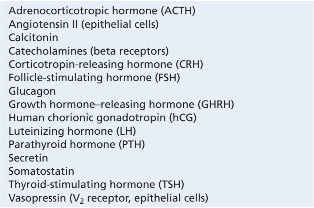
Table 75-3 shows a few of the many hormones that use the adenylyl cyclase-cAMP mechanism to stimulate their target tissues, and Figure 75-7 shows the adenylyl cyclase-cAMP second messenger system. Binding of the hormones with the receptor allows coupling of the receptor to a G protein. If the G protein stimulates the adenylyl cyclase-cAMP system, it is called a G, protein, denoting a stimulatory G protein. Stimulation of adenylyl cyclase, a membrane-bound enzyme, by the G, protein then catalyzes the conversion of a small amount of cytoplasmic adenosine triphosphate into cAMP inside the cell. This then activates cAMP-dependent protein kinase, which phosphorylates specific cell proteins, triggering biochemical reactions that ultimately lead to the cell’s response to the hormone.
سیستم پیام رسان دوم آدنیلیل سیکلاز-cAMP
جدول 75-3 تعدادی از هورمون های متعددی را نشان می دهد که از مکانیسم adenylyl cyclase-cAMP برای تحریک بافت های هدف خود استفاده می کنند و شکل 75-7 سیستم پیام رسان دوم adenylyl cyclase-cAMP را نشان می دهد. اتصال هورمون ها به گیرنده امکان جفت شدن گیرنده را با پروتئین G فراهم می کند. اگر پروتئین G سیستم آدنیلیل سیکلاز-cAMP را تحریک کند، پروتئین G نامیده می شود که نشان دهنده یک پروتئین G تحریک کننده است. تحریک آدنیلیل سیکلاز، یک آنزیم متصل به غشاء، توسط پروتئین G، سپس تبدیل مقدار کمی از آدنوزین تری فسفات سیتوپلاسمی به cAMP را در داخل سلول کاتالیز می کند. سپس پروتئین کیناز وابسته به cAMP را فعال می کند که پروتئین های سلولی خاص را فسفریله می کند و واکنش های بیوشیمیایی را تحریک می کند که در نهایت منجر به پاسخ سلول به هورمون می شود.
Once cAMP is formed inside the cell, it usually activates a cascade of enzymes. That is, first one enzyme is activated, which activates a second enzyme, which activates a third, and so forth. The importance of this mechanism is that only a few molecules of activated adenylyl cyclase immediately inside the cell membrane can cause many more molecules of the next enzyme to be activated, which can cause still more molecules of the third enzyme to be activated, and so forth. In this way, even the slightest amount of hormone acting on the cell surface can initiate a powerful cascading activating force for the entire cell.
هنگامی که cAMP در داخل سلول تشکیل می شود، معمولاً آبشاری از آنزیم ها را فعال می کند. یعنی اول یک آنزیم فعال می شود که آنزیم دوم را فعال می کند که سومی را فعال می کند و غیره. اهمیت این مکانیسم در این است که فقط چند مولکول آدنیلیل سیکلاز فعال شده بلافاصله در داخل غشای سلولی می تواند باعث شود که مولکول های بیشتری از آنزیم بعدی فعال شود، که می تواند باعث شود که مولکول های بیشتری از آنزیم سوم فعال شوند و غیره. به این ترتیب، حتی کوچکترین مقدار هورمونی که روی سطح سلول عمل می کند، می تواند نیروی فعال کننده آبشاری قدرتمندی را برای کل سلول ایجاد کند.
If binding of the hormone to its receptors is coupled to an inhibitory G protein (denoted G; protein), adenylyl cyclase will be inhibited, reducing formation of cAMP and ultimately leading to an inhibitory action in the cell. Thus, depending on the coupling of the hormone receptor to an inhibitory or a stimulatory G protein, a hormone can either increase or decrease the concentration of CAMP and phosphorylation of key proteins inside the cell.
اگر اتصال هورمون به گیرندههای آن به یک پروتئین مهارکننده G (پروتئین G نشان داده میشود) جفت شود، آدنیلیل سیکلاز مهار میشود و تشکیل cAMP را کاهش میدهد و در نهایت منجر به یک عمل بازدارنده در سلول میشود. بنابراین، بسته به جفت شدن گیرنده هورمون به یک پروتئین G بازدارنده یا محرک، یک هورمون می تواند غلظت CAMP و فسفوریلاسیون پروتئین های کلیدی را در داخل سلول افزایش یا کاهش دهد.
The specific action that occurs in response to increases or decreases of cAMP in each type of target cell depends on the nature of the intracellular machinery; some cells have one set of enzymes, and other cells have other enzymes. Therefore, different functions are elicited in different target cells, such as initiating synthesis of specific intracellular chemicals, causing muscle contraction or relaxation, initiating secretion by the cells, and altering cell permeability.
عمل خاصی که در پاسخ به افزایش یا کاهش cAMP در هر نوع سلول هدف رخ میدهد به ماهیت ماشینهای درون سلولی بستگی دارد. برخی از سلول ها دارای یک مجموعه آنزیم هستند و سلول های دیگر دارای آنزیم های دیگری هستند. بنابراین، عملکردهای مختلفی در سلول های هدف مختلف ایجاد می شود، مانند شروع سنتز مواد شیمیایی داخل سلولی خاص، ایجاد انقباض یا شل شدن ماهیچه، شروع ترشح توسط سلول ها و تغییر نفوذپذیری سلول.
Thus, a thyroid cell stimulated by CAMP forms the metabolic hormones thyroxine and triiodothyronine, whereas the same cAMP in an adrenocortical cell causes secretion of the adrenocortical steroid hormones. In some epithelial cells of the distal and collecting tubules of the kidney, cAMP increases their permeability to water.
بنابراین، یک سلول تیروئید تحریک شده توسط CAMP، هورمون های متابولیکی تیروکسین و تری یدوتیرونین را تشکیل می دهد، در حالی که همان cAMP در سلول قشر آدرنال باعث ترشح هورمون های استروئیدی قشر آدرنال می شود. در برخی از سلول های اپیتلیال لوله های دیستال و جمع کننده کلیه، cAMP نفوذپذیری آنها را در برابر آب افزایش می دهد.
Table 75-3 Hormones That Use the Adenylyl Cyclase- CAMP Second Messenger System
جدول 75-3 هورمون هایی که از سیستم پیام رسان دوم آدنیلیل سیکلاز- CAMP استفاده می کنند.
 cAMP , Cylic adenosine monophosphate.

Figure 75-7. The cyclic adenosine monophosphate (cAMP) mechanism by which many hormones exert their control of cell function. ADP, Adenosine diphosphate; ATP, adenosine triphosphate; GTP, guanosine triphosphate.
شکل 75-7. مکانیسم آدنوزین مونوفسفات حلقوی (cAMP) که توسط آن بسیاری از هورمون ها کنترل خود را بر عملکرد سلول اعمال می کنند. ADP، آدنوزین دی فسفات؛ ATP، آدنوزین تری فسفات؛ GTP، گوانوزین تری فسفات.
Cell Membrane Phospholipid Second Messenger System
Some hormones activate transmembrane receptors that activate the enzyme phospholipase C attached to the inside projections of the receptors (Table 75-4). This enzyme catalyzes the breakdown of some phospholipids in the cell membrane, especially phosphatidylinositol bi- phosphate (PIP2), into two different second messenger products: inositol triphosphate (IP) and diacylglycerol (DAG). The IP3 mobilizes calcium ions from mitochondria and the endoplasmic reticulum, and the calcium ions then have their own second messenger effects, such as smooth muscle contraction and changes in cell secretion.
سیستم پیام رسان دوم فسفولیپید غشای سلولی
برخی از هورمون ها گیرنده های گذرنده را فعال می کنند که آنزیم فسفولیپاز C متصل به برجستگی های داخلی گیرنده ها را فعال می کنند (جدول 75-4). این آنزیم تجزیه برخی از فسفولیپیدها در غشای سلولی، به ویژه فسفاتیدیل بی فسفات (PIP2) را به دو محصول پیام رسان دوم مختلف کاتالیز می کند: اینوزیتول تری فسفات (IP) و دی آسیل گلیسرول (DAG). IP3 یونهای کلسیم را از میتوکندری و شبکه آندوپلاسمی بسیج میکند و یونهای کلسیم اثرات پیامرسان دوم خود را دارند، مانند انقباض عضلات صاف و تغییرات در ترشح سلول.
DAG, the other lipid second messenger, activates the enzyme protein kinase C, which then phosphorylates a large number of proteins, leading to the cell’s response (Figure 75-8). In addition to these effects, the lipid portion of DAG is arachidonic acid, which is the precursor for the prostaglandins and other local hormones that cause multiple effects in tissues throughout the body.
DAG، پیام رسان دوم لیپیدی، آنزیم پروتئین کیناز C را فعال می کند، که سپس تعداد زیادی پروتئین را فسفریله می کند و منجر به پاسخ سلول می شود (شکل 75-8). علاوه بر این اثرات، بخش لیپیدی DAG اسید آراشیدونیک است که پیش ساز پروستاگلاندین ها و سایر هورمون های محلی است که باعث ایجاد اثرات متعدد در بافت های سراسر بدن می شود.
Table 75-4 Hormones That Use the Phospholipase C Second Messenger System
جدول 75-4 هورمون هایی که از سیستم پیام رسان دوم فسفولیپاز C استفاده می کنند.


Figure 75-8. The cell membrane phospholipid second messenger system by which some hormones exert their control of cell function. DAG, Diacylglycerol; IP3, inositol triphosphate; PIP2, phosphatidylinositol biphosphate.
شکل 75-8. سیستم پیام رسان دوم فسفولیپید غشای سلولی که توسط آن برخی از هورمون ها کنترل خود را بر عملکرد سلول اعمال می کنند. DAG، دی اسیل گلیسرول؛ IP3، اینوزیتول تری فسفات؛ PIP2، فسفاتیدیلینوزیتول بی فسفات.
Calcium-Calmodulin Second Messenger System
Another second messenger system operates in response to entry of calcium into the cells. Calcium entry may be initiated by (1) changes in membrane potential that open calcium channels or (2) a hormone interacting with membrane receptors that open calcium channels.
سیستم پیام رسان دوم کلسیم-کالمودولین
یک سیستم پیام رسان دوم دیگر در پاسخ به ورود کلسیم به سلول ها عمل می کند. ورود کلسیم ممکن است با (1) تغییر در پتانسیل غشایی که کانالهای کلسیم را باز میکند یا (2) هورمونی که با گیرندههای غشایی که کانالهای کلسیم را باز میکنند در تعامل است، آغاز شود.
Upon entering a cell, calcium ions bind with the protein calmodulin. This protein has four calcium sites, and when three or four of these sites have become bound with calcium, the calmodulin changes its shape and initiates multiple effects inside the cell, including activation or inhibition of protein kinases. Activation of calmodulin- dependent protein kinases causes, via phosphorylation, activation or inhibition of proteins involved in the cell’s response to the hormone. For example, one specific function of calmodulin is to activate myosin light chain kinase, which acts directly on the myosin of smooth muscle to cause smooth muscle contraction (see Figure 8-3).
پس از ورود به سلول، یون های کلسیم با پروتئین کالمودولین متصل می شوند. این پروتئین دارای چهار جایگاه کلسیمی است و زمانی که سه یا چهار محل از این محل ها با کلسیم پیوند می یابند، کالمودولین شکل خود را تغییر می دهد و اثرات متعددی را در داخل سلول آغاز می کند، از جمله فعال یا مهار پروتئین کینازها. فعال شدن پروتئین کینازهای وابسته به کالمودولین، از طریق فسفوریلاسیون، باعث فعال شدن یا مهار پروتئین های دخیل در پاسخ سلول به هورمون می شود. به عنوان مثال، یک عملکرد خاص کالمودولین فعال کردن زنجیره سبک کیناز میوزین است که مستقیماً بر روی میوزین عضله صاف عمل می کند و باعث انقباض عضلات صاف می شود (شکل 8-3 را ببینید).
The normal calcium ion concentration in most cells of the body is 10- to 10-7 mol/L, which is not enough to activate the calmodulin system. However, when the calcium ion concentration rises to 10-6 to 10-5 mol/L, enough binding occurs to cause all the intracellular actions of calmodulin. This is almost exactly the same amount of calcium ion change in skeletal muscle that is required to activate troponin C, which causes skeletal muscle contraction, as explained in Chapter 7. It is interesting that troponin C is similar to calmodulin in both function and protein structure.
غلظت نرمال یون کلسیم در اکثر سلول های بدن 10 تا 7-10 مول در لیتر است که برای فعال کردن سیستم کالمودولین کافی نیست. با این حال، هنگامی که غلظت یون کلسیم به 10-6 تا 10-5 مول در لیتر افزایش می یابد، اتصال کافی برای ایجاد تمام اعمال درون سلولی کالمودولین رخ می دهد. این تقریباً همان مقدار تغییر یون کلسیم در عضله اسکلتی است که برای فعال کردن تروپونین C مورد نیاز است، که باعث انقباض عضله اسکلتی می شود، همانطور که در فصل 7 توضیح داده شد. جالب است که تروپونین C هم از نظر عملکرد و هم از نظر ساختار پروتئین شبیه به کالمودولین است.
HORMONES THAT ACT MAINLY ON THE GENETIC MACHINERY OF THE CELL
Steroid Hormones Increase Protein Synthesis
Another means by which hormones act, especially steroid hormones, is to cause protein synthesis in target cells. These proteins then function as enzymes, transport proteins, or structural proteins, which in turn provide other functions of the cells.
هورمون هایی که عمدتا بر روی ماشین ژنتیکی سلول
هورمون های استروئیدی سنتز پروتئین را افزایش می دهند
وسیله دیگری که هورمون ها به ویژه هورمون های استروئیدی از طریق آن عمل می کنند، ایجاد سنتز پروتئین در سلول های هدف است. سپس این پروتئینها بهعنوان آنزیم، پروتئینهای انتقال یا پروتئینهای ساختاری عمل میکنند که به نوبه خود عملکردهای دیگر سلولها را فراهم میکنند.
The sequence of events in steroid function is essentially the following (see Figure 75-6):
توالی رویدادها در عملکرد استروئید اساساً به شرح زیر است (شکل 75-6 را ببینید):
1. The steroid hormone diffuses across the cell membrane and enters the cytoplasm of the cell, where it binds with a specific receptor protein.
1. هورمون استروئیدی در سراسر غشای سلولی منتشر می شود و وارد سیتوپلاسم سلول می شود، جایی که با پروتئین گیرنده خاصی متصل می شود.
2. The combined receptor protein-hormone then diffuses into or is transported into the nucleus.
2. پروتئین-هورمون گیرنده ترکیبی سپس به درون هسته منتشر می شود یا به درون هسته منتقل می شود.
3. The combination binds at specific points on the DNA strands in the chromosomes, which activates the transcription process of specific genes to form mRNA.
3. این ترکیب در نقاط خاصی از رشته های DNA در کروموزوم ها متصل می شود، که فرآیند رونویسی ژن های خاص را برای تشکیل mRNA فعال می کند.
4. The mRNA diffuses into the cytoplasm, where it promotes the translation process at the ribosomes to form new proteins.
4. mRNA به داخل سیتوپلاسم منتشر می شود، جایی که فرآیند ترجمه را در ریبوزوم ها برای تشکیل پروتئین های جدید ترویج می کند.
To give an example, aldosterone, one of the hormones secreted by the adrenal cortex, enters the cytoplasm of renal tubular cells, which contain a specific receptor protein often called the mineralocorticoid receptor. Therefore, in these cells, the sequence of events cited earlier ensues. After about 45 minutes, proteins begin to appear in the renal tubular cells and promote sodium reabsorption from the tubules and potassium secretion into the tubules. Thus, the full action of the steroid hormone is characteristically delayed for at least 45 minutes-up to several hours or even days. This action is in marked contrast to the rapid actions of some of the peptide and amino acid-derived hormones, such as vasopressin and norepinephrine.
برای مثال، آلدوسترون، یکی از هورمونهایی که از قشر آدرنال ترشح میشود، وارد سیتوپلاسم سلولهای لولهای کلیوی میشود که حاوی پروتئین گیرنده خاصی است که اغلب به نام گیرنده معدنی کورتیکوئید نامیده میشود. بنابراین، در این سلول ها، توالی رویدادهایی که قبلا ذکر شد، به وجود می آید. پس از حدود 45 دقیقه، پروتئینها در سلولهای لولهای کلیه ظاهر میشوند و باعث جذب مجدد سدیم از لولهها و ترشح پتاسیم به داخل لولهها میشوند. بنابراین، عملکرد کامل هورمون استروئیدی به طور مشخص حداقل 45 دقیقه تا چندین ساعت یا حتی چند روز به تعویق می افتد. این عمل در تضاد با عملکرد سریع برخی از پپتیدها و هورمون های مشتق از اسید آمینه مانند وازوپرسین و نوراپی نفرین است.
Thyroid Hormones Increase Gene Transcription in the Cell Nucleus
The thyroid hormones thyroxine and triiodothyronine cause increased transcription by specific genes in the nucleus. To accomplish this increased transcription, these hormones first bind directly with receptor proteins in the nucleus, as discussed in more detail in Chapter 77 (see Figure 77-5); these receptors are activated transcription factors located within the chromosomal complex, and they control the function of the gene promoters, as explained in Chapter 3.
هورمون های تیروئید رونویسی ژن را در هسته سلول افزایش می دهند
هورمون های تیروئید تیروکسین و تری یدوتیرونین باعث افزایش رونویسی توسط ژن های خاص در هسته می شوند. برای انجام این رونویسی افزایش یافته، این هورمون ها ابتدا به طور مستقیم با پروتئین های گیرنده در هسته متصل می شوند، همانطور که در فصل 77 با جزئیات بیشتر بحث شده است (شکل 77-5 را ببینید). این گیرنده ها فاکتورهای رونویسی فعالی هستند که در کمپلکس کروموزومی قرار دارند و عملکرد محرک های ژن را کنترل می کنند، همانطور که در فصل 3 توضیح داده شد.
The following two features of thyroid hormone function in the nucleus are important:
دو ویژگی زیر در عملکرد هورمون تیروئید در هسته مهم هستند:
1. They activate the genetic mechanisms for synthesizing many types of intracellular proteins-probably 100 or more. Many of these intracellular proteins are enzymes that promote enhanced intracellular metabolic activity in virtually all cells of the body.
1. آنها مکانیسم های ژنتیکی را برای سنتز بسیاری از انواع پروتئین های درون سلولی فعال می کنند – احتمالاً 100 یا بیشتر. بسیاری از این پروتئین های درون سلولی آنزیم هایی هستند که فعالیت متابولیک داخل سلولی را در تقریباً در تمام سلول های بدن افزایش می دهند.
2. Once bound to the intranuclear receptors, the thyroid hormones can continue to express their control functions for days or even weeks.
2. پس از اتصال به گیرنده های داخل هسته ای، هورمون های تیروئید می توانند برای روزها یا حتی هفته ها به بیان عملکردهای کنترلی خود ادامه دهند.
Measurement of Hormone Concentrations in the Blood
Most hormones are present in the blood in extremely minute quantities; some concentrations are as low as one billionth of a milligram (1 picogram) per milliliter. Therefore, it was difficult to measure these concentrations by the usual chemical means. However, an extremely sensitive method, called radioimmunoassay, was developed by Rosalyn Yalow and Solomon Berson in 1959 and revolutionized the measurement of hormones, their precursors, and their metabolic end products. More recently, additional methods, such as enzyme-linked immunosorbent assays, have been developed for accurate, high-throughput measurements of hormones.
اندازه گیری غلظت هورمون در خون
بیشتر هورمون ها در مقادیر بسیار کم در خون وجود دارند. برخی از غلظت ها به یک میلیاردم میلی گرم (1 پیکوگرم) در میلی لیتر می رسد. بنابراین، اندازه گیری این غلظت ها با روش های معمول شیمیایی دشوار بود. با این حال، یک روش بسیار حساس، به نام رادیوایمونواسی، توسط روزالین یالو و سولومون برسون در سال 1959 ایجاد شد و انقلابی در اندازه گیری هورمون ها، پیش سازهای آنها و محصولات نهایی متابولیک آنها ایجاد کرد. اخیراً، روشهای اضافی، مانند سنجشهای ایمونوسوربنت مرتبط با آنزیم، برای اندازهگیری دقیق و با کارایی بالا هورمونها توسعه یافتهاند.
Radioimmunoassay
The method of performing radioimmunoassay is as follows. First, an antibody that is highly specific for the hormone to be measured is produced.
رادیوایمونواسی
روش انجام رادیو ایمونواسی به شرح زیر است. ابتدا یک آنتی بادی که برای هورمون مورد اندازه گیری بسیار اختصاصی است تولید می شود.
Second, a small quantity of this antibody is (1) mixed with a quantity of fluid from the animal containing the hormone to be measured and (2) mixed simultaneously with an appropriate amount of purified standard hormone that has been tagged with a radioactive isotope. However, one specific condition must be met. There must be too little antibody to bind completely both the radioactively tagged hormone and the hormone in the fluid to be assayed. Therefore, the natural hormone in the assay fluid and the radioactive standard hormone compete for binding sites of the antibody. In the process of competing, the quantity of each of the two hormones, the natural and the radioactive, that binds is proportional to its concentration in the assay fluid.
دوم، مقدار کمی از این آنتی بادی (1) با مقداری مایع از حیوان حاوی هورمون مورد اندازه گیری مخلوط می شود و (2) به طور همزمان با مقدار مناسبی از هورمون استاندارد خالص شده که با ایزوتوپ رادیواکتیو برچسب گذاری شده است مخلوط می شود. با این حال، یک شرط خاص باید رعایت شود. باید آنتی بادی بسیار کمی وجود داشته باشد تا بتواند به طور کامل هم به هورمون برچسب گذاری شده با رادیواکتیو و هم به هورمون موجود در مایع مورد سنجش متصل شود. بنابراین، هورمون طبیعی در مایع سنجش و هورمون استاندارد رادیواکتیو برای مکانهای اتصال آنتیبادی با هم رقابت میکنند. در فرآیند رقابت، مقدار هر یک از دو هورمون طبیعی و رادیواکتیو که متصل میشوند، متناسب با غلظت آن در مایع سنجش است.
Third, after binding has reached equilibrium, the antibody- hormone complex is separated from the remainder of the solution, and the quantity of radioactive hormone bound in this complex is measured by radioactive counting techniques. If a large amount of radioactive hormone has bound with the antibody, it is clear that there was only a small amount of natural hormone to compete with the radioactive hormone, and therefore the concentration of the natural hormone in the assayed fluid was small. Conversely, if only a small amount of radioactive hormone has bound, it is clear that there was a large amount of natural hormone to compete for the binding sites.
سوم، پس از رسیدن به تعادل، کمپلکس آنتی بادی-هورمون از باقیمانده محلول جدا میشود و مقدار هورمون رادیواکتیو متصل در این کمپلکس با روشهای شمارش رادیواکتیو اندازهگیری میشود. اگر مقدار زیادی از هورمون رادیواکتیو به آنتی بادی متصل شده باشد، واضح است که فقط مقدار کمی از هورمون طبیعی برای رقابت با هورمون رادیواکتیو وجود دارد و بنابراین غلظت هورمون طبیعی در مایع مورد سنجش کم است. برعکس، اگر فقط مقدار کمی از هورمون رادیواکتیو متصل شده باشد، واضح است که مقدار زیادی هورمون طبیعی برای رقابت برای مکانهای اتصال وجود دارد.
Fourth, to make the assay highly quantitative, the radio- immunoassay procedure is also performed for “standard” solutions of untagged hormone at several concentration levels. Then a “standard curve” is plotted, as shown in Figure 75-9. By comparing the radioactive counts recorded from the “unknown” assay procedures with the standard curve, one can determine within an error of 10% to 15%, the concentration of the hormone in the “unknown” assayed fluid. As little as billionths or even trillionths of a gram of hormone can often be assayed in this way.
چهارم، برای اینکه سنجش بسیار کمی باشد، روش رادیو ایمونواسی نیز برای محلولهای «استاندارد» هورمون بدون برچسب در سطوح مختلف غلظت انجام میشود. سپس یک “منحنی استاندارد” همانطور که در شکل 75-9 نشان داده شده است ترسیم می شود. با مقایسه تعداد رادیواکتیو ثبتشده از روشهای سنجش «ناشناخته» با منحنی استاندارد، میتوان با خطای 10 تا 15 درصد، غلظت هورمون را در مایع سنجش شده «ناشناخته» تعیین کرد. معمولاً میتوان مقدار کمی از یک میلیاردم یا حتی تریلیونم یک گرم هورمون را با این روش ارزیابی کرد.

Figure 75-9. Standard curve for radioimmunoassay of aldosterone. (Courtesy Dr. Manis Smith.)
شکل 75-9. منحنی استاندارد برای سنجش رادیو ایمنی آلدوسترون. (با احترام از دکتر مانیس اسمیت.)
Enzyme-Linked Immunosorbent Assay
Enzyme-linked immunosorbent assays (ELISAs) can be used to measure almost any protein, including hormones. This test combines the specificity of antibodies with the sensitivity of simple enzyme assays. Figure 75-10 shows the basic elements of this method, which is often per- formed on plastic plates that each have 96 small wells. Each well is coated with an antibody (AB,) that is specific for the hormone being assayed. Samples or standards are added to each of the wells, followed by a second antibody (AB2) that is also specific for the hormone but binds to a different site of the hormone molecule. A third antibody (AB3) that is added recognizes AB2 and is coupled to an enzyme that converts a suitable substrate to a product that can be easily detected by colorimetric or fluorescent optical methods.
سنجش ایمونوسوربنت مرتبط با آنزیم
سنجشهای ایمونوسوربنت مرتبط با آنزیم (ELISAs) را میتوان برای اندازهگیری تقریباً هر پروتئینی، از جمله هورمونها، استفاده کرد. این آزمایش ویژگی آنتی بادی ها را با حساسیت سنجش های آنزیمی ساده ترکیب می کند. شکل 75-10 عناصر اساسی این روش را نشان می دهد که اغلب بر روی صفحات پلاستیکی که هر کدام دارای 96 چاه کوچک هستند اجرا می شود. هر چاهک با یک آنتی بادی (AB،) پوشیده شده است که برای هورمون مورد سنجش خاص است. نمونهها یا استانداردها به هر یک از چاهکها اضافه میشوند و به دنبال آن یک آنتیبادی دوم (AB2) که برای هورمون نیز خاص است، اما به محل متفاوتی از مولکول هورمون متصل میشود. آنتی بادی سوم (AB3) که اضافه می شود AB2 را می شناسد و با آنزیمی که یک بستر مناسب را به محصولی تبدیل می کند که به راحتی با روش های نوری رنگ سنجی یا فلورسنت قابل تشخیص است، جفت می شود.
Because each molecule of enzyme catalyzes the formation of many thousands of product molecules, even small amounts of hormone molecules can be detected. In contrast to competitive radioimmunoassay methods, ELISA methods use excess antibodies so that all hormone molecules are captured in antibody-hormone complexes. There- fore, the amount of hormone present in the sample or in the standard is proportional to the amount of product formed.
از آنجا که هر مولکول آنزیم تشکیل هزاران مولکول محصول را کاتالیز می کند، حتی مقادیر کمی از مولکول های هورمون را می توان تشخیص داد. برخلاف روشهای رادیوامونواسی رقابتی، روشهای الایزا از آنتیبادیهای اضافی استفاده میکنند تا تمام مولکولهای هورمون در کمپلکسهای آنتیبادی-هورمون جذب شوند. بنابراین، مقدار هورمون موجود در نمونه یا استاندارد متناسب با مقدار محصول تشکیل شده است.
The ELISA method has become widely used in clinical and research laboratories because (1) it does not use radioactive isotopes, (2) much of the assay can be automated using 96-well plates, and (3) it has proved to be a cost- effective and accurate method for assessing hormone levels.
روش الایزا به طور گسترده در آزمایشگاههای بالینی و تحقیقاتی مورد استفاده قرار گرفته است زیرا (1) از ایزوتوپهای رادیواکتیو استفاده نمیکند، (2) بسیاری از روشها را میتوان با استفاده از صفحات 96 چاهی خودکار کرد، و (3) ثابت کرده است که روشی مقرونبهصرفه و دقیق برای ارزیابی سطوح هورمونی است.

Figure 75-10. Basic principles of the enzyme-linked immunosorbent assay for measuring the concentration of a hormone (H). AB, and AB2 are antibodies that recognize the hormone at different binding sites, and AB3 is an antibody that recognizes AB2. E is an enzyme linked to AB; that catalyzes the formation of a colored fluorescent product (P) from a substrate (S). The amount of the product is measured using optical methods and is proportional to the amount of hormone in the well if there are excess antibodies in the well.
شکل 75-10. اصول اساسی سنجش ایمونوسوربنت متصل به آنزیم برای اندازه گیری غلظت یک هورمون (H). AB و AB2 آنتی بادی هایی هستند که هورمون را در مکان های مختلف اتصال شناسایی می کنند و AB3 آنتی بادی است که AB2 را تشخیص می دهد. E یک آنزیم مرتبط با AB است. که تشکیل یک محصول فلورسنت رنگی (P) از یک بستر (S) را کاتالیز می کند. مقدار محصول با استفاده از روش های نوری اندازه گیری می شود و در صورت وجود آنتی بادی های اضافی در چاه، متناسب با مقدار هورمون موجود در چاه است.
کلیک کنید «Bibliography: فهرست کتب مربوطه»
Aguiar-Oliveira MH, Bartke A: Growth hormone deficiency: health and longevity. Endocr Rev 40:575, 2019.
Baker ME: Steroid receptors and vertebrate evolution. Mol Cell Endocrinol Oct 1;496:110526. doi: 10.1016/j.mce.2019.110526
Bianco AC, Dumitrescu A, Gereben B, et al: Paradigms of dynamic control of thyroid hormone signaling. Endocr Rev 40:1000, 2019.
Deussing JM, Chen A: The corticotropin-releasing factor family: phys- iology of the stress response. Physiol Rev 98:2225, 2018.
Forrester SJ, Booz GW, Sigmund CD, et al: Angiotensin II signal trans- duction: an update on mechanisms of physiology and pathophysi- ology. Physiol Rev 98:1627, 2018.
Gamble KL, Berry R, Frank SJ, Young ME: Circadian clock control of endocrine factors. Nat Rev Endocrinol 10:466, 2014.
Gomez-Sanchez E, Gomez-Sanchez CE: The multifaceted mineralocorticoid receptor. Compr Physiol 4:965, 2014.
Haeusler RA, McGraw TE, Accili D: Biochemical and cellular properties of insulin receptor signalling. Nat Rev Mol Cell Biol 19:31, 2018.
Harno E, Gali Ramamoorthy T, Coll AP, White A: POMC: the physiological power of hormone processing. Physiol Rev 98:2381, 2018.
Hewitt SC, Korach KS: Estrogen receptors: new directions in the new millennium. Endocr Rev 39:664, 2018.
Hunter I, Hay CW, Esswein B, Watt K, McEwan IJ: Tissue control of androgen action: the ups and downs of androgen receptor expres- sion. Mol Cell Endocrinol 465:27, 2018.
urek B, Neumann ID: The oxytocin receptor: from intracellular signaling to behavior. Physiol Rev 98:1805, 2018.
Najjar SM, Perdomo G: Hepatic insulin clearance: mechanism and physiology. Physiology (Bethesda) 34:198, 2019.
Oster H, Challet É, Ott V, et al: The functional and clinical significance of the 24-hour rhythm of circulating glucocorticoids. Endocr Rev 38:3, 2017.
Pan WW, Myers MG Jr: Leptin and the maintenance of elevated body weight. Nat Rev Neurosci 19:95, 2018.
Petersen MC, Shulman Gl: Mechanisms of insulin action and insulin resistance. Physiol Rev 98:2133, 2018.
Sarfstein R, Werner H: Minireview: nuclear insulin and insulin-like growth factor-1 receptors: a novel paradigm in signal transduc- tion. Endocrinology 154:1672, 2013.
Stenvers DJ, Scheer FAJL, Schrauwen P, et al: Circadian clocks and insulin resistance. Nat Rev Endocrinol 15:75, 2019.
Yang Q, Vijayakumar A, Kahn BB: Metabolites as regulators of insulin sensitivity and metabolism. Nat Rev Mol Cell Biol 19:654, 2018.
🚀 با ما همراه شوید!
تازهترین مطالب و آموزشهای مغز و اعصاب را از دست ندهید. با فالو کردن کانال تلگرام آیندهنگاران مغز، از ما حمایت کنید!



